Contructs for enhancing immune responses
Ertl , et al.
U.S. patent number 10,328,146 [Application Number 15/639,296] was granted by the patent office on 2019-06-25 for contructs for enhancing immune responses. This patent grant is currently assigned to THE WISTAR INSTITUTE OF ANATOMY AND BIOLOGY. The grantee listed for this patent is THE WISTAR INSTITUTE OF ANATOMY AND BIOLOGY. Invention is credited to Hildegund C. J. Ertl, Luis C. S. Ferreira, Marcio O. Lasaro.


View All Diagrams
| United States Patent | 10,328,146 |
| Ertl , et al. | June 25, 2019 |
Contructs for enhancing immune responses
Abstract
Chimeric protein constructs including a herpesvirus glycoprotein D (gD) and a heterologous polypeptide that interact with herpes virus entry mediator (HVEM) and enhance and enhance an immune response against the heterologous polypeptide and methods for their use are provided.
| Inventors: | Ertl; Hildegund C. J. (Villanova, PA), Lasaro; Marcio O. (Maple Shade, NJ), Ferreira; Luis C. S. (Sao Paulo, BR) | ||||||||||
|---|---|---|---|---|---|---|---|---|---|---|---|
| Applicant: |
|
||||||||||
| Assignee: | THE WISTAR INSTITUTE OF ANATOMY AND
BIOLOGY (Philadelphia, PA) |
||||||||||
| Family ID: | 38779709 | ||||||||||
| Appl. No.: | 15/639,296 | ||||||||||
| Filed: | June 30, 2017 |
Prior Publication Data
| Document Identifier | Publication Date | |
|---|---|---|
| US 20170340730 A1 | Nov 30, 2017 | |
Related U.S. Patent Documents
| Application Number | Filing Date | Patent Number | Issue Date | ||
|---|---|---|---|---|---|
| 14628784 | Feb 23, 2015 | 9724406 | |||
| 13239771 | Feb 24, 2015 | 8962816 | |||
| 12438889 | |||||
| PCT/US2007/018939 | Aug 28, 2007 | ||||
| 60840526 | Aug 28, 2006 | ||||
| Current U.S. Class: | 1/1 |
| Current CPC Class: | A61K 39/245 (20130101); A61K 39/12 (20130101); C12N 7/00 (20130101); A61P 31/22 (20180101); C07K 14/005 (20130101); C12N 2710/16622 (20130101); A61K 2039/53 (20130101); C07K 2319/00 (20130101); Y02A 50/30 (20180101); C12N 2710/10043 (20130101); A61K 2039/57 (20130101); C12N 2710/16634 (20130101); C07K 2319/10 (20130101) |
| Current International Class: | C07K 14/005 (20060101); A61K 39/12 (20060101); C12N 7/00 (20060101); A61K 39/245 (20060101); A61K 39/00 (20060101) |
References Cited [Referenced By]
U.S. Patent Documents
| 5814486 | September 1998 | Cohen et al. |
| 6936255 | August 2005 | Wettendorff et al. |
| 8962816 | February 2015 | Ertl |
| 9724406 | August 2017 | Ertl |
| 1336619 | Aug 2003 | EP | |||
Other References
|
Altstein, et al., "Immunization with Influenza A NP-Expressing Vaccinia Virus Recombinant Protects Mice Against Experimental Infection with Hyuman and Avian Influenza Viruses", Archives of Virology, vol. 151, No. 5, May 2006, pp. 921-931. cited by applicant . Alves, et al., "Antibody Response in Mice Immunized with a Plasmid DNA Encoding the Colonization Factor Antigen I of Enterotoxigenic Escherichia coli", FEMS Immunology Medical Microbiology, vol. 23, No. 4, Apr. 1999, pp. 321-330. cited by applicant . Hazama, et al., "Adjuvant-independent enhanced immune responses to recombinant Herpes Simplex Virus Type 1 Glycoprotein D by fusion biologically active interlukin-2", Vaccine, vol. 11, No. 6, 1993, pp. 629-636. cited by applicant . Hinuma, et al., "A novel strategy for converting recombinant viral protein into high immunogeic antigen", FEBS Letters, vol. 288, No. 1/2, Aug. 1991, pp. 138-142. cited by applicant . Lasaro, et al., "Antibody-inducing properties of a prototype bivalent Herpes Simplex virus/Enterotoxigenic Escherichia coli DNA Vaccine", FEMS Immunology and Medical Microbiology, vol. 35, No. 1, Jan. 21, 2003, pp. 25-31. cited by applicant . Lasaro, et al., "Anti-Tumor DNA Vaccines based on the Expressiopn of Human Papillomavirus-16 E6/E7 Oncoproteins Genetically Fused With the Glycoprotein D from Herpes Simplex Virus-1", Microbes and Infection, vol. 7, No. 15, Dec. 2005, pp. 1541-1550. cited by applicant . Lasaro, et al., "Human papillomavirus-associated cervical cancer: Prophylactic and therapeutic vaccines.", Gene Therapy Molecular Biology, 2004, vol. 8, pp. 291-306. cited by applicant . Michel, et al., "Enhanced Immunogenicity of HPV 16 E7 Fusion Proteins in DNA Vaccination", Virology, 2002, vol. 294, pp. 47-59. cited by applicant . Saha, et al., "A Fused Gene of Nucleoprotein (NP) and Herpes Simplex Virus Genes (VP22) Induces Highly Protective Immunity Against Different Subtypes of Influenza Virus", Virology, vol. 354, No. 1, Oct. 10, 2006, pp. 48-57. cited by applicant . Watson, et al., "Herpes Simplex Virus Type=1 Glycoprotein D Gene: Nucleotide Sequence and Expression in Escherichia coli", Science, vol. 218, Oct. 22, 1982, pp. 381-384. cited by applicant . Zago, et al., "Use of herpes simplex virus and pseudorabies virus chimeric glycoprotein D molecules to identify regions critical for membrane fusion.", PNAS, 2004, vol. 101, No. 50, pp. 17498-17503. cited by applicant. |
Primary Examiner: Horning; Michelle S
Attorney, Agent or Firm: Saul Ewing Arnstein & Lehr LLP Doyle; Kathryn
Government Interests
STATEMENT REGARDING FEDERALLY SPONSORED RESEARCH OR DEVELOPMENT
This invention was made with government support under 5 P01 AI052271, awarded by the National Institutes of Health (NIH). The government has certain rights in the invention.
Parent Case Text
CROSS-REFERENCE TO RELATED APPLICATIONS
The present application is a continuation of U.S. patent application Ser. No. 14/628,784, filed on Feb. 23, 2015, which is a continuation of U.S. patent application Ser. No. 13/239,771, filed on Sep. 22, 2011, now U.S. Pat. No. 8,962,816, which is a continuation of U.S. patent application Ser. No. 12/438,889, filed on Feb. 25, 2009, abandoned, which is a National Stage application of PCT/US2007/018939, filed on Aug. 28, 2007, which claims priority under 35 U.S.C. .sctn. 119(e) to U.S. Provisional Patent Application No. 60/840,526, filed on Aug. 28, 2006.
Claims
What is claimed:
1. A vaccine comprising a nucleic acid molecule which encodes a fusion protein, wherein the fusion protein comprises: a. a first polypeptide segment comprising at least amino acids 1-240 of a mature Herpes simplex virus (HSV) glycoprotein D, wherein the first polypeptide segment does not comprise a full length mature glycoprotein D; b. a second polypeptide segment comprising at least one antigen, wherein the at least one antigen is not an HSV glycoprotein D antigen, wherein the N terminus of the second polypeptide segment is linked to the C terminus of the first polypeptide segment; and c. a third polypeptide segment comprising a C terminal portion of the HSV glycoprotein D, wherein the N terminus of the third polypeptide segment is linked to the C terminus of the second polypeptide segment.
2. The vaccine of claim 1, wherein the HSV is selected from the group consisting of HSV-1 and HSV-2.
3. The vaccine of claim 2, wherein the first polypeptide segment comprises an amino acid sequence selected from the group consisting of: a. amino acids 26-265 of SEQ ID NO:27; b. amino acids 26-265 of SEQ ID NO:29; c. amino acids 1-244 of the glycoprotein D; d. amino acids 1-288 of the glycoprotein D; and e. amino acids 1-294 of a mature HSV glycoprotein D with the exception that amino acid 294 is alanine instead of tryptophan.
4. The vaccine of claim 3, wherein the first polypeptide segment is encoded by a nucleic acid sequence comprising nucleotides 76-795 of SEQ ID NO:26; or nucleotides 350-1069 of SEQ ID NO:28.
5. The vaccine of claim 1, wherein the at least one antigen is selected from the group consisting of: a. an influenza virus antigen; b. a nucleoprotein P influenza virus antigen; c. a Plasmodium antigen; d. a Plasmodium antigen selected from the group consisting of thrombospondin-related anonymous protein (TRAP), ring-infected erythrocyte surface antigen (RESA), merozoite surface protein 1 (MSP1), merozoite surface protein 2 (MSP2), merozoite surface protein 3 (MSP3), and glutamate-rich antigen (GLURP); e. human papilloma virus (HPV) antigen; f. human papilloma virus HPV16 antigen; g. HPV E5 protein; h. HPV E6 protein; i. HPV E7 protein; j. a human immunodeficiency virus (HIV) antigen; and k. an HIV gag antigen.
6. The vaccine of claim 1, wherein the nucleic acid molecule encodes the amino acid sequence encoded SEQ ID NO:35.
7. The vaccine of claim 1, wherein the nucleic acid molecule comprises a nucleotide sequence selected from the group consisting of SEQ ID NO:31; SEQ ID NO:32; SEQ ID NO:34; SEQ ID NO:35; SEQ ID NO:36; and SEQ ID NO:37.
8. The vaccine of claim 1, wherein the fusion protein comprises an amino acid sequence selected from the group consisting of SEQ ID NO:22; SEQ ID NO:23; SEQ ID NO:24; and SEQ ID NO:33.
9. The vaccine of claim 1, wherein the nucleic acid molecule is in a viral vector.
10. The vaccine of claim 1, wherein the nucleic acid molecule is naked DNA.
11. The vaccine of claim 1, wherein the nucleic acid molecule is in a bacterial vector.
12. The vaccine of claim 5, wherein the second polypeptide segment comprises the HPV E5 protein, the HPV E6 protein, and the HPV E7 protein.
13. The vaccine of claim 1, wherein the third polypeptide segment comprises the transmembrane domain of the HSV glycoprotein D.
14. A vaccine comprising a fusion protein, wherein the fusion protein comprises: a. a first polypeptide segment comprising at least amino acids 1-240 of a mature Herpes simplex virus (HSV) glycoprotein D, wherein the first polypeptide segment does not comprise a full length glycoprotein D; b. a second polypeptide segment comprising at least one antigen, wherein the at least one antigen is not an HSV glycoprotein D antigen, wherein the N terminus of the second polypeptide segment is linked to the C terminus of the first polypeptide segment; and c. a third polypeptide segment comprising a C terminal portion of the HSV glycoprotein D, wherein the N terminus of the third polypeptide segment is linked to the C terminus of the second polypeptide segment.
15. The vaccine of claim 14, wherein the HSV is selected from the group consisting of HSV-1 and HSV-2.
16. The vaccine of claim 15, wherein the first polypeptide segment comprises an amino acid sequence selected from the group consisting of: a. amino acids 26-265 of SEQ ID NO:27; b. amino acids 26-265 of SEQ ID NO:29; c. amino acids 1-244 of the glycoprotein D; d. amino acids 1-288 of the glycoprotein D; and e. amino acids 1-294 of a mature HSV glycoprotein D with the exception that amino acid 294 is alanine instead of tryptophan.
17. The vaccine of claim 16, wherein the first polypeptide segment is encoded by a nucleic acid sequence comprising nucleotides 76-795 of SEQ ID NO:26; or nucleotides 350-1069 of SEQ ID NO:28.
18. The vaccine of claim 14, wherein the at least one antigen is selected from the group consisting of: a. an influenza virus antigen; b. a nucleoprotein P influenza virus antigen; c. a Plasmodium antigen; d. a Plasmodium antigen selected from the group consisting of thrombospondin-related anonymous protein (TRAP), ring-infected erythrocyte surface antigen (RESA), merozoite surface protein 1 (MSP1), merozoite surface protein 2 (MSP2), merozoite surface protein 3 (MSP3), and glutamate-rich antigen (GLURP); e. human papilloma virus (HPV) antigen; f. human papilloma virus HPV16 antigen; g. HPV E5 protein; h. HPV E6 protein; i. HPV E7 protein; j. a human immunodeficiency virus (HIV) antigen; and k. an HIV gag antigen.
19. The vaccine of claim 14, wherein the fusion protein comprises an amino acid sequence selected from the group consisting of SEQ ID NO:22; SEQ ID NO:23; SEQ ID NO:24; and SEQ ID NO:33.
20. The vaccine of claim 14, wherein the fusion protein is encoded by a nucleic acid sequence selected from the group consisting of SEQ ID NO:31; SEQ ID NO:32; SEQ ID NO:34; SEQ ID NO:35; SEQ ID NO:36; and SEQ ID NO:37.
21. The vaccine of claim 18, wherein the second polypeptide segment comprises the HPV E5 protein, the HPV E6 protein, and the HPV E7 protein.
22. The vaccine of claim 14, wherein the third polypeptide segment comprises the transmembrane domain of the HSV glycoprotein D.
Description
FIELD OF THE INVENTION
Embodiments of the present invention relate in general to chimeric (fusion) protein constructs including a herpesvirus glycoprotein D (gD) and a heterologous polypeptide (e.g., antigen) that enhance the immune response against the heterologous polypeptide (e.g., antigen) in a subject.
BACKGROUND OF THE INVENTION
gD is the receptor-binding glycoprotein of herpesviruses (Fusco et al. (2005) Proc. Natl. Acad. Sci. U.S.A. 102:9323). The gD ectodomain is organized in two structurally and functionally differentiated regions. The amino-terminus includes the signal sequence and receptor-binding sites, and the carboxy-terminus includes the pro-fusion domain and the transmembrane domain. gD interacts with two alternative receptors belonging to unrelated protein families, the herpesvirus entry mediator (HVEM) and the nectins (Geraghty et al. (1998) Science 280:1618; Montgomery et al. (1996) Cell 87:427; Cocchi et al. (1998) J. Virol. 72:9992; Warner et al. (1998) Virology 246:179; Lopez et al. (2000) J. Virol. 74:1267). HVEM is expressed on dendritic cells and the B and T lymphocyte attenuator (BTLA) is expressed on activated T and B lymphocytes. The interaction between HVEM and BTLA results in the down-regulation of immune responses.
BRIEF DESCRIPTION OF THE DRAWINGS
FIG. 1 depicts a schematic representation of the chimeric gene gDE7E6E5. The HPV-16 E5, E6 and E7 genes without respective start and stop codons were linked in tandem and incorporated into the HSV-1 gD gene ApaI site, which corresponds to amino acid 244 in the gD mature form.
FIG. 2 depicts a schematic representation of chimeric gene gDgag. The codon-optimized truncated form of gag from HIV-1 clade B was fused into the HSV-1 gD gene NarI site, which corresponds to amino acid 289 in the gD mature form.
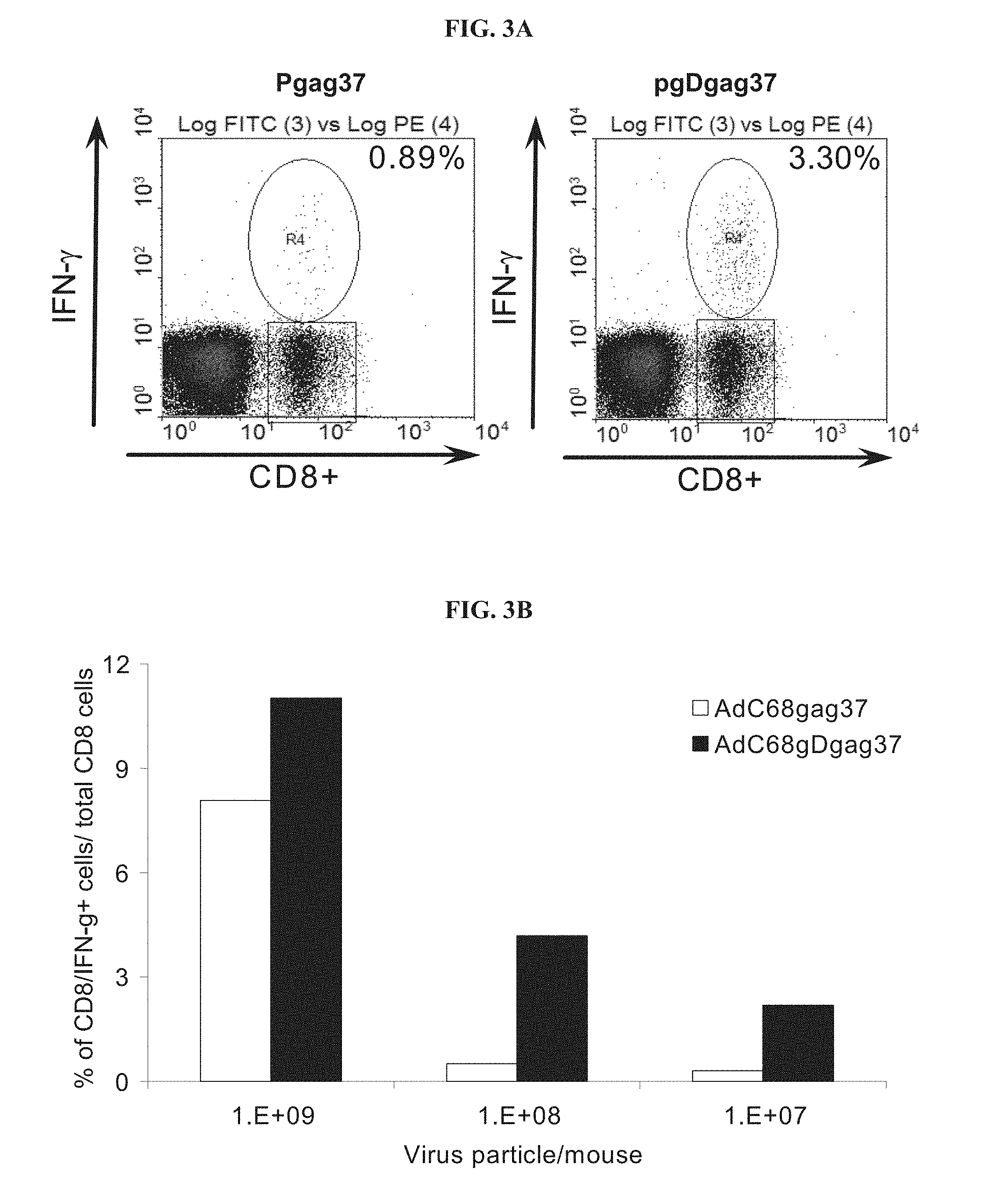
FIGS. 3A-3B depict the gag-specific CD8.sup.+ IFN-.gamma. response in mice immunized with vaccine constructs carrying the gDgag chimeric gene. FIG. 3A depicts a FACS analysis of the gag-specific CD8/IFN-.gamma..sup.+ response in peripheral blood mononuclear cells (PBMC) from mice immunized with DNA vaccine expressing either HIV-1 gag or HIV-1 gag fused to HSV-1 gD (gDgag). Numbers on the right corner represent percentage CD8.sup.+/IFN-.gamma..sup.+ cells over total of CD8.sup.+ cells. FIG. 3B graphically depicts PBMC from mice immunized with AdC68 vectors carrying either genes encoding gag or gDgag, inoculated with different amounts of virus particles per mouse.
FIG. 4 graphically depicts the effect of pre-existing immunity to the AdHu5 adenovirus vector on the transgene product-specific CD8.sup.+ T cell response to the AdC68 vector. AdC68 vectors carrying gag, gDgag or gDE7E65 were inoculated into naive mice and mice previously immunized with an AdHu5 expressing an unrelated antigen (rabies glycoprotein, AdHu5rab.gp). The percentage of reduction of CD8.sup.+ T cell response was defined as a percentage of CD8.sup.+/IFN-.gamma..sup.+ frequency in mice previously immunized with AdHu5 over the frequency found in mice that did not receive AdHu5 vector.
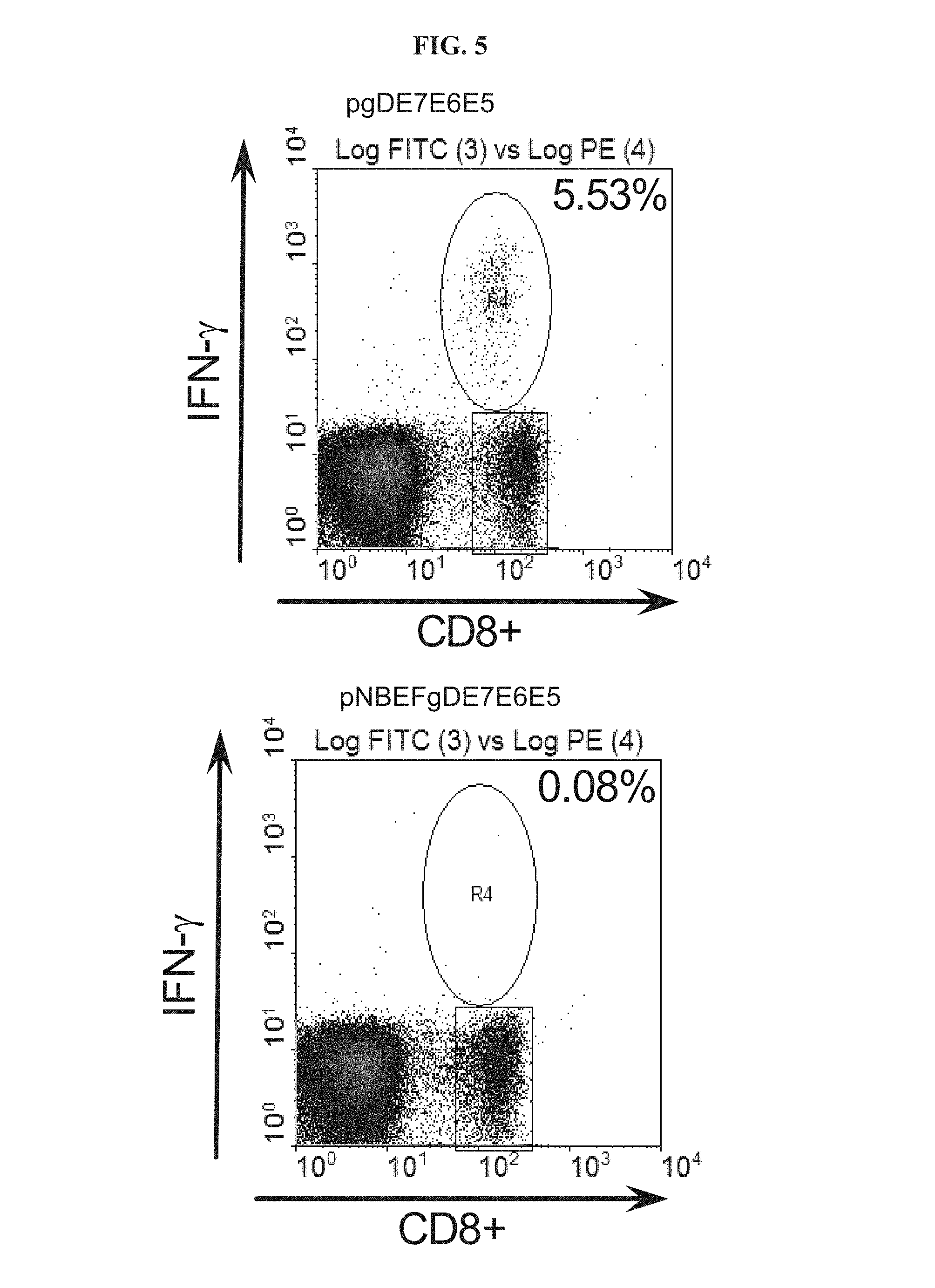
FIG. 5 depicts the E7-specific CD8.sup.+ IFN-.gamma. response in mice immunized with DNA vaccines expressing non-mutated or mutated gDE7E6E5. The mutation was designed to disrupt the HVEM binding site of gD. Mice were immunized with non-mutated gDE7E6E5 (pgDE7E6E5) or mutated gDE7E6E5 (pNBEFgDE7E6E5) and 14 days later peripheral blood mononuclear cells were investigated by intracellular cytokine staining for E7-specific CD8.sup.+ IFN-.gamma. responses. Numbers on the right corners represent percentage CD8.sup.+/IFN-.gamma..sup.+ cells over total of CD8.sup.+ cells.
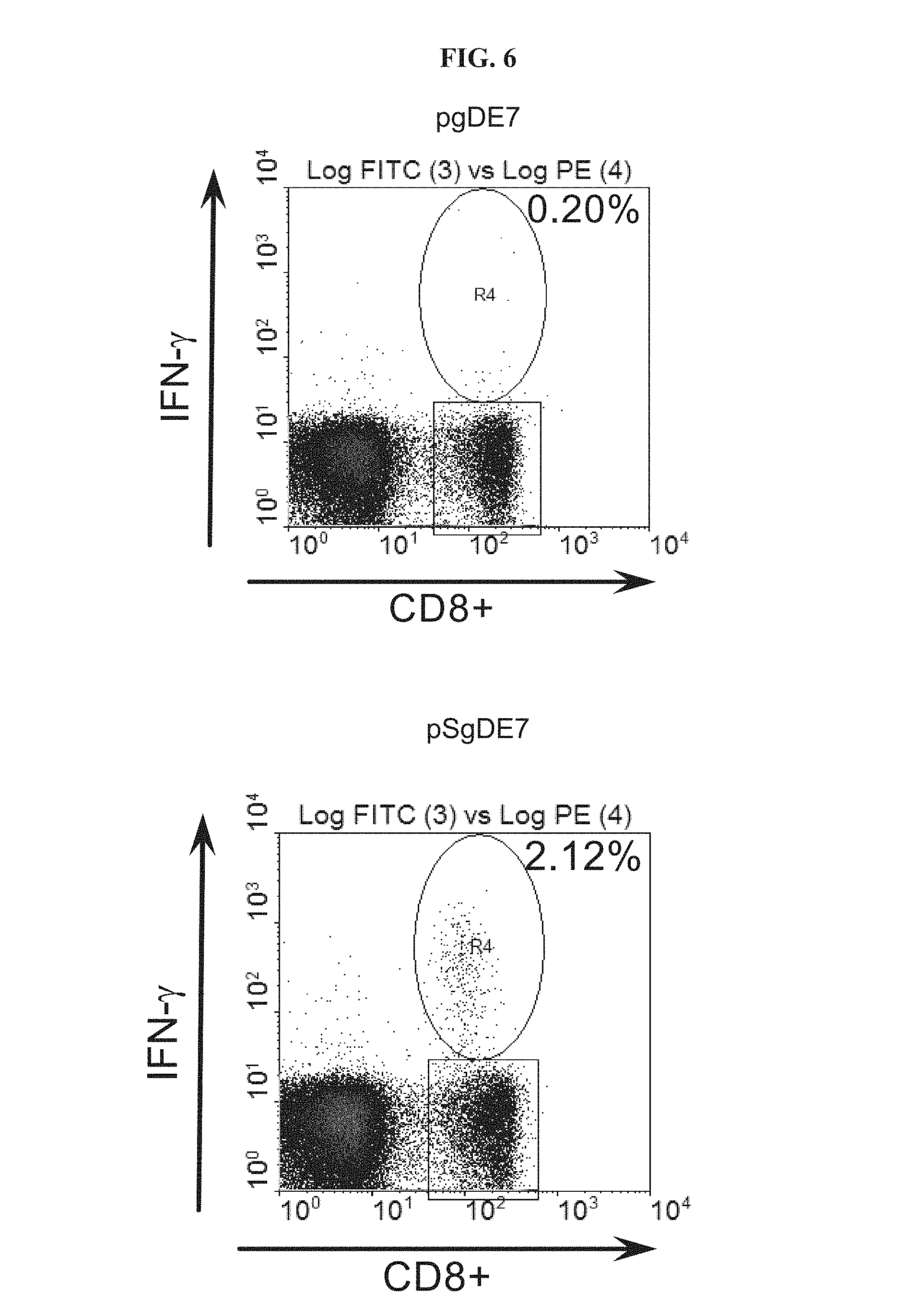
FIG. 6 depicts the E7-specific CD8.sup.+ IFN-.gamma. response in mice immunized with DNA vaccines expressing non-mutated (gDE7) or mutated gD (SgDE7). This mutation (SgD) was designed to increase binding between gD and HVEM. Mice were immunized with pgDE7 or pSgDE7 and 14 days later peripheral blood mononuclear cells were investigated by intracellular cytokine staining for E7-specific CD8.sup.+ IFN-.gamma. response. Numbers in the corners represent percentage CD8.sup.+/IFN-.gamma..sup.+ cells over total of CD8.sup.+ cells.
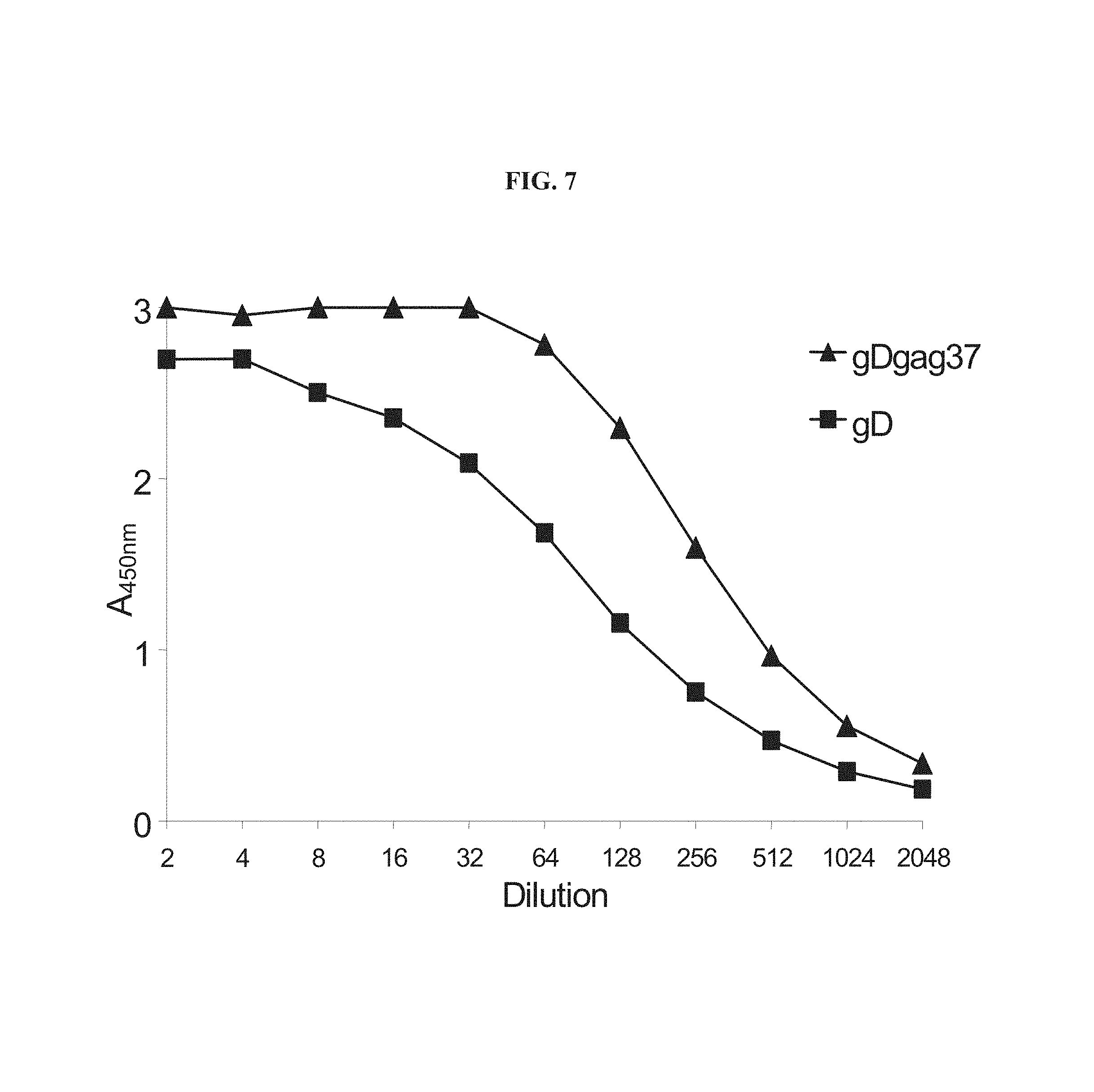
FIG. 7 graphically depicts an in vitro HVEM binding assay. CHO-CAR cells (Chinese hamster ovary-coxsackie-adenovirus receptor cells) were infected with either AdC68gD or AdC68gDgag and, after 48 hours, total protein was extracted. The amount of gD in each sample was quantified by capture ELISA and the protein extracts were diluted in extraction buffer to normalized levels of gD. Equalized extracts were diluted and added to 96-well plates coated with purified HVEM. The amount of gD bound to HVEM was detected by using anti-gD polyclonal antisera and anti-Rabbit IgG horseradish peroxidase. Data shown is one representative experiment from two performed.
FIGS. 8A-8B depict confocal microscopy for localization of gDgag and HVEM on AdC68gDgag infected cells. B78H1/3E5 cells, which expressed HVEM fused to enhanced green fluorescence protein (HVEM-EGFP), were infected with AdC68gDgag. After 48 hours, cells were either directly stained (FIG. 8A) or permeabilized and stained (FIG. 8B) with an anti-gD monoclonal antibody (DL-6) and anti-mouse IgG conjugated with Texas Red. Cells were examined with a Leica TCS SP2 Confocal Microscope at 400.times. final magnification.
FIG. 9 graphically depicts FACS analysis of gDgag expression on the surface of AdC68gDgag infected cells. B78H1/3E5 cells (darker line), which express HVEM-EGFP on the surface, and B78H1 cells (lighter line), which do not express HVEM, were infected with AdC68gDgag. Cells were cultivated for 48 hours, and then labeled with an anti-gD monoclonal antibody (DL-6) and anti-mouse IgG conjugated to phycoerythrin (PE). Cell suspensions were analyzed using an EPICS XL (Beckman-Coulter, Inc., Miami, Fla.) to determine presence of gDgag.
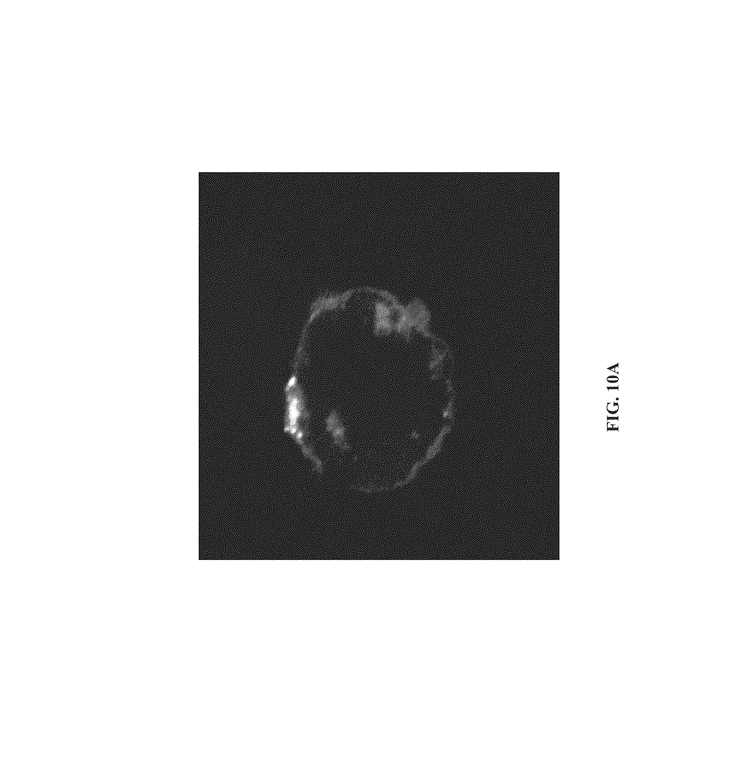
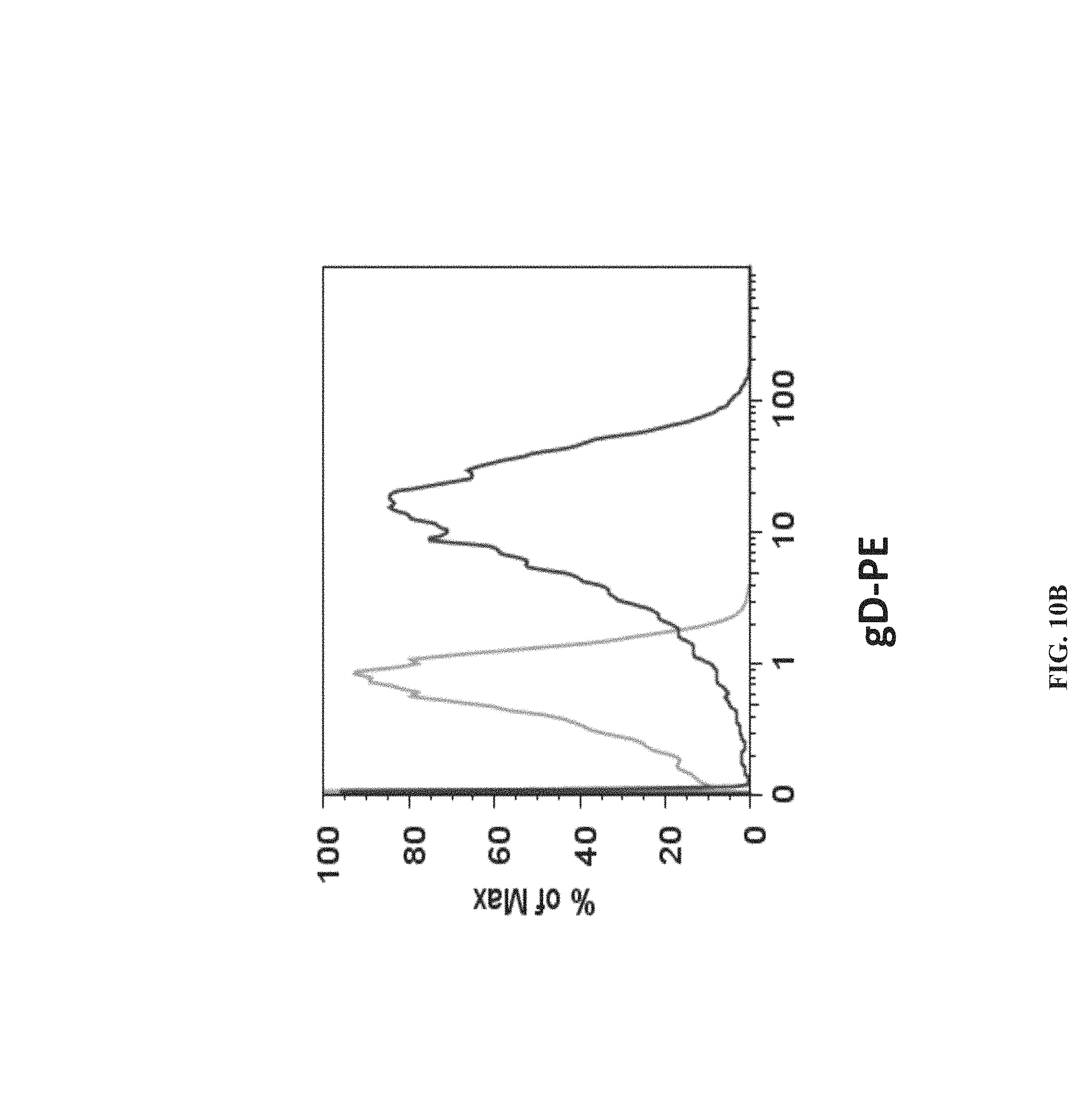
FIGS. 10A-10B depict that gDgag expressed by infected cells bound to HVEM expressed on non-infected cells. Confocal microscopy (FIG. 10A) and FACS analysis (FIG. 10B) were performed to localize gDgag on non-infected cells. CHO-CAR cells were infected with AdC68gDgag. After 48 hours, cells were harvested and washed extensively with cold PBS. AdC68gDgag-infected CHO-CAR cells were cultured with B78H1/3E5 cells, which expresses HVEM-EGFP on the surface, at 4:1 ratio. After 48 hours, cells were stained using the anti-gD monoclonal antibody DL-6 and anti-mouse IgG conjugated to Texas Red (microscopy) or PE (FACS). (FIG. 10A) Microscopy was performed with a Leica TCS SP2 Confocal Microscope at 400.times. final magnification. (FIG. 10B) Cell suspensions were analyzed using an EPICS XL (Beckman-Coulter, Inc., Miami, Fla.). B78H1/3E5 cells were cultured with either AdC68gDgag-infected CHO-CAR cells (darker line) or non-infected CHO-CAR cells (lighter line). Data on graph show cells which are positive for GFP.
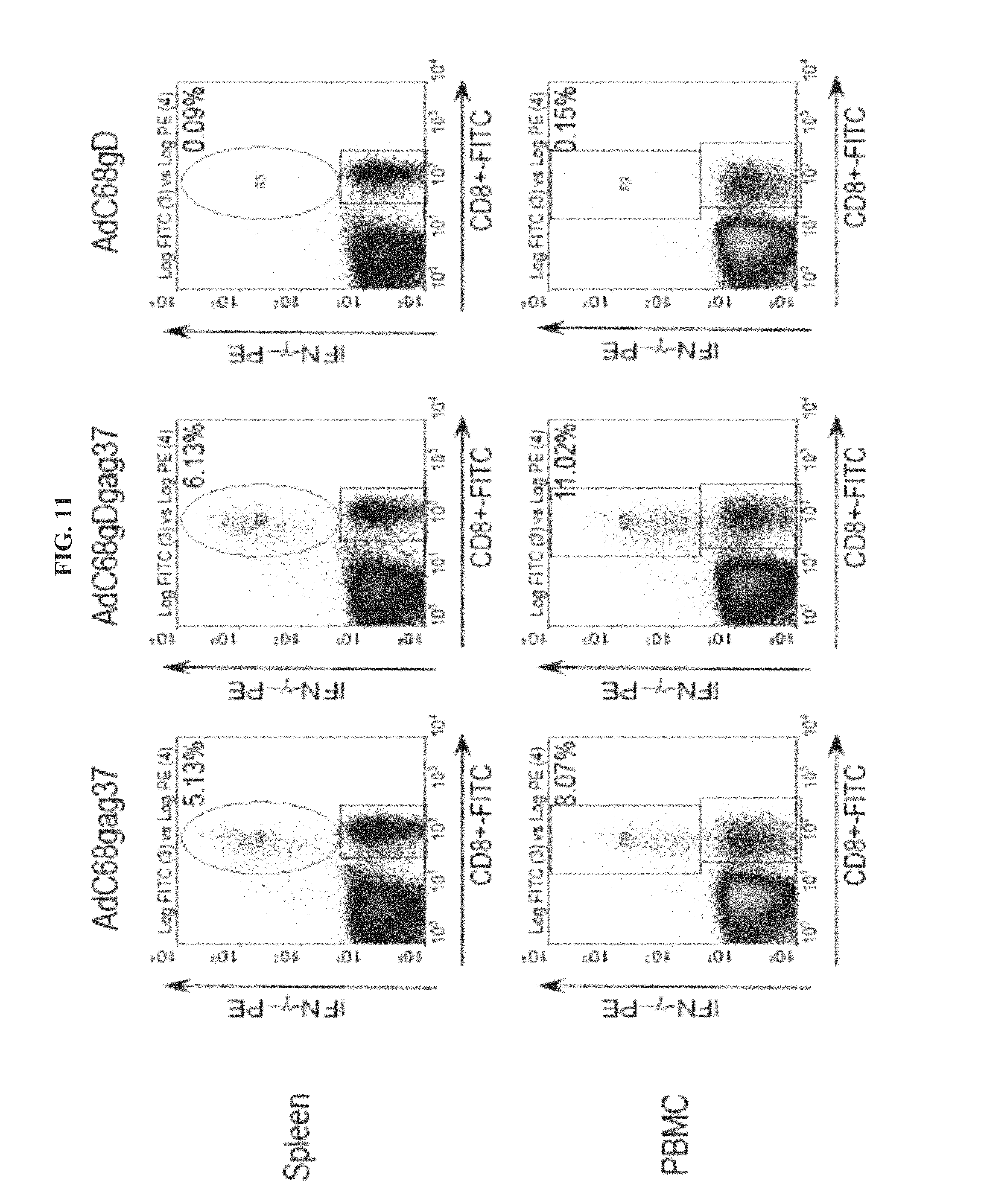
FIG. 11 depicts the gag-specific CD8.sup.+ T cell response in mice immunized with AdC68 vectors. PBMC and splenocytes from mice immunized with 1.times.10.sup.9 vp of AdC68 vectors carrying either gag, gDgag or gD were tested. Percentage represents CD8.sup.+/IFN-.gamma..sup.+ cells over total of CD8.sup.+ cells. CD8.sup.+/IFN-.gamma..sup.+ frequencies in all groups stimulated with an unrelated control peptide were below 0.20%. Data shown are representative of two performed experiments.
FIG. 12 graphically depicts the gag-specific IFN-.gamma. response of CD8.sup.+ T cells from mice immunized with AdC68 vectors. PBMC are from mice immunized with with different amounts of AdC68 vectors carrying either gag, gDgag or gD. Percentage represents CD8.sup.+/IFN-.gamma..sup.+ cells over total of CD8.sup.+ cells. CD8.sup.+/IFN-.gamma..sup.+ frequencies in all groups stimulated with an unrelated control peptide were 0.20%. Data shown are representative of two performed experiments.
FIG. 13 depicts the phenotypic profile of CD8.sup.+ cells activated by AdC68 vaccination. PBMC from naive mice and mice immunized with AdC68 carrying either gag or gDgag were stained with gag-tetramer-APC and anti-CD8-PerCP, in combination with anti-CD25-PE, anti-CD-122-PE, anti-CD127-PE, anti-CD27-PE, anti-CD62L-FITC, anti-CD69-PE, anti-CD103-PE, anti-CD43-PE, anti-CD44-FITC, anti-CD54-PE, anti-Bc12-PE, anti-BTLA-PE, anti-CTLA4-PE and anti-PD1-PE. Graphs show data from CD8.sup.+/gag-tet.sup.+ cells for AdC68gag (gray line) and AdC68gDgag (black line), and total CD8.sup.+ for naive (black dotted line). Data were analyzed on Flowjo software (Tree Star Inc.).
FIG. 14. Intracellular IFN-.gamma. staining of gag-specific and AAV-specific CD8.sup.+ T cells from mice immunized with AAV vectors expressing gD, gag or gDgag. Detection of gag-specific and AAV-specific CD8.sup.+ T cells was carried out after stimulation of peripheral blood mononuclear cells (PBMCs) with either MHC class I restricted gag peptide or AAV-capsid peptide and cell surface staining for CD8 and intracellular staining for IFN-.gamma.. Unspecific peptide was used as control. The numbers in the right upper corners show the frequencies of peptide-specific CD8.sup.+ T cells, as percentages of IFN-.gamma.-producing CD8.sup.+ T cells over all detected CD8.sup.+ T cells.
FIG. 15. Schematic representation of chimeric gene gD-NP. Nucleoprotein P (NP) from Influenza virus A/PR8 without its start and stop codons was incorporated into the HSV-1 gD ApaI site, which corresponds to amino acid 244 in the mature form of gD.
FIG. 16. NP-tetramer staining of CD8.sup.+ T cells isolated from blood of mice immunized with either pgD-NP or pNP DNA vaccine. Mice were immunized with 100 .mu.g of each DNA vaccine vector. Fourteen days after immunization peripheral blood mononuclear cells (PBMCs) were isolated and cell surface stained with the NP-tetramer and a labeled antibody to CD8. Naive mice were used as negative control. Data represent percentages of NP-tetramer.sup.+ CD8.sup.+ T cells over all detected CD8.sup.+ T cells.
FIG. 17. Schematic representation of chimeric gene gDTRAPTB. Thrombospondin-related anonymous protein (TRAP) from parasite Plasmodium falciparum and Mycobacterium tuberculosis epitope string (TB) without their start and stop codons are incorporated into the HSV-1 gD NarI site, which corresponds to amino acid 288 in the gD mature form.
FIGS. 18A-18D. Molecular modeling of the gD-gag chimeric protein. Ribbon representations of gD-gag in the unligated (FIGS. 18A, 18B) and HVEM ligated (FIGS. 18C, 18D) conformations. FIG. 18A, the gag insertion is connected to the gD ectodomain core by a long flexible linker. FIG. 18B, a superposition of the native gD X-ray structure (2C36) (darker ribbon) and that of the gD-gag chimera model (lighter ribbon) shows that the gag insert repositions the C-terminus of native gD away from the HVEM binding pocket. The dashed line indicates an 11 residue gD loop segment that like the first 22 N-terminal residues (not shown) is unresolved in the X-ray structure and presumed to be highly flexible (Krummenacher et al., EMBO 1 24, 4144-53, 2005). FIG. 18C, gD-gag chimera model in the HVEM ligated conformation with HVEM positioned as observed in the gD-HVEM complex X-ray structure (1JMA). The gD N-terminus changes conformation upon formation of the HVEM complex. FIG. 18D, a superposition of the native gD X-ray structure (2C36) (darker ribbon) with the gD-gag chimera model (lighter ribbon) in the HVEM-bound conformation shows that the gag insert does not disrupt the gD core domain.
FIG. 19. Confocal microscopy was carried out with B78-H1/3E5 cells, which express HVEM fused to Enhanced Green Fluorescence Protein (HVEM-EGFP). B78-H1/3E5 cells were infected with AdC68gag, AdC68gD or AdC68gD-gag, then stained anti-gD DL-6 MAb and anti-mouse IgG conjugated with Texas Red. AdC68gD-gag-infected cells were permeabilized then stained as above. Cells were examined with a Leica TCS SP2 Confocal Microscope at 400.times. final magnification.
FIG. 20. Comparison of gD expression on the surface of B78-H1 (black line) and B78-H1/3E5 (gray) cells infected with AdC68 vectors carrying gD-gag, gD or gag.
FIG. 21. Presence of gD on the surface of non-infected HVEM.sup.+ cells co-cultivated with HVEM.sup.- cells infected with either AdC68gD-gag, AdC68gD or AdC68gag. Non-infected cells were used as negative control.
FIG. 22. AdC68gD induces enhanced expansion of CD8.sup.+ T cells in vitro. Irradiated lymph nodes cells from naive mice and mice immunized with either AdC68E7E6E5 or AdC68gD were incubated with CFSE-labeled CD8.sup.+ OT-1 (V.alpha.2.sup.+) cells for 72 hrs. Total live cells (left) were analyzed for expression of CD8.sup.+V.alpha.2.sup.+. CSFE expression by these double positive populations (highlighted by the squares on the right graphs) is shown on the left graphs. The bars and numbers show from right to left the percentages of the population that underwent no replication, 1, 2, 3 or .ltoreq.4 cycles of replication. Graphs show data from one representative experiment of two performed.
FIGS. 23A-23C. CD8.sup.+ T cell responses to vectors expressing antigens fused to gD. FIG. 23A, intracellular cytokine staining of E7- and gag-specific CD8.sup.+ T cells were carried out on PBMCs from mice i.m. immunized with DNA vaccines (upper graphs) or AdC68 vectors (lower graphs) expressing either gD, E7E6E5, gD-E7E6E5, gag or gD-gag, after stimulation with E7 or gag peptide and cell surface staining for CD8 (FITC) and intracellular staining for IFN-.gamma. (PE). PBMCs were isolated from animals 14 days after DNA vaccination or 10 days after application of AdC68 vector. The numbers in the right upper corners show frequencies of IFN-.gamma.-producing CD8.sup.+ T cells as a percentage of all CD8.sup.+ T cells. Frequencies of IFN-.gamma..sup.+/CD8.sup.+ T cells stimulated with an unrelated control peptide were below 0.2% in all groups. FIG. 23B, Gag-specific CD8.sup.+ T cell frequencies were determined 10 days after immunization of mice with decreasing doses of either AdC68gag (open bars) or AdC68gD-gag (black bars) vectors. FIG. 23C, The kinetics of E7-specific CD8.sup.+ T cell responses induced by the AdC68gD-E7E6E5 vector were analyzed from BPMCs of mice immunized with either AdC68E7E6E5 (squares), AdC68gD (diamonds) or AdC68gD-E7E6E5 (triangles) vectors at different days after a single dose of 10.sup.10 vps of the vaccines.
FIGS. 24A-24B. The enhancement of CD8.sup.+ T cell responses requires binding of gD to HVEM. FIG. 24A, E7-specific IFN-.gamma..sup.+CD8.sup.+ responses were evaluated with splenocytes from mice immunized with one dose of DNA vaccines expressing the E7E6E5 polypeptide either within wild-type gD (pgD-E7E6E5), a mutated gD that shows loss of binding to HVEM (NBEFgD-E7E6E5) or that shows enhanced binding to HVEM (SgD-E7E6E5). FIG. 24B, splenocytes from mice immunized with one dose of DNA vaccines carrying E7 fused to either wild type gD (gD-E7) or mutated gD with high affinity to HVEM (SgD-E7) were evaluated for E7-specific IFN-.gamma..sup.+CD8.sup.+ response. PBMCs were isolated 14 days after DNA vaccine immunizations.
FIG. 25. Phenotypes of gag-specific CD8.sup.+ T cells were analyzed on PBMCs from mice immunized with either AdC68gD-gag or AdC68gag. PBMCs were isolated 10 days after immunization. Naive mice were used as controls (black dotted line). The graphs shown reflect expression levels of total CD8+ T cells from naive mice (black dotted line) and gag-tet.sup.+CD8.sup.+ T cells from mice immunized with either AdC68gag (grey line) or AdC68gD-gag (black line).
FIGS. 26A-26C. CD8.sup.+ T cells induced by gD-antigen chimeric protein are functional in vivo. Protection against TC-1 tumor challenge was evaluated in mice vaccinated with DNA (FIG. 26A) or AdC68vectors (FIG. 26B) expressing either gD (circles), E7E6E5 (diamonds) or gD-E7E6E5 (squares). FIG. 26C, Protection to TC-1 tumor challenge in mice vaccinated with DNA vaccine expressing either NBEFgD-E7E6E5 (diamonds), SgD-E7E6E5 (squares), gD-E7 (circles) or SgD-E7 (triangles) chimeric genes. Mice were challenged 14 and 10 days after vaccination with DNA and AdC68 vectors, respectively. Tumor development was followed for up to 60 days after challenge.
FIGS. 27A-27B. Quantification of specific mRNA copies and protein expression by cells infected in vitro with AdC68 vectors. FIG. 27A, RNA isolated from non-infected cells and from cells infected with AdC68gD (white bars), AdC68E7E6E5 (gray bars) or AdC68gD-E7E6E5 (black bars) were reverse transcribed and quantified by Real-Time PCR. After quantification of GAPDH mRNA copies, all samples were normalized to 10.sup.9 GAPDH mRNA copies. Specific mRNA copies were quantified using gD, E7, E6, and E5 specific primers. Neither E7, E6 nor E5 mRNA were detected in cells infected with AdC68gD, and no gD specific mRNA was detected in cells infected with AdC68E7E6E5. mRNA levels were assessed in three independent experiments and each sample was investigated in triplicates. p values from two-tail student's t test are shown on top of the bars. FIG. 27B, confocal microscopy was carried out with CHO/CAR cells infected with AdC68 expressing either gD, E7E6E5 or gD-E7E6E5, then permeabilized and stained with anti-gD DL-6 MAb and anti-mouse IgG conjugated with FITC. Immunoflorescence is shown on the top panel while differential interference contrast (DIC) microscopy is shown on the bottom panel. Cells were examined with a Leica TCS SP2 Confocal Microscope at 400.times. magnification.
FIGS. 28A-28C. Intracellular IFN-.gamma. staining of E7-specific CD8.sup.+ T cells from mice immunized with AdC68 and DNA vectors. FIG. 28A, frequencies of E7-specific CD8.sup.+ T cells in PBMC (top) or spleens (bottom) from naive mice or mice immunized with AdC68 vector expressing either gD, E7E6E5 or gD-E7E6E5 were determined 10 days after immunization. FIG. 28B, frequencies of E7-specific CD8.sup.+ T cells in PBMC (top) or spleens (bottom) from naive mice or mice immunized with DNA vaccines expressing either gD, E7E6E5 or gD-E7E6E5 were determined 14 days after immunization. FIG. 28C, prime and boost regimens with pgD-E7E6E5 and AdC68gD-E7E6E5 vectors. Mice immunized with one dose of pgD-E7E6E5 vector were boosted after 90 days with AdC68gD-E7E6E5 (open bars), while mice immunized with AdC68gD-E7E6E5 were boosted after 90 days with pgD-E7E6E5 (black bars). Detection of E7-specific CD8.sup.+T response was carried out after stimulation with a MHC class I restricted E7 peptide and cell surface staining for CD8 (FITC) and intracellular staining for IFN-.gamma. (PE). The numbers in the right upper corners show the frequencies of E7-specific CD8.sup.+ T cells, as percentages of IFN-.gamma.-producing CD8.sup.+ T cells over all detected CD8.sup.+ T cells. IFN-.gamma.-producing CD8.sup.+ cell frequencies in all groups stimulated with an unrelated peptide or in the absence of stimulus were below 0.2%. The data shown in a and b are from one representative experiment of four performed.
FIG. 29. Dose-response of the CD8+ T cell response to AdC68gD-E7E6E5. Frequencies of IFN-.gamma.-producing E7-specific CD8.sup.+T cells in spleens and PBMCs induced by 5.times.10.sup.10 to 1.times.10.sup.8 vp/animal of AdC68gD-E7E6E5 vector were determined as described on legend to FIGS. 28A-28C.
FIGS. 30A-30B. Anti-tumor effects of AdC68 vectors against TC-1 cell challenge. FIG. 30A, for post-challenge vaccination, groups of 10 mice were s.c. inoculated with TC-1, then 5 days later immunized with either AdC68gD (triangles), AdC68E7E6E5 (circles) or AdC68gD-E7E6E5 (squares). FIG. 30B, for pre-challenge vaccination, groups of 10 mice vaccinated one year earlier with AdC68 vectors carrying either gD (triangles), E7E6E5 (circles) or gD-E7E6E5 (squares) were s.c. challenged with TC-1. For all TC-1 challenge experiments animals were monitored 3 times per week for evidence of tumor growth over a period of 60 days.
FIGS. 31A-31B. CD8.sup.+ T cell response after challenge with TC-1 in mice vaccinated one year earlier. One year after vaccination with AdC68 vectors expressing either gD, E7E6E5 or gD-E7E6E5, mice were challenged with TC-1 cells and 10 days later E7-specific frequencies of E7-specific CD8.sup.+ T cells were determined. FIG. 31A, ICS of E7-specific CD8.sup.+ T cells in spleen from non-challenged mice (top) or mice challenged with TC-1 cells (top). Frequencies of IFN-.gamma.-producing E7-specific CD8.sup.+T cells in spleen were determined as in FIG. 2 legend. FIG. 31B, E7-tetramer staining of CD8.sup.+ T cells isolated from spleen, blood and liver of mice challenged (white bars) or not (black bars) with TC-1 cells. Data represent percentages of E7-tetramer.sup.+ CD8.sup.+ T cells over all detected CD8.sup.+ T cells. E7-tetramer.sup.+CD8.sup.+ cells were not detected in mice immunized with either AdC68gD or AdC68E7E6E5.
FIGS. 32A-32C. CD8.sup.+ T cell response and phenotypic profile in mice immunized with AdC68 vectors and subsequently challenged with TC-1 cells. Lymphocytes were isolated 3 days after TC-1 challenge from animals immunized with AdC68gD-E7E6E5 and 7 days after challenge from animals immunized with either AdC68gD or AdC68E7E6E5. E7-specific CD8.sup.+ T response were determined in spleen, PBMC and TIL by ICS (FIG. 32A) and E7-tetramer staining (FIG. 32B). ICS data was determined as in FIG. 2 legend, while E7-tetramer staining data represent percentages of E7-tetramer.sup.+ CD8.sup.+ T cells over all detected CD8.sup.+ T cells. E7-specific IFN-.gamma..sup.+CD8.sup.- cells and E7-tetramer.sup.+CD8.sup.+ cells were not detected in mice immunized with either AdC68gD or AdC68E7E6E5. FIG. 32C, phenotype analysis of splenocytes, PBMCs and TILs were determined with cells isolated from mice immunized with AdC68gD-E7E6E5 (black line) or AdC68gD (dotted black line) then challenged with TC-1. Cells were stained with E7-tetramer-APC and anti-CD8-PerCP, in combination with antibodies to CD44, CD62L, CD27, Bc12, BTLA, CTLA-4 and PD-1. CD8.sup.+ T cells isolated from either naive mice or mice immunized with AdC68E7E6E5 showed similar phenotype profiles as CD8.sup.+ T cells isolated from mice immunized with AdC68gD.
FIGS. 33A-33C. Comparison of CD8.sup.+ T cell responses and phenotype profiles induced by AdC68 vectors in wild-type and HPV-16 E6/E7-tg mice. One-year old E6/E7-tg mice were vaccinated with AdC68 vectors expressing either gD, E7E6E5 or gD-E7E6E5, and 1-year old C57B1/6 mice were vaccinated with AdC68gD-E7E6E5 vector. Ten days later frequencies and phenotypes of E7-specific CD8.sup.+ T cells were determined. FIG. 33A, E7-specific CD8.sup.+ T cells isolated from spleens of E6/E7-tg mice immunized with AdC68gD, AdC68E7E6E5 or AdC68gD-E7E6E5, or from spleen of wild-type mice immunized with AdC68gD-E7E6E5 were tested by ICS. Frequencies of IFN-.gamma.-producing E7-specific CD8.sup.+T cells in spleen were determined as described in legend to FIG. 2. FIG. 33B, E7-tetramer staining of CD8.sup.+ T cells was performed with splenocytes and PBMC from wild-type and E6/E7-tg mice, and with lymphocytes from thyroids of E6/E7-tg mice. Data show percentages of E7-tetramer.sup.+ CD8.sup.+ T cells over all detected CD8.sup.+ T cells. ND, not determined. FIG. 33C, phenotypic profile of E7-specific CD8.sup.+ T cells were determined using cells isolated from spleen and blood of AdC68gD-E7E6E5 vaccinated E6/E7 tg (filled gray) and wild-type (black line) mice, and from cells isolated from thyroid of E6/E7 tg mice vaccinated with AdC68gD-E7E6E5. CD8.sup.+ T cells isolated from E6/E7 tg naive mice were used as controls (black dotted line).
DETAILED DESCRIPTION
The invention provides chimeric, or fusion, proteins in which one or more antigens is inserted into the C terminal region of a mature HSV gD protein. Such fusion proteins enhance the immune response of a host against the antigen(s) to a much greater degree than is observed without the gD. The gD chimeric proteins of the present invention are particularly suitable for use as genetic vaccines (e.g., DNA vaccines or viral vector vaccines) to therapeutically or prophylactically treat a subject. Thus, the invention also provides nucleic acid molecules which encode fusion proteins of the invention.
Glycoprotein D
Glycoprotein D (gD) is an envelope glycoprotein found on Herpes simplex viruses such as HSV-1 or HSV-2 and is expressed in cells infected by the viruses. An HSV gD has a 25-amino acid amino-terminal signal sequence and a carboxy-terminal transmembrane domain. The signal sequence is typically cleaved in the mature form of the protein. The amino acid sequence of HSV-1 gD is shown in SEQ ID NO:27 (amino acids 1-25 are the signal sequence; amino acids 26-394 are mature HSV-1 gD); a coding sequence for SEQ ID NO:27 is shown in SEQ ID NO:26. The amino acid sequence of HSV-2 gD is shown in SEQ ID NO:29 (amino acids 1-25 are the signal sequence; amino acids 26-393 are mature HSV-2 gD); a coding sequence for SEQ ID NO:25 is shown in SEQ ID NO:28.
An HSV gD or mutant thereof which is useful in the present invention has the ability to interact with HVEM and, in addition, may have one or more of the following properties: 1) ability to stimulate a CD8.sup.+ T cell response to the fusion partner; 2) ability to disrupt an HVEM-BTLA pathway activity; 3) ability to interact with nectin-1; 4) ability to mediate cell entry by an HSV-1 and/or HSV-2 virus; and 5) ability to mediate cell-to-cell spread of HSV-1 and/or HSV-2. Thus, as used herein, a "gD" or an "immunostimulatory portion of a gD" refers to a polypeptide having an amino acid sequence of a wild-type gD or a mutant thereof which retains one or more gD activities.
gD chimeric (fusion) proteins of the invention comprise at least two, preferably three polypeptide segments. The first polypeptide segment comprises at least amino acids 1-240 of a mature Herpes simplex virus (HSV) glycoprotein D; in preferred embodiments the first polypeptide segment does not comprise a full length mature glycoprotein D; in this case a third polypeptide segment is included. The second polypeptide segment, the N terminus of which is linked to the C terminus of the first polypeptide segment, comprises at least one antigen which is not an HSV glycoprotein D antigen. The third polypeptide comprises a C terminal portion of the HSV glycoprotein D; the N terminus of the third polypeptide segment is linked to the C terminus of the second polypeptide segment. Thus, in certain embodiments an antigen is fused to the carboxy-terminal region of gD. In other aspects, an antigen is inserted within the carboxy-terminal amino acid sequence of gD such that the amino-terminal end of the gD chimeric protein is an amino-terminal gD amino acid sequence, fused to an internal antigenic sequence, fused to a carboxy-terminal amino acid sequence of gD.
In certain embodiments, the first polypeptide segment of a gD chimeric protein of the present invention includes the entire gD amino acid sequence (e.g., amino acids 1-394) or the mature gD amino acid sequence (e.g., amino acids 26-394, the carboxy-terminal 369 amino acids). In other embodiments, the first polypeptide segment includes less than full-length mature gD but includes 250, 260, 270, 280, 290, 300, 305, 310, 315, 320, 325, 330, 335, 340, 345, 350, 351, 352, 353, 354, 355, 356, 357, 358, 359, 360, 360, 361, 362, 363, 362, 365, 366, 367 or 368 amino acids of the mature gD sequence.
In certain aspects, an antigenic amino acid sequence is inserted within a region of a gD that is between amino acids 230 and 300, between amino acids 235 and 295, or between amino acids 240 and 290 of a mature gD amino acid sequence. In other aspects an antigenic amino acid sequence is inserted at a position carboxy-terminal to amino acid 230, 235, 240, 245, 250, 255, 260, 265, 270, 275, 280, 285 of a mature gD amino acid sequence. In certain aspects, an antigenic amino acid sequence is inserted immediately adjacent to amino acid 240, 241, 242, 243, 244, 245, 246, 247, 248, 249, 250, 251, 252, 253, 254, 255, 256, 257, 258, 259, 260, 261, 262, 263, 264, 265, 266, 267, 268, 269, 270, 271, 272, 273, 274, 275, 276, 277, 278, 279, 280, 282, 282, 283, 284, 285, 286, 287, 288, 289, 290, 291, 292, 293, 294, 295, 296, 297, 298, 299, or 300 of a mature gD amino acid sequence
In certain embodiments, a chimeric gD protein of the invention has a structure that is similar to the structure of the wild-type protein, that is, the chimeric gD protein has the ability to interact with HVEM and, in addition, may have one or more of the following activities: 1) stimulating a CD8.sup.+ T cell response to the fusion partner; 2) disrupting an HVEM-BTLA pathway activity; 3) interacting with nectin-1; 4) mediating cell entry by an HSV-1 and/or HSV-2 virus; and/or 5) mediating cell-to-cell spread of HSV-1 and/or HSV-2.
Antigens
As used herein, the term "antigen" (also termed "fusion partner" in relation to gD) is intended to include, but is not limited to, a substance that an immune response is specifically mounted against, such as a protein or a polypeptide. An antigen of the invention can be of any length, ranging in size from a few amino acids in length to hundreds of amino acids in length, provided that that the chimeric gD maintains a structure that is similar to that of the wild-type gD, e.g., the chimeric gD retains one or more activities of the wild-type gD, particularly the activities of interacting with HVEM and/or stimulating a CD8.sup.+ T cell response to the fusion partner in the host. In certain aspects of the invention, the antigen is 1000, 900, 800, 700, 600, 500, 400, 350, 340, 330, 320, 310, 300, 290, 280, 270, 260, 250, 240, 230, 220, 210, 200, 190, 180, 170, 160, 150, 140, 130, 120, 110, 100, 90, 80, 70, 60, 50, 40, 30, 20, 10 or less amino acids in length.
An antigen of the present invention includes a heterologous protein and/or polypeptide, such as a viral, bacterial, fungal, or parasite protein or polypeptide, and/or a host polypeptide and/or protein e.g., a tumor cell polypeptide or protein (such as an oncoprotein or a portion thereof) or a polypeptide or protein associated with inflammation. Antigens of particular interest include, but are not limited to, influenza virus antigens, such as nucleoprotein P (NP; see FIG. 16), matrixprotein (M), and hemagglutinin (HA); Plasmodium antigens such as thrombospondin-related anonymous protein (TRAP; see FIG. 17), ring-infected erythrocyte surface antigen (RESA), merozoite surface protein 1 (MSP1), merozoite surface protein 2 (MSP2), merozoite surface protein 3 (MSP3), and glutamate-rich antigen (GLURP); human papilloma virus (HPV) antigens, particularly HPV-16 antigens, such as E5 protein, E6 protein, and E7 protein; and HIV antigens, such as gag, pol, nef, tet, and env.
Viruses include DNA or RNA animal virus. As used herein, RNA viruses include, but are not limited to, virus families such as picornaviridae (e.g., polioviruses), reoviridae (e.g., rotaviruses), togaviridae (e.g., encephalitis viruses, yellow fever virus, rubella virus), orthomyxoviridae (e.g., influenza viruses), paramyxoviridae (e.g., respiratory syncytial virus (RSV), measles virus (MV), mumps virus (MuV), parainfluenza virus (PIV)), rhabdoviridae (e.g., rabies virus (RV)), coronaviridae, bunyaviridae, flaviviridae (e.g., hepatitis C virus (HCV)), filoviridae, arenaviridae, bunyaviridae, and retroviridae (e.g., human T-cell lymphotropic viruses (HTLV), human immunodeficiency viruses (HIV)). As used herein, DNA viruses include, but are not limited to, virus families such as papovaviridae (e.g., papilloma viruses), adenoviridae (e.g., adenovirus), herpesviridae (e.g., herpes simplex viruses, e.g., HSV-1, HSV-2; varicella zoster virus (VZV); Epstein-Barr virus (EBV); cytomegalovirus (CMV); human herpesviruses, e.g., HHV-6 and HHV-7; Kaposi's sarcoma-associated herpesvirus (KSHV) and the like), and poxviridae (e.g., variola viruses). These and other viruses and viral proteins are included in the present invention and are described further in Knipe et al., Field's Virology, 4.sup.th ed., Lippincott Williams & Wilkins, 2001, incorporated herein by reference in its entirety for all purposes.
Bacteria include, but are not limited to, gram positive bacteria, gram negative bacteria, acid-fast bacteria and the like. As used herein, gram positive bacteria include, but are not limited to, Actinomedurae, Actinomyces israelii, Bacillus anthracis, Bacillus cereus, Clostridium botulinum, Clostridium difficile, Clostridium perfringens, Clostridium tetani, Corynebacterium, Enterococcus faecalis, Listeria monocytogenes, Nocardia, Propionibacterium acnes, Staphylococcus aureus, Staphylococcus epiderm, Streptococcus mutans, Streptococcus pneumoniae and the like. As used herein, gram negative bacteria include, but are not limited to, Afipia felis, Bacteriodes, Bartonella bacilliformis, Bortadella pertussis, Borrelia burgdorferi, Borrelia recurrentis, Brucella, Calymmatobacterium granulomatis, Campylobacter, Escherichia coli, Francisella tularensis, Gardnerella vaginalis, Haemophilius aegyptius, Haemophilius ducreyi, Haemophilius influenziae, Heliobacter pylori, Legionella pneumophila, Leptospira interrogans, Neisseria meningitidia, Porphyromonas gingivalis, Providencia sturti, Pseudomonas aeruginosa, Salmonella enteridis, Salmonella typhi, Serratia marcescens, Shigella boydii, Streptobacillus moniliformis, Streptococcus pyogenes, Treponema pallidum, Vibrio cholerae, Yersinia enterocolitica, Yersinia pestis and the like. As used herein, acid-fast bacteria include, but are not limited to, Myobacterium avium, Myobacterium leprae, Myobacterium tuberculosis and the like.
Other bacteria not falling into the other three categories include, but are not limited to, Bartonella henseiae, Chlamydia psittaci, Chlamydia trachomatis, Coxiella burnetii, Mycoplasma pneumoniae, Rickettsia akari, Rickettsia prowazekii, Rickettsia rickettsii, Rickettsia tsutsugamushi, Rickettsia typhi, Ureaplasma urealyticum, Diplococcus pneumoniae, Ehrlichia chafensis, Enterococcus faecium, Meningococci and the like.
Fungi include, but are not limited to, Aspergilli, Candidae, Candida albicans, Coccidioides immitis, Cryptococci, and combinations thereof.
Parasites include, but are not limited to, Balantidium coli, Cryptosporidium parvum, Cyclospora cayatanensis, Encephalitozoa, Entamoeba histolytica, Enterocytozoon bieneusi, Giardia lamblia, Leishmaniae, Plasmodii, Toxoplasma gondii, Trypanosomae, trapezoidal amoeba and the like.
Oncoproteins are intended, without limitation, to refer to proteins and/or peptides that are capable of inducing cell transformation. Oncoproteins include, but are not limited to, cellular proteins such as PDGF, ERB-B, ERB-B2, K-RAS, N-RAS, C-MYC, N-MYC, L-MYC, BCL-2, BCL-1, MDM2 and the like. Oncoproteins also include, but are not limited to, viral proteins from RNA and/or DNA tumor viruses such as hepatitis B viruses, SV40 viruses, polyomaviruses, adenoviruses, herpes viruses, retroviruses and the like. Tumor suppressor proteins are intended, without limitation, to refer to proteins or polypeptides that can suppress or block aberrant cellular proliferation, as well as tumor suppressor proteins that have been mutated and, accordingly, no longer suppress or block aberrant cellular proliferation. Tumor suppressor proteins include, but are not limited to, cellular proteins such as APC, DPC4, NF-1, NF-2, MTS1, RB, p53 and the like. gD chimeric proteins of the present invention are useful for modulating disorders associated with aberrant cellular proliferation mediated by oncoproteins and/or tumor suppressor proteins, such as cancer. Aberrant cellular proliferation is intended to include, but is not limited to, inhibition of proliferation including rapid proliferation. As used herein, the term "disorder associated with aberrant cellular proliferation" includes, but is not limited to, disorders characterized by undesirable or inappropriate proliferation of one or more subset(s) of cells in a multicellular organism. The term "cancer" refers to various types of malignant neoplasms, most of which can invade surrounding tissues, and may metastasize to different sites (PDR Medical Dictionary 1st edition (1995)). The terms "neoplasm" and "tumor" refer to an abnormal tissue that grows by cellular proliferation more rapidly than normal and continues to grow after the stimuli that initiated proliferation is removed (PDR Medical Dictionary 1st edition (1995)). Such abnormal tissue shows partial or complete lack of structural organization and functional coordination with the normal tissue which may be either benign (benign tumor) or malignant (malignant tumor).
Polypeptides and proteins associated with inflammation include those that modulate a disease or disorder characterized by, caused by, resulting from, or becoming affected by inflammation. Examples of inflammatory diseases or disorders include, but not limited to, acute and chronic inflammation disorders such as asthma, psoriasis, rheumatoid arthritis, osteoarthritis, psoriatic arthritis, inflammatory bowel disease (Crohn's disease, ulcerative colitis), sepsis, vasculitis, and bursitis; autoimmune diseases such as lupus, polymyalgia, rheumatica, scleroderma, Wegener's granulomatosis, temporal arteritis, cryoglobulinemia, and multiple sclerosis; transplant rejection; reperfusion injury in strokes or myocardial infarction; osteoporosis; cancer, including solid tumors (e.g., lung, CNS, colon, kidney, and pancreas); Alzheimer's disease; atherosclerosis; viral (e.g., HIV or influenza) infections; chronic viral (e.g., Epstein-Barr, cytomegalovirus, herpes simplex virus) infection; and ataxia telangiectasia.
Nucleic Acid Molecules
The invention also provides nucleic acid molecules which encode fusion proteins of the invention. In certain embodiments of the invention, the nucleic acid molecule is a vector. As used herein, the term "vector" refers to a nucleic acid molecule, a protein, or a liquid structure capable of transporting another nucleic acid. One type of vector is a "plasmid," which refers to a circular double stranded DNA loop into which additional DNA segments can be ligated.
Another type of vector is a viral vector, wherein additional DNA segments can be ligated into the viral genome. Certain vectors are capable of autonomous replication in a host cell into which they are introduced (e.g., bacterial vectors having a bacterial origin of replication and episomal mammalian vectors). Other vectors are replication-defective and remain in the nucleus as episomes. Other vectors (e.g., non-episomal mammalian vectors) are integrated into the genome of a host cell upon introduction into the host cell, and thereby are replicated along with the host genome. Moreover, certain vectors are capable of directing the expression of genes to which they are operatively linked. Such vectors are referred to herein as "expression vectors." In general, expression vectors of utility in recombinant DNA techniques are often in the form of plasmids. The terms "plasmid" and "vector" can be used interchangeably as the plasmid is a commonly used form of vector.
In one embodiment, a recombinant virus is provided for eliciting an immune response in a host infected by the virus. In certain aspects, the recombinant virus is replication-incompetent. A recombinant virus may be constructed from any virus using methods known in the art, provided that the native progenitor is rendered replication incompetent. For example, replication-incompetent adenovirus, adeno-associated virus, SV40 virus, retrovirus, herpes simplex virus or vaccinia virus may be used to generate the recombinant virus by inserting the viral antigen into a region that is non-essential to the infectivity of the recombinant virus. A recombinant virus does not have the pathologic regions of the native progenitor of the benign virus but retains its infectivity to the host.
In a certain embodiment, the recombinant virus is a replication-incompetent chimpanzee-derived adenovirus. Chimpanzee-derived adenovirus vectors have distinct advantages over previously used adenoviral recombinants of the human serotype 5 that is typically used in the art. Most importantly, the efficacy of simian adenoviral vaccine carriers is not impaired by pre-existing neutralizing antibodies to human adenovirus serotype 5 that can be detected in up to 45% of the adult human population in the United States. Furthermore, simian adenoviral recombinants have interactions with cells of the innate immune system, most notably dendritic cells, which sponsor development of strongly biased Thl responses suited to induce potent responses of CD8.sup.+ T cells, a subset of immunocytes that is particularly important to control the spread of HIV-1. For a review of replication-incompetent chimpanzee-derived adenovirus, see U.S. Pat. No. 6,019,978, incorporated herein by reference in its entirety for all purposes.
A number of viral vectors suitable for in vivo expression of the gD chimeric proteins described herein are known. Such vectors include retroviral vectors (see, e.g., Miller (1992) Curr. Top. Microbiol. Immunol. 158:1; Salmons and Gunzburg (1993) Human Gene Therapy 4:129; Miller et al. (1994) Meth. Enz. 217:581) and adeno-associated vectors (reviewed in Carter (1992) Curr. Opinion Biotech. 3:533; Muzcyzka (1992) Curr. Top. Microbiol. Immunol. 158:97). Other viral vectors that are used include adenoviral vectors, alphavirus replicons, herpes virus vectors, pox virus vectors, and rhabdovirus vectors, as generally described in, e.g., Jolly (1994) Cancer Gene Therapy 1:51; Latchman (1994) Molec. Biotechnol. 2:179; Johanning et al. (1995) Nucl. Acids Res. 23:1495; Berencsi et al. (2001) J. Infect. Dis. 183:1171; Rosenwirth et al. (2001) Vaccine February 19:1661; Kittlesen et al. (2000) J. Immunol. 164:4204; Brown et al. (2000) Gene Ther. 7:1680; Kanesa-thasan et al. (2000) Vaccine 19:483; and Sten (2000) Drug 60:249. Compositions comprising vectors and an acceptable excipient are provided herein.
Nucleic Acid and Protein Variants
In certain aspects, gD nucleic acid molecules and polypeptides are "naturally occurring." As used herein, a "naturally-occurring" molecule refers to a gD molecule having a nucleotide sequence that occurs in nature (e.g., encodes a gD polypeptide sequence found in a herpes simplex virus, e.g., HSV-1 or HSV-2). In addition, naturally or non-naturally occurring variants of these polypeptides and nucleic acid molecules which retain the same functional activity, e.g., the ability to bind HVEM and/or stimulate an immune response to a fusion partner in a host. Such variants can be made, e.g., by mutation using techniques that are known in the art. Alternatively, variants can be chemically synthesized.
As used herein, the term "variant" is intended to include, but is not limited to, nucleic acid molecules or polypeptides that differ in sequence from a reference nucleic acid molecule or polypeptide, but retains its essential properties, that is, it retains the ability to interact with HVEM and, in addition, it may have one or more of the following activities: 1) stimulating a CD8.sup.+ T cell response to a fusion partner 2) disrupting an HVEM-BTLA pathway activity; 3) interacting with nectin-1; 4) mediating cell entry by an HSV-1 and/or HSV-2 virus; and/or 5) mediating cell-to-cell spread of HSV-1 and/or HSV-2. Changes in the nucleotide sequence of the variant may or may not alter the amino acid sequence of a polypeptide encoded by the reference nucleic acid molecule. Nucleotide changes may result in amino acid substitutions, additions, deletions, fusions and truncations in the polypeptide encoded by the reference sequence.
Variants can be made using mutagenesis techniques that are known in the art. Alternatively, variants can be chemically synthesized. Mutations can include one or more point mutations, deletions and/or insertions. In certain aspects of the invention, a mutant gD chimeric polypeptide has the ability to bind HVEM at a level that is the same as or greater than the ability of a wild-type gD protein to bind HVEM. In certain aspects of the invention, amino acid W 294 of the mature gD sequence is mutated to alanine.
Construction of Fusion Proteins
Fusion proteins of the invention typically are prepared recombinantly, as described in Example 1. Many kits for constructing fusion proteins are available from companies such as Promega Corporation (Madison, Wis.), Stratagene (La Jolla, Calif.), CLONTECH (Mountain View, Calif.), Santa Cruz Biotechnology (Santa Cruz, Calif.), MBL International Corporation (MIC; Watertown, Mass.), and Quantum Biotechnologies (Montreal, Canada; 1-888-DNA-KITS). Alternatively, a fusion protein can be synthesized chemically, for example, using solid phase techniques. See, e.g., Merrifield, J. Am. Chem. Soc. 85, 2149 54, 1963; Roberge et al., Science 269, 202 04, 1995. Protein synthesis can be performed using manual techniques or by automation. Automated synthesis can be achieved, for example, using Applied Biosystems 431A Peptide Synthesizer (Perkin Elmer). Optionally, fragments of a fusion protein can be separately synthesized and combined using chemical methods to produce a full-length molecule.
Methods of Using Fusion Proteins and Nucleic Acids of the Invention
Pharmaceutical Compositions
Dosage Regimens
Certain embodiments of the invention are directed to prophylactically treating an individual in need thereof. As used herein, the term "prophylactic treatment" includes, but is not limited to, the administration of a nucleic acid sequence encoding a gD chimeric protein to a subject who does not display signs or symptoms of a disease, pathology, or medical disorder, or displays only early signs or symptoms of a disease, pathology, or disorder, such that treatment is administered for the purpose of diminishing, preventing, or decreasing the risk of developing the disease, pathology, or medical disorder. A prophylactic treatment functions as a preventative treatment against a disease or disorder.
Certain embodiments of the invention are directed to therapeutically treating an individual in need thereof. As used herein, the term "therapeutically" includes, but is not limited to, the administration of a nucleic acid sequence encoding a gD chimeric protein to a subject who displays symptoms or signs of pathology, disease, or disorder, in which treatment is administered to the subject for the purpose of diminishing or eliminating those signs or symptoms of pathology, disease, or disorder.
Embodiments of the present invention are directed to compositions and methods for enhancing the immune response of a subject to one or more antigens. As used herein, the terms "subject" and "host" are intended to include living organisms such as mammals. Examples of subjects or hosts include, but are not limited to, horses, cows, sheep, pigs, goats, dogs, cats, rabbits, guinea pigs, rats, mice, gerbils, non-human primates, humans and the like, non-mammals, including, e.g., non-mammalian vertebrates, such as birds (e.g., chickens or ducks) fish or frogs (e.g., Xenopus), and a non-mammalian invertebrates, as well as transgenic species thereof.
As used herein, the term "immune response" is intended to include, but is not limited to, T and/or B cell responses, that is, cellular and/or humoral immune responses. In one embodiment, the claimed methods can be used to stimulate cytotoxic T cell responses. The claimed methods can be used to stimulate both primary and secondary immune responses. The immune response of a subject can be determined by, for example, assaying antibody production, immune cell proliferation, the release of cytokines, the expression of cell surface markers, cytotoxicity, and the like. In certain aspects, the claimed gD chimeric proteins increase the immune response in a subject when compared to the immune response by an untreated subject or a subject who receives a vaccine containing the same antigen but without gD.
As used herein, the term "enhancing an immune response" includes increasing T and/or B cell responses, that is, cellular and/or humoral immune responses, by treatment of a subject using the claimed gD chimeric proteins and/or methods. In one embodiment, the claimed gD chimeric proteins and/or methods can be used to enhance cytotoxic T cell responses to the antigen (fusion partner). In another embodiment, the claimed compounds and methods can be used to inhibit the ability of the HVEM to interact with B and T lymphocyte attenuator (BTLA), thus enhancing an immune response to the antigen (fusion partner). In another embodiment, the claimed gD chimeric protein interacts with HVEM.
As used herein, the term "immune cell" is intended to include, but is not limited to, cells that are of hematopoietic origin and play a role in an immune response. Immune cells include, but are not limited to, lymphocytes, such as B cells and T cells; natural killer cells; myeloid cells, such as monocytes, macrophages, eosinophils, mast cells, basophils, and granulocytes.
As used herein, the term "adjuvant" includes, but is not limited to, agents which potentiate the immune response to an antigen. Adjuvants can be administered in conjunction with a nucleic acid sequence encoding a gD chimeric protein of the invention to additionally augment the immune response.
Nucleic acid sequences encoding the gD chimeric proteins described herein can be administered to subjects in whom it is desirable to promote an immune response. In one embodiment, a nucleic acid sequence encoding a gD chimeric protein described herein is administered prophylactically, e.g., prior to infection with a pathogen or to a subject who is free of cancer or free of an autoimmune disease. In another embodiment, a nucleic acid sequence encoding a gD chimeric protein described herein is administered therapeutically, e.g., to a subjects who has a preexisting condition, e.g., a subject who is infected with a pathogen, who has cancer, or who suffers from an autoimmune disease.
In one embodiment, the gD chimeric protein is administered by "genetic immunization." In this embodiment, a DNA expression vector encoding the gD chimeric protein is injected into the subject animal, e.g., into the skin or into a muscle of the subject. The gene products are correctly synthesized, glycosylated, folded, and expressed by the subject to elicit the desired immune response. In one embodiment, DNA is injected into muscles or delivered into the skin coated onto gold microparticles by a particle bombardment device, a "gene gun." Genetic immunization has been shown to induce specific humoral responses and cellular immune responses (See, e.g., Mor et al. (1995) J. Immunol. 155:2039; Xu and Liew (1995) Immunology 84:173; Davis et al. (1994) Vaccine 12:1503).
A dosage regimen of administration of a gD chimeric protein or a nucleic acid sequence encoding a gD chimeric protein may be adjusted to provide the optimum therapeutic response for each subject without undue experimentation. For example, antibody titers to an antigen or cellular immune responses to an antigen can be measured to determine whether or not the subject is developing an immune response or is manifesting an enhanced immune response to the antigen and the dosage regimen can be adjusted accordingly.
The composition including a gD chimeric protein or a nucleic acid sequence encoding a gD chimeric protein may also be administered parenterally or intraperitoneally. The agent can be administered, for example, intranasally, orally, intravenously, intramuscularly, subcutaneously or mucosally. Dispersions can also be prepared in glycerol, liquid polyethylene glycols, and mixtures thereof and in oils. Under ordinary conditions of storage and use, these preparations may contain a preservative to prevent the growth of microorganisms.
Pharmaceutical compositions suitable for injection include sterile aqueous solutions (where water soluble) or dispersions and sterile powders for the extemporaneous preparation of sterile injectable solutions or dispersion. A pharmaceutical composition of the invention can be formulated to be suitable for a particular route of administration. For example, in various embodiments, a pharmaceutical composition of the invention can be suitable for injection, inhalation or insufflation (either through the mouth or the nose), or for intranasal, mucosal, oral, buccal, parenteral, rectal, intramuscular, intravenous, intraperitoneal, and subcutaneous delivery.
The composition including a gD chimeric protein or a nucleic acid sequence encoding a gD chimeric protein will be sterile. In addition, it will be stable under the conditions of manufacture and storage and preserved against the contaminating action of microorganisms such as bacteria and fungi. The carrier can be a solvent or dispersion medium containing, for example, water, ethanol, polyol (for example, glycerol, propylene glycol, and liquid polyethylene glycol, and the like), and suitable mixtures thereof. The proper fluidity can be maintained, for example, by the use of a coating such as lecithin, by the maintenance of the required particle size in the case of dispersion and by the use of surfactants. Prevention of the action of microorganisms can be achieved by various antibacterial and antifungal agents, for example, parabens, chlorobutanol, phenol, ascorbic acid, thimerosal, and the like. In many cases, it will be desirable to include isotonic agents, for example, sugars, polyalcohols such as mannitol, sorbitol, sodium chloride in the composition. Prolonged absorption of the injectable compositions can be brought about by including in the composition an agent which delays absorption, for example, aluminum monostearate and gelatin.
Sterile, injectable solutions can be prepared by incorporating a gD chimeric protein or a nucleic acid sequence encoding a gD chimeric protein in the required amount in an appropriate solvent with one or a combination of ingredients enumerated above, as required, followed by filter sterilization. Generally, dispersions are prepared by incorporating the gD chimeric protein or the nucleic acid sequence (with or without a carrier) encoding the gD chimeric protein into a sterile vehicle which contains a basic dispersion medium and the required other ingredients from those enumerated above. In the case of sterile powders for the preparation of sterile injectable solutions, the preferred methods of preparation are vacuum drying and freeze-drying which yields a powder of the active ingredient (e.g., agent or composition) plus any additional desired ingredient from a previously sterile-filtered solution thereof. The agent or composition can be administered in a form suitable for use with a needle-less injector device (such devices are known in the art (see, e.g., U.S. Pat. Nos. 5,383,851; 5,581,198; 5,846,233) for example as described in Mol. Med. (1998) 4:109.
When the composition including a gD chimeric protein or a nucleic acid sequence encoding a gD chimeric protein is suitably protected, as described above, the composition may be orally administered, for example, with an inert diluent or an assimilable edible carrier. As used herein "pharmaceutically acceptable carrier" includes any and all solvents, dispersion media, coatings, antibacterial and antifungal agents, isotonic and absorption delaying agents, and the like. The use of such media and agents for pharmaceutically active substances is well known in the art. Except insofar as any conventional media or agent is incompatible with the composition including a gD chimeric protein or a nucleic acid sequence encoding a gD chimeric protein, use thereof in the therapeutic compositions is contemplated. Supplementary active compounds can also be incorporated into the compositions.
It is especially advantageous to formulate parenteral compositions in dosage unit form for ease of administration and uniformity of dosage. Dosage unit form as used herein refers to physically discrete units suited as unitary dosages for the mammalian subjects to be treated; each unit containing a predetermined quantity of active compound calculated to produce the desired therapeutic effect in association with the required pharmaceutical carrier. The specification for the dosage unit forms of the invention are dictated by and directly dependent on (a) the unique characteristics of the active compound and the particular therapeutic effect to be achieved, and (b) the limitations inherent in the art of compounding such an active agent or composition for the treatment of individuals.
The composition including a gD chimeric protein or a nucleic acid sequence encoding a gD chimeric protein of the invention is administered to subjects in a biologically compatible form suitable for pharmaceutical administration in vivo to enhance immune responses. By "biologically compatible form suitable for administration in vivo" is meant a form of the gD chimeric protein or a nucleic acid sequence encoding a gD chimeric protein to be administered in which any toxic effects are outweighed by the therapeutic effects of the agent.
Administration of a therapeutically or prophylactically active amount of the compositions of a gD chimeric protein or a nucleic acid sequence encoding a gD chimeric protein is defined as an amount effective, at dosages and for periods of time necessary to achieve the desired result. The administration of a gD chimeric protein or a nucleic acid sequence encoding a gD chimeric protein can result in an enhanced immune response (e.g., a stimulation of CD8.sup.+ T cells) to an antigen (e.g., a viral or a tumor cell antigen).
As defined herein, a therapeutically or prophylactically effective amount of a composition of a gD chimeric protein or a nucleic acid sequence encoding a gD chimeric protein (an effective dosage) ranges from about 0.001 to 30 mg/kg body weight, about 0.01 to 25 mg/kg body weight, about 0.1 to 20 mg/kg body weight, or from about 1 to 10 mg/kg, 2 to 9 mg/kg, 3 to 8 mg/kg, 4 to 7 mg/kg, or 5 to 6 mg/kg body weight. The skilled artisan will appreciate that certain factors may influence the dosage required to effectively treat a subject, including but not limited to the severity of the disease or disorder, previous treatments, the general health and/or age of the subject, and other diseases present. Moreover, treatment of a subject with a therapeutically effective amount of an inhibitor can include a single treatment or can include a series of treatments. It will also be appreciated that the effective dosage of inhibitor used for treatment may increase or decrease over the course of a particular treatment. Changes in dosage may result from the results of diagnostic assays as described herein.
The practice of the present invention will employ, unless otherwise indicated, conventional techniques of cell biology, cell culture, molecular biology, microbiology, recombinant DNA and immunology, which are within the skill of the art. Such techniques are explained fully in the literature. See, for example, Molecular Cloning A Laboratory Manual, 2nd Ed., ed. by Sambrook, J. et al. (Cold Spring Harbor Laboratory Press (1989)); Short Protocols in Molecular Biology, 3rd Ed., ed. by Ausubel, F. et al. (Wiley, NY (1995)); Current Protocols in Molecular Biology, John Wiley & Sons, Inc., 1998; DNA Cloning, Volumes I and II (D. N. Glover ed., 1985); Oligonucleotide Synthesis (M. J. Gait ed. (1984)); Mullis et al. U.S. Pat. No. 4,683,195; Nucleic Acid Hybridization (B. D. Hames & S. J. Higgins eds. (1984)); the treatise, Methods In Enzymology (Academic Press, Inc., N.Y.); Immunochemical Methods in Cell and Molecular Biology (Mayer and Walker, eds., Academic Press, London (1987)); Handbook of Experimental Immunology, Volumes I IV (D. M. Weir and C. C. Blackwell, eds. (1986)); and Miller, J. Experiments in Molecular Genetics (Cold Spring Harbor Press, Cold Spring Harbor, N.Y. (1972)).
The following examples are set forth as being representative of the present invention. These examples are not to be construed as limiting the scope of the invention as these and other equivalent embodiments will be apparent in view of the present disclosure, figures, tables, and accompanying claims. The contents of all references, patents and published patent applications cited throughout this application are hereby incorporated by reference in their entirety for all purposes.
EXAMPLE 1
Materials and Methods
Construction of HSV-1 gD Fused Genes
A number of DNA and adenovirus vector vaccines were constructed and tested (Table 1). The chimeric gene gDE7E6E5 was constructed based on the fusion of the HPV-16 E7, E6 and E5 oncoproteins and the HSV-1 gD protein. Although in this example the HPV proteins are in the order E7, E6, and E5, they can be used in any order. The E7, E6 and E5 genes, without their respective stop codons, were amplified by PCR using the HPV-16 complete genome as a template. The gD gene is from HSV-1. E7, E6, and E5 genes are from HPV-16. Gag is a codon-optimized truncated form of gag from HIV-1 clade B. SgD is a mutated form (W294A) of HSV-1 gD, which shows high affinity to HVEM (Krummenacher et al., 2005). NBEF is a mutated HSV-1 gD, which contains mutations that has been described to prevent gD-HVEM interaction (Connelly et al, 2003); see SEQ ID NO:37. pRE4 was provide by Drs. Gary Cohen and Roselyn Eisenberg (Cohen et al., 1988). AdC68gag was previously described by Fitzgerald and collaborators (2003).
TABLE-US-00001 TABLE 1 List of vaccine vectors used. Vector name Genes encoded Vaccine carrier pRE4 (pgD) gD DNA vaccine pE7E6E5 E7, E6, and E5 DNA vaccine pgDE7E6E5 gD, E7, E6, and E5 DNA vaccine pgDE7 gD and E7 DNA vaccine pSgDE7 SgD (gDW294A) and E7 DNA vaccine pSgDE7E6E5 SgD, E7, E6, and E5 DNA vaccine pNBEFgDE7E6E5 NBEFgD, E7, E6 and E5 DNA vaccine pgag gag DNA vaccine pgDgag gD and gag DNA vaccine AdC68gD gD E1-deleted adenovirus vector, chimpanzee serotype 68 AdC68E7 E7 E1-deleted adenovirus vector, chimpanzee serotype 68 AdC68E7E6E5 E7, E6, and E5 E1-deleted adenovirus vector, chimpanzee serotype 68 AdC68gDE7E6E5 gD, E7, E6, and E5 E1-deleted adenovirus vector, chimpanzee serotype 68 AdC68gag gag E1-deleted adenovirus vector, chimpanzee serotype 68 AdC68gDgag gD and gag E1-deleted adenovirus vector, chimpanzee serotype 68
Separate amplification reactions were carried out with the following primers: E7FwApaI and E7RvNarI, E6FwNarI and E6RvNotI, and E5FwNotI and E5RvApaI (Table 2). The DNA fragment of the E7 gene was cleaved with ApaI and Notl. The E6 DNA fragment was cleaved with Notl and NarI, and the E5 was cleaved with NarI and ApaI. All DNA fragments were cloned into the ApaI site in the pRE4 vector, provided by Drs. Gary Cohen and Roselyn Eisenberg (University of Pennsylvania, USA) (Cohen et al., 1988). The correct in-frame cloning of E7-, E6- and E5-encoding genes was confirmed after nucleotide sequencing at Wistar Sequencing Facility. Control vectors pE7E6E5 and AdC68E7E6E5 were generated by PCR using pgDE7E6E5 as template and primers E7FwHindIII and E5RvHindIII (Table 2). AdC68gD control vector was generated using pRE4 as template and primers gDFwXbaI and gDRvXbaI (Table 2).
TABLE-US-00002 TABLE 2 List of primers used Primer Sequence (5'-3') E7FwApaI GCTGTAGGGCCCCATGGAGATACACCTAC (SEQ ID NO: 1) E7RvNarI CATGGTGGCGCCTGGTTTCTGAGAACAG (SEQ ID NO: 2) E6FwNarI AGACATGGCGCCCACCAAAAGAGAACTGC (SEQ ID NO: 3) E6RvNotI CTCCATGCGGCCGCCCAGCTGGGTTTCTCTACG (SEQ ID NO: 4) E5FwNotI GACAAAGCGGCCGCCTGCATCCACAACATTAC (SEQ ID NO: 5) E5RvApaI ACATATGGGCCCTGTAATTAAAAAGCGTGC (SEQ ID NO: 6) E7FwHindIII GGGTGGAAGCTTATGGGAGATACACCTAC (SEQ ID NO: 7) E5RvHindIII TGGGGCAAGCTTTTAAATTAAAAAGCGTGC (SEQ ID NO: 8) gDFwXbaI CCCTAGTCTAGAATGGGGGGGGCTGCCGCC (SEQ ID NO: 9) gDRvXbaI CCCTAGTCTAGACTAGTAAAACAAGGGCTGGTG (SEQ ID NO: 10) gagFwNarI AAGAAGGGCGCCGGTGCGAGAGCGTCAG (SEQ ID NO: 11) gagRvNarI AAGGGTGGCGCCCAAAACTCTTGCCTTATGGC (SEQ ID NO: 12) gDFwHindIII AAGCCCAAGCTTATGGGGGGGGCTGCCGCC (SEQ ID NO: 13) gDRvHindIII AAGCCCAAGCTTCTAGTAAAACAAGGGCTGGTG (SEQ ID NO: 14) NBEFgDRv GACCGGAAGGTCTTTGCCGCGAAAGCGAGCGGG GTCGGCCGCCTTGAG (SEQ ID NO: 15) NBEFgDFw CGCTTTCGCGGCAAAGACCTTCCGGTCGCGGAC GCGGCGGCCGCCCC (SEQ ID NO: 16) SgDFw CAAATCCAACAAAACGCGCACATAGGCTCGATCC (SEQ ID NO: 17) SgDRv GATCGACGGTATGTGCGCGTTTGGTGGGATTTGC (SEQ ID NO: 18)
To construct the AdC68gDE7E6E5 vector, the gDE7E6E5 chimeric gene was amplified by PCR using the pgDE7E6E5 vector as a template. The PCR reaction was carried out with gDFwXbaI and gDRvXbaI primers (Table 2). The DNA fragment of the gDE7E6E5 chimeric gene was cleaved with XbaI and cloned into XbaI site on the shuttle vector (BD PharMingen, San Diego, Calif.). The pShuttlegDE7E6E5 clone was confirmed by restriction analysis and sub-cloned into E1-deleted chimpanzee-derived adenovirus vector serotype 68 using PI-Scel and I-CeuI sites as described (Fitzgerald et al. 2003).
The gDgag chimeric gene was generated by insertion of the codon-optimized truncated form of gag from HIV-1 clade B into the HSV-1 gD NarI site. The gag gene was amplified by PCR using the pCMVgag vector as a template and primers gagFwNarI and gagRvNarI (Table 2). The DNA fragment corresponding to gag gene was cleaved with NarI, cloned into pShuttlegD, and then sub-cloned into AdC68 vector as described above.
Construction of gD Mutants
The SgDE7 mutated gene was constructed using QUICKCHANGE.RTM. site-directed mutagenesis kit (Stratagene Cloning Systems, La Jolla, Calif.) as recommended by the manufacturer. Briefly, SgDFw and SgDRv primers (Table 2), designed to mutate the amino acid residue 294 of gD, were used to PCR amplify the entire pgDE7 vector. The reaction products were then treated with DpnI and used to transform DH5.alpha. E. coli cells. The NBEFgDE7E6E5 gene (see SEQ ID NO:37) was generated by mutation of residues crucial for HVEM-gD interaction. HSV-1 gD residues 11, 15, 25, 27, 28, 29, and 30, were mutated to alanine by gene splicing by overlap extension (i.e., gene SOEing). Briefly, two PCR reactions were carried out using set of primers, (i) with gDFwHindIII and NBEFgDRv, and (ii) with NBEFgDFw and gDRvHindIII (Table 2). Vector pgDE7E6E5 was used as a template in both PCR reactions. Two amplified fragments were used as template to PCR reaction with gDfWHindIII and gDRvHindIII primers (Table 2). The NBEFgDE7E6E5 DNA fragment was cloned into the same pgDE7E6E5 backbone vector. Both mutant gD sequences were confirmed by sequencing the entire gene at Wistar Sequencing Facility.
DNA Vaccine and E1-Deleted Chimpanzee-Derived Adenovirus Purification
DNA vaccines were propagated in E. coli K12 DH5a cells in LB medium supplemented with ampicillin and purified with the Maxi Prep Kit (QIAGEN.RTM., Valencia, Calif.). The DNA concentration was determined by spectrophotometry at 260 nm and confirmed by visual inspection of ethidium bromide-stained 1% agarose gels in comparison to DNA fragments of known concentration (Invitrogen, Carlsbad, Calif.). Plasmids were kept at -20.degree. C. until use, at which time the DNA concentration was adjusted to 1 .mu.g/.mu.l in phosphate-buffered saline (PBS). AdC68 vectors were propagated using E1-transfected HEK 293 cells and purified by CsCl gradient centrifugation as previously described (Fitzgerald et al., 2003). Upon purification, the concentration of each virus vector batch was determined by measuring virus particles (vp) by spectrophotometry at 260 nm.
Cell Lines
TC-1 tumor cells, derived from C57BL/6 origin lung epithelial cells transformed with v-Ha-ras and HPV-16 E6 and E7 genes, were provided by Dr. T. C. Wu, Johns Hopkins University, USA (Lin et al., 1996). Mouse melanoma cells B78H1 and B78H1/3E5, which express HVEM fused to EGFP, were provided by Drs. Gary Cohen and Roselyn Eisenberg (University of Pennsylvania, USA). E1-transfected HEK 293 cells were used to propagate E1-deleted chimpanzee derived adenovirus vectors. All cells were propagated in DMEM supplemented with glutamine, sodium pyruvate, nonessential amino acids, HEPES buffer, antibiotic, and 10% FBS (TC-1, CHO/CAR and E1-transfected HEK 293 cells) or 5% FBS (B78H1 and B78H1/3E5 cells). CHO cells stably transfected to express the coxsackie adenovirus receptor (CHO/CAR) were obtained from J. Bergelson (Childrens Hospital of Philadelphia).
Animals and Immunization
Female BALB/c and C57B1/6 mice at 6-8 weeks of age were purchased from Jackson Laboratory (Bar Harbor, ME) or Charles River Laboratories (Boston, Mass.) and housed at the Animal Facility of the Wistar Institute. All procedures involving handling and sacrifice of animals were performed according to approved protocols in accordance with recommendations for the proper use and care of laboratory animals at the Wistar Institute. Groups of 5 to 10 BALB/c and C57B.sup.1/.sub.6 mice were intramuscularly (i.m.) vaccinated with the DNA vaccines or E1-deleted chimpanzee derived adenovirus vectors into the tibialis anterior muscle of each hind limb. DNA vaccine was given at 100 .mu.g divided in two 50 .mu.l aliquots. E1-deleted chimpanzee-derived adenovirus vectors (AdC68) were inoculated from 1.times.10.sup.8 to at 5.times.10.sup.10 vp per mouse. For most experiments AdC68 vectors were inoculated at 10.sup.10 viral particles per mouse.
E7 Transgenic Mouse
The E7 transgenic mouse was based on a similar mouse where E7 is expressed in the thymus under the control of the thyroglobulin promoter (Ledent et al., Oncogene 1995; 10:1789-97). The plasmid used to create the E7 mouse was a very generous gift from Dr. Catherine Ledent, Universite Libre de Bruxelles. The plasmid, constructed in the pSG5 vector, contained the bovine thyroglobulin promoter, a rabbit .beta.-intron, the E7 gene, and a polyadenylation signal in a gene cassette. The bovine thyroglobulin promoter was used as it has been shown to be tightly regulated and expressed in thyrocytes (Ledent et al., Proc Natl Acad Sci USA 87:6176, 1990). The rabbit .beta.-intron was used to increase the expression of the transgenes (Palmiter et al., Proc Natl Acad Sci USA 88:478, 1991). This cassette was removed and purified by gel electrophoresis followed by a Geneclean kit (Q-Biogen, Morgan Irvine, Calif.). DNA was concentrated by ethanol precipitation. The cut DNA was then microinjected by the University of Pennsylvania School of Medicine Transgenic Facility under the direction of Dr. Jean Richa. The founder mouse strain was C57BL/6. The founder mice were thus mated to wild type C57BL/6, and the progeny back crossed and screened by the .DELTA.CT real time PCR method for homozygosity. The E7 tg mice were bred at the Animal Facility of the Wistar Institute from a pair provided by Dr. Y. Patterson (University of Pennsylvania). All animal procedures were performed in accordance with recommendations for the proper use and care of laboratory animals at the Wistar Institute. Groups of 3 to 10 animals were vaccinated i.m. with the E1-deleted AdC68 vectors into the tibialis anterior muscle of each hind limb. AdC68 vectors were inoculated at 5.times.10.sup.1.degree. vp per mouse unless stated otherwise.
Intracellular Cytokine Staining
Intracellular IFN-.gamma. staining was performed using peripheral blood mononuclear cells (PBMC) and cells from the spleen two weeks after the DNA vaccine dose or 10 days after E1-deleted chimpanzee-derived adenoviral vector administration unless stated otherwise. Samples were washed twice with L-15 medium. Cells were then treated for 5 minutes on ice with ACK lysis buffer (Invitrogen) to rupture red blood cells, washed, and suspended in DMEM supplement with 2% FBS. Samples were cultured at a concentration of 10.sup.6 cells/well for 5 hours at 37.degree. C. in a 96-well round bottom microtiter plate (Costar) in 200 .mu.l of DMEM supplemented with 2% FBS and 10 .sup.-6 M 2-mercaptoethanol. Brefeldin A (GolgiPlug; BD PharMingen) was added at 1 .mu.l/ml. The E7-specific RAHYNIVTF (SEQ ID NO:19) peptide, which carries the immunodominant epitope of E7 for mice of the H-2b haplotype, or the AMQMLKETI (SEQ ID NO:20) peptide, which carries the immunodominant MHC class I epitope of gag for mice of the H-2d haplotype, were used for peptide stimulation at a concentration of 3 .mu.g/ml. The V3 control peptide delineated from the sequence of the envelope protein of HIV-1 clade B (VVEDEGCTNLSGF; SEQ ID NO:21) and the SIINFEKL peptide (SEQ ID NO:30) were used as control peptides. After washing, cells were incubated for 30 min at 4.degree. C. with 100 .mu.l of a 1:100 dilution of a fluorescein (FITC)-conjugated monoclonal antibody to mouse CD8a (BD PharMingen). Cells were washed once with PBS followed by permeabilization with Cytofix/Cytoperm (BD PharMingen) for 20 min at 4.degree. C., washed twice with Perm/Wash buffer (BD PharMingen) and incubated in the same buffer for 30 min at 4.degree. C. with 50.mu.1 of a 1:100 dilution of a phycoerythrin (PE)-labeled monoclonal antibody to mouse IFN-.gamma. (BD PharMingen). After washing, cells were suspended in PBS and examined by two-color flow cytometry using an EPICS Elite XL (Beckman Coulter). Data were analyzed by WinMDi software. The percentages of antigen specific CD8.sup.+ T cells that stained positive for IFN-.gamma. over all CD8.sup.+ T cells were determined.
TC-1 Challenge
C57B1/6 Mice were challenged subcutaneously (s.c.) with 1.times.10.sup.5 TC-1 cells suspended in 100 .mu.l of serum-free media, and injected at one rear flank. To determine the protection of pre-challenge vaccination mice were challenged 10 and 14 days after vaccination with DNA vaccine or E1-deleted chimpanzee derived adenovirus vector, respectively. Post-challenge vaccination was evaluated with mice challenged five days before vaccination (Example 3 and 15). Tumor growth in pre- and post-challenge vaccinated mice was monitored by visual inspection and palpation three times a week. Animals were scored as tumor-bearing when tumors attained sizes of approximately 1-2 mm in diameter. Mice were euthanized once tumors exceeded a diameter of 1 cm. Tumor growth was followed for a period of 60 days after the challenge.
Statistical Analysis
Experiments were conducted using 3-10 mice per group. Samples tested by ELISA were assayed in triplicates. Results show the means.+-.standard deviation (SD). Intracellular cytokine staining was conducted with PBMC from individual mice, while tetramer and markers staining were performed with pooled samples. Significances between two groups were analyzed by one-tailed student's t-test.
HVEM Binding Assay
CHO/CAR cells were infected with either AdC68gD, AdC68gag, or AdC68gDgag. After 72 hrs, cells were harvested, suspend in lml of extraction buffer (10 mM Tris, 150 mM NaCl, 10 mM EDTA, 1% NP-40, 0.5% sodium deoxycholate, 1mM PMSF [pH 8.0]) supplement with Complete Protease inhibitor (Roche, Basel, Switzerland), and incubated at 4.degree. C. for 1 hour. After spinning at 12,000 g for 15 min at 4.degree. C., protein extracts were kept at -80.degree. C. until use. A capture enzyme-linked immunosorbent assay (ELISA) was used to normalize the amount of gD in the extracts. ELISA plates were coated with 50 .mu.l of a 10 .mu.g/ml concentration of ID3 monoclonal antibody (MAb) diluted in PBS/well. After an overnight incubation at 4.degree. C., plates were exposed to blocking solution for 1 hour and then to extracts diluted in blocking solution for 2 hours at room temperature. Captured gD was detected by adding 50 .mu.l of a 1 .mu.g/m1 concentration of Pab R7/well followed by goat anti-rabbit antibody coupled to horseradish peroxidase. Plates were rinsed with 20 mM citrate buffer (pH 4.5), ABTS peroxidase substrate was added, and the absorbance at 405 nm was recorded by using a microtiter plate reader. The level of gD in each extract was normalized by dilution in extraction buffer. To assess receptor binding of the gD mutants, ELISA plates were coated overnight with 50 .mu.l of human-HVEM (5 .mu.g/ml), exposed to blocking solution, and incubated with normalized cell extracts diluted in blocking solution for 2 hours at room temperature. Bound gD was detected as described above. soluble gD306 (Nicola et al., J. Virol. 71, 2940-46, 1997; Sisk et al., J. Virol. 68, 766-75, 1994) and gD285 (Whitbeck et al., J. Virol. 71, 6083-93, 1997), purified as described previously, were used at 1 .mu.M.
Detection of Transcripts by Real-Time PCR
Total RNA was isolated from CHO/CAR cells 48 hrs after infection with the Ultraspec RNA solution system kit (Biotex). The mRNA was reversed transcribed in vitro using MEGAscript transcription kit (Ambion). Remaining DNA was removed by treatment with DNase I (Ambion) for 1 h at 37.degree. C. Quantification of the housekeeping gene glyceraldehyde-3-phosphate dehydrogenase (GAPDH) was performed to normalize the amount of cDNA in each sample. Normalized cDNA samples were used for amplification with E5-, E6-, E7- and gD-specific primers. Quantitative real-time PCR reactions were conducted using the LightCycler-RNA amplification kit SYBR Green 1 (Boehinger Mannheim), following manufacture's conditions. Samples were quantified in triplicate by three independent experiments.
Confocal Microscopy and FACS Analysis
B78H1/3E5 cells, which express HVEM fused to Enhanced Green Fluorescence Protein (HVEM-EGFP), or B78H1 cells (both obtained from Drs. Gary Cohen and Roselyn Eisenberg, University of Pennsylvania) were infected with either AdC68gD, AdC68gag, or AdC68gDgag. CHO/CAR cells were infected with AdC68gD, AdC68E7E6E5, or AdC68gD-E7E6E5. After 48 hours, cells were directly stained or permeabilized with Cytofix/Cytoperm (BD PharMingen) and then stained with anti-gD MAb (DL-6) followed by anti-mouse IgG conjugated with Texas Red or PE. Confocal microscopy was performed with a Leica TCS SP2 Confocal Microscope at 400.times. final magnification. Cell suspensions were analyzed using an EPICS XL (Beckman-Coulter, Inc., Miami, Fla.) to determine presence of gDgag. Data were analyzed by Flowjo software (Tree Star, Inc.).
Molecular Modeling of gD-gag
The 3-dimensional models of the gD-gag were constructed with the MODELLER package.sup.44,45 by combining the structures of individual protein domains as determined by X-ray crystallography. The receptor bound form of gD-gag model was based upon the HSV-1 gD HVEM complex (1JMA).sup.12, chain A, residues 1:259; SIV gag (1ECW).sup.46, residues 1:119; and HIV-1 gag (1E6J).sup.47, chain P, residues 11:220. The gD-gag unligated form was based upon the Cyclophilin A/HIV-1Chimera Complex (1M9D).sup.48, chain A, residues 1:15; HSV-1 gD (2C36).sup.15, chain A, residues 23:256; SIV gag (lECW), residues 1:119; and HIV-1 gag (1E6J), chain P, residues 11:220. Ribbon representations were prepared within the Swiss-PdbViewer program.sup.49 and rendered with the Persistence of Vision Ray Tracer program (POV-Ray 2004, version 3.6).
In Vitro T Cell Proliferation Assay
Cells were harvested from draining popliteal lymph node of naive and mice i.m. immunized 24 hrs earlier with 10.sup.11 vps of AdC68gD or AdC68E7E6E5 then irradiated with 2000 RADs. CD8.sup.+ cells were isolated from the spleens of OT-1 mice by negative selection using magnetic beads (Miltenyi Biotec) and labeled with 2 .mu.M CFSE (Molecular Probes). A total of 1.times.10.sup.6 irradiated lymph node cells were cultured with 1.times.10.sup.5 CD8.sup.+ CF SE-labeled OT-1 cells in presence of either SIINFEKL peptide or control peptide AMQMLKETI (both at 10.sup.-8M, Alpha Diagnostic International) in 96-well plate wells for 72 hours. Cells were stained with anti-CD8-PerCP and anti-V.alpha.2-PE (both BD Pharmingen) for 30 min on ice. Cells were examined on aFACSCalibur using CellQuest software (BD Biosciences Pharmingen) and were analyzed using FlowJo software version 7.1.2 (TriStar, Inc).
ELISA for Antibodies to Gag
Sera from the vaccinated or naive mice were tested on plates coated with purified gag protein. Briefly, 96-well round-bottom Maxisorb (Nunc) plate wells were coated overnight with 0.2 .mu.g of gag p24 HIV-1 (Immuno Diagnostics, Inc.) diluted in 100 .mu.l of coating buffer (15 mM Na.sub.2CO.sub.3, 35 mM NaHCO.sub.3 and 3 mM NaN.sub.3, pH 9.6). The next day plates were blocked with 200 .mu.l of PBS containing 3% BSA for 2 hours. Serum samples were serially diluted in PBS supplemented with 3% BSA and in triplicates at 100 .mu.l/well on the gag-coated plates for 1 h at room temperature. Plates were washed, and a 1:200 dilution of alkaline phosphatase-conjugated goat anti-mouse Igs (Cappel) was added to each well for 1 h at room temperature. After washing, plates were incubated with substrate (10 mg d-nitrophenyl phosphate disodium dissolved in 10 ml of 1 mM MgCl.sub.2, 3 mM NaN.sub.3, and 0.9 M diethanolamine, pH 9.8), and then read in an automated ELISA reader at 405 nm (model EL311, Bio-Tek Instruments).
Isolation of Lymphocytes
Peripheral blood mononuclear cells (PBMC), spleen and livers were harvested as described (Lasaro et al., Microbes Infect 2005;7:1541-50; Lin et al., Cancer Res 1996;56:21-6). Tumor-infiltrating lymphocytes (TILs) were isolated from matrigel-tumors 3 or 10 days after challenge as described (22). TILs from the thyroid of E7-tg mice were harvested upon treatment of thyroid tissue fragments with 2 mg/ml collagenase P (Roche) and 1 mg/ml DNase I (Invitrogen). After 1 hour, the thyroid was homogenized and filtrated through a 70-mm cell strainer. Cells were washed with RPMI (Cellgro) media supplemented with 5% FBS, treated for 5 min on ice with Ack lysing buffer (Invitrogen) to rupture red blood cells, then suspended in 7 mL 40% percoll (Amersham Bioscience, Piscataway, N.J.), and applied on top of 3 mL 70% percoll. After centrifugation at 2200 rpm for 20 min at room temperature, the cells at the interface were harvested and resuspended in media.
Tetramer and Lymphocyte Markers Staining
Antigen-specific CD8.sup.+ T cells were detected by APC-labeled MHC class I tetramers carrying the AMQMLKETI peptide (SEQ ID NO:20) of gag or the RAHYNIVTF peptide of E7 (E7tet; SEQ ID NO:19) (MHC Tetramer Core Facility, Emory University Vaccine Center, Atlanta, Ga.). PBMC and splenocytes isolated 10 days after immunization with AdC68 vectors were treated as described for intracellular cytokine staining. Samples were stained for 30 minutes at room temperature with gag-tet and anti-CD8a-PerCP in combination with the following antibodies: CD25-PE, CD122-PE, CD127-PE, CD27-PE, BTLA-PE (eBioscience), PD1-PE, CD62L-FITC, CD69-FITC, CD103-FITC, CD43-FITC, CD44-FITC and CD54-FITC (all from BD Biosciences Pharmingen, unless indicated otherwise). For Bc12 and CTLA-4 staining, cells were washed, permeabilized for 30 min at 4.degree. C. with Cytofix/Cytoperm (BD Biosciences Pharmingen), and then stained with antibodies Bc12-PE or CTLA-PE (BD Biosciences Pharmingen).
Lymphocytes were stained for 30 minutes at room temperature with E7-tet and anti-CD8a-PerCP together with the following antibodies: BTLA-PE (eBioscience), CD44-FITC, CD62L-PE, CD27-PE, CD127-PE, CD122-PE and PD1-PE (BD Biosciences Pharmingen). For Bc12 and CTLA-4 staining, cells were washed, permeabilized for 30 min at 4.degree. C. with Cytofix/Cytoperm (BD Biosciences Pharmingen), and then stained with antibodies Bcl2-PE or CTLA-PE (BD Biosciences Pharmingen). Flow cytometry analyses were performed with at least 100,000 viable cells live-gated on FACSCalibur using CellQuest software (BD Biosciences Pharmingen) and were analyzed using FlowJo software version 7.1.2 (TriStar, Inc).
EXAMPLE 2
Fusion Protein Constructs
The HPV-16/HSV-1 glycoprotein D (gD) chimeric gene, called gDE7E6E5, was composed of the complete open reading frame of gD, which had incorporated into the ApaI site a fusion gene composed of HPV-16 E5, E6 and E7 genes without respective start and stop codons (FIG. 1). The HIV-1/gD chimeric gene, named gDgag, was composed of the complete open reading frame of gD which had incorporated into the NarI site a codon-optimized truncated form of gag HIV-1 clade B (FIG. 2).
EXAMPLE 3
Effects of Fusion Protein Constructs on Host Immune System
Mice were immunized with 100 .mu.g/mouse of DNA vaccines or 5.times.10.sup.10 virus particles/mouse of E1-deleted adenovirus vectors. After 14 days (DNA vaccines) or 10 days (E1-deleted adenovirus vaccines) peripheral blood mononuclear cells (PBMC) were stained for CD8 and IFN-.gamma. by intracellular cytokine staining. Percentage represents number of CD8.sup.+/IFN-.gamma..sup.+ cells over total number of CD8.sup.+ cells.
Pre-challenge vaccination: ten days after immunization mice were challenged with 5.times.10.sup.5 TC-1 cells (10 mice per group), in vitro transformed syngeneic cells which express E7 and E6 and induce tumors in C57B1/6 mice, and tumor growth was following per 60 days. Percentage represents number of tumor-free mice over total mice challenged at day 60.
Post-challenge vaccination: Mice (10 per group) were vaccinated 5 days after challenged with 5.times.10.sup.5 TC-1 cells. Tumor growth was followed for 60 days.
The results are shown in Table 3. Percentage represents number of tumor-free mice over total mice challenged at day 60. The fusion gene gDE7E6E5 expressed by a DNA vaccine (pgDE7E6E5) or an E1-deleted chimpanzee-derived adenovirus vector (AdC68gDE7E6E5) induced high frequencies of E7-specific CD8.sup.+ T cells and complete protection against tumor cell challenge after a single dose. However, neither E7-specific CD8.sup.+ T cell responses nor protection to challenge were developed when gD was not present, indicating that this fusion had dramatically improved the efficacy of the vaccines.
TABLE-US-00003 TABLE 3 Protection upon Protection upon E7-specific CD8.sup.+ pre-challenge post-challenge Immunization T cell response.sup.b vaccination.sup.c vaccination.sup.d pRE4 (gD only) 0.11% 0% 0% pE7E6E5 0.18% 0% 0% pgDE7E6E5 5.53% 100% 70% AdC68gD 0.05% 0% 0% AdC68E7E6E5 0.05% 0% 0% AdC68gDE7E6E5 21.94% 100% 100%
An E1-deleted chimpanzee-derived adenovirus carrying a codon-optimized, truncated form of gag HIV-1 clade B (AdC68gag) has been shown to induce a strong specific CD8.sup.- T cell response (Fitzgerald et al. (2003) J. Immunol. 170:1416). However, gag-specific CD8.sup.+ T cell frequencies induced by the DNA vaccine (pgDgag) and the E1-deleted chimpanzee-derived adenovirus vector (AdC68gDgag) were higher when gag was fused into gD (FIGS. 3A-3B). The effect of gD fusion was more remarkable when lower amounts of adenovirus vector were used for vaccination (FIG. 3B).
Although chimpanzee-derived adenovirus vectors circumvented the expected negative effect of pre-existing immunity to common human serotypes of adenovirus, such as serotype 5 (AdHu5), the efficacy of AdC68 vaccination was decreased by approximately 50% when neutralizing antibodies to AdHu5 were present. Although not wishing to be bound by this explanation, this reduction likely is caused by pre-existing T cell that cross-react between adenovirus of the human serotype 5 and the chimpanzee adenoviruses. However, AdC68 vaccinations carrying antigens fused with HSV-1 gD were only weakly influenced by those antibodies (FIG. 4).
EXAMPLE 4
Interaction of Constructs Which Encode Fusion Proteins With HVEM
Efficacy of the vaccine was related to the localization of the transgene in relationship the N-terminus of gD. The specific region localized in the N-terminal portion of gD, which interacts with the herpes virus entry mediator (HVEM), was crucial in enhancing stimulation of specific CD8.sup.+ T cell mediated immune response. A mutated form of gD chimeric protein gDE7E6E5, called NBEFgDE7E6E5, was generated altering specific gD amino acids (M11A, N15A, L25, Q27A, L28A, T29A, D30A), which promote gD-HVEM interaction. The mutated form of gDE7E6E5 expressed by DNA vaccine was not able to induce an E7-specific CD8.sup.+ T cell response, although mutated and non-mutated gDE7E6E5 genes were transcribed at the same level (FIG. 5). On the other hand, a single-amino acid modification on HSV-1 gD (W294A), was able to enhance the efficacy of a gD/E7 fusion DNA vaccine (FIG. 6).
The interaction of HSV-1 gD with HVEM interferes with down-regulation of immune responses associated with HVEM-BTLA (B and T lymphocyte attenuator) pathway. Without intending to be bound by theory, maintenance of gD interference in HVEM-BTLA pathway is a important key to enhance a specific T cell mediated immune response to one or more heterologous antigens. To this end, it was demonstrated that a fusion of an antigen, such as HIV-1 gag, into the C-terminal region of HSV-1 gD did not decrease its affinity to bind to HVEM (FIG. 7). The ability of HSV-1 gD to bind HVEM was not affected by fusion with HIV-1 gag. In fact, the affinity to HVEM of HIV-1 gag/HSV-1 gD chimeric protein was slight higher than non-fused gD.
Confocal microscopy and FACS analyses of cells infected in vitro with AdC68gDgag provided additional evidence that gDgag bound HVEM (FIGS. 8A-8B and FIG. 9). Confocal microscopy indicated that gDgag was co-localized with HVEM on the membrane (FIG. 8A) and inside (FIG. 8B) cells infected with AdC68gDgag. Also, cell surface gD staining on AdC68gDgag-infected cells demonstrated that the amount of gD was greater on cells that expressed HVEM than on cells that did not express HVEM (FIG. 9). These results indicate that gDgag bound HVEM, that part of gDgag expressed by the infected cell bound HVEM inside the cell, and that the complex of gDgag-HVEM was transported from the inside the cell to the cell surface. In addition, confocal microscopy and gD staining indicate that gDgag encoded by AdC68gDgag-infected cell was able to bind to HVEM expressed on the surface of other non-infected cells (FIGS. 10A-10B).
Human derived serotype 2 adeno-associated virus (AAV) vectors encoding the gDgag chimeric protein induced higher CD8+ T cell response against gag than AAV vector encoding only gag (FIG. 14). In addition, immunization with AAVgDgag enhanced CD8+ T cell response against AAV capsid.
EXAMPLE 5
CD8.sup.+ T Cell Response Against NP
The chimeric protein gDNP is composed of the complete open reading frame of HSV-1 gD, into which we incorporated into the ApaI site the nucleoprotein P (NP) gene from Influenza virus A/PR8 without respective start and stop codons (FIG. 15).
Mice were immunized with 100 .mu.g of DNA vaccine vector pgDNP or pNP. Fourteen days after immunization peripheral blood mononuclear cells (PBMCs) were isolated and cell surface stained with the NP-tetramer and a labeled antibody to CD8. Naive mice were used as negative control. Data are shown in FIG. 16 and represent percentages of NP-tetramer.sup.+ CD8.sup.+ T cells over all detected CD8.sup.+ T cells. This experiment demonstrated that the gDNP chimeric protein expressed by DNA vaccine vector induced NP-specific CD8+ T cell response in mice after a single immunization.
EXAMPLE 6
Molecular Modeling
To examine how insertion of a foreign sequence within the C terminus may modify folding of gD we modeled the structure of unligated gD-gag (FIGS. 18A, 18B) and of gD-gag bound to HVEM (FIGS. 18C, 18D). The C-terminus of the native unligated gD structure is anchored near the N-terminal region and thus masks the HVEM binding site (Krummenacher et al., EMBO 1 24, 4144-53, 2005; FIG. 19). However, computational modeling of the gD-gag structure predicts that the C-terminus would be shifted away from the N-terminal portion without altering the core structure or the N terminus, which is required to form the HVEM binding site (FIGS. 18A, 18B). A superposition of X-ray structure of gD upon the gD-gag model indicates that insertion of gag into gD does not disrupt the integrity of the HVEM binding surface (FIGS. 18B, 18D).
EXAMPLE 7
Cellular Localization of Chimeric gD Proteins
Binding of gD to HVEM presumably depends on secretion or cell surface expression of gD. To determine the cellular localization of the chimeric gD-gag protein, B78-H1/3E5 cells, which express human HVEM-EGFP on their surface, were infected with AdC68gag, AdC68gD or AdC68gD-gag, stained with a monoclonal antibody (MAb) to gD and analyzed by confocal microscopy (FIG. 19). Both gD and gD-gag co-localize with HVEM on the surface of infected cells as well as within infected cells. Similar results were obtained upon staining of cells with antibodies to gag. Absolute levels of gD-gag expressed on the surface of infected cells were below those of native gD (FIG. 20). Analyses of mRNA levels showed that all vectors transcribed the chimeric genes at equal amounts to their corresponding non-chimeric versions. Thus the lower levels of cell surface expression of the chimeric protein suggest inefficient secretion, rapid re-internalization of gD-gag or accelerated proteolytic degradation. The presence of HVEM on the cell surface increased the amount of gD-gag (FIG. 20) on the cell surface suggesting that intracellular binding of HVEM to gD-gag stabilized gD-gag or facilitated export of the chimeric protein.
When AdC68gD or AdC68gD-gag infected HVEM negative (HVEM.sup.-) cells were mixed with uninfected cells expressing HVEM (HVEM.sup.+), both gD and gD-gag co-localized with HVEM on the uninfected HVEM.sup.+ cells indicating that some of the protein was released and then bound to HVEM. This was more pronounced with gD-gag than with gD, and may reflect either increased secretion of the chimeric protein or its superior binding to HVEM (FIG. 21). To ensure that the observed transfer of gD or gD-gag from HVEM.sup.- to HVEM.sup.+ cells was not a result of cell fusion, cells were stained for an additional marker, the coxsackie adenovirus receptor (CAR), expressed only by the HVEM.sup.- cell line. Uninfected HVEM.sup.+ cells which co-stained for gD failed to express CAR thus ruling out cell fusion.
EXAMPLE 8
Expression of Antigens Within gD Augments Antigen-Specific CD8.sup.+ T Cell Responses
To determine if expression of an antigen within gD affects stimulation of CD8.sup.+ T cells in general to epitopes presented on gD transduced antigen presenting cells we initially performed proliferation assays. Irradiated cells from draining lymph nodes of mice intramuscularly (i.m.) immunized with AdC68 carrying either gD or E7E6E5 were pulsed with low amounts of the SIINFEKL peptide and served as antigen presenting cells for carboxyfluorescein diacetate succinimidyl diester (CFSE)-labeled CD8.sup.+ T cells from OT-1 mice, which carry CD8.sup.+ T cells with a transgenic receptor for SIINFEKL in the context of Kb. We had shown previously that i.m. application of an AdC68 vector causes an accumulation of vector transduced mature dendritic cells within draining lymph nodes.sup.26. We thus expected that upon application of the AdC68gD vector some of the mature dendritic cells would express gD, which in turn may modulate the response of OT-1 derived CD8.sup.+ T cells to their cognate antigen. As shown in FIG. 22, OT-1 CD8+ T cells proliferated more vigorously upon co-culture with lymph node cells from AdC68gD injected mice than upon co-culture with cells from AdC68E7E6E5 injected mice.
To evaluate if blockade of the HVEM inhibitory pathway enhances adaptive immune responses in vivo, we vaccinated mice with DNA and AdC68 vectors expressing the gD-antigen chimeric proteins and tested T and B cell responses in comparison to those of mice injected with vectors expressing E7, E6 and E5 or gag without gD. Mice fail to mount detectable E7-specific CD8.sup.+ T cell responses following vaccination with DNA or AdC68 vectors encoding either E7E6E5 (FIG. 23A) or E7 alone. In contrast, the DNA vaccine and the AdC68 vector expressing E7 or the fusion polypeptide of E7, E6 and E5 within gD induce robust E7-specific CD8.sup.+ T cell responses. Similarly, the DNA vaccine expressing the gD-gag chimeric protein stimulate more potent gag-specific CD8.sup.+ T cell responses compared to vectors expressing gag only (FIG. 23A). The AdC68 vector expressing the gD-gag chimeric protein also elicits stronger gag-specific CD8.sup.+ T cells than those expressing gag only, which was especially pronounced at low vector doses (FIGS. 23A, 23B). To determine the longevity of vaccine-induced responses, E7-specific CD8.sup.+ T cells were monitored for over a year following vaccination with the AdC68gD-E7E6E5 vectors. CD8.sup.+ T cell responses were maintained at stable frequencies indicating that the enhancement of the initial primary T cell response resulted in an increase of the memory T cell population (FIG. 23C).
EXAMPLE 9
Expression of Antigens Within gD Increases Antigen-Specific Antibody Responses
It is well established that neutralizing antibodies are the primary correlate of vaccine-induced protection against most virus infections. LIGHT stimulation enhances both B cell proliferation and immunoglobulin production.sup.27 and might balance inhibition exerted by ligation of BTLA to HVEM. To analyze whether antibody responses can be enhanced by the gD-antigen chimeric proteins, sera of mice immunized with the AdC68 vectors expressing gag or gD-gag were analyzed for antibodies to gag (Table 4).
TABLE-US-00004 TABLE 4 Gag-specific antibody response after immunization with AdC68 vectors expressing gD, gag, or gD-gag. Anti-gag antibody titer .+-. SD.sup.a (p value).sup.b Immunization 10 days 7 weeks 20 weeks 49 weeks AdC68gag 26 .+-. 8 35 .+-. 8 152 .+-. 19 6 .+-. 4 AdC68gD 3 .+-. 4 (0.007) 9 .+-. 4 (0.0037) 28 .+-. 15 (0.0004) 8 .+-. 2 (0.2573) AdC68gD-gag 287 .+-. 7 (2.6E-7) 740 .+-. 2 (6.8E-9) 1257 .+-. 21 (4.8E-6) 294 .+-. 8 (3.3E-7) .sup.aAnti-gag Ig titers were established as the reciprocal of the serum dilution that gave an adsorbance twice above that of the sera from naive mice. SD--standard deviation. .sup.bp-values were determined using one-tailed Student t test comparing titers either from mice immunized with AdC68gD or AdC68gD-gag with titers from mice immunized with AdC68gag.
The AdC68gag vector induces only marginal levels of gag-specific antibodies while AdC68gD-gag vector elicits a significantly higher response that remained detectable for at least 11 months. Antibody responses to gD, measured in parallel, were comparable in mice vaccinated with the AdC68gD or the AdC68gD-gag vector.
EXAMPLE 10
gD-HVEM Interaction is Needed to Augment Immune Responses
To determine if enhancement of the CD8.sup.+ T cell responses by expression of an antigen within gD requires binding of gD to HVEM, we constructed DNA vaccines expressing the E7E6E5 sequence within two modified versions of gD; In one construct, termed NBEFgD-E7E6E5, 7 amino acids at the N-terminus of gD, i.e., M11, N15, L25, Q27, L28, T29 and D30, were replaced with alanine residues. Previous studies have shown that these 7 amino acids on wild type gD are crucial to gD-HVEM interaction.sup.13. In a second construct, termed SgD-E7E6E5, the tryptophan (W) in position 294 of gD was changed to alanine (A). This modification, which is thought to destabilize the native conformation of the gD C-terminus, was shown to increase binding of gD306 to HVEM.sup.15. Mice immunized with one dose of a DNA vaccine expressing NBEFgD-E7E6E5 failed to mount detectable frequencies of E7-specific CD8.sup.+ T cells (FIG. 24A). In contrast, frequencies of E7-specific IFN-.gamma..sup.+CD8.sup.+ T cells induced by pSgD-E7E6E5 were higher compared to those induced by pgD-E7E6E5 (FIG. 24A), although this difference did not reach statistical significance. To confirm this observation we also modified the pgD-E7 DNA vaccine.sup.28, which carries only E7 inserted into gD, by changing the tryptophan in position 294 to alanine. This new vector, termed pSgD-E7, stimulated significantly higher frequencies of E7-specific CD8.sup.+ T cells compared to a plasmid vector expressing E7 within the wild-type form of gD (FIG. 24B). Taken together, these results demonstrate that binding to HVEM is essential for the immunopotentiating effect of gD and that gD mutations that increase its binding to HVEM can under some conditions further augment CD8.sup.+ T cell responses.
EXAMPLE 11
Functionality of CD8.sup.+ T Cells Induced by gD Chimeric Proteins
Modulation of regulatory pathways may affect the functionality of the resultant T cell responses. Phenotypes of vaccine-induced CD8.sup.+ T cells were analyzed from mice immunized with AdC68 vectors expressing gD-gag or gag (FIG. 25). We measured expression of the following phenotypic markers, i.e., CD25, CD122, CD127, CD27, CD62L, CD69, CD103, CD43, CD44, CD54, Bc12, BTLA, CTLA-4 and PD-1 on gag-specific CD8.sup.+ T cells (FIG. 25). Most of the markers tested (CD122, CD127, CD27, CD62L, CD69, CD103, CD43, CD44, CD54, Bc12, BTLA, CTLA-4 and PD-1) are modulated on antigen-specific CD8.sup.+ T cells as compared to naive CD8.sup.+ T cells and for most markers expression levels on CD8.sup.+ T cells induced by gag or gD-gag are identical. CD27 is increased on a subpopulation of gD-gag induced gag-specific CD8.sup.+ T cells, while CTLA expression is marginally lower when compared to CD8.sup.+ T cells induced by gag alone. Overall, although AdC68gD-gag elicits higher frequencies of gag-specific CD8.sup.+ T cells than AdC68gag, the phenotypic profiles of the resultant effector cells are very similar. IFN-.gamma..sup.+CD8.sup.+ T cells induced by gD-gag or gag respond to the same epitope when tested against a panel of gag peptides, indicating that expression of gag within gD does not increase the breadth of the CD8+ response.
T cell functionally was further accessed by testing if mice vaccinated with either DNA (FIG. 26A) or AdC68 vectors (FIG. 26B) expressing E7E6E5 with or without gD were protected against challenge with TC-1 cells, which are lung epithelial cells derived from C57B1/6 mice that are transformed with v-Ha-ras and the E6 and E7 oncoproteins of HPV-16.sup.26. Animals immunized with pgD-E7E6E5 or AdC68gD-E7E6E5 were completely protected against TC-1 tumor progression, and protection was only seen upon vaccination with constructs carrying gD fusion proteins. Mice vaccinated with DNA vaccines expressing E7 or E7E6E5 within mutant forms of gD were also tested for protection against TC-1 tumor formation (FIG. 26C). Protection correlated with CD8.sup.+ T cell responses; mice immunized with a vector expressing E7E6E5 within the form of gD that can not bind to HVEM were not protected while mice immunized with the same antigen expressed within the gDW294A variant were fully protected. Additionally, animals immunized once with a DNA vaccine expressing only E7 within gD developed tumors while those that expressed E7 within the gDW294A variant were completely protected.
Discussion of Examples 2-11
Incorporation of the antigens into the extracellular C-terminal domain of gD markedly increases vaccine-induced CD8.sup.+ T and B cell responses. As demonstrated in the specific examples, above, this is especially pronounced for CD8.sup.+ T cell responses to E7, which expresses a T cell epitope with low to moderate affinity to H-2 K.sup.b (He et al., virol. 270, 146-61, 2000). A single dose of DNA vaccines or AdC68 vectors expressing the E7E6E5 polypeptide fails to elicit detectable CD8.sup.+ T cell responses or protective immunity to challenge with an E7-expressing tumor cell line, while the same antigen expressed within gD by either vector results in high and sustained frequencies of E7-specific CD8.sup.+ T cells and complete protection against tumor cell challenge. Gag of HIV-1 carries a high affinity epitope for mice of the H-2.sup.d haplotype and DNA vaccines or AdC68 vectors (Fitzgerald et al., J. Immunol. 170, 1416-22, 2003) expressing gag induce readily detectable CD8.sup.+ T cell responses. Responses to gag are also increased upon expression of gag within gD.
The immunopotentiating effect of gD on the response to gag is more impressive when the gD-gag chimeric protein is delivered by a DNA vaccine rather than by the highly immunogenic AdC68 vector. Upon dose reduction of the AdC68 vector, results clearly demonstrate that an induction of gag-specific CD8.sup.+ T cells can be achieved with an approximately 100 fold lower vaccine dose than needed for the AdC68 vector expressing gag only. This is important because Ad vectors of the common human serotype 5 (AdHu5) when tested in clinical trials as vaccine carriers for antigens of HIV-1 encountered dose limiting toxicity (Kresge, IAVI rep. 9, 18-20, 2005). Reactogenicity at high doses in humans is also anticipated with the chimpanzee-origin Ad vectors such as AdC68, which was developed to circumvent the effect of neutralizing antibodies to common human serotypes of adenovirus. Thus, further improvement of the immunogenicity of Ad vector vaccines that allows for a substantial dose reduction while maintaining efficacy could lower vaccine-related side effects, reduce the overall cost of the vaccine and facilitate production for mass vaccination. Increased efficacy may also lessen the need for complex prime boost regimens that are currently being tested in clinical HIV-1 vaccine trials but that may be unmanageable and too costly for developing countries.
Insertion of antigens into gD does not affect the functionality of antigen-specific CD8.sup.+ T cells; T cells induced by the gD chimeric proteins protect against tumor challenge in the E7 model, are phenotypically similar to those induced in absence of gD, and efficiently differentiate into memory cells as shown with gag-expressing AdC68 vaccines, confirming a previous study with BTLA-deficient T cells (Krieg et al., Nat. Immunol. 8, 162-71, 2007).
Antibody responses are also augmented by expressing the antigen within gD. LIGHT participates in B cell expansion as a co-stimulus of CD40 and induces antibody production (Duhen et al., Eur. J. Immunol. 34, 3534-41, 2004). The control of LIGHT-induced B cell activation appears to be provided by down-regulation of HVEM following its engagement by LIGHT (Duhen et al., 2004). BTLA is constitutively expressed on B cells and BTLA deficient mice mount higher antibody responses compared to wild-type mice although this effect was shown previously to be modest (Hurchla et al., J. Immunol. 174, 3377-85, 2005). In contrast, in our vaccine model, antibody responses were markedly enhanced upon expressing gag within gD. While not wishing to be bound by the explanation, this may reflect that binding of gD to HVEM augments CD4.sup.+ T cell responses, which in turn promote activation of B cells. Alternative pathways such as an enhancement of the co-stimulatory HVEM-LIGHT pathway may also have contributed.
Binding of gD to HVEM is essential for augmentation of CD8.sup.+ T cell responses to the fusion partner as vaccines expressing antigen within a modified gD in which the HVEM binding site had been obliterated fail to induce enhanced CD8.sup.+ T cell responses. In the native unligated structure form of gD, the C-terminus largely obstructs movement of the N-terminus into its HVEM-binding conformation (Krummenacher et al., EMBO 1 24, 4144-53, 2005). According to our molecular model, insertion of foreign sequences of certain lengths such as gag or the E7E6E5 polypeptide may effect a structural change of gD which improves its binding to HVEM. This may not be achieved by short sequences such as E7 alone, for which the immunogenicity and efficacy of the gD chimeric vaccines can be improved further through a single amino acid exchange in position 294 of gD which had been described previously to improve binding of gD to HVEM (Krummenacher et al., 2005).
Manipulation or disruption of negative regulatory pathways has been shown previously to augment T cell responses. Recent reports showed that antibody-mediated interruption of the PD1-PD-L1 pathway reverses T cell exhaustion caused by chronic infections and allows for increased T cell proliferation (Barber et al., Nature 439, 682-87, 2006; Day et al., Nature 443, 350-54, 2006). Manipulation of this pathway may not readily affect primary T cell responses as PD1 requires induction by activation and is not expressed on naive T cells (Barber et al., 2006). In contrast, low levels of BTLA are expressed on naive T cells; the levels rapidly increase upon T cell activation and then decline (Hurchla et al., 2005; Han et al., J. Immunol. 172, 5931-39, 2004).
Medicinal targeting of immunoregulatory pathways can result in immunopathology such as the devastating cytokine storms observed upon application of anti-CD28 antibodies to human volunteers (Suntharalingam et al., NEJM 355, 1018-28, 2006) or auto-immunity commonly seen in genetically modified mice (Watanabe et al., Nat. Immunol. 4, 670-79, 2003). The use of a gD-antigen chimeric protein to enhance the immunogenicity of vaccines has the advantage that it does not involve systemic interruption of an inhibitory pathway but rather exerts its effects locally to the site of antigen presentation. We confirmed the spatially limited effect of gD experimentally by injecting two Ad vectors expressing either gD or E7E6E5 into distant anatomical sites (left leg versus right leg) and failed to observe enhancement of antigen-specific CD8.sup.+ T cell responses. It has been suggested previously that targeting of BTLA through inhibitory antibody or small molecules may enhance vaccine immunogenicity. Results shown here suggest that such novel adjuvants may indeed be useful; nevertheless, their effect, unlike that of gD chimeric antigens, would be systemic and thus carry a higher likelihood of unwanted side effects.
EXAMPLE 12
In Vitro Characterization of the Vaccine Vectors
We compared levels of transgene in cells infected in vitro with AdC68 vectors. Total RNA isolated from CHO/CAR cells infected with 10.sup.9 virus particles (vps) of AdC68gD, AdC68E7E6E5 and AdC68gD-E7E6E5 vectors was reversed transcribed and gD, E7, E6 and E5 specific mRNA were quantified by real-time PCR (FIG. 27A). To minimize inherent differences in RNA isolation procedures, mRNA of a housekeeping gene (GAPDH) was used to normalize the amount of cDNA in each sample. The amount of gD-specific mRNA transcripts in cells infected with AdC68gD-E7E6E5 was not statistically different from that in cells infected with AdC68gD. The mRNA levels of E7, E6 and E5 were also similar in samples infected with AdC68gD-E7E6E5 and AdC68E7E6E5. Protein expression in cells infected with AdC68 vectors was evaluated by immunofluorescence with a monoclonal antibody (MAb) against gD (FIG. 27B). CHO/CAR cells infected with AdC68gD expressed gD mainly on their surface although some of the protein could be detected within the cells. Cells infected with AdC68E76E5 did not stain with the gD-specific MAb. In cells infected with AdC68gD-E7E6E5, gD was mainly detected within the cells; levels of gD on the cell surface were markedly reduced when compared to those on cells infected with AdC68gD. The lower levels of cell surface expression of the chimeric protein might be due to inefficient secretion, rapid re-internalization of gD-E7E6E5 or accelerated proteolytic degradation.
EXAMPLE 13
E7-Specific CD8.sup.+ T Cell Responses Induced by AdC68 Vectors
The induction of immune responses by the gD-E7E6E5 expressed by AdC68 and DNA vectors were described above. Here, we confirmed our studies by testing splenocytes and PBMCs from mice immunized with either AdC68 or DNA by ICS for frequencies of CD8.sup.+ T cells producing IFN-.gamma. in response to a peptide expressing the immunodominant epitope of E7 (FIGS. 28A and 28B, respectively). We confirm that immunization with AdC68gD-E7E6E5 induced a potent E7-specific CD8.sup.+ T cell response detectable from spleens and blood while no specific CD8.sup.+ T cell response was found upon immunization with AdC68gD or AdC68E7E6E5. Also, DNA vaccines carrying either the E7E6E5 fusion protein alone or gD failed to induce CD8.sup.+ T cells to E7, while such cells were elicited by the DNA vaccine expressing E7E6E5 within gD.
To investigate whether the CD8.sup.+ T cell response induced by AdC68gD-E7E6E5 could be enhanced further, AdC68gD-E7E6E5 was tested in a prime and boost regimen with the DNA vaccine expressing the same transgene product (FIG. 28C). Groups of mice were primed with the DNA vaccine and then boosted with the AdC68 vector 90 days later. In other animals the order of the vaccines was reversed. T cell frequencies were tested from blood 14, 30 and 90 days after priming and then on days 7, 10 and 14 after the boost. After priming, the AdC68 vector induced higher frequencies of E7-specific CD8.sup.+ T cells compared to the DNA vaccine. Upon booster immunization the AdC68 prime DNA boost regimen performed poorly, and although CD8.sup.+ T cell frequencies increased they remained below the peak frequencies seen upon AdC68 priming. In contrast, in the group primed with the DNA vaccine, a boost with the AdC68 vector induced a pronounced increase in E7-specific CD8.sup.+ T cell frequencies.
Once we defined the most efficient prime boost regimen, we tested whether the DNA vaccine or the AdC68E7E6E5 vector could prime or boost a CD8.sup.+ T cell response to E7. Mice primed with the DNA vaccines carrying E7E6E5 with or without gD were boosted 90 days later with either AdC68E7E6E5 or AdC68gD-E7E6E5, respectively. See Table 5, below.
TABLE-US-00005 TABLE 5 AdC68 vectors boost after prime with DNA vaccines. Percentage of IFN-.gamma. CD8+ cells over total CD8+ cells.sup.b Boost.sup.c Prime.sup.a No Boost AdC68E7E6E5 AdC68gD-E7E6E5 No prime 0.1 0.1 21.9 pE7E6E5 0.1 0.1 21.2 pgD-E7E6E5 0.3 2.5 47.4 .sup.aMice were primed i.m. immunized with 100 .mu.g of DNA vaccine. .sup.bFrequencies of E7-specific CD8+ T cells over all CD8+ cells were determined 10 days after boost. .sup.cMice were boosted with 5 .times. 10.sup.10 vps of AdC68 vectors 60 days after prime.
Ten days after immunization with AdC68gD-E7E6E5, E7-specific CD8.sup.+ T-cell frequencies were similar in mice primed with pE7E6E5 and unprimed control mice, indicating that the DNA vaccine expressing the oncoproteins of HPV-16 was non-immunogenic. Again, mice primed with pgDE7E6E5 and boosted with AdC68gD-E7E6E5 developed high frequencies of E7-specific IFN-.gamma.-producing CD8.sup.+ T cells. A low but significant increase in E7-specific CD8.sup.+ T cells was observed in mice primed with the DNA vaccine expressing gD-E7E6E5 and then boosted with AdC68E7E6E5, indicating that within this more immunogenic vaccine vehicle, the oncoproteins could trigger expansion of a memory response.
To test whether the effect of gD required that the antigen was expressed within gD or if concomitant presence of gD and the antigen sufficed, we immunized mice with a mixture of equal doses of AdC68gD and AdC68E7E6E5. This mixture failed to elicit a detectable E7-specific CD8.sup.+ T cell response, suggesting that the immunopotentiating effect of gD requires that the antigen is expressed within gD or that the same cells that express gD have to express the antigen.
EXAMPLE 14
Dose Response Curve of the CD8.sup.+ T Cell Response to AdC68gD-E7E6E5
In humans, Ad vectors cause dose limiting toxicity, as has been established for E1-deleted AdHu5 vector tested as vaccine carriers for antigens of HIV-1 in clinical trials (Kresge, K. J et al., 2005). Although the AdC68 vector has not yet undergone clinical testing, we anticipate that this vector would also cause significant toxicity if used at high doses. We therefore tested the E7-specific CD8.sup.+ T cell response elicited by varied doses of AdC68gD-E7E6E5 (FIG. 29A).
Mice were immunized i.m. with 1.times.10.sup.8 to 5.times.10.sup.10 vps of AdC68gD-E7E6E5. Frequencies of E7-specific CD8.sup.+ T cells were measured 10 days later from blood and spleens. The specific CD8.sup.+ T response declined with decreasing doses of adenovirus vector. A response could still be detected upon immunization with 5.times.10.sup.8 vps of the vaccine but then became undetectable at 1.times.10.sup.8 vps. Comparable results were obtained with other immunogens, as described above.
EXAMPLE 15
AdC68gD-E7E6E5 Protects Against Challenge With an E7-Expressing Tumor Cell Line
To formally demonstrate efficacy of AdC68gD-E7E6E5 we conducted challenge experiments with the TC-1 cell line. To determine the effectiveness of the AdC68 vector-induced immune response in causing regression of already established tumors, groups of C57B1/6 mice were first injected with TC-1 cells then vaccinated 5 days later with AdC68gD, AdC68E7E6E5, or AdC68gD-E7E6E5 (FIG. 30A). Mice immunized with AdC68gD-E7E6E5 rejected the tumors and remained tumor-free for at least 60 days after challenge. In contrast, mice immunized with AdC68gD and AdC68E7E6E5 showed progressive growth of the TC-1 tumors.
Induction of long-term memory by a therapeutic vaccine is advantageous, since longevity of T cells may prevent resurgence of virus-infected cells that escape the initial wave of the immune response. Recently, we showed that E7-specific CD8.sup.+ T response in mice immunized with a single dose of AdC68gD-E7E6E5 was detected over a year after immunization. We challenged mice that had been vaccinated 1 year earlier with AdC68gD-E7E6E5, AdC68gD or AdC68E7E6E5 with TC-1 cells (FIG. 30B). Mice immunized with AdC68gD-E7E6E5 were protected against TC-1 challenge given one year later, while mice injected with AdC68gD or AdC68E7E6E5 developed tumors after challenge.
To determine if tumor cell challenge boosted the vaccine-induced E7-specific CD8.sup.+ T cell response, we investigated E7-specific CD8.sup.+ T cell response after challenge (FIGS. 31A-31B). Frequencies of E7-specific CD8.sup.+ T cells as detected by E7/D.sup.b tetramers increased slightly in mice that were challenged compared to those that had only been vaccinated (FIGS. 31A and 31B).
EXAMPLE 16
Phenotypic Profile of E7-Specific CD8.sup.+ T Cells
We analyzed CD8.sup.+ T cells from blood, spleens and tumors of vaccinated mice. Mice were immunized with AdC68gD, AdC68E7E6E5 or AdC68gD-E7E6E5. They were challenged 10 days later with TC-1 cells in matrigel. Lymphocytes were isolated 3 days later from spleens, blood and tumors. A pronounced cellular infiltrate was seen in tumors from AdC68gD-E7E6E5 vaccinated animals while comparatively few cells could be isolated from tumors of the other groups. We therefore isolated cells from additional mice on day 10 after challenge. At this time point there was a pronounced infiltrate in the tumors of AdC68gD and AdC68E7E6E5 vaccinated mice. In mice vaccinated with AdC68gD-E7E6E5, tumors had resolved and only a few cells could be recovered. We therefore compared cells isolated from day 3 tumors from AdC68gD-E7E6E5 vaccinated mice with those isolated on day 10 from tumors of AdC68gD and AdC68E7E6E5 vaccinated mice. In addition we analyzed PBMCs and splenocytes harvested on the corresponding days.
Tumors from mice vaccinated with AdC68gD-E7E6E5 had frequencies of E7-specific CD8.sup.+ T cells that exceeded those in spleens or blood of the same mice, indicating a rapid recruitment or retention of E7-specific CD8.sup.+ T cells within the tumors. E7-specific CD8.sup.+ T cells from spleens, blood and tumors were analyzed phenotypically for CD44, CD62L, CD27, Bc12, BTLA, CTLA-4 and PD-1 in comparison to tetramer negative (tet-) T cells from naive mice or mice immunized with AdC68gD or AdC68E7E6E5. The phenotypic profiles of tet-T cells from either of these groups were identical. E7-specific CD8.sup.+ T cells from spleens and blood up-regulated CD44 and down-regulated CD62L, CD27, Bcl2 and BTLA. There was no change of CTLA-4 or PD1 expression compared to tet-CD8.sup.+ T cells. CD8.sup.+ T cells isolated from tumors showed a distinct phenotype. Tet.sup.+ CD8.sup.+ cells up-regulated CD44 and down-regulated CD62L, CD27 and BTLA. Unlike cells from blood and spleens, they failed to down-regulate Bcl2 and strongly upregulated CTLA-4 and PD1, two molecules involved in negative immunoregulation.
EXAMPLE 17
AdC68gD-E7E6E5 Induces an E7-Specific CD8.sup.+ T Cell Response in E7-Transgenic Mice
Women with HPV-16-associated cancers are expected to respond poorly to E7 as a progressing tumor would impair the adaptive immune response directed against its antigen. To determine if AdC68gD-E7E6E5 induces an E7-specific CD8.sup.+ T cell response in mice that are tolerant to E7 we tested transgenic (tg) mice that constitutively express E7 under a tissue-specific promoter in the thyroid. These mice develop with age large goiters and thyroid carcinomas. E7-tg mice as well as age-matched control mice were vaccinated at one year of age with 5.times.10.sup.10 vps of AdC68gD-E7E6E5 or as a control with AdC68gD or AdC68E7E6E5. Ten days later lymphocytes were isolated from blood and spleens.
E7-tg mice had markedly enlarged thyroids and lymphocytes were also isolated from their thyroids. The thyroids of wild-type mice were comparatively small and we were unsuccessful to obtain lymphocytes from them. T cells from individual animals were tested for frequencies of E7-specific CD8.sup.+ T cells. Mice failed to develop E7-specific CD8.sup.+ T cells upon vaccination with AdC68gD or AdC68E7E6E5 (FIG. 33A). Both E7-tg and wild-type mice developed vigorous responses in blood and spleens to E7 upon vaccination with AdC68gD-E7E6E5. Responses were slightly higher in samples from wild-type mice compared to those from E7-tg mice. This difference was statistically not significant by ICS although it was significant upon analysis by tetramer staining. E7-specific CD8.sup.+ T cells could also be detected at high frequencies in the thyroid of AdC68gD-E7E6E5-vaccinated E7-tg mice. E7-specific CD8.sup.+ T cells isolated from blood and spleen of wild-type and E7-tg mice as well as from thyroids of E7-tg mice were analyzed for expression of phenotypic markers.
CD8.sup.+ T cells from naive E7-tg mice isolated from spleens, blood or thyroids were analyzed for comparison. In spleen and blood expression of CD62, CD127, BcL2, BTLA, and CTLA-4 on E7-specific CD8.sup.+ T cells from E6/E7-tg and wild-type mice were indistinguishable. CD27, CD44 and PD-1 were slightly higher on E7-specific CD8.sup.+ T cells isolated from blood and to a lesser degree from spleens of E7-tg mice. E7-specific CD8.sup.+ T cells isolated from the thyroids expressed markedly higher levels of CD44, CD27 and PD-1 in comparison to CD8.sup.+ T cells isolated from the thyroids of naive E7-tg mice. The other markers were similar to those seen on E7-specific T cells from blood and spleens. None of the CD8.sup.+ T cells isolated from the thyroids of naive E7-tg mice stained with the tetramer to E7, phenotypes of the vaccine-induced E7-specific CD8.sup.+ T cells could thus not be compared to phenotypes of T cells induced by the transgenic E7 protein.
Discussion of Examples 12-17
A preventative vaccine for HPV-16 based on L1 virus like particles, which induces neutralizing antibodies, has recently been licensed by FDA and has been recommended for use in teenage girls and young women. Once an infection has occurred, however, neutralizing antibodies do not affect viral clearance nor do they inhibit development of malignancies. Consequently, women with humoral immunodeficiency do not have an increased susceptibility to cervical cancer, while women with cell-mediated immunodeficiency, such as HIV-1-infected patients, renal transplant patients or patients with genetic T cell deficiencies, have increased incidence rates (Moscicki et al., J Infect Dis 2004; 190:37-45; Matas et al., Lancet 1975; 1:883-6; Frisch et al., J Natl Cancer Inst 2000; 92:1500-10; Lowy et al., J Natl Cancer Inst 2003; 95:1648-50. This together with extensive studies in animal models and clinical trials (reviewed in Galloway, Lancet Infect Dis 2003; 3:469-75) implicates a crucial role for T cells in eliminating cells persistently infected with oncogenic types of HPVs.
The vaccine described here expressed three of the oncoproteins of HPV-16, i.e., E7, E6 and E5 to broaden responses in human outbred populations. Mice of the H-2.sup.b haplotype respond to a low affinity epitope of E7, but according to our results fail to develop CD8.sup.+ T cells to E6. Our CD8.sup.+ T cell analyses thus focused on responses to E7. To enhance immune responses we incorporated the oncoproteins into gD of HSV-1 which binds to the herpes virus entry mediator (HVEM) (Montgomery et al., Cell 1996; 87:427-36; Whitbeck et al., J Virol 1997; 71:6083-93). HVEM, which is expressed on dendritic cells, is a member of the tumor-necrosis factor receptor (TNFR) family and interacts with LIGHT (Marsters et al., J Biol Chem 1997; 272:14029-32; Granger & Rickert, Cytokine Growth Factor Rev 2003;14:289-96) and lymphotoxin-.alpha. (LT-.alpha.) (Mauri et al., Immunity 1998; 8:21-30; Sarrias et al., Mol Immunol 2000; 37:665-73). Also, HVEM binds to B and T lymphocyte attenuator (BTLA), a recently described member of the B7-family (Gonzalez et al., Proc Natl Acad Sci USA 2005; 102:1116-21; Sedy et al., Nat Immunol 2005; 6:90-8). The HVEM-BTLA interaction inhibits T cell activation in vitro thus defining these molecules as part of an inhibitory pathway (Sedy et al., 2005). Expression of BTLA is upregulated on tumor-infiltrating T cells as was shown in cancer patients (Wang et al., Tissue Antigens 2007; 69:62-72). HSV-1 gD competes with BTLA for binding to HVEM (Compaan et al., J Biol Chem 2005; 280:39553-61) and would thus be expected to enhance activation of naive T cells by blockade of this negative immunoregulatory pathway. Recently, we showed that viral antigens expressed within gD induced CD8.sup.+ T and B cell responses to the antigens that are far more potent than those elicited by the same antigen expressed without gD.
Our data confirm a very strong increase of CD8.sup.+ T cell responses to E7 expressed within HSV-1 gD. Responses to E7 expressed by the DNA vaccine or the AdC68 vector without gD were below the level of detection, while E7 expressed within gD induced frequencies of E7-specific CD8+ T cells of 1-3% upon DNA vaccination and 10-24% upon Ad vector immunization. As expected, responses were markedly higher upon vaccination with the AdC68 vector than the DNA vaccine. Most of our studies therefore focused on the AdC68 vector vaccine. Mice vaccinated with AdC68gD-E7E6E5 were completely protected against challenge with the E7 and E6 expressing TC-1 tumor cells given shortly after vaccination or one year later. More importantly, mice with pre-existing TC-1 tumors rejected the tumors upon vaccination and then remained disease-free. Vaccine-induced E7-specific CD8.sup.+ T cells rapidly enriched within the TC-1 tumors. Phenotypically E7-specific CD8.sup.+ T cells isolated from TC-1 tumors upregulated CD27, CTLA-4 and PD-1 and down-regulated Bcl2, which may have been a consequence of the engagement of their receptors by the tumor cells. Expression of BTLA, which was previously reported to become upregulated on TILs cells in humans, was not increased on E7-specific CD8.sup.+ T cells isolated from TC-1 tumors.
Numerous studies have shown efficacy of HPV-16 E7 vaccines against TC-1 tumors. Transplantable tumors grow very rapidly in mice and vaccines are thus applied before or shortly after challenge. At this early stage, T cells to the tumor antigens are probably not yet compromised. We therefore tested AdC68gD-E7E6E5 in E7-tg mice, which express the oncoprotein under a tissue specific promoter within their thyroid. Listeria based E7 vaccines have been tested in E6/E7-tg mice in which the transgenes were similarly expressed with a bovine thyroglobulin promoter (Souders et al., Cancer Immun 2007; 7:2). The Listeria vaccine was shown to induce lower frequencies of E7-specific CD8.sup.+ T cells compared to wild-type mice and the average avidity of CD8.sup.+ T cells that were induced was ten fold lower than those isolated from wild-type mice. Nevertheless the Listeria vaccine could eradicate 7 day established (5 mm) transplanted tumors in some E6/E7 transgenic mice, albeit at lower frequency than in wild-type mice. We tested AdC68gD-E7E6E5 in one-year-old E7-tg with thyroid hyperplasia. AdC68gD-E7E6E5 induced an E7-specific CD8.sup.+ T cell response in the E7-tg mice that was only slightly below that induced in age-matched wild-type mice. The vaccine-induced E7-specific CD8.sup.+ T cells infiltrated the thyroid. Phenotypically, the thyroid-infiltrating vaccine-induced E7-specific CD8.sup.+ T cells in E7-tg mice showed only minor difference to those isolated from TC-1 tumors and these differences, i.e., an overall decrease in CD44, CD27, Bc12, and CTLA4 may be a reflection of difference in sampling time rather than T cell functionality. Accordingly E7-specific CD8.sup.+ T cells isolated from blood or spleens of vaccinated wild-type or E7-tg mice showed virtually identical phenotypes.
Our studies show that E7 of HPV-16 expressed as a fusion protein together with E6 and E5 within gD induces a robust CD8.sup.+ T cell response even in animals with developing E7-associated malignancies. Without further studies directly comparing the effect of similar numbers of E7-specific CD8.sup.+ T cells induced by a vaccine expressing E7 without gD to those induced by a vaccine expressing E7 within gD we can only speculate that the high efficacy of the latter is linked to blockade of an immunoinhibitory pathway. The Listeria vector used previously induces frequencies of E7-specific CD8.sup.+ T cells that in wild-type mice are comparable in magnitude to those induced by AdC68gD-E7E6E5. In E6/E7-tg mice the Listeria vector induced markedly lower frequencies of T cells (Souders et al., 2007), while the response to AdC68gD-E7E6E5 is only slightly reduced. Although it is tempting to speculate that this is caused by an immunopotentiating effect of gD, other differences in the vaccine delivery vehicles such as their interactions with antigen presenting cells, in addition to differences between the E6/E7 and E7 transgenic mice such as transgene copy number and E7 expression may also have contributed to these results.
Blockade of immunoinhibitory pathways that are upregulated in cancer patients such as regulatory T cells or PD-1 expression on T cells has been shown pre-clinically to increase T cell responses to tumor antigens. In these systems regulatory T cells were depleted or rendered dysfunctional by antibodies and using a similar approach, the PD-1 pathway was blocked by antibodies to PD-1 or its ligand. These interventions, which have shown promise in animal models, exert a global effect on the immune system, which poses the risk of augmenting auto-immune reactivity. In contrast, blockade of the BTLA-HVEM pathway only exerts a local effect on T cells that are being activated to the antigen expressed within gD and should thus not subject patients to unwanted immune responses.
EXAMPLE 18
Immunization of Rhesus Macaques
Two groups of rhesus macaques (4 per group) are enrolled into the study. Animals are screened for antibodies to AdC68. Only seronegative animals are used. Sera and peripheral PBMCs are harvested 4 and 2 weeks before vaccination and preserved to serve as controls.
Animals are vaccinated once with 500 .mu.g of pgag (group 1) or pgD-gag (group 2). They are boosted 2 months later with 5.times.10.sup.10 vps of purified and quality controlled AdC68gag (group 1) or AdC68gD-gag (group 2) vector given i.m. in saline. Animals are bled 2, 4, and 8 weeks after priming and 2, 6 and 12 weeks after booster vaccination. PBMCs are tested for T cell responses to a pool of gag peptides by ELISpot for IFN-.gamma. and IL-2 and by ICS for CD3, CD8, CD4, IFN-.gamma. and IL-2 as described (Reyes-Sandoval, et al. J.Virol. 78:7392-7399).
Sera are tested for antibodies to gag (ELISA) and neutralizing antibodies to the vaccine carrier. The experiments are controlled by samples collected prior to vaccination. Animals are euthanized .about.4 months after the boost, and lymphocytes are isolated from various compartments (spleen, blood, lymph nodes, liver, intestine) and tested by ELISpot for T cell responses to gag including analyses for IFN-.gamma., IL-2, TNF-.alpha. and MIP-1.beta.. In addition they are analyzed by ICS for secretion of IFN-.gamma. and expression of T cell markers (CD3, CD4, CD8, included into each panel), activation (CD69, CD25, CD95, CD71) and proliferation (Ki67) markers, markers that identify T cell subsets (naive, central memory T cells, effector memory T cells and effector T cells) (CD28, CD95, CD45RA, CD62L, CCR7, CD27, CD127); chemokine and homing receptors (CCRS, CCR9, CXCR4, C11a-c, a4b7, CD103, CD49d); and markers indicative for lytic potential (CD107, perforin, granzyme B).
Immunophenotypical studies are performed by multicolor (7-8 colors) flow cytometry on mononuclear cells. The analyses are restricted to compartments that that allow for isolation of sufficient numbers of lymphocytes. The experiment is controlled using cryopreserved splenocytes from sham-vaccinated rhesus.
It is to be understood that the embodiments of the present invention which have been described are merely illustrative of some of the applications of the principles of the present invention. Numerous modifications may be made by those skilled in the art based upon the teachings presented herein without departing from the true spirit and scope of the invention.
SEQUENCE LISTINGS
1
37129DNAArtificial Sequenceprimer 1gctgtagggc cccatggaga tacacctac 29228DNAArtificial Sequenceprimer 2catggtggcg cctggtttct gagaacag 28329DNAArtificial Sequenceprimer 3agacatggcg cccaccaaaa gagaactgc 29433DNAArtificial Sequenceprimer 4ctccatgcgg ccgcccagct gggtttctct acg 33532DNAArtificial Sequenceprimer 5gacaaagcgg ccgcctgcat ccacaacatt ac 32630DNAArtificial Sequenceprimer 6acatatgggc cctgtaatta aaaagcgtgc 30729DNAArtificial Sequenceprimer 7gggtggaagc ttatgggaga tacacctac 29830DNAArtificial Sequenceprimer 8tggggcaagc ttttaaatta aaaagcgtgc 30930DNAArtificial Sequenceprimer 9ccctagtcta gaatgggggg ggctgccgcc 301033DNAArtificial Sequenceprimer 10ccctagtcta gactagtaaa acaagggctg gtg 331128DNAArtificial Sequenceprimer 11aagaagggcg ccggtgcgag agcgtcag 281232DNAArtificial Sequenceprimer 12aagggtggcg cccaaaactc ttgccttatg gc 321330DNAArtificial Sequenceprimer 13aagcccaagc ttatgggggg ggctgccgcc 301433DNAArtificial Sequenceprimer 14aagcccaagc ttctagtaaa acaagggctg gtg 331548DNAArtificial Sequenceprimer 15gaccggaagg tctttgccgc gaaagcgagc ggggtcggcc gccttgag 481647DNAArtificial Sequenceprimer 16cgctttcgcg gcaaagacct tccggtcgcg gacgcggcgg ccgcccc 471734DNAArtificial Sequenceprimer 17caaatccaac aaaacgcgca cataggctcg atcc 341834DNAArtificial Sequenceprimer 18gatcgacggt atgtgcgcgt ttggtgggat ttgc 34199PRTHPV 19Arg Ala His Tyr Asn Ile Val Thr Phe1 5209PRTHIV-1 20Ala Met Gln Met Leu Lys Glu Thr Ile1 52113PRTHIV-1 21Val Val Glu Asp Glu Gly Cys Thr Asn Leu Ser Gly Phe1 5 1022758PRTArtificial SequencegDgag 22Met Gly Gly Ala Ala Ala Arg Leu Gly Ala Val Ile Leu Phe Val Val1 5 10 15Ile Val Gly Leu His Gly Val Arg Gly Lys Tyr Ala Leu Ala Asp Ala 20 25 30Ser Leu Lys Met Ala Asp Pro Asn Arg Phe Arg Gly Lys Asp Leu Pro 35 40 45Val Leu Asp Gln Leu Thr Asp Pro Pro Gly Val Arg Arg Val Tyr His 50 55 60Ile Gln Ala Gly Leu Pro Asp Pro Phe Gln Pro Pro Ser Leu Pro Ile65 70 75 80Thr Val Tyr Tyr Ala Val Leu Glu Arg Ala Cys Arg Ser Val Leu Leu 85 90 95Asn Ala Pro Ser Glu Ala Pro Gln Ile Val Arg Gly Ala Ser Glu Asp 100 105 110Val Arg Lys Gln Pro Tyr Asn Leu Thr Ile Ala Trp Phe Arg Met Gly 115 120 125Gly Asn Cys Ala Ile Pro Ile Thr Val Met Glu Tyr Thr Glu Cys Ser 130 135 140Tyr Asn Lys Ser Leu Gly Ala Cys Pro Ile Arg Thr Gln Pro Arg Trp145 150 155 160Asn Tyr Tyr Asp Ser Phe Ser Ala Val Ser Glu Asp Asn Leu Gly Phe 165 170 175Leu Met His Ala Pro Ala Phe Glu Thr Ala Gly Thr Tyr Leu Arg Leu 180 185 190Val Lys Ile Asn Asp Trp Thr Glu Ile Thr Gln Phe Ile Leu Glu His 195 200 205Arg Ala Lys Gly Ser Cys Lys Tyr Ala Leu Pro Leu Arg Ile Pro Pro 210 215 220Ser Ala Cys Leu Ser Pro Gln Ala Tyr Gln Gln Gly Val Thr Val Asp225 230 235 240Ser Ile Gly Met Leu Pro Arg Phe Ile Pro Glu Asn Gln Arg Thr Val 245 250 255Ala Val Tyr Ser Leu Lys Ile Ala Gly Trp His Gly Pro Lys Ala Pro 260 265 270Tyr Thr Ser Thr Leu Leu Pro Pro Glu Leu Ser Glu Thr Pro Asn Ala 275 280 285Thr Gln Pro Glu Leu Ala Pro Glu Asp Pro Glu Asp Ser Ala Leu Leu 290 295 300Glu Asp Pro Val Gly Thr Val Ala Pro Gly Ala Arg Ala Ser Val Leu305 310 315 320Ser Gly Gly Glu Leu Asp Arg Trp Glu Lys Ile Arg Leu Arg Pro Gly 325 330 335Gly Lys Lys Lys Tyr Lys Leu Lys His Ile Val Trp Ala Ser Arg Glu 340 345 350Leu Glu Arg Phe Ala Val Asn Pro Gly Leu Leu Glu Thr Ser Glu Gly 355 360 365Cys Arg Gln Ile Leu Gly Gln Leu Gln Pro Ser Leu Gln Thr Gly Ser 370 375 380Glu Glu Leu Arg Ser Leu Tyr Asn Thr Val Ala Thr Leu Tyr Cys Val385 390 395 400His Gln Arg Ile Glu Ile Lys Asp Thr Lys Glu Ala Leu Asp Lys Ile 405 410 415Glu Glu Glu Gln Asn Lys Ser Lys Lys Lys Ala Gln Gln Ala Ala Ala 420 425 430Asp Thr Gly His Ser Asn Gln Val Ser Gln Asn Tyr Pro Ile Val Gln 435 440 445Asn Ile Gln Gly Gln Met Val His Gln Ala Ile Ser Pro Arg Thr Leu 450 455 460Asn Ala Trp Val Lys Val Val Glu Glu Lys Ala Phe Ser Pro Glu Val465 470 475 480Ile Pro Met Phe Ser Ala Leu Ser Glu Gly Ala Thr Pro Gln Asp Leu 485 490 495Asn Thr Met Leu Asn Thr Val Gly Gly His Gln Ala Ala Met Gln Met 500 505 510Leu Lys Glu Thr Ile Asn Glu Glu Ala Ala Asp Trp Asp Arg Val His 515 520 525Pro Val His Ala Gly Pro Ile Ala Pro Gly Gln Met Arg Glu Pro Arg 530 535 540Gly Ser Asp Ile Ala Gly Thr Thr Ser Thr Leu Gln Glu Gln Ile Gly545 550 555 560Trp Met Thr Asn Asn Pro Pro Ile Pro Val Gly Glu Ile Tyr Lys Arg 565 570 575Trp Ile Ile Leu Gly Leu Asn Lys Ile Val Arg Met Tyr Ser Pro Thr 580 585 590Ser Ile Leu Asp Ile Arg Gln Gly Pro Lys Glu Pro Phe Arg Asp Tyr 595 600 605Val Asp Arg Phe Tyr Lys Thr Leu Arg Ala Glu Gln Ala Ser Gln Glu 610 615 620Val Lys Asn Trp Met Thr Glu Thr Leu Leu Val Gln Asn Ala Asn Pro625 630 635 640Asp Cys Lys Thr Ile Leu Lys Ala Leu Gly Pro Ala Ala Thr Leu Glu 645 650 655Glu Met Met Thr Ala Cys Gln Gly Val Gly Gly Pro Gly His Lys Ala 660 665 670Arg Val Leu Ala Pro Gln Ile Pro Pro Asn Trp His Ile Pro Ser Ile 675 680 685Gln Asp Ala Ala Thr Pro Tyr His Pro Pro Ala Thr Pro Asn Asn Met 690 695 700Gly Leu Ile Ala Gly Ala Val Gly Gly Ser Leu Leu Ala Ala Leu Val705 710 715 720Ile Cys Gly Ile Val Tyr Trp Met Arg Arg Arg Thr Gln Lys Ala Pro 725 730 735Lys Arg Ile Arg Leu Pro His Ile Arg Glu Asp Asp Gln Pro Ser Ser 740 745 750His Gln Pro Leu Phe Tyr 75523732PRTArtificial SequencegDE7E6E5 23Met Gly Gly Ala Ala Ala Arg Leu Gly Ala Val Ile Leu Phe Val Val1 5 10 15Ile Val Gly Leu His Gly Val Arg Gly Lys Tyr Ala Leu Ala Asp Ala 20 25 30Ser Leu Lys Met Ala Asp Pro Asn Arg Phe Arg Gly Lys Asp Leu Pro 35 40 45Val Leu Asp Gln Leu Thr Asp Pro Pro Gly Val Arg Arg Val Tyr His 50 55 60Ile Gln Ala Gly Leu Pro Asp Pro Phe Gln Pro Pro Ser Leu Pro Ile65 70 75 80Thr Val Tyr Tyr Ala Val Leu Glu Arg Ala Cys Arg Ser Val Leu Leu 85 90 95Asn Ala Pro Ser Glu Ala Pro Gln Ile Val Arg Gly Ala Ser Glu Asp 100 105 110Val Arg Lys Gln Pro Tyr Asn Leu Thr Ile Ala Trp Phe Arg Met Gly 115 120 125Gly Asn Cys Ala Ile Pro Ile Thr Val Met Glu Tyr Thr Glu Cys Ser 130 135 140Tyr Asn Lys Ser Leu Gly Ala Cys Pro Ile Arg Thr Gln Pro Arg Trp145 150 155 160Asn Tyr Tyr Asp Ser Phe Ser Ala Val Ser Glu Asp Asn Leu Gly Phe 165 170 175Leu Met His Ala Pro Ala Phe Glu Thr Ala Gly Thr Tyr Leu Arg Leu 180 185 190Val Lys Ile Asn Asp Trp Thr Glu Ile Thr Gln Phe Ile Leu Glu His 195 200 205Arg Ala Lys Gly Ser Cys Lys Tyr Ala Leu Pro Leu Arg Ile Pro Pro 210 215 220Ser Ala Cys Leu Ser Pro Gln Ala Tyr Gln Gln Gly Val Thr Val Asp225 230 235 240Ser Ile Gly Met Leu Pro Arg Phe Ile Pro Glu Asn Gln Arg Thr Val 245 250 255Ala Val Tyr Ser Leu Lys Ile Ala Gly Trp His Gly Pro His Gly Asp 260 265 270Thr Pro Thr Leu His Glu Tyr Met Leu Asp Leu Gln Pro Glu Thr Thr 275 280 285Asp Leu Tyr Cys Tyr Glu Gln Leu Asn Asp Ser Ser Glu Glu Glu Asp 290 295 300Glu Ile Asp Gly Pro Ala Gly Gln Ala Glu Pro Asp Arg Ala His Tyr305 310 315 320Asn Ile Val Thr Phe Cys Cys Lys Cys Asp Ser Thr Leu Arg Leu Cys 325 330 335Val Gln Ser Thr His Val Asp Ile Arg Thr Leu Glu Asp Leu Leu Met 340 345 350Gly Thr Leu Gly Ile Val Cys Pro Ile Cys Ser Gln Lys Pro Gly Ala 355 360 365His Gln Lys Arg Thr Ala Met Phe Gln Asp Pro Gln Glu Arg Pro Arg 370 375 380Lys Leu Pro Gln Leu Cys Thr Glu Leu Gln Thr Thr Ile His Asp Ile385 390 395 400Ile Leu Glu Cys Val Tyr Cys Lys Gln Gln Leu Leu Arg Arg Glu Val 405 410 415Tyr Asp Phe Ala Phe Arg Asp Leu Cys Ile Val Tyr Arg Asp Gly Asn 420 425 430Pro Tyr Ala Val Cys Asp Lys Cys Leu Lys Phe Tyr Ser Lys Ile Ser 435 440 445Glu Tyr Arg His Tyr Cys Tyr Ser Leu Tyr Gly Thr Thr Leu Glu Gln 450 455 460Gln Tyr Asn Lys Pro Leu Cys Asp Leu Leu Ile Arg Cys Ile Asn Cys465 470 475 480Gln Lys Pro Leu Cys Pro Glu Glu Lys Gln Arg His Leu Asp Lys Lys 485 490 495Gln Arg Phe His Asn Ile Arg Gly Arg Trp Thr Gly Arg Cys Met Ser 500 505 510Cys Cys Arg Ser Ser Arg Thr Arg Arg Glu Thr Gln Leu Ala Ala Ala 515 520 525Cys Ile His Asn Ile Thr Gly Val Leu Phe Ala Leu Leu Cys Val Leu 530 535 540Leu Cys Val Cys Leu Leu Ile Arg Pro Leu Leu Leu Ser Val Ser Thr545 550 555 560Tyr Thr Ser Leu Ile Ile Leu Val Leu Leu Leu Trp Ile Thr Ala Ala 565 570 575Ser Ala Phe Arg Cys Phe Ile Val Tyr Ile Ile Phe Val Tyr Ile Pro 580 585 590Leu Phe Leu Ile His Thr His Ala Arg Phe Leu Ile Thr Gly Pro Lys 595 600 605Ala Pro Tyr Thr Ser Thr Leu Leu Pro Pro Glu Leu Ser Glu Thr Pro 610 615 620Asn Ala Thr Gln Pro Glu Leu Ala Pro Glu Asp Pro Glu Asp Ser Ala625 630 635 640Leu Leu Glu Asp Pro Val Gly Thr Val Ala Pro Gln Ile Pro Pro Asn 645 650 655Trp His Ile Pro Ser Ile Gln Asp Ala Ala Thr Pro Tyr His Pro Pro 660 665 670Ala Thr Pro Asn Asn Met Gly Leu Ile Ala Gly Ala Val Gly Gly Ser 675 680 685Leu Leu Ala Ala Leu Val Ile Cys Gly Ile Val Tyr Trp Met Arg Arg 690 695 700Arg Thr Gln Lys Ala Pro Lys Arg Ile Arg Leu Pro His Ile Arg Glu705 710 715 720Asp Asp Gln Pro Ser Ser His Gln Pro Leu Phe Tyr 725 73024732PRTArtificial SequencegDE7E6E5 W294A 24Met Gly Gly Ala Ala Ala Arg Leu Gly Ala Val Ile Leu Phe Val Val1 5 10 15Ile Val Gly Leu His Gly Val Arg Gly Lys Tyr Ala Leu Ala Asp Ala 20 25 30Ser Leu Lys Met Ala Asp Pro Asn Arg Phe Arg Gly Lys Asp Leu Pro 35 40 45Val Leu Asp Gln Leu Thr Asp Pro Pro Gly Val Arg Arg Val Tyr His 50 55 60Ile Gln Ala Gly Leu Pro Asp Pro Phe Gln Pro Pro Ser Leu Pro Ile65 70 75 80Thr Val Tyr Tyr Ala Val Leu Glu Arg Ala Cys Arg Ser Val Leu Leu 85 90 95Asn Ala Pro Ser Glu Ala Pro Gln Ile Val Arg Gly Ala Ser Glu Asp 100 105 110Val Arg Lys Gln Pro Tyr Asn Leu Thr Ile Ala Trp Phe Arg Met Gly 115 120 125Gly Asn Cys Ala Ile Pro Ile Thr Val Met Glu Tyr Thr Glu Cys Ser 130 135 140Tyr Asn Lys Ser Leu Gly Ala Cys Pro Ile Arg Thr Gln Pro Arg Trp145 150 155 160Asn Tyr Tyr Asp Ser Phe Ser Ala Val Ser Glu Asp Asn Leu Gly Phe 165 170 175Leu Met His Ala Pro Ala Phe Glu Thr Ala Gly Thr Tyr Leu Arg Leu 180 185 190Val Lys Ile Asn Asp Trp Thr Glu Ile Thr Gln Phe Ile Leu Glu His 195 200 205Arg Ala Lys Gly Ser Cys Lys Tyr Ala Leu Pro Leu Arg Ile Pro Pro 210 215 220Ser Ala Cys Leu Ser Pro Gln Ala Tyr Gln Gln Gly Val Thr Val Asp225 230 235 240Ser Ile Gly Met Leu Pro Arg Phe Ile Pro Glu Asn Gln Arg Thr Val 245 250 255Ala Val Tyr Ser Leu Lys Ile Ala Gly Trp His Gly Pro His Gly Asp 260 265 270Thr Pro Thr Leu His Glu Tyr Met Leu Asp Leu Gln Pro Glu Thr Thr 275 280 285Asp Leu Tyr Cys Tyr Glu Gln Leu Asn Asp Ser Ser Glu Glu Glu Asp 290 295 300Glu Ile Asp Gly Pro Ala Gly Gln Ala Glu Pro Asp Arg Ala His Tyr305 310 315 320Asn Ile Val Thr Phe Cys Cys Lys Cys Asp Ser Thr Leu Arg Leu Cys 325 330 335Val Gln Ser Thr His Val Asp Ile Arg Thr Leu Glu Asp Leu Leu Met 340 345 350Gly Thr Leu Gly Ile Val Cys Pro Ile Cys Ser Gln Lys Pro Gly Ala 355 360 365His Gln Lys Arg Thr Ala Met Phe Gln Asp Pro Gln Glu Arg Pro Arg 370 375 380Lys Leu Pro Gln Leu Cys Thr Glu Leu Gln Thr Thr Ile His Asp Ile385 390 395 400Ile Leu Glu Cys Val Tyr Cys Lys Gln Gln Leu Leu Arg Arg Glu Val 405 410 415Tyr Asp Phe Ala Phe Arg Asp Leu Cys Ile Val Tyr Arg Asp Gly Asn 420 425 430Pro Tyr Ala Val Cys Asp Lys Cys Leu Lys Phe Tyr Ser Lys Ile Ser 435 440 445Glu Tyr Arg His Tyr Cys Tyr Ser Leu Tyr Gly Thr Thr Leu Glu Gln 450 455 460Gln Tyr Asn Lys Pro Leu Cys Asp Leu Leu Ile Arg Cys Ile Asn Cys465 470 475 480Gln Lys Pro Leu Cys Pro Glu Glu Lys Gln Arg His Leu Asp Lys Lys 485 490 495Gln Arg Phe His Asn Ile Arg Gly Arg Trp Thr Gly Arg Cys Met Ser 500 505 510Cys Cys Arg Ser Ser Arg Thr Arg Arg Glu Thr Gln Leu Ala Ala Ala 515 520 525Cys Ile His Asn Ile Thr Gly Val Leu Phe Ala Leu Leu Cys Val Leu 530 535 540Leu Cys Val Cys Leu Leu Ile Arg Pro Leu Leu Leu Ser Val Ser Thr545 550 555 560Tyr Thr Ser Leu Ile Ile Leu Val Leu Leu Leu Trp Ile Thr Ala Ala 565 570 575Ser Ala Phe Arg Cys Phe Ile Val Tyr Ile Ile Phe Val Tyr Ile Pro 580 585 590Leu Phe Leu Ile His Thr His Ala Arg Phe Leu Ile Thr Gly Pro Lys 595 600 605Ala Pro Tyr Thr Ser Thr Leu Leu Pro Pro Glu Leu Ser Glu Thr Pro 610 615 620Asn Ala Thr Gln Pro Glu Leu Ala Pro Glu Asp Pro Glu Asp Ser Ala625 630 635 640Leu Leu Glu Asp Pro Val Gly Thr Val Ala Pro Gln Ile Pro Pro Asn 645 650 655Ala His Ile Pro Ser Ile Gln Asp Ala Ala Thr Pro Tyr His Pro Pro 660 665
670Ala Thr Pro Asn Asn Met Gly Leu Ile Ala Gly Ala Val Gly Gly Ser 675 680 685Leu Leu Ala Ala Leu Val Ile Cys Gly Ile Val Tyr Trp Met Arg Arg 690 695 700Arg Thr Gln Lys Ala Pro Lys Arg Ile Arg Leu Pro His Ile Arg Glu705 710 715 720Asp Asp Gln Pro Ser Ser His Gln Pro Leu Phe Tyr 725 73025394PRTArtificial SequencegD W294A 25Met Gly Gly Ala Ala Ala Arg Leu Gly Ala Val Ile Leu Phe Val Val1 5 10 15Ile Val Gly Leu His Gly Val Arg Gly Lys Tyr Ala Leu Ala Asp Ala 20 25 30Ser Leu Lys Met Ala Asp Pro Asn Arg Phe Arg Gly Lys Asp Leu Pro 35 40 45Val Leu Asp Gln Leu Thr Asp Pro Pro Gly Val Arg Arg Val Tyr His 50 55 60Ile Gln Ala Gly Leu Pro Asp Pro Phe Gln Pro Pro Ser Leu Pro Ile65 70 75 80Thr Val Tyr Tyr Ala Val Leu Glu Arg Ala Cys Arg Ser Val Leu Leu 85 90 95Asn Ala Pro Ser Glu Ala Pro Gln Ile Val Arg Gly Ala Ser Glu Asp 100 105 110Val Arg Lys Gln Pro Tyr Asn Leu Thr Ile Ala Trp Phe Arg Met Gly 115 120 125Gly Asn Cys Ala Ile Pro Ile Thr Val Met Glu Tyr Thr Glu Cys Ser 130 135 140Tyr Asn Lys Ser Leu Gly Ala Cys Pro Ile Arg Thr Gln Pro Arg Trp145 150 155 160Asn Tyr Tyr Asp Ser Phe Ser Ala Val Ser Glu Asp Asn Leu Gly Phe 165 170 175Leu Met His Ala Pro Ala Phe Glu Thr Ala Gly Thr Tyr Leu Arg Leu 180 185 190Val Lys Ile Asn Asp Trp Thr Glu Ile Thr Gln Phe Ile Leu Glu His 195 200 205Arg Ala Lys Gly Ser Cys Lys Tyr Ala Leu Pro Leu Arg Ile Pro Pro 210 215 220Ser Ala Cys Leu Ser Pro Gln Ala Tyr Gln Gln Gly Val Thr Val Asp225 230 235 240Ser Ile Gly Met Leu Pro Arg Phe Ile Pro Glu Asn Gln Arg Thr Val 245 250 255Ala Val Tyr Ser Leu Lys Ile Ala Gly Trp His Gly Pro Lys Ala Pro 260 265 270Tyr Thr Ser Thr Leu Leu Pro Pro Glu Leu Ser Glu Thr Pro Asn Ala 275 280 285Thr Gln Pro Glu Leu Ala Pro Glu Asp Pro Glu Asp Ser Ala Leu Leu 290 295 300Glu Asp Pro Val Gly Thr Val Ala Pro Gln Ile Pro Pro Asn Ala His305 310 315 320Ile Pro Ser Ile Gln Asp Ala Ala Thr Pro Tyr His Pro Pro Ala Thr 325 330 335Pro Asn Asn Met Gly Leu Ile Ala Gly Ala Val Gly Gly Ser Leu Leu 340 345 350Ala Ala Leu Val Ile Cys Gly Ile Val Tyr Trp Met Arg Arg Arg Thr 355 360 365Gln Lys Ala Pro Lys Arg Ile Arg Leu Pro His Ile Arg Glu Asp Asp 370 375 380Gln Pro Ser Ser His Gln Pro Leu Phe Tyr385 390261206DNAHerpes simplex 1CDS(1)...(1185) 26atg ggg ggg gct gcc gcc agg ttg ggg gcc gtg att ttg ttt gtc gtc 48Met Gly Gly Ala Ala Ala Arg Leu Gly Ala Val Ile Leu Phe Val Val 1 5 10 15ata gtg ggc ctc cat ggg gtc cgc ggc aaa tat gcc ttg gcg gat gcc 96Ile Val Gly Leu His Gly Val Arg Gly Lys Tyr Ala Leu Ala Asp Ala 20 25 30tct ctc aag atg gcc gac ccc aat cgc ttt cgc ggc aaa gac ctt ccg 144Ser Leu Lys Met Ala Asp Pro Asn Arg Phe Arg Gly Lys Asp Leu Pro 35 40 45gtc ctg gac cag ctg acc gac cct ccg ggg gtc cgg cgc gtg tac cac 192Val Leu Asp Gln Leu Thr Asp Pro Pro Gly Val Arg Arg Val Tyr His 50 55 60atc cag gcg ggc cta ccg gac ccg ttc cag ccc ccc agc ctc ccg atc 240Ile Gln Ala Gly Leu Pro Asp Pro Phe Gln Pro Pro Ser Leu Pro Ile 65 70 75 80acg gtt tac tac gcc gtg ttg gag cgc gcc tgc cgc agc gtg ctc cta 288Thr Val Tyr Tyr Ala Val Leu Glu Arg Ala Cys Arg Ser Val Leu Leu 85 90 95aac gca ccg tcg gag gcc ccc cag att gtc cgc ggg gcc tcc gaa gac 336Asn Ala Pro Ser Glu Ala Pro Gln Ile Val Arg Gly Ala Ser Glu Asp 100 105 110gtc cgg aaa caa ccc tac aac ctg acc atc gct tgg ttt cgg atg gga 384Val Arg Lys Gln Pro Tyr Asn Leu Thr Ile Ala Trp Phe Arg Met Gly 115 120 125ggc aac tgt gct atc ccc atc acg gtc atg gag tac acc gaa tgc tcc 432Gly Asn Cys Ala Ile Pro Ile Thr Val Met Glu Tyr Thr Glu Cys Ser 130 135 140tac aac aag tct ctg ggg gcc tgt ccc atc cga acg cag ccc cgc tgg 480Tyr Asn Lys Ser Leu Gly Ala Cys Pro Ile Arg Thr Gln Pro Arg Trp145 150 155 160aac tac tat gac agc ttc agc gcc gtc agc gag gat aac ctg ggg ttc 528Asn Tyr Tyr Asp Ser Phe Ser Ala Val Ser Glu Asp Asn Leu Gly Phe 165 170 175ctg atg cac gcc ccc gcg ttt gag acc gcc ggc acg tac ctg cgg ctc 576Leu Met His Ala Pro Ala Phe Glu Thr Ala Gly Thr Tyr Leu Arg Leu 180 185 190gtg aag ata aac gac tgg acg gag att aca cag ttt atc ctg gag cac 624Val Lys Ile Asn Asp Trp Thr Glu Ile Thr Gln Phe Ile Leu Glu His 195 200 205cga gcc aag ggc tcc tgt aag tac gcc ctc ccg ctg cgc atc ccc ccg 672Arg Ala Lys Gly Ser Cys Lys Tyr Ala Leu Pro Leu Arg Ile Pro Pro 210 215 220tca gcc tgc ctg tcc ccc cag gcc tac cag cag ggg gtg acg gtg gac 720Ser Ala Cys Leu Ser Pro Gln Ala Tyr Gln Gln Gly Val Thr Val Asp225 230 235 240agc atc ggg atg ctg ccc cgc ttc atc ccc gag aac cag cgc acc gtc 768Ser Ile Gly Met Leu Pro Arg Phe Ile Pro Glu Asn Gln Arg Thr Val 245 250 255gcc gta tac agc ttg aag atc gcc ggg tgg cac ggg ccc aag gcc cca 816Ala Val Tyr Ser Leu Lys Ile Ala Gly Trp His Gly Pro Lys Ala Pro 260 265 270tac acg agc acc ctg ctg ccc ccg gag ctg tcc gag acc ccc aac gcc 864Tyr Thr Ser Thr Leu Leu Pro Pro Glu Leu Ser Glu Thr Pro Asn Ala 275 280 285acg cag cca gaa ctc gcc ccg gaa gac ccc gag gat tcg gcc ctc ttg 912Thr Gln Pro Glu Leu Ala Pro Glu Asp Pro Glu Asp Ser Ala Leu Leu 290 295 300gag gac ccc gtg ggg acg gtg gcg ccg caa atc cca cca aac tgg cac 960Glu Asp Pro Val Gly Thr Val Ala Pro Gln Ile Pro Pro Asn Trp His305 310 315 320ata ccg tcg atc cag gac gcc gcg acg cct tac cat ccc ccg gcc acc 1008Ile Pro Ser Ile Gln Asp Ala Ala Thr Pro Tyr His Pro Pro Ala Thr 325 330 335ccg aac aac atg ggc ctg atc gcc ggc gcg gtg ggc ggc agt ctc ctg 1056Pro Asn Asn Met Gly Leu Ile Ala Gly Ala Val Gly Gly Ser Leu Leu 340 345 350gca gcc ctg gtc att tgc gga att gtg tac tgg atg cgc cgc cgc act 1104Ala Ala Leu Val Ile Cys Gly Ile Val Tyr Trp Met Arg Arg Arg Thr 355 360 365caa aaa gcc cca aag cgc ata cgc ctc ccc cac atc cgg gaa gac gac 1152Gln Lys Ala Pro Lys Arg Ile Arg Leu Pro His Ile Arg Glu Asp Asp 370 375 380cag ccg tcc tcg cac cag ccc ttg ttt tac tag ataccccccc ttaatgggtg 1205Gln Pro Ser Ser His Gln Pro Leu Phe Tyr *385 390c 120627394PRTHerpes simplex 1 27Met Gly Gly Ala Ala Ala Arg Leu Gly Ala Val Ile Leu Phe Val Val1 5 10 15Ile Val Gly Leu His Gly Val Arg Gly Lys Tyr Ala Leu Ala Asp Ala 20 25 30Ser Leu Lys Met Ala Asp Pro Asn Arg Phe Arg Gly Lys Asp Leu Pro 35 40 45Val Leu Asp Gln Leu Thr Asp Pro Pro Gly Val Arg Arg Val Tyr His 50 55 60Ile Gln Ala Gly Leu Pro Asp Pro Phe Gln Pro Pro Ser Leu Pro Ile65 70 75 80Thr Val Tyr Tyr Ala Val Leu Glu Arg Ala Cys Arg Ser Val Leu Leu 85 90 95Asn Ala Pro Ser Glu Ala Pro Gln Ile Val Arg Gly Ala Ser Glu Asp 100 105 110Val Arg Lys Gln Pro Tyr Asn Leu Thr Ile Ala Trp Phe Arg Met Gly 115 120 125Gly Asn Cys Ala Ile Pro Ile Thr Val Met Glu Tyr Thr Glu Cys Ser 130 135 140Tyr Asn Lys Ser Leu Gly Ala Cys Pro Ile Arg Thr Gln Pro Arg Trp145 150 155 160Asn Tyr Tyr Asp Ser Phe Ser Ala Val Ser Glu Asp Asn Leu Gly Phe 165 170 175Leu Met His Ala Pro Ala Phe Glu Thr Ala Gly Thr Tyr Leu Arg Leu 180 185 190Val Lys Ile Asn Asp Trp Thr Glu Ile Thr Gln Phe Ile Leu Glu His 195 200 205Arg Ala Lys Gly Ser Cys Lys Tyr Ala Leu Pro Leu Arg Ile Pro Pro 210 215 220Ser Ala Cys Leu Ser Pro Gln Ala Tyr Gln Gln Gly Val Thr Val Asp225 230 235 240Ser Ile Gly Met Leu Pro Arg Phe Ile Pro Glu Asn Gln Arg Thr Val 245 250 255Ala Val Tyr Ser Leu Lys Ile Ala Gly Trp His Gly Pro Lys Ala Pro 260 265 270Tyr Thr Ser Thr Leu Leu Pro Pro Glu Leu Ser Glu Thr Pro Asn Ala 275 280 285Thr Gln Pro Glu Leu Ala Pro Glu Asp Pro Glu Asp Ser Ala Leu Leu 290 295 300Glu Asp Pro Val Gly Thr Val Ala Pro Gln Ile Pro Pro Asn Trp His305 310 315 320Ile Pro Ser Ile Gln Asp Ala Ala Thr Pro Tyr His Pro Pro Ala Thr 325 330 335Pro Asn Asn Met Gly Leu Ile Ala Gly Ala Val Gly Gly Ser Leu Leu 340 345 350Ala Ala Leu Val Ile Cys Gly Ile Val Tyr Trp Met Arg Arg Arg Thr 355 360 365Gln Lys Ala Pro Lys Arg Ile Arg Leu Pro His Ile Arg Glu Asp Asp 370 375 380Gln Pro Ser Ser His Gln Pro Leu Phe Tyr385 390281730DNAHerpes simplex 2CDS(275)...(1456) 28agagcggtgg gggggggggg gggaagaaac taaaaacaca tcaagcccac aaccctccac 60aaggggggtt atggcggacc caccgcacca ccatactccg attcgaccac atatgcaacc 120aaatcacccc cagaggggaa ggttccattt ttacgaggag gaggagtata atagagtctt 180tgtgtttaaa acccggggtc ggtgtggtgt tcggtcataa gctgcattgc gaacgactag 240tcggccgttt ttcgtgtgca tcgcgtatca cggc atg ggg cgt ttg acc tcc ggc 295 Met Gly Arg Leu Thr Ser Gly 1 5gtc ggg acg gcg gcc ctg cta gtt gtc gcg gtg gga ctc cgc gtc gtc 343Val Gly Thr Ala Ala Leu Leu Val Val Ala Val Gly Leu Arg Val Val 10 15 20tgc gcc aaa tac gcc tta gca gac ccc tcg ctt aag atg gcc gat ccc 391Cys Ala Lys Tyr Ala Leu Ala Asp Pro Ser Leu Lys Met Ala Asp Pro 25 30 35aat cga ttt cgc ggg aag aac ctt ccg gtt ttg gac cag ctg acc gac 439Asn Arg Phe Arg Gly Lys Asn Leu Pro Val Leu Asp Gln Leu Thr Asp 40 45 50 55ccc ccc ggg gtg aag cgt gtt tac cac att cag ccg agc ctg gag gac 487Pro Pro Gly Val Lys Arg Val Tyr His Ile Gln Pro Ser Leu Glu Asp 60 65 70ccg ttc cag ccc ccc agc atc ccg atc act gtg tac tac gca gtg ctg 535Pro Phe Gln Pro Pro Ser Ile Pro Ile Thr Val Tyr Tyr Ala Val Leu 75 80 85gaa cgt gcc tgc cgc agc gtg ctc cta cat gcc cca tcg gag gcc ccc 583Glu Arg Ala Cys Arg Ser Val Leu Leu His Ala Pro Ser Glu Ala Pro 90 95 100cag atc gtg cgc ggg gct tcg gac gag gcc cga aag cac acg tac aac 631Gln Ile Val Arg Gly Ala Ser Asp Glu Ala Arg Lys His Thr Tyr Asn 105 110 115ctg acc atc gcc tgg tat cgc atg gga gac aat tgc gct atc ccc atc 679Leu Thr Ile Ala Trp Tyr Arg Met Gly Asp Asn Cys Ala Ile Pro Ile120 125 130 135acg gtt atg gaa tac acc gag tgc ccc tac aac aag tcg ttg ggg gtc 727Thr Val Met Glu Tyr Thr Glu Cys Pro Tyr Asn Lys Ser Leu Gly Val 140 145 150tgc ccc atc cga acg cag ccc cgc tgg agc tac tat gac agc ttt agc 775Cys Pro Ile Arg Thr Gln Pro Arg Trp Ser Tyr Tyr Asp Ser Phe Ser 155 160 165gcc gtc agc gag gat aac ctg gga ttc ctg atg cac gcc ccc gcc ttc 823Ala Val Ser Glu Asp Asn Leu Gly Phe Leu Met His Ala Pro Ala Phe 170 175 180gag acc gcg ggt acg tac ctg cgg cta gtg aag ata aac gac tgg acg 871Glu Thr Ala Gly Thr Tyr Leu Arg Leu Val Lys Ile Asn Asp Trp Thr 185 190 195gag atc aca caa ttt atc ctg gag cac cgg gcc cgc gcc tcc tgc aag 919Glu Ile Thr Gln Phe Ile Leu Glu His Arg Ala Arg Ala Ser Cys Lys200 205 210 215tac gct ctc ccc ctg cgc atc ccc ccg gca gcg tgc ctc acc tcg aag 967Tyr Ala Leu Pro Leu Arg Ile Pro Pro Ala Ala Cys Leu Thr Ser Lys 220 225 230gcc tac caa cag ggc gtg acg gtc gac agc atc ggg atg tta ccc cgc 1015Ala Tyr Gln Gln Gly Val Thr Val Asp Ser Ile Gly Met Leu Pro Arg 235 240 245ttt act ccc gaa aac cag cgc acc gtc gcc cta tac agc tta aaa atc 1063Phe Thr Pro Glu Asn Gln Arg Thr Val Ala Leu Tyr Ser Leu Lys Ile 250 255 260gcc ggg tgg cac ggc ccc aag ccc ccg tac acc agc acc ctg ctg ccg 1111Ala Gly Trp His Gly Pro Lys Pro Pro Tyr Thr Ser Thr Leu Leu Pro 265 270 275ccg gag ctg tcc gac acc acc aac gcc acg caa ccc gaa ctc gtt ccg 1159Pro Glu Leu Ser Asp Thr Thr Asn Ala Thr Gln Pro Glu Leu Val Pro280 285 290 295gaa gac ccc gag gac tcg gcc ctc tta gag gat ccc gcc ggg acg gtg 1207Glu Asp Pro Glu Asp Ser Ala Leu Leu Glu Asp Pro Ala Gly Thr Val 300 305 310tct tcg cag atc ccc cca aac tgg cac atc ccg tcg atc cag gac gtc 1255Ser Ser Gln Ile Pro Pro Asn Trp His Ile Pro Ser Ile Gln Asp Val 315 320 325gcg ccg cac cac gcc ccc gcc gcc cca gcc aac ccg ggc ctg atc atc 1303Ala Pro His His Ala Pro Ala Ala Pro Ala Asn Pro Gly Leu Ile Ile 330 335 340ggc gcg ctg gcc ggc agt acc ctg gcg gcg ctg gtc atc ggc ggt att 1351Gly Ala Leu Ala Gly Ser Thr Leu Ala Ala Leu Val Ile Gly Gly Ile 345 350 355gcg ttt tgg gta cgc cgc cgg cgc tca gtg gcc ccc aag cgc cta cgt 1399Ala Phe Trp Val Arg Arg Arg Arg Ser Val Ala Pro Lys Arg Leu Arg360 365 370 375ctc ccc cac atc cgg gat gac gac gcg ccc ccc tcg cac cag cca ttg 1447Leu Pro His Ile Arg Asp Asp Asp Ala Pro Pro Ser His Gln Pro Leu 380 385 390ttt tac tag aggagtttcc ccgctcccgt gtacctctga ggcccgtgtg 1496Phe Tyr *gagggtggct ggggtattta gggtgggact tggactccgc ataaaggagt ctcgaaggag 1556ggaaactagg acagttcata ggccgggagc gtggggcgcg cgccgcgtgt cccgacgatt 1616agccagccgc gcccacagcc acctcgaccc ggtccgatcc gcggtatgcc cggccgctcg 1676ctgcagggcc tggcgatcct gggcctgtgg gtctgcgcca ccggcctggt ccgt 173029393PRTHerpes simplex 2 29Met Gly Arg Leu Thr Ser Gly Val Gly Thr Ala Ala Leu Leu Val Val1 5 10 15Ala Val Gly Leu Arg Val Val Cys Ala Lys Tyr Ala Leu Ala Asp Pro 20 25 30Ser Leu Lys Met Ala Asp Pro Asn Arg Phe Arg Gly Lys Asn Leu Pro 35 40 45Val Leu Asp Gln Leu Thr Asp Pro Pro Gly Val Lys Arg Val Tyr His 50 55 60Ile Gln Pro Ser Leu Glu Asp Pro Phe Gln Pro Pro Ser Ile Pro Ile65 70 75 80Thr Val Tyr Tyr Ala Val Leu Glu Arg Ala Cys Arg Ser Val Leu Leu 85 90 95His Ala Pro Ser Glu Ala Pro Gln Ile Val Arg Gly Ala Ser Asp Glu 100 105 110Ala Arg Lys His Thr Tyr Asn Leu Thr Ile Ala Trp Tyr Arg Met Gly 115 120 125Asp Asn Cys Ala Ile Pro Ile Thr Val Met Glu Tyr Thr Glu Cys Pro 130 135 140Tyr Asn Lys Ser Leu Gly Val Cys Pro Ile Arg Thr Gln Pro Arg Trp145 150 155 160Ser Tyr Tyr Asp Ser Phe Ser Ala Val Ser Glu Asp Asn Leu Gly Phe 165 170 175Leu Met His Ala Pro Ala Phe Glu Thr Ala Gly Thr Tyr Leu Arg Leu 180 185 190Val Lys Ile Asn Asp Trp Thr Glu Ile Thr Gln Phe Ile Leu Glu His 195 200 205Arg Ala Arg Ala Ser Cys Lys Tyr Ala Leu Pro Leu Arg Ile Pro Pro 210 215 220Ala Ala Cys Leu Thr Ser Lys Ala Tyr Gln Gln Gly Val Thr Val
Asp225 230 235 240Ser Ile Gly Met Leu Pro Arg Phe Thr Pro Glu Asn Gln Arg Thr Val 245 250 255Ala Leu Tyr Ser Leu Lys Ile Ala Gly Trp His Gly Pro Lys Pro Pro 260 265 270Tyr Thr Ser Thr Leu Leu Pro Pro Glu Leu Ser Asp Thr Thr Asn Ala 275 280 285Thr Gln Pro Glu Leu Val Pro Glu Asp Pro Glu Asp Ser Ala Leu Leu 290 295 300Glu Asp Pro Ala Gly Thr Val Ser Ser Gln Ile Pro Pro Asn Trp His305 310 315 320Ile Pro Ser Ile Gln Asp Val Ala Pro His His Ala Pro Ala Ala Pro 325 330 335Ala Asn Pro Gly Leu Ile Ile Gly Ala Leu Ala Gly Ser Thr Leu Ala 340 345 350Ala Leu Val Ile Gly Gly Ile Ala Phe Trp Val Arg Arg Arg Arg Ser 355 360 365Val Ala Pro Lys Arg Leu Arg Leu Pro His Ile Arg Asp Asp Asp Ala 370 375 380Pro Pro Ser His Gln Pro Leu Phe Tyr385 390308PRTHIV-1 30Ser Ile Ile Asn Phe Glu Lys Leu1 5312199DNAArtificial Sequencenucleotide sequence encoding gDE7E6E5 31atgggggggg ctgccgccag gttgggggcc gtgattttgt ttgtcgtcat agtgggcctc 60catggggtcc gcggcaaata tgccttggcg gatgcctctc tcaagatggc cgaccccaat 120cgctttcgcg gcaaagacct tccggtcctg gaccagctga ccgaccctcc gggggtccgg 180cgcgtgtacc acatccaggc gggcctaccg gacccgttcc agccccccag cctcccgatc 240acggtttact acgccgtgtt ggagcgcgcc tgccgcagcg tgctcctaaa cgcaccgtcg 300gaggcccccc agattgtccg cggggcctcc gaagacgtcc ggaaacaacc ctacaacctg 360accatcgctt ggtttcggat gggaggcaac tgtgctatcc ccatcacggt catggagtac 420accgaatgct cctacaacaa gtctctgggg gcctgtccca tccgaacgca gccccgctgg 480aactactatg acagcttcag cgccgtcagc gaggataacc tggggttcct gatgcacgcc 540cccgcgtttg agaccgccgg cacgtacctg cggctcgtga agataaacga ctggacggag 600attacacagt ttatcctgga gcaccgagcc aagggctcct gtaagtacgc cctcccgctg 660cgcatccccc cgtcagcctg cctgtccccc caggcctacc agcagggggt gacggtggac 720agcatcggga tgctgccccg cttcatcccc gagaaccagc gcaccgtcgc cgtatacagc 780ttgaagatcg ccgggtggca cgggccccat ggagatacac ctacattgca tgaatatatg 840ttagatttgc aaccagagac aactgatctc tactgttatg agcaattaaa tgacagctca 900gaggaggagg atgaaataga tggtccagct ggacaagcag aaccggacag agcccattac 960aatattgtaa ccttttgttg caagtgtgac tctacgcttc ggttgtgcgt acaaagcaca 1020cacgtagaca ttcgtacttt ggaagacctg ttaatgggca cactaggaat tgtgtgcccc 1080atctgttctc agaaaccagg cgcccaccaa aagagaactg caatgtttca ggacccacag 1140gagcgaccca gaaagttacc acagttatgc acagagctgc aaacaactat acatgatata 1200atattagaat gtgtgtactg caagcaacag ttactgcgac gtgaggtata tgactttgct 1260tttcgggatt tatgcatagt atatagagat gggaatccat atgctgtatg tgataaatgt 1320ttaaagtttt attctaaaat tagtgagtat agacattatt gttatagttt gtatggaaca 1380acattagaac agcaatacaa caaaccgttg tgtgatttgt taattaggtg tattaactgt 1440caaaagccac tgtgtcctga agaaaagcaa agacatctgg acaaaaagca aagattccat 1500aatataaggg gtcggtggac cggtcgatgt atgtcttgtt gcagatcatc aagaacacgt 1560agagaaaccc agctggcggc cgcctgcatc cacaacatta ctggcgtgct ttttgctttg 1620ctttgtgtgc ttttgtgtgt ctgcctatta atacgtccgc tgcttttgtc tgtgtctaca 1680tacacatcat taataatatt ggtattacta ttgtggataa cagcagcctc tgcgtttagg 1740tgttttattg tatatattat atttgtttat ataccattat ttttaataca tacacatgca 1800cgctttttaa ttacagggcc caaggcccca tacacgagca ccctgctgcc cccggagctg 1860tccgagaccc ccaacgccac gcagccagaa ctcgccccgg aagaccccga ggattcggcc 1920ctcttggagg accccgtggg gacggtggcg ccgcaaatcc caccaaactg gcacataccg 1980tcgatccagg acgccgcgac gccttaccat cccccggcca ccccgaacaa catgggcctg 2040atcgccggcg cggtgggcgg cagtctcctg gcagccctgg tcatttgcgg aattgtgtac 2100tggatgcgcc gccgcactca aaaagcccca aagcgcatac gcctccccca catccgggaa 2160gacgaccagc cgtcctcgca ccagcccttg ttttactag 2199322277DNAArtificial Sequencenucleotide sequence encoding gDgag 32atgggggggg ctgccgccag gttgggggcc gtgattttgt ttgtcgtcat agtgggcctc 60catggggtcc gcggcaaata tgccttggcg gatgcctctc tcaagatggc cgaccccaat 120cgctttcgcg gcaaagacct tccggtcctg gaccagctga ccgaccctcc gggggtccgg 180cgcgtgtacc acatccaggc gggcctaccg gacccgttcc agccccccag cctcccgatc 240acggtttact acgccgtgtt ggagcgcgcc tgccgcagcg tgctcctaaa cgcaccgtcg 300gaggcccccc agattgtccg cggggcctcc gaagacgtcc ggaaacaacc ctacaacctg 360accatcgctt ggtttcggat gggaggcaac tgtgctatcc ccatcacggt catggagtac 420accgaatgct cctacaacaa gtctctgggg gcctgtccca tccgaacgca gccccgctgg 480aactactatg acagcttcag cgccgtcagc gaggataacc tggggttcct gatgcacgcc 540cccgcgtttg agaccgccgg cacgtacctg cggctcgtga agataaacga ctggacggag 600attacacagt ttatcctgga gcaccgagcc aagggctcct gtaagtacgc cctcccgctg 660cgcatccccc cgtcagcctg cctgtccccc caggcctacc agcagggggt gacggtggac 720agcatcggga tgctgccccg cttcatcccc gagaaccagc gcaccgtcgc cgtatacagc 780ttgaagatcg ccgggtggca cgggcccaag gccccataca cgagcaccct gctgcccccg 840gagctgtccg agacccccaa cgccacgcag ccagaactcg ccccggaaga ccccgaggat 900tcggccctct tggaggaccc cgtggggacg gtggcgccgg gtgcgagagc gtcagtatta 960agcgggggag aattagatcg atgggaaaaa attcggttaa ggccaggggg aaagaagaag 1020tacaagctaa agcacatcgt atgggcaagc agggagctag aacgattcgc agttaatcct 1080ggcctgttag aaacatcaga aggctgtaga caaatactgg gacagctaca accatccctt 1140cagacaggat cagaggagct tcgatcacta tacaacacag tagcaaccct ctattgtgtg 1200caccagcgga tcgagatcaa ggacaccaag gaagctttag acaagataga ggaagagcaa 1260aacaagtcca agaagaaggc ccagcaggca gcagctgaca caggacacag caatcaggtc 1320agccaaaatt accctatagt gcagaacatc caggggcaaa tggtacatca ggccatatca 1380cctagaactt taaatgcatg ggtaaaagta gtagaagaga aggctttcag cccagaagtg 1440atacccatgt tttcagcatt atcagaagga gccaccccac aggacctgaa cacgatgttg 1500aacaccgtgg ggggacatca agcagccatg caaatgttaa aagagaccat caatgaggaa 1560gctgcagatt gggatagagt gcatccagtg catgcagggc ctattgcacc aggccagatg 1620agagaaccaa ggggaagtga catagcagga actactagta cccttcagga acaaatagga 1680tggatgacaa ataatccacc tatcccagta ggagagatct acaagaggtg gataatcctg 1740ggattgaaca agatcgtgag gatgtatagc cctaccagca ttctggacat aagacaagga 1800ccaaaggaac cctttagaga ctatgtagac cggttctata aaactctaag agctgagcaa 1860gcttcacagg aggtaaaaaa ttggatgaca gaaaccttgt tggtccaaaa tgcgaaccca 1920gattgtaaga ccatcctgaa ggctctcggc ccagcggcta cactagaaga aatgatgaca 1980gcatgtcagg gagtaggagg acccggccat aaggcaagag ttttggcgcc gcaaatccca 2040ccaaactggc acataccgtc gatccaggac gccgcgacgc cttaccatcc cccggccacc 2100ccgaacaaca tgggcctgat cgccggcgcg gtgggcggca gtctcctggc agccctggtc 2160atttgcggaa ttgtgtactg gatgcgccgc cgcactcaaa aagccccaaa gcgcatacgc 2220ctcccccaca tccgggaaga cgaccagccg tcctcgcacc agcccttgtt ttactag 227733758PRTArtificial Sequenceamino acid sequence of gDgagW294A 33Met Gly Gly Ala Ala Ala Arg Leu Gly Ala Val Ile Leu Phe Val Val1 5 10 15Ile Val Gly Leu His Gly Val Arg Gly Lys Tyr Ala Leu Ala Asp Ala 20 25 30Ser Leu Lys Met Ala Asp Pro Asn Arg Phe Arg Gly Lys Asp Leu Pro 35 40 45Val Leu Asp Gln Leu Thr Asp Pro Pro Gly Val Arg Arg Val Tyr His 50 55 60Ile Gln Ala Gly Leu Pro Asp Pro Phe Gln Pro Pro Ser Leu Pro Ile65 70 75 80Thr Val Tyr Tyr Ala Val Leu Glu Arg Ala Cys Arg Ser Val Leu Leu 85 90 95Asn Ala Pro Ser Glu Ala Pro Gln Ile Val Arg Gly Ala Ser Glu Asp 100 105 110Val Arg Lys Gln Pro Tyr Asn Leu Thr Ile Ala Trp Phe Arg Met Gly 115 120 125Gly Asn Cys Ala Ile Pro Ile Thr Val Met Glu Tyr Thr Glu Cys Ser 130 135 140Tyr Asn Lys Ser Leu Gly Ala Cys Pro Ile Arg Thr Gln Pro Arg Trp145 150 155 160Asn Tyr Tyr Asp Ser Phe Ser Ala Val Ser Glu Asp Asn Leu Gly Phe 165 170 175Leu Met His Ala Pro Ala Phe Glu Thr Ala Gly Thr Tyr Leu Arg Leu 180 185 190Val Lys Ile Asn Asp Trp Thr Glu Ile Thr Gln Phe Ile Leu Glu His 195 200 205Arg Ala Lys Gly Ser Cys Lys Tyr Ala Leu Pro Leu Arg Ile Pro Pro 210 215 220Ser Ala Cys Leu Ser Pro Gln Ala Tyr Gln Gln Gly Val Thr Val Asp225 230 235 240Ser Ile Gly Met Leu Pro Arg Phe Ile Pro Glu Asn Gln Arg Thr Val 245 250 255Ala Val Tyr Ser Leu Lys Ile Ala Gly Trp His Gly Pro Lys Ala Pro 260 265 270Tyr Thr Ser Thr Leu Leu Pro Pro Glu Leu Ser Glu Thr Pro Asn Ala 275 280 285Thr Gln Pro Glu Leu Ala Pro Glu Asp Pro Glu Asp Ser Ala Leu Leu 290 295 300Glu Asp Pro Val Gly Thr Val Ala Pro Gly Ala Arg Ala Ser Val Leu305 310 315 320Ser Gly Gly Glu Leu Asp Arg Trp Glu Lys Ile Arg Leu Arg Pro Gly 325 330 335Gly Lys Lys Lys Tyr Lys Leu Lys His Ile Val Trp Ala Ser Arg Glu 340 345 350Leu Glu Arg Phe Ala Val Asn Pro Gly Leu Leu Glu Thr Ser Glu Gly 355 360 365Cys Arg Gln Ile Leu Gly Gln Leu Gln Pro Ser Leu Gln Thr Gly Ser 370 375 380Glu Glu Leu Arg Ser Leu Tyr Asn Thr Val Ala Thr Leu Tyr Cys Val385 390 395 400His Gln Arg Ile Glu Ile Lys Asp Thr Lys Glu Ala Leu Asp Lys Ile 405 410 415Glu Glu Glu Gln Asn Lys Ser Lys Lys Lys Ala Gln Gln Ala Ala Ala 420 425 430Asp Thr Gly His Ser Asn Gln Val Ser Gln Asn Tyr Pro Ile Val Gln 435 440 445Asn Ile Gln Gly Gln Met Val His Gln Ala Ile Ser Pro Arg Thr Leu 450 455 460Asn Ala Trp Val Lys Val Val Glu Glu Lys Ala Phe Ser Pro Glu Val465 470 475 480Ile Pro Met Phe Ser Ala Leu Ser Glu Gly Ala Thr Pro Gln Asp Leu 485 490 495Asn Thr Met Leu Asn Thr Val Gly Gly His Gln Ala Ala Met Gln Met 500 505 510Leu Lys Glu Thr Ile Asn Glu Glu Ala Ala Asp Trp Asp Arg Val His 515 520 525Pro Val His Ala Gly Pro Ile Ala Pro Gly Gln Met Arg Glu Pro Arg 530 535 540Gly Ser Asp Ile Ala Gly Thr Thr Ser Thr Leu Gln Glu Gln Ile Gly545 550 555 560Trp Met Thr Asn Asn Pro Pro Ile Pro Val Gly Glu Ile Tyr Lys Arg 565 570 575Trp Ile Ile Leu Gly Leu Asn Lys Ile Val Arg Met Tyr Ser Pro Thr 580 585 590Ser Ile Leu Asp Ile Arg Gln Gly Pro Lys Glu Pro Phe Arg Asp Tyr 595 600 605Val Asp Arg Phe Tyr Lys Thr Leu Arg Ala Glu Gln Ala Ser Gln Glu 610 615 620Val Lys Asn Trp Met Thr Glu Thr Leu Leu Val Gln Asn Ala Asn Pro625 630 635 640Asp Cys Lys Thr Ile Leu Lys Ala Leu Gly Pro Ala Ala Thr Leu Glu 645 650 655Glu Met Met Thr Ala Cys Gln Gly Val Gly Gly Pro Gly His Lys Ala 660 665 670Arg Val Leu Ala Pro Gln Ile Pro Pro Asn Ala His Ile Pro Ser Ile 675 680 685Gln Asp Ala Ala Thr Pro Tyr His Pro Pro Ala Thr Pro Asn Asn Met 690 695 700Gly Leu Ile Ala Gly Ala Val Gly Gly Ser Leu Leu Ala Ala Leu Val705 710 715 720Ile Cys Gly Ile Val Tyr Trp Met Arg Arg Arg Thr Gln Lys Ala Pro 725 730 735Lys Arg Ile Arg Leu Pro His Ile Arg Glu Asp Asp Gln Pro Ser Ser 740 745 750His Gln Pro Leu Phe Tyr 755342277DNAArtificial Sequencenucleotide sequence encoding gDgagW294A 34atgggggggg ctgccgccag gttgggggcc gtgattttgt ttgtcgtcat agtgggcctc 60catggggtcc gcggcaaata tgccttggcg gatgcctctc tcaagatggc cgaccccaat 120cgctttcgcg gcaaagacct tccggtcctg gaccagctga ccgaccctcc gggggtccgg 180cgcgtgtacc acatccaggc gggcctaccg gacccgttcc agccccccag cctcccgatc 240acggtttact acgccgtgtt ggagcgcgcc tgccgcagcg tgctcctaaa cgcaccgtcg 300gaggcccccc agattgtccg cggggcctcc gaagacgtcc ggaaacaacc ctacaacctg 360accatcgctt ggtttcggat gggaggcaac tgtgctatcc ccatcacggt catggagtac 420accgaatgct cctacaacaa gtctctgggg gcctgtccca tccgaacgca gccccgctgg 480aactactatg acagcttcag cgccgtcagc gaggataacc tggggttcct gatgcacgcc 540cccgcgtttg agaccgccgg cacgtacctg cggctcgtga agataaacga ctggacggag 600attacacagt ttatcctgga gcaccgagcc aagggctcct gtaagtacgc cctcccgctg 660cgcatccccc cgtcagcctg cctgtccccc caggcctacc agcagggggt gacggtggac 720agcatcggga tgctgccccg cttcatcccc gagaaccagc gcaccgtcgc cgtatacagc 780ttgaagatcg ccgggtggca cgggcccaag gccccataca cgagcaccct gctgcccccg 840gagctgtccg agacccccaa cgccacgcag ccagaactcg ccccggaaga ccccgaggat 900tcggccctct tggaggaccc cgtggggacg gtggcgccgg gtgcgagagc gtcagtatta 960agcgggggag aattagatcg atgggaaaaa attcggttaa ggccaggggg aaagaagaag 1020tacaagctaa agcacatcgt atgggcaagc agggagctag aacgattcgc agttaatcct 1080ggcctgttag aaacatcaga aggctgtaga caaatactgg gacagctaca accatccctt 1140cagacaggat cagaggagct tcgatcacta tacaacacag tagcaaccct ctattgtgtg 1200caccagcgga tcgagatcaa ggacaccaag gaagctttag acaagataga ggaagagcaa 1260aacaagtcca agaagaaggc ccagcaggca gcagctgaca caggacacag caatcaggtc 1320agccaaaatt accctatagt gcagaacatc caggggcaaa tggtacatca ggccatatca 1380cctagaactt taaatgcatg ggtaaaagta gtagaagaga aggctttcag cccagaagtg 1440atacccatgt tttcagcatt atcagaagga gccaccccac aggacctgaa cacgatgttg 1500aacaccgtgg ggggacatca agcagccatg caaatgttaa aagagaccat caatgaggaa 1560gctgcagatt gggatagagt gcatccagtg catgcagggc ctattgcacc aggccagatg 1620agagaaccaa ggggaagtga catagcagga actactagta cccttcagga acaaatagga 1680tggatgacaa ataatccacc tatcccagta ggagagatct acaagaggtg gataatcctg 1740ggattgaaca agatcgtgag gatgtatagc cctaccagca ttctggacat aagacaagga 1800ccaaaggaac cctttagaga ctatgtagac cggttctata aaactctaag agctgagcaa 1860gcttcacagg aggtaaaaaa ttggatgaca gaaaccttgt tggtccaaaa tgcgaaccca 1920gattgtaaga ccatcctgaa ggctctcggc ccagcggcta cactagaaga aatgatgaca 1980gcatgtcagg gagtaggagg acccggccat aaggcaagag ttttggcgcc gcaaatccca 2040ccaaacgcgc acataccgtc gatccaggac gccgcgacgc cttaccatcc cccggccacc 2100ccgaacaaca tgggcctgat cgccggcgcg gtgggcggca gtctcctggc agccctggtc 2160atttgcggaa ttgtgtactg gatgcgccgc cgcactcaaa aagccccaaa gcgcatacgc 2220ctcccccaca tccgggaaga cgaccagccg tcctcgcacc agcccttgtt ttactag 2277352682DNAArtificial Sequencenucleotide sequence encoding gDNP 35atgggggggg ctgccgccag gttgggggcc gtgattttgt ttgtcgtcat agtgggcctc 60catggggtcc gcggcaaata tgccttggcg gatgcctctc tcaagatggc cgaccccaat 120cgctttcgcg gcaaagacct tccggtcctg gaccagctga ccgaccctcc gggggtccgg 180cgcgtgtacc acatccaggc gggcctaccg gacccgttcc agccccccag cctcccgatc 240acggtttact acgccgtgtt ggagcgcgcc tgccgcagcg tgctcctaaa cgcaccgtcg 300gaggcccccc agattgtccg cggggcctcc gaagacgtcc ggaaacaacc ctacaacctg 360accatcgctt ggtttcggat gggaggcaac tgtgctatcc ccatcacggt catggagtac 420accgaatgct cctacaacaa gtctctgggg gcctgtccca tccgaacgca gccccgctgg 480aactactatg acagcttcag cgccgtcagc gaggataacc tggggttcct gatgcacgcc 540cccgcgtttg agaccgccgg cacgtacctg cggctcgtga agataaacga ctggacggag 600attacacagt ttatcctgga gcaccgagcc aagggctcct gtaagtacgc cctcccgctg 660cgcatccccc cgtcagcctg cctgtccccc caggcctacc agcagggggt gacggtggac 720agcatcggga tgctgccccg cttcatcccc gagaaccagc gcaccgtcgc cgtatacagc 780ttgaagatcg ccgggtggca cgggcccgcg tcccaaggca ccaaacggtc ttacgaacag 840atggagactg atggagaacg ccagaatgcc actgaaatca gagcatccgt cggaaaaatg 900attggtggaa ttggacgatt ctacatccaa atgtgcaccg aactcaaact cagtgattat 960gagggacggt tgatccaaaa cagcttaaca atagagagaa tggtgctctc tgcttttgac 1020gaaaggagaa ataaatacct ggaagaacat cccagtgcgg ggaaagatcc taagaaaact 1080ggaggaccta tatacaggag agtaaacgga aagtggatga gagaactcat cctttatgac 1140aaagaagaaa taaggcgaat ctggcgccaa actaataatg gtgacgatgc aacggctggt 1200ctgactcaca tgatgatctg gcattccaat ttgaatgatg caacttatca gaggacaaga 1260gctcttgttc gcaccggaat ggatcccagg atgtgctctc tgatgcaagg ttcaactctc 1320cctaggaggt ctggagccgc aggtgctgca gtcaaaggag ttggaacaat ggtgatggaa 1380ttggtcagga tgatcaaacg tgggatcaat gatcggaact tctggagggg cgagaatgga 1440cgaaaaacaa gaattgctta tgaaagaatg tgcaacattc tcaaagggaa atttcaaact 1500gctgcacaaa aagcaatgat ggatcaagtg agagagagcc ggaacccagg gaatgctgag 1560ttcgaagatc tcacttttct agcacggtct gcactcatat tgagagggtc ggttgctcac 1620aagtcctgcc tgcctgcctg tgtgtatgga cctgccgtag ccagtgggta cgactttgaa 1680agagagggat actctctagt cggaatagac cctttcagac tgcttcaaaa cagccaagtg 1740tacagcctaa tcagaccaaa tgagaatcca gcacacaaga gtcaactggt gtggatggca 1800tgccattctg ccgcatttga agatctaaga gtattaagct tcatcaaagg gacgaaggtg 1860ctcccaagag ggaagctttc cactagagga gttcaaattg cttccaatga aaatatggat 1920gctatggaat caagtacact tgaactgaga agcaggtact gggccataag gaccagaagt 1980gggggaaaca ccaatcaaca gagggcatct gcgggccaaa tcagcataca acctacgttc 2040tcagtacaga gaaatctccc ttttgacaga acaaccatta tggcagcatt caatgggaat 2100acagagggaa gaacatctga catgaggacc gaaatcataa ggatgatgga aagtgcaaga 2160ccagaagatg tgtctttcca ggggcgggga gtcttcgagc tctcggacga aaaggcagcg 2220agcccgatcg tgccttcctt tgacatgagt aatgaaggat cttatttctt cggagacaat 2280gcagaggagt acgacaatgg gcccaaggcc ccatacacga gcaccctgct gcccccggag
2340ctgtccgaga cccccaacgc cacgcagcca gaactcgccc cggaagaccc cgaggattcg 2400gccctcttgg aggaccccgt ggggacggtg gcgccgcaaa tcccaccaaa ctggcacata 2460ccgtcgatcc aggacgccgc gacgccttac catcccccgg ccaccccgaa caacatgggc 2520ctgatcgccg gcgcggtggg cggcagtctc ctggcagccc tggtcatttg cggaattgtg 2580tactggatgc gccgccgcac tcaaaaagcc ccaaagcgca tacgcctccc ccacatccgg 2640gaagacgacc agccgtcctc gcaccagccc ttgttttact ag 2682361185DNAArtificial Sequencenucleotide sequence encoding gDW294A 36atgggggggg ctgccgccag gttgggggcc gtgattttgt ttgtcgtcat agtgggcctc 60catggggtcc gcggcaaata tgccttggcg gatgcctctc tcaagatggc cgaccccaat 120cgctttcgcg gcaaagacct tccggtcctg gaccagctga ccgaccctcc gggggtccgg 180cgcgtgtacc acatccaggc gggcctaccg gacccgttcc agccccccag cctcccgatc 240acggtttact acgccgtgtt ggagcgcgcc tgccgcagcg tgctcctaaa cgcaccgtcg 300gaggcccccc agattgtccg cggggcctcc gaagacgtcc ggaaacaacc ctacaacctg 360accatcgctt ggtttcggat gggaggcaac tgtgctatcc ccatcacggt catggagtac 420accgaatgct cctacaacaa gtctctgggg gcctgtccca tccgaacgca gccccgctgg 480aactactatg acagcttcag cgccgtcagc gaggataacc tggggttcct gatgcacgcc 540cccgcgtttg agaccgccgg cacgtacctg cggctcgtga agataaacga ctggacggag 600attacacagt ttatcctgga gcaccgagcc aagggctcct gtaagtacgc cctcccgctg 660cgcatccccc cgtcagcctg cctgtccccc caggcctacc agcagggggt gacggtggac 720agcatcggga tgctgccccg cttcatcccc gagaaccagc gcaccgtcgc cgtatacagc 780ttgaagatcg ccgggtggca cgggcccaag gccccataca cgagcaccct gctgcccccg 840gagctgtccg agacccccaa cgccacgcag ccagaactcg ccccggaaga ccccgaggat 900tcggccctct tggaggaccc cgtggggacg gtggcgccgc aaatcccacc aaacgcgcac 960ataccgtcga tccaggacgc cgcgacgcct taccatcccc cggccacccc gaacaacatg 1020ggcctgatcg ccggcgcggt gggcggcagt ctcctggcag ccctggtcat ttgcggaatt 1080gtgtactgga tgcgccgccg cactcaaaaa gccccaaagc gcatacgcct cccccacatc 1140cgggaagacg accagccgtc ctcgcaccag cccttgtttt actag 1185372199DNAArtificial Sequencenucleotide sequence encoding NBEF gDE7E6E5 37atgggggggg ctgccgccag gttgggggcc gtgattttgt ttgtcgtcat agtgggcctc 60catggggtcc gcggcaaata tgccttggcg gatgcctctc tcaaggcggc cgaccccgct 120cgctttcgcg gcaaagacct tccggtcgcg gacgcggcgg ccgcccctcc gggggtccgg 180cgcgtgtacc acatccaggc gggcctaccg gacccgttcc agccccccag cctcccgatc 240acggtttact acgccgtgtt ggagcgcgcc tgccgcagcg tgctcctaaa cgcaccgtcg 300gaggcccccc agattgtccg cggggcctcc gaagacgtcc ggaaacaacc ctacaacctg 360accatcgctt ggtttcggat gggaggcaac tgtgctatcc ccatcacggt catggagtac 420accgaatgct cctacaacaa gtctctgggg gcctgtccca tccgaacgca gccccgctgg 480aactactatg acagcttcag cgccgtcagc gaggataacc tggggttcct gatgcacgcc 540cccgcgtttg agaccgccgg cacgtacctg cggctcgtga agataaacga ctggacggag 600attacacagt ttatcctgga gcaccgagcc aagggctcct gtaagtacgc cctcccgctg 660cgcatccccc cgtcagcctg cctgtccccc caggcctacc agcagggggt gacggtggac 720agcatcggga tgctgccccg cttcatcccc gagaaccagc gcaccgtcgc cgtatacagc 780ttgaagatcg ccgggtggca cgggccccat ggagatacac ctacattgca tgaatatatg 840ttagatttgc aaccagagac aactgatctc tactgttatg agcaattaaa tgacagctca 900gaggaggagg atgaaataga tggtccagct ggacaagcag aaccggacag agcccattac 960aatattgtaa ccttttgttg caagtgtgac tctacgcttc ggttgtgcgt acaaagcaca 1020cacgtagaca ttcgtacttt ggaagacctg ttaatgggca cactaggaat tgtgtgcccc 1080atctgttctc agaaaccagg cgcccaccaa aagagaactg caatgtttca ggacccacag 1140gagcgaccca gaaagttacc acagttatgc acagagctgc aaacaactat acatgatata 1200atattagaat gtgtgtactg caagcaacag ttactgcgac gtgaggtata tgactttgct 1260tttcgggatt tatgcatagt atatagagat gggaatccat atgctgtatg tgataaatgt 1320ttaaagtttt attctaaaat tagtgagtat agacattatt gttatagttt gtatggaaca 1380acattagaac agcaatacaa caaaccgttg tgtgatttgt taattaggtg tattaactgt 1440caaaagccac tgtgtcctga agaaaagcaa agacatctgg acaaaaagca aagattccat 1500aatataaggg gtcggtggac cggtcgatgt atgtcttgtt gcagatcatc aagaacacgt 1560agagaaaccc agctggcggc cgcctgcatc cacaacatta ctggcgtgct ttttgctttg 1620ctttgtgtgc ttttgtgtgt ctgcctatta atacgtccgc tgcttttgtc tgtgtctaca 1680tacacatcat taataatatt ggtattacta ttgtggataa cagcagcctc tgcgtttagg 1740tgttttattg tatatattat atttgtttat ataccattat ttttaataca tacacatgca 1800cgctttttaa ttacagggcc caaggcccca tacacgagca ccctgctgcc cccggagctg 1860tccgagaccc ccaacgccac gcagccagaa ctcgccccgg aagaccccga ggattcggcc 1920ctcttggagg accccgtggg gacggtggcg ccgcaaatcc caccaaactg gcacataccg 1980tcgatccagg acgccgcgac gccttaccat cccccggcca ccccgaacaa catgggcctg 2040atcgccggcg cggtgggcgg cagtctcctg gcagccctgg tcatttgcgg aattgtgtac 2100tggatgcgcc gccgcactca aaaagcccca aagcgcatac gcctccccca catccgggaa 2160gacgaccagc cgtcctcgca ccagcccttg ttttactag 2199
* * * * *
D00000
D00001
D00002
D00003
D00004
D00005
D00006
D00007
D00008
D00009
D00010
D00011
D00012

D00013
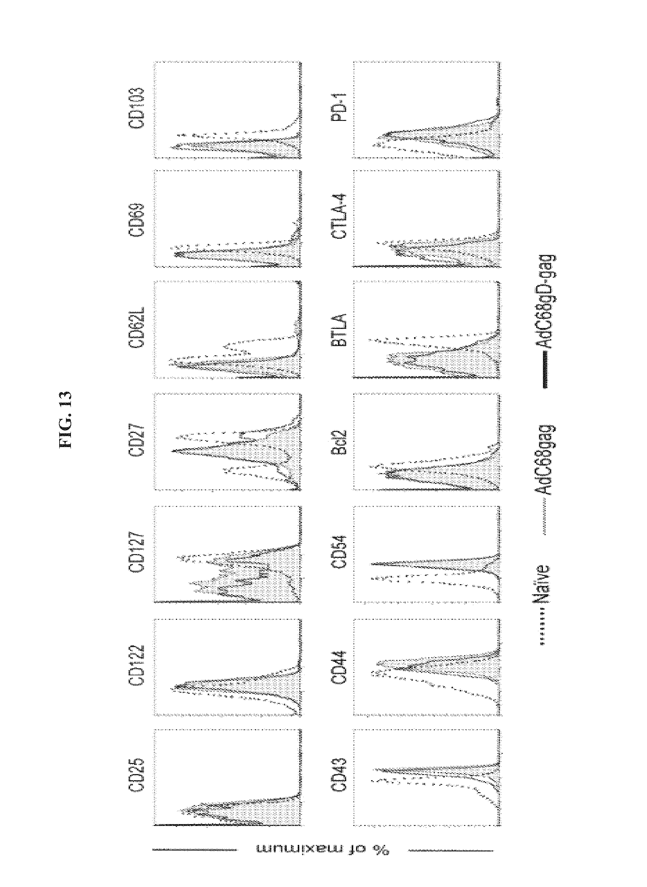
D00014
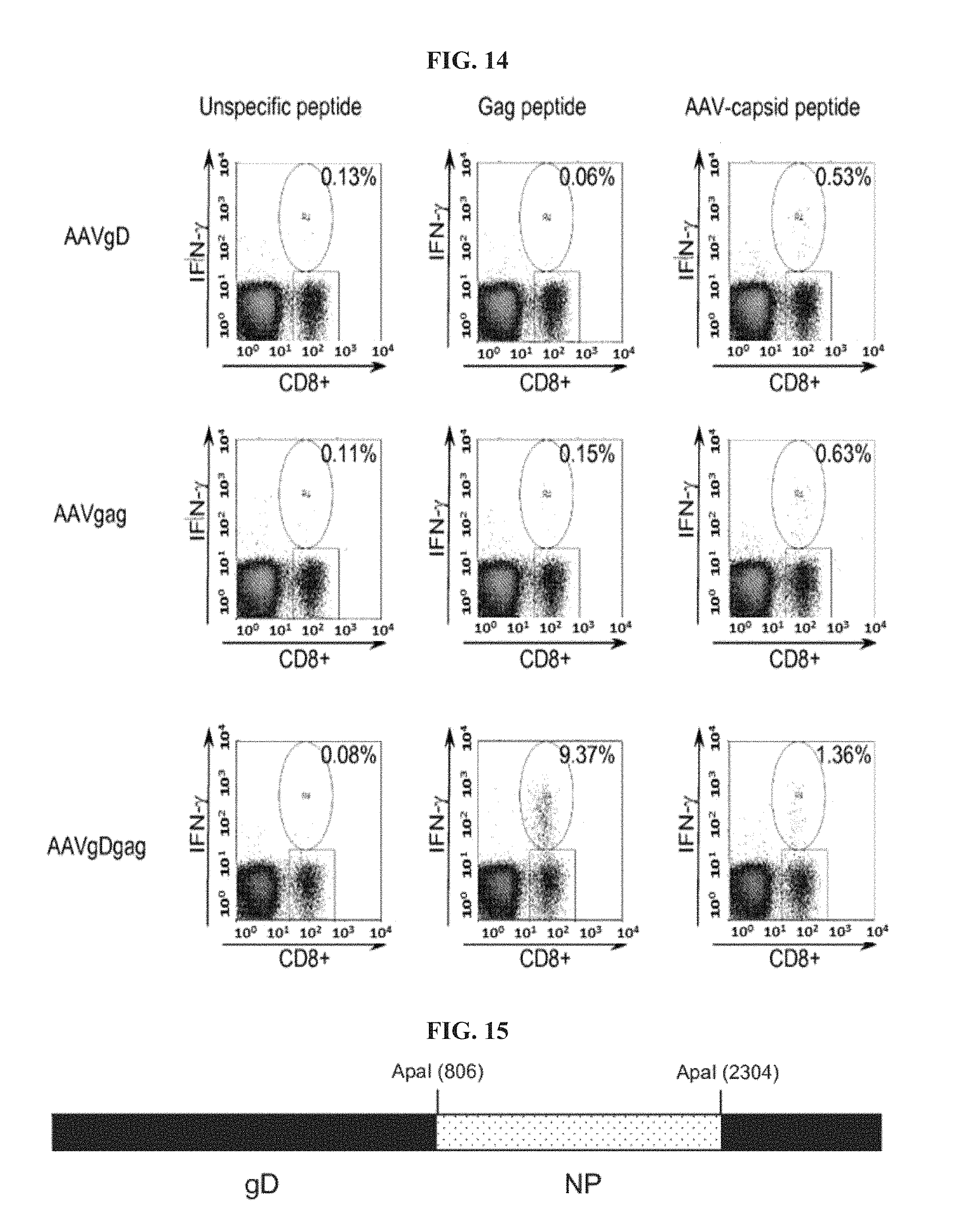
D00015

D00016
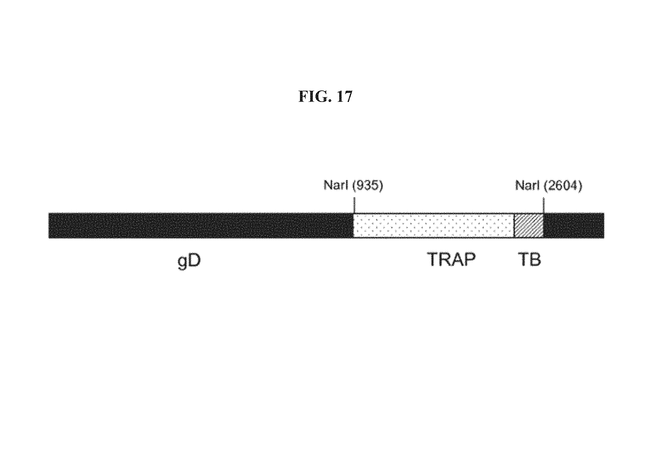
D00017
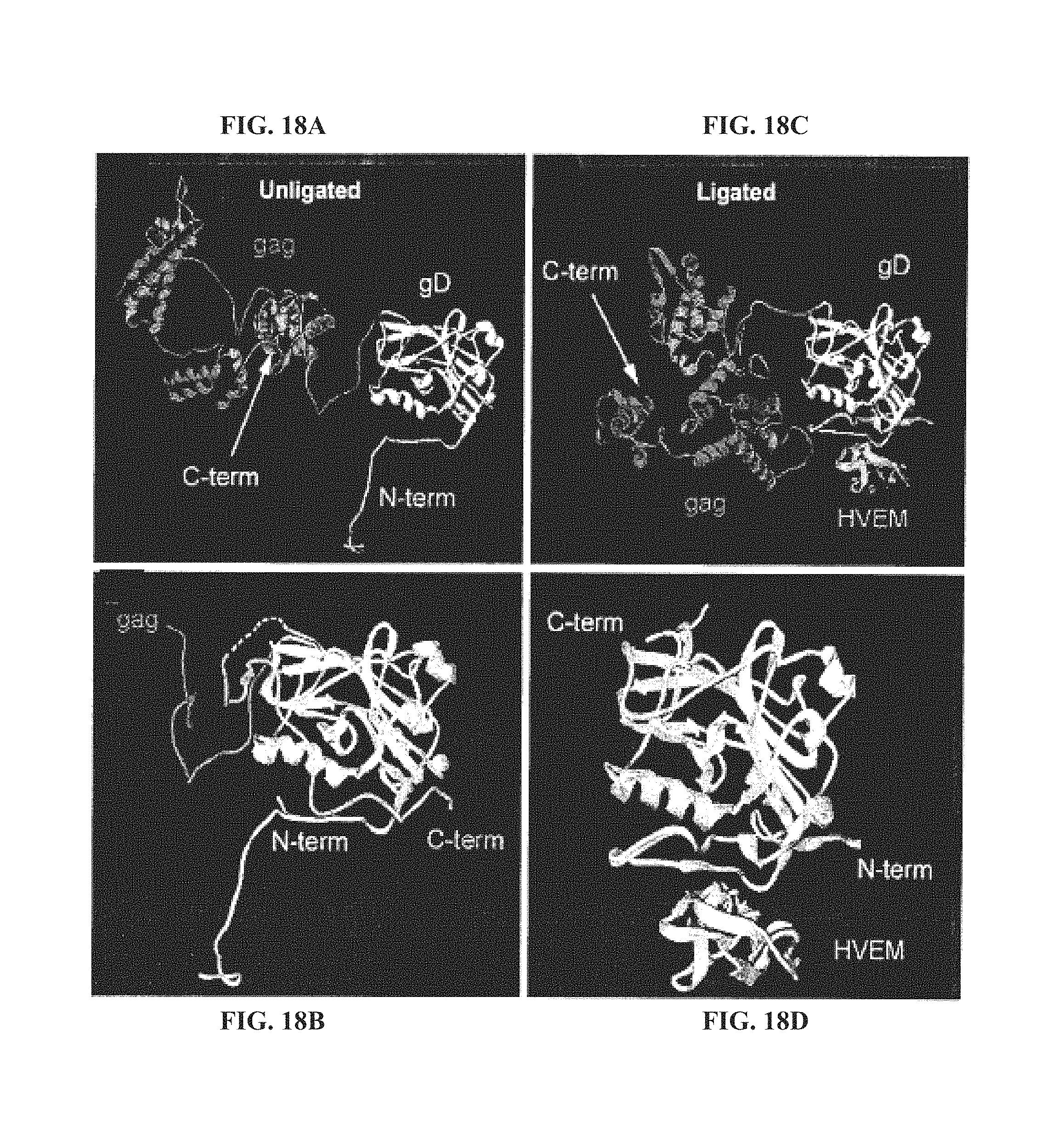
D00018
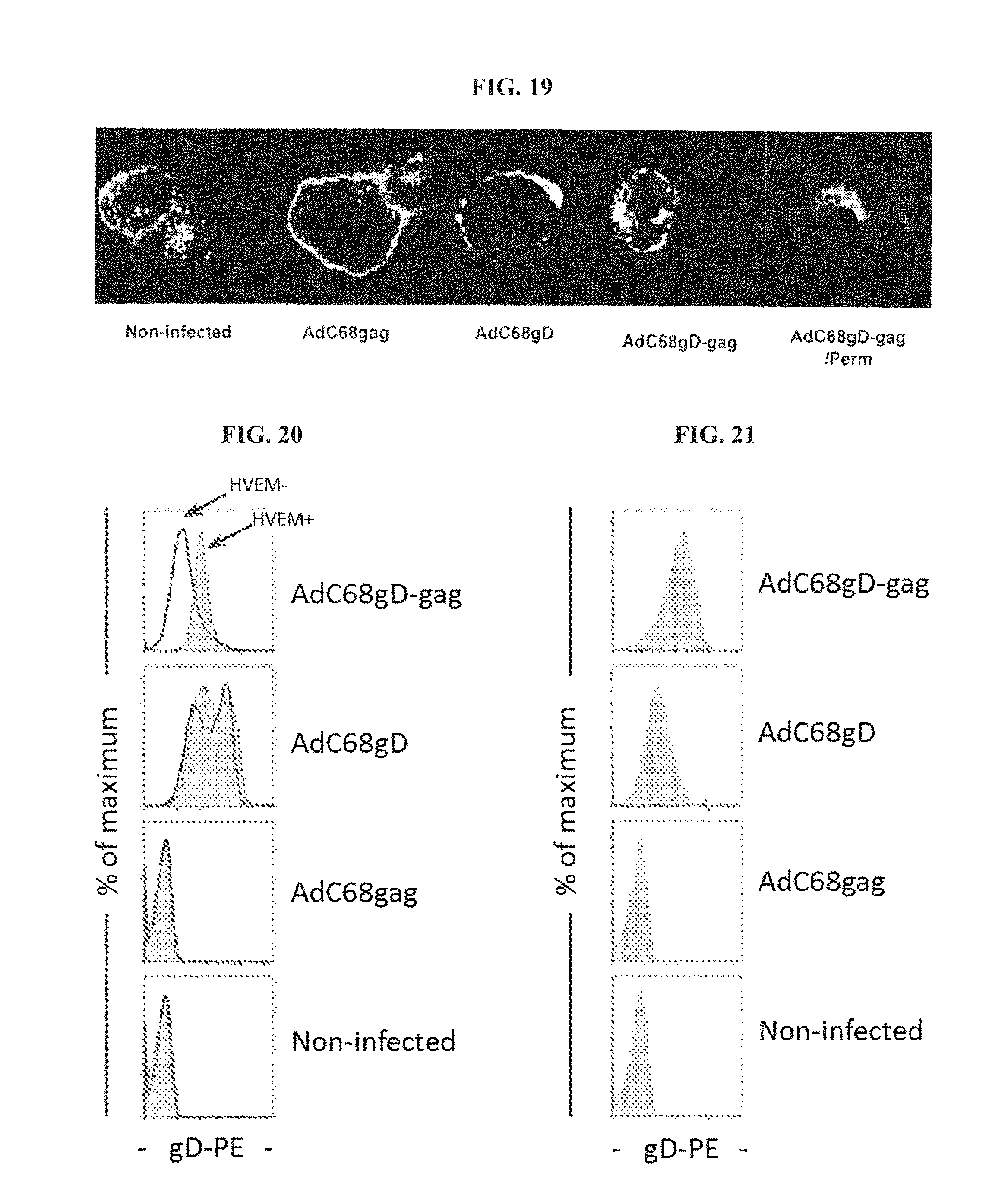
D00019
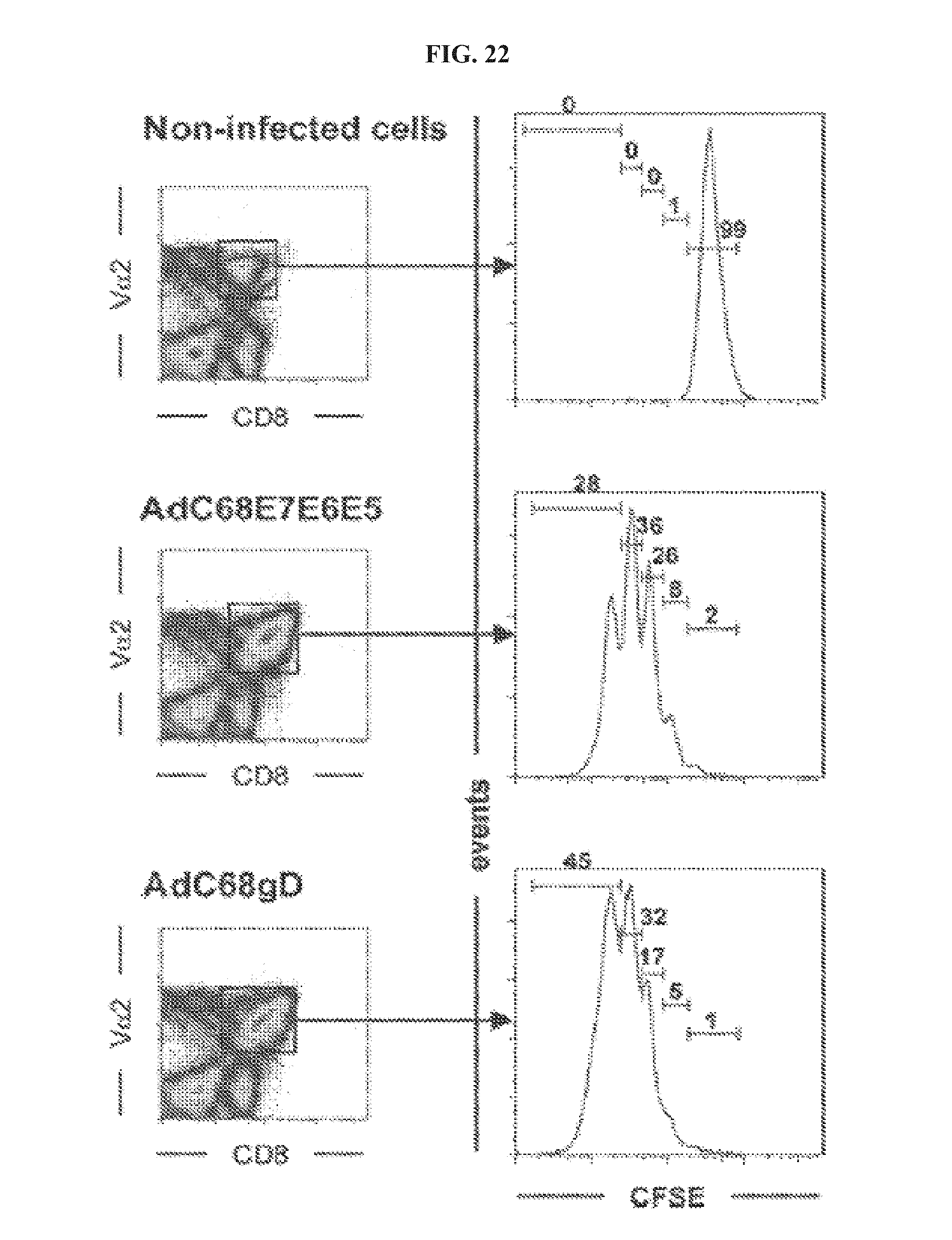
D00020
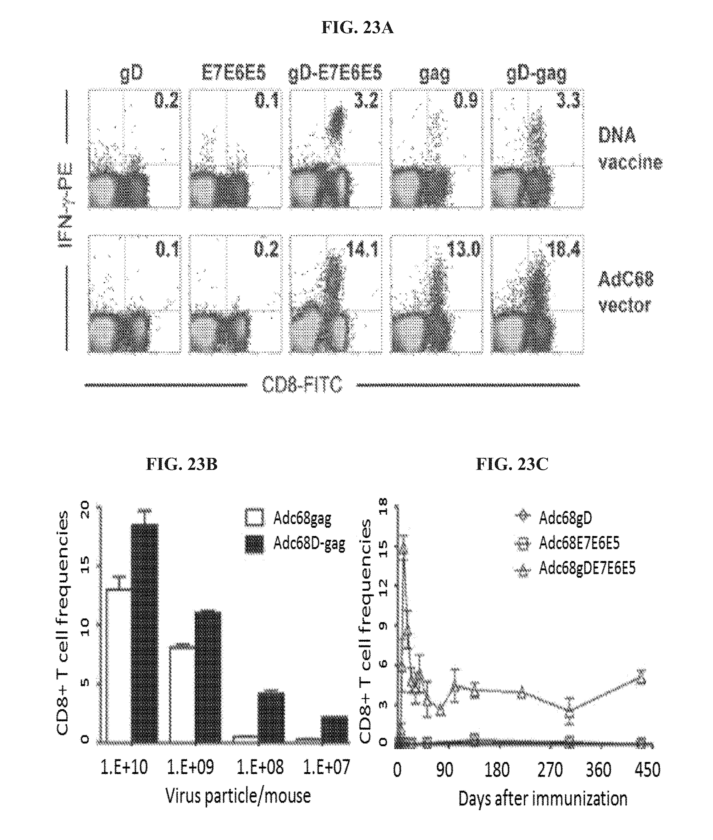
D00021

D00022

D00023

D00024
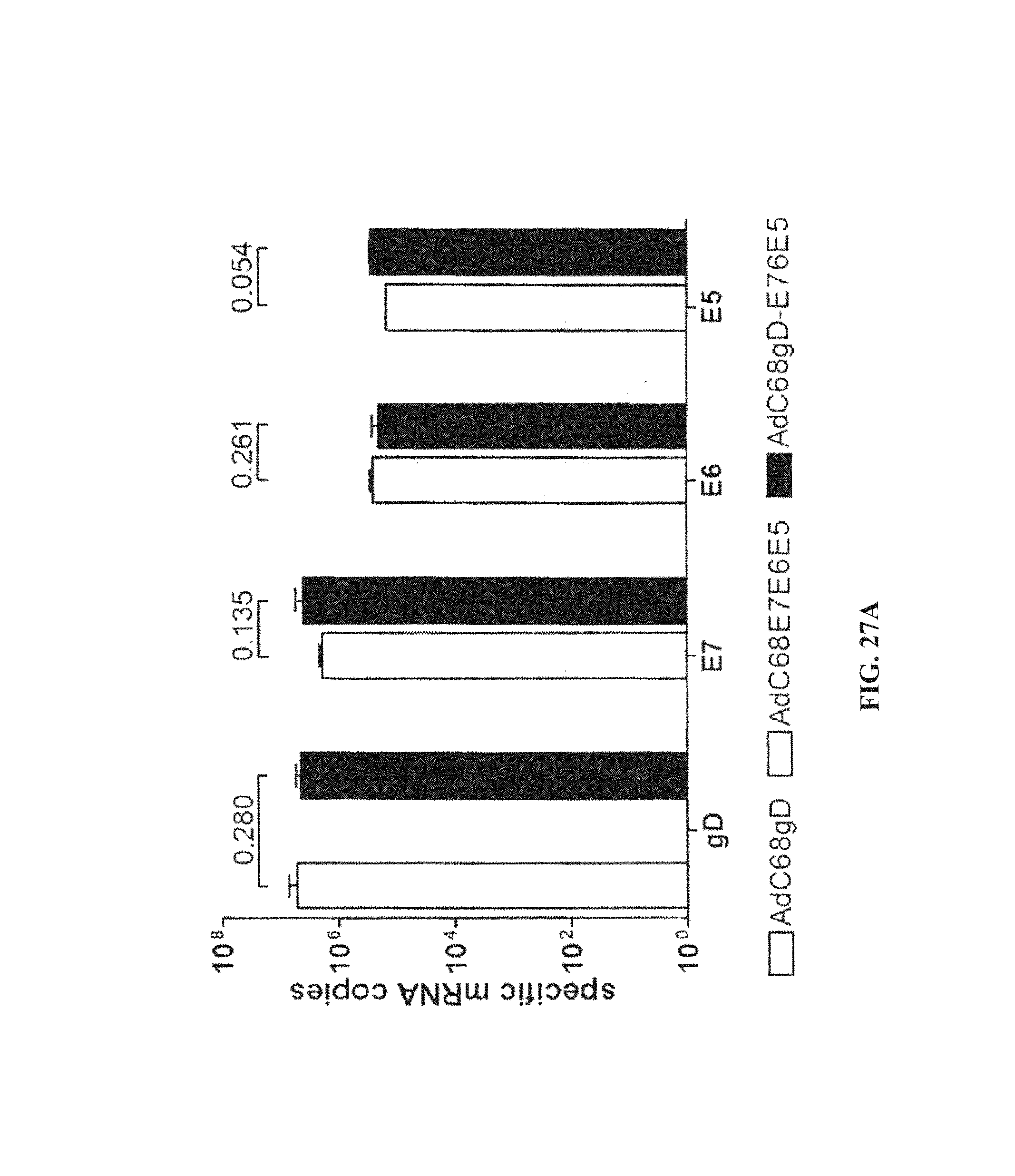
D00025
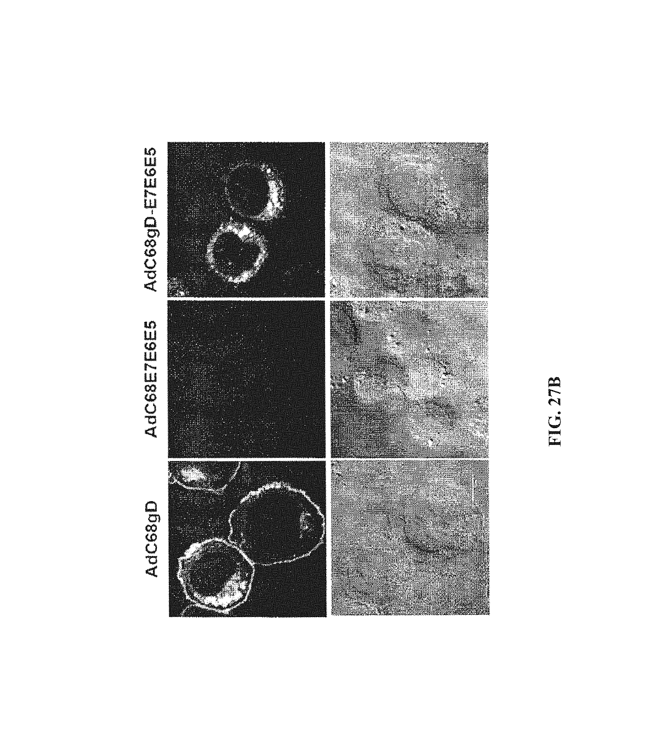
D00026

D00027

D00028
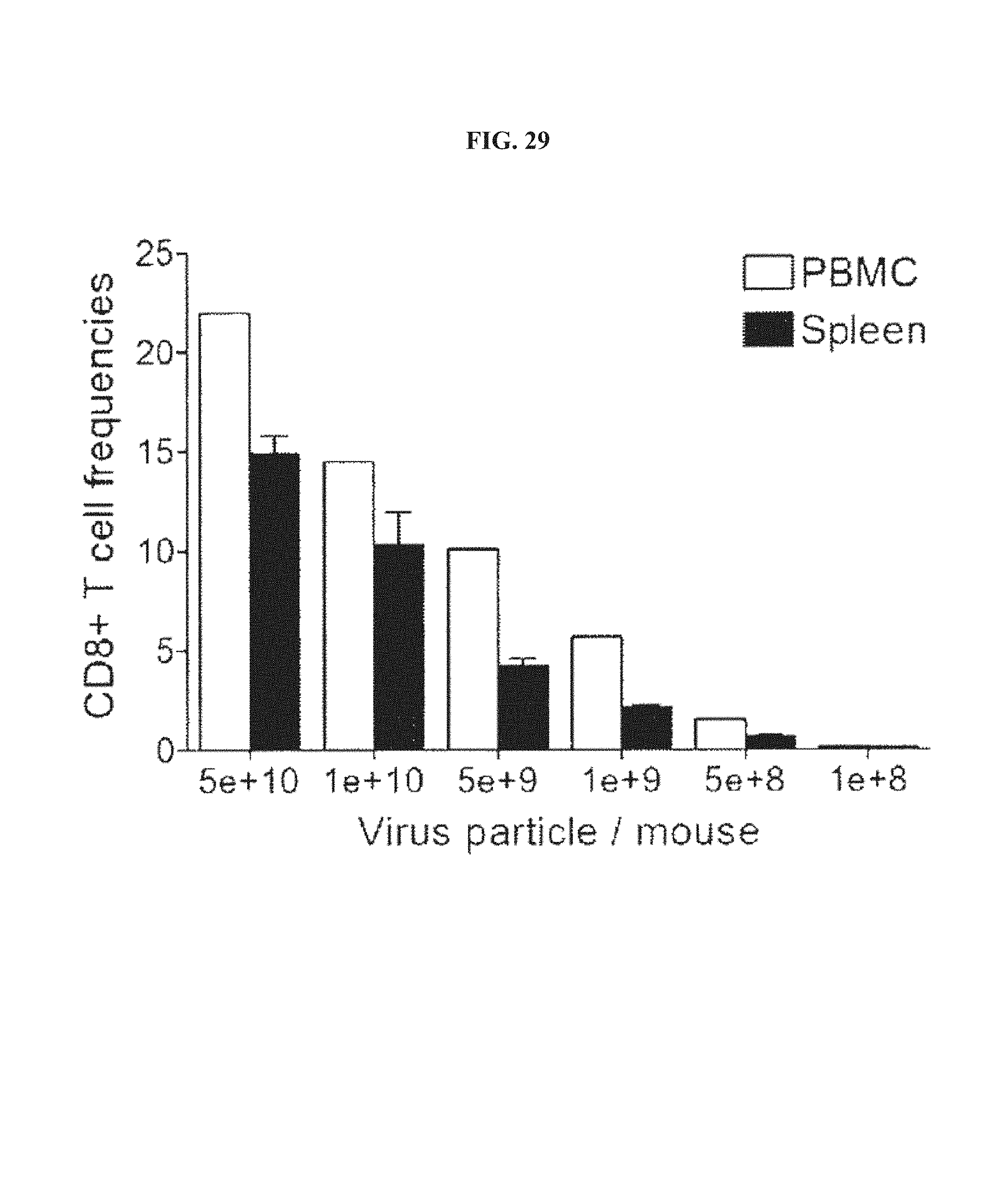
D00029
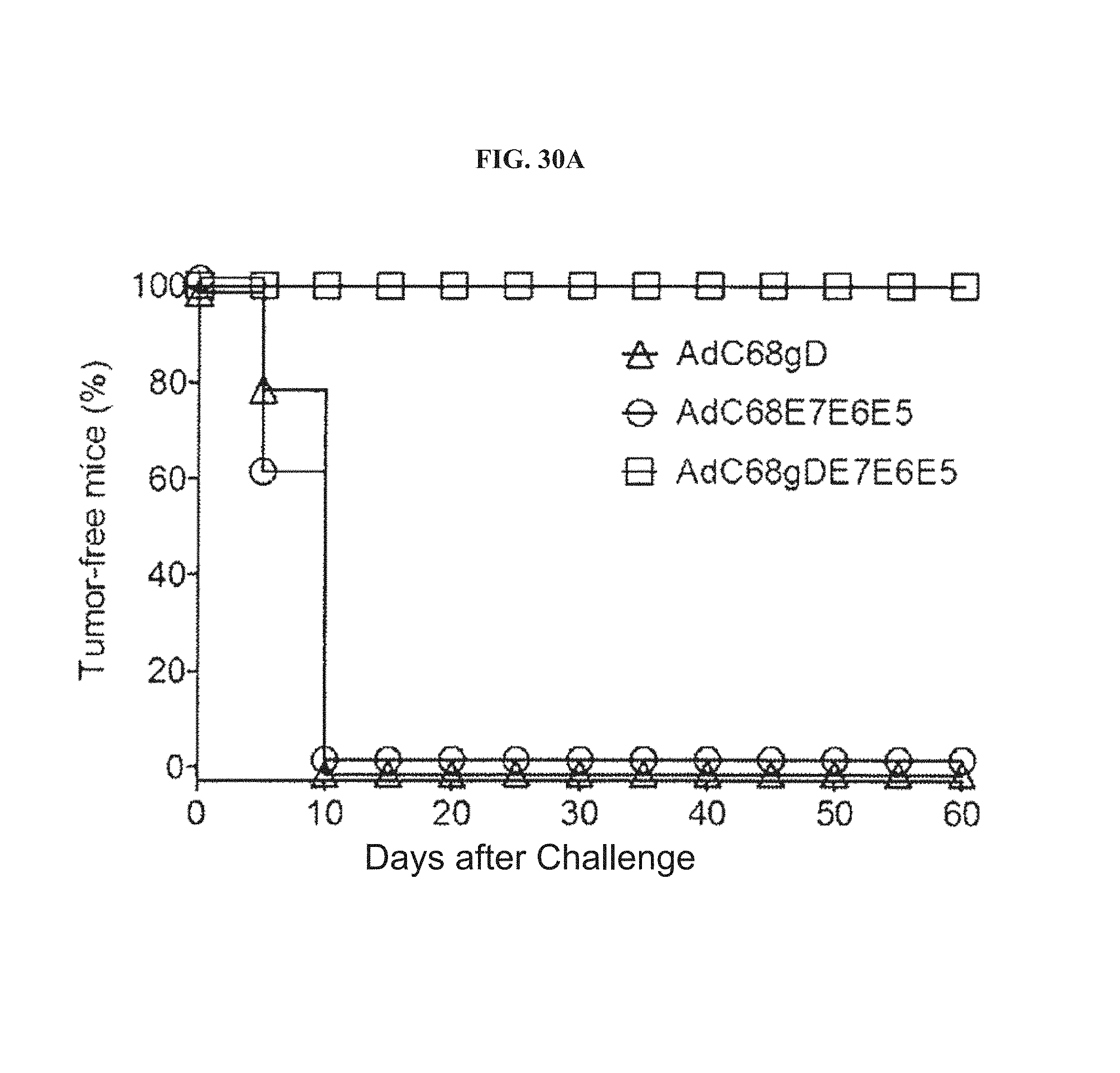
D00030

D00031

D00032
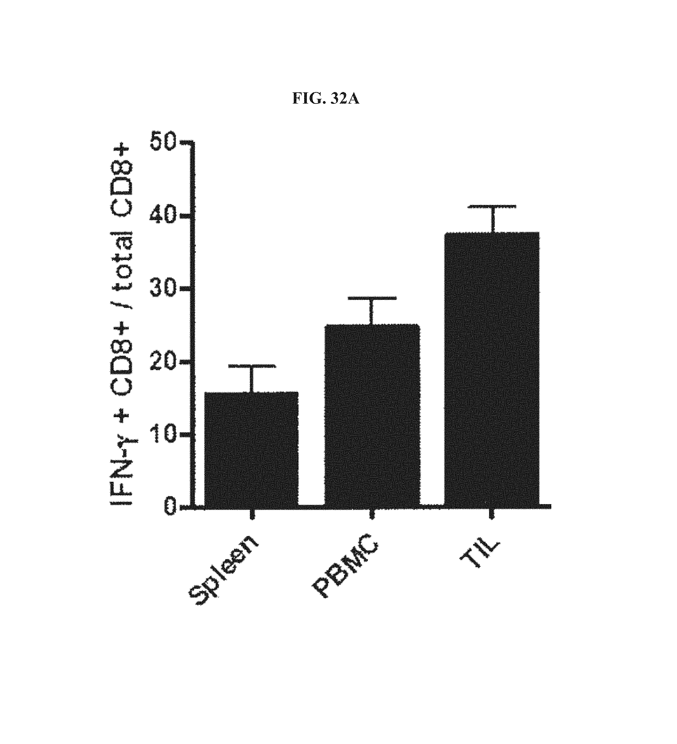
D00033

D00034
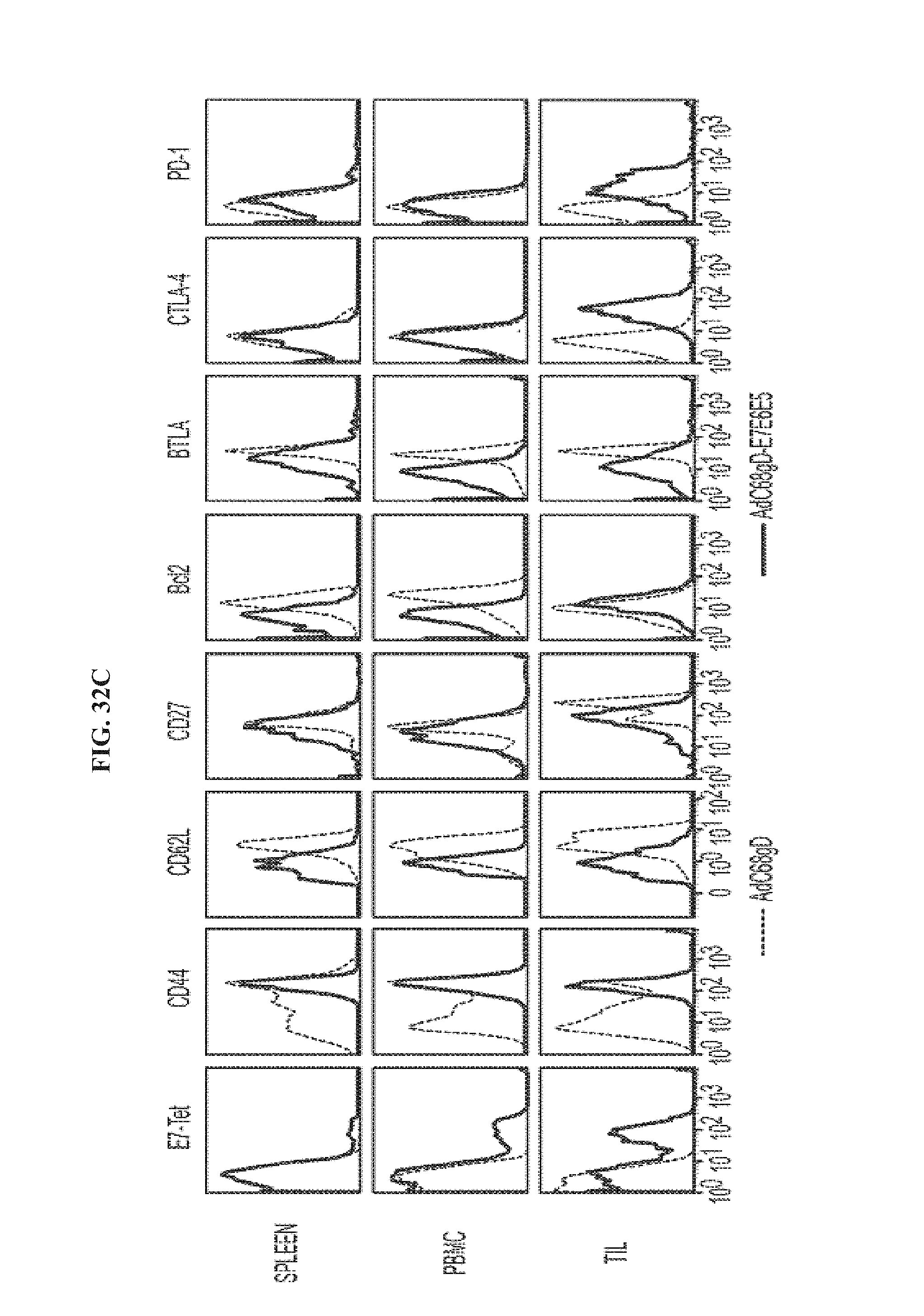
D00035
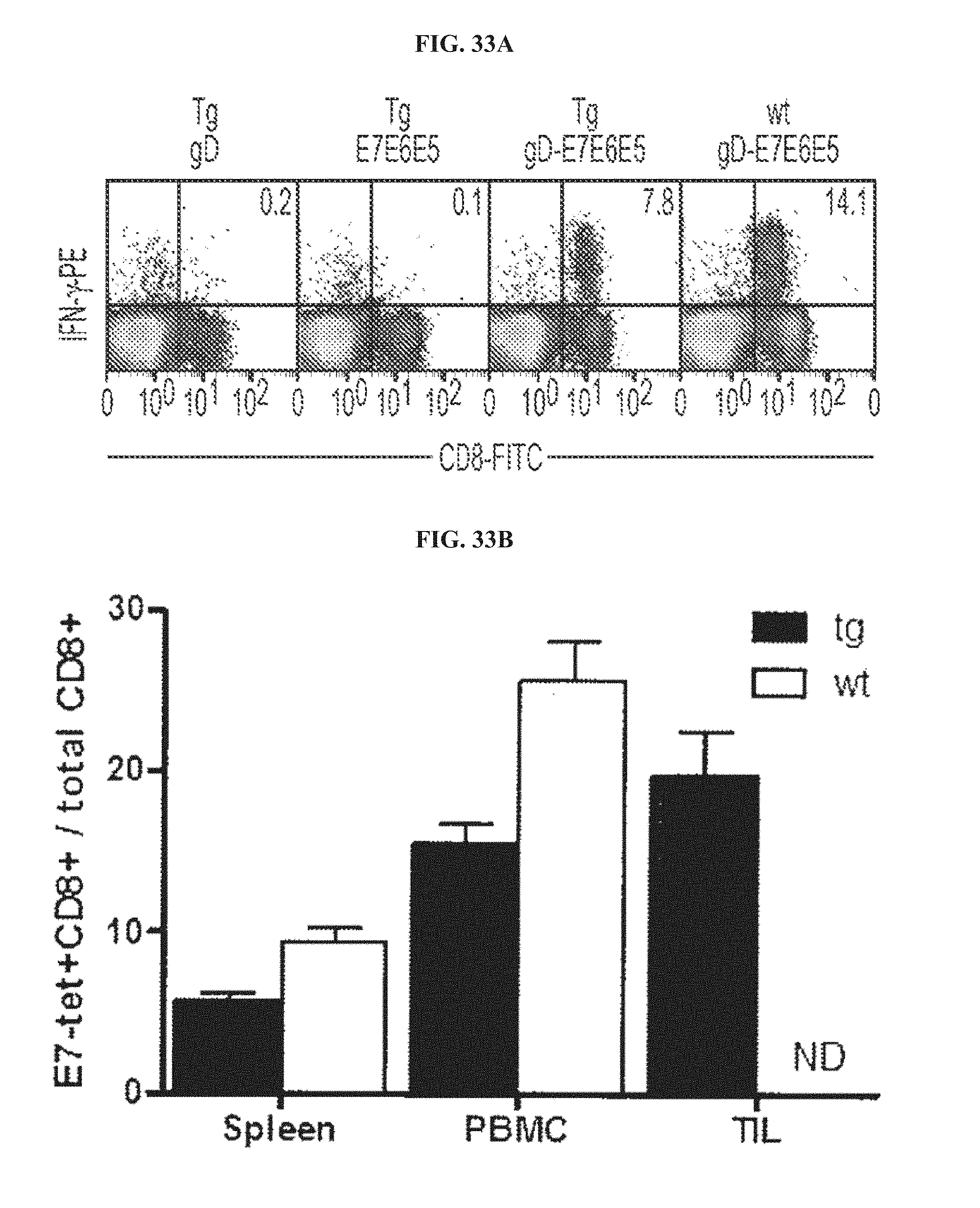
S00001
XML
uspto.report is an independent third-party trademark research tool that is not affiliated, endorsed, or sponsored by the United States Patent and Trademark Office (USPTO) or any other governmental organization. The information provided by uspto.report is based on publicly available data at the time of writing and is intended for informational purposes only.
While we strive to provide accurate and up-to-date information, we do not guarantee the accuracy, completeness, reliability, or suitability of the information displayed on this site. The use of this site is at your own risk. Any reliance you place on such information is therefore strictly at your own risk.
All official trademark data, including owner information, should be verified by visiting the official USPTO website at www.uspto.gov. This site is not intended to replace professional legal advice and should not be used as a substitute for consulting with a legal professional who is knowledgeable about trademark law.