Mutated immunoglobulin-binding polypeptides
Rodrigo , et al.
U.S. patent number 10,308,690 [Application Number 14/961,164] was granted by the patent office on 2019-06-04 for mutated immunoglobulin-binding polypeptides. This patent grant is currently assigned to GE HEALTHCARE BIOPROCESS R&D AB. The grantee listed for this patent is GE HEALTHCARE BIOPROCESS R&D AB. Invention is credited to Mats Ander, Goran Bauren, Tomas Bjorkman, Gustav Rodrigo.


| United States Patent | 10,308,690 |
| Rodrigo , et al. | June 4, 2019 |
Mutated immunoglobulin-binding polypeptides
Abstract
The invention discloses a polypeptide with improved alkaline stability, which polypeptide comprises a mutant of a B or C domain of Staphylococcus Protein A (SpA), as specified by SEQ ID NO 1 or SEQ ID NO 2, or of Protein Z, as specified by SEQ ID NO 3, comprising at least the mutation wherein the glutamine residue at position 9 has been mutated to a tryptophan, leucine, glutamic acid, valine or lysine. The invention also discloses multimers of the polypeptide, as well as separation matrices comprising the multimers or polypeptides.
| Inventors: | Rodrigo; Gustav (Uppsala, SE), Ander; Mats (Uppsala, SE), Bauren; Goran (Uppsala, SE), Bjorkman; Tomas (Uppsala, SE) | ||||||||||
|---|---|---|---|---|---|---|---|---|---|---|---|
| Applicant: |
|
||||||||||
| Assignee: | GE HEALTHCARE BIOPROCESS R&D
AB (Uppsala, SE) |
||||||||||
| Family ID: | 52280382 | ||||||||||
| Appl. No.: | 14/961,164 | ||||||||||
| Filed: | December 7, 2015 |
Prior Publication Data
| Document Identifier | Publication Date | |
|---|---|---|
| US 20160159857 A1 | Jun 9, 2016 | |
Related U.S. Patent Documents
| Application Number | Filing Date | Patent Number | Issue Date | ||
|---|---|---|---|---|---|
| PCT/SE2014/050872 | Jul 8, 2014 | ||||
Foreign Application Priority Data
| Jul 10, 2013 [SE] | 1350859 | |||
| Jul 10, 2013 [SE] | 1350860 | |||
| Current U.S. Class: | 1/1 |
| Current CPC Class: | B01J 20/286 (20130101); B01J 20/3212 (20130101); B01J 20/3219 (20130101); C07K 1/22 (20130101); B01D 15/3809 (20130101); B01J 20/3204 (20130101); B01J 20/3274 (20130101); C07K 14/31 (20130101); C07K 14/745 (20130101); C07K 16/00 (20130101) |
| Current International Class: | C07K 14/31 (20060101); C07K 1/22 (20060101); B01J 20/286 (20060101); C07K 14/745 (20060101); C07K 16/00 (20060101); B01J 20/32 (20060101); B01D 15/38 (20060101) |
References Cited [Referenced By]
U.S. Patent Documents
| 2013/0046056 | February 2013 | Spector et al. |
| 101775069 | Jul 2010 | CN | |||
| 2202310 | Jun 2010 | EP | |||
| 2014/050872 | Jul 2014 | SE | |||
| 200063243 | Oct 2000 | WO | |||
| WO-2007019376 | Feb 2007 | WO | |||
| 2012083425 | Jun 2012 | WO | |||
| 2012087231 | Jun 2012 | WO | |||
| 2012165544 | Dec 2012 | WO | |||
Other References
|
Supplementary European Search Report from European Patent Application No. 14 82 3321 dated Feb. 2, 2017. cited by applicant . Chinese Office Action and Search Report Received for Chinese Patent Application 201480039292.8 dated Aug. 3, 2018, 11 pages (English Translation). cited by applicant . International Search Report, PCT/SE2014/050872, filed Jul. 8, 2014 (dated Jan. 1, 2015). cited by applicant. |
Primary Examiner: Kolker; Daniel E
Assistant Examiner: Rogers; James L
Attorney, Agent or Firm: Wood IP LLC
Parent Case Text
This is a continuation-in-part of International Application PCT/SE2014/050872, with an international filing date of Jul. 8, 2014.
Claims
The invention claimed is:
1. An Fc-binding polypeptide with improved alkaline stability, which polypeptide comprises a mutant of a parental polypeptide, as defined by, or having at least 90% identity to, SEQ ID NO 28, SEQ ID NO 29 or SEQ ID NO 30, comprising at least the mutation wherein the glutamine residue at position 3 has been mutated to leucine, glutamic acid, valine or lysine.
2. A multimer with improved alkaline stability comprising or consisting essentially of a plurality of polypeptide units, which polypeptide comprises a mutant of a parental polypeptide, as defined by, or having at least 90% identity to, SEQ ID NO 28, SEQ ID NO 29 or SEQ ID NO 30, comprising at least the mutation wherein the glutamine residue at position 3 has been mutated to tryptophan, leucine, glutamic acid, valine or lysine.
3. The multimer of claim 2, wherein the polypeptide units are linked by elements comprising up to 15 amino acids.
4. The multimer of claim 3, further comprising at the C-terminal or N-terminal one or more coupling element, selected from the group consisting of a cysteine residue, a plurality of lysine residues and a plurality of histidine residues.
5. A separation matrix, comprising a plurality of polypeptides according to claim 1 coupled to a solid support.
6. A separation matrix, comprising a plurality of multimers according to claim 4 coupled to a solid support.
7. The separation matrix of claim 6, wherein the multimers have been coupled to the solid support via thioether bonds.
8. A method of isolating an immunoglobulin, using a separation matrix according to claim 6, the method comprising, a) contacting a liquid sample comprising an immunoglobulin with the separation matrix according to claim 6, b) washing said separation matrix with a washing liquid, c) eluting the immunoglobulin from the separation matrix with an elution liquid, and d) cleaning the separation matrix with a cleaning liquid.
9. The method of claim 8, wherein the cleaning liquid is alkaline.
10. The method of claim 8, wherein the cleaning liquid comprises 0.1-1.0 M NaOH or KOH.
11. The method of claim 8, wherein steps a)-d) are repeated at least 50 times.
12. An Fc-binding polypeptide with improved alkaline stability, which polypeptide comprises a mutant of a B or C domain of Staphylococcus Protein A (SpA), as defined by, or having at least 90% identity to, SEQ ID NO 1 or SEQ ID NO 2, of Protein Z, as defined by, or having at least 90% identity to, SEQ ID NO 3, or of a Protein Z variant as defined by, or having at least 90% identity to, SEQ ID NO 4, comprising at least the mutation wherein the glutamine residue at position 9 has been mutated to leucine, glutamic acid, valine or lysine.
13. A multimer comprising or consisting essentially of a plurality of polypeptide units, which polypeptide comprises a mutant of a B or C domain of Staphylococcus Protein A (SpA), as defined by, or having at least 90% identity to, SEQ ID NO 1 or SEQ ID NO 2, of Protein Z, as defined by, or having at least 90% identity to, SEQ ID NO 3, or of a Protein Z variant as defined by, or having at least 90% identity to, SEQ ID NO 4, comprising at least the mutation wherein the glutamine residue at position 9 has been mutated to tryptophan, leucine, glutamic acid, valine or lysin.
14. A separation matrix, having a plurality of multimers according to claim 13 coupled to a solid support.
15. A method of isolating an immunoglobulin, comprising the steps of: a) contacting a liquid sample comprising an immunoglobulin with a separation matrix according to claim 14, b) washing said separation matrix with a washing liquid, c) eluting the immunoglobulin from the separation matrix with an elution liquid, and d) cleaning the separation matrix with a cleaning liquid.
16. The method of claim 15, wherein the cleaning liquid comprises 0.1-1.0 M NaOH or KOH.
17. A polypeptide, comprising or consisting essentially of an amino acid sequence selected from the group consisting of: SEQ ID NO 31, SEQ ID NO 32, SEQ ID NO 33, SEQ ID NO 34 and SEQ ID NO 35.
18. A separation matrix, having a plurality of polypeptides according to claim 17 coupled to a solid support.
19. A method of isolating an immunoglobulin, comprising the steps of: a) contacting a liquid sample comprising an immunoglobulin with a separation matrix according to claim 18, b) washing said separation matrix with a washing liquid, c) eluting the immunoglobulin from the separation matrix with an elution liquid, and d) cleaning the separation matrix with a cleaning liquid.
20. A multimer with improved alkaline stability comprising or consisting essentially of a plurality of polypeptide units as defined by claim 17.
21. The multimer of claim 20, wherein the polypeptide units are linked by elements comprising up to 15 amino acids.
22. The multimer of claim 21, further comprising at the C-terminal or N-terminal one or more coupling element, selected from the group consisting of a cysteine residue, a plurality of lysine residues and a plurality of histidine residues.
23. A separation matrix, comprising a plurality of multimers according to claim 20 coupled to a solid support.
24. The separation matrix of claim 23, wherein the multimers have been coupled to the solid support via thioether bonds.
25. A method of isolating an immunoglobulin, using a separation matrix according to claim 23, the method comprising, a) contacting a liquid sample comprising an immunoglobulin with the separation matrix according to claim 23, b) washing said separation matrix with a washing liquid, c) eluting the immunoglobulin from the separation matrix with an elution liquid, and d) cleaning the separation matrix with a cleaning liquid.
26. The method of claim 25, wherein the cleaning liquid comprises 0.1-1.0 M NaOH or KOH.
27. The method of claim 23, wherein steps a)-d) are repeated at least 50 times.
Description
TECHNICAL FIELD OF THE INVENTION
The present invention relates to the field of affinity chromatography, and more specifically to mutated immunoglobulin-binding domains of Protein A, which are useful in affinity chromatography of immunoglobulins. The invention also relates to multimers of the mutated domains and to separation matrices containing the mutated domains or multimers.
BACKGROUND OF THE INVENTION
Immunoglobulins represent the most prevalent biopharmaceutical products in either manufacture or development worldwide. The high commercial demand for and hence value of this particular therapeutic market has led to the emphasis being placed on pharmaceutical companies to maximize the productivity of their respective mAb manufacturing processes whilst controlling the associated costs.
Affinity chromatography is used in most cases, as one of the key steps in the purification of these immunoglobulin molecules, such as monoclonal or polyclonal antibodies. A particularly interesting class of affinity reagents is proteins capable of specific binding to invariable parts of an immunoglobulin molecule, such interaction being independent on the antigen-binding specificity of the antibody. Such reagents can be widely used for affinity chromatography recovery of immunoglobulins from different samples such as but not limited to serum or plasma preparations or cell culture derived feed stocks. An example of such a protein is staphylococcal protein A, containing domains capable of binding to the Fc and Fab portions of IgG immunoglobulins from different species.
Staphylococcal protein A (SpA) based reagents have due to their high affinity and selectivity found a widespread use in the field of biotechnology, e.g. in affinity chromatography for capture and purification of antibodies as well as for detection or quantification. At present, SpA-based affinity medium probably is the most widely used affinity medium for isolation of monoclonal antibodies and their fragments from different samples including industrial cell culture supernatants. Accordingly, various matrices comprising protein A-ligands are commercially available, for example, in the form of native protein A (e.g. Protein A SEPHAROSE.TM., GE Healthcare, Uppsala, Sweden) and also comprised of recombinant protein A (e.g. rProtein A-SEPHAROSE.TM., GE Healthcare). More specifically, the genetic manipulation performed in the commercial recombinant protein A product is aimed at facilitating the attachment thereof to a support.
These applications, like other affinity chromatography applications, require comprehensive attention to definite removal of contaminants and impurities. Such contaminants/impurities can for example be non-eluted molecules adsorbed to the stationary phase or matrix in a chromatographic procedure, such as non-desired biomolecules or microorganisms, including for example proteins, carbohydrates, lipids, bacteria and viruses. The removal of such contaminants from the matrix is usually performed after a first elution of the desired product in order to regenerate the matrix before subsequent use. Such removal usually involves a procedure known as cleaning-in-place (CIP), wherein agents capable of eluting contaminants from the stationary phase are used. One such class of agents often used is alkaline solutions that are passed over said stationary phase. At present the most extensively used cleaning and sanitizing agent is NaOH, and the concentration thereof can range from 0.1 up to e.g. 1 M, depending on the degree and nature of contamination. This strategy is associated with exposing the matrix for pH-values above 13. For many affinity chromatography matrices containing proteinaceous affinity ligands such alkaline environment is a very harsh condition and consequently results in decreased capacities owing to instability of the ligand to the high pH involved.
An extensive research has therefore been focused on the development of engineered protein ligands that exhibit an improved capacity to withstand alkaline pH-values. For example, Gulich et al. (Susanne Gulich, Martin Linhult, Per-.ANG.ke Nygren, Mathias Uhlen, Sophia Hober, Journal of Biotechnology 80 (2000), 169-178) suggested protein engineering to improve the stability properties of a Streptococcal albumin-binding domain (ABD) in alkaline environments. Gulich et al. created a mutant of ABD, wherein all the four asparagine residues have been replaced by leucine (one residue), aspartate (two residues) and lysine (one residue). Further, Gulich et al. report that their mutant exhibits a target protein binding behavior similar to that of the native protein, and that affinity columns containing the engineered ligand show higher binding capacities after repeated exposure to alkaline conditions than columns prepared using the parental non-engineered ligand. Thus, it is concluded therein that all four asparagine residues can be replaced without any significant effect on structure and function.
Recent work shows that changes can also be made to protein A (SpA) to effect similar properties. US patent application publication US 2005/0143566 discloses that when at least one asparagine residue is mutated to an amino acid other than glutamine or aspartic acid, the mutation confers an increased chemical stability at pH-values of up to about 13-14 compared to the parental SpA, such as the B-domain of SpA, or Protein Z, a synthetic construct derived from the B-domain of SpA (U.S. Pat. No. 5,143,844). The authors show that when these mutated proteins are used as affinity ligands, the separation media as expected can better withstand cleaning procedures using alkaline agents. Further mutations of protein A domains with the purpose of increasing the alkali stability have also been published in WO 2008/039141, JP 2006304633A, EP 1992692A1, EP2202310A2, WO 2010/110288, WO 2012/086660 and WO 2012/083425. However, the currently available mutations are still sensitive to alkaline pH and the NaOH concentration during cleaning is usually limited to 0.1 M, which means that complete cleaning is difficult to achieve. Higher NaOH concentrations, which would improve the cleaning, lead to unacceptable capacity losses.
There is thus still a need in this field to obtain a separation matrix containing protein ligands having a further improved stability towards alkaline cleaning procedures.
SUMMARY OF THE INVENTION
One aspect of the invention is to provide a polypeptide with improved alkaline stability. This is achieved with a polypeptide as defined in the claims.
One advantage is that the alkaline stability is improved over the parental polypeptides. A further advantage is a highly selective binding towards immunoglobulins and other Fc-containing proteins.
A second aspect of the invention is to provide a multimer with improved alkaline stability, comprising a plurality of polypeptides. This is achieved with a multimer as defined in the claims.
A third aspect of the invention is to provide a nucleic acid or a vector encoding a polypeptide or multimer with improved alkaline stability. This is achieved with a nucleic acid or vector as defined in the claims.
A fourth aspect of the invention is to provide an expression system capable of expressing a polypeptide or multimer with improved alkaline stability. This is achieved with an expression system as defined in the claims.
A fifth aspect of the invention is to provide a separation matrix capable of selectively binding immunoglobulins and other Fc-containing proteins and exhibiting an improved alkaline stability. This is achieved with a separation matrix as defined in the claims.
A sixth aspect of the invention is to provide an efficient and economical method of isolating an immunoglobulin or other Fc-containing protein. This is achieved with a method as defined in the claims.
Further suitable embodiments of the invention are described in the dependent claims.
DEFINITIONS
The terms "antibody" and "immunoglobulin" are used interchangeably herein, and are understood to include also fragments of antibodies, fusion proteins comprising antibodies or antibody fragments and conjugates comprising antibodies or antibody fragments.
The terms an "Fc-binding polypeptide" and "Fc-binding protein" mean a polypeptide or protein respectively, capable of binding to the crystallisable part (Fc) of an antibody and includes e.g. Protein A and Protein G, or any fragment or fusion protein thereof that has maintained said binding property.
The term "linker" herein means an element linking two polypeptide units, monomers or domains to each other in a multimer.
The term "spacer" herein means an element connecting a polypeptide or a polypeptide multimer to a support.
BRIEF DESCRIPTION OF FIGURES
FIG. 1 shows results from Example 1 for the alkali stability of mutated and non-mutated monomeric Zvar (SEQ ID NO 4) polypeptide variants coupled to an SPR biosensor chip.
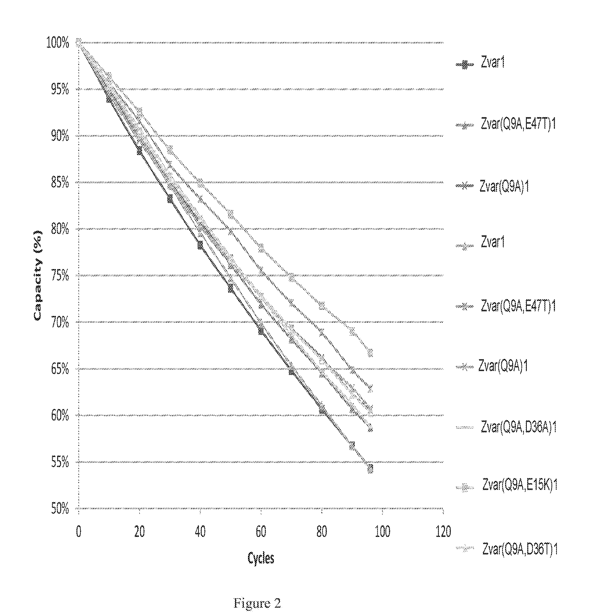
FIG. 2 shows an enlargement of FIG. 1 for polypeptide variants with high alkali stabilities.
DETAILED DESCRIPTION OF EMBODIMENTS
In one aspect the present invention discloses a polypeptide, which comprises, or consists essentially of, one or more mutants of a B or C domain of Staphylococcus Protein A (SpA), as defined by, or having at least 90%, at least 95% or at least 98% identity to, SEQ ID NO 1 or SEQ ID NO 2, or of Protein Z, as specified by SEQ ID NO 3 or SEQ ID NO 4, wherein at least the glutamine residue at position 9 has been mutated to (i.e. substituted with) an amino acid other than asparagine. SEQ ID NO 4 denotes a variant of Protein Z, here called Zvar, with the mutations N3A,N6D,N23T. The mutation of Q9 in these domains confers an improved alkali stability in comparison with the parental domain/polypeptide (i.e. SEQ ID NO 1, 2 or 3), without impairing the immunoglobulin-binding properties. Hence, the polypeptide can also be described as an Fc- or immunoglobulin-binding polypeptide.
SEQ ID NO 1 (SpA B domain)
ADNKFNKEQQ NAFYEILHLP NLNEEQRNGF IQSLKDDPSQ SANLLAEAKK LNDAQAPK
SEQ ID NO 2 (SpA C domain)
ADNKFNKEQQ NAFYEILHLP NLTEEQRNGF IQSLKDDPSV SKEILAEAKK LNDAQAPK
SEQ ID NO 3 (Protein Z)
VDNKFNKEQQ NAFYEILHLP NLNEEQRNAF IQSLKDDPSQ SANLLAEAKK LNDAQAPK
SEQ ID NO 4 (Zvar)
VDAKFDKEQQ NAFYEILHLP NLTEEQRNAF IQSLKDDPSQ SANLLAEAKK LNDAQAPK
In some embodiments, at least the glutamine residue at position 9 of SEQ ID NO 1-4 has been mutated to (i.e. substituted with) an amino acid other than asparagine, proline or cysteine, such as to a tryptophan, leucine, glutamic acid, valine or lysine. This has the advantage that deamidation-sensitive asparagines are not introduced, that the chain conformation is not disturbed by introduction of prolines and no sites for disulfide bridges are introduced. The Q9 mutation (e.g. a Q9 W,L,V,E or K mutation) may be the only mutation or the polypeptide may also comprise further mutations, such as in at least one of the N3, N6, Q10, E15, H18, N21, N23, N28, G/A29, D36, Q/V40, A/K42, N/E43, L/I44, E47, Q55 and P57 positions. In one or more of these positions, the original amino acid residue may e.g. be substituted with an amino acid which is not asparagine, proline or cysteine. The original amino acid residue may e.g. be substituted with an alanine, a valine, a threonine, a serine, a lysine or an aspartic acid.
The invention can also be expressed as an Fc-binding polypeptide with improved alkaline stability, which polypeptide comprises, or consists essentially of, a mutant of a parental polypeptide, as defined by, or having at least 90%, at least 95% or at least 98% identity to, by SEQ ID NO 28, SEQ ID NO 29 or SEQ ID NO 30, wherein at least the glutamine residue at position 3 (corresponding to pos. 9 in SEQ ID NO 2, 3 or 4) has been mutated to (i.e. substituted with) an amino acid other than asparagine, in particular to tryptophan, leucine, glutamic acid, valine or lysine. SEQ ID NO 28 is Protein Z without the VDNKFN linker region, which has low influence on the polypeptide properties, while SEQ ID NO 29 is Zvar without the VDAKFD linker region and SEQ ID NO 30 is the C domain without the ADNKFN linker region. The mutation of Q3 confers an improved alkali stability in comparison with the parental polypeptides, without impairing the immunoglobulin-binding properties.
SEQ ID NO 28--Truncated Protein Z
KEQQ NAFYEILHLP NLNEEQRNAF IQSLKDDPSQ SANLLAEAKK LNDAQAPK
SEQ ID NO 29--Truncated Zvar
KEQQ NAFYEILHLP NLTEEQRNAF IQSLKDDPSQ SANLLAEAKK LNDAQAPK
SEQ ID NO 30--Truncated SpA C domain
KEQQ NAFYEILHLP NLTEEQRNGF IQSLKDDPSV SKEILAEAKK LNDAQAPK
In certain embodiments, the amino acid residue at position 23 (or pos. 17 in SEQ ID NO 28-30) is a threonine or an alanine.
In certain embodiments, the serine residue at position 33 (or pos. 27 in SEQ ID NO 28-30) has been mutated to an amino acid other than asparagine, glutamine, proline or cysteine, such as to a lysine. In alternative embodiments, the amino acid residue at position 33 is a serine.
In some embodiments, the glutamine residue at position 10 (or pos. 4 in SEQ ID NO 28-30) has been mutated to an amino acid other than asparagine, glutamine, proline or cysteine, such as to an alanine. In alternative embodiments, the amino acid residue at position 10 is a glutamine.
In certain embodiments, the glutamine residue at position 32 (or pos. 26 in SEQ ID NO 28-30) has been mutated to an amino acid other than asparagine, glutamine, proline or cysteine, such as to an alanine. In alternative embodiments, the amino acid residue at position 32 is a glutamine.
In some embodiments, the glutamine residue at position 40 (or pos. 34 in SEQ ID NO 28-30) has been mutated to an amino acid other than asparagine, glutamine, proline or cysteine, such as to an alanine or valine. In alternative embodiments, the amino acid residue at position 40 is a glutamine.
In certain embodiments, the glutamine residue at position 55 (or pos. 49 in SEQ ID NO 28-30) has been mutated to an amino acid other than asparagine, glutamine, proline or cysteine, such as to an alanine, a serine or glutamic acid. In alternative embodiments, the amino acid residue at position 55 is a glutamine.
In some embodiments, the amino acid residue at position 26 (or pos. 20 in SEQ ID NO 28-30) is a glutamine. It appears that the alkali stability of the polypeptides with Q9 mutations is better when Q26 has not been mutated to a threonine or an alanine. In alternative embodiments, the amino acid residue at position 26 may be mutated to an amino acid other than asparagine, glutamine, proline, cysteine, threonine or alanine.
In certain embodiments, the glutamic acid residue at position 15 (or pos. 9 in SEQ ID NO 28-30) has been mutated to an amino acid other than asparagine, glutamine, proline or cysteine. In particular embodiments, the glutamic acid residue at position 15 has been mutated to a lysine. This accidental mutation further improves the alkaline stability of the polypeptides with a Q9 mutation. In alternative embodiments, the amino acid residue at position 15 is a glutamic acid.
In some embodiments, the glutamic acid residue at position 47 (or pos. 41 in SEQ ID NO 28-30) has been mutated to an amino acid other than asparagine, glutamine, proline or cysteine, such as to an alanine or a threonine. In alternative embodiments, the amino acid residue at position 47 is a glutamic acid.
In some embodiments, the asparagine residue at position 21 (or pos. 15 in SEQ ID NO 28-30) has been mutated to an amino acid other than glutamine, proline or cysteine, such as to an aspartic acid. In alternative embodiments, the amino acid residue at position 21 is an asparagine.
In certain embodiments, the aspartic acid residue at position 36 (or pos. 30 in SEQ ID NO 28-30) has been mutated to an amino acid other than glutamine, proline or cysteine. In particular embodiments, the aspartic acid residue at position 36 has been mutated to an alanine or a threonine. In alternative embodiments, the amino acid at position 36 is an aspartic acid.
In some embodiments, the mutation is selected from the group consisting of Q9W, Q9L, Q9E, Q9K and Q9V with respect to the parental domains SEQ ID NO 1-4 or from the group consisting of Q3W, Q3L, Q3E, Q3K and Q3V with respect to the parental domains SEQ ID NO 28-30.
In certain embodiments, the polypeptide comprises a sequence selected from the group consisting of SEQ ID NO 6, SEQ ID NO 7, SEQ ID NO 8, SEQ ID NO 9, SEQ ID NO 10, SEQ ID NO 20, SEQ ID NO 21, SEQ ID NO 22 and SEQ ID NO 23, and additionally SEQ ID NO 25, SEQ ID NO 26 and SEQ ID NO 27. It may e.g. be defined by a sequence selected from the group consisting of SEQ ID NO 6-10 and 20-23, but it may also comprise additional amino acid residues at the C- and/or N-terminal end.
In certain embodiments, the polypeptide comprises a sequence selected from the group consisting of SEQ ID NO 31, SEQ ID NO 32, SEQ ID NO 33, SEQ ID NO 34 and SEQ ID NO 35. It may e.g. be defined by a sequence selected from the group consisting of SEQ ID NO 31-35, but it may also comprise additional amino acid residues at the C- and/or N-terminal end.
TABLE-US-00001 Zvar(Q9A) SEQ ID NO 6 VDAKFDKEAQ NAFYEILHLP NLTEEQRNAF IQSLKDDPSQ SANLLAEAKK LNDAQAPK Zvar(Q9A,E15K) SEQ ID NO 7 VDAKFDKEAQ NAFYKILHLP NLTEEQRNAF IQSLKDDPSQ SANLLAEAKK LNDAQAPK Zvar(Q9A,E47T) SEQ ID NO 8 VDAKFDKEAQ NAFYEILHLP NLTEEQRNAF IQSLKDDPSQ SANLLATAKK LNDAQAPK Zvar(Q9A,D36T) SEQ ID NO 9 VDAKFDKEAQ NAFYEILHLP NLTEEQRNAF IQSLKTDPSQ SANLLAEAKK LNDAQAPK Zvar(Q9A,D36A) SEQ ID NO 10 VDAKFDKEAQ NAFYEILHLP NLTEEQRNAF IQSLKADPSQ SANLLAEAKK LNDAQAPK Zvar(Q9T,E47T) SEQ ID NO 20 VDAKFDKETQ NAFYEILHLP NLTEEQRNAF IQSLKDDPSQ SANLLATAKK LNDAQAPK SEQ ID NO 21 VDAKFDKEAQ NAFYKILHLP NLTEEQRNAF IQSLKTDPSV SKNILAAAKK LNDAQAPK SEQ ID NO 22 VDNKFNKEAQ NAFYKILHLP NLTEEQRNAF IQSLKTDPSV SKNILAAAKK LNDAQAPK SEQ ID NO 23 VDNKFNKEAQ NAFYKILHLP NLTEEQRAAF IQSLKTDPSV SKNILAAAKK LNDAQAPK Zvar4 SEQ ID NO 24 AQVDAKFDKEQQ NAFYEILHLP NLTEEQRNAF IQSLKDDPSQ SANLLAEAKK LNDAQAPK VDAKFDKEQQ NAFYEILHLP NLTEEQRNAF IQSLKDDPSQ SANLLAEAKK LNDAQAPK VDAKFDKEQQ NAFYEILHLP NLTEEQRNAF IQSLKDDPSQ SANLLAEAKK LNDAQAPK VDAKFDKEQQ NAFYEILHLP NLTEEQRNAF IQSLKDDPSQ SANLLAEAKK LNDAQAPKC Zvar(Q9A)4 SEQ ID NO 25 AQVDAKFDKEAQ NAFYEILHLP NLTEEQRNAF IQSLKDDPSQ SANLLAEAKK LNDAQAPK VDAKFDKEAQ NAFYEILHLP NLTEEQRNAF IQSLKDDPSQ SANLLAEAKK LNDAQAPK VDAKFDKEAQ NAFYEILHLP NLTEEQRNA IQSLKDDPSQ SANLLAEAKK LNDAQAPK VDAKFDKEAQ NAFYEILHLP NLTEEQRNAF IQSLKDDPSQ SANLLAEAKK LNDAQAPKC Zvar(Q9A,E15K)4 SEQ ID NO 26 AQ VDAKFDKEAQ NAFYKILHLP NLTEEQRNAF IQSLKDDPSQ SANLLAEAKK LNDAQAPK VDAKFDKEAQ NAFYKILHLP NLTEEQRNAF IQSLKDDPSQ SANLLAEAKK LNDAQAPK VDAKFDKEAQ NAFYKILHLP NLTEEQRNAF IQSLKDDPSQ SANLLAEAKK LNDAQAPK VDAKFDKEAQ NAFYKILHLP NLTEEQRNAF IQSLKDDPSQ SANLLAEAKK LNDAQAPKC Zvar(Q9A,E47T)4 SEQ ID NO 27 AQ VDAKFDKEAQ NAFYEILHLP NLTEEQRNAF IQSLKDDPSQ SANLLATAKK LNDAQAPK VDAKFDKEAQ NAFYEILHLP NLTEEQRNAF IQSLKDDPSQ SANLLATAKK LNDAQAPK VDAKFDKEAQ NAFYEILHLP NLTEEQRNAF IQSLKDDPSQ SANLLATAKK LNDAQAPK VDAKFDKEAQ NAFYEILHLP NLTEEQRNAF IQSLKDDPSQ SANLLATAKK LNDAQAPKC Zvar(Q9W) SEQ ID NO 31 VDAKFDKEWQ NAFYEILHLP NLTEEQRNAF IQSLKDDPSQ SANLLAEAKK LNDAQAPK Zvar(Q9L) SEQ ID NO 32 VDAKFDKELQ NAFYEILHLP NLTEEQRNAF IQSLKDDPSQ SANLLAEAKK LNDAQAPK Zvar(Q9E) SEQ ID NO 33 VDAKFDKEEQ NAFYEILHLP NLTEEQRNAF IQSLKDDPSQ SANLLAEAKK LNDAQAPK Zvar(Q9K) SEQ ID NO 34 VDAKFDKEKQ NAFYEILHLP NLTEEQRNAF IQSLKDDPSQ SANLLAEAKK LNDAQAPK Zvar(Q9V) SEQ ID NO 35 VDAKFDKEVQ NAFYEILHLP NLTEEQRNAF IQSLKDDPSQ SANLLAEAKK LNDAQAPK
In a second aspect the present invention discloses a multimer comprising, or consisting essentially of, a plurality of polypeptide units as defined by any embodiment disclosed above. The multimer can e.g. be a dimer, a trimer, a tetramer, a pentamer or a hexamer. It can be a homomultimer, where all the units in the multimer are identical or it can be a heteromultimer, where at least one unit differs from the others. Advantageously, all the units in the multimer are alkali stable, such as by comprising the mutations disclosed above. The polypeptides can be linked to each directly by peptide bonds between the C-terminal and N-terminal ends of the polypeptides. Alternatively, two or more units in the multimer can be linked by elements comprising oligomeric or polymeric species, such as elements comprising up to 15 or 30 amino acids, such as 1-5, 1-10 or 5-10 amino acids. This applies in particular to mutations of SEQ ID NO 28-30, where examples of such elements include VDAKFD, VDNKFN and ADNKFN. The nature of such a link should preferably not destabilize the spatial conformation of the protein units. This can e.g. be achieved by avoiding the presence of proline in the links. Furthermore, said link should preferably also be sufficiently stable in alkaline environments not to impair the properties of the mutated protein units. For this purpose, it is advantageous if the links do not contain asparagine. It can additionally be advantageous if the links do not contain glutamine. The multimer may further at the N-terminal end comprise a plurality of amino acid residues originating from the cloning process or constituting a residue from a cleaved off signaling sequence. The number of additional amino acid residues may e.g. be 15 or less, such as 10 or less or 5 or less. As a specific example, the multimer may comprise an AQ sequence at the N-terminal end.
In some embodiments, the polypeptide and/or multimer, as disclosed above, further comprises at the C-terminal or N-terminal end one or more coupling elements, selected from the group consisting of a cysteine residue, a plurality of lysine residues and a plurality of histidine residues. The coupling element may e.g. be a single cysteine at the C-terminal end. The coupling element(s) may be directly linked to the C- or N-terminal end, or it/they may be linked via a linker comprising up to 15 amino acids, such as 1-5, 1-10 or 5-10 amino acids. This stretch should preferably also be sufficiently stable in alkaline environments not to impair the properties of the mutated protein. For this purpose, it is advantageous if the stretch does not contain asparagine. It can additionally be advantageous if the stretch does not contain glutamine. An advantage of having a C-terminal cysteine is that endpoint coupling of the protein can be achieved through reaction of the cysteine thiol with an electrophilic group on a support. This provides excellent mobility of the coupled protein which is important for the binding capacity.
The alkali stability of the polypeptide or multimer can be assessed by coupling it to an SPR chip, e.g. to Biacore CM5 sensor chips as described in the examples, using e.g. NETS- or maleimide coupling chemistries, and measuring the immunoglobulin-binding capacity of the chip, typically using polyclonal human IgG, before and after incubation in alkaline solutions at a specified temperature, e.g. 22+/-2.degree. C. The incubation can e.g. be performed in 0.5 M NaOH for a number of 10 min cycles, such as 100, 200 or 300 cycles. The IgG capacity of the matrix after 96-100 10 min incubation cycles in 0.5 M NaOH at 22+/-2.degree. C. can be at least 46, such as at least 50, or at least 55% of the IgG capacity before the incubation. Alternatively, the remaining IgG capacity after 96-100 cycles for a particular mutant measured as above can be compared with the remaining IgG capacity for the parental polypeptide/multimer. In this case, the remaining IgG capacity for the mutant may be at least 102%, such as at least 105%, or at least 110% of the parental polypeptide/multimer.
In a third aspect the present invention discloses a nucleic acid encoding a polypeptide or multimer according to any embodiment disclosed above. Thus, the invention encompasses all forms of the present nucleic acid sequence such as the RNA and the DNA encoding the polypeptide or multimer. The invention embraces a vector, such as a plasmid, which in addition to the coding sequence comprises the required signal sequences for expression of the polypeptide or multimer according the invention. In one embodiment, the vector comprises nucleic acid encoding a multimer according to the invention, wherein the separate nucleic acids encoding each unit may have homologous or heterologous DNA sequences.
In a fourth aspect the present invention discloses an expression system, which comprises, a nucleic acid or a vector as disclosed above. The expression system may e.g. be a gram-positive or gram-negative prokaryotic host cell system, e.g. E. coli or Bacillus sp. which has been modified to express the present polypeptide or multimer. In an alternative embodiment, the expression system is a eukaryotic host cell system, such as a yeast, e.g. Pichea pastoris or Saccharomyces cerevisiae.
In a fifth aspect, the present invention discloses a separation matrix, wherein a plurality of polypeptides or multimers according to any embodiment disclosed above have been coupled to a solid support. Such a matrix is useful for separation of immunoglobulins or other Fc-containing proteins and, due to the improved alkali stability of the polypeptides/multimers, the matrix will withstand highly alkaline conditions during cleaning, which is essential for long-term repeated use in a bioprocess separation setting. The alkali stability of the matrix can be assessed by measuring the immunoglobulin-binding capacity, typically using polyclonal human IgG, before and after incubation in alkaline solutions at a specified temperature, e.g. 22+/-2.degree. C. The incubation can e.g. be performed in 0.5 M or 1.0 M NaOH for a number of 15 min cycles, such as 100, 200 or 300 cycles, corresponding to a total incubation time of 25, 50 or 75 h.
As the skilled person will understand, the expressed polypeptide or multimer should be purified to an appropriate extent before been immobilized to a support. Such purification methods are well known in the field, and the immobilization of protein-based ligands to supports is easily carried out using standard methods. Suitable methods and supports will be discussed below in more detail.
The solid support of the matrix according to the invention can be of any suitable well-known kind. A conventional affinity separation matrix is often of organic nature and based on polymers that expose a hydrophilic surface to the aqueous media used, i.e. expose hydroxy (--OH), carboxy (--COOH), carboxamido (--CONH.sub.2, possibly in N-substituted forms), amino (--NH.sub.2, possibly in substituted form), oligo- or polyethylenoxy groups on their external and, if present, also on internal surfaces. The solid support can suitably be porous. The porosity can be expressed as a Kav or Kd value (the fraction of the pore volume available to a probe molecule of a particular size) measured by inverse size exclusion chromatography, e.g. according to the methods described in Gel Filtration Principles and Methods, Pharmacia LKB Biotechnology 1991, pp 6-13. By definition, both Kd and Kav values always lie within the range 0-1. The Kav value can advantageously be 0.6-0.95, e.g. 0.7-0.90 or 0.6-0.8, as measured with dextran of Mw 110 kDa as a probe molecule. An advantage of this is that the support has a large fraction of pores able to accommodate both the polypeptides/multimers of the invention and immunoglobulins binding to the polypeptides/multimers and to provide mass transport of the immunoglobulins to and from the binding sites.
The polypeptides or multimers may be attached to the support via conventional coupling techniques utilising e.g. thiol, amino and/or carboxy groups present in the ligand. Bisepoxides, epichlorohydrin, CNBr, N-hydroxysuccinimide (NETS) etc are well-known coupling reagents. Between the support and the polypeptide/multimer, a molecule known as a spacer can be introduced, which improves the availability of the polypeptide/multimer and facilitates the chemical coupling of the polypeptide/multimer to the support. Alternatively, the polypeptide/multimer may be attached to the support by non-covalent bonding, such as physical adsorption or biospecific adsorption.
In some embodiments the matrix comprises 5-20, such as 5-15 mg/ml, 5-11 mg/ml or 6-11 mg/ml of said polypeptide or multimer coupled to said support. The amount of coupled polypeptide/multimer can be controlled by the concentration of polypeptide/multimer used in the coupling process, by the coupling conditions used and/or by the pore structure of the support used. As a general rule the absolute binding capacity of the matrix increases with the amount of coupled polypeptide/multimer, at least up to a point where the pores become significantly constricted by the coupled polypeptide/multimer. The relative binding capacity per mg coupled polypeptide/multimer will decrease at high coupling levels, resulting in a cost-benefit optimum within the ranges specified above.
In certain embodiments the polypeptides or multimers are coupled to the support via thioether bonds. Methods for performing such coupling are well-known in this field and easily performed by the skilled person in this field using standard techniques and equipment. Thioether bonds are flexible and stable and generally suited for use in affinity chromatography. In particular when the thioether bond is via a terminal or near-terminal cysteine residue on the polypeptide or multimer, the mobility of the coupled polypeptide/multimer is enhanced which provides improved binding capacity and binding kinetics. In some embodiments the polypeptide/multimer is coupled via a C-terminal cysteine provided on the protein as described above. This allows for efficient coupling of the cysteine thiol to electrophilic groups, e.g. epoxide groups, halohydrin groups etc. on a support, resulting in a thioether bridge coupling.
In certain embodiments the support comprises a polyhydroxy polymer, such as a polysaccharide. Examples of polysaccharides include e.g. dextran, starch, cellulose, pullulan, agar, agarose etc. Polysaccharides are inherently hydrophilic with low degrees of nonspecific interactions, they provide a high content of reactive (activatable) hydroxyl groups and they are generally stable towards alkaline cleaning solutions used in bioprocessing.
In some embodiments the support comprises agar or agarose. The supports used in the present invention can easily be prepared according to standard methods, such as inverse suspension gelation (S Hjerten: Biochim Biophys Acta 79(2), 393-398 (1964). Alternatively, the base matrices are commercially available products, such as SEPHAROSE.TM. FF (GE Healthcare). In an embodiment, which is especially advantageous for large-scale separations, the support has been adapted to increase its rigidity using the methods described in U.S. Pat. No. 6,602,990 or 7,396,467, which are hereby incorporated by reference in their entirety, and hence renders the matrix more suitable for high flow rates.
In certain embodiments the support, such as a polysaccharide or agarose support, is crosslinked, such as with hydroxyalkyl ether crosslinks. Crosslinker reagents producing such crosslinks can be e.g. epihalohydrins like epichlorohydrin, diepoxides like butanediol diglycidyl ether, allylating reagents like allyl halides or allyl glycidyl ether. Crosslinking is beneficial for the rigidity of the support and improves the chemical stability. Hydroxyalkyl ether crosslinks are alkali stable and do not cause significant nonspecific adsorption.
Alternatively, the solid support is based on synthetic polymers, such as polyvinyl alcohol, polyhydroxyalkyl acrylates, polyhydroxyalkyl methacrylates, polyacrylamides, polymethacrylamides etc. In case of hydrophobic polymers, such as matrices based on divinyl and monovinyl-substituted benzenes, the surface of the matrix is often hydrophilised to expose hydrophilic groups as defined above to a surrounding aqueous liquid. Such polymers are easily produced according to standard methods, see e.g. "Styrene based polymer supports developed by suspension polymerization" (R Arshady: Chimica e L'Industria 70(9), 70-75 (1988)). Alternatively, a commercially available product, such as SOURCE.TM. (GE Healthcare) is used. In another alternative, the solid support according to the invention comprises a support of inorganic nature, e.g. silica, zirconium oxide etc.
In yet another embodiment, the solid support is in another form such as a surface, a chip, capillaries, or a filter (e.g. a membrane or a depth filter matrix).
As regards the shape of the matrix according to the invention, in one embodiment the matrix is in the form of a porous monolith. In an alternative embodiment, the matrix is in beaded or particle form that can be porous or non-porous. Matrices in beaded or particle form can be used as a packed bed or in a suspended form. Suspended forms include those known as expanded beds and pure suspensions, in which the particles or beads are free to move. In case of monoliths, packed bed and expanded beds, the separation procedure commonly follows conventional chromatography with a concentration gradient. In case of pure suspension, batch-wise mode will be used.
In a sixth aspect, the present invention discloses a method of isolating an immunoglobulin, wherein a separation matrix as disclosed above is used
In certain embodiments, the method comprises the steps of:
a) contacting a liquid sample comprising an immunoglobulin with a separation matrix as disclosed above,
b) washing said separation matrix with a washing liquid,
c) eluting the immunoglobulin from the separation matrix with an elution liquid, and
d) cleaning the separation matrix with a cleaning liquid.
The method may also comprise steps of before step a) providing an affinity separation matrix according to any of the embodiments described above and providing a solution comprising an immunoglobulin and at least one other substance as a liquid sample and after step c) recovering the eluate and optionally subjecting the eluate to further separation steps, e.g. by anion or cation exchange chromatography, multimodal chromatography and/or hydrophobic interaction chromatography. Suitable compositions of the liquid sample, the washing liquid and the elution liquid, as well as the general conditions for performing the separation are well known in the art of affinity chromatography and in particular in the art of Protein A chromatography. The liquid sample comprising an Fc-containing protein and at least one other substance may comprise host cell proteins (HCP), such as CHO cell or E. coli proteins. Contents of CHO cell and E. coli proteins can conveniently be determined by immunoassays directed towards these proteins, e.g. the CHO HCP or E. coli HCP ELISA kits from Cygnus Technologies. The host cell proteins or CHO cell/E. coli proteins may be desorbed during step b).
The elution may be performed by using any suitable solution used for elution from Protein A media. This can e.g. be a solution or buffer with pH 5 or lower, such as pH 2.5-5 or 3-5. It can also in some cases be a solution or buffer with pH 11 or higher, such as pH 11-14 or pH 11-13. In some embodiments the elution buffer or the elution buffer gradient comprises at least one mono- di- or trifunctional carboxylic acid or salt of such a carboxylic acid. In certain embodiments the elution buffer or the elution buffer gradient comprises at least one anion species selected from the group consisting of acetate, citrate, glycine, succinate, phosphate, and formiate.
In some embodiments, the cleaning liquid is alkaline, such as with a pH of 13-14. Such solutions provide efficient cleaning of the matrix, in particular at the upper end of the interval
In certain embodiments, the cleaning liquid comprises 0.1-2.0 M NaOH or KOH, such as 0.5-2.0 or 0.5-1.0 M NaOH or KOH.
In some embodiments, steps a)-d) are repeated at least 10 times, such as at least 50 times or 50-200 times.
EXAMPLES
Mutagenesis of Protein
Site-directed mutagenesis was performed by a two-step PCR using oligonucleotides coding for the asparagine replacement. As template a plasmid containing a single domain of either Z or C was used. The PCR fragments were ligated into an E. coli expression vector (pGO). DNA sequencing was used to verify the correct sequence of inserted fragments.
To form multimers of mutants an Acc I site located in the starting codons (GTA GAC) of the C or Z domain was used, corresponding to amino acids VD. pGO for the monomeric domain were digested with Acc I and CIP treated. Acc I sticky-ends primers were designed, specific for each variant, and two overlapping PCR products were generated from each template. The PCR products were purified and the concentration was estimated by comparing the PCR products on a 2% agarose gel. Equal amounts of the pair wise PCR products were hybridized (90.degree. C..fwdarw.25.degree. C. in 45 min) in ligation buffer. The resulting product consists approximately to 1/4 of fragments likely to be ligated into an Acc I site (correct PCR fragments and/or the digested vector). After ligation and transformation colonies were PCR screened to identify constructs containing the desired mutant. Positive clones were verified by DNA sequencing.
Construct Expression and Purification
The constructs were expressed in the bacterial periplasm by fermentation of E. coli K12 in standard media. After fermentation the cells were heat-treated to release the periplasm content into the media. The constructs released into the medium were recovered by microfiltration with a membrane having a 0.2 .mu.m pore size.
Each construct, now in the permeate from the filtration step, was purified by affinity. The permeate was loaded onto a chromatography medium containing immobilized IgG. The loaded product was washed with phosphate buffered saline and eluted by lowering the pH.
The elution pool was adjusted to a neutral pH and reduced by addition of dithiothreitol. The sample was then loaded onto an anion exchanger. After a wash step the construct was eluted in a NaCl gradient to separate it from any contaminants. The elution pool was concentrated by ultrafiltration to 40-50 mg/ml. It should be noted that the successful affinity purification of a construct on an immobilized IgG medium indicates that the construct in question has a high affinity to IgG.
The purified ligands were analyzed with LC-MS to determine the purity and to ascertain that the molecular weight corresponded to the expected (based on the amino acid sequence).
Example 1
The purified monomeric ligands listed in Table 1 were immobilized on Biacore CM5 sensor chips (GE Healthcare, Sweden), using the amine coupling kit of GE Healthcare (for carbodiimide coupling of amines on the carboxymethyl groups on the chip) in an amount sufficient to give a signal strength of about 1000RU in a Biacore instrument (GE Healthcare, Sweden). To follow the IgG binding capacity of the immobilized surface 1 mg/ml human polyclonal IgG (Gammanorm) was flowed over the chip and the signal strength was noted. The surface was then cleaned-in-place (CIP), i.e. flushed with 500 mM NaOH for 10 minutes at room temperature (22+/-2.degree. C.). This was repeated for 96 cycles and the immobilized ligand alkaline stability was followed as the relative loss of IgG binding capacity (signal strength) after each cycle. The results are shown in FIG. 1 and Table 1 and indicate that at least the ligands Zvar(Q9A)1, Zvar(Q9A,E15K)1, Zvar(Q9A,E47T)1, Zvar(Q9A,D36T)1, Zvar(Q9A,D36A)1 and Zvar(Q9T,E47T)1 have an improved alkali stability compared to the parental structure Zvar1.
TABLE-US-00002 TABLE 1 Remaining Remaining Ratio capacity capacity prototype SEQ after 96 of Zvar1 ligand/ ID cycles reference Zvar1 Ligand NO. (%) (%) reference Zvar(Q9A)1 6 61 54 1.13 Zvar(Q9A, E15K)1 7 67 54 1.24 Zvar(Q9A, E47T)1 8 63 54 1.17 Zvar(Q9A, D36T)1 9 60 54 1.11 Zvar(Q9A, D36A)1 10 65 54 1.20 Zvar(Q9T, Q26A, E47T)1 11 5 54 0.09 Zvar(Q9A, Q26T, D36T, 12 15 54 0.28 E47T)1 Zvar(Q9T, Q26T, N43T)1 13 16 54 0.30 Zvar(Q9T, Q26A)1 14 20 54 0.37 Zvar(Q9A, Q26T, D36T, 15 23 54 0.65 K58R)1 Zvar(Q9T, D36A, E47T)1 16 41 54 0.76 Zvar(Q9T, D36T)1 17 42 54 0.78 Zvar(Q9A, Q26A, D36A, 18 45 54 0.83 E47A)1 Zvar(Q9A, E47T, K49E, 19 47 54 0.87 N52S)1 Zvar(Q9T, E47T)1 20 58 54 1.07 Zvar(Q9W)1 31 50 45 1.11 Zvar(Q9L)1 32 46 45 1.02 Zvar(Q9E)1 33 50 46 1.09 Zvar(Q9K)1 34 48 46 1.04 Zvar(Q9V)1 35 47 45 1.04
Example 2
The purified tetrameric ligands of Table 2 (all with an additional N-terminal cysteine) were immobilized on agarose beads using the methods described below and assessed for capacity and stability.
TABLE-US-00003 TABLE 2 Remaining Remaining Initial IgG IgG IgG capacity capacity SEQ Ligand capacity after 100 after 100 ID content Qb10 cycles cycles Ligand NO. (mg/ml) (mg/ml) (mg/ml) (%) Zvar4 24 6.4 47.6 34.1 71.7 Zvar(Q9A)4 25 6.3 46.3 38.8 83.9 Zvar(Q9A, E15K)4 26 6.2 49.8 41.0 82.3 Zvar(Q9A, E47T)4 27 7.2 50.5 40.8 80.8
Activation
The base matrix used was rigid cross-linked agarose beads of 85 micrometers (volume-weighted) median diameter, prepared according to the methods of U.S. Pat. No. 6,602,990 and with a pore size corresponding to an inverse gel filtration chromatography Kav value of 0.70 for dextran of Mw 110 kDa, according to the methods described in Gel Filtration Principles and Methods, Pharmacia LKB Biotechnology 1991, pp 6-13.
25 mL (g) of drained base matrix, 10.0 mL distilled water and 2.02 g NaOH (s) was mixed in a 100 mL flask with mechanical stirring for 10 min at 25.degree. C. 4.0 mL of epichlorohydrin was added and the reaction progressed for 2 hours. The activated gel was washed with 10 gel sediment volumes (GV) of water.
Coupling
To 20 mL of ligand solution (50 mg/mL) in a 50 ml Falcon tube, 169 mg NaHCO.sub.3, 21 mg Na.sub.2CO.sub.3, 175 mg NaCl and 7 mg EDTA, was added. The Falcon tube was placed on a roller table for 5-10 min, and then 77 mg of DTE was added. Reduction proceeded for >45 min. The ligand solution was then desalted on a PD10 column packed with Sephadex G-25. The ligand content in the desalted solution was determined by measuring the 276 nm UV absorption.
The activated gel was washed with 3-5 GV {0.1 M phosphate/1 mM EDTA pH 8.6} and the ligand was then coupled according to the method described in U.S. Pat. No. 6,399,750. All buffers used in the experiments had been degassed by nitrogen gas for at least 5-10 min. The ligand content of the gels could be controlled by varying the concentration of the ligand solution.
After immobilization the gels were washed 3.times. GV with distilled water. The gels+1 GV {0.1 M phosphate/1 mM EDTA/10% thioglycerol pH 8.6} was mixed and the tubes were left in a shaking table at room temperature overnight. The gels were then washed alternately with 3.times. GV {0.1 M TRIS/0.15 M NaCl pH 8.6} and 0.5 M HAc and then 8-10.times. GV with distilled water. Gel samples were sent to an external laboratory for amino acid analysis and the ligand content (mg/ml gel) was calculated from the total amino acid content.
2 ml of resin was packed in TRICORN.TM. 5 100 columns.
Protein
Gammanorm 165 mg/ml (Octapharma), diluted to 1 mg/ml in Equilibration buffer.
Equilibration Buffer
APB Phosphate buffer 20 mM+0.15 M NaCl, pH 7.4 (Elsichrom AB)
Adsorption Buffer
APB Phosphate buffer 20 mM+0.15 M NaCl, pH 7.4 (Elsichrom AB).
Elution Buffers
Citrate buffer 0.1 M, pH 6.
Citrate buffer 0.1 M, pH 3.
Dynamic Binding Capacity
The breakthrough capacity was determined with an AKTAExplorer 10 system at a residence time of 2.4 minutes. Equilibration buffer was run through the bypass column until a stable baseline was obtained. This was done prior to auto zeroing. Sample was applied to the column until a 100% UV signal was obtained. Then, equilibration buffer was applied again until a stable baseline was obtained.
Sample was loaded onto the column until a UV signal of 85% of maximum absorbance was reached. The column was then washed with equilibration buffer until a UV signal of 20% of maximum absorbance at flow rate 0.5 ml/min. The protein was eluted with a linear gradient over 10 column volumes starting at pH 6.0 and ending at pH 3.0 at a flow rate of 0.5 ml/min. Then the column was cleaned with 0.1M NaOH at flow rate 0.5 ml/min and re-equilibrated with equilibration buffer prior to cleaning with 20% ethanol. The last step was to check the sample concentration by loading sample through the bypass column until a 100% UV signal was obtained.
For calculation of breakthrough capacity at 10%, the equation below was used. That is the amount of IgG that is loaded onto the column until the concentration of IgG in the column effluent is 10% of the IgG concentration in the feed.
.times..function..intg..times..function..times..times. ##EQU00001##
A.sub.100%=100% UV signal;
A.sub.sub=absorbance contribution from non-binding IgG subclass;
A(V)=absorbance at a given applied volume;
V.sub.c=column volume;
V.sub.app=volume applied until 10% breakthrough;
V.sub.sys=system dead volume;
C.sub.0=feed concentration.
The dynamic binding capacity (DBC) at 10% breakthrough was calculated and the appearance of the curve was studied. The curve was also studied regarding binding, elution and CIP peak. The dynamic binding capacity (DBC) was calculated for 10 and 80% breakthrough.
CIP 0.5 M NaOH
The 10% breakthrough DBC (Qb10) was determined both before and after repeated exposures to alkaline cleaning solutions. Each cycle included a CIP step with 0.5 M NaOH pumped through the column at a rate of 5 microliters/min, resulting in 10 min exposure time per cycle. The exposure took place at room temperature (22+/-2.degree. C.). Table 2 shows the remaining capacity after 100 cycles (i.e. 1000 min cumulative exposure time to 0.5 M NaOH), both in absolute numbers and relative to the initial capacity.
This written description uses examples to disclose the invention, including the best mode, and also to enable any person skilled in the art to practice the invention, including making and using any devices or systems and performing any incorporated methods. The patentable scope of the invention is defined by the claims, and may include other examples that occur to those skilled in the art. Such other examples are intended to be within the scope of the claims if they have structural elements that do not differ from the literal language of the claims, or if they include equivalent structural elements with insubstantial differences from the literal languages of the claims.
SEQUENCE LISTINGS
1
35158PRTStaphylococcus aureus 1Ala Asp Asn Lys Phe Asn Lys Glu Gln Gln Asn Ala Phe Tyr Glu Ile 1 5 10 15 Leu His Leu Pro Asn Leu Asn Glu Glu Gln Arg Asn Gly Phe Ile Gln 20 25 30 Ser Leu Lys Asp Asp Pro Ser Gln Ser Ala Asn Leu Leu Ala Glu Ala 35 40 45 Lys Lys Leu Asn Asp Ala Gln Ala Pro Lys 50 55 258PRTStaphylococcus aureus 2Ala Asp Asn Lys Phe Asn Lys Glu Gln Gln Asn Ala Phe Tyr Glu Ile 1 5 10 15 Leu His Leu Pro Asn Leu Thr Glu Glu Gln Arg Asn Gly Phe Ile Gln 20 25 30 Ser Leu Lys Asp Asp Pro Ser Val Ser Lys Glu Ile Leu Ala Glu Ala 35 40 45 Lys Lys Leu Asn Asp Ala Gln Ala Pro Lys 50 55 358PRTEscherichia coli 3Val Asp Asn Lys Phe Asn Lys Glu Gln Gln Asn Ala Phe Tyr Glu Ile 1 5 10 15 Leu His Leu Pro Asn Leu Asn Glu Glu Gln Arg Asn Ala Phe Ile Gln 20 25 30 Ser Leu Lys Asp Asp Pro Ser Gln Ser Ala Asn Leu Leu Ala Glu Ala 35 40 45 Lys Lys Leu Asn Asp Ala Gln Ala Pro Lys 50 55 458PRTEscherichia coli 4Val Asp Ala Lys Phe Asp Lys Glu Gln Gln Asn Ala Phe Tyr Glu Ile 1 5 10 15 Leu His Leu Pro Asn Leu Thr Glu Glu Gln Arg Asn Ala Phe Ile Gln 20 25 30 Ser Leu Lys Asp Asp Pro Ser Gln Ser Ala Asn Leu Leu Ala Glu Ala 35 40 45 Lys Lys Leu Asn Asp Ala Gln Ala Pro Lys 50 55 558PRTEscherichia coli 5Val Asp Asn Lys Phe Asn Lys Glu Gln Gln Asn Ala Phe Tyr Glu Ile 1 5 10 15 Leu His Leu Pro Asn Leu Thr Glu Glu Gln Arg Asn Ala Phe Ile Gln 20 25 30 Ser Leu Lys Asp Asp Pro Ser Gln Ser Ala Asn Leu Leu Ala Glu Ala 35 40 45 Lys Lys Leu Asn Asp Ala Gln Ala Pro Lys 50 55 658PRTEscherichia coli 6Val Asp Ala Lys Phe Asp Lys Glu Ala Gln Asn Ala Phe Tyr Glu Ile 1 5 10 15 Leu His Leu Pro Asn Leu Thr Glu Glu Gln Arg Asn Ala Phe Ile Gln 20 25 30 Ser Leu Lys Asp Asp Pro Ser Gln Ser Ala Asn Leu Leu Ala Glu Ala 35 40 45 Lys Lys Leu Asn Asp Ala Gln Ala Pro Lys 50 55 758PRTEscherichia coli 7Val Asp Ala Lys Phe Asp Lys Glu Ala Gln Asn Ala Phe Tyr Lys Ile 1 5 10 15 Leu His Leu Pro Asn Leu Thr Glu Glu Gln Arg Asn Ala Phe Ile Gln 20 25 30 Ser Leu Lys Asp Asp Pro Ser Gln Ser Ala Asn Leu Leu Ala Glu Ala 35 40 45 Lys Lys Leu Asn Asp Ala Gln Ala Pro Lys 50 55 858PRTEscherichia coli 8Val Asp Ala Lys Phe Asp Lys Glu Ala Gln Asn Ala Phe Tyr Glu Ile 1 5 10 15 Leu His Leu Pro Asn Leu Thr Glu Glu Gln Arg Asn Ala Phe Ile Gln 20 25 30 Ser Leu Lys Asp Asp Pro Ser Gln Ser Ala Asn Leu Leu Ala Thr Ala 35 40 45 Lys Lys Leu Asn Asp Ala Gln Ala Pro Lys 50 55 958PRTEscherichia coli 9Val Asp Ala Lys Phe Asp Lys Glu Ala Gln Asn Ala Phe Tyr Glu Ile 1 5 10 15 Leu His Leu Pro Asn Leu Thr Glu Glu Gln Arg Asn Ala Phe Ile Gln 20 25 30 Ser Leu Lys Thr Asp Pro Ser Gln Ser Ala Asn Leu Leu Ala Glu Ala 35 40 45 Lys Lys Leu Asn Asp Ala Gln Ala Pro Lys 50 55 1058PRTEscherichia coli 10Val Asp Ala Lys Phe Asp Lys Glu Ala Gln Asn Ala Phe Tyr Glu Ile 1 5 10 15 Leu His Leu Pro Asn Leu Thr Glu Glu Gln Arg Asn Ala Phe Ile Gln 20 25 30 Ser Leu Lys Ala Asp Pro Ser Gln Ser Ala Asn Leu Leu Ala Glu Ala 35 40 45 Lys Lys Leu Asn Asp Ala Gln Ala Pro Lys 50 55 1158PRTEscherichia coli 11Val Asp Ala Lys Phe Asp Lys Glu Thr Gln Asn Ala Phe Tyr Glu Ile 1 5 10 15 Leu His Leu Pro Asn Leu Thr Glu Glu Ala Arg Asn Ala Phe Ile Gln 20 25 30 Ser Leu Lys Asp Asp Pro Ser Gln Ser Ala Asn Leu Leu Ala Thr Ala 35 40 45 Lys Lys Leu Asn Asp Ala Gln Ala Pro Lys 50 55 1258PRTEscherichia coli 12Val Asp Ala Lys Phe Asp Lys Glu Ala Gln Asn Ala Phe Tyr Glu Ile 1 5 10 15 Leu His Leu Pro Asn Leu Thr Glu Glu Thr Arg Asn Ala Phe Ile Gln 20 25 30 Ser Leu Lys Thr Asp Pro Ser Gln Ser Ala Asn Leu Leu Ala Thr Ala 35 40 45 Lys Lys Leu Asn Asp Ala Gln Ala Pro Lys 50 55 1358PRTEscherichia coli 13Val Asp Ala Lys Phe Asp Lys Glu Thr Gln Asn Ala Phe Tyr Glu Ile 1 5 10 15 Leu His Leu Pro Asn Leu Thr Glu Glu Thr Arg Asn Ala Phe Ile Gln 20 25 30 Ser Leu Lys Asp Asp Pro Ser Gln Ser Ala Thr Leu Leu Ala Glu Ala 35 40 45 Lys Lys Leu Asn Asp Ala Gln Ala Pro Lys 50 55 1458PRTEscherichia coli 14Val Asp Ala Lys Phe Asp Lys Glu Thr Gln Asn Ala Phe Tyr Glu Ile 1 5 10 15 Leu His Leu Pro Asn Leu Thr Glu Glu Ala Arg Asn Ala Phe Ile Gln 20 25 30 Ser Leu Lys Asp Asp Pro Ser Gln Ser Ala Asn Leu Leu Ala Glu Ala 35 40 45 Lys Lys Leu Asn Asp Ala Gln Ala Pro Lys 50 55 1558PRTEscherichia coli 15Val Asp Ala Lys Phe Asp Lys Glu Ala Gln Asn Ala Phe Tyr Glu Ile 1 5 10 15 Leu His Leu Pro Asn Leu Thr Glu Glu Thr Arg Asn Ala Phe Ile Gln 20 25 30 Ser Leu Lys Thr Asp Pro Ser Gln Ser Ala Asn Leu Leu Ala Glu Ala 35 40 45 Lys Lys Leu Asn Asp Ala Gln Ala Pro Arg 50 55 1658PRTEscherichia coli 16Val Asp Ala Lys Phe Asp Lys Glu Thr Gln Asn Ala Phe Tyr Glu Ile 1 5 10 15 Leu His Leu Pro Asn Leu Thr Glu Glu Gln Arg Asn Ala Phe Ile Gln 20 25 30 Ser Leu Lys Ala Asp Pro Ser Gln Ser Ala Asn Leu Leu Ala Thr Ala 35 40 45 Lys Lys Leu Asn Asp Ala Gln Ala Pro Lys 50 55 1758PRTEscherichia coli 17Val Asp Ala Lys Phe Asp Lys Glu Thr Gln Asn Ala Phe Tyr Glu Ile 1 5 10 15 Leu His Leu Pro Asn Leu Thr Glu Glu Gln Arg Asn Ala Phe Ile Gln 20 25 30 Ser Leu Lys Thr Asp Pro Ser Gln Ser Ala Asn Leu Leu Ala Glu Ala 35 40 45 Lys Lys Leu Asn Asp Ala Gln Ala Pro Lys 50 55 1858PRTEscherichia coli 18Val Asp Ala Lys Phe Asp Lys Glu Ala Gln Asn Ala Phe Tyr Glu Ile 1 5 10 15 Leu His Leu Pro Asn Leu Thr Glu Glu Ala Arg Asn Ala Phe Ile Gln 20 25 30 Ser Leu Lys Ala Asp Pro Ser Gln Ser Ala Asn Leu Leu Ala Ala Ala 35 40 45 Lys Lys Leu Asn Asp Ala Gln Ala Pro Lys 50 55 1958PRTEscherichia coli 19Val Asp Ala Lys Phe Asp Lys Glu Ala Gln Asn Ala Phe Tyr Glu Ile 1 5 10 15 Leu His Leu Pro Asn Leu Thr Glu Glu Gln Arg Asn Ala Phe Ile Gln 20 25 30 Ser Leu Lys Asp Asp Pro Ser Gln Ser Ala Asn Leu Leu Ala Thr Ala 35 40 45 Glu Lys Leu Ser Asp Ala Gln Ala Pro Lys 50 55 2058PRTEscherichia coli 20Val Asp Ala Lys Phe Asp Lys Glu Thr Gln Asn Ala Phe Tyr Glu Ile 1 5 10 15 Leu His Leu Pro Asn Leu Thr Glu Glu Gln Arg Asn Ala Phe Ile Gln 20 25 30 Ser Leu Lys Asp Asp Pro Ser Gln Ser Ala Asn Leu Leu Ala Thr Ala 35 40 45 Lys Lys Leu Asn Asp Ala Gln Ala Pro Lys 50 55 2158PRTEscherichia coli 21Val Asp Ala Lys Phe Asp Lys Glu Ala Gln Asn Ala Phe Tyr Lys Ile 1 5 10 15 Leu His Leu Pro Asn Leu Thr Glu Glu Gln Arg Asn Ala Phe Ile Gln 20 25 30 Ser Leu Lys Thr Asp Pro Ser Val Ser Lys Asn Ile Leu Ala Ala Ala 35 40 45 Lys Lys Leu Asn Asp Ala Gln Ala Pro Lys 50 55 2258PRTEscherichia coli 22Val Asp Asn Lys Phe Asn Lys Glu Ala Gln Asn Ala Phe Tyr Lys Ile 1 5 10 15 Leu His Leu Pro Asn Leu Thr Glu Glu Gln Arg Asn Ala Phe Ile Gln 20 25 30 Ser Leu Lys Thr Asp Pro Ser Val Ser Lys Asn Ile Leu Ala Ala Ala 35 40 45 Lys Lys Leu Asn Asp Ala Gln Ala Pro Lys 50 55 2358PRTEscherichia coli 23Val Asp Asn Lys Phe Asn Lys Glu Ala Gln Asn Ala Phe Tyr Lys Ile 1 5 10 15 Leu His Leu Pro Asn Leu Thr Glu Glu Gln Arg Ala Ala Phe Ile Gln 20 25 30 Ser Leu Lys Thr Asp Pro Ser Val Ser Lys Asn Ile Leu Ala Ala Ala 35 40 45 Lys Lys Leu Asn Asp Ala Gln Ala Pro Lys 50 55 24235PRTEscherichia coli 24Ala Gln Val Asp Ala Lys Phe Asp Lys Glu Gln Gln Asn Ala Phe Tyr 1 5 10 15 Glu Ile Leu His Leu Pro Asn Leu Thr Glu Glu Gln Arg Asn Ala Phe 20 25 30 Ile Gln Ser Leu Lys Asp Asp Pro Ser Gln Ser Ala Asn Leu Leu Ala 35 40 45 Glu Ala Lys Lys Leu Asn Asp Ala Gln Ala Pro Lys Val Asp Ala Lys 50 55 60 Phe Asp Lys Glu Gln Gln Asn Ala Phe Tyr Glu Ile Leu His Leu Pro 65 70 75 80 Asn Leu Thr Glu Glu Gln Arg Asn Ala Phe Ile Gln Ser Leu Lys Asp 85 90 95 Asp Pro Ser Gln Ser Ala Asn Leu Leu Ala Glu Ala Lys Lys Leu Asn 100 105 110 Asp Ala Gln Ala Pro Lys Val Asp Ala Lys Phe Asp Lys Glu Gln Gln 115 120 125 Asn Ala Phe Tyr Glu Ile Leu His Leu Pro Asn Leu Thr Glu Glu Gln 130 135 140 Arg Asn Ala Phe Ile Gln Ser Leu Lys Asp Asp Pro Ser Gln Ser Ala 145 150 155 160 Asn Leu Leu Ala Glu Ala Lys Lys Leu Asn Asp Ala Gln Ala Pro Lys 165 170 175 Val Asp Ala Lys Phe Asp Lys Glu Gln Gln Asn Ala Phe Tyr Glu Ile 180 185 190 Leu His Leu Pro Asn Leu Thr Glu Glu Gln Arg Asn Ala Phe Ile Gln 195 200 205 Ser Leu Lys Asp Asp Pro Ser Gln Ser Ala Asn Leu Leu Ala Glu Ala 210 215 220 Lys Lys Leu Asn Asp Ala Gln Ala Pro Lys Cys 225 230 235 25235PRTEscherichia coli 25Ala Gln Val Asp Ala Lys Phe Asp Lys Glu Ala Gln Asn Ala Phe Tyr 1 5 10 15 Glu Ile Leu His Leu Pro Asn Leu Thr Glu Glu Gln Arg Asn Ala Phe 20 25 30 Ile Gln Ser Leu Lys Asp Asp Pro Ser Gln Ser Ala Asn Leu Leu Ala 35 40 45 Glu Ala Lys Lys Leu Asn Asp Ala Gln Ala Pro Lys Val Asp Ala Lys 50 55 60 Phe Asp Lys Glu Ala Gln Asn Ala Phe Tyr Glu Ile Leu His Leu Pro 65 70 75 80 Asn Leu Thr Glu Glu Gln Arg Asn Ala Phe Ile Gln Ser Leu Lys Asp 85 90 95 Asp Pro Ser Gln Ser Ala Asn Leu Leu Ala Glu Ala Lys Lys Leu Asn 100 105 110 Asp Ala Gln Ala Pro Lys Val Asp Ala Lys Phe Asp Lys Glu Ala Gln 115 120 125 Asn Ala Phe Tyr Glu Ile Leu His Leu Pro Asn Leu Thr Glu Glu Gln 130 135 140 Arg Asn Ala Phe Ile Gln Ser Leu Lys Asp Asp Pro Ser Gln Ser Ala 145 150 155 160 Asn Leu Leu Ala Glu Ala Lys Lys Leu Asn Asp Ala Gln Ala Pro Lys 165 170 175 Val Asp Ala Lys Phe Asp Lys Glu Ala Gln Asn Ala Phe Tyr Glu Ile 180 185 190 Leu His Leu Pro Asn Leu Thr Glu Glu Gln Arg Asn Ala Phe Ile Gln 195 200 205 Ser Leu Lys Asp Asp Pro Ser Gln Ser Ala Asn Leu Leu Ala Glu Ala 210 215 220 Lys Lys Leu Asn Asp Ala Gln Ala Pro Lys Cys 225 230 235 26235PRTEscherichia coli 26Ala Gln Val Asp Ala Lys Phe Asp Lys Glu Ala Gln Asn Ala Phe Tyr 1 5 10 15 Lys Ile Leu His Leu Pro Asn Leu Thr Glu Glu Gln Arg Asn Ala Phe 20 25 30 Ile Gln Ser Leu Lys Asp Asp Pro Ser Gln Ser Ala Asn Leu Leu Ala 35 40 45 Glu Ala Lys Lys Leu Asn Asp Ala Gln Ala Pro Lys Val Asp Ala Lys 50 55 60 Phe Asp Lys Glu Ala Gln Asn Ala Phe Tyr Lys Ile Leu His Leu Pro 65 70 75 80 Asn Leu Thr Glu Glu Gln Arg Asn Ala Phe Ile Gln Ser Leu Lys Asp 85 90 95 Asp Pro Ser Gln Ser Ala Asn Leu Leu Ala Glu Ala Lys Lys Leu Asn 100 105 110 Asp Ala Gln Ala Pro Lys Val Asp Ala Lys Phe Asp Lys Glu Ala Gln 115 120 125 Asn Ala Phe Tyr Lys Ile Leu His Leu Pro Asn Leu Thr Glu Glu Gln 130 135 140 Arg Asn Ala Phe Ile Gln Ser Leu Lys Asp Asp Pro Ser Gln Ser Ala 145 150 155 160 Asn Leu Leu Ala Glu Ala Lys Lys Leu Asn Asp Ala Gln Ala Pro Lys 165 170 175 Val Asp Ala Lys Phe Asp Lys Glu Ala Gln Asn Ala Phe Tyr Lys Ile 180 185 190 Leu His Leu Pro Asn Leu Thr Glu Glu Gln Arg Asn Ala Phe Ile Gln 195 200 205 Ser Leu Lys Asp Asp Pro Ser Gln Ser Ala Asn Leu Leu Ala Glu Ala 210 215 220 Lys Lys Leu Asn Asp Ala Gln Ala Pro Lys Cys 225 230 235 27235PRTEscherichia coli 27Ala Gln Val Asp Ala Lys Phe Asp Lys Glu Ala Gln Asn Ala Phe Tyr 1 5 10 15 Glu Ile Leu His Leu Pro Asn Leu Thr Glu Glu Gln Arg Asn Ala Phe 20 25 30 Ile Gln Ser Leu Lys Asp Asp Pro Ser Gln Ser Ala Asn Leu Leu Ala 35 40 45 Thr Ala Lys Lys Leu Asn Asp Ala Gln Ala Pro Lys Val Asp Ala Lys 50 55 60 Phe Asp Lys Glu Ala Gln Asn Ala Phe Tyr Glu Ile Leu His Leu Pro 65 70 75 80 Asn Leu Thr Glu Glu Gln Arg Asn Ala Phe Ile Gln Ser Leu Lys Asp 85 90 95 Asp Pro Ser Gln Ser Ala Asn Leu Leu Ala Thr Ala Lys Lys Leu Asn 100 105 110 Asp Ala Gln Ala Pro Lys Val Asp Ala Lys Phe Asp Lys Glu Ala Gln 115 120 125 Asn Ala Phe Tyr Glu Ile Leu His Leu Pro Asn Leu Thr Glu Glu Gln 130 135 140 Arg Asn Ala Phe Ile Gln Ser Leu Lys Asp Asp Pro Ser Gln Ser Ala 145 150 155 160 Asn Leu Leu Ala Thr Ala Lys Lys Leu Asn Asp Ala Gln Ala Pro Lys 165 170 175 Val Asp Ala
Lys Phe Asp Lys Glu Ala Gln Asn Ala Phe Tyr Glu Ile 180 185 190 Leu His Leu Pro Asn Leu Thr Glu Glu Gln Arg Asn Ala Phe Ile Gln 195 200 205 Ser Leu Lys Asp Asp Pro Ser Gln Ser Ala Asn Leu Leu Ala Thr Ala 210 215 220 Lys Lys Leu Asn Asp Ala Gln Ala Pro Lys Cys 225 230 235 2852PRTEscherichia coli 28Lys Glu Gln Gln Asn Ala Phe Tyr Glu Ile Leu His Leu Pro Asn Leu 1 5 10 15 Asn Glu Glu Gln Arg Asn Ala Phe Ile Gln Ser Leu Lys Asp Asp Pro 20 25 30 Ser Gln Ser Ala Asn Leu Leu Ala Glu Ala Lys Lys Leu Asn Asp Ala 35 40 45 Gln Ala Pro Lys 50 2952PRTEscherichia coli 29Lys Glu Gln Gln Asn Ala Phe Tyr Glu Ile Leu His Leu Pro Asn Leu 1 5 10 15 Thr Glu Glu Gln Arg Asn Ala Phe Ile Gln Ser Leu Lys Asp Asp Pro 20 25 30 Ser Gln Ser Ala Asn Leu Leu Ala Glu Ala Lys Lys Leu Asn Asp Ala 35 40 45 Gln Ala Pro Lys 50 3052PRTStaphylococcus aureus 30Lys Glu Gln Gln Asn Ala Phe Tyr Glu Ile Leu His Leu Pro Asn Leu 1 5 10 15 Thr Glu Glu Gln Arg Asn Gly Phe Ile Gln Ser Leu Lys Asp Asp Pro 20 25 30 Ser Val Ser Lys Glu Ile Leu Ala Glu Ala Lys Lys Leu Asn Asp Ala 35 40 45 Gln Ala Pro Lys 50 3158PRTEscherichia coli 31Val Asp Ala Lys Phe Asp Lys Glu Trp Gln Asn Ala Phe Tyr Glu Ile 1 5 10 15 Leu His Leu Pro Asn Leu Thr Glu Glu Gln Arg Asn Ala Phe Ile Gln 20 25 30 Ser Leu Lys Asp Asp Pro Ser Gln Ser Ala Asn Leu Leu Ala Glu Ala 35 40 45 Lys Lys Leu Asn Asp Ala Gln Ala Pro Lys 50 55 3258PRTEscherichia coli 32Val Asp Ala Lys Phe Asp Lys Glu Leu Gln Asn Ala Phe Tyr Glu Ile 1 5 10 15 Leu His Leu Pro Asn Leu Thr Glu Glu Gln Arg Asn Ala Phe Ile Gln 20 25 30 Ser Leu Lys Asp Asp Pro Ser Gln Ser Ala Asn Leu Leu Ala Glu Ala 35 40 45 Lys Lys Leu Asn Asp Ala Gln Ala Pro Lys 50 55 3358PRTEscherichia coli 33Val Asp Ala Lys Phe Asp Lys Glu Glu Gln Asn Ala Phe Tyr Glu Ile 1 5 10 15 Leu His Leu Pro Asn Leu Thr Glu Glu Gln Arg Asn Ala Phe Ile Gln 20 25 30 Ser Leu Lys Asp Asp Pro Ser Gln Ser Ala Asn Leu Leu Ala Glu Ala 35 40 45 Lys Lys Leu Asn Asp Ala Gln Ala Pro Lys 50 55 3458PRTEscherichia coli 34Val Asp Ala Lys Phe Asp Lys Glu Lys Gln Asn Ala Phe Tyr Glu Ile 1 5 10 15 Leu His Leu Pro Asn Leu Thr Glu Glu Gln Arg Asn Ala Phe Ile Gln 20 25 30 Ser Leu Lys Asp Asp Pro Ser Gln Ser Ala Asn Leu Leu Ala Glu Ala 35 40 45 Lys Lys Leu Asn Asp Ala Gln Ala Pro Lys 50 55 3558PRTEscherichia coli 35Val Asp Ala Lys Phe Asp Lys Glu Val Gln Asn Ala Phe Tyr Glu Ile 1 5 10 15 Leu His Leu Pro Asn Leu Thr Glu Glu Gln Arg Asn Ala Phe Ile Gln 20 25 30 Ser Leu Lys Asp Asp Pro Ser Gln Ser Ala Asn Leu Leu Ala Glu Ala 35 40 45 Lys Lys Leu Asn Asp Ala Gln Ala Pro Lys 50 55
* * * * *
uspto.report is an independent third-party trademark research tool that is not affiliated, endorsed, or sponsored by the United States Patent and Trademark Office (USPTO) or any other governmental organization. The information provided by uspto.report is based on publicly available data at the time of writing and is intended for informational purposes only.
While we strive to provide accurate and up-to-date information, we do not guarantee the accuracy, completeness, reliability, or suitability of the information displayed on this site. The use of this site is at your own risk. Any reliance you place on such information is therefore strictly at your own risk.
All official trademark data, including owner information, should be verified by visiting the official USPTO website at www.uspto.gov. This site is not intended to replace professional legal advice and should not be used as a substitute for consulting with a legal professional who is knowledgeable about trademark law.