Method of preventing polycystic kidney disease and PKD animal model with exogenous neutrophil gelatinase-associated lipocalin
Hsieh , et al.
U.S. patent number 10,307,461 [Application Number 15/220,687] was granted by the patent office on 2019-06-04 for method of preventing polycystic kidney disease and pkd animal model with exogenous neutrophil gelatinase-associated lipocalin. This patent grant is currently assigned to NATIONAL TAIWAN NORMAL UNIVERSITY. The grantee listed for this patent is NATIONAL TAIWAN NORMAL UNIVERSITY. Invention is credited to Yuan-Yow Chiou, Hsiu-Mei Hsieh, Wen-Yih Jeng, Si-Tse Jiang, Yi-Ren Wang.


View All Diagrams
| United States Patent | 10,307,461 |
| Hsieh , et al. | June 4, 2019 |
Method of preventing polycystic kidney disease and PKD animal model with exogenous neutrophil gelatinase-associated lipocalin
Abstract
Disclosed is a method of treating or preventing polycystic kidney disease (PKD) by administration of neutrophil geleatinase-associated lipocalin (Ngal) protein. Also, a transgenic non-human animal model is established to investigate the effect of overexpression of exogenous Ngal on PKD progression.
| Inventors: | Hsieh; Hsiu-Mei (Taipei, TW), Wang; Yi-Ren (Taipei, TW), Jiang; Si-Tse (Taipei, TW), Jeng; Wen-Yih (Taipei, TW), Chiou; Yuan-Yow (Tainan, TW) | ||||||||||
|---|---|---|---|---|---|---|---|---|---|---|---|
| Applicant: |
|
||||||||||
| Assignee: | NATIONAL TAIWAN NORMAL
UNIVERSITY (Taipei, TW) |
||||||||||
| Family ID: | 59088203 | ||||||||||
| Appl. No.: | 15/220,687 | ||||||||||
| Filed: | July 27, 2016 |
Prior Publication Data
| Document Identifier | Publication Date | |
|---|---|---|
| US 20170182120 A1 | Jun 29, 2017 | |
Foreign Application Priority Data
| Dec 23, 2015 [TW] | 104143323 A | |||
| Current U.S. Class: | 1/1 |
| Current CPC Class: | A61K 38/16 (20130101); C07K 14/47 (20130101); A61P 13/12 (20180101); A61K 38/1709 (20130101); A01K 2267/0306 (20130101); A01K 67/0275 (20130101); A01K 2227/105 (20130101); A01K 2217/075 (20130101); A01K 67/0276 (20130101) |
| Current International Class: | A61K 38/17 (20060101); C07K 14/47 (20060101); A61K 38/16 (20060101); A01K 67/027 (20060101) |
| Field of Search: | ;514/15.4,2 ;530/350 |
References Cited [Referenced By]
U.S. Patent Documents
| 8247376 | August 2012 | Barasch |
| 2008/0090765 | April 2008 | Schmidt-Ott et al. |
| 2009/0170143 | July 2009 | Uttenthal et al. |
| 2011/0091912 | April 2011 | Barasch et al. |
| 2014/0079769 | March 2014 | Terzi et al. |
| 2007536260 | Dec 2007 | JP | |||
| WO 2005/107793 | Nov 2005 | WO | |||
Other References
|
Wei et al. (2008) Kidney Int., vol. 74(10), 1310-1318. cited by examiner . Devarajan (2010) Kidney Int., vol. 77, 755-757. cited by examiner . Feng Wei, et al., "Neutrophil gelatinase-associated lipocalin suppresses cyst growth by Pkd1 null cells in vitro and in vivo", Kidney Int., Nov. 2008, vol. 74, No. 10, pp. 1310-1318. cited by applicant . Rahul D. Pawar, et al., "Neutrophil Gelatinase Associated Lipocalin is Instrumental in the Pathogenesis of Antibody-Mediated Nephritis", Arthritis Rheum., May 2012, vol. 64, No. 5, pp. 1620-1631. cited by applicant . Jaya Mishra et al; "Amelioration of Ischemic Acute Renal Injury by Neutrophil Gelatinase-Associated Lipocalin," Journal of the American Society of Nephrology, vol. 15, p. 3073-3082, Dec. 1, 2004. cited by applicant . Kamura, Koichi, "Polycystic Kidney Disease: Clinical Advances in the Past 10 Years and Topics in 2002," IRYO vol. 57 No. 9, pp. 551-557), 2003. cited by applicant. |
Primary Examiner: Wehbe; Anne Marie S
Attorney, Agent or Firm: Muncy, Geissler, Olds & Lowe, P.C.
Claims
What is claimed is:
1. A method of preventing polycystic kidney disease (PKD), comprising: administering an effective amount of neutrophil gelatinase-associated lipocalin (NGAL) to a subject in need, wherein preventing PKD is statistically significantly improving survival and lifespan of a patient through at least four of the following pathophysiological mechanisms or features: retarding renal cyst growth, reducing renal cyst volume and kidney/body weight, reducing renal interstitial fibrosis, reducing hypoxia in the interstitial compartment of kidney, increasing apoptosis of cystic epithelial cells and decreasing proliferation of cystic epithelial cells in the kidney; wherein the PKD is autosomal dominant polycystic kidney disease (ADPKD), and administering the effective amount of neutrophil gelatinase-associated lipocalin (NGAL) is before statistically significant ischemic injury.
2. The method of claim 1, wherein the NGAL induces at least one of the following: increased expression of NGAL and NGAL receptor (NGAL-R), increased active-caspase-3 and decreased pro-caspase 3 in preventing PKD.
3. The method of claim 1, wherein the NGAL inhibits the expression of at least one of: .alpha.-SMA, collagen, hypoxia-inducible factor 1-.alpha. (HIF-1.alpha.), pro-caspase 3, proliferating cell nuclear antigen (PCNA), Akt, mammalian target of rapamycin (mTOR) and S6K in preventing PKD.
4. The method of claim 1, wherein the NGAL is dose-dependent.
5. The method of claim 1, wherein the NGAL induces at least four of the following: reduced .alpha.-SMA, reduced hypoxia-inducible factor 1-.alpha. (HIF-1.alpha.), reduced Akt, reduced mammalian target of rapamycin (mTOR), reduced S6K, reduced pro-caspase 3, reduced proliferating cell nuclear antigen (PCNA), increased expression of NGAL and NGAL receptor (NGAL-R), and increased active-caspase-3 in preventing PKD.
Description
CROSS REFERENCE TO RELATED APPLICATIONS
This application claims the benefits of the Taiwan Patent Application Serial Number 104143323, filed on Dec. 23, 2015, the subject matter of which is incorporated herein by reference.
FIELD OF THE INVENTION
The present invention relates to a method of treating or preventing polycystic kidney disease (PKD) and a PKD animal model with exogenous neutrophil gelatinase-associated lipocalin (NGAL).
DESCRIPTION OF RELATED ART
Autosomal dominant polycystic kidney disease (ADPKD) is one of the most common genetic diseases with a prevalence of 1:400 to 1:1000. Mutations in PKD1 on chromosome 16 accounts for approximately 85% of the ADPKD cases (Tones V E, et al., Lancet, 369: 1287-1301, 2007; Grantham J J, et al., Nat Rev Nephrol, 7: 556-566, 2011). The PKD1 gene encodes polycystin-1 (PC1) and the downregulation of PC1 is associated with an early cyst formation at embryonic day 15.5 (E15.5) and disease severity in laboratory mice (Piontek K, et al., Nat Med, 13: 1490-1495, 2007; Hopp K, et al., J Clin Invest, 122: 4257-4273, 2012; Fedeles S V, et al., Trends Mol Med, 20: 251-260, 2014; Lu W, et al., Nat Genet, 17: 179-181, 1997; Magenheimer B S, et al., J Am Soc Nephrol, 17: 3424-3437, 2006).
Patients with ADPKD usually begin to experience symptoms by the age of 40 and progress to renal failure by the age of 50, and require dialysis (such as hemodialysis or peritoneal dialysis) or kidney transplantation to maintain life. ADPKD is inherited as an autosomal dominant trait in families. The phrase "autosomal dominant" means that if one parent has the disease, there is a 50-percent chance that the disease will pass to a child. ADPKD, the fourth leading cause of chronic kidney failure, affects an estimated 12 million people worldwide and six hundred thousand people in the U.S (approximately 1/500 Americans). Approximately 50% of ADPKD patients have renal failure by the age of 50.
Tolvaptan, discovered and developed in Japan by Otsuka Pharmaceutical, was a vasopressin V2-receptor antagonist used to treat hyponatremia (low blood sodium levels) associated with congestive heart failure. The drug was also expected to be effective in the treatment of ADPKD because of its success in animal models using rats and mice. The clinical trials in 1,445 adults with ADPKD have been completed worldwide in support of the ADPKD indication. Tolvaptan has been proven to effectively slow both the increase in total kidney volume and the decline in kidney function in PKD patients. However, a risk of irreversible liver injury was identified in patients with ADPKD taking tolvaptan.
Neutrophil gelatinase-associated lipocalin (NGAL) was a 22-kD secreted lipocalin-family protein and expressed in the nephrons of embryonic kidney. NGAL was involved in kidney development (Yang J, et al., Mol Cell, 10: 1045-1056, 2002) and in innate immune response to bacterial infection (Flo T H, et al., Nature, 432: 917-921, 2004; Berger T, et al., Proc Natl Acad Sci, 103: 1834-1839, 2006). Increased level of NGAL was regarded as an earlier and more sensitive biomarker than creatinine and blood urea nitrogen (BUN) for acute kidney ischemia (AKI) (Mishra J, et al., J Am Soc Nephrol, 14: 2534-2543, 2003; Mishra J, et al., Lancet 365: 1231-1238, 2005; Urbschat A, et al., Eur J Clin Invest, 44: 652-659, 2014; Di Grande A, et al., Eur Rev Med Pharmacol Sci, 13: 197-200, 2009; Devarajan P, Biomark Med, 8: 217-219, 2014), progression of chronic kidney disease (CKD) (Nickolas T L, et al., Kidney Int, 82: 718-722, 2012; Shen S J, et al., Nephrology (Carlton), 19: 129-135, 2014) and severity of PKD (Parikh C R, et al., Kidney Int, 81: 784-790, 2012; Meijer E, et al., Am J Kidney Dis, 56: 883-895, 2010).
NGAL receptor (NGAL-R, Slc22a17) was expressed in the apical membranes of the distal tubules and collecting ducts and was involved in endocytic iron delivery (Langelueddecke C, et al., J Biol Chem, 287: 159-169, 2012). NGAL and NGAL-R coupling contributed to the depletion of intracellular iron, stimulation of apoptosis, and reduction of proliferation (Devireddy L R, et al., Cell, 123: 1293-1305, 2005; Schmidt-Ott K M, et al., J Am Soc Nephrol, 18: 407-413, 2007 Devarajan P., Cancer Ther, 5: 463-470, 2007).
Disclosed in U.S. Patent Publication No. 2014/0079769 A1 is to predict the progression of chronic kidney disease (CKD) by determining the expression level of Ngal gene and to prevent or treat CKD by inhibiting Ngal gene expression. Additionally, U.S. Patent Publication No. 2009/0170143 A1 discloses methods for monitoring or determining the likelihood of a renal disorder by measuring the concentration of NGAL in a sample of bodily fluid from a subject. Further, the method for differentiating between CKD and acute kidney injury or disease (AKI or AKD) is disclosed in U.S. Patent Publication No. 2011/0091912 A1, which relies on detecting the amount of NGAL in the high molecular weight fraction.
U.S. Pat. No. 8,247,376 B2 discloses intravenous, subcutaneous or intraperitoneal administration of NGAL to a patient suffering from ischemic, ischemic-reperfusion, toxin-induced injury, acute or chronic kidney injury. This patent asserts that NGAL can directly target renal proximal tubule cells and enhance the renal re-epithelialization and tubular cell proliferation, and reduce tubule cell apoptosis and high levels of serum or plasma creatinine after ischemia-reperfusion injury.
Although the use of NGAL as therapeutic agents has been disclosed for treatment in patients suffering from ischemic, ischemic-reperfusion, toxin-induced injury, acute or chronic kidney injury, no therapeutic agents or methods without side effects are given for PKD.
SUMMARY OF THE INVENTION
In view of the deficiency of potent anti-PKD drug with no side effect on liver, the inventors of the present invention were devoted to the development of anti-PKD drugs without liver toxicity and thus completed the present invention. In the present invention, PKD mouse model (Pkd1.sup.L3/L3) and NGAL mouse model (Ngal.sup.Tg/Tg with overexpression of kidney-specific NGAL) are established and crossbred to generate Pkd1.sup.L3/L3; Ngal.sup.Tg/Tg mice with PKD and overexpression of NGAL in renal tubules of embryonic kidney and adult kidney. Further, those mouse models can be used to investigate the effect of overexpression of exogenous kidney-specific NGAL on PKD progression. Based on the therapeutic result of overexpressed exogenous kidney-specific NGAL on the prevention or treatment of PKD, a protein medicament for the treatment of PKD can be successfully developed.
The present invention relates to use of neutrophil gelatinase-associated lipocalin (NGAL) in the manufacture of a medicament for the prevention or treatment of polycystic kidney disease (PKD).
The present invention also relates to a method of treating or preventing PKD that includes of a step of administering an effective amount of neutrophil gelatinase-associated lipocalin (NGAL) to a subject with PKD gene mutation. Further, the present invention is directed to a method of genetically treating or preventing PKD in which a foreign gene for overexpression of exogenous NGAL proteins is transferred into a subject with PKD gene mutation by genetic transformation. The subject with PKD gene mutation refers to a subject having a potential for developing PKD, including a potential patient who is not yet ill, but who is susceptible to, or otherwise at risk of, developing PKD, a patient who is progressing towards PKD, and a patient who is diagnosed with PKD. Here, the subject may be a human being or an animal.
Additionally, the present invention establishes Pkd1.sup.L3/L3; Ngal.sup.Tg/Tg mouse model with C57BL/6J genetic background that expresses a low level of full-length PC1 and overexpresses exogenous NGAL. Here, the exogenous Ngal gene can express NGAL under the control of kidney tubular-specific cadherin 16 (Ksp-Cdh16) promoter. The Ngal gene may be any Ngal gene of known organisms which can express fully functional NGAL regardless of whether it is modified. The overexpression of exogenous NGAL is based on rental NGAL/.beta.-actin ratio. For example, the renal level of NGAL in Pkd1.sup.L3/L3; Ngal.sup.Tg/Tg mice is 1.8-fold and 3.7-fold greater than those in Pkd1.sup.L3/L3 and Ngal.sup.Tg/Tg mice, respectively. The low level of PC1 refers to the level of PC1 being 20% or less, particularly to 15% or less and more particularly to 10% or less, of wild type. The animal model can be used to investigate the effect of NGAL or other enzymes, receptors, factors or drugs on PKD.
Furthermore, the lifespan of Pkd1.sup.L3/L3; Ngal.sup.Tg/Tg mice can be 1.6-6 folds, and more particularly to 2-5 folds, longer than that of Pkd1.sup.L3/L3 mice. The kidney weight to body weight ratio in Pkd1.sup.L3/L3; Ngal.sup.Tg/Tg mice can be 10%-17%, and more particularly to 12.2%-15.5%.
The NGAL can be overexpressed in milk or the eggs of chicken or duck by animal genetic transformation, or in edible components of plants, such as leaves, fruits, seeds, roots or stems, by plant genetic transformation. Alternatively, in the manufacture of NGAL medicament, NGAL expression vector may be transformed into Escherichia coli or transfected into CHO cells to produce the NGAL in large quantities, followed by protein purification.
NGAL is versatile in its mode of application, and may be delivered, for example, by oral administration, intravenous injection, intraperitoneal injection or subcutaneous injection. By intravenous injection, NGAL or protein drugs derived therefrom can be delivered into the blood circulatory system and absorbed in kidneys so as to reduce enzymatic degradation or hepatic first-pass effect and maintain efficacy. Additionally, the NGAL or protein drugs derived therefrom in accordance with the present invention is more advantageous than traditional small molecule drug, such as tolvaptan, owing to its high specificity, reduced side effects and toxicity, and high biocompatibility.
The aforementioned PKD includes autosomal dominant polycystic kidney disease (ADPKD) and autosomal recessive polycystic kidney disease (ARPKD). In embodiments of the present invention, the PKD is ADPKD.
The prevention or treatment of PKD refers to preventing, inhibiting, alleviating, treating or ameliorating the disease. In accordance with embodiments of the present invention, the disease is treated by retarding cyst growth, reducing interstitial fibrosis, increasing apoptosis of cystic epithelial cells and decreasing proliferation of cystic epithelial cells in the kidney so as to prolong lifespan of patients. Also, for a subject with PKD gene mutation, the disease can be prevented by administrating exogenous NGAL to the subject having a potential for developing PKD when the subject is not yet ill or is in the embryonic period before the expression of endogenous NGAL.
The delivery of exogenous NGAL to the PKD patients can be realized by genetic transformation for NGAL overexpression or by directly administrating NGAL medicament to patients. NGAL is effective in the prevention or treatment of PKD because it can induce increased active-caspase-3 and reduced .alpha.-SMA and hypoxia-inducible factor 1-.alpha. (HIF-1.alpha.) as well as downregulation of Akt-mTOR-S6K signaling (i.e. reduced proliferating cell nuclear antigen (PCNA), Akt, mammalian target of rapamycin (mTOR) and S6K).
Based on embodiments of the present invention, it can be recognized that NGAL exhibits therapeutic effects in a dose-dependent manner. In other words, for the prevention or treatment of PKD, the level of total NGAL preferably is higher than the level of endogenous NGAL.
BRIEF DESCRIPTION OF THE DRAWINGS
The patent or application file contains at least one drawing executed in color. Copies of this patent or patent application publication with color drawings will be provided by the Office upon request and payment of the necessary fee.
FIGS. 1A-1D show generation of Ngal knockout (KO) and Cdh16-mNgal transgenic (Tg) mice: <FIG. 1A> targeting of the Ngal locus by homologous recombination; <FIG. 1B> schematic map of the Ngal transgene, in which overexpression controlled by a murine Cdh16 kidney-specific promoter; <FIGS. 1C and 1D> PCR-based genotyping of Ngal.sup.-/- and Ngal.sup.Tg/Tg;
FIGS. 2A-2D show temporal expression of NGAL in kidneys of Pkd1.sup.L31-3 and wild-type mice identified by Western-blot analyses: <FIG. 2A> western blotting results of NGAL expression in kidneys, urine, and serum of Pkd1.sup.L3/L3 and wild-type mice; <FIGS. 2B, 2C and 2D> quantitation of western blot, in which .beta.-actin was used as loading control, and data are means.+-.SEM; n=6; *p<0.05; ** p.ltoreq.0.01;*** p.ltoreq.0.001);
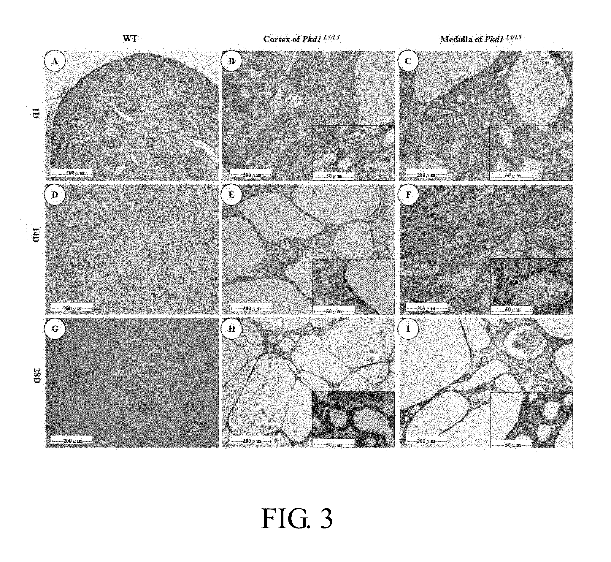
FIG. 3 shows temporal expression of NGAL in kidneys of Pkd1.sup.L3/L3 and wild-type mice identified by immunohistochemical analysis, in which renal sections at different ages of wild-type and homozygous kidneys were stained with NGAL antibody: (A-C) on day 1 (1D) of renal section, brown staining of NGAL was apparent in medulla but not in glomerulus and cortical tubules in wild-type (WT) mice (A), and in part of the renal cortex (B) and medulla (C) in Pkd1.sup.L3/L3 mice; (D-F) on 14D of renal section, the expression of NGAL was decreased than 1D in wild-type mice (D), but increased than 1D in the renal cortex (E) and medulla (F) in Pkd1.sup.L3/L3 mice; (G-I) on 28D of renal section, cyst enlargement was greater than 14D (E, F) in Pkd1.sup.L3/L3 mice, in which (i) cuboidal epithelium transformed into a squamous pattern during cyst enlargement, and expression of NGAL was also found (E, F, H, I), (ii) all images are representative of at least three mice per genotype in two independent experiments, and (iii) bars=200 .mu.m figures (A-I) and 50 .mu.m in insets (B, C, E, F, H, I);
FIG. 4 shows NGAL expression in renal tissue of patients with ADPKD and ARPKD: NGAL expression in human renal tissues of normal (A, B), ADPKD (C, D), and ARPKD (E, F), in which (i) the arrowheads indicate dark brown staining for NGAL in both ADPKD and ARPKD sections, (ii) the arrows indicate staining of endogenous NGAL expression in mononuclear cells in the interstitium area of ADPKD section, and (iii) bars=200 .mu.m (A), 50 .mu.m (B), 500 .mu.m (C, E), and 100 .mu.m (D, F);
FIGS. 5A-5C show that the renal level of Ngal is greater in Pkd1.sup.L3/L3; Ngal.sup.Tg/Tg mice than in Pkd1.sup.L3/L3 and Ngal.sup.Tg/Tg mice: <FIG. 5A> representative Western blotting of NGAL, NGAL-R, and .beta.-actin (control) at 21 days after birth (21D) in the mice with different genotypes; <FIGS. 5B and 5C> quantification of Western blotting results for NGAL and NGAL-R, in which bars show the means.+-.standard errors of the means (SEM) of three individual mice; *** p<0.001;
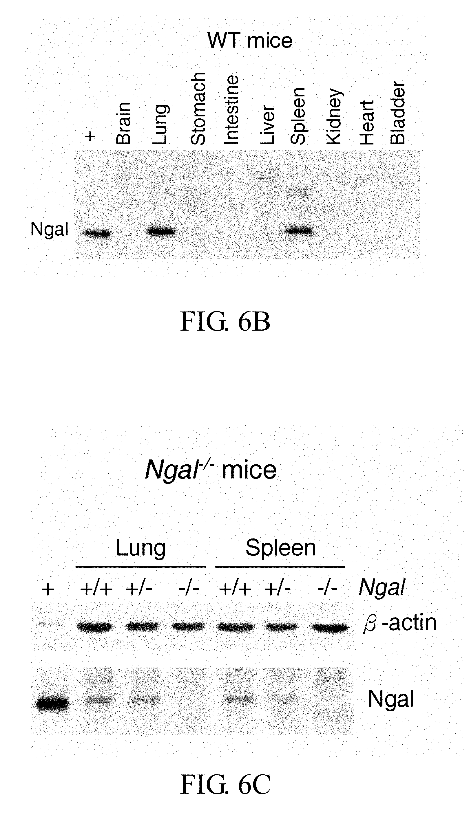
FIGS. 6A-6F show expression of NGAL in kidney, urine, and serum of Ngal.sup.Tg/0 mice, expression of NGAL in major organs of different mice, and glomerulus number in kidney sections of Ngal.sup.Tg/Tg and Ngal.sup.-/- mice as compared with wild type mice: <FIG. 6A> western blotting results of NGAL expression in kidneys, urine, and serum of Ngal.sup.Tg/0 mice; <FIGS. 6B, 6C, 6D and 6E> western blotting results of NGAL expression in the major organs of wild type, Ngal.sup.-/-, Ngal.sup.Tg/Tg, and Pkd1.sup.L3/L3; Ngal.sup.Tg/Tg mice, in which the cystic fluid was as a positive loading control (+); <FIG. 6F> the number of renal glomeruli of 21-day-old- and sex-matched in wild type (filled circle), Ngal.sup.Tg/Tg (filled square), and Ngal.sup.-/- (filled triangle) mice by Kruskal-Wallis test, in which all results represent the median with interquartile range (25%-75%);
FIGS. 7A-7B show that the overexpression of exogenous kidney-specific NGAL prolongs the survival of Pkd1.sup.L3/L3 mice: <FIG. 7A> Kaplan-Meier analysis of survival in Pkd1.sup.L3/L3 (open triangles), Pkd1.sup.L3/L3; Ngal.sup.Tg/Tg (filled circles), and Pkd1.sup.L3/L3; Ngal.sup.-/- (open squares) mice, in which the log-rank test showed significant differences between Pkd1.sup.L3/L3; Ngal.sup.Tg/Tg and Pkd1.sup.L3/L3 mice, and between Pkd1.sup.L3/L3; Ngal.sup.Tg/Tg and Pkd1.sup.L3/L3; Ngal.sup.-/- mice (both p<0.001); <FIG. 7B> the median survival days in Pkd1.sup.L3/L3 (n=31, median=25 days), Pkd1.sup.L3/L3; Ngal.sup.Tg/Tg (n=45, 46 days), and Pkd1.sup.L3/L3; Ngal.sup.-/- (n=32, 26 days) mice, in which all results represent the median with interquartile range (25%-75%); *** p<0.001;
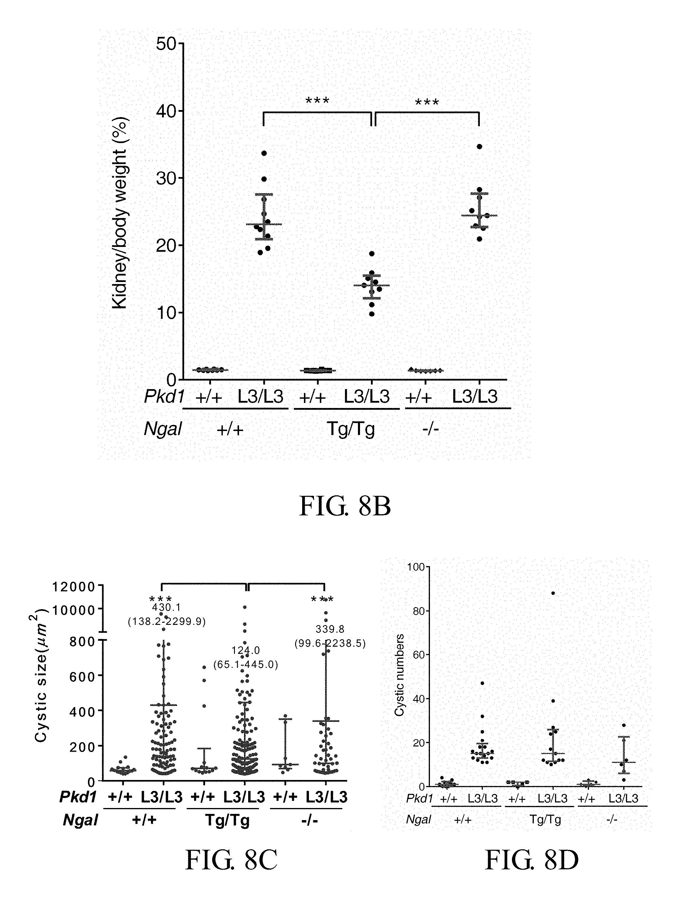
FIGS. 8A-8D show that the overexpression of exogenous kidney-specific NGAL reduces the renal cysts of Pkd1.sup.L3/L3 mice: <FIG. 8A> representative images of the kidneys of each genotype; <FIG. 8B> the median percentage of kidney weight to body weight in Pkd1.sup.L3/L3 (n=10, median=23.1%), Pkd1.sup.L3/L3; Ngal.sup.Tg/Tg (n=9, 14.1%), and Pkd1.sup.L3/L3; Ngal.sup.-/- (n=9, 24.4%) mice at 21D by Dunn's multiple comparisons test; <FIG. 8C> the size of renal cysts in Pkd1.sup.L3/L3 (430.1 .mu.m.sup.2), Pkd1.sup.L3/L3; Ngal.sup.Tg/Tg (124.0 .mu.m.sup.2), and Pkd1.sup.L3/L3; Ngal.sup.-/- (339.8 .mu.m.sup.2) mice; <FIG. 8D> the number of renal cysts, in which all results represent the median with interquartile range (25%-75%); *** p<0.001;
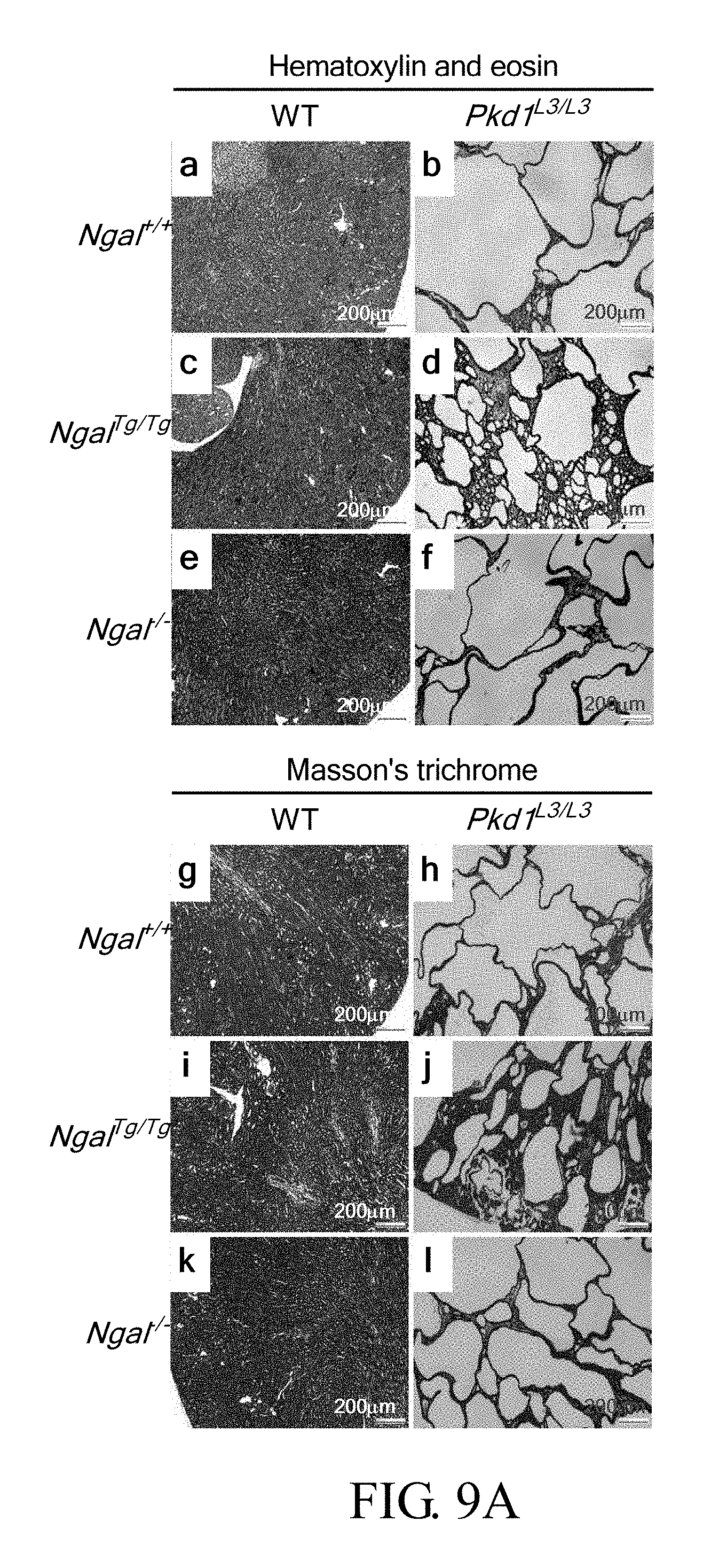
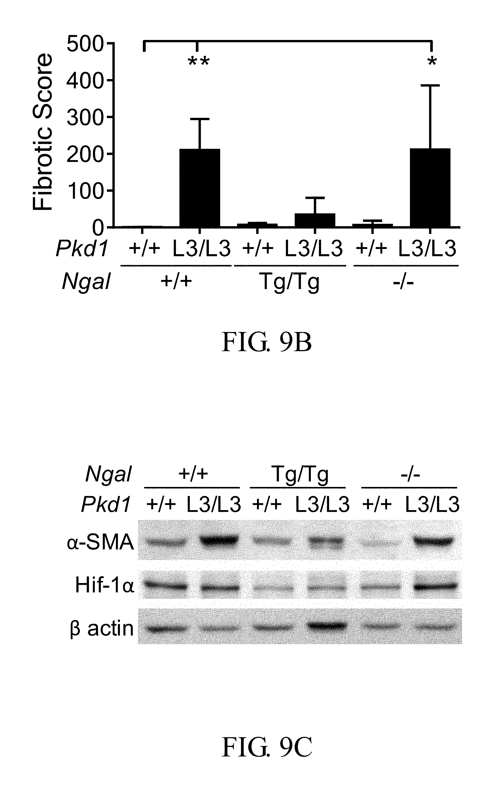
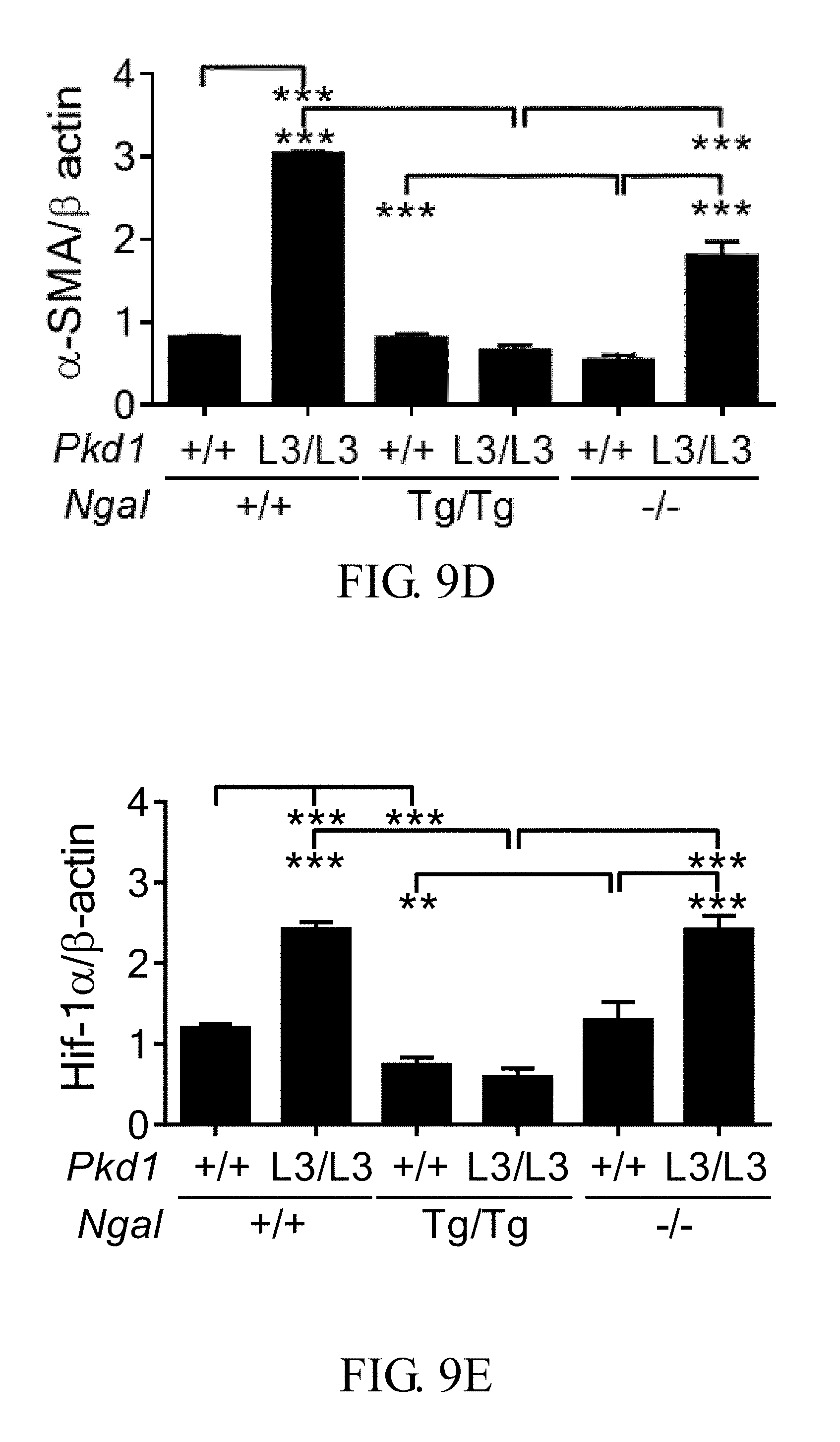
FIGS. 9A-9E show that the overexpression of exogenous kidney-specific NGAL reduces the interstitial fibrosis of Pkd1.sup.L3/L3 mice: <FIG. 9A> hematoxylin and eosin staining (upper) and Masson's trichrome staining (lower, scale bar: 200 .mu.m; <FIG. 9B> renal fibrosis scores, determined from Masson's trichrome-stained kidneys and calculated as described in the embodiments, in which all results represent the median with interquartile range (25%-75%); <FIG. 9C-9E> representative Western blotting and quantification of results for .alpha.-SMA and HIF-la, in which bar charts show means.+-.SEM of three samples; *p<0.05; **p<0.01; *** p<0.001;
FIGS. 10A-10C show that the overexpression of exogenous kidney-specific NGAL reduces the renal levels of PCNA and pro-caspase-3 of Pkd1.sup.L3/L3 mice: <FIG. 10A> representative Western blotting results of PCNA, caspase-3, and Glyceraldehyde 3-phosphate dehydrogenase (GAPDH, loading control) at 21D in the different genotypes; <FIG. 10B> quantification of Western blotting results for PCNA/GAPDH; <FIG. 10C> quantification of Western blotting results for pro-caspase-3/GAPDH, in which bar charts show means.+-.SEM of three samples; **p<0.01; ***p<0.001;
FIGS. 11A-11C show that the overexpression of exogenous kidney-specific NGAL reduces the levels of phospho-mTOR (p-mTOR) and mTOR of Pkd1.sup.L3/L3 mice: <FIG. 11A> representative Western blotting results of phospho-mTOR (Ser2448), mTOR, and .beta.-actin (loading control) at 21D in the different genotypes; <FIGS. 11B and 11C> quantification of Western blotting results for phospho-mTOR/.beta.-actin, and mTOR/.beta.-actin, respectively, in which bar charts show the means.+-.SEM of three samples; *p<0.05; **p<0.01; ***p<0.001;
FIGS. 12A-12C show that the overexpression of exogenous kidney-specific NGAL reduces the renal levels of phospho-Akt, Akt, phospho-S6K and S6K of Pkd1.sup.L3/L3 mice: <FIG. 12A> representative Western blotting results of phospho-Akt (Ser473), Akt, phospho-S6K (Thr389), S6K and .beta.-actin (loading control) at 21D in the different genotypes; <FIGS. 12B and 12C> quantification of Western blotting results for phospho-Akt/.beta.-actin and phospho-S6K/.beta.-actin, respectively, in which bar charts show means.+-.SEM of three samples; *p<0.05; **p<0.01; ***p<0.001;
FIGS. 13A-13C show that the renal levels of phospho-EGFR and EGFR are greater in Pkd1.sup.L3/L3; Ngal.sup.Tg/Tg, Pkd1.sup.L3/L3 and Pkd1.sup.L3/L3; Ngal.sup.-/- mice: <FIG. 13A> representative Western blotting results of phospho-EGFR (Tyr1068), EGFR and GAPDH (loading control) at 21D in the different genotypes; <FIGS. 13B and 13C> quantification of Western blotting results for phospho-EGFR/GAPDH, and EGFR/GAPDH, respectively, in which bar charts show the means.+-.SEM of three samples; **p<0.01; ***p<0.001; and
FIGS. 14A-14D show SDS-PAGE of the purified recombinant mNGAL.DELTA.N (mouse N-terminally truncated NGALA protein) and therapeutic effect of mNGAL-injection: <FIG. 14A> initial purification of recombinant mNGAL.DELTA.N via Ni.sup.2+-NTA chromatography; Lane M, protein marker; Lane T, total cell lysate; Lane P, insoluble pellet; Lane S, soluble fraction; Lane FT, flow through; Lane 3-39, eluted fractions; <FIG. 14B> further purification of recombinant mNGAL.DELTA.N via Q anion exchange chromatography; <FIG. 14C> Kaplan-Meier analysis of survival in Pkd1.sup.L3/L3 (open triangles), Pkd1.sup.L3/L3; Ngal.sup.Tg/Tg (filled circles), Pkd1.sup.L3/L3; Ngal.sup.-/- (open squares), mNGAL-injected Pkd1.sup.L3/L3 (filled triangles) and mNGAL-injected Pkd1.sup.L3/L3; Ngal.sup.-/- mice (filled squares); <FIG. 14D> representative images of the kidneys of untreated Pkd1.sup.L3/L3 (a-b), and mNGAL-treated Pkd1.sup.L3/L3 mice (c-d).
DETAILED DESCRIPTION OF THE PREFERRED EMBODIMENTS
Embodiments
Clinical Human Kidney Specimens and Patients
All kidney tissues were obtained from the human biobank of National Cheng Kung Hospital. This study was approved by the institutional review board of National Cheng Kung University Medical Center (A-ER-101-228). NGAL protein levels were examined by immunohostochemistry in 3 human kidney specimens (2 PKD cases, 1 normal kidney) obtained from the Department of Pathology, National Cheng Kung University Hospital, Taiwan. Normal tissue was obtained from a deceased patient whose autopsy had confirmed the absence of non-genitourinary tract disease. Case 1 had ADPKD with chronic renal failure due to severe tubulointerstitial nephritis, while case 2 had been diagnosed at 30 weeks of gestational age as premature ARPKD with polycystic mega-kidney, and the pregnancy had been terminated.
Immunohistochemistry
Kidneys were removed and fixed in 10% formalin at 4.degree. C. overnight, then dehydrated and embedded in paraffin, and sectioned at 4 .mu.m for immunostaining. To detect NGAL, kidney sections were blocked with Avidin/Biotin Blocking Kit (Vector Laboratories, Burlingame, Calif.) after deparaffinization and rehydration, and incubated overnight at 4.degree. C. with rabbit anti-NGAL antibody or goat anti-human NGAL antibody. For other immunostaining, a standard immunoperoxidase protocol (Vectastain ABC kit; Vector Laboratories) was used. After blocking with goat serum, sections were incubated with primary antibodies for 1 hour at room temperature, rinsed in PBS, incubated with biotinylated goat anti-rabbit antibodies, rinsed, then incubated with streptavidin-conjugated peroxidase, rinsed, then incubated with 3-amino-9-ethyl-carbazole as a chromogen, counterstained with hematoxylin, and examined by light microscopy.
Animal Experiments
All mice were housed in the National Laboratory Animal Center (NLAC), Tainan, Taiwan, under a 12 h light:dark cycle, and all experiments were conducted according to protocols approved by the Institutional Animal Care and Use Committee of NLAC. In this study, the inventors identified no significant difference between male and female mice prior to experimental execution. Several literatures also conducted their experiments using animals in both sexes. This suggests that the sexes of animals have little impact on the PKD pathological and molecular analyses. Therefore, both sexes of mice were combined and used in the experiments.
PKD Mice
The inventors' previous study described a murine model of PKD. Pkd1.sup.L3/L3 mice produced low levels of full-length PC1, and progressively developed polycystic kidney disease (Jiang S T, et al., Am J Pathol, 168: 205-220, 2006). The original Pkd1.sup.L3/+ mice had a mixed C57BL6-129 background and were backcrossed with C57BL/6J mice for more than ten generations to obtain a uniform genetic background. The Pkd1.sup.L3/+ mice with stable C57BL/6J genetic background were then used to generate the homozygous mutants Pkd1.sup.L3/L3.
Ngal Knockout Mice
The Ngal conventional knockout mice were generated using VelociGene biotechnology. The murine Ngal gene was disrupted by a PGK/Neo cassette, and lacZ was used as the reporter gene. The Ngal.sup.-/- mice were originally bred on a mixed C57BL6-129 background and were backcrossed with C57BL/6J mice for more than ten generations prior to intercrossing to generate Ngal.sup.-/-, ensuring a uniform genetic background. NGAL conventional knockout can prevent the production of NGAL from kidney and other tissues so as to characterize the renal function of NGAL protein in PKD.
Cdh16-mNgal Transgenic Mice
The Cdh16-mNgal transgenic mice overexpress the murine NGAL under the control of the mouse Cdh16 promoter (Shao X, et al., J Am Soc Nephrol, 13: 1824-1836, 2002). Ksp-Cdh16-mNgal ensured an early expression of exogenous NGAL at a time before the expression of endogenous NGAL and cyst formation in ADPKD. Cdh16-mNgal transgenic mice have a C57BL/6J genetic background. The protocol for DNA recombination was approved by the Institutional Biosafety Committee of the NLAC. Here, the Ngal is NCBI Gene No. 16819 or EMBL-EBI No. ENSMUSG00000026822.
Genotyping
Polymerase chain reaction (PCR) was used to genotype all mutant mice with genomic DNA extracted from the tails. Each sample contained 20 .mu.L of the PCR reaction mixture, and .beta.-actin was used as the internal control. The primer sequences were murine Ngal forward primer sequence, SEQ ID No. 1 (5'-ATGGCCCTGAGTGTCATGTGTC-3'), and murine Ngal reverse primer sequence, SEQ ID No. 2 (5'-GCTCCAGATGCTCCTTGGTATG-3'); and .beta.-actin forward primer sequence, SEQ ID No. 3 (5'-GGCATTGTTACCAACTGGGA CG-3'), and .beta.-actin reverse primer sequence, SEQ ID No. 4 (5'-AGG A AGGCTGGAAAAGAGCC-3'). Ngal knockout mutants were analyzed using the following primers: murine Ngal 5' UTR forward primer sequence, SEQ ID No. 5 (5'-TTCCTCCTCCAGCACACA TCAGAC-3'), lacZ reverse primer sequence, SEQ ID No. 6 (5'-GAGTAACAACCCGTCGGATTCTC-3'), and murine Ngal reverse primer sequence, SEQ ID No. 7 (5'-AGGGGTTACTGTC AGAGTGGCTATC-3').
Western-Blot Analysis
Total proteins (50 .mu.g) extracted from mouse kidneys were subjected to Western-blot analysis. The pQE protein expression system (Qiagen) was used to express mouse full-length NGAL and purified NGAL protein with 6.times.His-tags. The purified NGAL was then used to generate the NGAL antibody by immunizing the rabbit with mouse full-length NGAL protein. The other antibodies were rabbit polyclonal antibody to Slc22A17 (GTX85032; GeneTex), rabbit polyclonal antibody to GAPDH (631401; BioLegend), mouse monoclonal antibody to .alpha.-smooth muscle actin (.alpha.-SMA) (A2547; Sigma-Aldrich), rabbit polyclonal antibody to hypoxia-inducible factor 1-.alpha. (HIF-1.alpha.) (GTX 127309; GeneTex), rabbit monoclonal antibody to mTOR (phospho Ser2448) (5536; Cell Signaling Technology), rabbit monoclonal antibody to mTOR (2983; Cell Signaling Technology), mouse monoclonal antibody to .beta.-actin (8H10D10) (12262; Cell Signaling Technology), rabbit polyclonal antibody to S6K1 (phospho Thr389) (ab2571; Abcam), rabbit monoclonal antibody to p70 S6 Kinase (2708; Cell Signaling Technology), rabbit polyclonal antibody to Akt (phospho Ser473) (9271; Cell Signaling Technology), rabbit polyclonal antibody to Akt (9272; Cell Signaling Technology), rabbit polyclonal antibody to caspase-3 (9662; Cell Signaling Technology), mouse monoclonal antibody to proliferating cell nuclear antigen (PCNA) (307902; Biolegend), rabbit polyclonal antibody to epidermal growth factor receptor (EGFR) (phospho Tyr1068) (2234; Cell Signaling Technology), and rabbit monoclonal antibody to EGFR (GTX61503; GeneTex).
Histology and Histomorphometric Analyses
Specimens were fixed in formalin, embedded in paraffin, cut into 4 sections, and stained with hematoxylin and eosin (H&E) or Masson's trichrome for examination by light microscopy (Eclipse E600 Nikon, Japan), as described previously (Wang, E. et al., J Pathol, 222: 238-248, 2010). Quantification of cyst number and size (H&E staining) and staining of collagen fiber (Masson's trichrome) was performed by who was blinded to the mouse genotypes. The Image-Pro Plus v. 4.5.0.29 software (Media Cybernetics, Rockville, Md., 20850 USA) was used to calculate mean quick scores (QSs). For each slide, the software randomly selected five fields (200.times.) for analysis. After adequate white balance, the software allows the operator to gate areas of special interest and to examine them separately by grabbing digital images. Each slide was analyzed to determine the label index (LI; ratio of positively stained areas to the total area) and the mean optical density (MOD; stain concentration, based on counts of positive pixels). The QS was calculated as the mathematical product of LI and MOD. For counting of cysts, representative images of H&E-stained transverse kidney sections, including the cortex, medulla, and papilla, were used. A grid was placed over the images, and the percentage of grid intersection points that bisected cystic and non-cystic areas was calculated.
Statistical Analysis
Comparisons were made among three groups: Pkd1.sup.L3/L3, Pkd1.sup.L3/L3; Ngal.sup.Tg/Tg, and Pkd1.sup.L3/L3; Ngal.sup.-/-. Parametric data were compared all groups with one-way ANOVA, and followed with Tukey multiple comparison tests of every pair. Due to the non-normal distributions, survival times are presented as medians and interquartile ranges (25%-75%), and groups were compared by the non-parametric Kruskal-Wallis test with Dunn's multiple comparisons test. Survival curves were constructed, and Kaplan-Meier estimates were used to compare survival rates of the different groups using the log-rank test. A p-value less than 0.05 was considered statistically significant.
<Results>
Generation of Ngal Conventional Knockout and Cdh16-mNgal Transgenic Mice
The mouse Ngal targeting strategy and construction of the Cdh16-mNgal transgene were shown in FIGS. 1A and 1B, respectively. PCR analysis confirmed the genotyping of these mice (FIG. 1C). The ratio of the heterozygous Ngal.sup.Tg/o mice also confirmed the homozygosity of the Ngal.sup.Tg/Tg mice after backcrossing with wild type C57BL/6J mice. The expression level of Ngal was positively correlated with the copy number of Ngal in Ngal.sup.Tg/o and Ngal.sup.Tg/Tg mice (FIG. 1D).
The Level of NGAL is Increased in the Kidneys of Pkd1.sup.L3/L3; Ngal.sup.Tg/Tg Mice
Pkd1.sup.L3/L3; Ngal.sup.Tg/Tg mice were generated by crossbreeding Pkd1.sup.L3/+; Ngal.sup.Tg/Tg mice with Pkd1.sup.L3/+; Ngal.sup.Tg/Tg mice. Western-blot analysis detected the differential expression of endogenous NGAL in urine, serum and kidneys of Pkd1.sup.L3/L3 and wild type mice (FIGS. 2A-2D). The level was gradually increased along the course of cyst enlargement in Pkd1.sup.L3/L3 mice; whereas its level was decreased with age in wild type mice. Immunohistochemical analysis revealed a temporal expression pattern of renal NGAL at 1 day (D), 14D and 28D of age in both wild type and Pkd1.sup.L3/L3 mice (FIG. 3). Endogenous NGAL was mainly localized at the apical surface of cystic epithelia in Pkd1.sup.L3/L3 mice at 14D and 28D, and cysts were enlarged with age in the renal medulla in Pkd1.sup.L3/L3 mice. Similar patterns were observed as well in the kidneys of patients with ADPKD or autosomal recessive polycystic kidney disease (ARPKD) (FIG. 4). The results indicated that an upregulation of endogenous NGAL in cystic epithelia might represent an abnormal renal development, and this argument is supported by the role of NGAL in the severity of PKD. The renal expression of NGAL and NGAL-R in these three PKD mice (namely Pkd1.sup.L3/L3 mice, Pkd1.sup.L3/L3; Ngal.sup.Tg/Tg mice, Pkd1.sup.L3/L3; Ngal.sup.-/- mice) at 21D was next examined by Western-blot analysis (FIG. 5A). The renal level of NGAL in Pkd1.sup.L3/L3; Ngal.sup.Tg/Tg mice was 1.8-fold and 3.7-fold greater than thoses in the Pkd1.sup.L3/L3 and Ngal.sup.Tg/Tg mice, respectively (FIG. 5B). In addition, the renal expression of NGAL-R in these three PKD mice was significantly upregulated as compared to those in the control littermates without PKD (p<0.001) (FIGS. 5A and 5C). The results demonstrated that exogenous NGAL is continuously overexpressed in the embryonic kidney under the control of Ksp-Cdh16 promoter before the expression of endogenous NGAL, and this suggests that exogenous NGAL can prevent progression of PKD in Pkd1.sup.L3/L3 mice at embryonic stage.
Kidney-Specific Overexpression of Exogenous NGAL in Ngal.sup.Tg/Tg and Pkd1.sup.L3/L3; Ngal.sup.Tg/Tg Mice
Western-blot analysis detected the expression of exogenous NGAL in urine, serum and kidneys of Ngal.sup.Tg/0 mice (FIG. 6A). Comparing wild type and Ngal.sup.-/- mice, it was observed that NGAL was expressed in the tissue of lung and spleen in wild type (WT) mice (FIG. 6B) but not seen in Ngal.sup.-/- mice (FIG. 6C). Additionally, the kidney-specific overexpression of exogenous NGAL was detected in Ngal.sup.Tg/Tg (FIG. 6D), and Pkd1.sup.L3/L3; Ngal.sup.Tg/Tg (FIG. 6E) mice. Furthermore, the effects of NGAL on the renal development was examined by counting glomeruli in the kidney sections of Ngal.sup.Tg/Tg mice and Ngal.sup.-/- mice as compared with wild type mice (FIG. 6F). These results showed that the number of renal glomeruli had no significant differences among these age- and sex-matched mice (p=0.294).
Overexpression of Cdh16-mNgal Prolongs the Survival of Pkd1.sup.L3/L3; Ngal.sup.Tg/Tg Mice
By the differential expression of NGAL, the effect of NGAL on the survival of Pkd1.sup.L3/L3, Pkd1.sup.L3/L3; Ngal.sup.Tg/Tg and Pkd1.sup.L3/L3; Ngal.sup.-/- mice can be observed (FIG. 7A). The log-rank tests showed that the lifespan of Pkd1.sup.L3/L3; Ngal.sup.Tg/Tg mice was a significantly longer (median=46 days, Interquartile-Range IQR 37-60 days) than those of Pkd1.sup.L3/L3 (25 days, IQR 23-26 days) and Pkd1.sup.L3/L3; Ngal.sup.-/- mice (26 days, IQR 24-31 days) (both p<0.001 versus Pkd1.sup.L3/L3; Ngal.sup.Tg/Tg mice) (FIG. 7B). Additionally, there was no significant difference between the lifespan of Pkd1.sup.L3/L3 and Pkd1.sup.L343; Ngal.sup.-/- mice (p=0.2988).
Overexpression of Cdh16-mNgal Retards Cyst Enlargement in Pkd1.sup.L3/L3; Ngal.sup.Tg/Tg Mice
The total kidney volume (TKV) was known to be positively correlated with increased BUN and disease progression of PKD. Polycystic kidneys progressed to the terminal stage at 21D in Pkd1.sup.L3/L3 and Pkd1.sup.L3/L3; Ngal.sup.-/- mice. The kidneys of Pkd1.sup.L3/L3; Ngal.sup.Tg/Tg mice were smaller than those of Pkd1.sup.L3/L3 and Pkd1.sup.L3/L3; Ngal.sup.-/- mice (FIG. 8A). Additionally, the median kidney weight to body weight ratio at 21D was significantly lower in Pkd1.sup.L3/L3; Ngal.sup.Tg/Tg (median=14.1%, IQR 12.2-15.5%) than in Pkd1.sup.L3/L3 (23.1%, IQR 20.9-27.6%) and Pkd1.sup.L3/L3; Ngal.sup.-/- (24.4%, IQR 22.7-27.7%) mice (both p<0.001 versus Pkd1.sup.L3/L3; Ngal.sup.Tg/Tg) (FIG. 8B). Renal cyst size was also significantly smaller in Pkd1.sup.L3/L3; Ngal.sup.Tg/Tg mice than in other two kinds of PKD mice (both p<0.001 versus Pkd1.sup.L3/L3; Ngal.sup.Tg/Tg) (FIG. 8C), whereas there was no significant difference in cyst numbers (FIG. 8D). These results demonstrated that overexpression of exogenous kidney-specific NGAL could prevent rapid progression of PKD by reducing cyst size but not cyst number of Pkd1.sup.L3/L3 mice. Based on the results, it can be recognized that the deletion of Ngal has no effect on PKD possible, which suggests that the decreased PC1 but not NGAL determines the PKD progression. Additionally, upregulation of endogenous NGAL of Pkd1.sup.L3/L3 mice cannot protect mice from early death.
Overexpression of Cdh16-mNgal Reduces Interstitial Fibrosis in Pkd1.sup.L3/L3; Ngal.sup.Tg/Tg Mice
Hematoxylin and eosin-stained (H&E, FIG. 9A, upper panels) and Masson's trichrome-stained (FIG. 9A, lower panels) kidney sections were examined at 21D. Wild type mice had a significant lower renal fibrosis scores than both Pkd1.sup.L3/L3 (p=0.002) and Pkd1.sup.L3/L3; Ngal.sup.-/- (p=0.015) mice but not Pkd1.sup.L3/L3; Ngal.sup.Tg/Tg mice (p=0.417) (FIG. 9B). The renal levels of .alpha.-SMA and HIF-1.alpha., the molecules associated with progressive fibrosis of PKD were further examined. Western-blot analyses demonstrated that the levels of .alpha.-SMA in Pkd1.sup.L3/L3; Ngal.sup.Tg/Tg mice was comparable to those of wild type mice (FIG. 9C); however, the expression of .alpha.-SMA and HIF-1.alpha. were significantly lower than those of other two kind of PKD mice (both p<0.001 versus Pkd1.sup.L3/L3; Ngal.sup.Tg/Tg) (FIGS. 9D and 9E). Thus, the expressions of .alpha.-SMA and HIF-la in these mice were highly correlated with their fibrotic score.
The Changes of Proliferating Cell Nuclear Antigen (PCNA), and Caspase-3 in Pkd1.sup.L3/L3; Ngal.sup.Tg/Tg Mice
Both proliferation and apoptosis were increased in early cystic formation of polycystic kidneys. Western-blot analyses (FIG. 10A) revealed that both the expression of pro-caspase-3 and PCNA were reduced and that active caspase-3 increased in Pkd1.sup.L3/L3; Ngal.sup.Tg/Tg mice as compared to other two kinds of PKD mice (both p<0.001 versus Pkd1.sup.L3/L3; Ngal.sup.Tg/Tg) (FIGS. 10B and 10C).
Renal Level of Mammalian Target of Rapamycin (mTOR) is Decreased in Pkd1.sup.L3/L3; Ngal.sup.Tg/Tg Mice with Downregulation of Akt and S6 Kinase Signaling
All of the three PKD mice exhibited increased expressions of mTOR and p-mTOR as compared to their respective control littermates without PKD (FIG. 11A). Additionally, Pkd1.sup.L3/L3; Ngal.sup.Tg/Tg mice had the lowest mTOR and p-mTOR levels compared to Pkd1.sup.L3/L3 and Pkd1.sup.L3/L3; Ngal.sup.-/- mice (both p<0.001 versus Pkd1.sup.L3/L3; Ngal.sup.Tg/Tg) (FIGS. 11B-11C). A major upstream regulator (Akt) and a downstream target [ribosomal protein S6 Kinase (S6K)] of mTOR are involved in cell proliferation. Subsequent Western-blot analyses (FIG. 12A) confirmed that the expression of phosphorylated Akt (p-Akt) and phosphorylated S6K (p-S6K) were significantly reduced in Pkd1.sup.L3/L3; Ngal.sup.Tg/Tg mice as compared to the other two kinds of PKD mice (both p<0.001 versus Pkd1.sup.L3/L3; Ngal.sup.Tg/Tg) (FIGS. 12B and 12C).
Renal Levels of Epidermal Growth Factor Receptor (EGFR) and Phosphorylated EGFR (p-EGFR) in Pkd1.sup.L3/L3; Ngal.sup.Tg/Tg Mice
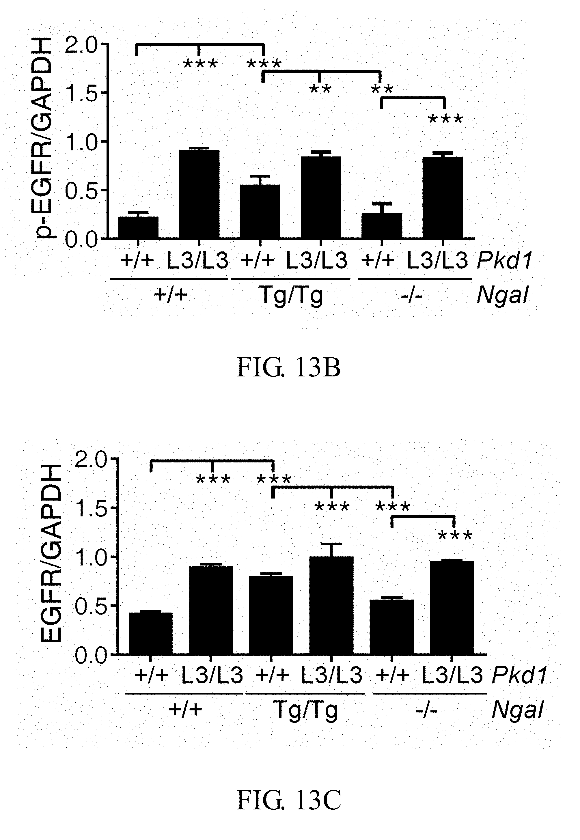
Overexpression and apical mislocation of EGFR and p-EGFR is known to promote cyst growth, and inhibition of EGFR tyrosine kinase activity can attenuate the development of PKD (Orellana S A, et al., Kidney Int, 47: 490-499, 1995; Du J, et al., Am J Physiol, 269: C487-495, 1995; Sweeney W E, et al., Kidney Int, 57: 33-40, 2000; Tones V E, et al., Kidney Int, 64: 1573-1579, 2003). Western-blot analyses (FIG. 13A) demonstrated that the expression of both EGFR and p-EGFR (Tyr1068) were significantly increased in these three PKD mice as compared with their control littermates without PKD (p<0.001 versus control) (FIGS. 13B and 13C). However, the ratio of p-EGFR/EGFR revealed no difference among the three PKD mice.
Preparation of Recombinant Mouse Ngal Protein (mNGAL) and Treatment PKD Mice
In our study, we emphasized the therapeutic effect of secretory Ngal on a reduction of cyst growth. We further prepared the recombinant mouse Ngal protein (mNGAL), and then performed intraperitoneal injection of functional recombinant mNgal protein with low endotoxin and high purity (100 .mu.g/day) into the PKD mice. The coding sequences of mNGAL (residues from 23 to 200) was subcloned into pET21a vector with an N-terminal His.sub.6-tag to facilitate purification. The correct construct by sequencing was subsequently transformed to E. coli BL21 (DE3) for protein expression. The 60 ml overnight culture of a single transformant was used to inoculate 6 liters of fresh LB medium containing 100 .mu.g/ml ampicillin. The cells were grown to A600 nm=0.6.about.1.0 and induced with 0.5 mM isopropyl .beta.-thiogalactopyranoside (IPTG) at 20.degree. C. After 16 hr, the cells were harvested by centrifugation at 7,000.times.g for 15 mM to collect the cell paste. The cell pellet was resuspended immediately in the lysis buffer containing 20 mM Tris-HCl, 400 mM NaCl, 10 mM imidazole, pH 8.0. The cell suspension was disrupted by Constant Cell Disruption System (CONSTANT SYSTEM Ltd., UK) and centrifuged at 17,000.times.g to remove cell debris. The cell-free extract was loaded onto a Ni.sup.2+-NTA column, which had been previously equilibrated with lysis buffer. The column was washed with lysis buffer, subsequently the His.sub.6-tagged mNGAL was eluted by a linear gradient from 10 mM to 500 mM imidazole (FIG. 14A). The purified His.sub.6-tagged mNGAL was diluted with four-fold 20 mM Tris, pH 8.0 buffer and subsequently was loaded onto a Q anion exchange column, which had been previously equilibrated with 20 mM Tris, pH 8.0 buffer. The low lipopolysaccharides (LPS) containing His.sub.6-tagged mNGAL was collected from the flow through of Q anion exchange column (FIG. 14B). Finally the His.sub.6-tagged mNGAL was concentrated and exchanged into PBS buffer with 10 kDa cut-off size membrane of Vivaspin 20 (Sartorius, Germany) for storage at -80.degree. C. The Pkd1.sup.L3/L3 (n=3) and Pkd1.sup.L3/L3; Ngal.sup.-/- mice (n=6) were injected with mNGAL (100 .mu.l/day) after postnatal day 7 via subcutaneous (SC) or intraperitoneal injection (IP). Our data demonstrated that the survival rate was markedly increased in all mNGAL-injected Pkd1.sup.L3/L3 and Pkd1.sup.L3/L3; Ngal.sup.-/- mice as compared with untreated group (FIG. 14C). The cystic kidneys of Pkd1.sup.L3/L3 mice was also significantly retarded in mNGAL-treated Pkd1.sup.L3/L3 mice as compared with untreated group (FIG. 14D and Table 1).
TABLE-US-00001 TABLE 1 (a) (b) (c) (d) mNGAL treatment -- -- + + Dosage/day -- -- 100 .mu.g 100 .mu.g Treatment period -- -- 12-35D 9-32D Kidney/body weight ratio (%) 30.96% 29.35% 13.72% 12.74%
SEQUENCE LISTINGS
1
7122DNAArtificial SequencePrimer 1atggccctga gtgtcatgtg tc 22222DNAArtificial SequencePrimer 2gctccagatg ctccttggta tg 22322DNAArtificial SequencePrimer 3ggcattgtta ccaactggga cg 22420DNAArtificial SequencePrimer 4aggaaggctg gaaaagagcc 20524DNAArtificial SequencePrimer 5ttcctcctcc agcacacatc agac 24623DNAArtificial SequencePrimer 6gagtaacaac ccgtcggatt ctc 23725DNAArtificial SequencePrimer 7aggggttact gtcagagtgg ctatc 25
* * * * *
D00001
D00002
D00003
D00004
D00005
D00006
D00007
D00008
D00009
D00010
D00011
D00012
D00013
D00014
D00015
D00016
D00017

D00018

D00019

D00020

D00021

D00022
D00023

D00024

P00001

P00002

S00001
XML
uspto.report is an independent third-party trademark research tool that is not affiliated, endorsed, or sponsored by the United States Patent and Trademark Office (USPTO) or any other governmental organization. The information provided by uspto.report is based on publicly available data at the time of writing and is intended for informational purposes only.
While we strive to provide accurate and up-to-date information, we do not guarantee the accuracy, completeness, reliability, or suitability of the information displayed on this site. The use of this site is at your own risk. Any reliance you place on such information is therefore strictly at your own risk.
All official trademark data, including owner information, should be verified by visiting the official USPTO website at www.uspto.gov. This site is not intended to replace professional legal advice and should not be used as a substitute for consulting with a legal professional who is knowledgeable about trademark law.