Therapeutic renal neuromodulation for treating dyspnea and associated systems and methods
Sobotka
U.S. patent number 10,292,754 [Application Number 13/884,602] was granted by the patent office on 2019-05-21 for therapeutic renal neuromodulation for treating dyspnea and associated systems and methods. This patent grant is currently assigned to MEDTRONIC ARDIAN LUXEMBOURG S.A.R.L.. The grantee listed for this patent is Paul A. Sobotka. Invention is credited to Paul A. Sobotka.


| United States Patent | 10,292,754 |
| Sobotka | May 21, 2019 |
Therapeutic renal neuromodulation for treating dyspnea and associated systems and methods
Abstract
Methods for treating dyspnea with therapeutic renal neuromodulation and associated system and methods are disclosed herein. One aspect of the present technology, for example, is directed to methods that block, reduce, and/or inhibit renal sympathetic nerve activity to achieve a reduction in central sympathetic tone. Renal sympathetic nerve activity may be altered or modulated along the afferent and/or efferent pathway. The achieved reduction in central sympathetic tone may carry therapeutic benefits for patients with dyspnea.
| Inventors: | Sobotka; Paul A. (West St. Paul, MN) | ||||||||||
|---|---|---|---|---|---|---|---|---|---|---|---|
| Applicant: |
|
||||||||||
| Assignee: | MEDTRONIC ARDIAN LUXEMBOURG
S.A.R.L. (Luxembourg, LU) |
||||||||||
| Family ID: | 46084621 | ||||||||||
| Appl. No.: | 13/884,602 | ||||||||||
| Filed: | November 16, 2011 | ||||||||||
| PCT Filed: | November 16, 2011 | ||||||||||
| PCT No.: | PCT/US2011/061019 | ||||||||||
| 371(c)(1),(2),(4) Date: | October 03, 2013 | ||||||||||
| PCT Pub. No.: | WO2012/068268 | ||||||||||
| PCT Pub. Date: | May 24, 2012 |
Prior Publication Data
| Document Identifier | Publication Date | |
|---|---|---|
| US 20140024959 A1 | Jan 23, 2014 | |
Related U.S. Patent Documents
| Application Number | Filing Date | Patent Number | Issue Date | ||
|---|---|---|---|---|---|
| 61414499 | Nov 17, 2010 | ||||
| Current U.S. Class: | 1/1 |
| Current CPC Class: | A61B 5/082 (20130101); A61B 18/02 (20130101); A61B 18/1492 (20130101); A61B 18/14 (20130101); A61B 5/4848 (20130101); A61B 2018/00613 (20130101); A61B 2018/00434 (20130101); A61B 2018/00589 (20130101); A61B 2018/00577 (20130101) |
| Current International Class: | A61B 18/14 (20060101); A61B 18/02 (20060101); A61B 5/08 (20060101); A61B 5/00 (20060101); A61B 18/00 (20060101) |
References Cited [Referenced By]
U.S. Patent Documents
| 4602624 | July 1986 | Naples et al. |
| 4649936 | March 1987 | Ungar et al. |
| 4709698 | December 1987 | Johnston et al. |
| 4764504 | August 1988 | Johnson et al. |
| 4890623 | January 1990 | Cook et al. |
| 4976711 | December 1990 | Parins et al. |
| 5300068 | April 1994 | Rosar et al. |
| 5358514 | October 1994 | Schulman et al. |
| 5368591 | November 1994 | Lennox et al. |
| 5423744 | June 1995 | Gencheff et al. |
| 5425364 | June 1995 | Imran |
| 5484400 | January 1996 | Edwards et al. |
| 5571147 | November 1996 | Sluijter et al. |
| 5588964 | December 1996 | Imran et al. |
| 5599345 | February 1997 | Edwards et al. |
| 5626576 | May 1997 | Janssen |
| 5672174 | September 1997 | Gough et al. |
| 5688266 | November 1997 | Edwards et al. |
| 5700282 | December 1997 | Zabara |
| 5707400 | January 1998 | Terry, Jr. et al. |
| 5772590 | June 1998 | Webster, Jr. |
| 5860974 | January 1999 | Abele |
| 5865787 | February 1999 | Shapland et al. |
| 5893885 | April 1999 | Webster et al. |
| 5944710 | August 1999 | Dev et al. |
| 5954719 | September 1999 | Chen et al. |
| 5983141 | November 1999 | Sluijter et al. |
| 6004269 | December 1999 | Crowley et al. |
| 6009877 | January 2000 | Edwards |
| 6024740 | February 2000 | Lesh et al. |
| 6036687 | March 2000 | Laufer et al. |
| 6066134 | May 2000 | Eggers et al. |
| 6091995 | July 2000 | Ingle et al. |
| 6099524 | August 2000 | Lipson et al. |
| 6117101 | September 2000 | Diederich et al. |
| 6135999 | October 2000 | Fanton et al. |
| 6142993 | November 2000 | Whayne et al. |
| 6149620 | November 2000 | Baker et al. |
| 6161048 | December 2000 | Sluijter et al. |
| 6219577 | April 2001 | Brown, III et al. |
| 6224592 | May 2001 | Eggers et al. |
| 6246912 | June 2001 | Sluijter et al. |
| 6273886 | August 2001 | Edwards et al. |
| 6283951 | September 2001 | Flaherty et al. |
| 6292695 | September 2001 | Webster, Jr. et al. |
| 6314325 | November 2001 | Fitz |
| 6322558 | November 2001 | Taylor et al. |
| 6322559 | November 2001 | Daulton et al. |
| 6405732 | June 2002 | Edwards et al. |
| 6413255 | July 2002 | Stern |
| 6488679 | December 2002 | Swanson et al. |
| 6506189 | January 2003 | Rittman, III et al. |
| 6514226 | February 2003 | Levin et al. |
| 6522926 | February 2003 | Kieval et al. |
| 6542781 | April 2003 | Koblish et al. |
| 6562034 | May 2003 | Edwards et al. |
| 6616624 | September 2003 | Kieval |
| 6622731 | September 2003 | Daniel et al. |
| 6635054 | October 2003 | Fjield et al. |
| 6640120 | October 2003 | Swanson et al. |
| 6685648 | February 2004 | Flaherty et al. |
| 6711444 | March 2004 | Koblish |
| 6736835 | May 2004 | Pellegrino et al. |
| 6752805 | June 2004 | Maguire et al. |
| 6845267 | January 2005 | Harrison et al. |
| 6850801 | February 2005 | Kieval et al. |
| 6869431 | March 2005 | Maguire et al. |
| 6885888 | April 2005 | Rezai |
| 6893436 | May 2005 | Woodard et al. |
| 6917834 | July 2005 | Koblish et al. |
| 6939346 | September 2005 | Kannenberg et al. |
| 6949097 | September 2005 | Stewart et al. |
| 6951217 | October 2005 | Berthon-Jones |
| 7149574 | December 2006 | Yun et al. |
| 7162303 | January 2007 | Levin et al. |
| 7221979 | May 2007 | Zhou et al. |
| 7381200 | June 2008 | Katoh et al. |
| 7390894 | June 2008 | Weinshilboum et al. |
| 7617005 | November 2009 | Demarais et al. |
| 7620451 | November 2009 | Demarais et al. |
| 7622304 | November 2009 | Tonnessen et al. |
| 7647115 | January 2010 | Levin et al. |
| 7653438 | January 2010 | Deem et al. |
| 7717948 | May 2010 | Demarais et al. |
| 7778703 | August 2010 | Gross et al. |
| 8131371 | March 2012 | Demarais et al. |
| 8131372 | March 2012 | Levin et al. |
| 8140170 | March 2012 | Rezai et al. |
| 8145317 | March 2012 | Demarais et al. |
| 8150518 | April 2012 | Levin et al. |
| 8150519 | April 2012 | Demarais et al. |
| 8150520 | April 2012 | Demarais et al. |
| 8175711 | May 2012 | Demarais et al. |
| 2002/0087208 | July 2002 | Koblish et al. |
| 2002/0107515 | August 2002 | Edwards et al. |
| 2002/0139379 | October 2002 | Edwards et al. |
| 2002/0165532 | November 2002 | Hill et al. |
| 2002/0183682 | December 2002 | Darvish et al. |
| 2003/0050635 | March 2003 | Truckai et al. |
| 2003/0050681 | March 2003 | Pianca et al. |
| 2003/0060858 | March 2003 | Kieval et al. |
| 2003/0074039 | April 2003 | Puskas |
| 2003/0125790 | July 2003 | Fastovsky et al. |
| 2003/0181897 | September 2003 | Thomas et al. |
| 2003/0195507 | October 2003 | Stewart et al. |
| 2003/0199863 | October 2003 | Swanson et al. |
| 2003/0216792 | November 2003 | Levin et al. |
| 2003/0229340 | December 2003 | Sherry et al. |
| 2003/0233099 | December 2003 | Danaek et al. |
| 2004/0010289 | January 2004 | Biggs et al. |
| 2004/0167509 | August 2004 | Taimisto |
| 2004/0215186 | October 2004 | Cornelius et al. |
| 2005/0065567 | March 2005 | Lee et al. |
| 2005/0080409 | April 2005 | Young et al. |
| 2005/0096647 | May 2005 | Steinke et al. |
| 2005/0187579 | August 2005 | Danek et al. |
| 2005/0228460 | October 2005 | Levin et al. |
| 2006/0085054 | April 2006 | Zikorus et al. |
| 2006/0095029 | May 2006 | Young et al. |
| 2006/0100618 | May 2006 | Chan et al. |
| 2006/0206150 | September 2006 | Demarais et al. |
| 2006/0212076 | September 2006 | Demarais et al. |
| 2006/0271111 | November 2006 | Demarais et al. |
| 2007/0129720 | June 2007 | Demarais et al. |
| 2007/0265687 | November 2007 | Deem et al. |
| 2008/0077187 | March 2008 | Levin et al. |
| 2008/0086185 | April 2008 | Amurthur et al. |
| 2008/0255642 | October 2008 | Zarins |
| 2008/0319513 | December 2008 | Pu et al. |
| 2009/0036948 | February 2009 | Levin et al. |
| 2010/0070004 | March 2010 | Hlavka |
| 2010/0137860 | June 2010 | Demarais et al. |
| 2010/0137952 | June 2010 | Demarais et al. |
| 2010/0191112 | July 2010 | Demarais et al. |
| 2010/0222851 | September 2010 | Deem et al. |
| 2010/0222854 | September 2010 | Demarais et al. |
| 2012/0130289 | May 2012 | Demarais et al. |
| 2012/0130345 | May 2012 | Levin et al. |
| 2012/0172837 | July 2012 | Demarais et al. |
| 20090116492 | Nov 2009 | KR | |||
| WO-199407446 | Apr 1994 | WO | |||
| WO-1995025472 | Sep 1995 | WO | |||
| WO-9531142 | Nov 1995 | WO | |||
| WO-1997036548 | Oct 1997 | WO | |||
| WO-1999/00060 | Jan 1999 | WO | |||
| WO-2001022897 | Apr 2001 | WO | |||
| WO-2001070114 | Sep 2001 | WO | |||
| WO-2003022167 | Mar 2003 | WO | |||
| WO-2004032727 | Apr 2004 | WO | |||
| WO-2005030072 | Apr 2005 | WO | |||
| WO-2005041748 | May 2005 | WO | |||
| WO-2005110528 | Nov 2005 | WO | |||
| WO-2006041881 | Apr 2006 | WO | |||
| WO-2006105121 | Oct 2006 | WO | |||
| WO-2007008954 | Jan 2007 | WO | |||
| WO-2007078997 | Jul 2007 | WO | |||
| WO-2008049084 | Apr 2008 | WO | |||
| WO-2012068268 | May 2012 | WO | |||
Other References
|
A Vazir et al. "A high prevalence of sleep disorded breathing in men with mild symptomatic chronic heart failure due to left ventricular systolic dysfunction". European Journal of Heart Failure. vol. 9. 2007. p. 243-250. Online. Oct. 6, 2016. <https://onlinelibrary.wiley.com/doi/abs/10.1016/j.ejheart.2006.08.001- >. cited by examiner . Miller, Reed, "Finding a Future for Renal Denervation with Better Controlled Trials." Pharma & Medtech Business Intelligence, Article # 01141006003, Oct. 6, 2014, 4 pages. cited by applicant . Papademetriou, Vasilios, "Renal Denervation and Symplicity HTN-3: "Dubium Sapientiae Initium" (Doubt is the Beginning of Wisdom)", Circulation Research, 2014; 115: 211-214. cited by applicant . Papademetriou, Vasilios et al., "Renal Nerve Ablation for Resistant Hypertension: How Did We Get Here, Present Status, and Future Directions." Circulation. 2014; 129: 1440-1450. cited by applicant . Papademetriou, Vasilios et al., "Catheter-Based Renal Denervation for Resistant Hypertension: 12-Month Results of the EnligHTN I First-in-Human Study Using a Multielectrode Ablation System." Hypertension. 2014; 64: 565-572. cited by applicant . Doumas, Michael et al., "Renal Nerve Ablation for Resistant Hypertension: The Dust Has Not Yet Settled." The Journal of Clinical Hypertension. 2014; vol. 16, No. 6, 2 pages. cited by applicant . Messerli, Franz H. et al. "Renal Denervation for Resistant Hypertension: Dead or Alive?" Healio: Cardiology today's Intervention, May/Jun. 2014, 2 pages. cited by applicant . Dodge, et al., "Lumen Diameter of Normal Human Coronary Arteries Influence of Age, Sex, Anatomic Variation, and Left Ventricular Hypertrophy or Dilation", Circulation, 1992, vol. 86 (1), pp. 232-246. cited by applicant . Opposition to European Patent No. 2465470, Granted Oct. 28, 2015, Date of Opposition Jul. 27, 2016, 34 pp. cited by applicant . Pieper, et al., "Design and Implementation of a New Computerized System for Intraoperative Cardiac Mapping" Journal of Applied Physiology, 1991, vol. 71 (4), pp. 1529-1539. cited by applicant . Remo, et al., "Safety and Efficacy of Renal Denervation as a Novel Treatment of Ventricular Tachycardia Storm in Patients with Cardiomyopathy" Heart Rhythm, 2014, 11(4), pp. 541-546. cited by applicant . U.S. Appl. No. 11/363,867, filed Feb. 27, 2006, 70 pp. cited by applicant . U.S. Appl. No. 60/813,589, filed Dec. 29, 2005, 62 pgs. cited by applicant . U.S. Appl. No. 60/852,787, filed Oct. 18, 2006, 112 pgs. cited by applicant . Ureter, https://en.wikipedia.org/wiki/Ureter, Jun. 2016, 6 pgs. cited by applicant . Supplementary European Search Report dated Apr. 13, 2015; European Application No. 11841336.8; 5 pages. cited by applicant . Kara et al., "Chemoreflexes--physiology and clinical implications." Acta Physio Scand, 2003, 8 pages. cited by applicant . Kentaro et al., "Assessment of central chemosensitivity and cardiac sympathetic nerve activity using I-123 MIBG imaging in central sleep apnea syndrome in patients with dilated cardiomyopathy." Annals of Nuclear Medicine, vol. 21, No. 1, 73-78, 2007. cited by applicant . Pieper et al., "Design and Implementation of a New Computerized System for Intraoperative Cardiac Mapping." Journal of Applied Physiology, 1991, vol. 71, No. 4, pp. 1529-1539. cited by applicant . Remo, Benjamin F. et al., "Safety and Efficacy of Renal Denervation as a Novel Treatment of Ventricular Tachycardia Storm in Patients with Cardiomyopathy." Heart Rhythm, 2014, 11(4), 541-6. cited by applicant . Sobotka, Paul et al., "Impaired Hypoxis Ventilatory Drive in Diabetic Patients with Autonomic Neuropathy." J. Clin. Endocrinol. Metab., vol. 62, 1986, 7 pages. cited by applicant . Solin, Peter et al., "Peripheral and Central Ventilatory Responses in Central Sleep Apnea with and without Congestive Heart Failure." Am. J. Respir. Crit. Care. Med., vol. 162, 2000, 7 pages. cited by applicant . U.S. Appl. No. 60/852,787, filed Oct. 18, 2006, 112 pages. cited by applicant . U.S. Appl. No. 95/002,110, filed Aug. 29, 2012, Demarais et al. cited by applicant . U.S. Appl. No. 95/002,209, filed Sep. 13, 2012, Levin et al. cited by applicant . U.S. Appl. No. 95/002,233, filed Sep. 13, 2012, Levin et al. cited by applicant . U.S. Appl. No. 95/002,24, filed Sep. 13, 2012, Levin et al. cited by applicant . U.S. Appl. No. 95/002,253, filed Sep. 13, 2012, Demarais et al. cited by applicant . U.S. Appl. No. 95/002,255, filed Sep. 13, 2012, Demarais et al. cited by applicant . U.S. Appl. No. 95/002,292, filed Sep. 14, 2012, Demarais et al. cited by applicant . U.S. Appl. No. 95/002,327, filed Sep. 14, 2012, Demarais et al. cited by applicant . U.S. Appl. No. 95/002,335, filed Sep. 14, 2012, Demarais et al. cited by applicant . U.S. Appl. No. 95/002,336, filed Sep. 14, 2012, Levin et al. cited by applicant . U.S. Appl. No. 95/002,356, filed Sep. 14, 2012, Demarais et al. cited by applicant . "2011 Edison Award Winners." Edison Awards: Honoring Innovations & Innovators, 2011, 6 pages, <http://www.edisonawards.com/BestNewProduct_2011.php>. cited by applicant . "2012 top 10 advances in heart disease and stroke research: American Heart Association/America Stroke Association Top 10 Research Report." American Heart Association, Dec. 17, 2012, 5 pages, <http://newsroom.heart.org/news/2012-top-10-advances-in-heart-241901&g- t;. cited by applicant . "Ardian(R) Receives 2010 EuroPCR Innovation Award and Demonstrates Further Durability of Renal Denervation Treatment for Hypertension." PR Newswire, Jun. 3, 2010, 2 pages, <http://www.prnewswire.com/news-releases/ardianr-receives-2010-europer- -innovation-award-and-demonstrates-further-durability-of-renal-denervation- -treatment-for-hypertension-95545014.html>. cited by applicant . "Boston Scientific to Acquire Vessix Vascular, Inc.: Company to Strengthen Hypertension Program with Acquisition of Renal Denervation Technology." Boston Scientific: Advancing science for life--Investor Relations, Nov. 8, 2012, 2 pages, <http://phx.corporate-ir.net/phoenix.zhtml?c=62272&p=irol-newsArticle&- id=1756108>. cited by applicant . "Cleveland Clinic Unveils Top 10 Medical Innovations for 2012: Experts Predict Ten Emerging Technologies that will Shape Health Care Next Year." Cleveland Clinic, Oct. 6, 2011, 2 pages. <http://my.clevelandclinic.org/media_relations/library/2011/2011-10-6-- cleveland-clinic-unveils-top-10-medical-innovations-for-2012.aspx>. cited by applicant . "Does renal denervation represent a new treatment option for resistant hypertension?" Interventional News, Aug. 3, 2010, 2 pages. <http://www.cxvascular.com/in-latest-news/interventional-news---latest- -news/does-renal-denervation-represent-a-new-treatment-option-for-resistan- t-hypertension>. cited by applicant . "Iberis--Renal Sympathetic Denervation System: Turning innovation into quality care." [Brochure], Terumo Europe N.V., 2013, Europe, 3 pages. cited by applicant . "Neurotech Reports Announces Winners of Gold Electrode Awards." Neurotech business report, 2009. 1 page. <http://www.neurotechreports.com/pages/goldelectrodes09.html>. cited by applicant . "Quick. Consistent. Controlled. OneShot renal Denervation System" [Brochure], Covidien: positive results for life, 2013, (n.l.), 4 pages. cited by applicant . "Renal Denervation Technology of Vessix Vascular, Inc. been acquired by Boston Scientific Corporation (BSX) to pay up to $425 Million." Vessix Vascular Pharmaceutical Intelligence: A blog specializing in Pharmaceutical Intelligence and Analytics, Nov. 8, 2012, 21 pages, <http://pharmaceuticalintelligence.com/tag/vessix-vascular/>. cited by applicant . "The Edison Awards.sup.IM" Edison Awards: Honoring Innovations & Innovators, 2013, 2 pages, <http://www.edisonawards.com/Awards.php>. cited by applicant . "The Future of Renal denervation for the Treatment of Resistant Hypertension." St. Jude Medical, Inc., 2012, 12 pages. cited by applicant . "Vessix Renal Denervation System: So Advanced It's Simple." [Brochure], Boston Scientific: Advancing science for life, 2013, 6 pages cited by applicant . Asbell, Penny, "Conductive Keratoplasty for the Correction of Hyperopia." Tr Am Ophth Soc, 2001, vol. 99, 10 pages. cited by applicant . Badoer, Emilio, "Cardiac afferents play the dominant role in renal nerve inhibition elicited by volume expansion in the rabbit." Am J Physiol Regul Integr Comp Physiol, vol. 274, 1998, 7 pages. cited by applicant . Bengel, Frank, "Serial Assessment of Sympathetic Reinnervation After Orthotopic Heart Transplantation: A longitudinal Study Using PET and C-11 Hydroxyephedrine." Circulation, vol. 99, 1999,7 pages cited by applicant . Benito, F., et al. "Radiofrequency catheter ablation of accessory pathways in infants." Heart, 78:160-162 (1997). cited by applicant . Bettmann, Michael, Carotid Stenting and Angioplasty: A Statement for Healthcare Professionals from the Councils on Cardiovascular Radiology, Stroke, Cardio-Thoracic and Vascular Surgery, Epidemiology and Prevention, and Clinical Cardiology, American Heart Association, Circulation, vol. 97, 1998, 4 pages. cited by applicant . Bohm, Michael et al., "Rationale and design of a large registry on renal denervation: the Global SYMPLICITY registry." EuroIntervention, vol. 9, 2013, 9 pages. cited by applicant . Brosky, John, "EuroPCR 2013: CE-approved devices line up for renal denervation approval." Medical Device Daily, May 28, 2013, 3 pages, <http://www.medicaldevicedaily.com/servlet/com.accumedia.web.Dispatche- r?next=bioWorldHeadlines_article&forceid=83002>. cited by applicant . Davis, Mark et al., "Effectiveness of Renal Denervation Therapy for Resistant Hypertension." Journal of the American College of Cardiology, vol. 62, No. 3, 2013, 11 pages. cited by applicant . Dibona, G. F. "Sympathetic nervous system and kidney in hypertension." Nephrol and Hypertension, 11: 197-200 (2002). cited by applicant . Dubuc, M., et al., "Feasibility of cardiac cryoablation using a transvenous steerable electrode catheter." J Interv Cardiac Electrophysiol, 2:285-292 (1998). cited by applicant . Final Office Action; U.S. Appl. No. 12/827,700; dated Feb. 5, 2013, 61 pages. cited by applicant . Geisler, Benjamin et al., "Cost-Effectiveness and Clinical Effectiveness of Catheter-Based Renal Denervation for Resistant Hypertension." Journal of the American College of Cardiology, col. 60, No. 14, 2012, 7 pages. cited by applicant . Gelfand, M., et al., "Treatment of renal failure and hypertension." U.S. Appl. No. 60/442,970, dated Jan. 29, 2003, 23 pages. cited by applicant . Gertner, Jon, "Meet the Tech Duo That's Revitalizing the Medical Device Industry." FAST COMPANY, Apr. 15, 2013, 6:00 AM, 17 pages, <http://www.fastcompany.com/3007845/meet-tech-duo-thats-revitalizing-m- edical-device-industry>. cited by applicant . Golwyn, D. H., Jr., et al. "Percutaneous Transcatheter Renal Ablation with Absolute Ethanol for Uncontrolled Hypertension or Nephrotic Syndrome: Results in 11 Patients with End-Stage Renal Disease." JVIR, 8: 527-533 (1997). cited by applicant . Hall, W. H., et al. "Combined embolization and percutaneous radiofrequency ablation of a solid renal tumor." Am. J. Roentgenol,174: 1592-1594 (2000). cited by applicant . Han, Y.-M, et al., "Renal artery ebolization with diluted hot contrast medium: An experimental study." J Vasc Intery Radiol, 12: 862-868 (2001). cited by applicant . Hansen, J. M., et al. "The transplanted human kidney does not achieve functional reinnervation." Clin. Sci, 87: 13-19 (1994). cited by applicant . Hendee, W. R. et al. "Use of Animals in Biomedical Research: The Challenge and Response." American Medical Association White Paper (1988) 39 pages. cited by applicant . Hering, Dagmara et al., "Chronic kidney disease: role of sympathetic nervous system activation and potential benefits of renal denervation." EuroIntervention, vol. 9, 2013, 9 pages. cited by applicant . Huang et al., "Renal denervation prevents and reverses hyperinsulinemia-induced hypertension in rats." Hypertension 32 (1998) pp. 249-254. cited by applicant . Imimdtanz, "Medtronic awarded industry's highest honor for renal denervation system." The official blog of Medtronic Australasia, Nov. 12, 2012, 2 pages, <http://97waterlooroad.wordpress.com/2012/11/12/medtronic-awarded-indu- strys-highest-honour-for-renal-denervation-system/>. cited by applicant . Kaiser, Chris, AHA Lists Year's Big Advances in CV Research, medpage Today, Dec. 18, 2012, 4 pages, <http://www.medpagetoday.com/Cardiology/PCI/36509>. cited by applicant . Kompanowska, E., et al., "Early Effects of renal denervation in the anaesthetised rat: Natriuresis and increased cortical blood flow." J Physiol, 531. 2:527-534 (2001). cited by applicant . Lee, S.J., et al. "Ultrasonic energy in endoscopic surgery." Yonsei Med J, 40:545-549 (1999). cited by applicant . Linz, Dominik et al., "Renal denervation suppresses ventricular arrhythmias during acute ventricular ischemia in pigs." Heart Rhythm, vol. 0, No. 0, 2013, 6 pages. cited by applicant . Lustgarten, D.L.,et al., "Cryothermal ablation: Mechanism of tissue injury and current experience in the treatment of tachyarrhythmias." Progr Cardiovasc Dis, 41:481-498 (1999). cited by applicant . Mabin, Tom et al., "First experience with endovascular ultrasound renal denervation for the treatment of resistant hypertension." Eurolntervention, vol. 8, 2012, 5 pages. cited by applicant . Mahfoud, Felix et al., "Ambulatory Blood Pressure Changes after Renal Sympathetic Denervation in Patients with Resistant Hypertension." Circulation, 2013, 25 pages. cited by applicant . Mahfoud, Felix et al., "Expert consensus document from the European Society of Cardiology on catheter-based renal denervation." European Heart Journal, 2013, 9 pages. cited by applicant . Mahfoud, Felix et al., "Renal Hemodynamics and Renal Function After Catheter-Based Renal Sympathetic Denervation in Patients with Resistant Hypertension." Hypertension, 2012, 6 pages. cited by applicant . Medical-Dictionary.com, Definition of "Animal Model," http://medical-dictionary.com (search "Animal Model"), 2005, 1 page. cited by applicant . Medtronic, Inc., Annual Report (Form 10-K) (Jun. 28, 2011) 44 pages. cited by applicant . Millard, F. C., et al, "Renal Embolization for ablation of function in renal failure and hypertension." Postgraduate Medical Journal, 65, 729-734, (1989). cited by applicant . Oliveira, V., et al., "Renal denervation normalizes pressure and baroreceptor reflex in high renin hypertension in conscious rats." Hypertension, 19:II-17-II-21 (1992). cited by applicant . Ong, K. L., et al. "Prevalence, Awareness, Treatment, and Control of Hypertension Among United States Adults 1999-2004." Hypertension, 49: 69-75 (2007) (originally published online Dec. 11, 2006). cited by applicant . Ormiston, John et al., "First-in-human use of the OneShot.sup.IM renal denervation system from Covidien." Eurolntervention, vol. 8, 2013, 4 pages. cited by applicant . Ormiston, John et al., "Renal denervation for resistant hypertension using an irrigated radiofrequency balloon: 12-month results from the Renal Hypertension Ablation System (RHAS) trial." Eurolntervention, vol. 9, 2013, 5 pages. cited by applicant . Pedersen, Amanda, "TCT 2012: Renal denervation device makers play show and tell." Medical Device Daily, Oct. 26, 2012, 2 pages, <http://www.medicaldevicedaily.com/servlet/com.accumedia.web.Dispatche- r?next=bioWorldHeadlines_article&forceid=80880>. cited by applicant . Peet, M., "Hypertension and its Surgical Treatment by bilateral supradiaphragmatic splanchnicectomy" Am J Surgery (1948) pp. 48-68. cited by applicant . Renal Denervation (RDN), Symplicity RDN System Common Q&A (2011), 4 pages, http://www.medtronic.com/rdn/mediakit/RDN%20FAQ.pdf. cited by applicant . Schauerte, P., et al. "Catheter ablation of cardiac autonomic nerves for prevention of vagal atrial fibrillation." Circulation, 102:2774-2780 (2000). cited by applicant . Schlaich, Markus et al., "Renal Denervation in Human Hypertension: Mechanisms, Current Findings, and Future Prospects." Curr Hypertens Rep, vol. 14, 2012, 7 pages. cited by applicant . Schmid, Axel et al., "Does Renal Artery Supply Indicate Treatment Success of Renal Denervation." Cardiovasc Intervent Radiol, vol. 36, 2013, 5 pages. cited by applicant . Schmieder, Roland E. et al., "Updated ESH position paper on interventional therapy of resistant hypertension." Eurolntervention, vol. 9, 2013, 9 pages. cited by applicant . Sievert, Horst, "Novelty Award EuroPCR 2010." Euro PCR, 2010, 15 pages. cited by applicant . Solis-Herruzo et al., "Effects of lumbar sympathetic block on kidney function in cirrhotic patients with hepatorenal syndrome," J. Hepatol. 5 (1987), pp. 167-173. cited by applicant . Stella, A., et al., "Effects of reversible renal denervation on haemodynamic and excretory functions on the ipsilateral and contralateral kidney in the cat." Hypertension, 4:181-188 (1986). cited by applicant . Stouffer, G. A. et al., "Catheter-based renal denervation in the treatment of resistant hypertension." Journal of Molecular and Cellular Cardiology, vol. 62, 2013, 6 pages. cited by applicant . Swartz, J. F., et al., "Radiofrequency endocardial catheter ablation of accessory atrioventricular pathway atrial insertion sites." Circulation, 87: 487-499 (1993). cited by applicant . Uchida, F., et al., "Effect of radiofrequency catheter ablation on parasympathetic denervation: A comparison of three different ablation sites." PACE, 21:2517-2521 (1998). cited by applicant . Verloop, W. L. et al., "Renal denervation: a new treatment option in resistant arterial hypertension." Neth Heart J., Nov. 30, 2012, 6 pages, <http://www.ncbi.nlm.nih.gov/pmc/articles/PMC3547427/>. cited by applicant . Weinstock, M., et al., "Renal denervation prevents sodium retention and hypertension in salt sensitive rabbits with genetic baroreflex impairment." Clinical Science, 90:287-293 (1996). cited by applicant . Wilcox, Josiah N., Scientific Basis Behind Renal Denervation for the Control of Hypertension, ICI 2012, Dec. 5-6, 2012. 38 pages. cited by applicant . Worthley, Stephen et al., "Safety and efficacy of a multi-electrode renal sympathetic denervation system in resistant hypertension: The EnligHTN I trial." European Heart Journal, vol. 34, 2013, 9 pages. cited by applicant . Worthley, Stephen, "The St. Jude Renal Denervation System Technology and Clinical Review." The University of Adelaide Australia, 2012, 24 pages. cited by applicant . Zuern, Christine S., "Impaired Cardiac Baroflex Sensitivity Predicts Response to Renal Sympathetic Denervation in Patients with Resistant Hypertension." Journal of the American College of Cardiology, 2013, doi: 10.1016/j.jacc.2013.07.046, 24 pages. cited by applicant . Allen, E. V., Sympathectomy for essential hypertension, Circulation, 1952, 6:131-140. cited by applicant . Bello-Reuss, E. et al., "Effects of Acute Unilateral Renal Denervation in the Rat," Journal of Clinical Investigation, vol. 56, Jul. 1975, pp. 208-217. cited by applicant . Bello-Reuss, E. et al., "Effects of Renal Sympathetic Nerve Stimulation on Proximal Water and Sodium Reabsorption," Journal of Clinical Investigation, vol. 57, Apr. 1976, pp. 1104-1107. cited by applicant . Bhandari, A. and Ellias, M., "Loin Pain Hemaluria Syndrome: Pain Control with RFA to the Splanchanic Plexus," The Pain Clinc, 2000, vol. 12, No. 4, pp. 323-327. cited by applicant . Curtis, John J. et al., "Surgical Therapy for Persistent Hypertension After Renal Transplantation" Transplantation, 31:125-128 (1981). cited by applicant . Dibona, Gerald F. et al., "Neural Control of Renal Function," Physiological Reviews, vol. 77, No. 1, Jan. 1997, The American Physiological Society 1997, pp. 75-197. cited by applicant . Dibona, Gerald F., "Neural Control of the Kidney--Past, Present and Future," Nov. 4, 2002, Novartis Lecture, Hypertension 2003, 41 part 2, 2002 American Heart Association, Inc., pp. 621-624. cited by applicant . Janssen, Ben J.A. et al., "Effects of Complete Renal Denervation and Selective Afferent Renal Denervation on the Hypertension Induced by Intrarenal Norepinephrine Infusion in Conscious Rats", Journal of Hypertension 1989, 7: 447-455. cited by applicant . Katholi, Richard E., "Renal Nerves in the Pathogenesis of Hypertension in Experimental Animals and Humans," Am J. Physiol. vol. 245, 1983, the American Physiological Society 1983, pp. F1-F14. cited by applicant . Krum, Henry et al., "Catheter-Based Renal Sympathetic Denervation for Resistant Hypertension: A Mulitcentre Safety and Proof-of Principle Cohort Study," Lancet 2009; 373:1275-81. cited by applicant . Krum, et al., "Renal Sympathetic-Nerve Ablation for Uncontrolled Hypertension." New England Journal of Med, Aug. 2009, 361 ;9. cited by applicant . Luippold, Gerd et al., "Chronic Renal Denervation Prevents Glomerular Hyperfiltration in Diabetic Rats", Nephrol Dial Transplant, vol. 19, No. 2, 2004, pp. 342-347. cited by applicant . Mahfoud et al. "Treatment strategies for resistant arterial hypertension" Dtsch Arztebl Int. 2011;108:725-731. cited by applicant . Osborn, et al., "Effect of Renal Nerve Stimulation on Renal Blood Flow Autoregulation and Antinatriuresis During Reductions in Renal Perfusion Pressure," Proceedings of the Society for Experimental Biology and Medicine, vol. 168, 77-81, 1981. cited by applicant . Page, I.H. et al., "The Effect of Renal Denervation on Patients Suffering From Nephritis," Feb. 27, 1935;443-458. cited by applicant . Page, I. H. et al., "The Effect of Renal Denervation on the Level of Arterial Blood Pressure and Renal Function in Essential Hypertension," J. Clin Invest. 1934;14:27-30. cited by applicant . Rocha-Singh, "Catheter-Based Sympathetic Renal Denervation," Endovascular Today, Aug. 2009. cited by applicant . Schlaich, M. P. et al., "Renal Denervation as a Therapeutic Approach for Hypertension: Novel Implications for an Old Concept," Hypertension, 2009; 54:1195-1201. cited by applicant . Schlaich, M. P. et al., "Renal Sympathetic-Nerve Ablation for Uncontrolled Hypertension," N Engl J Med 2009; 361(9): 932-934. cited by applicant . Smithwick, R. H. et al., "Splanchnicectomy for Essential Hypertension," Journal Am Med Assn, 1953; 152:1501-1504. cited by applicant . Symplicity HTN-1 Investigators; Krum H, Barman N, Schlaich M, et al. Catheter-based renal sympathetic denervation for resistant hypertension: durability of blood pressure reduction out to 24 months. Hypertension. 2011 ;57(5):91 1-917. cited by applicant . Symplicity HTN-2 Investigators, "Renal Sympathetic Denervation in Patients with Treatment-Resistant Hypertension (The Symplicity HTN-2 Trial): A Randomised Controlled Trial"; Lancet, Dec. 4, 2010, vol. 376, pp. 1903-1909. cited by applicant . United States Renal Data System, USRDS 2003 Annual Data Report: Atlas of End-Stage Renal Disease in the United States, National Institutes of Health, National Institute of Diabetes and Digestive and Kidney Diseases, 2003, 593 pages. cited by applicant . Valente, John F. et al., "Laparoscopic Renal Denervation for Intractable ADPKD-Related Pain", Nephrol Dial Transplant (2001) 16:160. cited by applicant . Wagner, C.D. et al., "Very Low Frequency Oscillations in Arterial Blood Pressure After Autonomic Blockade in Conscious Dogs," Feb. 5, 1997, Am J Physiol Regul lntegr Comp Physiol 1997, vol. 272, 1997 the American Physiological Society, pp. 2034-2039. cited by applicant . Ahmed, Humera et al., Renal Sympathetic Denervation Using an Irrigated Radiofrequency Ablation Catheter for the Management of Drug-Resistant Hypertension, JACC Cardiovascular Interventions, vol. 5, No. 7, 2012, pp. 758-765. cited by applicant . Avitall et al., "The creation of linear contiguous lesions in the atria with an expandable loop catheter, "Journal of the American College of Cardiology, 1999; 33; pp. 972-984. cited by applicant . Beale et al., "Minimally Invasive Treatment for Varicose Veins: A Review of Endovenous Laser Treatment and Radiofrequency Ablation". Lower Extremity Wounds 3(4), 2004, 10 pages. cited by applicant . Blessing, Erwin et al., Cardiac Ablation and Renal Denervation Systems Have Distinct Purposes and Different Technical Requirements, JACC Cardiovascular Interventions, vol. 6, No. 3, 2013, 1 page. cited by applicant . ClinicalTrials.gov, Renal Denervation in Patients with uncontrolled Hypertension in Chinese (2011), 6pages. www.clinicaltrials.gov/ct2/show/NCT01390831. cited by applicant . Excerpt of Operator's Manual of Boston Scientific's EPT-1000 XP Cardiac Ablation Controller & Accessories, Version of Apr. 2003, (6 pages). cited by applicant . Excerpt of Operator's Manual of Boston Scientific's Maestro 30000 Cardiac Ablation System, Version of Oct. 17, 2005 , (4 pages). cited by applicant . Holmes et al., Pulmonary Vein Stenosis Complicating Ablation for Atrial Fibrillation: Clinical Spectrum and Interventional Considerations, JACC: Cardiovascular Interventions, 2: 4, 2009, 10 pages. cited by applicant . Kandarpa, Krishna et al., "Handbook of Interventional Radiologic Procedures", Third Edition, pp. 194-210 (2002). cited by applicant . Mount Sinai School of Medicine clinical trial for Impact of Renal Sympathetic Denervation of Chronic Hypertension, Mar. 2013, 11 pages. http://clinicaltrials.gov/ct2/show/NCT01628198. cited by applicant . Opposition to European Patent No. EP1802370, Granted Jan. 5, 2011, Date of Opposition Oct. 5, 2011, 20 pages. cited by applicant . Opposition to European Patent No. EP2037840, Granted Dec. 7, 2011, Date of Opposition Sep. 7, 2012, 25 pages. cited by applicant . Opposition to European Patent No. EP2092957, Granted Jan. 5, 2011, Date of Opposition Oct. 5, 2011, 26 pages. cited by applicant . Oz, Mehmet, Pressure Relief, Time, Jan. 9, 2012, 2 pages, <www.time.come/time/printout/0,8816,2103278,00.html>. cited by applicant . Papademetriou, Vasilios, Renal Sympathetic Denervation for the Treatment of Difficult-to-Control or Resistant Hypertension, Int. Journal of Hypertension, 2011, 8 pages. cited by applicant . Prochnau, Dirk et al., Catheter-based renal denervation for drug-resistant hypertension by using a standard electrophysiology catheter; Euro Intervention 2012, vol. 7, pp. 1077-1080. cited by applicant . Purerfellner, Helmut et al., Incidence, Management, and Outcome in Significant Pulmonary Vein Stenosis Complicating Ablation for Atrial Fibrillation, Am. J. Cardiol , 93, Jun. 1, 2004, 4 pages. cited by applicant . Purerfellner, Helmut et al., Pulmonary Vein Stenosis Following Catheter Ablation of Atrial Fibrillation, Curr. Opin. Cardio. 20 :484-490, 2005. cited by applicant . Schneider, Peter A., "Endovascular Skills--Guidewire and Catheter Skills for Endovascular Surgery," Second Edition Revised and Expanded, 10 pages, (2003). cited by applicant . ThermoCool Irrigated Catheter and Integrated Ablation System, Biosense Webster (2006), 6 pages. cited by applicant . Tsao, Hsuan-Ming, Evaluation of Pulmonary Vein Stenosis after Catheter Ablation of Atrial Fibrillation, Cardiac Electrophysiology Review, 6, 2002, 4 pages. cited by applicant . Wittkampf et al., "Control of radiofrequency lesion size by power regulation," Journal of the American Heart Associate, 1989, 80: pp. 962-968. cited by applicant . Zheng et al., "Comparison of the temperature profile and pathological effect at unipolar, bipolar and phased radiofrequency current configurations," Journal of Interventional Cardiac Electrophysiology, 2001, pp. 401-410. cited by applicant . European Search Report for European Application No. 13159256, dated Oct. 17, 2013, 6 pages. cited by applicant . International Search Report and Written Opinion for International App. No. PCT/US2011/061019, dated Feb. 28, 2012, 10 pages. cited by applicant. |
Primary Examiner: Fowler; Daniel W
Assistant Examiner: Collins; Sean W
Parent Case Text
CROSS-REFERENCE TO RELATED APPLICATION(S)
This application claims the benefit of U.S. Provisional Application No. 61/414,499, filed Nov. 17, 2010, and incorporated herein by reference in its entirety.
Claims
I claim:
1. A method of treating a human patient diagnosed with dyspnea, the method comprising: intravascularly positioning a catheter carrying an energy transfer element within a renal artery and at least proximate renal nerves of the patient with diagnosed dyspnea, wherein, before therapy, the patient has a measured hypercapnic ventilatory response above a threshold value of 3 L/min/mmHgCO.sub.2; ablating the renal nerves of the patient via energy from the energy transfer element, wherein ablating the renal nerves at least partially reduces sympathetic neural activity in the renal nerves of the patient; and reducing central sympathetic drive in the patient in a manner that treats the patient for the dyspnea and reduces the patient's sensitivity to pCO.sub.2 such that the patient has a measured hypercapnic ventilatory response below the threshold value.
2. The method of claim 1 wherein reducing central sympathetic drive in a manner that treats the patient for the dyspnea comprises altering the patient's chemoreceptor set point.
3. The method of claim 1 wherein reducing central sympathetic drive in a manner that treats the patient for the dyspnea comprises changing the patient's gain in respiratory effort associated with increased pCO.sub.2.
4. The method of claim 1 wherein the dyspnea is associated with a condition comprising at least one of systolic heart failure, diastolic heart failure, congestive heart failure, panic disorders, chronic lung disease, acute lung disease, renal disease, deconditioning, and hypertension.
5. The method of claim 1 wherein ablating the renal nerves comprises thermally ablating the renal nerves.
6. The method of claim 5 wherein thermally ablating the renal nerves comprises necrosing the renal nerves via a cryoablation device.
7. The method of claim 5 wherein thermally ablating the renal nerves comprises delivering an energy field to the renal nerves via the energy transfer element.
8. The method of claim 1 wherein at least partially reducing sympathetic neural activity in the renal nerves of the patient comprises blocking afferent neural activity.
9. The method of claim 1 wherein at least partially reducing sympathetic neural activity in the renal nerves of the patient comprises blocking efferent neural activity.
10. The method of claim 1 wherein, before therapy, the patient has a measured hypercapnic ventilatory response above 12 L/min/mm/HgCO.sub.2.
11. The method of claim 1 wherein, before therapy, the patient has a measured hypercapnic ventilatory response from about 5 L/min/mm/HgCO.sub.2 to about 12 L/min/mmHgCO.sub.2.
12. The method of claim 1 wherein, before therapy, the patient has a measured hypercapnic ventilatory response above 3 L/min/mmHgCO.sub.2 to about 5 L/min/mmHgCO.sub.2.
Description
TECHNICAL FIELD
The present technology relates generally to renal neuromodulation and associated systems and methods. In particular, several embodiments are directed to therapeutic renal neuromodulation to treat dyspnea and using a measure of chemosensitivity as an indication of central sympathetic hyperactivation.
BACKGROUND
Dyspnea is the subjective sensation of breathlessness. More specifically, dyspnea is defined by the American Thoracic Society as the "subjective experience of breathing discomfort that consists of qualitatively distinct sensations that vary in intensity. The experience derives from interactions among multiple physiological, psychological, social, and environmental factors, and may induce secondary physiological and behavioral responses." Dyspnea itself is a limiting symptom or a significant component of the clinical features associated with systolic heart failure, diastolic heart failure, most phases of chronic kidney disease, panic disorders, chronic lung disease, asthma, hypertension, and both acute and chronic deconditioning.
Current therapies for dyspnea include non-pharmacological and pharmacological approaches. Examples include breathing and meditation techniques, administration of oxygen, morphine, or anti-anxiety medications such as lorazepam. A therapy that substantially ameliorates dyspnea would have broadly applicable, clinically significant, and physiologically important implications.
BRIEF DESCRIPTION OF THE DRAWINGS
Many aspects of the present disclosure can be better understood with reference to the following drawings. The components in the drawings are not necessarily to scale. Instead, emphasis is placed on illustrating clearly the principles of the present disclosure.
FIG. 1 is a schematic illustration of a human neuron.
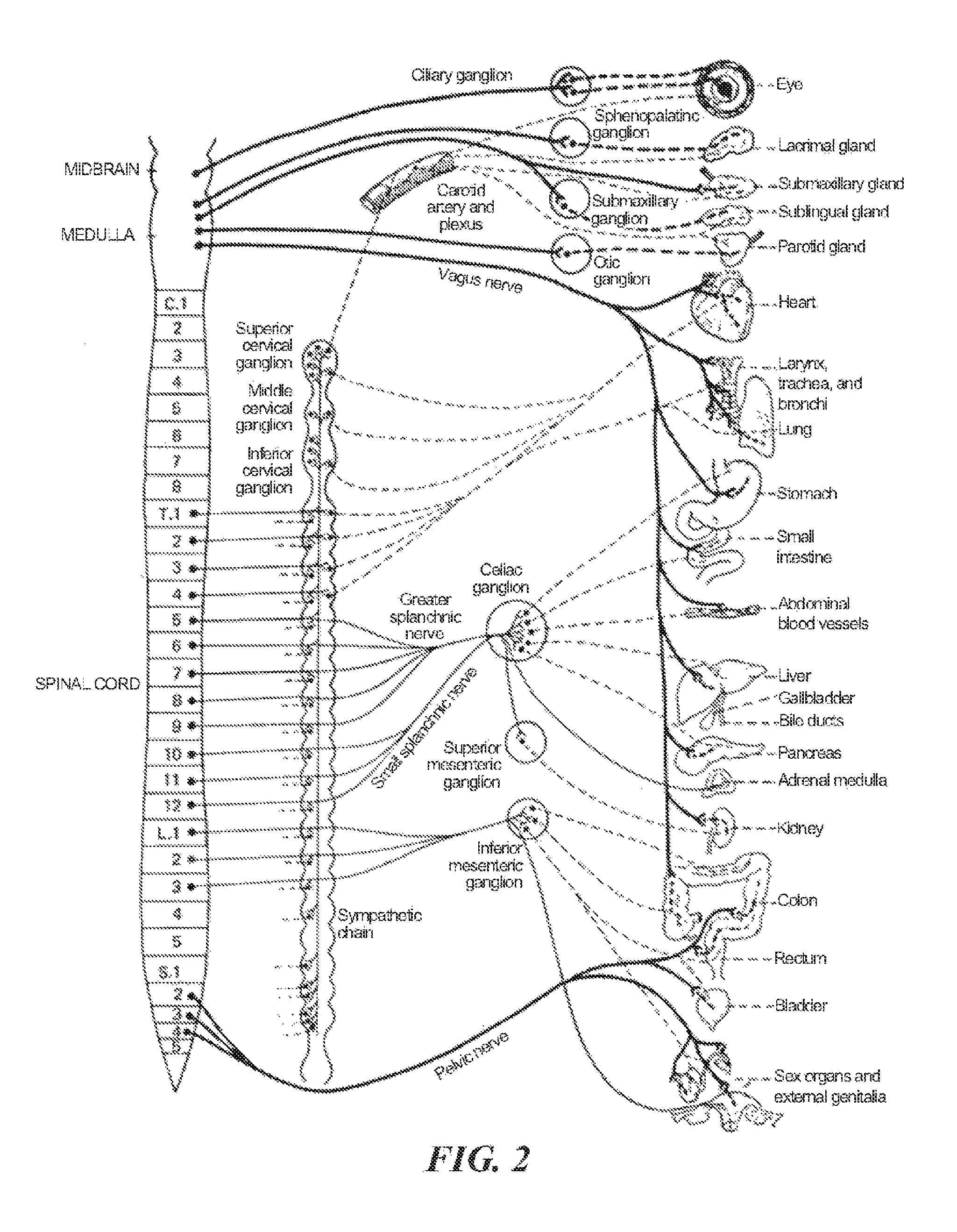
FIG. 2 is a conceptual illustration of the sympathetic nervous system (SNS) and how the brain communicates with the body via the SNS.
FIG. 3 is an enlarged anatomic view of nerves innervating a left kidney to form the renal plexus surrounding the left renal artery.
FIG. 4 is a partially schematic view of a catheter-based treatment device positioned within a renal artery and configured for therapeutic renal neuromodulation in accordance with an embodiment of the technology.
FIG. 5 is a schematic diagram illustrating a test apparatus to monitor ventilator response to pCO.sub.2 in accordance with an embodiment of the technology.
FIG. 6 is a display diagram illustrating ventilator response to pCO.sub.2 for elevated and normal central sympathetic drives in accordance with an embodiment of the technology.
DETAILED DESCRIPTION
The present technology is directed to methods for therapeutic renal neuromodulation for the treatment of dyspnea and associated systems and devices. The present disclosure further describes methods for indicating a measure of central sympathetic state by measuring pCO.sub.2, sensitivity. The following disclosure includes five sections directed to various aspects of the disclosed technology. Section I, for example, describes renal neuromodulation, and Section II focuses on the pertinent anatomy and physiology. Section III describes techniques for measuring sympathetic activity and associated systems and methods, and Section IV is directed to chronic sympathetic activation and its relationship to dyspnea. Section V describes using therapeutic renal neuromodulation to reduce central sympathetic drive and sympathetic neural activity in a manner that may treat patient for dyspnea and how hypercapnic ventilatory response can be used as an indicator of sympathetic activation.
Specific details of several embodiments of the technology are described below with reference to FIGS. 1-6. Although many of the embodiments are described below with respect to devices, systems, and methods for therapeutic renal neuromodulation, other applications and other embodiments in addition to those described herein are within the scope of the technology. Additionally, several other embodiments of the technology can have different configurations, components, or procedures than those described herein. A person of ordinary skill in the art, therefore, will accordingly understand that the technology can have other embodiments with additional elements, or the technology can have other embodiments without several of the features shown and described below with reference to FIGS. 1-6.
The terms "distal" and "proximal" are used in the following description with respect to a position or direction relative to the treating clinician. "Distal" or "distally" are a position distant from or in a direction away from the clinician. "Proximal" and "proximally" are a position near or in a direction toward the clinician.
I. Renal Neuromodulation
Renal neuromodulation is the partial or complete incapacitation or other effective disruption of nerves innervating the kidneys. In particular, renal neuromodulation comprises inhibiting, reducing, and/or blocking neural communication along neural fibers (i.e., efferent and/or afferent nerve fibers) innervating the kidneys. Such incapacitation can be long-term (e.g., permanent or for periods of months, years, or decades) or short-term (e.g., for periods of minutes, hours, days, or weeks). As described in greater detail herein, renal neuromodulation is expected to efficaciously treat several clinical conditions characterized by increased overall sympathetic activity, and in particular conditions associated with central sympathetic over stimulation such as hypertension, heart failure, acute myocardial infarction, metabolic syndrome, insulin resistance, diabetes, left ventricular hypertrophy, chronic and end stage renal disease, inappropriate fluid retention in heart failure, cardio-renal syndrome, and sudden death. The reduction of afferent neural signals contributes to the systemic reduction of sympathetic tone/drive, and renal neuromodulation is expected to be useful in treating several conditions associated with systemic sympathetic over activity or hyperactivity. Renal neuromodulation can potentially benefit a variety of organs and bodily structures innervated by sympathetic nerves. For example, a reduction in central sympathetic drive may reduce insulin resistance that afflicts patients with metabolic syndrome and Type II diabetics. Additionally, osteoporosis can be sympathetically activated and might benefit from the downregulation of sympathetic drive that accompanies renal neuromodulation.
Various techniques can be used to partially or completely incapacitate neural pathways, such as those innervating the kidney. The purposeful application of energy (e.g., electrical energy, thermal energy) to tissue can induce one or more desired thermal heating effects on localized regions of the renal artery and adjacent regions of the renal plexus, which lay intimately within or adjacent to the adventitia of the renal artery. The purposeful application of the thermal heating and cooling effects can achieve neuromodulation along all or a portion of the renal plexus.
II. Pertinent Anatomy and Physiology
The following discussion provides various details regarding pertinent patient anatomy and physiology. This section is intended to provide additional context regarding the disclosed technology and the therapeutic benefits associated with renal denervation, and to supplement and expand upon the disclosure herein regarding the relevant anatomy and physiology. For example, as mentioned below, several properties of the renal vasculature may inform the design of treatment devices and associated methods for achieving renal neuromodulation via intravascular access, and impose specific design requirements for such devices. Specific design requirements may include accessing the renal artery, facilitating stable contact between the energy delivery elements of such devices and a luminal surface or wall of the renal artery, and/or effectively modulating the renal nerves with the neuromodulatory apparatus.
A. Autonomic Nervous System
The autonomic nervous system (ANS) is comprised of the parasympathetic and sympathetic nervous systems. These systems work together to regulate visceral body functions including heart rate, blood pressure, respiration, digestion, body temperature, and urination. The ANS is always active at a basal level, primarily acting in an involuntary, reflexive manner to maintain homeostasis. The sympathetic and parasympathetic nervous systems involve networks of nerves connecting the brain, the spinal cord and the peripheral organs. These two systems regulate visceral body functions including respiration, cardiovascular activity, and energy balance.
B. Sympathetic Nervous System
The Sympathetic Nervous System (SNS) is a branch of the autonomic nervous system along with the enteric nervous system and parasympathetic nervous system. Activation of the SNS is typically associated with a "fight or flight" quick alarm or stress response that enables the body to perform strenuous physical activity, such as when fleeing from danger. Within seconds, the heart pumps more forcefully, the heart rate increases, blood is shunted from the GI tract to active muscles and the brain, and blood glucose increases to provide energy for increased cellular metabolism. Sympathetic drive is also a key regulator of the body's blood pressure and fluid balance, ensuring adequate blood supply for vital organs such as the brain when the body is fleeing from danger.
The SNS is balanced by the functions of the "rest and digest" parasympathetic nervous system (PNS), which promotes nutrient absorption from the GI tract and energy storage. While the SNS responds within seconds to environmental triggers, some effects of the parasympathetic nervous system may not be seen for hours. Most visceral organs have both sympathetic and parasympathetic innervation, though one system can dominate control of a given organ. The response to activation of the SNS and PNS is both neuronally and hormonally mediated. The hormonal contribution comes from the adrenal gland, which is activated by the SNS and PNS to release hormones such as epinephrine (adrenaline) into the bloodstream that can amplify the body's response to the neural stimulation. Together, the functions of the sympathetic and parasympathetic nervous systems enable the body to respond to environmental stimuli in a graded fashion instead of simply on or off.
The SNS is composed primarily of neurons. As shown in FIG. 1, for example, neurons 100 are composed of three parts: the cell body 102 where information is integrated, specialized projections 104 (i.e., dendrites) that bring information into the cell body 102, and a single projection 106 (i.e., axon) that takes information away from the cell body information is passed between neurons electrochemically across synapses, small gaps between axons 106 and dendrites 104. At a distal end of the pre-synaptic neuron's axon 106, chemicals termed neurotransmitters 108 are released, cross the synapse, and bind to cell surface receptors at a post-synaptic neuron (not shown). An electric potential is generated in the post-synaptic dendrite and spreads to the cell body, where the signal is integrated. The signal relayed to the next neuron (not shown) by generating an electrical potential that travels down the corresponding axon, activating release of neurotransmitters at the distal end of the axon into the next synapse.
Axons are typically bundled together like the ropes of a cable; a large bundle can be visible to the naked eye and is often called a nerve fiber. A cluster of neurons and synapses is called a ganglion. Ganglions provide key relay points throughout the sympathetic nervous system. Although nerve signals may travel from one ganglia to another, many signals pass through only one ganglion. When considering the general ANS architecture, post-ganglionic neurons are those neurons that have their cell bodies in the ganglia and send axons directly out to the peripheral organs. All other neurons are termed pre-ganglionic neurons.
FIG. 2 is a conceptual illustration of a human SNS illustrating how the brain communicates with the body via the SNA. The nerves comprising the SNS enable bidirectional signal communication between the brain, spinal cord, and nearly every organ system. For example, signals from the periphery to the brain, termed afferent signals, travel within one neuron and carry information primarily about, temperature or pain. In the opposite direction, efferent signals are primarily transmitted by a two neuron system; the first neuron originates in the brain and spinal cord, exits at the mid-lower back at spinal levels T1-L2 (the sympathetic thoracolumbar outflow) and synapses in a ganglia. The most prominent ganglia are those found parallel to the vertebral column at spinal levels T1-L2. These are grouped together as the sympathetic trunk. Post-ganglionic nerves from the sympathetic trunk primarily regulate the abdominal and thoracic visceral organs. Other important ganglia of the SNS include the cervical ganglion (regulates organs in the head and thorax), the celiac ganglion, and the mesenteric ganglia (regulates abdominal organs) Post-ganglionic nerves then transmit the signal directly to the peripheral organs.
Efferent neuronal signaling in the SNS is carried by two primary small molecule neurotransmitters; acetylcholine and norepinephrine. All preganglionic signals are mediated by acetylcholine, a chemical messenger that binds and activates cholinergic receptors on postganglionic neurons. Acetylcholine is primarily an activating neurotransmitter. In the brain, for example, acetylcholine improves attention, enhances sensory perceptions, and enhances memory and learning. Preganglionic release of acetylcholine stimulates postganglionic neurons, thereby promoting generation of electric potentials in the postganglionic neurons. Once stimulated, postganglionic neurons primarily use the neurotransmitter noradrenaline (norepinephrine). Norepinephrine binds to adrenergic receptors to directly stimulate peripheral organs. In the adrenal gland, SNS stimulation causes norepinephrine release into the blood, heightening the body's arousal and enhancing the SNS response.
FIG. 3 is an enlarged anatomic view of nerves innervating a left kidney to form a renal plexus surrounding the left renal artery. Sympathetic communication between the CNS and the kidney is achieved via many neurons that travel from the sympathetic chain to innervate the kidney. Many of these nerves arise primarily from the celiac ganglion, the superior mesenteric ganglion, and the aorticorenal ganglion. From the ganglia, these fibers join together into a plexus of nerves that surround the renal artery. This is typically termed the renal plexus or renal nerve. The renal plexus or renal nerve is embedded within the adventitia (i.e., the outer wall) of the renal artery extending along the renal artery until it arrives at the substance of the kidney. There is also rich innervation of the kidney vasculature and of the tubular structures (nephrons) that comprise the filtering and concentrating functions of the kidney.
The renal plexus carries both afferent and efferent signals. As mentioned previously, afferent signals increase with temperature, pain, decreased renal blood flow, and intra-renal pathologies such as kidney hypoxia or ischemia. They are also influenced by the chemical composition of the urine; small signaling molecules such as adenosine are released into the urine when the kidneys are hemodynamically (i.e. too much or too little blood flow) or metabolically stressed. Afferent signals are carried by several different neurotransmitters including substance P, a molecule well known to participate in pain signaling. Signals from one kidney impact the renal sympathetic outflow and the functioning of both that kidney and the opposite (contralateral) kidney and also affect the brain. Central integration of the afferent signals in the posterior hypothalamus of the brain and in the spinal cord causes increased central sympathetic outflow.
Efferent renal nerve activity is stimulated by numerous inputs. As mentioned above, afferent signals from one kidney can cause increased efferent activity in that kidney as well as the contralateral kidney. This latter effect is known as the renorenal reflex. In addition, most stimuli of central sympathetic outflow also increase efferent renal nerve activity. These stimuli include infection, inflammation, and acute stress, which release chemical mediators that can act directly on the brain to increase central sympathetic outflow. In addition, feedback mechanisms such as the baroreceptor reflex can increase central sympathetic outflow. Baroreceptor sensors in the carotid arteries of the neck are sensitive to blood pressure. A fall in blood pressure causes a corresponding fall in baroreceptor activity, which stimulates increased sympathetic outflow.
C. Dyspnea
The most rigorous physiologic definitions of dyspnea capture the concept that afferent signals from peripheral or central chemoreceptors ascend to the brain. Afferent neurons significant in dyspnea arise from a large number of sources including the carotid bodies, medulla, lungs, and chest wall. Chemoreceptors in the carotid bodies and medulla supply information regarding the blood gas levels of O.sub.2, CO.sub.2 and H.sup.+. In the lungs, juxtacapillary (J) receptors are sensitive to pulmonary interstitial edema, while stretch receptors signal bronchoconstriction. Muscle spindles in the chest wall signal the stretch and tension of the respiratory muscles. Thus the following conditions may contribute to a feeling of dyspnea: (a) poor ventilation leading to hypercapnia; (b) left heart failure leading to interstitial edema (impairing gas exchange); and (c) asthma causing bronchoconstriction (limiting airflow) and muscle fatigue leading to ineffective respiratory muscle action. Thus, dyspnea is attributed to inadequate respiration (failure to remove pCO.sub.2 from blood), increased interstitial lung water content or alveolar water content causing increased lung stiffness and pulmonary edema respectively, lung stiffness of any etiology causing stimulation of interstitial pulmonary j nerve fibers.
Another phenomenon that is associated with or that may contribute to dyspnea is an altered sensitivity of chemoreceptors. For example, altered chemoreceptor sensitivity may manifest as a change in the set point of the chemoreceptor (point at which afferent signals begin and dyspnea is sensed). This changed set point would result in a gain in respiratory effort, which is associated with dyspnea (hypothalamic integration of the signal with efferent signals to the respiratory tree). It has been demonstrated that pCO.sub.2 sensitivity may be heightened in patients having certain disease states (e.g. diabetics with autonomic neuropathy). However, in the absence of a tool to modify set point and gain, the idea that heightened sensitivity to pCO.sub.2 could be corrected has previously remained untested.
iii. Measuring Sympathetic Activity
SNS activity is often measured using methods including microneurography or norepinephrine spillover. Microneurography is the more direct method of the two to measure the level of sympathetic activity. It involves insertion of an electrode into the nerve to measure directly the action potentials from axons of sympathetic nerves. The electrode picks up signals from all neurons in the nerve bundle. An increased number and frequency of action potentials correlates with higher sympathetic outflow in that nerve bundle. Because this method requires a macroscopic nerve bundle into which the electrode can be placed, it cannot be used to represent the sympathetic stimulation to whole organs, which are often innervated by multiple nerves arranged in a mesh-like plexus. Nevertheless, this method is well suited for measurement of sympathetic stimulation to peripheral muscles, which are often innervated by a single identifiable nerve. When microneurography is used in this case, the technique and measurable quantity is often termed. "muscle sympathetic nerve activity," or MSNA.
Measurement of norepinephrine spillover is a less direct method of estimating SNS activity, but can be used to aggregate SNS outflow to whole organs and in the body as a whole. This method involves measuring the levels of the neurotransmitter norepinephrine released at a target organ. Increased neuronal firing corresponds with increased release of the neurotransmitter norepinephrine, which then can be measured via arterial and venous sampling of norepinephrine (a radioisotope of norepinephrine is also commonly used). For example, samples of blood from the renal artery can be measured for norepinephrine content and compared to the norepinephrine content in samples taken from the renal vein. Higher norepinephrine levels in the venous sample represent increased efferent sympathetic signaling to the kidney.
Overall sympathetic activity is estimated by measuring norepinephrine levels in the central veins draining from the body into the heart, termed "whole body norepinephrine levels." It can be especially useful to measure norepinephrine spillover in specific organs as sympathetic outflow is non-uniform and can vary significantly to different organs. While the above described methods can provide fairly accurate assessments of elevated central sympathetic tone, these methods are not utilized often because of the invasiveness, inconvenience and cost associate with such tests.
IV. Chronic Sympathetic Activation in Dyspnea
While acute activation of the SNS is an appropriate response to maintaining survival, chronic sympathetic activation is a maladaptive response. Without being bound by theory, it is thought that sensory afferent signals originating from the kidneys are often major contributors to initiate and sustain elevated central sympathetic outflow. With chronic stimulation, the body sets a new homeostasis where higher SNS outflow is the norm. This new homeostasis, however, is harmful to the body. Malfunction of the renal sympathetic nervous system and chronic sympathetic activation play a key role in the development and progression of diseases such as essential hypertension, chronic kidney disease, heart failure, insulin resistance, metabolic syndrome and diabetes, among others.
Abnormal sensitivity to pCO.sub.2 (set point and/or gain) as well as increased sympathetic nerve activity are seen together in multiple disease states. This correlation suggests that pCO.sub.2 sensitivity may be under central autonomic influence. More specifically, this correlation suggests that changes in set point of chemoreceptors may be linked to central sympathetic state, and, for example, increased central sympathetic drive itself may cause increased chemoreceptor sensitivity to partial pressure of carbon dioxide (pCO.sub.2) and respiratory muscle response to afferent signaling from these chemoreceptors.
V. Therapeutic Renal Neuromodulation
The physiology described above suggests an integral role between central sympathetic activity and the renal nerves in the development of dyspnea. It is accordingly expected that renal neuromodulation, e.g., via denervation of tissue containing, renal nerves, may be valuable in the treatment of this disease. More specifically, neuromodulation of afferent sensory nerves can reduce the systemic sympathetic drive through direct effect on the brain, thus altering chemosensor sensitivity to pCO.sub.2.
Medtronic Ardian LLC has discovered that reduction of sympathetic neural activity in nerves that innervate a kidney of a patient with hypertension can lower blood pressure. For example, sympathetic neural activity can be blocked or reduced by creating thermal lesions of at least a portion of renal sympathetic nerves. This procedure may provide relief of hypertensive conditions by reducing central sympathetic tone in patients having hyperactive central sympathetic tone. Similarly, such a procedure could also correct or improve sensitivity to pCO.sub.2 by reducing central sympathetic tone. This could reduce subjective dyspnea that is concomitant with various disease states, for example and not limited to systolic heart failure, diastolic heart failure, congestive heart failure, panic disorders, chronic and acute lung diseases, chronic and acute renal diseases, central sleep apnea, and deconditioning.
Medtronic Ardian LLC has further developed systems and associated methods to selectively modulate the renal afferent and efferent sympathetic nerves that lie within and alongside the adventitia (i.e., outer wall) of the renal arteries. Modulation of such nerves may be achieved using a variety of techniques. For example, the purposeful application of energy (e.g., electrical energy, thermal energy) to tissue can initiate renal neuromodulation via denervation caused by irreversible electroporation, electrofusion, apoptosis, necrosis, ablation, cryogenic ablation, thermal alteration, alteration of gene expression, or another suitable modality.
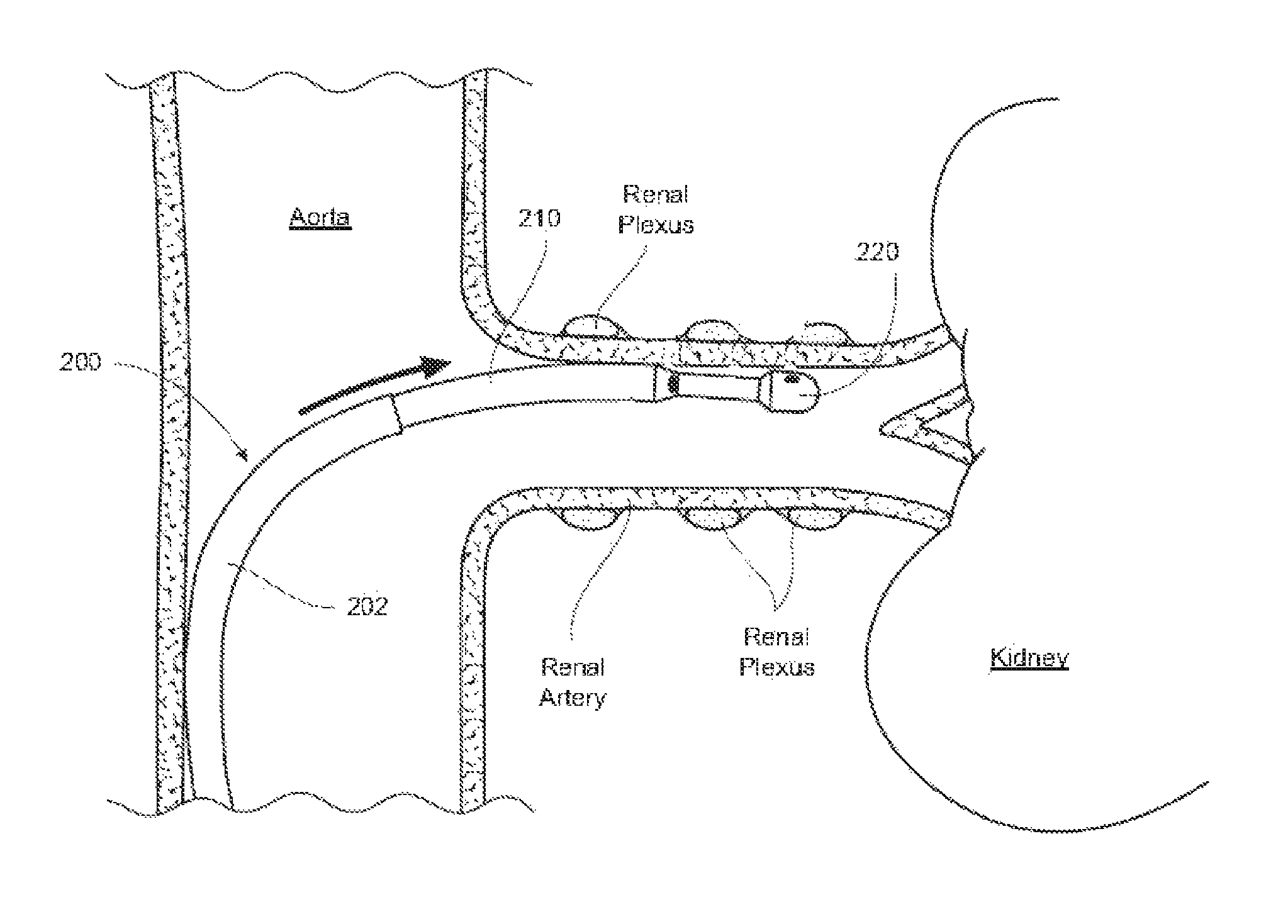
Several embodiments of this procedure involve discrete, low-dose radiofrequency ablation of the target nerves via a radiofrequency (RF) emitting catheter placed on the inside wall of the renal artery. FIG. 4, for example, is a detailed anatomic view of a catheter-based treatment device 200 positioned within a renal artery of a patient and configured for renal neuromodulation in accordance with one embodiment of the disclosure. The device 200 can be deployed using a conventional guide catheter or pre-curved renal guide catheter 202. The device 200 can be introduced via the guide catheter 202 through the common femoral artery or, alternatively, through a brachial/radial approach, and advanced to the renal artery under guidance (e.g., fluoroscopic imaging guidance).
A flexible, controllable elongated shaft 210 of the treatment device 200 carries an energy delivery element 220 (e.g., a thermal heating element) and energy can be applied via the energy delivery element 220 to one or more target treatment sites along a length of the renal artery. The target treatment sites can be spaced longitudinally and rotationally along the length of the renal artery. Individual treatments can include, for example, ramped low power RF energy delivery (e.g., about 5 to 8 watts) for a selected period of time (e.g., two minutes). Blood flow through the renal artery can help minimize surface and/or endothelial injury to the target treatment sites. Further, focal ablations spaced apart from each other along the vessel allow for rapid healing. In one embodiment, up to six treatments are applied along the length of the renal artery beginning from where the renal artery branches off the aorta and ending at the kidney itself. In other embodiments, however, a different number of treatments may be applied and the treatment sites may have a different arrangement relative to each other. After all the treatments are completed, the treatment device 200 is removed from the patient. Various embodiments of methods, apparatuses, and systems for performing renal neuromodulation are described in greater detail in U.S. patent application Ser. No. 12/545,648, filed Aug. 21, 2009, and Patent Cooperation Treaty (PCT) Application No. PCT/US09/69334, filed Dec. 22, 2009, both of which are incorporated herein by reference in their entireties.
Other techniques or approaches for renal neuromodulation may also be administered to achieve the therapeutic benefits described herein. For example, renal neuromodulation can be achieved via a pulsed electric field or intravascular electroporation. In still another example, U.S. Pat. No. 6,978,174 describes neuromodulation via delivery of neuromodulatory agents. In yet another example, U.S. Pat. No. 7,620,451 describes neuromodulation via an intra-to-extravascular approach. These patent references are incorporated herein by reference in their entireties.
A. Therapeutic Renal Neuromodulation in the Treatment of Subjective Dyspnea
The recognition that feelings of dyspnea may be substantially a result of altered chemoreceptor sensitivity could change clinical understanding of the many diseases where quality of life is impacted by this subjective symptom. Interestingly, it is exactly this subjective symptom that plays so heavily into quality of life measures and the grading of severity of disease states. For example, in heart failure, the New York Heart Association's functional classification of disease severity is entirely based on subjective classification of dyspnea at rest, with moderate exertion, with mild exertion or without dyspnea. While the functional classification has provided the basis for separation of morbidity and mortality curves, the failure to recognize a common basis or physiologic basis for its etiology has been frustrating, leading to the development of alternative tests, such as peak VO.sub.2 during exercise. Nonetheless, hospitalization and clinical decisions are often predicated on failure or success in treatment of dyspnea. Similarly, in all forms of heart failure, panic disorders, chronic kidney diseases, and many chronic lung diseases, subjective dyspnea and subsequent hyperventilatory response underlie the disease pathology.
Clinical efforts have been focused on changing underlying ventricular function in heart failure, congestion status in heart failure, dialysis frequency in end stage renal disease (ESRD) and breathing dynamics in chronic lung disorders. However, subjective dyspnea may be principally a neurologic consequence to heightened central sympathetic drive. Consequently, it is expected that subjective dyspnea could be modified with correct therapy targeting the underlying sympathetic hyperactivity. The potential impact of this invention is that the basis of treatment for the aforementioned associated diseases could be altered.
Proposed herein s the idea that dyspnea is a consequence of heightened central and peripheral chemoreceptor sensitivity to pCO.sub.2, which is under central sympathetic control. The consequence of this relation is that a therapeutic change in the set point (pCO.sub.2 level at which dyspnea is sensed) and/or the gain (minute ventilatory change in response to exposure to pCO.sub.2) could be achieved if central sympathetic tone is altered. As previously discussed this may be achieved with therapeutic renal neuromodulation.
B. Measuring Minute Ventilation/pCO.sub.2 as an Indicator of Central Sympathetic Drive in Conjunction with Therapeutic Renal Neuromodulation
Central hypercapnic chemosensitivity may be assessed with a test that involves breathing air with varying percentages of CO.sub.2 and comparing the responding drive to breath. An example of such test may include having a patient rebreath air into a bag (e.g., a 6 liter bag) for a predetermined period of time (e.g., about 4 minutes). The air in the bag 500 may contain a gas mixture of, for example, about 7% CO.sub.2 in the beginning of the test rising to about 10% at the end of the test. Measurements of gas contents (e.g., pCO.sub.2, % CO.sub.2, in exhaled or inhaled gas) and tidal volume (e.g., minute ventilation) may be monitored continuously or intermittently using a gas analyzer 501 (e.g., a Beckman LB-2 CO.sub.2 analyzer, available from Beckman Coulter of Brea, Calif.) and flow meter 502 (e.g. a Series 3800 pneumotach, available from Han Rudolph, Inc. of Shawnee, Kans.) (see FIG. 5). Other examples of equipment that may facilitate monitoring pCO.sub.2 include suitable capnography and capnometry devices. These devices provide both waveform (capnograph) and digital readout or end-tidal CO.sub.2. Most of the commonly used capnography and capnometry devices utilize infrared absorption of CO.sub.2 as a principle of operation. The relationship between ventilation and pCO.sub.2 may be expressed in terms of volume of breathed air per unit time per unit of partial pressure of CO.sub.2 (e.g. liters per minute per millimeters of mercury). A plot of this relationship may characterize a patients sensitivity to pCO.sub.2. Such a test assesses hypercapnic ventilator response.
A similar apparatus may be modified to measure mouth occlusion pressure, which may be an alternative or additional indication of drive to breathe. For example, in addition the above mentioned apparatus used to measure minute ventilation vs. pCO.sub.2, a mouth valve (e.g., a nasal and mouth breathing mask available from Han Rudolph, Inc. of Shawnee Kans.) may separate expiratory flow and inspiratory flow. The expiratory flow may pass through the flow meter and gas analyzer to measure minute ventilation vs. pCO2 and then be returned to the breathing circuit. The inspiratory side of the circuit may be silently occluded during exhalation, and the mouth occlusion pressure may he measured by a pressure sensor a short time (e.g., about 100 msec) after the start of an inspiratory effort against the completely occluded airway periodically (e.g., about every 8-15 breaths). The airway may he reopened once a pressure measurement is taken so breathing can resume.
A plot of ventilator response (i.e., minute ventilation and/or mouth occlusion pressure) to inhaled pCO.sub.2 is expected to show distinct characteristics for various states of central sympathetic drive. For example, as shown in FIG. 6, a patient with a normal central sympathetic drive may demonstrate a pCO.sub.2 sensitivity characterized by the solid line 600, while a patient with an elevated central sympathetic drive may demonstrate a pCO.sub.2 sensitivity characterized by the dotted line 601. These plats may identify various aspects of a patient's sensitivity to pCO.sub.2. For example, a set point (i.e., a level of pCO.sub.2 at which afferent signals begin and dyspnea is sensed) may be indicated by an upward inflection point in the plot at 602 and 603; gain in respiratory effort associated with dyspnea may be represented by an increase in minute ventilation and/or mouth occlusion pressure for a given increase in pCO.sub.2. Alternatively, ventilatory response vs. pCO.sub.2 data may be represented by a linear model and gain may be represented by a slope of the linear plot. Such characteristics may indicate an enhanced chemoreceptor reflex. For example, a patient with a normal sympathetic state may have a hypercapnic ventilatory response in a range of about 1.5 to 3 L/min/mmHgCO.sub.2, while a patient with an excited sympathetic state may have a hypercapnic ventilatory response above about 3 L/min/mmHgCO.sub.2. Furthermore, a scale of severity of heightened sympathetic state may be correlated to ranges of hypercapnic ventilatory responses (e.g., about 3 to 5
L/min/mmHgCO.sub.2 may represent a mildly excited sympathetic state, above 12 L/min/mmHgCO.sub.2 may represent a severely excited sympathetic state, and in between may represent progressive severities.)
Data from the sensors may be monitored by software that may record, process, analyze, and display information about a patient's hypercapnic ventilator response. Non-limiting examples of how software may be implemented follow: Software may analyze flow rate of a breath and characterize various phases of the respiratory cycle; end tidal values of pCO.sub.2 may be isolated by correlating the pCO.sub.2 measurements with the phases of a respiratory cycle; outlier data may be filtered out, for example when a patient coughs or swallows; repeat tests could be averaged or compared; a patient's test results could be stored and retrieved at a later date or compared to another test; data may be analyzed to create best fit curves or plots, for example with linear regression analysis; plots can be displayed on a graph or numerically; results can be displayed with comparison to normal ranges of ventilatory response; evaluation of results may be displayed showing if a patient has normal, heightened or along a scale of pCO.sub.2 sensitivities or sympathetic drive. Such software may be provided as an installation program to be installed on a computer or laptop or the software may be provided in a computerized medical device.
The suggestion that ventilator response to inhaled pCO.sub.2 may identity heightened central sympathetic tone provides a clinical analogy to the use of muscle sympathetic nerve activity (MSNA). It is possible that one or both could be used for the purpose of diagnosing and/or selecting patients, monitoring success of therapy, or identification of late failures of therapeutic renal denervation. A test for ventilator response to inhaled pCO.sub.2 could be easier to administer, require simpler and cheaper test equipment, and be less stressful to the patient compared to an MSNA test or a norepinephrine spillover test.
The concept is that, like MSNA, elevated hyperventilatory response to inhaled pCO.sub.2 may represent elevated central sympathetic state, a condition that could be important for the success of renal denervation for the treatment of several disease states. Thus, the test might be used to screen candidates for a renal denervation procedure. In one embodiment, for example, a patient who has a disease state, such as hypertension, but does not show an elevated central sympathetic state as determined by a test that compares minute ventilation to pCO.sub.2 may not be a suitable candidate for a renal denervation procedure. However, a patient that tests positive fur elevated central sympathetic state using a pCO.sub.2 sensitivity test may be more likely to have a therapeutic response to renal denervation. Similarly, when performed after a renal denervation procedure, it is expected that a pCO.sub.2 sensitivity test may be a tool to assess success of initial denervation and/or subsequent monitoring of late failure of the therapy.
Knowing that treatment has not successfully altered chemoreceptor function does not necessarily imply that the treatment failed (it is possible that additional factors are operative in coordinating respiratory response to inhaled pCO.sub.2). Further, it is possible that the treatment was only partially successful, which may suggest the potential for a repeat renal denervation treatment.
VI. Conclusion
The above detailed descriptions of embodiments of the technology are not intended to be exhaustive or to limit the technology to the precise form disclosed above. Although specific embodiments of, and examples for, the technology are described above for illustrative purposes, various equivalent modifications are possible within the scope of the technology, as those skilled in the relevant art will recognize. For example, while steps are presented in a given order, alternative embodiments may perform steps in a different order. The various embodiments described herein may also be combined to provide further embodiments.
From the foregoing, it will be appreciated that specific embodiments of the technology have been described herein for purposes of illustration, but well-known structures and functions have not been shown or described in detail to avoid unnecessarily obscuring the description of the embodiments of the technology. Where the context permits, singular or plural terms may also include the plural or singular term, respectively.
Moreover, unless the word "or" is expressly limited to mean only a single item exclusive from the other items in reference to a list of two or more items, then the use of "or" in such a list is to be interpreted as including (a) any single item in the list, (b) all of the items in the list, or (c) any combination of the items in the list. Additionally, the term "comprising" is used throughout to mean including at least the recited feature(s) such that any greater number of the same feature and/or additional types of other features are not precluded. It will also be appreciated that specific embodiments have been described herein for purposes of illustration, but that various modifications may be made without deviating from the technology. Further, while advantages associated with certain embodiments of the technology have been described in the context of those embodiments, other embodiments may also exhibit such advantages, and not all embodiments need necessarily exhibit such advantages to fall within the scope of the technology. Accordingly, the disclosure and associated technology can encompass other embodiments not expressly shown or described herein.
The disclosure may be defined by one or more of the following clauses:
1. A method of treating a human patient diagnosed with dyspnea, the method comprising: at least partially inhibiting sympathetic neural activity in nerves that innervate a kidney of the patient; and reducing central sympathetic drive in the patient in a manner that treats the patient for the dyspnea.
2. The method of clause 1 wherein reducing central sympathetic drive in a manner that treats the patient for the dyspnea comprises reducing the patient's sensitivity to pCO.sub.2.
3. The method of clause 1 wherein reducing central sympathetic drive in a manner that treats the patient for the dyspnea comprises altering the patient's chemoreceptor set point.
4. The method of clause 1 wherein reducing central sympathetic drive in a manner that treats the patient for the dypnea comprises changing the patient's gain in respiratory effort associated with increased pCO.sub.2.
5. The method of clause 1 wherein the dyspnea is associated with a condition comprising at least one of systolic heart failure, diastolic heart failure, congestive heart failure, panic disorders, chronic lung disease, acute lung disease, renal disease, deconditioning, and hypertension.
6. The method of clause 1 wherein the dyspnea comprises subjective feelings of breathlessness.
7. The method of clause 1 wherein at least partially inhibiting sympathetic neural activity in nerves that innervate a kidney of the patient comprises ablating a renal nerve of the patient with an energy transfer element positioned at least proximate to the renal nerve.
8. The method of clause 7 wherein ablating a renal nerve comprises thermally ablating the renal nerve.
9. The method of clause 8 wherein thermally ablating the renal nerve comprises necrosing the renal nerve via a cryoablation device.
10. The method of clause 8 wherein thermally ablating the renal nerve comprises delivering an energy field to the renal nerve via the energy transfer element.
11. The method of clause 7 wherein ablating a renal nerve comprises ablating the renal nerve via an intravascularly positioned catheter carrying the energy transfer element.
12. The method of clause 11 wherein ablating the renal nerve via an intravascularly positioned catheter comprises ablating the renal nerve via the energy transfer element from within a renal artery of the patient.
13. The method of clause 1 wherein at least partially inhibiting sympathetic neural activity in nerves that innervate a kidney of the patient comprises blocking afferent neural activity.
14. The method of clause 1 wherein at least partially inhibiting sympathetic neural activity in nerves that innervate a kidney of the patient comprises blocking efferent neural activity
15. A method of determining whether a human patient will respond to a renal denervation treatment, the method comprising: measuring pCO.sub.2 sensitivity of the patient; evaluating a sympathetic drive state of the patient based on the measured pCO.sub.2 sensitivity; and based at least in part on the measured pCO.sub.2 sensitivity and the evaluation, determining whether the patient is likely to respond to a renal denervation treatment.
16. The method of clause 15 wherein evaluating the sympathetic drive state of the patient comprises comparing the measured pCO.sub.2 sensitivity of the patient to a representative pCO.sub.2 sensitivity of a patient having relatively normal central sympathetic tone.
17. A method of evaluating effectiveness of a renal denervation procedure to reduce central sympathetic drive, the method comprising: measuring a hypercapnic ventilatory response of a patient after the patient has undergone a renal denervation procedure; and evaluating a central sympathetic state of the patient based, at least in part, on the hypercapnic ventilatory response.
18. The method of clause 18, further comprising measuring the hypercapnic ventilatory response of the patient prior to administering the renal denervation procedure, and wherein the hypercapnic ventilatory response of the patient following the renal denervation procedure is compared to the hypercapnic ventilatory response of the patient prior to the procedure.
19. A system for determining central sympathetic drive in a patient, the system comprising: a breathing tube configured for respiratory communication with the patient; a container configured to receive respiratory communication from the patient via the breathing tube; a first sensor configured to measure content of air within the breathing tube; a second sensor configured to measure a characteristic or parameter associated with respiration of the patient; and a component configured to determine a minute ventilation of the patient and plat the determined minute ventilation as a function of measured pCO.sub.2 of the patient.
20. The system of clause 19 wherein the first sensor comprises a gas analyzer.
21. The system of clause 19 wherein the first sensor is configured to measure at least one of CO.sub.2, O.sub.2, N.sub.2, pCO.sub.2, and pO.sub.2.
22. The system of clause 19 wherein the second sensor is configured to measure a characteristic or parameter from a group comprising volumetric flow rate, mass flow rate, and pressure.
23. The system of clause 19, further comprising a third sensor configured to measure content of air in the container.
* * * * *
References
-
onlinelibrary.wiley.com/doi/abs/10.1016/j.ejheart.2006.08.001
-
en.wikipedia.org/wiki/Ureter
-
edisonawards.com/BestNewProduct_2011.php
-
newsroom.heart.org/news/2012-top-10-advances-in-heart-241901
-
prnewswire.com/news-releases/ardianr-receives-2010-europer-innovation-award-and-demonstrates-further-durability-of-renal-denervation-treatment-for-hypertension-95545014.html
-
phx.corporate-ir.net/phoenix.zhtml?c=62272&p=irol-newsArticle&id=1756108
-
my.clevelandclinic.org/media_relations/library/2011/2011-10-6-cleveland-clinic-unveils-top-10-medical-innovations-for-2012.aspx
-
cxvascular.com/in-latest-news/interventional-news---latest-news/does-renal-denervation-represent-a-new-treatment-option-for-resistant-hypertension
-
neurotechreports.com/pages/goldelectrodes09.html
-
pharmaceuticalintelligence.com/tag/vessix-vascular
-
-
medicaldevicedaily.com/servlet/com.accumedia.web.Dispatcher?next=bioWorldHeadlines_article&forceid=83002
-
fastcompany.com/3007845/meet-tech-duo-thats-revitalizing-medical-device-industry
-
97waterlooroad.wordpress.com/2012/11/12/medtronic-awarded-industrys-highest-honour-for-renal-denervation-system
-
medpagetoday.com/Cardiology/PCI/36509
-
medical-dictionary.com
-
-
medtronic.com/rdn/mediakit/RDN%20FAQ.pdf
-
ncbi.nlm.nih.gov/pmc/articles/PMC3547427
-
clinicaltrials.gov/ct2/show/NCT01390831
-
clinicaltrials.gov/ct2/show/NCT01628198
D00000
D00001
D00002
D00003
D00004
D00005
D00006
XML
uspto.report is an independent third-party trademark research tool that is not affiliated, endorsed, or sponsored by the United States Patent and Trademark Office (USPTO) or any other governmental organization. The information provided by uspto.report is based on publicly available data at the time of writing and is intended for informational purposes only.
While we strive to provide accurate and up-to-date information, we do not guarantee the accuracy, completeness, reliability, or suitability of the information displayed on this site. The use of this site is at your own risk. Any reliance you place on such information is therefore strictly at your own risk.
All official trademark data, including owner information, should be verified by visiting the official USPTO website at www.uspto.gov. This site is not intended to replace professional legal advice and should not be used as a substitute for consulting with a legal professional who is knowledgeable about trademark law.