Method for purifying immunoglobulin
Son , et al.
U.S. patent number 10,287,315 [Application Number 15/123,869] was granted by the patent office on 2019-05-14 for method for purifying immunoglobulin. This patent grant is currently assigned to GREEN CROSS HOLDINGS CORPORATION. The grantee listed for this patent is GREEN CROSS HOLDINGS CORPORATION. Invention is credited to Sung Min Choi, Yong Kang, Ki-Yong Kim, Dong-Hwarn Park, Kang Yun Seo, Ki-Hwan Son.
| United States Patent | 10,287,315 |
| Son , et al. | May 14, 2019 |
Method for purifying immunoglobulin
Abstract
The present invention relates to a method for purifying an immunoglobulin, and more particularly, to a method for purifying an immunoglobulin, which comprises: dialyzing and concentrating an immunoglobulin-containing plasma protein fraction II paste; removing thrombotic substances from the dialyzed and concentrated fraction by a purification process using ceramic cation exchange resin; and performing elution while maintaining salt concentration at a constant level to maintain the polymer content of the immunoglobulin at a low level. When the immunoglobulin purification method according to the present invention is used, the efficiency with which impurities and thrombotic substances are removed can be increased and the polymer content of the immunoglobulin can be maintained, and thus a stable immunoglobulin with improved quality can be produced.
| Inventors: | Son; Ki-Hwan (Chungcheongbuk-do, KR), Kang; Yong (Gyeonggi-do, KR), Park; Dong-Hwarn (Gyeonggi-do, KR), Choi; Sung Min (Gyeonggi-do, KR), Seo; Kang Yun (Gyeonggi-do, KR), Kim; Ki-Yong (Gyeonggi-do, KR) | ||||||||||
|---|---|---|---|---|---|---|---|---|---|---|---|
| Applicant: |
|
||||||||||
| Assignee: | GREEN CROSS HOLDINGS
CORPORATION (Gyeonggi-Do, KR) |
||||||||||
| Family ID: | 54071961 | ||||||||||
| Appl. No.: | 15/123,869 | ||||||||||
| Filed: | March 11, 2014 | ||||||||||
| PCT Filed: | March 11, 2014 | ||||||||||
| PCT No.: | PCT/KR2014/002020 | ||||||||||
| 371(c)(1),(2),(4) Date: | September 06, 2016 | ||||||||||
| PCT Pub. No.: | WO2015/137530 | ||||||||||
| PCT Pub. Date: | September 17, 2015 |
Prior Publication Data
| Document Identifier | Publication Date | |
|---|---|---|
| US 20170022248 A1 | Jan 26, 2017 | |
| Current U.S. Class: | 1/1 |
| Current CPC Class: | C07K 1/34 (20130101); C07K 1/36 (20130101); C07K 1/1136 (20130101); C07K 16/065 (20130101); C07K 1/30 (20130101); C07K 1/18 (20130101) |
| Current International Class: | C07K 1/18 (20060101); C07K 1/34 (20060101); C07K 1/30 (20060101); C07K 1/36 (20060101); C07K 16/06 (20060101); C07K 1/113 (20060101) |
References Cited [Referenced By]
U.S. Patent Documents
| 4124576 | November 1978 | Coval |
| 4665159 | May 1987 | Dobkin |
| 4764369 | August 1988 | Neurath et al. |
| 6069236 | May 2000 | Burnouf-Radosevich |
| 6124437 | September 2000 | Hirao |
| 9023994 | May 2015 | Mintz |
| 2006/0194953 | August 2006 | Bonnerjea |
| 2011/0014203 | January 2011 | Nilsson |
| 2013/0058961 | March 2013 | Teschner et al. |
| 2014/0116941 | May 2014 | Thorm |
| 2014/0271669 | September 2014 | Hofbauer |
| 2017/0218051 | August 2017 | Gnauer |
| 2018/0140699 | May 2018 | Shin |
| 2604759 | Sep 1977 | DE | |||
| 07-267997 | Oct 1995 | JP | |||
| 10-1983-0007083 | Dec 1983 | KR | |||
| 10-2001-0052733 | Jul 2005 | KR | |||
| 10-2007-0009995 | Jan 2007 | KR | |||
| 10-2013-0069515 | Mar 2015 | KR | |||
| WO-9518155 | Jul 1995 | WO | |||
| WO1999033484 | Jul 1999 | WO | |||
| WO2000076534 | Dec 2000 | WO | |||
Other References
|
PALL Life Sciences Product Note USD 2744 "Q, S, DEAE, CM Ceramic HyperD F ion exchange sorbents" pp. 1-6 (Year: 2011). cited by examiner . Schreiner et al. J Allergy Clin Immunol Feb. 2003 Abstract 620 entiteld Stability of new liquid intravenous immunoglobulin preparation (IGIV-C 10%) manufactured using a novel production process (Year: 2003). cited by examiner . Cohn, E.J., et al., "Preparation and Properties of Serum and Plasma Proteins. IV. A System for the Separation into Fractions of the Protein and Lipoprotein Components of Biological Tissues and Fluids", "Journal of the American Chemical Society", Mar. 1, 1946, pp. 459-475, vol. 68. cited by applicant . Kistler, P., et al., "Large Scale Production of Human Plasma Fractions", "Vox Sanguinis", Jul. 8, 1962, pp. 414-424, vol. 7. cited by applicant . Polson, A., et al., "The Fractionation of Protein Mixtures by Linear Polymers of High Molecular Weight", "Biochimica et Biophysica Acta", 1964, pp. 463-475, vol. 82. cited by applicant . Polson, A., et al., "Fractionation of Plasma with Polyethylene Glycol", "Vox Sanguinis", Jul. 8, 1972, pp. 107-118, vol. 23. cited by applicant . Note: For the non-patent literature citations that no month of publication is indicated, the year of publication is more than 1 year prior to the effective filing date of the present application. cited by applicant . Staby, A., et al., "Comparison of Chromatographic Ion-Exchange Resins V. Strong and Weak Cation-Exchanage Resins", "Journal of Chromatography A", 2006, pp. 168-179, vol. 1118. cited by applicant. |
Primary Examiner: Kolker; Daniel E
Assistant Examiner: Rogers; James L
Attorney, Agent or Firm: Hultquist, PLLC Hultquist; Steven J.
Claims
The invention claimed is:
1. A method for purifying an immunoglobulin, comprising the steps of: (a) dissolving an immunoglobulin-containing plasma protein fraction II paste, followed by filtration to obtain a fraction II solution; (b) dialyzing and/or concentrating the obtained fraction II solution, subjecting the dialyzed and/or concentrated solution to anion exchange chromatography, and recovering a fraction not attached to a column of the anion exchange chromatography; (c) treating the recovered fraction with a solvent and/or a detergent to inactivate viruses, followed by subjecting the fraction to cation exchange chromatography to remove the solvent and/or the detergent, and thrombotic substances, wherein the cation exchange chromatography is performed at a salt concentration of 400-600 mM; (d) dialyzing and/or concentrating an eluate obtained from the cation exchange chromatography; and (e) filtering the dialyzed and/or concentrated solution, thereby obtaining a purified immunoglobulin.
2. The method of claim 1, wherein the dissolving of the plasma protein fraction II paste in step (a) is performed by adding a sodium chloride solution in an amount equivalent to 2-10 times the volume of the plasma protein fraction.
3. The method of claim 1, wherein the filtration in step (a) is clarifying filtration, and comprises adjusting pH to 4.5-5.5.
4. The method of claim 1, wherein the dialysis and/or concentration in step (b) is performed using an ultrafiltration/diafiltration (UF/DF) system at an osmotic pressure of 10 mOsmol/kg or lower, and then adjusting the pH to 5.5-6.5.
5. The method of claim 1, wherein the anion exchange chromatography in step (b) is performed at a pH of 5.5-6.5 and a flow rate of 95-145 cm/hr, and a fraction not attached to the column of the anion exchange chromatography is obtained with 1.5-2.0 loading volumes (LV).
6. The method of claim 1, wherein the solvent in step (c) is tri-n-butyl phosphate (TNBP), and the detergent is at least one selected from among polysorbate 80, Triton X-100 and Triton X-45.
7. The method of claim 1, wherein the cation-exchange chromatography in step (c) is performed at a pH of 4.5-5.5 and a flow rate of 110-130 cm/hr.
8. The method of claim 1, wherein an adsorption amount of immunoglobulin adsorbed into the cation-exchange resin is 90-130 mg per ml of the cation-exchange resin in the cation-exchange chromatography in step (c).
9. The method of claim 1, wherein the cation-exchange chromatography in step (c) uses a ceramic-based cation exchange resin.
10. The method of claim 1, wherein the salt concentration of the eluate obtained from the cation exchange chromatography column in the step (c) is maintained at 50-150 mM in order to maintain the content of polymers during dialysis and/or concentration in step (d).
11. The method of claim 1, wherein the dialysis and/or concentration in step (d) is performed using an ultrafiltration/diafiltration (UF/DF) system, at an osmotic pressure of 10 mOsmol/kg or lower, and then the pH is adjusted to 4.0-5.0.
12. The method of claim 1, wherein the filtration in step (e) is performed using a nanofiltration system at a pressure of 1.5-2.5 bar.
13. The method of claim 1, further comprising a step of adding a stabilizer to prepare an immunoglobulin for intravenous injection after step (e).
14. The method of claim 13, wherein the stabilizer is at least one selected from the group consisting of sugar alcohol, maltose, sorbitol, mannose, glucose, trehalose, albumin, lysine, glycine, PEG and Tween 80.
15. The method of claim 13, wherein the stabilizer is added to a concentration of 90-110 g/l.
16. The method of claim 13, wherein pH of the immunoglobulin solution is adjusted to 3.5-4.0, after addition of the stabilizer.
Description
CROSS-REFERENCE TO RELATED APPLICATION
This application is a U.S. national phase application under the provisions of 35 U.S.C. .sctn. 371 of International Patent Application No. PCT/KR14/02020 filed Mar. 11, 2014. The disclosure of such international patent application is hereby incorporated herein by reference in its entirety, for all purposes.
TECHNICAL FIELD
The present invention relates to a method for purifying an immunoglobulin, and more particularly, to a method for purifying an immunoglobulin, which comprises: dialyzing and concentrating an immunoglobulin-containing plasma protein fraction II paste, followed by removing thrombotic substances from the dialyzed and concentrated fraction by a purification process using ceramic cation exchange resin, and performing elution while maintaining salt concentration at a constant level to maintain the polymer content of the immunoglobulin at a low level.
BACKGROUND ART
Immunoglobulins that are plasma proteins containing antibodies against various viruses and bacteria are used as drugs to prevent or treat diseases by administration to either subjects who naturally lack antibodies or patients who are in need of artificial supplement of antibodies because of viral or bacterial diseases.
In order to use such immunoglobulins as drugs, immunoglobulins for subcutaneous or intramuscular injection have been prepared according to the cold ethanol fractionation process (Cohn E. et al., J. Am. Chem. Soc., 68:459, 1946) developed by Cohn and Oncley or the modified cold ethanol fractionation process (Kistler P, Nitschmann H S, Vox Sang, 7:414. 1952) developed by Kistler and Nitschmann.
However, immunoglobulins for intramuscular injection have the following problems: 1) the doses of such immunoglobulins are limited, making it impossible to administer the immunoglobulins in large amounts; 2) the immunoglobulins cause pain at the site injected with the immunoglobulins; 3) the immunoglobulins have a low content of natural immunoglobulin G (IgG) having antibody activity; 4) the antibody activity of the immunoglobulins is reduced by protease at the injected site; and 5) the time taken to reach peak plasma concentrations is 24 hours or more.
In order to solve the problems of intramuscular injection, administration of immunoglobulins by intravenous injection was attempted. However, when immunoglobulin preparations were administered intravenously, a variety of immediate side effects, including difficult breathing and circulatory system shock, appeared due to a serious side effect (anaphylactic reaction) attributable to aggregates with anti-complementary activity. Such symptoms appeared mainly in immunoglobulin-deficient patients. Particularly, a side effect of serious hypersensitivity was observed in patients in which anti-IgA antibodies appeared.
Thus, as intravenous injection of immunoglobulins is impossible due to the above-described problems, development of immunoglobulin preparations for intravenous injection has been required, and methods capable of removing the above-described aggregates and/or preventing aggregate formation during preparation processes have been developed. Intravenous injection of immunoglobulins has become possible as a result of treating immunoglobulins with proteases such as pepsin, papain or Plasmin, or chemical substances such as .beta.-propiolactone, to change their structure so as to suppress the formation of immunoglobulin aggregates or destroy immunoglobulin aggregates, thereby reducing the anti-complementary activities of the immunoglobulins.
The first-generation intravenous immunoglobulin (IVIG) products were prepared by treating a starting material (Cohn fraction II) with pepsin to remove immunoglobulin aggregates. The preparation process did not comprise a column chromatography step, and the prepared product was lyophilized so as to be stably maintained over a suitable period of time, and was dissolved immediately before use. However, it was found that IVIG products manufactured by some manufacturers caused viral infections such as viral hepatitis C. For this reason, one or more steps of inactivating and/or removing known virus were added to the preparation process. Thereafter, the second-generation IVIG products with low anti-complementary activity and higher stability were disclosed in the mid-1980s, and the IVIG products were purified by several chromatography steps.
Such preparations were injected intravenously, and thus overcame the disadvantages of intramuscular immunoglobulins, including limited dose, pain at the injected site, and the reduction in antibody activity of immunoglobulins by protease, and the time taken to reach peak plasma concentrations was also reduced to several hours or less.
However, the intravenous immunoglobulin products as described above have little or no natural IgG with antibody activity due to their structural change, and thus have reduced or no complement binding ability and also have a blood half-life as short as about 4-12 days, suggesting that they exhibit no satisfactory effects on the prevention and treatment of diseases. Furthermore, the first-generation and second-generation IVIG products prepared in the form of lyophilized powder require an additional process for dissolving them, and have low dissolution rates. For this reason, liquid IVIG products have been developed, and improved processes have been required to obtain more stable and pure IVIG products.
In connection with this, German Patent No. 2,604,759 and U.S. Pat. No. 4,124,576 discloses methods of obtaining pure IgG (third-generation IVIG) with antibody activity by using a non-ionic surfactant such as polyethylene glycol, unlike the above-described gamma-immunoglobulin for intravenous injection. Such IgG preparations have complement binding ability and increased blood half-lives, and thus can show good effects on the prevention and treatment of diseases. However, these preparations produced by treatment with polyethylene glycol can still cause side effects, because it is difficult to completely remove aggregates with anti-complementary activity from these preparations (showing an anti-complementary activity of about 0.02 U/mg).
In addition, Korean Laid-Open Publication No. 1983-0007083 discloses a method of preparing an intravenous immunoglobulin from Cohn fraction II or fraction II+III, isolated from human plasma, by treatment with polyethylene glycol. However, there are problems in that the process is complicated and the yield is low.
Accordingly, the present inventors have made extensive efforts to solve the above-described problems occurring in the prior art, and as a result, have found that, when a fraction II paste isolated from plasma is dialyzed and concentrated, and then purified by anion exchange chromatography and cation exchange chromatography while controlling salt concentration during cation exchange chromatography and elution, thrombotic substances in the plasma fraction are efficiently removed and the stability of the immunoglobulin product is improved, thereby completing the present invention.
DISCLOSURE OF INVENTION
Technical Problem
It is an object of the present invention to provide a method for purifying immunoglobulins, which can efficiently remove impurities and thrombotic substances in order to produce a stable and high-purity immunoglobulin.
Technical Solution
To achieve the above object, the present invention provides a method for purifying an immunoglobulin, comprising the steps of:
(a) dissolving an immunoglobulin-containing plasma protein fraction II paste, followed by filtration to obtain a fraction II solution;
(b) dialyzing and/or concentrating the obtained fraction II solution, subjecting the dialyzed and/or concentrated solution to anion exchange chromatography, and recovering a fraction not attached to a column of the anion exchange chromatography;
(c) treating the recovered fraction with a solvent and/or a detergent to inactivate viruses, followed by subjecting the fraction to cation exchange chromatography to remove the solvent and/or the detergent, and thrombotic substances;
(d) dialyzing and/or concentrating an eluate obtained from the cation exchange chromatography; and
(e) filtering the dialyzed and/or concentrated solution, thereby obtaining a purified immunoglobulin.
BRIEF DESCRIPTION OF THE DRAWINGS
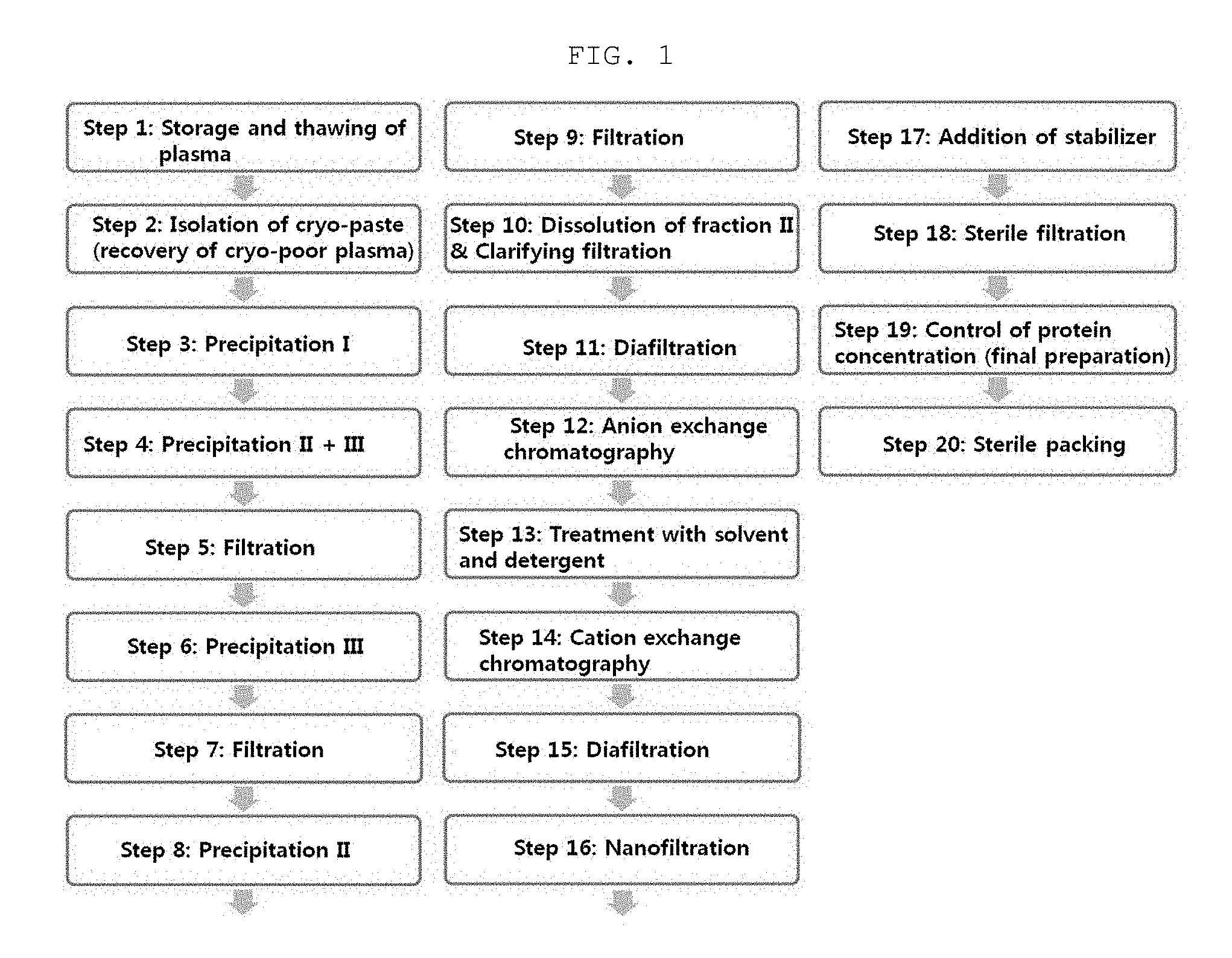
FIG. 1 is a schematic view showing a process for preparing an intravenous immunoglobulin according to the present invention.
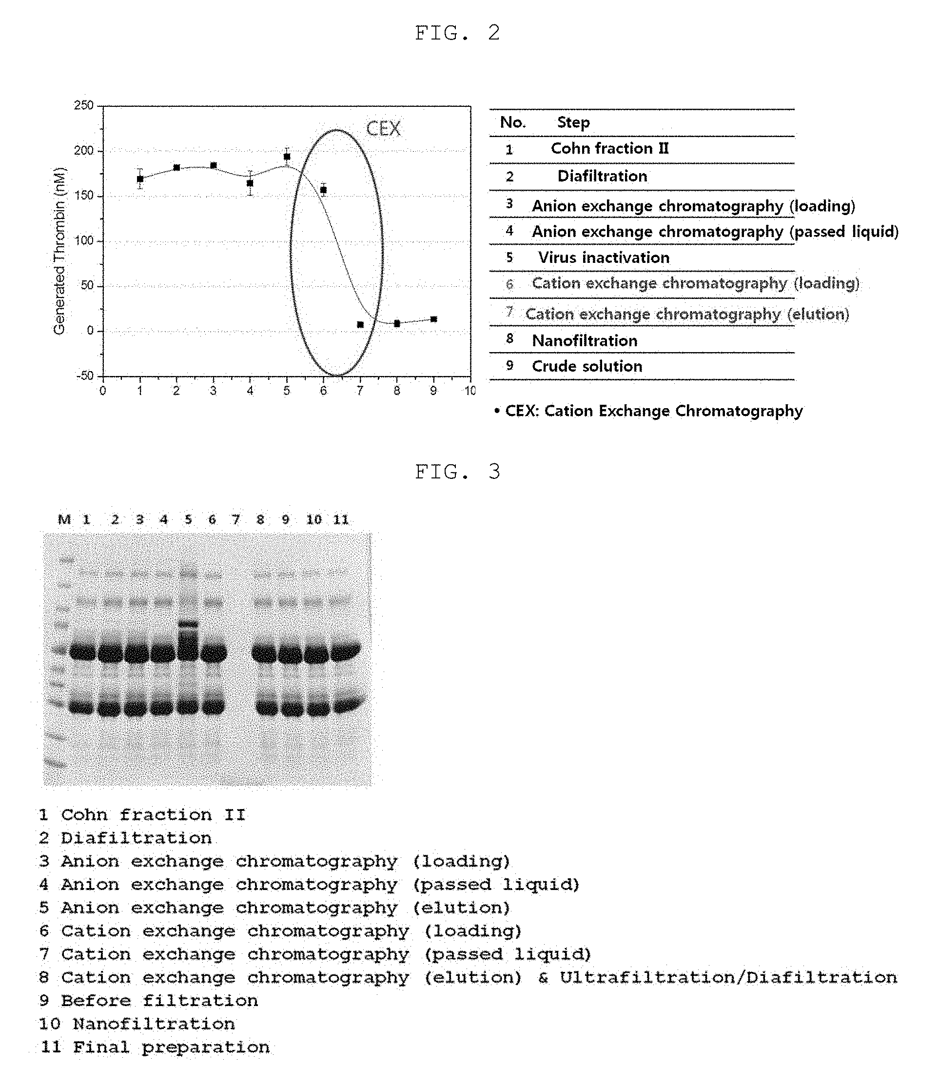
FIG. 2 shows the results of measuring the purity (thrombin/IgG) of an immunoglobulin in each preparation step.
FIG. 3 shows the results of measuring the concentration of FXI (human coagulation factor XI) contained in a filtrate or precipitate in each preparation step by SDS-PAGE.
BEST MODE FOR CARRYING OUT THE INVENTION
Unless defined otherwise, all technical and scientific terms used herein have the same meaning as commonly understood by one of ordinary skill in the art to which the invention pertains. Generally, the nomenclature used herein and the experiment methods, which will be described below, are those well known and commonly employed in the art.
As used herein, the expression "immunoglobulin-containing plasma protein" is meant to encompass cryoprecipitate-free plasma obtained by removing various plasma proteins such as Factor IX and antithrombin from human plasma or human placental plasma, various Cohn fractions, and fractions obtained by ammonium sulfate or PEG (Polson et al., Biochem Biophys Acta, 82:463, 1964); Polson and Ruiz-Bravo, Vox Sang, 23:107. 1972) precipitation. Preferably, the plasma protein fraction that is used in the present invention may be Cohn fraction II, Cohn fraction I, II and III or Cohn fraction II+III.
In the present invention, the fraction II paste obtained from human plasma according to a conventional Cohn plasma fraction method was used. A subsequent purification process for removing various lipoproteins, fibrinogens, .alpha.-globulin, .beta.-globulin and various coagulation factors from the fraction II paste was performed.
In the present invention, the human plasma used was FDA-approved and American Red Cross-derived plasma subjected to Biotests, including nucleic acid amplification tests on human immunodeficiency virus (HIV), hepatitis C virus (HCV), hepatitis B virus (HBV) and parvovirus B19, and serological tests. The plasma stored at -20.degree. C. or below was thawed by incubation in a jacketed vessel at 1 to 6.degree. C. for 12-72 hours.
While the plasma was thawed under the above-described conditions, a cryoprecipitate including fibrinogen and coagulation factors was produced. The produced cryoprecipitate was removed by centrifugation, and the remaining cryo-poor plasma was recovered. Then, precipitation and filtration processes were repeated, thereby obtaining a fraction II paste.
In the filtration process for isolating immunoglobulin-containing plasma, a filter aid was added to and mixed with the cryo-poor plasma which was then separated into a supernatant and a precipitate by means of a filter press. As the filter aid, Celite(STD) or Harbolite(Expanded perlite product) was used.
In the present invention, the dissolving of the plasma protein fraction II paste in step (a) may be performed by adding a sodium chloride solution in an amount equivalent to 2-10 times the volume of the plasma protein fraction.
The plasma protein fraction is preferably suspended (dissolved) in water and/or buffer at a substantially non-denaturing temperature and pH. The term "substantially non-denaturing"implies that the condition to which the term refers does not cause substantial irreversible loss of functional activity of the IgG molecules, e.g. loss of antigen binding activity and/or loss of biological Fc-function.
Advantageously, the plasma protein fraction is dissolved in water acidified with at least one non-denaturing buffer at volumes of from 2 to 5, preferably from 3 to 4, times that of the plasma protein fraction. The pH of the immunoglobulin-containing suspension is preferably maintained at a pH below 6, such as within the range of 4.0-6.0, preferably 4.9-5.1, in order to ensure optimal solubility of the immunoglobulin. Any acidic buffer known in the art can be used, but sodium phosphate, sodium acetate, sodium chloride, acetic acid, hydrochloric acid, or water (distilled water) may preferably used as the acidic buffer. In the present invention, sodium chloride solution was used.
The immunoglobulin suspension is maintained at a low temperature so as to prevent protein denaturation and minimize protease activity, and the immunoglobulin suspension, and water or buffer added to the immunoglobulin suspension, are maintained at a temperature ranging from 0 to 12.degree. C., preferably from 0 to 7.degree. C., more preferably from 1 to 4.degree. C.
In the present invention, the filtration in step (a) of obtaining the fraction II solution may be clarifying filtration, and may comprise adjusting pH to 4.5-5.5. Preferably, the pH is adjusted to 4.9-5.1.
In the present invention, the fraction II paste was transferred into a jacketed vessel at 10.degree. C. or below, and dissolved by adding thereto a 0.6% sodium chloride solution in an amount of 4 times equivalent to the volume of the fraction II paste, and then 1M acetic acid was added to the solution to adjust the pH to 5.0.+-.1. Next, the solution was subjected to clarifying filtration using a depth filter cartridge, thereby obtaining a fraction II solution.
In the present invention, the dialysis and/or concentration in step (b) may be performed using an ultrafiltration/diafiltration (UF/DF) system until the osmotic pressure of the dialyzed and concentrated solution reaches 10 mOsmol/kg or lower, followed by adjusting the pH to 5.5-6.5, preferably 5.9-6.1.
Diafiltration is a process of removing only a certain solute from a fluid containing a solvent and two or more solutes having different molecular sizes by simultaneously performing dialysis and ultrafiltration. It is effective for purification of polymer materials and has advantages over general dialysis processes in that it is time-saving and cost-effective.
In an example of the present invention, the fraction II solution was filtered using an ultrafiltration/diafiltration (UF/DF) system at an osmotic of 10 mOsmol/kg or lower, and 1M sodium acetate was added to the filtrate to a concentration of 5.0.+-.1.0 mM, followed by adjusting the pH to 6.0.+-.0.1, thereby obtaining a dialyzed and/or concentrated immunoglobulin-containing solution.
In the present invention, the anion exchange chromatography in step (b) may be performed at a pH of 5.5-6.5 and a flow rate of 95-145 cm/hr, and a fraction not attached to the anion exchange chromatography column may be recovered with 1.5-2.0 loading volumes (LV).
The anion exchange resin that is used in the anion-exchange chromatography step may be one substituted with diethylaminoethyl (DEAE) or quaternary ammonium groups, but is not limited thereto. Preferably, the anion exchange resin may be any one selected from among anion exchange resins having a strongly basic quaternary ammonium group or a weakly basic diethylaminoethyl (DEAE) group.
For example, as a strongly basic anion exchange resin, Q Sepharose Fast Flow, Q Sepharose High Performance, Resource Q, Source 15Q, Source 30Q, Mono Q, Mini Q, Capto Q, Capto Q ImpRes, Q HyperCel, Q Cermic HyperD F, Nuvia Q, UNOsphere Q, Macro-Prep High Q, Macro-Prep 25 Q, Fractogel EMD TMAE(S), Fractogel EMD TMAE Hicap (M), Fractogel EMD TMAE (M), Eshmono Q, Toyopearl QAE-550C, Toyopearl SuperQ-650C, Toyopearl GigaCap Q-650M, Toyopearl Q-600C AR, Toyopearl SuperQ-650M, Toyopearl SuperQ-650S, TSKgel SuperQ-5PW (30), TSKgel SuperQ-5PW (20), TSKgel SuperQ-5PW or the like may be used, but is not limited thereto, and any anion exchange resin known in the art may be used.
The appropriate volume of resin used in the anion exchange chromatography is reflected by the dimensions of the column, i.e., the diameter of the column and the height of the resin, and varies depending on, for example, the amount of the immunoglobulin in the applied solution and the binding capacity of the resin used. Before performing anion exchange chromatography, the anion exchange resin is preferably equilibrated with a buffer which allows the resin to bind its counterions.
In the present invention, the anion exchange resin used is DEAE-Sepharose gel, and the column buffers used may be equilibration buffer known in the art, for example, sodium phosphate buffer, citrate buffer, acetate buffer or the like, wash buffer and elution buffer.
In the anion exchange chromatography, the column was equilibrated with 5.+-.1.0 mM sodium acetate buffer such that the pH would be 6.0.+-.0.1, and the flow rate of the mobile phase was controlled to 95-145 cm/hr. The fraction not attached to the anion exchange chromatography column was recovered with 1.5-2.0 loading volumes (LV).
In the present invention, step (c) is a step of inactivating viruses such as potential lipid enveloped viruses in the immunoglobulin-containing solution and then removing a substance used for the inactivation. In this step, a virus-inactivating agent, preferably a solvent and/or a detergent, may be used. Most preferably, solvent & detergent treatment employing a solvent-detergent mixture may be used.
Through step (c), lipid enveloped viruses (e.g. HIV1 and HIV2, hepatitis type C and non A-B-C, HTLV 1 and 2, the herpes virus family, including CMV and Epstein Barr virus) can be inactivated, and thus the safety of the final product can be increased.
In step (c), any solvent and detergent may be used without limitation, as long as they have the capability to inactivate viruses, particularly lipid enveloped viruses. The detergent may be selected from the group consisting of non-ionic and ionic detergents and is preferably selected to be substantially non-denaturing. Particularly, a non-ionic detergent is preferable in terms of easy removal. The solvent is most preferably tri-n-butyl phosphate (TNBP) as disclosed in U.S. Pat. No. 4,764,369, but is not limited thereto.
The virus-inactivating agent that is used in the present invention is preferably a mixture of TNBP and at least one selected from among polysorbate 80 (Tween 80), Triton X-100 and Triton X-45, but is not limited thereto.
The preferred solvent/detergent mixture is added such that the concentration of TNBP in the immunoglobulin-containing solution is 0.2-0.6 wt %, preferably 0.24-0.36 wt %, and such that the concentration of Tween 80 is 0.8-1.5 wt %, preferably 0.8-1.2 wt %.
The virus-inactivation step is performed under conditions that inactivate enveloped viruses, resulting in a substantially virus-safe immunoglobulin-containing solution. Such conditions include a temperature of 4-30.degree. C., preferably 19-28.degree. C., most preferably 24-26.degree. C., and an incubation time of 1-24 hours, preferably 4-12 hours, most preferably about 8 hours, to ensure sufficient virus inactivation.
In the present invention, the cation-exchange chromatography in step (c) may be performed at a pH of 4.5-5.5 and a flow rate of 110-130 cm/hr. Preferably, the pH is adjusted to 4.9-5.1. The amount of immunoglobulin loaded onto the cation-exchange resin is 90-130 mg per ml of the cation-exchange resin, preferably 95-105 mg per ml of the resin. After adsorption of the immunoglobulin, washing with equilibration buffer is performed. The equilibration buffer that is used in the washing may be used in an amount of at least three column volumes, preferably at least five column volumes. After washing, the immunoglobulin is eluted with at least 8 column volumes of elution buffer.
The cation exchange resin that is used in the present invention may be Sephardex, Sepharose, HyperCell or Source, but is not limited thereto, and other cation exchange resins known in the art may also be used. In the present invention, a ceramic-based cation exchange resin may preferably be used. In an example of the present invention, CM Hyper D gel that is a ceramic-based resin was used as the cation exchange resin, and equilibration buffer known in the art, such as sodium phosphate buffer, citrate buffer or acetate buffer, wash buffer and elution buffer were used as the column buffers.
The elution of the immunoglobulin from the cation exchange resin is performed with a substantially non-denaturing buffer having a pH and ionic strength sufficient to cause efficient elution of the IgG, thereby recovering an immunoglobulin-containing eluate. Herein, "efficient elution" means that at least 75%, such as at least 80%, for example, at least 85%, of the immunoglobulin solution loaded onto the cation exchange resin is eluted from the cation exchange resin.
In the present invention, the cation exchange chromatography in step (c) may be performed at the salt concentration of the eluting buffer, which is sufficiently high to displace the immunoglobulin from the cation exchange resin. It may be performed at a salt concentration of 400-600 mM, preferably 500 mM.
In addition, in order to maintain the polymer content of the immunoglobulin during the dialysis and/or concentration in step (d), an eluate obtained from the cation exchange chromatography column may be added to a calculated dialyzed and/or concentrated solution to maintain the salt concentration at a constant level of 50-150 mM, preferably 100 mM or less. When an elution method enabling a low salt concentration to be maintained is used in the protein elution step, the polymer content of the immunoglobulin can be minimized, and thus the immunoglobulin with improved quality can be purified.
In the present invention, the dialysis and/or concentration in step (d) may be performed using an ultrafiltration/diafiltration (UF/DF) system. It is performed at an osmotic pressure of 10 mOsmol/kg or lower, and then the pH is adjusted to 4.0-5.0.
In the present invention, diafiltration was performed in order to remove low-molecular-weight ions from the cation exchange chromatography eluate, and the osmotic pressure in the UF/DF system was controlled to 10 mOsmol/kg or lower. Then, hydrochloric acid (HCl) was added to the filtrate to adjust the pH to 4.5.+-.0.1.
In the present invention, the filtration in step (e) may be performed using a nanofiltration system. The nanofiltration may be performed at a pressure of 1.5-2.5 bar.
The method of the present invention may further comprise, after step (e), a step of adding a stabilizer to prepare an immunoglobulin for intravenous injection.
After completion of the nanofiltration, at least one protein stabilizer known in the art may be added, and examples thereof include various sugar alcohols and saccharides (such as sorbitol, mannose, glucose, trehalose, maltose), proteins (such as albumin), amino acids (such as lysine, glycine, etc.) and organic agents (such as PEG and Tween 80).
In the present invention, after step (e), a stabilizer that can be added may be at least one selected from among sugar alcohol, maltose, sorbitol, mannose, glucose, trehalose, albumin, lysine, glycine, PEG and Tween 80. Preferably, maltose is used as the stabilizer.
The stabilizer may be added to a concentration of 90-110 g/l. After addition of the stabilizer, the pH of the immunoglobulin solution may be adjusted to 3.5-4.0. Preferably, the pH may be adjusted to 3.7-3.9 by adding an acid, preferably sulfuric acid or hydrochloric acid.
Nanofiltration is an important virus-removing step. In the present invention, the dialyzed and concentrated immunoglobulin solution was filtered through a Pall DVD prefilter and a DV20 virus filter at a pressure of 2.0.+-.0.5 bar to remove viruses from the immunoglobulin solution. Then, maltose was added to the nanofiltered solution to a final concentration of 100 g/l to stabilize the immunoglobulin. The stabilized immunoglobulin solution was adjusted to a pH of 3.8.+-.0.1 by addition of hydrochloric acid. Then, it was sterilized using a 0.2 .mu.m filter and stored.
The sterilized immunoglobulin preparation for intravenous injection may be diluted or concentrated such that the concentration of the protein (purified immunoglobulin) is 1-30 wt %. In the present invention, the sterilized immunoglobulin preparation was diluted with WFI or concentrated by ultrafiltration such that the protein concentration would be 40-60 g/l, preferably 45-55 g/l, more preferably 49.5-50.5 g/l. Then, maltose was added to the immunoglobulin solution to a final concentration of 100 g/l and thoroughly mixed, and then the pH of the immunoglobulin preparation was measured, and hydrochloric acid was added to the immunoglobulin solution to adjust the pH to 3.8.+-.0.1, thereby preparing an intravenous immunoglobulin preparation.
In an example of the present invention, the purity (thrombin/IgG) of an immunoglobulin solution in each preparation step and the concentration of FXI (human coagulation factor XI) in a filtrate or precipitate in each preparation step were measured. As a result, it could be seen that an immunoglobulin solution with a purity of 99% or higher was purified (FIG. 2) and that the coagulation factor FXI was mostly removed (Table 2 and FIG. 3).
EXAMPLES
Hereinafter, the present invention will be described in further detail with reference to examples. It will be obvious to a person having ordinary skill in the art that these examples are illustrative purposes only and are not to be construed to limit the scope of the present invention. Thus, the substantial scope of the present invention will be defined by the appended claims and equivalents thereof.
Example 1: Preparation of Intravenous Immunoglobulin
1-1: Preparation of Plasma
As plasma, FDA-approved plasma was used which was subjected to Biotests, including nucleic acid amplification tests on human immunodeficiency virus (HIV), hepatitis C virus (HCV), hepatitis B virus (HBV) and parvovirus B19, and serological tests.
In the present invention, plasma (Batch No. 600A9008) obtained from the Red Cross was used. The plasma was stored at -20.degree. C. or below until use. A bottle containing the plasma was opened with a bottle cutting machine, and the plasma was thawed by incubation in a jacketed vessel at 1-6.degree. C. for 12-72 hours.
While the plasma was thawed under the above-described conditions, a cryoprecipitate containing fibrinogen and coagulation factors was produced. The produced cryoprecipitate was removed by centrifugation, and the remaining cryo-poor plasma was recovered.
1-2: Precipitation I Step
To additionally remove coagulation factors from the cryo-poor plasma, a precipitation I step was performed.
96% ethanol was added to the cryo-poor plasma recovered in Example 1-1 such that the final ethanol concentration would be 8.+-.0.8% at -3.+-.1.degree. C., and then the pH of the solution was adjusted to a 7.2.+-.0.2 using acetate buffer. The precipitate was removed by centrifugation, and the supernatant (addition I supernatant) was recovered. For total viable count, a portion of the supernatant was sampled and stored.
1-3: Precipitation II+III Step and Filtration
To precipitate an immunoglobulin contained in the supernatant recovered in Example 1-2, a precipitation II+III step was performed.
96% ethanol was additionally added to the supernatant recovered in Example 1-2 such that the final ethanol concentration would be 20.+-.2% at -5.+-.1.0.degree. C. Then, the pH of the solution was adjusted to 6.9.+-.0.1 using acetate buffer.
Next, a filter aid (Celite(STD) or Harbolite(Expanded perlite product)) was added to the solution in an amount of 0.0284 kg per kg of the plasma and mixed for 30.+-.10 minutes. The mixture was separated into a supernatant and a precipitate on a filter press (DG800K) in a cold room maintained at a temperature of 2 to 8.degree. C.
The supernatant was named "supernatant I+II+III (or II+III)", and the precipitate was named "fraction I+II+IIIw (or II+IIIw)" (w; wash). Fraction I+II+IIIw (or II+IIIw) was immediately used or was stored at -20.degree. C. or below.
1-4: Precipitation III Step and Filtration
To additionally remove albumin, lipoprotein, thrombin and other unwanted proteins from immunoglobulin-containing fraction I+II+IIIw (or II+IIIw), a precipitation III step was performed.
The fraction I+II+IIIw (or II+IIIw) recovered in Example 1-3 was dissolved in cold distilled water, and a portion of the solution was sampled and stored for total viable count. Then, 96% ethanol was added to the dissolved fraction I+II+IIIw (or II+IIIw) such that the final ethanol concentration would be 18.+-.1.8% at -5.+-.1.0.degree. C. Next, the solution was adjusted to a pH of 5.2.+-.0.1 by addition of an acetate buffer prepared at -6.degree. C.
Next, the mixture was separated into a supernatant and a precipitate by means of a filter press (DG800K). The supernatant was named "filtrate I+III (or III)", and the precipitate was named "fraction I+III (or III)". Fraction I+III (or III) was discarded, and a portion of filtrate I+III (or III) was sampled and stored for total viable count.
1-5: Precipitation II Step and Filtration
To precipitate the immunoglobulin in filtrate I+III (or III) obtained in Example 1-4, a precipitation II step was performed.
96% ethanol was added to filtrate I+III (or III) such that the final ethanol concentration would be 25.+-.2.5% at -10.+-.2.0.degree. C. Next, the solution was adjusted to a pH of 7.4.+-.0.2 by addition of 1M sodium bicarbonate.
Then, the mixture was separated into a supernatant and a precipitate by means of a filter press (DG800K). The precipitate was named "fraction II paste", and a portion of the precipitate was sampled and stored for measurement of the contents of bacterial endotoxin and protein and the composition thereof.
1-6: Fraction II Paste Dissolution and Clarifying Filtration
Isolation/purification processes for increasing the content of the immunoglobulin isolated from the plasma and removing thrombotic substances were performed.
First, the immunoglobulin-containing fraction II paste isolated in Example 1-5 was dissolved to have conditions suitable for dialysis. The fraction II paste was transferred into a jacketed vessel at 10.degree. C. or lower and dissolved by adding thereto a 0.6% sodium chloride solution in an amount equivalent to 4 times the volume of the fraction II paste, and a portion of the solution was sampled and stored in order to the protein content and composition thereof.
Next, the solution was adjusted to a pH of 5.0.+-.1 by addition of 1M acetic acid, and then the solution was subjected to clarifying filtration using depth filter cartridges (BecodiskBP01), thereby obtaining a fraction II solution
1-7: Diafiltration
The immunoglobulin-containing fraction II solution obtained in Example 1-6 was diafiltered to remove ethanol and low-molecular-weight ions, and the pH thereof was adjusted so that the solution would have conditions suitable for anion exchange chromatography.
The immunoglobulin-containing fraction II solution was diafiltered using an ultrafiltration/diafiltration system (Millipore Pellicon2 (50K)) at an osmotic pressure of 10 mOsmol/kg or lower, and a portion of the obtained filtrate was sampled and stored in order to measure the protein content and composition thereof and count viable cells.
The filtrate was adjusted to a pH of 6.0.+-.0.1 by adding 1M sodium acetate to a concentration of 5.0.+-.1.0 mM, thereby obtaining a dialyzed and/or concentrated immunoglobulin solution.
1-8: Anion Exchange Chromatography
In order to remove polymer and other plasma proteins from the dialyzed and/or concentrated immunoglobulin solution obtained in Example 1-7, anion exchange chromatography was performed.
The anion exchange resin DEAE-Sepharose gel (GE Healthcare, Catalog No. 17-0709) was packed into a column, and then equilibrated with equilibration buffer such that the pH would be 6.0.+-.0.1. Next, the dialyzed and/or concentrated immunoglobulin solution obtained in Example 1-7 was loaded into the column at a flow rate of 120.+-.25 cm/hr. Next, a fraction not attached to the anion exchange chromatography column was recovered with 1.5-2.0 loading volumes (LV).
1-9: Solvent/Detergent Treatment for Virus Inactivation
To inactivate potential lipid enveloped viruses in the immunoglobulin-containing solution, a step of treating the solution with a solvent and a detergent was performed.
First, to adjust the pH of the fraction to 5.0.+-.0.1, acetic acid was added to the fraction not attached to the anion exchange chromatography column and recovered in Example 1-8. Then, tri(n-butyl)-phosphate (TNBP) and polysorbate 80 (Tween 80) were added to the fraction to concentrations of 0.3.+-.0.06% and 1.+-.0.2%, respectively, followed by stirring at 200.+-.50 RPM for 20-30 minutes. In order to whether TNBP and Tween 80 in the solution were uniformly mixed, a portion of the solution was sampled and analyzed. Thereafter, the solution was continuously stirred at 25.+-.1.0.degree. C. and 200.+-.50 RPM for 8 hours. The immunoglobulin-containing solution was transferred to another tank (that is a viral secure area (VSA)) through a hard pipeline.
1-10: Cation Exchange Chromatography
To remove TNBP, Tween 80 and other thrombotic substances such as coagulation factors from the immunoglobulin solution treated with the solvent/detergent, cation exchange chromatography was performed.
The cation exchange resin CM Hyper D gel (Pall Corporation; Catalog No. 20050) that is a ceramic material was packed into a column, and then equilibrated with equilibration buffer such that the pH would be 5.0.+-.0.1. Next, the immunoglobulin solution treated with the solvent/detergent in Example 1-9 was loaded into the column a flow rate of 120.+-.10 cm/hr. Thereafter, after washing with at least 5 column volumes of wash buffer, the immunoglobulin was eluted with elution buffer (elution buffer composition: 20 mM NaOAc pH 4.5 w/0.5M NaCl) and recovered (adsorption rate: 100 mg immunoglobulin per ml of cation exchange resin).
1-11: Diafiltration
To remove low-molecular-weight ions from the cation exchange chromatography eluate, diafiltration was performed.
The eluate obtained in Example 1-10 was diafiltered using an ultrafiltration/diafiltration system (Millipore Pellicon2 (50K)) at an osmotic pressure of 10 mOsmol/kg or lower. In order to maintain the immunoglobulin polymer content, the cation exchange chromatography eluate was added to the calculated dialysate concentrate, and the ultrafiltration/diafiltration (UF/DF) was continuously performed while a sodium chloride concentration of 100 mM or lower was maintained.
A portion of the obtained filtrate was sampled and stored in order to measure the protein content and composition thereof and count viable cells, and the remaining filtrate was adjusted to a pH of 4.5.+-.0.1 by addition of hydrochloric acid (HCl).
1-12: Nanofiltration and Addition of Stabilizer
Nanofiltration is an important virus-removing step. The dialyzed/concentrated immunoglobulin solution obtained in Example 1-11 was filtered through a FlorodyneII prefilter (AB1DJL7PH4) and filtered through a virus filter (DV20, AB3DV207PH4) at a pressure of 2.0.+-.0.5 bar to thereby remove viruses from the immunoglobulin solution.
In order to stabilize the immunoglobulin, maltose was added to the nanofiltered filtrate to a final concentration of 100 g/l and thoroughly mixed. Then, the pH of the stabilized immunoglobulin solution was measured, and the immunoglobulin solution was adjusted to a pH of 3.8.+-.0.1 by addition of hydrochloric acid.
Next, the filtrate was sterilized using a 0.2 .mu.m filter and stored in a stainless steel storage tank.
1-13: Preparation of Final Preparation of Immunoglobulin Preparation for Intravenous Injection and Aseptic Filling
The resulting immunoglobulin preparation for intravenous injection was diluted with WFI or concentrated by ultrafiltration such that the protein concentration would be 50.+-.1 g/l. Next, maltose was added thereto to a final concentration of 100 g/l and thoroughly mixed. Then, the stabilized immunoglobulin preparation was measured for its pH and adjusted to a pH of 3.8.+-.0.1 by addition of hydrochloric acid.
After pH adjustment, the immunoglobulin preparation was sterilized, and transferred to a packing room to prepare a product which was in turn stored at a temperature of 2-8.degree. C.
Example 2: Measurement of Generated Thrombin/IgG (Thromboembolic Risk) in Immunoglobulin Solution in Each Preparation Step
Generated thrombin/IgG (thromboembolic risk) in the immunoglobulin preparation sampled in each preparation step of Example 1 was measured.
2-1: Experimental Method
In the present invention, the measurement of thromboembolic risk in the immunoglobulin solution in each step of Example 1 was performed in accordance with the Thrombin Generation protocol (CBER Thrombin Generation protocol 01 Experiment (100916)a) provided by the CBER (Center for Biologics Evaluation and Research) that is one of the six affiliated analytical organizations of the FDA.
2-2: Experimental Results
The immunoglobulin purification process according to the present invention includes the Cohn plasma fractionation method and the ion exchange chromatography purification techniques. As shown in FIG. 2 and Table 1 below, it could be seen that, in the cation exchange chromatography among the chromatographic purification processes, the amount of generated thrombin (that is a thrombotic substance) was effectively reduced to approximately 95% compared to an initial stage.
TABLE-US-00001 TABLE 1 Analysis of product obtained in each preparation process Batch No. 654C13171 654C13172 654C13173 Average Standard Processes Remark Thrombin (nM) deviation 1. Cohn II paste 182.2 163.5 162.7 169.5 11.0 2. Dia-filtration 184.3 178.6 182.6 181.8 2.9 3. Anion exchange Loaded 185.7 182.2 186.0 184.6 2.1 (AEX) portion chromatography Passed 159.3 154.4 180.2 164.6 13.7 portion 4. Virus 190.6 187.3 204.9 194.3 9.4 inactivation 5. Cation exchange Loaded 156.0 150.9 165.0 157.3 7.1 (CEX) portion chromatography Passed 6.3 7.5 8.9 7.6 1.3 portion 6. Nanofilitration 5.0 9.8 12.1 9.0 3.6 7. Crude solution 14.1 10.7 16.5 13.8 2.9
The results in Table 1 above show that thrombosis that can be caused by intravenous injection of the immunoglobulin can be minimized so that thromboembolism caused by thrombosis can be effectively prevented, thereby maximizing the safety of the immunoglobulin.
Example 3: Measurement of Concentration of FXI (Human Coagulation Factor XI) in Filtrate or Precipitate in Each Preparation Step
In order to examine the degree of removal of coagulants, the concentration of FXI (Human Coagulation Factor XI) in the filtrate or precipitate sampled in each preparation step of Example 1 was measured by ELISA (AssayMax Human Factor XI (FXI) ELISA Kit; ssaypro, Catalog No. EF1011-1) and SDS-PAGE.
TABLE-US-00002 TABLE 2 FXI contents of purification process products FXI (EIA) Processes Remark (ng/mL) 1 Cohn II paste 2.32 2 Dia-filtration 3.06 3 Anion exchange (AEX) Loaded portion 2.41 4 chromatography) Passed portion 2.56 5 Cation exchange (CEX) Loaded portion 2.96 6 chromatography Passed portion N.D 7 Eluted portion N.D 8 Nanofilitration N.D 9 Crude solution N.D
The FXI contents of the products of the purification process according to the present invention were measured by ELISA and SDS-PAGE. As a result, as could be seen from Table 2 above and FIG. 3, the amounts of thrombin and FXI did not change in the anion exchange chromatography process, but the amounts of thrombin and FXI in the eluate obtained from the cation exchange chromatography process were below the limit of detection, and FXI was completely removed in the cation exchange chromatography process.
INDUSTRIAL APPLICABILITY
When the immunoglobulin purification method according to the present invention is used, the efficiency with which impurities and thrombotic substances are removed can be increased and the immunoglobulin polymer content can be maintained, and thus a stable immunoglobulin with increased quality can be produced.
Although the present invention has been described in detail with reference to the specific features, it will be apparent to those skilled in the art that this description is only for a preferred embodiment and does not limit the scope of the present invention. Thus, the substantial scope of the present invention will be defined by the appended claims and equivalents thereof.
* * * * *
D00001
D00002
XML
uspto.report is an independent third-party trademark research tool that is not affiliated, endorsed, or sponsored by the United States Patent and Trademark Office (USPTO) or any other governmental organization. The information provided by uspto.report is based on publicly available data at the time of writing and is intended for informational purposes only.
While we strive to provide accurate and up-to-date information, we do not guarantee the accuracy, completeness, reliability, or suitability of the information displayed on this site. The use of this site is at your own risk. Any reliance you place on such information is therefore strictly at your own risk.
All official trademark data, including owner information, should be verified by visiting the official USPTO website at www.uspto.gov. This site is not intended to replace professional legal advice and should not be used as a substitute for consulting with a legal professional who is knowledgeable about trademark law.