Methods for treating airways
Danek , et al.
U.S. patent number 10,278,766 [Application Number 14/171,973] was granted by the patent office on 2019-05-07 for methods for treating airways. This patent grant is currently assigned to Boston Scientific Scimed, Inc.. The grantee listed for this patent is Asthmatx, Inc.. Invention is credited to Michael Biggs, Christopher J. Danek, Gary S. Kaplan, Michael D. Laufer, Bryan E. Loomas, Kelly M. Shriner, William J. Wizeman.


| United States Patent | 10,278,766 |
| Danek , et al. | May 7, 2019 |
Methods for treating airways
Abstract
This relates to treating airways in a lung to decrease asthmatic symptoms. The also includes steps of measuring a parameter of an airway at a plurality of locations in a lung, identifying at least one treatment site from at least one of the plurality of locations based on the parameter, and applying energy to the treatment site to reduce the ability of the site to narrow.
| Inventors: | Danek; Christopher J. (San Carlos, CA), Biggs; Michael (Denver, CO), Loomas; Bryan E. (Los Gatos, CA), Laufer; Michael D. (Menlo Park, CA), Kaplan; Gary S. (Mountain View, CA), Shriner; Kelly M. (Arlington, MA), Wizeman; William J. (Mountain View, CA) | ||||||||||
|---|---|---|---|---|---|---|---|---|---|---|---|
| Applicant: |
|
||||||||||
| Assignee: | Boston Scientific Scimed, Inc.
(Maple Grove, MN) |
||||||||||
| Family ID: | 37417920 | ||||||||||
| Appl. No.: | 14/171,973 | ||||||||||
| Filed: | February 4, 2014 |
Prior Publication Data
| Document Identifier | Publication Date | |
|---|---|---|
| US 20140148635 A1 | May 29, 2014 | |
Related U.S. Patent Documents
| Application Number | Filing Date | Patent Number | Issue Date | ||
|---|---|---|---|---|---|
| 13557518 | Jul 25, 2012 | ||||
| 11398353 | Aug 28, 2012 | 8251070 | |||
| 10640967 | Sep 25, 2007 | 7273055 | |||
| 09535856 | Oct 21, 2003 | 6634363 | |||
| Current U.S. Class: | 1/1 |
| Current CPC Class: | A61B 5/0538 (20130101); A61B 5/6858 (20130101); A61B 10/04 (20130101); A61B 5/6853 (20130101); A61B 5/1076 (20130101); A61B 5/0809 (20130101); A61B 18/18 (20130101); A61B 5/0036 (20180801); A61B 5/08 (20130101); A61B 5/053 (20130101); A61B 18/14 (20130101); A61N 5/1001 (20130101); A61B 1/00085 (20130101); A61B 18/06 (20130101); A61B 2018/00773 (20130101); A61B 2018/0022 (20130101); A61B 2018/00898 (20130101); A61B 2018/1472 (20130101); A61B 2010/045 (20130101); A61B 2090/064 (20160201); A61B 2017/00106 (20130101); A61B 2018/00875 (20130101); A61B 5/4836 (20130101); A61B 18/1492 (20130101); A61B 2090/061 (20160201); A61B 2018/00214 (20130101); A61B 5/4878 (20130101); A61B 5/411 (20130101); A61B 2018/00244 (20130101); A61B 5/4519 (20130101); A61B 2018/00541 (20130101); A61B 2017/00809 (20130101) |
| Current International Class: | A61B 18/18 (20060101); A61B 5/00 (20060101); A61B 18/14 (20060101); A61B 1/00 (20060101); A61N 5/10 (20060101); A61B 18/06 (20060101); A61B 10/04 (20060101); A61B 5/107 (20060101); A61B 5/08 (20060101); A61B 5/053 (20060101); A61B 18/00 (20060101); A61B 90/00 (20160101); A61B 17/00 (20060101) |
References Cited [Referenced By]
U.S. Patent Documents
| 612724 | October 1898 | Hamilton |
| 1155169 | September 1915 | Starkweather |
| 1207479 | December 1916 | Bisgaard |
| 1216183 | February 1917 | Swingle |
| 2072346 | March 1937 | Smith |
| 3320957 | May 1967 | Sokolik |
| 3568659 | March 1971 | Karnegis |
| 3667476 | June 1972 | Muller |
| 3692029 | September 1972 | Adair |
| 3995617 | December 1976 | Watkins et al. |
| 4095602 | June 1978 | Leveen |
| 4116589 | September 1978 | Rishton |
| 4129129 | December 1978 | Amrine |
| 4154246 | May 1979 | Leveen |
| 4461283 | July 1984 | Doi |
| 4502490 | March 1985 | Evans et al. |
| 4503855 | March 1985 | Maslanka |
| 4512762 | April 1985 | Spears |
| 4522212 | June 1985 | Gelinas et al. |
| 4557272 | December 1985 | Carr |
| 4565200 | January 1986 | Cosman |
| 4567882 | February 1986 | Heller |
| 4584998 | April 1986 | McGrail |
| 4612934 | September 1986 | Borkan |
| 4621642 | November 1986 | Chen |
| 4621882 | November 1986 | Krumme |
| 4625712 | December 1986 | Wampler |
| 4643186 | February 1987 | Rosen et al. |
| 4646737 | March 1987 | Hussein et al. |
| 4674497 | June 1987 | Ogasawara |
| 4683890 | August 1987 | Hewson |
| 4704121 | November 1987 | Moise |
| 4706688 | November 1987 | Don Michael et al. |
| 4709698 | December 1987 | Johnston et al. |
| 4729385 | March 1988 | Juncosa et al. |
| 4739759 | April 1988 | Rexroth et al. |
| 4754065 | June 1988 | Levenson et al. |
| 4754752 | July 1988 | Ginsburg et al. |
| 4765959 | August 1988 | Fukasawa |
| 4772112 | September 1988 | Zider et al. |
| 4773899 | September 1988 | Spears |
| 4779614 | October 1988 | Moise |
| 4784135 | November 1988 | Blum et al. |
| 4790305 | December 1988 | Zoltan et al. |
| 4799479 | January 1989 | Spears |
| 4802492 | February 1989 | Grunstein |
| 4817586 | April 1989 | Wampler |
| 4825871 | May 1989 | Cansell |
| 4827935 | May 1989 | Geddes et al. |
| 4846152 | July 1989 | Wampler et al. |
| 4862886 | September 1989 | Clarke et al. |
| 4895557 | January 1990 | Moise et al. |
| 4906229 | March 1990 | Wampler |
| 4907589 | March 1990 | Cosman |
| 4908012 | March 1990 | Moise et al. |
| 4920978 | May 1990 | Colvin |
| 4944722 | July 1990 | Carriker et al. |
| 4955377 | September 1990 | Lennox et al. |
| 4967765 | November 1990 | Turner et al. |
| 4969865 | November 1990 | Hwang et al. |
| 4976709 | December 1990 | Sand |
| 4985014 | January 1991 | Orejola |
| 4991603 | February 1991 | Cohen et al. |
| 5009636 | April 1991 | Wortley et al. |
| 5009936 | April 1991 | Yamanaka et al. |
| 5010892 | April 1991 | Colvin et al. |
| 5019075 | May 1991 | Spears et al. |
| 5027829 | July 1991 | Larsen |
| 5030645 | July 1991 | Kollonitsch |
| 5036848 | August 1991 | Hewson |
| 5053033 | October 1991 | Clarke |
| 5056519 | October 1991 | Vince |
| 5074860 | December 1991 | Gregory et al. |
| 5078716 | January 1992 | Doll |
| 5084044 | January 1992 | Quint |
| 5096916 | March 1992 | Skupin |
| 5100388 | March 1992 | Behl et al. |
| 5100423 | March 1992 | Fearnot |
| 5103804 | April 1992 | Abele et al. |
| 5105826 | April 1992 | Smits et al. |
| 5106360 | April 1992 | Ishiwara et al. |
| 5107830 | April 1992 | Younes |
| 5114423 | May 1992 | Kasprzyk et al. |
| 5116864 | May 1992 | March et al. |
| 5117828 | June 1992 | Metzger et al. |
| 5135517 | August 1992 | McCoy |
| 5152286 | October 1992 | Sitko et al. |
| 5165420 | November 1992 | Strickland |
| 5167223 | December 1992 | Koros et al. |
| 5170803 | December 1992 | Hewson et al. |
| 5174288 | December 1992 | Bardy et al. |
| 5188602 | February 1993 | Nichols |
| 5191883 | March 1993 | Lennox et al. |
| 5213576 | May 1993 | Abiuso et al. |
| 5215103 | June 1993 | Desai |
| 5231996 | August 1993 | Bardy et al. |
| 5232444 | August 1993 | Just et al. |
| 5234456 | August 1993 | Silvestrini |
| 5254088 | October 1993 | Lundquist et al. |
| 5255678 | October 1993 | Deslauriers et al. |
| 5255679 | October 1993 | Imran |
| 5265604 | November 1993 | Vince |
| 5269758 | December 1993 | Taheri |
| 5281218 | January 1994 | Imran |
| 5290550 | March 1994 | Fisher et al. |
| 5292331 | March 1994 | Boneau |
| 5293869 | March 1994 | Edwards et al. |
| 5309910 | May 1994 | Edwards et al. |
| 5313943 | May 1994 | Houser et al. |
| 5324284 | June 1994 | Imran |
| 5343936 | September 1994 | Beatenbough et al. |
| 5345936 | September 1994 | Pomeranz et al. |
| 5366443 | November 1994 | Eggers et al. |
| 5368591 | November 1994 | Lennox et al. |
| 5370644 | December 1994 | Langberg |
| 5370679 | December 1994 | Atlee, III |
| 5374287 | December 1994 | Rubin |
| 5383917 | January 1995 | Desai et al. |
| 5393207 | February 1995 | Maher et al. |
| 5394880 | March 1995 | Atlee, III |
| 5396887 | March 1995 | Imran |
| 5400778 | March 1995 | Jonson et al. |
| 5400783 | March 1995 | Pomeranz et al. |
| 5411025 | May 1995 | Webster |
| 5415166 | May 1995 | Imran |
| 5415656 | May 1995 | Tihon et al. |
| 5417687 | May 1995 | Nardella et al. |
| 5422362 | June 1995 | Vincent et al. |
| 5423744 | June 1995 | Gencheff et al. |
| 5423811 | June 1995 | Imran et al. |
| 5425023 | June 1995 | Haraguchi et al. |
| 5425703 | June 1995 | Feiring |
| 5425811 | June 1995 | Mashita |
| 5431696 | July 1995 | Atlee, III |
| 5433730 | July 1995 | Alt |
| 5437665 | August 1995 | Munro |
| 5443470 | August 1995 | Stern et al. |
| 5454782 | October 1995 | Perkins |
| 5456667 | October 1995 | Ham et al. |
| 5458596 | October 1995 | Lax et al. |
| 5465717 | November 1995 | Imran et al. |
| 5471982 | December 1995 | Edwards et al. |
| 5474530 | December 1995 | Passafaro et al. |
| 5478309 | December 1995 | Sweezer et al. |
| 5485841 | January 1996 | Watkin et al. |
| 5496271 | March 1996 | Burton et al. |
| 5496311 | March 1996 | Abele et al. |
| 5496312 | March 1996 | Klicek |
| 5500011 | March 1996 | Desai |
| 5505728 | April 1996 | Ellman et al. |
| 5505730 | April 1996 | Edwards |
| 5507791 | April 1996 | Sit'ko |
| 5509419 | April 1996 | Edwards et al. |
| 5522862 | June 1996 | Testerman et al. |
| 5531779 | July 1996 | Dahl et al. |
| 5540681 | July 1996 | Strul et al. |
| 5545161 | August 1996 | Imran |
| 5545193 | August 1996 | Fleischman et al. |
| 5547469 | August 1996 | Rowland et al. |
| 5549559 | August 1996 | Eshel |
| 5549655 | August 1996 | Erickson |
| 5549661 | August 1996 | Kordis et al. |
| RE35330 | September 1996 | Malone et al. |
| 5558073 | September 1996 | Pomeranz et al. |
| 5562608 | October 1996 | Sekins et al. |
| 5570683 | November 1996 | Zapol |
| 5571074 | November 1996 | Buckman et al. |
| 5571088 | November 1996 | Lennox et al. |
| 5574059 | November 1996 | Regunathan et al. |
| 5578072 | November 1996 | Barone et al. |
| 5582609 | December 1996 | Swanson et al. |
| 5588432 | December 1996 | Crowley |
| 5588812 | December 1996 | Taylor et al. |
| 5595183 | January 1997 | Swanson et al. |
| 5598848 | February 1997 | Swanson et al. |
| 5599345 | February 1997 | Edwards et al. |
| 5601088 | February 1997 | Swanson et al. |
| 5605157 | February 1997 | Panescu et al. |
| 5607419 | March 1997 | Amplatz et al. |
| 5607462 | March 1997 | Imran |
| 5620438 | April 1997 | Amplatz et al. |
| 5623940 | April 1997 | Daikuzono |
| 5624439 | April 1997 | Edwards et al. |
| 5626618 | May 1997 | Ward et al. |
| 5630425 | May 1997 | Panescu et al. |
| 5630794 | May 1997 | Lax et al. |
| 5634471 | June 1997 | Fairfax et al. |
| 5641326 | June 1997 | Adams |
| 5647870 | July 1997 | Kordis et al. |
| 5660175 | August 1997 | Dayal |
| 5674483 | October 1997 | Tu et al. |
| 5678535 | October 1997 | Dimarco |
| 5680860 | October 1997 | Imran |
| 5681280 | October 1997 | Rusk et al. |
| 5681308 | October 1997 | Edwards et al. |
| 5687723 | November 1997 | Avitall |
| 5688267 | November 1997 | Panescu et al. |
| 5693078 | December 1997 | Desai et al. |
| 5694934 | December 1997 | Edelman |
| 5695471 | December 1997 | Wampler |
| 5699799 | December 1997 | Xu et al. |
| 5702386 | December 1997 | Stern et al. |
| 5707218 | January 1998 | Maher et al. |
| 5707336 | January 1998 | Rubin |
| 5707352 | January 1998 | Sekins et al. |
| 5722401 | March 1998 | Pietroski et al. |
| 5722403 | March 1998 | McGee et al. |
| 5722416 | March 1998 | Swanson et al. |
| 5725525 | March 1998 | Kordis |
| 5727569 | March 1998 | Benetti et al. |
| 5728094 | March 1998 | Edwards |
| 5730128 | March 1998 | Pomeranz et al. |
| 5730704 | March 1998 | Avitall |
| 5730726 | March 1998 | Klingenstein |
| 5730741 | March 1998 | Horzewski et al. |
| 5735846 | April 1998 | Panescu et al. |
| 5740808 | April 1998 | Panescu et al. |
| 5741248 | April 1998 | Stern et al. |
| 5752518 | May 1998 | McGee et al. |
| 5755714 | May 1998 | Murphy-Chutorian |
| 5755753 | May 1998 | Knowlton |
| 5759158 | June 1998 | Swanson |
| 5765568 | June 1998 | Sweezer et al. |
| 5769846 | June 1998 | Edwards |
| 5772590 | June 1998 | Webster |
| 5779669 | July 1998 | Haissaguerre et al. |
| 5779698 | July 1998 | Clayman et al. |
| 5782239 | July 1998 | Webster |
| 5782797 | July 1998 | Schweich et al. |
| 5782827 | July 1998 | Gough et al. |
| 5782848 | July 1998 | Lennox |
| 5782899 | July 1998 | Imran |
| 5792064 | August 1998 | Panescu et al. |
| 5795303 | August 1998 | Swanson et al. |
| 5800375 | September 1998 | Sweezer et al. |
| 5807306 | September 1998 | Shapland et al. |
| 5810757 | September 1998 | Sweezer et al. |
| 5810807 | September 1998 | Ganz et al. |
| 5817028 | October 1998 | Anderson |
| 5817073 | October 1998 | Krespi |
| 5820554 | October 1998 | Davis et al. |
| 5823189 | October 1998 | Kordis |
| 5827277 | October 1998 | Edwards |
| 5833651 | November 1998 | Donovan et al. |
| 5836905 | November 1998 | Lemelson et al. |
| 5836947 | November 1998 | Fleischman et al. |
| 5837001 | November 1998 | Mackey |
| 5843075 | December 1998 | Taylor |
| 5843077 | December 1998 | Edwards |
| 5846238 | December 1998 | Jackson et al. |
| 5848969 | December 1998 | Panescu et al. |
| 5848972 | December 1998 | Triedman et al. |
| 5849026 | December 1998 | Zhou et al. |
| 5855577 | January 1999 | Murphy-Chutorian et al. |
| 5860974 | January 1999 | Abele |
| 5863291 | January 1999 | Schaer |
| 5865791 | February 1999 | Whayne et al. |
| 5868740 | February 1999 | Leveen et al. |
| 5871443 | February 1999 | Edwards et al. |
| 5871523 | February 1999 | Fleischman et al. |
| 5873852 | February 1999 | Vigil et al. |
| 5873865 | February 1999 | Horzewski et al. |
| 5876340 | March 1999 | Tu et al. |
| 5876399 | March 1999 | Chia et al. |
| 5881727 | March 1999 | Edwards |
| 5882346 | March 1999 | Pomeranz et al. |
| 5891135 | April 1999 | Jackson et al. |
| 5891136 | April 1999 | McGee et al. |
| 5891138 | April 1999 | Tu et al. |
| 5893847 | April 1999 | Kordis |
| 5897554 | April 1999 | Chia et al. |
| 5899882 | May 1999 | Waksman et al. |
| 5904651 | May 1999 | Swanson et al. |
| 5904711 | May 1999 | Flom et al. |
| 5906636 | May 1999 | Casscells, III et al. |
| 5908445 | June 1999 | Whayne et al. |
| 5908446 | June 1999 | Imran |
| 5908839 | June 1999 | Levitt et al. |
| 5911218 | June 1999 | Dimarco |
| 5916235 | June 1999 | Guglielmi |
| 5919147 | July 1999 | Jain |
| 5919172 | July 1999 | Golba |
| 5924424 | July 1999 | Stevens et al. |
| 5928228 | July 1999 | Kordis et al. |
| 5931835 | August 1999 | Mackey |
| 5935079 | August 1999 | Swanson et al. |
| 5941869 | August 1999 | Patterson et al. |
| 5951494 | September 1999 | Wang et al. |
| 5951546 | September 1999 | Lorentzen |
| 5954661 | September 1999 | Greenspon et al. |
| 5954662 | September 1999 | Swanson et al. |
| 5954717 | September 1999 | Behl et al. |
| 5957961 | September 1999 | Maguire et al. |
| 5964753 | October 1999 | Edwards |
| 5964796 | October 1999 | Imran |
| 5971983 | October 1999 | Lesh |
| 5972026 | October 1999 | Laufer et al. |
| 5976175 | November 1999 | Hirano et al. |
| 5976709 | November 1999 | Kageyama et al. |
| 5979456 | November 1999 | Magovern |
| 5980563 | November 1999 | Tu et al. |
| 5984917 | November 1999 | Fleischman et al. |
| 5984971 | November 1999 | Faccioli et al. |
| 5991650 | November 1999 | Swanson et al. |
| 5992419 | November 1999 | Sterzer et al. |
| 5993462 | November 1999 | Pomeranz et al. |
| 5997534 | December 1999 | Tu et al. |
| 5999855 | December 1999 | Dimarco |
| 6001054 | December 1999 | Regulla et al. |
| 6003517 | December 1999 | Sheffield et al. |
| 6004269 | December 1999 | Crowley et al. |
| 6006755 | December 1999 | Edwards |
| 6008211 | December 1999 | Robinson et al. |
| 6009877 | January 2000 | Edwards |
| 6010500 | January 2000 | Sherman et al. |
| 6014579 | January 2000 | Pomeranz et al. |
| 6016437 | January 2000 | Tu et al. |
| 6023638 | February 2000 | Swanson |
| 6024740 | February 2000 | Lesh et al. |
| 6029091 | February 2000 | De La Rama et al. |
| 6033397 | March 2000 | Laufer et al. |
| 6036687 | March 2000 | Laufer et al. |
| 6036689 | March 2000 | Tu et al. |
| 6039731 | March 2000 | Taylor et al. |
| 6045549 | April 2000 | Smethers et al. |
| 6045550 | April 2000 | Simpson et al. |
| 6050992 | April 2000 | Nichols |
| 6053172 | April 2000 | Hovda et al. |
| 6053909 | April 2000 | Shadduck |
| 6056744 | May 2000 | Edwards |
| 6056769 | May 2000 | Epstein et al. |
| 6063078 | May 2000 | Wittkampf |
| 6071280 | June 2000 | Edwards et al. |
| 6071281 | June 2000 | Burnside et al. |
| 6071282 | June 2000 | Fleischman |
| 6083255 | July 2000 | Laufer et al. |
| 6090104 | July 2000 | Webster |
| 6092528 | July 2000 | Edwards |
| 6102886 | August 2000 | Lundquist et al. |
| 6106524 | August 2000 | Eggers et al. |
| 6123702 | September 2000 | Swanson et al. |
| 6123703 | September 2000 | Tu et al. |
| 6139527 | October 2000 | Laufer et al. |
| 6139571 | October 2000 | Fuller et al. |
| 6142993 | November 2000 | Whayne et al. |
| 6143013 | November 2000 | Samson et al. |
| 6149647 | November 2000 | Tu et al. |
| 6152143 | November 2000 | Edwards |
| 6152899 | November 2000 | Farley et al. |
| 6159194 | December 2000 | Eggers et al. |
| 6179833 | January 2001 | Taylor |
| 6183468 | February 2001 | Swanson et al. |
| 6198970 | March 2001 | Freed et al. |
| 6200311 | March 2001 | Danek et al. |
| 6200332 | March 2001 | Del Giglio |
| 6200333 | March 2001 | Laufer |
| 6210367 | April 2001 | Carr |
| 6212433 | April 2001 | Behl |
| 6214002 | April 2001 | Fleischman et al. |
| 6216043 | April 2001 | Swanson et al. |
| 6216044 | April 2001 | Kordis |
| 6217576 | April 2001 | Tu et al. |
| 6235024 | May 2001 | Tu |
| 6241727 | June 2001 | Tu et al. |
| 6245065 | June 2001 | Panescu et al. |
| 6254598 | July 2001 | Edwards et al. |
| 6258087 | July 2001 | Edwards et al. |
| 6264653 | July 2001 | Falwell |
| 6269813 | August 2001 | Fitzgerald et al. |
| 6270476 | August 2001 | Santoianni et al. |
| 6273907 | August 2001 | Laufer |
| 6283988 | September 2001 | Laufer et al. |
| 6283989 | September 2001 | Laufer et al. |
| 6287290 | September 2001 | Perkins et al. |
| 6287304 | September 2001 | Eggers et al. |
| 6296639 | October 2001 | Truckai et al. |
| 6299633 | October 2001 | Laufer |
| 6322559 | November 2001 | Daulton et al. |
| 6322584 | November 2001 | Ingle et al. |
| 6327505 | December 2001 | Medhkour et al. |
| 6338727 | January 2002 | Noda et al. |
| 6338836 | January 2002 | Kuth et al. |
| 6346104 | February 2002 | Daly et al. |
| 6355031 | March 2002 | Edwards et al. |
| 6379352 | April 2002 | Reynolds et al. |
| 6409723 | June 2002 | Edwards |
| 6411852 | June 2002 | Danek et al. |
| 6416511 | July 2002 | Lesh et al. |
| 6416740 | July 2002 | Unger |
| 6423105 | July 2002 | Iijima et al. |
| 6425895 | July 2002 | Swanson et al. |
| 6433040 | August 2002 | Dellamary et al. |
| 6440129 | August 2002 | Simpson |
| 6442435 | August 2002 | King et al. |
| 6458121 | October 2002 | Rosenstock et al. |
| 6460545 | October 2002 | Kordis |
| 6488673 | December 2002 | Laufer et al. |
| 6488679 | December 2002 | Swanson et al. |
| 6493589 | December 2002 | Medhkour et al. |
| 6494880 | December 2002 | Swanson et al. |
| 6496738 | December 2002 | Carr |
| 6514246 | February 2003 | Swanson et al. |
| 6526320 | February 2003 | Mitchell |
| 6529756 | March 2003 | Phan et al. |
| 6544226 | April 2003 | Gaiser et al. |
| 6544262 | April 2003 | Fleischman |
| 6547788 | April 2003 | Maguire et al. |
| 6558378 | May 2003 | Sherman et al. |
| 6572612 | June 2003 | Stewart et al. |
| 6575623 | June 2003 | Werneth |
| 6575969 | June 2003 | Rittman, III et al. |
| 6582427 | June 2003 | Goble et al. |
| 6582430 | June 2003 | Hall |
| 6589235 | July 2003 | Wong et al. |
| 6610043 | August 2003 | Ingenito |
| 6610054 | August 2003 | Edwards et al. |
| 6620159 | September 2003 | Hegde |
| 6626903 | September 2003 | McGuckin et al. |
| 6634363 | October 2003 | Danek et al. |
| 6635056 | October 2003 | Kadhiresan et al. |
| 6638273 | October 2003 | Farley et al. |
| 6640120 | October 2003 | Swanson et al. |
| 6645200 | November 2003 | Koblish et al. |
| 6652548 | November 2003 | Evans et al. |
| 6669693 | December 2003 | Friedman |
| 6673068 | January 2004 | Berube |
| 6692492 | February 2004 | Simpson et al. |
| 6699243 | March 2004 | West et al. |
| 6714822 | March 2004 | King et al. |
| 6723091 | April 2004 | Goble et al. |
| 6743197 | June 2004 | Edwards |
| 6749604 | June 2004 | Eggers et al. |
| 6749606 | June 2004 | Keast et al. |
| 6767347 | July 2004 | Sharkey et al. |
| 6770070 | August 2004 | Balbierz |
| 6802843 | October 2004 | Truckai et al. |
| 6805131 | October 2004 | Kordis |
| 6837888 | January 2005 | Ciarrocca et al. |
| 6840243 | January 2005 | Deem et al. |
| 6849073 | February 2005 | Hoey et al. |
| 6852091 | February 2005 | Edwards et al. |
| 6852110 | February 2005 | Roy et al. |
| 6866662 | March 2005 | Fuimaono et al. |
| 6881213 | April 2005 | Ryan et al. |
| 6893436 | May 2005 | Woodard et al. |
| 6893439 | May 2005 | Fleischman |
| 6895267 | May 2005 | Panescu et al. |
| 6904303 | June 2005 | Phan et al. |
| 6917834 | July 2005 | Koblish et al. |
| 6939346 | September 2005 | Kannenberg et al. |
| 6954977 | October 2005 | Maguire et al. |
| 7027869 | April 2006 | Danek et al. |
| 7043307 | May 2006 | Zelickson et al. |
| 7104987 | September 2006 | Biggs et al. |
| 7104990 | September 2006 | Jenkins et al. |
| 7118568 | October 2006 | Hassett et al. |
| 7122033 | October 2006 | Wood |
| 7131445 | November 2006 | Amoah |
| 7186251 | March 2007 | Malecki et al. |
| 7198635 | April 2007 | Danek et al. |
| 7200445 | April 2007 | Dalbec et al. |
| 7241295 | July 2007 | Maguire |
| 7255693 | August 2007 | Johnston et al. |
| 7264002 | September 2007 | Danek et al. |
| 7266414 | September 2007 | Cornelius et al. |
| 7273055 | September 2007 | Danek et al. |
| 7425212 | September 2008 | Danek et al. |
| 7542802 | June 2009 | Biggs et al. |
| 7556624 | July 2009 | Laufer et al. |
| 7740017 | June 2010 | Danek et al. |
| 7921855 | April 2011 | Danek et al. |
| 8161978 | April 2012 | Danek et al. |
| 8584681 | November 2013 | Danek et al. |
| 8640711 | February 2014 | Danek et al. |
| 9027564 | May 2015 | Danek et al. |
| 2001/0031985 | October 2001 | Gilboa et al. |
| 2003/0050631 | March 2003 | Mody et al. |
| 2003/0051733 | March 2003 | Kotmel et al. |
| 2003/0055331 | March 2003 | Kotmel et al. |
| 2003/0065371 | April 2003 | Satake |
| 2003/0069570 | April 2003 | Witzel et al. |
| 2003/0187430 | October 2003 | Vorisek |
| 2003/0236455 | December 2003 | Swanson et al. |
| 2004/0153056 | August 2004 | Muller et al. |
| 2004/0249401 | December 2004 | Rabiner et al. |
| 2005/0010270 | January 2005 | Laufer |
| 2005/0096644 | May 2005 | Hall et al. |
| 2005/0171396 | August 2005 | Pankratov et al. |
| 2005/0193279 | September 2005 | Daners |
| 2005/0203503 | September 2005 | Edwards et al. |
| 2005/0240176 | October 2005 | Oral et al. |
| 2005/0251128 | November 2005 | Amoah |
| 2006/0062808 | March 2006 | Laufer et al. |
| 2006/0079887 | April 2006 | Buysse et al. |
| 2006/0089637 | April 2006 | Werneth et al. |
| 2006/0135953 | June 2006 | Kania et al. |
| 2006/0137698 | June 2006 | Danek et al. |
| 2006/0247617 | November 2006 | Danek et al. |
| 2006/0247618 | November 2006 | Kaplan et al. |
| 2006/0247619 | November 2006 | Kaplan et al. |
| 2006/0247726 | November 2006 | Biggs et al. |
| 2006/0247727 | November 2006 | Biggs et al. |
| 2006/0247746 | November 2006 | Danek et al. |
| 2006/0278243 | December 2006 | Danek et al. |
| 2006/0278244 | December 2006 | Danek et al. |
| 2006/0282071 | December 2006 | Utley et al. |
| 2007/0074719 | April 2007 | Danek et al. |
| 2007/0083194 | April 2007 | Kunis et al. |
| 2007/0083197 | April 2007 | Danek et al. |
| 2007/0100390 | May 2007 | Danaek et al. |
| 2007/0102011 | May 2007 | Danek et al. |
| 2007/0106292 | May 2007 | Kaplan et al. |
| 2007/0106296 | May 2007 | Laufer et al. |
| 2007/0106348 | May 2007 | Laufer |
| 2007/0118184 | May 2007 | Danek et al. |
| 2007/0118190 | May 2007 | Danek et al. |
| 2007/0123958 | May 2007 | Laufer |
| 2007/0123961 | May 2007 | Danek et al. |
| 2007/0129720 | June 2007 | Demarais et al. |
| 2008/0004596 | January 2008 | Yun et al. |
| 2008/0097424 | April 2008 | Wizeman et al. |
| 2008/0255642 | October 2008 | Zarins et al. |
| 2009/0018538 | January 2009 | Webster et al. |
| 2009/0030477 | January 2009 | Jarrard |
| 2009/0043301 | February 2009 | Jarrard et al. |
| 2009/0069797 | March 2009 | Danek et al. |
| 2009/0112203 | April 2009 | Danek et al. |
| 2009/0143705 | June 2009 | Danek et al. |
| 2009/0143776 | June 2009 | Danek et al. |
| 2009/0192505 | July 2009 | Askew et al. |
| 2009/0192508 | July 2009 | Laufer et al. |
| 2009/0306644 | December 2009 | Mayse et al. |
| 2013/0035747 | February 2013 | Danek et al. |
| 19529634 | Feb 1997 | DE | |||
| 189329 | Jun 1987 | EP | |||
| 280225 | Mar 1989 | EP | |||
| 286145 | Oct 1990 | EP | |||
| 282225 | Jun 1992 | EP | |||
| 908713 | Apr 1999 | EP | |||
| 908150 | May 2003 | EP | |||
| 768091 | Jul 2003 | EP | |||
| 1297795 | Aug 2005 | EP | |||
| 2659240 | Jul 1997 | FR | |||
| 2233293 | Jan 1991 | GB | |||
| 59167707 | Sep 1984 | JP | |||
| 7289557 | Nov 1995 | JP | |||
| 9047518 | Feb 1997 | JP | |||
| 9243837 | Sep 1997 | JP | |||
| 10026709 | Jan 1998 | JP | |||
| 2053814 | Feb 1996 | RU | |||
| 2091054 | Sep 1997 | RU | |||
| 545358 | Feb 1977 | SU | |||
| WO-1989011311 | Nov 1989 | WO | |||
| WO-9502370 | Mar 1995 | WO | |||
| WO-1995010322 | Apr 1995 | WO | |||
| WO-1996004860 | Feb 1996 | WO | |||
| WO-1996010961 | Apr 1996 | WO | |||
| WO-1997032532 | Sep 1997 | WO | |||
| WO-1997033715 | Sep 1997 | WO | |||
| WO-1997037715 | Oct 1997 | WO | |||
| WO-9740751 | Nov 1997 | WO | |||
| WO 9844854 | Oct 1998 | WO | |||
| WO-1998044854 | Oct 1998 | WO | |||
| WO-1998052480 | Nov 1998 | WO | |||
| WO-9856234 | Dec 1998 | WO | |||
| WO-1998056324 | Dec 1998 | WO | |||
| WO-1999003413 | Jan 1999 | WO | |||
| WO-1998058681 | Mar 1999 | WO | |||
| WO-1999013779 | Mar 1999 | WO | |||
| WO-9932040 | Jul 1999 | WO | |||
| WO-1999034741 | Jul 1999 | WO | |||
| WO-1999044506 | Sep 1999 | WO | |||
| WO-1999045855 | Sep 1999 | WO | |||
| WO-9964109 | Dec 1999 | WO | |||
| WO-2000051510 | Sep 2000 | WO | |||
| WO-0062699 | Oct 2000 | WO | |||
| WO-2001003642 | Jan 2001 | WO | |||
| WO-0232333 | Apr 2002 | WO | |||
| WO-0232334 | Apr 2002 | WO | |||
| WO-2009082433 | Jul 2009 | WO | |||
| WO-2009137819 | Nov 2009 | WO | |||
Other References
|
Mitchell et al. "Video-imaging of lumen narrowing; muscle shortening and flow responsiveness in isolated bronchial segments of the pig." Eur Respir J. Jul. 1994;7(7):1317-25. cited by examiner . Templeton et al. "Effects of S-salbutamol on Human Isolated Bronchus". 11(1): 1-6. Pulmonary Pharmacology & Therapeutics Feb. 1998; 11(1): 1-6. cited by examiner . An S.S., et al., "Airway Smooth Muscle Dynamics: A Common Pathway of Airway Obstruction in Asthma," European Respiratory Journal, 2007, 29 (5), 834-860. cited by applicant . Bel E.H., ""Hot stuff": Bronchial Thermoplasty for Asthma," American Journal of Respiratory and Critical Care Medicine, 2006, 173 (9), 941-943. cited by applicant . Brown R.H., et al., "Effect of Bronchial Thermoplasty on Airway Distensibility," European Respiratory Journal, 2005, 26 (2), 277-282. cited by applicant . Brown R.H., et al., "In Vivo evaluation of the Effectiveness of Bronchial Thermoplasty with Computed Tomography," Journal of Applied Physiology, 2005, 98 (5), 1603-1606. cited by applicant . Chhajed P.N., et al., "Will there be a Role for Bronchoscopic Radiofrequency Ablation", Journal of Bronchology, 2005, 12 (3), 184-186. cited by applicant . Co-pending U.S. Appl. No. 09/095323, filed Jun. 10, 1998. cited by applicant . Co-pending U.S. Appl. No. 09/244173, filed Feb. 4, 1999. cited by applicant . Co-pending U.S. Appl. No. 12/640644, filed Dec. 17, 2009. cited by applicant . Co-pending U.S. Appl. No. 12/727156, filed Mar. 18, 2010. cited by applicant . Co-pending U.S. Appl. No. 12/765704, filed Apr. 22, 2010. cited by applicant . Cox G., et al., "Asthma Control during the Year after Bronchial Thermoplasty," New England journal of medicine, 2007, 356 (13), 1327-1337. cited by applicant . Cox G., et al., "Asthma Intervention Research (AIR) Trial Evaluating Bronchial Thermoplasty: Early Results," American Thoracic Society Annual Meeting, 2002, 1 page. cited by applicant . Cox G., et al., "Bronchial Thermoplasty for Asthma," American Journal of Respiratory and Critical Care Medicine, 2006, 173 (9), 965-969. cited by applicant . Cox G., et al., "Bronchial Thermoplasty: Long-Term Follow-Up and Patient Satisfaction," Chest, 2004, 126 (4), 822s. cited by applicant . Cox G., et al., "Bronchial Thermoplasty: One-Year Update, American Thoracic Society Annual Meeting," American Journal of Respiratory and Critical Care Medicine, 2004, 169, A313. cited by applicant . Cox G., et al., "Clinical Experience with Bronchial Thermoplasty for the Treatment of Asthma," Chest, 2003, 124, 106S. cited by applicant . Cox G., et al., "Development of a Novel Bronchoscopic Therapy for Asthma," Journal of Allergy and Clinical Immunology, 2003, 113 (2), S33. cited by applicant . Cox G., et al., "Early Clinical Experience with Bronchial Thermoplasty for the Treatment of Asthma," American Thoracic Society Annual Meeting, 2002, 1068. cited by applicant . Cox G., et al., "Impact of Bronchial Thermoplasty on Asthma Status: Interim Results from the AIR Trial," 2006, 1 page. cited by applicant . Cox G., et al., "Radiofrequency Ablation of Airway Smooth Muscle for Sustained Treatment of Asthma: Preliminary Investigations," European Respiratory Journal, 2004, 24 (4), 659-663. cited by applicant . Danek C.J., et al., "Bronchial Thermoplasty Reduces Canine Airway Responsiveness to Local Methacholine Challenge," American Thoracic Society Annual Meeting, 2002, 1 page. cited by applicant . Danek C.J., et al., "Reduction in Airway Hyperresponsiveness to Methacholine by the Application of RF Energy in Dogs," Journal of Applied Physiology, 2004, 97 (5), 1946-1953. cited by applicant . Dierkesmann R., "Indication and Results of Endobronchial Laser Therapy," Lung, 1990, 168, 1095-1102. cited by applicant . Fazio F., et al., "Assessment of Regional Ventilation by Continuous Inhalation of Radioactive Krypton-81m," British Medical Journal, 1975, vol. 3 (5985), pp. 673-676. cited by applicant . Global Strategy for Asthma Management and Prevention, National Institute of Health, National Heart, Lung and Blood Institute, 2002, 192 pages. cited by applicant . Hogg J. C., "The Pathology of Asthma," APMIS, 1997, 105 (10), 735-745. cited by applicant . International Search Report for Application No. PCT/US00/05412, dated Jun. 20, 2000, 2 pages. cited by applicant . International Search Report for Application No. PCT/US00/18197, dated Oct. 3, 2000, 1 page. cited by applicant . International Search Report for Application No. PCT/US00/28745, dated Mar. 28, 2001, 6 pages. cited by applicant . International Search Report for Application No. PCT/US01/32321, dated Jan. 18, 2002, 2 pages. cited by applicant . International Search Report for Application No. PCT/US98/03759, dated Jul. 30, 1998, 1 page. cited by applicant . International Search Report for Application No. PCT/US98/26227, dated Mar. 25, 1999, 1 page. cited by applicant . International Search Report for Application No. PCT/US99/00232, dated Mar. 4, 1999, 1 page. cited by applicant . International Search Report for Application No. PCT/US99/12986, dated Sep. 29, 1999, 1 page. cited by applicant . Ivanyuta O.M., et al., "Effect of Low-Power Laser Irradiation of Bronchial Mucosa on the State of Systemic and Local Immunity in Patients with Chronic Bronchitis," Problemy Tuberkuleza, 1991, 6, 26-29. cited by applicant . James A.L., et al., "The Mechanics of Airway Narrowing in Asthma," American Review of Respiratory Diseases, 1989, 139 (1), 242-246. cited by applicant . Janssen L.J., "Asthma Therapy: How Far have We Come, Why did We Fail and Where should We Go Next", European Respiratory Journal, 2009, 33 (1), 11-20. cited by applicant . Jeffery P.K, "Remodeling in Asthma and Chronic Obstructive Lung Disease," American Journal of Respiratory and Critical Care Medicine, 2001, 164 (10), S28-S38. cited by applicant . Johnson S. R., et al., "Synthetic Functions of Airway Smooth Muscle in Asthma," Trends Pharmacol. Sci., 1997, 18 (8), 288-292. cited by applicant . Kitamura S., "Color Atlas of Clinical Application of Fiberoptic Bronchoscopy," 1990, Year Book Medical Publishers, 2 pages. cited by applicant . Kraft M., "The Distal Airways: Are they Important in Asthma", European Respiratory Journal, 1999, 14 (6), 1403-1417. cited by applicant . Laviolette M., et al., "Asthma Intervention Research (Air) Trial: Early Safety Assessment of Bronchial Thermoplasty," American Journal of Respiratory and Critical Care Medicine, 2004, 169, A314. cited by applicant . Leff A., et al., "Bronchial Thermoplasty Alters Airway Smooth Muscle and Reduces Responsiveness in Dogs: A Possible Procedure for the Treatment of Asthma," American Thoracic Society Annual Meeting, 2002, 1 page. cited by applicant . Lim E.C., et al., "Botulinum Toxin: A Novel Therapeutic Option for Bronchial Asthma", Medical Hypotheses, 2006, 66 (5), 915-919. cited by applicant . Lombard C.M., et al., "Histologic Effects of Bronchial Thermoplasty of Canine and Human Airways," American Thoracic Society Annual Meeting, 2002, 1 page. cited by applicant . Macklem P. T., "Mechanical Factors Determining Maximum Bronchoconstriction," European Respiratory Journal, 1989, 6, 516s-519s. cited by applicant . Mayse M.L., et al., "Clinical Pearls for Bronchial Thermoplasty," Journal of Bronchology, 2007, 14 (2), 115-123. cited by applicant . Miller J.D., et al., "A Prospective Feasibility Study of Bronchial Thermoplasty in the Human Airway," Chest, 2005, 127 (6), 1999-2006. cited by applicant . Miller J.D., et al., "Bronchial Thermoplasty is Well Tolerated by Non-Asthmatic Patients Requiring Lobectomy," American Thoracic Society Annual Meeting, 2002, 1 page. cited by applicant . Mitzner W., "Airway Smooth Muscle the Appendix of the Lung," American Journal of Respiratory and Critical Care Medicine, 2004, 169 (7), 787-790. cited by applicant . Mitzner W., "Bronchial Thermoplasty in Asthma," Allergology International, 2006, 55 (3), 225-234. cited by applicant . Netter F.H., "Respiratory System: A Compilation of Paintings Depicting Anatomy and Embryology, Physiology, Pathology, Pathophysiology, and Clinical Features and Treatment of Diseases,In the CIBA Collection of Medical Illustrations M.B. Divertie, ed., Summit: New Jerse," 1979, 7, 119-135. cited by applicant . Non-Final Office Action for U.S. Appl. No. 11/361,564, dated Apr. 29, 2010. 12 pages. cited by applicant . Non-Final Office Action for U.S. Appl. No. 11/361,564, dated Jan. 22, 2009. 8 pages. cited by applicant . Notice of final Rejection, Japanese Patent Application No. 2000-553172, dated Sep. 2, 2008, 5 pages. cited by applicant . Pitris C., et al., "High Resolution Imaging of the Upper Respiratory Tract with Optical Coherence Tomography: A Feasibility Study," American Journal of Respiratory and Critical Care Medicine, 1998, vol. 157 (5 Pt 1), pp. 1640-1644. cited by applicant . Provotorov V.M., et al., "The Clinical Efficacy of Treating Patients with Nonspecific Lung Diseases Using Low-energy Laser Irradiation and Intrapulmonary Drug Administration," Terapevticheskii Arkhiv, 1991, 62 (12), 18-23. cited by applicant . Rubin A., et al., "Bronchial Thermoplasty Improves Asthma Status of Moderate to Severe Perisstent Asthmatics Over and Above Current Standard-of-Care," American College of Chest Physicians, 2006, 2 pages. cited by applicant . Seow C.Y., et al., "Historical Perspective on Airway Smooth Muscle: The Saga of a Frustrated Cell," Journal of Applied Physiology, 2001, 91 (2), 938-952. cited by applicant . Shesterina M.V., et al., "Effect of Laser Therapy on Immunity in Patients with Bronchial Asthma and Pulmonary Tuberculosis," Problemy Tuberkuleza, 1994, 5, 23-26. cited by applicant . Shore S.A., "Airway Smooth Muscle in Asthma--Not Just More of the Same," New England Journal of Medicine, 2004, 351 (6), 531-532. cited by applicant . Solway J., et al., "Airway Smooth Muscle as a Target for Asthma Therapy," New England Journal of medicine, 2007, 356 (13), 1367-1369. cited by applicant . Sterk P.J., et al., "Heterogeneity of Airway Hyperresponsiveness: Time for Unconventional, But Traditional, Studies," Journal of Applied Physiology, 2004, 96 (6), 2017-2018. cited by applicant . Toma T.P., et al., "Brave New World for Interventional Bronchoscopy," Thorax, 2005, 60 (3), 180-181. cited by applicant . Trow T.K., "Clinical Year in Review I: Diagnostic Imaging, Asthma, Lung Transplantation, and Interventional Pulmonology," Proceedings of the American Thoracic Society, 2006, 3 (7), 553-556. cited by applicant . UNSW Embryo--Respiratory System [online], Embryology, 2007, [retrieved on Dec. 10, 2007]. Retrieved from the internet: (URL:http://embryology.med.unsw.edu.au/Refer/respire/sclect.htm). cited by applicant . Vasilotta P.L., et al., "I-R Laser: A New Therapy in Rhino-Sino-Nasal Bronchial Syndrome with Asthmatic Component," American Society for Laser Medicine and Surgery Abstracts, 74. 1993. cited by applicant . Vorotnev A.I., et al., "The Treatment of Patients with Chronic Obstructive Bronchitis by Using a Low-power Laser at a General Rehabilitation Center," Terapevticheskii Arkhiv, 1997, 69 (3), 17-19. cited by applicant . Wiggs B.R., et al., "On the Mechanism of Mucosal Folding in Normal and Asthmatic Airways," Journal of Applied Physiology, 1997, 83 (6), 1814-1821. cited by applicant . Wilson S.R., et al., "Global Assessment after Bronchial Thermoplasty: The Patients Perspective," Journal of Outcomes Research, 2006, 10, 37-46. cited by applicant . Wizeman W., et al., "A Computer Model of Thermal Treatment of Airways by Radiofrequency (RF) Energy Delivery," American Thoracic Society Annual Meeting, 2007, 1 page. cited by applicant. |
Primary Examiner: Iwamaye; Andrew M
Attorney, Agent or Firm: Bookoff McAndrews, PLLC
Parent Case Text
CROSS REFERENCE TO RELATED APPLICATIONS
This application is a continuation of U.S. patent application Ser. No. 13/557,518 filed Jul. 25, 2012, which is a continuation of U.S. patent application Ser. No. 11/398,353 filed Apr. 4, 2006, now U.S. Pat. No. 8,251,070, which is a continuation-in-part of U.S. patent application Ser. No. 10/640,967 filed Aug. 13, 2003, now U.S. Pat. No. 7,273,055, which is a continuation of U.S. patent application Ser. No. 09/535,856 filed Mar. 27, 2000, now U.S. Pat. No. 6,634,363, the contents of each of which are incorporated herein by reference in their entirety.
Claims
The invention claimed is:
1. A method of treating a lung, the method comprising: stimulating an airway in the lung; after stimulating the airway, measuring a parameter of the airway; delivering a liquid to an enclosed volume via an energy delivery device; and based on the measured parameter, applying energy from an electrode of the energy delivery device, through the enclosed volume of liquid, to tissue surrounding the airway to damage nerve tissue.
2. The method of claim 1, wherein applying energy includes applying a radio frequency modality, and the energy delivery device is a monopolar device or a bipolar device.
3. The method of claim 1, further including monitoring electrical impedance of the tissue surrounding the airway.
4. The method of claim 1, wherein stimulating the airway causes the lung to produce at least one symptom of reversible obstructive pulmonary disease, and stimulating the airway increases the resistance to airflow in the lung or constricts one or more airways in the lung.
5. The method of claim 1, wherein stimulating the airway includes introducing a pharmacological agent into the airway to induce constriction of the airway.
6. The method of claim 1, wherein damaging nerve tissue reduces the resistance of the lung to airflow.
7. The method of claim 1, wherein, after applying energy, the method further includes determining an effectiveness of applying energy by determining whether the lung has a reduced ability to produce at least one symptom of reversible obstructive pulmonary disease.
8. The method of claim 1, wherein the energy delivery device includes a balloon that prevents the liquid from travelling distal of the energy delivery device within the airway.
9. The method of claim 1, wherein stimulating the airway and measuring the parameter are both performed with the energy delivery device.
10. A method of treating a lung, the method comprising: stimulating an airway in the lung to constrict the airway; after stimulating the airway, measuring a parameter of the airway; identifying a treatment site based on the measured parameter; applying energy to the identified treatment site from an electrode, through an enclosed volume of liquid, to damage nerve tissue to reduce the ability of the identified treatment site to respond to a stimulus; and after applying energy to the identified treatment site, determining an effectiveness of applying energy to the identified treatment site by determining whether the lung has a reduced ability to produce at least one symptom of reversible obstructive pulmonary disease.
11. The method of claim 10, wherein stimulating the airway includes contacting a portion of the airway with a pharmacological agent to induce constriction of the airway at the contacted portion of the airway, and identifying the treatment site based on the measured parameter includes selecting the contacted portion of the airway to receive energy from the electrode.
12. The method of claim 10, wherein identifying the treatment site based on the measured parameter includes determining one or more portions of the airway to deliver energy from the electrode.
13. The method of claim 12, wherein the enclosed volume of liquid is delivered to the airway via an energy delivery device that includes the electrode, and the energy delivery device includes a balloon that prevents the liquid from travelling distal of the energy delivery device within the airway.
14. The method of claim 13, wherein stimulating the airway and measuring the parameter are both performed with the energy delivery device.
15. A method of treating a lung, the method comprising: applying one or more diagnostic stimulations to the lung to constrict one or more airways of the lung; locating one or more treatment sites in the one or more airways of the lung by evaluating a reaction to the diagnostic stimulations; and damaging nerve tissue via applying energy from an electrode, through an enclosed volume of liquid, in a manner that reduces the ability of the lung to produce a symptom of a lung disease in response to a stimulus.
16. The method of claim 15, wherein applying diagnostic stimulations to the lung further includes exposing tissue surrounding the one or more airways to an electrical field stimulation.
17. The method of claim 15, wherein locating the one or more treatment sites further includes measuring an electrical property of tissue surrounding the one or more airways before and after applying the diagnostic stimulations to the lung.
18. The method of claim 15, wherein the electrode is a radio frequency electrode.
19. The method of claim 15, wherein applying one or more diagnostic stimulations to the lung further includes introducing a pharmacological agent into the one or more airways to induce constriction of the one or more airways.
20. The method of claim 15, wherein locating one or more treatment sites includes determining one or more portions of the one or more airways to deliver energy from the electrode.
Description
BACKGROUND OF THE INVENTION
The invention relates to a method of treating a lung having at least one symptom of reversible obstructive pulmonary disease, and more particularly, methods of treating airways in a lung to decrease asthmatic symptoms of the lung, by measuring a parameter of an airway at a plurality of locations in a lung, identifying at least one treatment site from at least one of the plurality of locations based on the parameter; and applying energy to the treatment site to reduce the ability of the site to narrow.
Reversible obstructive pulmonary disease includes asthma and reversible aspects of chronic obstructive pulmonary disease (COPD). Asthma is a disease in which (i) bronchoconstriction, (ii) excessive mucus production, and (iii) inflammation and swelling of airways occur, causing widespread but variable airflow obstruction thereby making it difficult for the asthma sufferer to breathe. Asthma is further characterized by acute episodes of airway narrowing via contraction of hyper-responsive airway smooth muscle.
The reversible aspects of COPD include excessive mucus production and partial airway occlusion, airway narrowing secondary to smooth muscle contraction, and bronchial wall edema and inflation of the airways. Usually, there is a general increase in bulk (hypertrophy) of the large bronchi and chronic inflammatory changes in the small airways. Excessive amounts of mucus are found in the airways and semisolid plugs of mucus may occlude some small bronchi. Also, the small airways are narrowed and show inflammatory changes.
In asthma, chronic inflammatory processes in the airway play a central role in increasing the resistance to airflow within the lungs. Many cells and cellular elements are involved in the inflammatory process, particularly mast cells, eosinophils T lymphocytes, neutrophils, epithelial cells, and even airway smooth muscle itself. The reactions of these cells result in an associated increase in sensitivity and hyper-responsiveness of the airway smooth muscle cells lining the airways to particular stimuli.
The chronic nature of asthma can also lead to remodeling of the airway wall (i.e., structural changes such as airway wall thickening or chronic edema) that can further affect the function of the airway wall and influence airway hyper-responsiveness. Epithelial denudation exposes the underlying tissue to substances that would not normally otherwise contact the underlying tissue, further reinforcing the cycle of cellular damage and inflammatory response.
In susceptible individuals, asthma symptoms include recurrent episodes of shortness of breath (dyspnea), wheezing, chest tightness, and cough. Currently, asthma is managed by a combination of stimulus avoidance and pharmacology.
Stimulus avoidance is accomplished via systematic identification and minimization of contact with each type of stimuli. It may, however, be impractical and not always helpful to avoid all potential stimuli.
Asthma is managed pharmacologically by: (1) long term control through use of anti-inflammatories and long-acting bronchodilators and (2) short term management of acute exacerbations through use of short-acting bronchodilators. Both of these approaches require repeated and regular use of the prescribed drugs. High doses of corticosteroid anti-inflammatory drugs can have serious side effects that require careful management. In addition, some patients are resistant to steroid treatment. The difficulty involved in patient compliance with pharmacologic management and the difficulty of avoiding stimulus that triggers asthma are common barriers to successful asthma management.
Asthma is a serious disease with growing numbers of sufferers. Current management techniques are neither completely successful nor free from side effects.
Accordingly, it would be desirable to provide an asthma treatment which improves airflow without the need for patient compliance.
In addition to the airways of the lungs, other body conduits such as the esophagus, ureter, urethra, and coronary arteries, are also subject to inflammation and periodic reversible spasms that produce obstruction to flow.
SUMMARY OF THE INVENTION
The present invention relates to methods for treating a lung, preferably having at least one symptom of reversible obstructive pulmonary disease, comprising the steps of advancing a treatment device into the lung and treating the lung with the device to at least reduce the ability of the lung to produce at least one symptom of reversible obstructive pulmonary disease and to decrease the resistance to the flow of air through the lung.
A variation of the invention includes the method described above further comprising the step of locating one or more treatment sites within an airway of the lung, selecting at least one of the treatment sites and treating at least one of the treatment sites selected in the selecting step. The invention may further include performing the steps while the lung is experiencing at least one symptom of either natural or artificially induced reversible obstructive pulmonary disease.
A further variation of the invention includes the method described above and further includes the steps of testing the lung for at least one pre-treatment pulmonary function value prior to the treating step, and re-testing the lung for at least one post-treatment pulmonary function value subsequent to the treating step.
A further variation of the invention includes the method described above further comprising identifying treatment sites within the airway being highly susceptible to either airway inflammation, airway constriction, excessive mucus secretion, or any other symptom of reversible obstructive pulmonary disease.
Another variation of the invention includes the method described above and the additional step of stimulating the lung to produce at least one artificially induced symptom of reversible obstructive pulmonary disease. The invention may further comprise the step of evaluating the results of the stimulating step.
Another variation of the invention includes the method described above where treating at least airway tissue within the lung further comprises the step of determining the effect of the treatment by visually observing the airway for blanching, or a change in appearance, of airway tissue.
Another variation of the invention includes the method described above where treating at least airway tissue at a treatment site within the lung further comprises the step of monitoring electrical impedance of tissue at one or more points.
Another variation of the invention includes the method described above where treating the lung includes sub-mucosal treatment of at least airway tissue in the lung.
Another variation of the invention includes the method described above where the treating step includes treating the lung by depositing a radioactive substance in at least one treatment site within the lung.
Another variation of the invention include the method described above further including the step of scraping tissue from a wall of an airway within the lung prior to the treating step. The invention may further comprise depositing a substance on the scraped wall of the airway.
Another variation of the invention includes the method described above where the treating step uses a modality selected from the group consisting of mechanical, chemical, radio frequency, radioactive energy, heat, and ultrasound.
Another variation of the invention includes the method described above further comprising pre-treating the lung to at least reduce the ability of the lung to produce at least one symptom of reversible obstructive pulmonary disease prior to the treating step, where at least one parameter of the pre-treating step is lesser than at least one parameter of the treating step.
Another variation of the invention comprises the method described above where the treating step includes separating the treating step into stages to reduce the healing load on the lung. The separating step may comprise treating different regions of the lung at different times or dividing the number of treatment sites into a plurality of groups of treatment sites and treating each group at a different time.
Another variation of the invention includes the method described above further comprising sensing movement of the lung and repositioning the treatment device in response to said sensing step.
Another variation of the invention includes the method described above further comprising reducing the temperature of lung tissue adjacent to a treatment site.
Another variation of the invention includes the method described above further comprising the step of providing drug therapy, exercise therapy, respiratory therapy, and/or education on disease management techniques to further reduce the effects of reversible obstructive pulmonary disease.
The invention further includes the method for reversing a treatment to reduce the ability of the lung to produce at least one symptom of reversible obstructive pulmonary disease comprising the step of stimulating re-growth of smooth muscle tissue in the lung.
The invention further includes the method of evaluating an individual having reversible obstructive pulmonary disease as a candidate for a procedure to reduce the ability of the individual's lung to produce at least one reversible obstructive pulmonary disease symptom by treating an airway within the lung of the individual, the method comprising the steps of assessing the pulmonary condition of the individual, comparing the pulmonary condition to a corresponding predetermined state; and evaluating the individual based upon the comparing step. The method may additionally comprise the steps of performing pulmonary function tests on the individual to obtain at least one pulmonary function value, comparing the at least one pulmonary function value to a corresponding predetermined pulmonary function value, and evaluating the individual based upon the comparing step.
The invention further comprises a method of evaluating the effectiveness of a procedure to reduce the ability of lung to produce at least one symptom of reversible obstructive pulmonary disease previously performed on an individual having reversible obstructive pulmonary disease, the method comprising the steps of assessing the pulmonary condition of the individual, comparing the pulmonary condition to a corresponding predetermined state; and evaluating the effectiveness of the procedure based upon the comparing step. The method may additionally comprise the steps of performing pulmonary function tests on the individual to obtain at least one pulmonary function value, treating the lung to at least reduce the ability of the lung to produce at least one symptom of reversible obstructive pulmonary disease, performing post-procedure pulmonary function tests on the individual to obtain at least one post-procedure pulmonary function value; and comparing the pulmonary function value with the post-procedure pulmonary function value to determine the effect of the treating step.
BRIEF DESCRIPTION OF THE DRAWINGS
The invention will now be described in greater detail with reference to the various embodiments illustrated in the accompanying drawings:
FIG. 1 is a cross sectional view of an airway in a healthy lung.
FIG. 2 shows a section through a bronchiole having an airway diameter smaller than that shown in FIG. 1.
FIG. 3 illustrates the airway of FIG. 1 in which the smooth muscle 14 has hypertrophied and increased in thickness causing reduction of the airway diameter.
FIG. 4A is a schematic view of the lungs being treated with a treatment device as described herein.
FIG. 4B illustrates one example of a treatment system for use with the methods described herein.
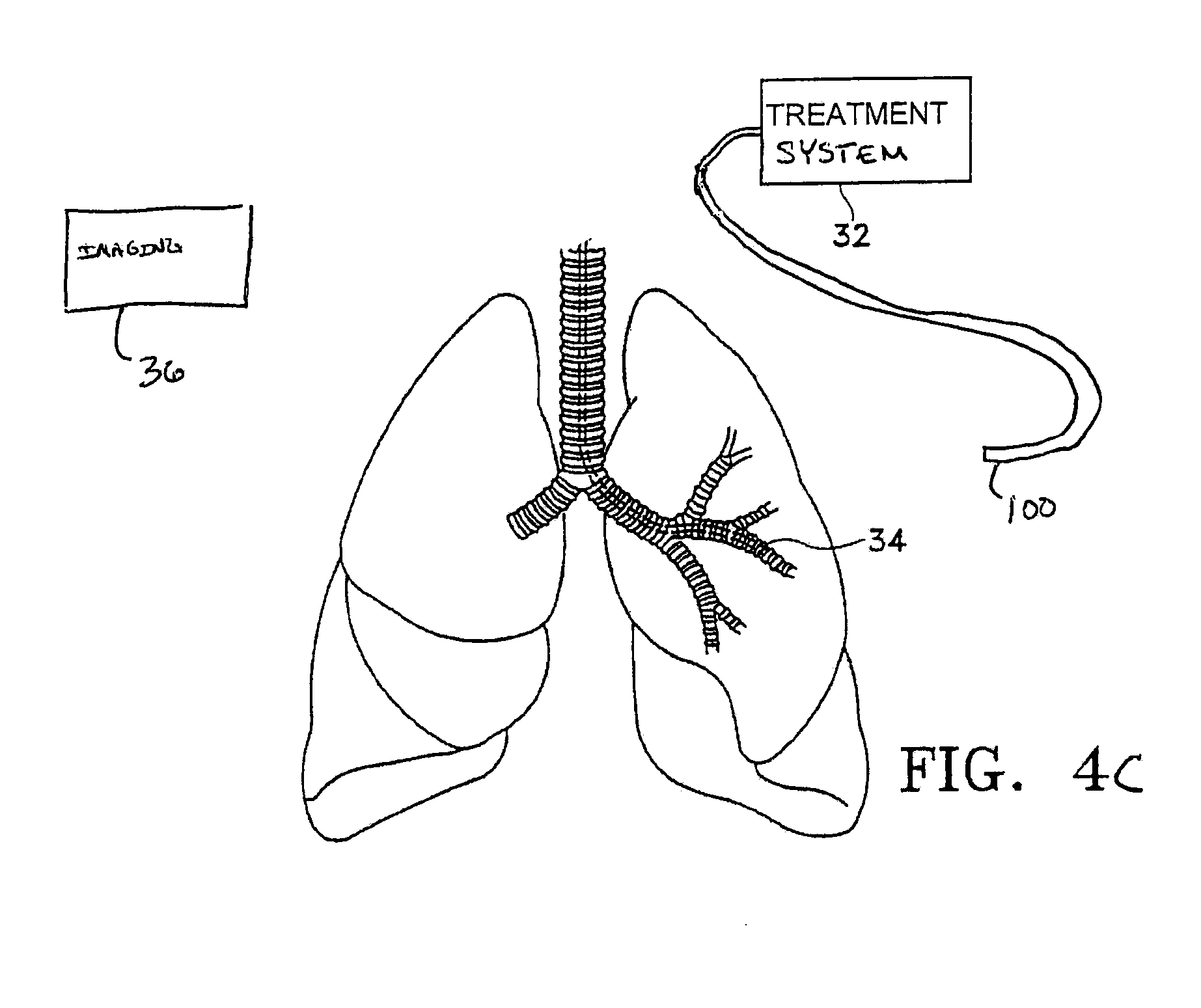
FIG. 4C illustrates another variation of a treatment system that applies the treatment externally to the lungs.
FIG. 5 illustrates a map to aid in treatment of the airways.
FIG. 6A illustrates a device that stimulates the airway into contracting.
FIGS. 6B-6F illustrate various modes of measuring parameters within the lungs to identify treatment sites.
FIG. 7 is a schematic side view of an embodiment of a treatment device with a balloon for heating of tissue.
DETAILED DESCRIPTION
The invention relates to methods for improving airflow through the airways of a lung having reversible obstructive pulmonary disease. It is intended that the invention is applicable to any aspect of reversible obstructive pulmonary disease, including but not limited to asthma. One way of improving airflow is to decrease the resistance to airflow within the lungs. There are several approaches to reducing this resistance, including but not limited to reducing the ability of the airway to contract, increasing the airway diameter, reducing the inflammation of airway tissues, and/or reducing the amount of mucus plugging of the airway. Another approach to reducing resistance is to increase the resting airway diameter of an airway such that any subsequent narrowing will not reduce the airway to a diameter such that obstruction to airflow is discernable by the patient. The present invention includes advancing a treatment device into the lung and treating the lung to at least reduce the ability of the lung to produce at least one symptom of reversible obstructive pulmonary disease. The following is a brief discussion of some causes of increased resistance to airflow within the lungs and the inventive treatment of the invention described herein. As such, the following discussion is not intended to limit the aspects or objective of the inventive method as the inventive method may cause physiological changes not described below but such changes still contributing to reducing or eliminating at least one of the symptoms of reversible obstructive pulmonary disease.
Reducing the Ability of the Airway to Contract
The inventive treatment reduces the ability of the airways to narrow or to reduce in diameter due to airway smooth muscle contraction. The inventive treatment uses a modality of treatments including, but not limited to the following: chemical, radio frequency, radioactivity, heat, ultrasound, radiant, laser, microwave, or mechanical energy (such as in the form of cutting, punching, abrading, rubbing, or dilating). This treatment reduces the ability of the smooth muscle to contract thereby lessening the severity of an asthma attack. The reduction in the ability of the smooth muscle to contract may be achieved by treating the smooth muscle itself or by treating other tissues which in turn influence smooth muscle contraction or the response of the airway to the smooth muscle contraction. Treatment may also reduce airway responsiveness or the tendency of the airway to narrow or to constrict in response to a stimulus.
The amount of smooth muscle surrounding the airway can be reduced by exposing the smooth muscle to energy which either kills the muscle cells or prevents these cells from replicating. The reduction in smooth muscle reduces the ability of the smooth muscle to contract and to narrow the airway during a spasm. The reduction in smooth muscle and surrounding tissue has the added potential benefit of increasing the caliber or diameter of the airways, which further reduces the resistance to airflow through the airways. In addition to the use of debulking smooth muscle tissue to open up the airways, the device used in the present invention may also eliminate smooth muscle altogether by damaging or destroying the muscle. The elimination of the smooth muscle prevents the contraction or spasms of hyper-reactive airways of a patient having reversible obstructive pulmonary disease. By doing so, the elimination of the smooth muscle may reduce some symptoms of reversible obstructive pulmonary disease.
The ability of the airway to contract can also be altered by treatment of the smooth muscle in particular patterns. The smooth muscle is arranged around the airways in a generally helical pattern with pitch angles ranging from about -38 to about +38 degrees. Thus, the treatment of the smooth muscle in appropriate patterns interrupts or cuts through the helical pattern of the smooth muscle at a proper pitch and prevents the airway from constricting. This procedure of patterned treatment application eliminates contraction of the airways without completely eradicating smooth muscle and other airway tissue. A pattern for treatment may be chosen from a variety of patterns including longitudinal or axial stripes, circumferential bands, helical stripes, and the like as well as spot patterns having rectangular, elliptical, circular or other shapes. The size, number, and spacing of the treatment bands, stripes, or spots are chosen to provide a desired clinical effect of reduced airway responsiveness while limiting insult to the airway to a clinically acceptable level.
The patterned treatment of the tissues surrounding the airways with energy provides various advantages. The careful selection of the portion of the airway to be treated allows desired results to be achieved while reducing the total healing load. Patterned treatment can also achieve desired results with decreased morbidity, preservation of epithelium, and preservation of a continuous or near continuous ciliated inner surface of the airway for mucociliary clearance. The pattern of treatment may also be chosen to achieve desired results while limiting total treatment area and/or the number of airways treated, thereby improving speed and ease of treatment.
Application of energy to the tissue surrounding the airways may also cause the DNA of the cells to become cross linked. The treated cells with cross linked DNA are incapable of replicating. Accordingly, over time, as the smooth muscle cells die, the total thickness of smooth muscle decreases because of the inability of the cells to replicate. The programmed cell death causing a reduction in the volume of tissue is called apoptosis. This treatment does not cause an immediate effect but causes shrinking of the smooth muscle and opening of the airway over time and substantially prevents re-growth. The application of energy to the walls of the airway may also be used to cause a cross linking of the DNA of the mucus gland cells thereby preventing them from replicating and reducing excess mucus plugging or production over time.
The ability of the airways to contract may also be reduced by altering mechanical properties of the airway wall, such as by increasing stiffness of the wall or by increasing parenchymal tethering of the airway wall. Both of these methods increase the strength of the airway wall and further oppose contraction and narrowing of the airway.
There are several ways to increase the stiffness of the airway wall. One way to increase stiffness is to induce fibrosis or a wound healing response by causing trauma to the airway wall. The trauma can be caused by delivery of therapeutic energy to the tissue in the airway wall, by mechanical insult to the tissue, or by chemically affecting the tissue. The energy is preferably delivered in such a way that it minimizes or limits the intra-luminal thickening that may occur.
Another way to increase the effective stiffness of the airway wall is to alter the submucosal folding of the airway upon narrowing. The mucosal layer includes the epithelium, its basement membrane, and the lamina propria, a subepithelial collagen layer. The submucosal layer may also play a role in airway folding. As an airway narrows, its perimeter remains relatively constant, with the mucosal layer folding upon itself. As the airway narrows further, the mucosal folds mechanically interfere with each other, effectively stiffening the airway. In asthmatic patients, the number of folds is fewer and the size of the folds is larger, and thus, the airway is free to narrow with less mechanical interference of mucosal folds than in a healthy patient. Thus, asthmatic patients have a decrease in airway stiffness and the airways have less resistance to narrowing.
The mucosal folding in asthmatic patients can be improved by treatment of the airway in a manner which encourages folding. Preferably, a treatment will increase the number of folds and/or decrease the size of the folds in the mucosal layer. For example, treatment of the airway wall in a pattern such as longitudinal stripes can encourage greater number of smaller mucosal folds and increase airway stiffness.
The mucosal folding can also be increased by encouraging a greater number of smaller folds by reducing the thickness of the mucosa and/or submucosal layer. The decreased thickness of the mucosa or submucosa may be achieved by application of energy which either reduces the number of cells in the mucosa or submucosal layer or which prevents replication of the cells in the mucosa or submucosal layer. A thinner mucosa or submucosal layer will have an increased tendency to fold and increased mechanical stiffening caused by the folds.
Another way to reduce the ability of the airways to contract is to improve parenchymal tethering. The parenchyma surrounds airways and includes the alveolus and tissue connected to and surrounding the outer portion of the airway wall. The parenchyma includes the alveolus and tissue connected to and surrounding the cartilage that supports the larger airways. In a healthy patient, the parenchyma provides a tissue network which connects to and helps to support the airway. Edema or accumulation of fluid in lung tissue in patients with asthma or COPD is believed to decouple the airway from the parenchyma reducing the restraining force of the parenchyma which opposes airway constriction. Energy can be used to treat the parenchyma to reduce edema and/or improve parenchymal tethering.
In addition, the applied energy may be used to improve connection between the airway smooth muscle and submucosal layer to the surrounding cartilage, and to encourage wound healing, collagen deposition, and/or fibrosis in the tissue surrounding the airway to help support the airway and prevent airway contraction.
Increasing the Airway Diameter
Hypertrophy of smooth muscle, chronic inflammation of airway tissues, and general thickening of all parts of the airway wall can reduce the airway diameter in patients with reversible obstructive pulmonary disease. Increasing the overall airway diameter using a variety of techniques can improve the passage of air through the airways. Application of energy to the airway smooth muscle of an asthmatic patient can debulk or reduce the volume of smooth muscle. This reduced volume of smooth muscle increases the airway diameter for improved air exchange.
Reducing inflammation and edema of the tissue surrounding the airway can also increase the diameter of an airway. Inflammation and edema (accumulation of fluid) of the airway are chronic features of asthma. The inflammation and edema can be reduced by application of energy to stimulate wound healing and regenerate normal tissue. Healing of the epithelium or sections of the epithelium experiencing ongoing denudation and renewal allows regeneration of healthy epithelium with less associated airway inflammation. The less inflamed airway has an increased airway diameter both at a resting state and in constriction. The wound healing can also deposit collagen which improves parenchymal tethering.
Inflammatory mediators released by tissue in the airway wall may serve as a stimulus for airway smooth muscle contraction. Therapy that reduces the production and release of inflammatory mediator can reduce smooth muscle contraction, inflammation of the airways, and edema. Examples of inflammatory mediators are cytokines, chemokines, and histamine. The tissues which produce and release inflammatory mediators include airway smooth muscle, epithelium, and mast cells. Treatment of these structures with energy can reduce the ability of the airway structures to produce or release inflammatory mediators. The reduction in released inflammatory mediators will reduce chronic inflammation, thereby increasing the airway inner diameter, and may also reduce hyper-responsiveness of the airway smooth muscle.
A further process for increasing the airway diameter is by denervation. A resting tone of smooth muscle is nerve regulated by release of catecholamines. Thus, by damaging or eliminating nerve tissue in the airways the resting tone of the smooth muscle is reduced, and the airway diameter is increased. Resting tone may also be reduced by directly affecting the ability of smooth muscle tissue to contract.
Reducing Plugging of the Airway
Excess mucus production and mucus plugging are common problems during both acute asthma exacerbation and in chronic asthma management. Excess mucus in the airways increases the resistance to airflow through the airways by physically blocking all or part of the airway. Excess mucus may also contribute to increased numbers of leukocytes found in airways of asthmatic patients by trapping leukocytes. Thus, excess mucus can increase chronic inflammation of the airways.
One type of asthma therapy involves treatment of the airways with energy to target and reduce the amount of mucus producing cells, ducts, and glands and to reduce the effectiveness of the remaining mucus producing cells and glands. The treatment can eliminate all or a portion of the mucus producing cells, ducts, and glands, can prevent the cells from replicating or can inhibit their ability to secrete mucus. This treatment will have both chronic benefits in increasing airflow through the airways and will lessen the severity of acute exacerbation of the symptoms of reversible obstructive pulmonary disease.
Application of Treatment
The following illustrations are examples of the invention described herein. It is contemplated that combinations of aspects of specific embodiments or combinations of the specific embodiments themselves are within the scope of this disclosure.
FIGS. 1 and 2 illustrate cross sections of two different airways in a healthy patient. The airway of FIG. 1 is a medium sized bronchus having an airway diameter D1 of about 3 mm. FIG. 2 shows a section through a bronchiole having an airway diameter D2 of about 1.5 mm. Each airway includes a folded inner surface or epithelium 10 surrounded by stroma 12 and smooth muscle tissue 14. The larger airways including the bronchus shown in FIG. 1 also have mucous glands 16 and cartilage 18 surrounding the smooth muscle tissue 14. Nerve fibers 20 and blood vessels 24 also surround the airway.
FIG. 3 illustrates the bronchus of FIG. 1 in which the smooth muscle 14 has hypertrophied and increased in thickness causing the airway diameter to be reduced from the diameter D1 to a diameter D3.
FIG. 4A is a schematic side view of the lungs being treated with a treatment device 38 according to the present invention. The treatment device 100 is an elongated member for treating tissue at a treatment site 34 within a lung. Although the invention discusses treatment of tissue at the airway wall surface it is also intended that the invention include treatment below an epithelial layer of the lung tissue. The invention may also rely on the use of an imaging device 36 to enable the identification of at least one treatments site from the plurality of possible treatment site locations. The imaging device may employ radiographic visualization such as fluoroscopy or other external visualization means such as computer aided tomography (CT), magnetic resonance imaging (MRI), positron emission tomography (PET), optical coherence tomography, or ultrasonic imaging. The imaging device may be external as shown. Alternatively, the imaging device may have a component that is affixed to the treatment system 32 or otherwise is inserted in to the body.
FIG. 4B represents one example of a treatment system 32 according to the present invention. In this variation, the system 32 delivers therapeutic energy to tissue of a patient via a device 100. Variations of devices are described in U.S. application Ser. Nos. 11/255,796 and 11/256,295 both filed Oct. 21, 2005 and the entirety of each of which is incorporated by reference.
FIG. 4B shows a schematic diagram of one example of a system 32 for delivering therapeutic energy to tissue of a patient for use with the device described herein. The illustrated variation shows, the system 32 having a power supply (e.g., consisting of an energy generator 112, a controller 114 coupled to the energy generator, a user interface surface 116 in communication with the controller 114). It is noted that the device may be used with a variety of systems (having the same or different components). For example, although variations of the device shall be described as RF energy delivery devices, variations of the device may include resistive heating systems, infrared heating elements, microwave energy systems, focused ultrasound, cryo-ablation, or any other energy deliver system. It is noted that the devices described should have sufficient length to access the tissue targeted for treatment. For example, it is presently believed necessary to treat airways as small as 3 mm in diameter to treat enough airways for the patient to benefit from the described treatment (however, it is noted that the invention is not limited to any particular size of airways and airways smaller than 3 mm may be treated). Accordingly, devices for treating the lungs must be sufficiently long to reach deep enough into the lungs to treat these airways. Accordingly, the length of the sheath/shaft of the device that is designed for use in the lungs should preferably be between 1.5-3 ft long in order to reach the targeted airways.
The particular system 32 depicted in FIG. 4B is one having a user interface as well as safety algorithms that are useful for the asthma treatment discussed above. Addition information on such a system may be found in U.S. Provisional application Nos. 60/674,106, and 60/673,876 both filed Apr. 21, 2005 the entirety of each of which is incorporated by reference herein.
Referring again to FIG. 4B, a variation of a device 100 described herein includes a flexible sheath 202, an elongate shaft 204 (in this example, the shaft extends out from the distal end of the sheath 202), and a handle or other operator interface 206 (optional) secured to a proximal end of the sheath 202. The distal portion of the device 100 includes an energy transfer element 208 (e.g., an electrode, a basket electrode, a resistive heating element, cryoprobe, etc.). Additionally, the device includes a connector 210 common to such energy delivery devices. The connector 210 may be integral to the end of a cable 212 as shown, or the connector 210 may be fitted to receive a separate cable 212. In any case, the device is configured for attachment to the power supply via some type connector 210. The elongate portions of the device 202 and 204 may also be configured and sized to permit passage through the working lumen of a commercially available bronchoscope or endoscope. As discussed herein, the device is often used within an endoscope, bronchoscope or similar device. However, the device may also be advanced into the body with or without a steerable catheter, in a minimally invasive procedure or in an open surgical procedure, and with or without the guidance of various vision or imaging systems.
FIG. 4B also illustrates additional components used in variations of the system. Although the depicted systems are shown as RF type energy delivery systems, it is noted that the invention is not limited as such. Other energy delivery configurations contemplated may include or not require some of the elements described below. The power supply (usually the user interface portion 116) shall have connections 120, 128, 130 for the device 100, return electrode 124 (if the system 32 employs a monopolor RF configuration), and actuation pedal(s) 126 (optional). The power supply and controller may also be configured to deliver RF energy to an energy transfer element configured for bipolar RF energy delivery. The user interface 116 may also include visual prompts 132, 160, 168, 174 for user feedback regarding setup or operation of the system. The user interface 116 may also employ graphical representations of components of the system, audio tone generators, as well as other features to assist the user with system use.
In many variations of the system, the controller 114 includes a processor 122 that is generally configured to accept information from the system and system components, and process the information according to various algorithms to produce control signals for controlling the energy generator 112. The processor 122 may also accept information from the system 110 and system components, process the information according to various algorithms and produce information signals that may be directed to the visual indicators, digital display or audio tone generator of the user interface in order to inform the user of the system status, component status, procedure status or any other useful information that is being monitored by the system. The processor 122 of the controller 114 may be digital IC processor, analog processor or any other suitable logic or control system that carries out the control algorithms.
In one variation of the system shown in FIG. 4B, the RF generator 112 generates RF energy at a frequency of about 400 kHz to about 500 kHz in with a wattage output sufficient to maintain a target tissue temperature of about 60 degrees C. to about 80 degrees C., specifically, about 60 degrees C. to about 70 degrees C. (when measuring at a surface of the electrode). The duration of the activation state for an embodiment of a single treatment cycle may be about 1 seconds to about 15 seconds, specifically, about 8 seconds to about 12 seconds. Alternatively, the duration of the activation state of the RF generator may also be set to not more than the duration required to deliver about 150 Joules of energy to the target tissue, specifically, not more than the duration required to deliver about 125 Joules of RF energy to target tissue.
Additional examples of devices for use with the methods of this invention are found in the following U.S. patent application Ser. Nos. 09/095,323 and 09/436,455; U.S. Pat. Nos. 6,488,675 and 6,411,852. The entirety of each of the aforementioned applications is incorporated by reference herein.
FIG. 4C represents another schematic side view of lungs being treated with a treatment device 100 according to the present invention. In this variation, the treatment device 100 and system 32 are external to the lungs and/or body but still applies energy to within the lungs. For example, such a treatment may use a high frequency ultrasound (commonly referred to as HIFU). As discussed above, the invention may also rely on the use of an imaging device 36.
The treatment of an airway with the treatment device may involve placing a visualization system such as an endoscope or bronchoscope into the airways. The treatment device is then inserted through or next to the bronchoscope or endoscope while visualizing the airways. Alternatively, the visualization system may be built directly into the treatment device using fiber optic imaging and lenses or a CCD and lens arranged at the distal portion of the treatment device. The treatment device may also be positioned using radiographic visualization such as fluoroscopy or other external visualization means. The treatment device which has been positioned with a distal end within an airway to be treated is energized so that energy is applied to the tissue of the airway walls in a desired pattern and intensity. The distal end of the treatment device may be moved through the airway in a uniform painting like motion to expose the entire length of an airway to be treated to the energy. The treatment device may be passed axially along the airway one or more times to achieve adequate treatment. The "painting-like" motion used to expose the entire length of an airway to the energy may be performed by moving the entire treatment device from the proximal end either manually or by motor. Alternatively, segments, stripes, rings or other treatment patterns may be used.
According to one variation of the invention, the energy is transferred to or from an airway wall in the opening region of the airway, preferably within a length of approximately two times the airway diameter or less, and to wall regions of airways distal to bifurcations and side branches, preferably within a distance of approximately twice the airway diameter or less. The invention may also be used to treat long segments of un-bifurcated airway.
The invention includes a method of advancing a treatment device into a lung and treating the lung with the device to, at least, reduce the ability of the lung to produce at least one symptom of reversible obstructive pulmonary disease. It is contemplated that the treatment may reduce all of the symptoms of reversible obstructive disease. Alternatively, the treatment may be selected to address specific symptoms of the disease. It is also intended that the treatment of the lung may sufficiently reduce the symptoms of reversible obstructive pulmonary disease such that the patient is able to function as those free from the disease. Alternatively, the treatment may be such that the symptoms are reduced to allow the patient to more easily manage the disease. It is also intended that the effects of the treatment may be either long term or short term with repeating treatment necessary to suppress the symptoms.
The methods of the invention described herein may be performed while the lung is experiencing natural symptoms of reversible obstructive pulmonary disease. One such example is where an individual, experiencing an asthma attack, or acute exacerbation of asthma or COPD, undergoes treatment to improve the individual's ability to breath. In such a case, the treatment, called `rescue,` seeks to provide immediate relief for the patient.
The method may also include the steps of locating one or more treatment sites within an airway of the lung, selecting one of the treatment sites from the locating step and treating at least one of the selected treatment sites. As mentioned above, these steps may be, but are not necessarily, performed while the lung is experiencing symptoms of reversible obstructive pulmonary disease.
The invention may further comprise the step of stimulating the lung to produce at least one artificially induced symptom of reversible obstructive pulmonary disease. For example, stimulation of the lung would preferably increase the resistance to airflow within the lung, constrict airways within the lung, inflame/irritate airway tissues, increase edema and/or increase the amount of mucus plugging of the airway. Stimulation of the lung may occur at any point during the procedure or before the procedure. For example, the lung may be stimulated either prior to or after, the step of locating a treatment site. If the lung is stimulated prior to the step of locating a treatment site, the reaction of the stimulated tissue within the lung may be useful in determining which locations are to be selected as treatment sites. The lung tissue or airway tissue within the lung may be stimulated by a variety of methods including but not limited to pharmacological stimulation, (e.g., histamine, methacholine, or other bronchoconstricting agents, etc.), electrical stimulation, mechanical stimulation, or any other stimuli causing obstructive pulmonary symptoms. For example, electrical stimulation may comprise exposing airway tissue to electrical field stimulation. An example of such parameters include 15 VDC, 0.5 ms pulses, 0.5-16 Hz, and 70 VDC, 2-3 ms pulses, 20 HZ.
The locating step described above may be performed using a non-invasive imaging technique, including but not limited to, a bronchogram, magnetic resonance imaging, computed tomography, radiography (e.g., x-ray), and ventilation perfusion scans.
The invention further includes the steps of testing the lung for at least one pre-treatment pulmonary function value prior to treating the lung with the device. After the lung is treated, the lung is re-tested for at least one post-treatment pulmonary function value. Naturally, the two pulmonary function values may be compared to estimate the effect of the treatment. The invention may also include treating additional sites in the lung after the re-testing step to at least reduce the effect of at least one symptom of reversible obstructive pulmonary disease. The invention may also include stimulating the lung to produce at least one artificially induced symptom of reversible obstructive pulmonary disease. As mentioned above, the stimulation of the lung may occur at any point during, or prior to, the procedure. For example, stimulation of the lung may occur prior to the step of testing the lung for pre-treatment pulmonary values. In this case, the values would be determinative of pulmonary function values of a lung experiencing symptoms of reversible obstructive pulmonary disease. Accordingly, the objective is to treat the lung until acceptable pulmonary function values are obtained. One benefit of such a procedure is that the effect of the treatment on the patient is more readily observed as compared to the situation where a patient, having previously been treated, must wait for an attack of reversible obstructive pulmonary disease to determine the efficacy of the treatment.
Pulmonary function values are well known in the art. The following is an example of pulmonary function values that may be used. Other pulmonary function values, or combinations thereof, are intended to be within the scope of this invention. The values include, but are not limited to, FEV (forced expiratory volume), FVC (forced vital capacity), FEF (forced expiratory flow), Vmax (maximum flow), PEFR (peak expiratory flow rate), FRC (functional residual capacity), RV (residual volume), TLC (total lung capacity).
FEV measures the volume of air exhaled over a pre-determined period of time by a forced expiration immediately after a full inspiration. FVC measures the total volume of air exhaled immediately after a full inspiration. Forced expiratory flow measures the volume of air exhaled during a FVC divided by the time in seconds. Vmax is the maximum flow measured during FVC. PEFR measures the maximum flow rate during a forced exhale starting from full inspiration. RV is the volume of air remaining in the lungs after a full expiration.
The locating step described above may also comprise identifying treatment sites within the airway being susceptible to a symptom of reversible obstructive pulmonary disease. For example, symptoms may include, but are not limited to, airway inflammation, airway constriction, excessive mucous secretion, or any other asthmatic symptom. Stimulation of the lung to produce symptoms of reversible obstructive pulmonary disease may assist in identifying ideal treatment sites.
As noted above, the method of the present invention may include stimulating the lung to produce at least one artificially induced symptom of reversible obstructive pulmonary disease and further include the step of evaluating the result of stimulation of the lung. For example, the evaluating step may include visually evaluating the effect of the stimulating step on the airway using a bronchoscope with a visualization system or by non-invasive imaging techniques, such as those describe herein. The evaluating step may include measuring pressure changes in the airway before and after the stimulating step. Pressure may be measured globally (e.g., within the entire lung), or locally (e.g., within a specific section of the lung such as an airway or alveolar sac.) Also, the evaluating step may comprise measuring the electrical properties of the tissue before and after the stimulating step. The invention may also include evaluating the results of the stimulating step by combining any of the methods previously mentioned. Also, the invention may further comprise the step of selecting at least one treatment parameter based upon the results of the evaluating step. Such treatment parameters may include, but are not limited to, duration of treatment, intensity of treatment, temperature, amount of tissue treated, depth of treatment, etc.
The method may also include the step of determining the effect of the treatment by visually observing lung, airway or other such tissue for blanching of the tissue. The term "blanching" is intended to include any physical change in tissue that is usually, but not necessarily, accompanied by a change in the color of the tissue. One example of such blanching is where the tissue turns to a whitish color after the treatment of application of energy.
The invention may also include the step of monitoring impedance across a treated area of tissue within the lung. Measuring impedance may be performed in cases of monopolar or bipolar energy delivery devices. Additionally, impedance may be monitored at more than one site within the lungs. The measuring of impedance may be, but is not necessarily, performed by the same electrodes used to deliver the energy treatment to the tissue. Furthermore, the invention includes adjusting the treatment parameters based upon the monitoring of the change in impedance after the treatment step. For example, as the energy treatment affects the properties of the treated tissue, measuring changes in impedance may provide information useful in adjusting treatment parameters to obtain a desired result.
Another aspect of the invention includes advancing a treatment device into the lung and treating lung tissue to at least reduce the ability of the lung to produce at least one symptom of reversible obstructive pulmonary disease and further comprising the step of sub-mucosal sensing of the treatment to the lung tissue. The sub-mucosal sensing may be invasive such as when using a probe equipped to monitor temperature, impedance, and/or blood flow. Or, the sub-mucosal sensing may be non-invasive in such cases as infra-red sensing.
The invention may also include using the treatment device to deposit radioactive substances at select treatment sites within the lung. The radioactive substances, including, but not limited to Iridium (e.g. 192Ir.) either treat the lung tissue over time or provide treatment upon being deposited.
The invention also includes scraping epithelial tissue from the wall of an airway within the lung prior to advancing a treatment device into the lung to treat the lung tissue. The removal of the epithelial tissue allows the device to treat the walls of an airway more effectively. The invention further comprises the step of depositing a substance on the scraped wall of the airway after the device treats the airway wall. The substance may include epithelial tissue, collagen, growth factors, or any other bio-compatible tissue or substance, which promotes healing, prevents infection, and/or assists in the clearing of mucus. Alternatively, the treatment may comprise the act of scraping epithelial tissue to induce yield the desired response.
The invention includes using the treating device to pre-treat the lung to at least reduce the ability of the lung to produce at least one symptom of reversible obstructive pulmonary disease prior to the treating step. At least one of the parameters of the pre-treating step may differ than one of the parameters of the treating step. Such parameters may include time, temperature, amount of tissue over which treatment is applied, amount of energy applied, depth of treatment, etc.
The invention may also include advancing the treatment device into the lung and treating the lung tissue in separate stages. One of the benefits of dividing the treating step into separate stages is that the healing load of the patient is lessened. Dividing of the treating step may be accomplished by treating different regions of the lung at different times. Or, the total number of treatment sites may be divided into a plurality of groups of treatment sites, where each group of treatment sites is treated at a different time. The amount of time between treatments may be chosen such that the healing load placed on the lungs is minimized.
The invention may also include advancing a treatment device into the lung, treating the lung with the device and sensing movement of the lung to reposition the treatment device in response to the movement. This sensing step accounts for the tidal motion of the lung during breathing cycles or other movement. Taking into account the tidal motion allows improved accuracy in repositioning of the device at a desired target.
The invention may also include the additional step of reducing or stabilizing the temperature of lung tissue near to a treatment site. This may be accomplished for example, by injecting a cold fluid into lung parenchyma or into the airway being treated, where the airway is proximal, distal, or circumferentially adjacent to the treatment site. The fluid may be sterile normal saline, or any other bin-compatible fluid. The fluid may be injected into treatment regions within the lung while other regions of the lung normally ventilated by gas. Or, the fluid may be oxygenated to eliminate the need for alternate ventilation of the lung. Upon achieving the desired reduction or stabilization of temperature the fluid may be removed from the lungs. In the case where a gas is used to reduce temperature, the gas may be removed from the lung or allowed to be naturally exhaled. One benefit of reducing or stabilizing the temperature of the lung may be to prevent excessive destruction of the tissue, or to prevent destruction of certain types of tissue such as the epithelium, or to reduce the systemic healing load upon the patient's lung.
Also contemplated as within the scope of the invention is the additional step of providing therapy to further reduce the effects of reversible obstructive pulmonary disease or which aids the healing process after such treatment. Some examples of therapy include, drug therapy, exercise therapy, and respiratory therapy. The invention further includes providing education on reversible obstructive pulmonary disease management techniques to further reduce the effects of the disease. For example, such techniques may be instruction on lifestyle changes, self-monitoring techniques to assess the state of the disease, and/or medication compliance education.
There may be occurrences where it is necessary to reverse the effects of the treatment described herein. Accordingly, the invention further includes a method for reversing a treatment to reduce the ability of the lung to produce at least one symptom of reversible obstructive pulmonary disease comprising the step of stimulating re-growth of smooth muscle tissue. The re-stimulation of the muscle may be accomplished by the use of electro-stimulation, exercising of the muscle and/or drug therapy.
The invention further includes methods of evaluating individuals having reversible obstructive pulmonary disease, or a symptom thereof, as a candidate for a procedure to reduce the ability of the individual's lung to produce at least one symptom of reversible obstructive pulmonary disease. The method comprises the steps of assessing the pulmonary condition of the individual, comparing the pulmonary condition to a corresponding pre-determined state, and evaluating the individual as a candidate based upon the comparison.
In assessing the pulmonary condition, the method may comprise the steps of performing pulmonary function tests on the individual to obtain a pulmonary function value which is compared to a predetermined value. Examples of pre-determined values are found above.
The method of evaluating may further include the step of determining how the individual's tissue will react to treatment allowing the treatment to be tailored to the expected tissue response.
The method of evaluating may further comprises the step of pulmonary function testing using a gas, a mixture of gases, or a composition of several mixtures of gases to ventilate the lung. The difference in properties of the gases may aid in the pulmonary function testing. For example, comparison of one or more pulmonary function test values that are obtained with the patient breathing gas mixtures of varying densities may help to diagnose lung function. Examples of such mixtures include air, at standard atmospheric conditions, and a mixture of helium and oxygen. Additional examples of pulmonary testing include tests that measure capability and evenness of ventilation given diffusion of special gas mixtures. Other examples of gases used in the described tests, include but are not limited to, nitrogen, carbon monoxide, carbon dioxide, and a range of inert gases.
The invention may also comprise the step of stimulating the lung to produce at least one artificially induced symptom of reversible obstructive pulmonary disease. Stimulating the symptoms of the disease in an individual allows the individual to be evaluated as the individual experiences the symptoms thereby allowing appropriate adjustment of the treatment.
The method of evaluating may also comprise the step of obtaining clinical information from the individual and accounting for the clinical information for treatment.
The method may further comprise the selection of a patient for treatment based upon a classification of the subtype of the patient's disease. For example, in asthma there are a number of ways to classify the disease state. One such method is the assessment of the severity of the disease. An example of a classification scheme by severity is found in the NHLBI Expert Panel 2 Guidelines for the Diagnosis and Treatment of Asthma. Another selection method may include selecting a patient by the type of trigger that induces the exacerbation. Such triggers may be classified further by comparing allergic versus non-allergic triggers. For instance, an exercise induced bronchospasm (EIB) is an example of a non-allergenic trigger. The allergic sub-type may be further classified according to specific triggers (e.g., dust mites, animal dander, etc.). Another classification of the allergic sub-type may be according to characteristic features of the immune system response such as levels of IgE (a class of antibodies that function in allergic reactions, also called immunoglobulin). Yet another classification of allergic sub-types may be according to the expression of genes controlling certain interleukins (e.g., IL-4, IL-5, etc.) which have been shown to play a key role in certain types of asthma.
The invention further comprises methods to determine the completion of the procedure and the effectiveness of the reduction in the lung's ability to produce at least one symptom of reversible obstructive pulmonary disease. This variation of the invention comprises assessing the pulmonary condition of the individual, comparing the pulmonary condition to a corresponding predetermined state, and evaluating the effectiveness of the procedure based on the comparison. The invention may also comprise the steps of performing pulmonary function tests on the individual to obtain at least one pulmonary function value, treating the lung to at least reduce the ability of the lung to produce at least one symptom of reversible obstructive pulmonary disease, performing a post-procedure pulmonary function tests on the individual to obtain at least one post pulmonary function value and comparing the two values.
This variation of the invention comprises obtaining clinical information, evaluating the clinical information with the results of the test to determine the effectiveness of the procedure. Furthermore, the variation may include stimulating the lung to produce a symptom of reversible obstructive pulmonary disease, assessing the pulmonary condition of the patient, then repeating the stimulation before the post-procedure pulmonary therapy. These steps allow comparison of the lung function when it is experiencing symptoms of reversible obstructive pulmonary disease, before and after the treatment, thereby allowing for an assessment of the improved efficiency of the lung during an attack of the disease.
The medical practitioners performing the treatments described herein may wish to treat a limited number of sites in the lung to produce acceptable results in lessening the severity of asthmatic or reversible obstructive pulmonary disease symptoms. For example, if a patient requires an increasing amount of medication (e.g., sedatives or anesthesia) to remain under continued control for performance of the procedure, then a medical practitioner may limit the procedure time rather than risk overmedicating the patient. As a result, rather than treating the patient continuously to complete the procedure, the practitioner may plan to break the procedure in two or more sessions. Subsequently, increasing the number of sessions poses additional consequences on the part of the patient in cost, the residual effects of any medication, adverse effects of the non-therapeutic portion of the procedure, etc.
Accordingly, the invention includes methods of treating airways in a lung to decrease asthmatic symptoms. The methods include measuring a parameter of an airway at a plurality of locations in a lung, identifying at least one treatment site from at least one of the plurality of locations based on the parameter, and applying energy to the treatment site to reduce the ability of the site to narrow.
Identification of the treatment sites may comprise comparing the parameter to known or studied parameters and selecting those sites that meet or exceed specific criteria. Alternatively, or in combination, identification of treatment sites may comprise selecting the sites with the most significant parameters, or delivering a treatment specifically tailored to the parameters measured at each individual site. For example, if the parameter comprises measuring contractile force or the amount of contraction, then sites having the highest quantitative parameters may be selected as treatment sites. In another variation, the medical practitioner may simply rank the parameters in a desired order of value and treat those sites that are believed to provide the most benefit. For example, the medical practitioner may chose to treat the top 10% of sites having the most contraction, smooth muscle tissue, or other parameters as described herein. It is also contemplated that the diametrical size of the airway or the length of the airway segment may be correlated to the ability for that site to narrow. For example, when measuring a narrowed airway diameter relative to its natural diameter, the percentage change for a large diameter airway may be different than a percentage change for a smaller airway. In such a case, the medical practitioner may measure the airway diameter at each site for determining the course of treatment. In other words, the practitioner may deliver treatment to specific sites that meet certain criteria (e.g., sites having a certain diameter). Alternatively, or in combination, the practitioner may delivery treatment specifically tailored to each individual site based on a characteristic of the site (e.g., its diameter). For example, when delivering energy, the practitioner could deliver more energy to larger diameter airway, and less energy to smaller diameter airways (or vice versa).
In those cases where more than one treatment site is identified, the method may include the act of treating the new treatment site.
The method may also include correlating the treatment sites to a map 90, as shown in FIG. 5, the map may provide a graphical representation of a bronchial tree. As shown, the various bronchioles 92 decrease in size and have many branches 96 as they extend into the right and left bronchi 94. Accordingly, an efficient treatment may require identification of potential treatment sites, treated sites, and other areas of the bronchial tree.
This mapping may be performed for a variety of reasons. For example, prior to treatment, the correlation may identify general areas for treatment by the medical practitioner. Once the area is treated, the map may then be marked to indicate a completed treatment. The treatment plan provided by the map should allow the medical practitioner a guide so that it is possible to treat less than all of the lungs. The treatment plan or map also may assist in avoiding double treatment of a particular treatment site. It is contemplated that the map 90 may be an actual chart, whether in tangible form or electronic form. Furthermore, the map may be incorporated into the treatment system 32 or the user interface 116 as discussed above. It is also contemplated that the map may be a three dimensional computer model, wherein the position of completed treatment sites are recorded by storing the spatial coordinates of these sites as each treatment is completed. As subsequent treatments are made, the user may compare the current position of the catheter to the map, which will aid in determining which site to treat next.
The parameters to be measured in accordance with the methods described herein may be any parameter that is an indicator of or associated with symptoms of asthma. For example, the parameter may be a measure of pulmonary function values (see above), a measure of the contractile force at which the airway contracts, a thickness or amount of the airway smooth muscle at a particular location, eosinophil counts near or at the actual or potential treatment site, degree of airflow within the airway, degree of contraction of the airway during an asthma episode or after stimulation of the airway, metabolic rate to assess the presence of smooth muscle, electrical impedance to assess the nature of the airway tissue, and/or degree of wheezing at a particular location, etc. Other parameters indicative of asthma or a lack of airflow due to asthmatic symptoms are also intended to be within the scope of this disclosure.
Methods of the present invention include first stimulating the airway and then subsequently measuring the parameter. The stimulation may be performed electrically (such as by placing a device within the airway and stimulating using the settings described above). Alternatively, or in combination, the stimulation may be artificially induced using an agent, such as methacholine. For example, as shown in FIG. 6A, a device 150 may deliver the agent 152 into the airway 2.
Once the airway contracts, the contraction may be measured or assessed. Although not shown, the contraction may be measured or assessed without making contact with the airway wall (e.g., visually with a retical; or optically, via a camera). Alternatively, or in combination, as shown in FIG. 6B, a contraction measurement device 154 may be placed against the airway (either prior or during contraction) and expanded to measure a natural state of the airway. The contraction measurement device 154 then transmits information regarding the contraction of the airway using, for example, strain gauges 156 placed on moveable arms of the device. The contraction measurement device 154 may also deliver an agent to cause contraction of the airways. In this manner, the device 154 will be in place while the airway contracts.
FIG. 6C illustrates another variation where a balloon catheter 158 measures contraction of the airway. As the airway constricts, the balloon 160 increases in pressure. The pressure may then be characterized to determine the degree of contractile force of the airway. It should be noted that the balloon catheter 158 may also include fluid delivery ports to deliver an agent or have electrodes to induce the contraction. Other examples of devices that may be used to measure contraction of the airways are devices that measure the airway diameter mechanically or optically.
In another variation as shown in FIG. 6D, a device 162 (e.g., an ultrasound balloon catheter, a non-balloon ultrasound catheter, a catheter (balloon or non-balloon) that is equipped to measure impedance, etc.), may be expanded within the airway to measure the thickness of the adjacent airway smooth muscle 14. Alternatively, as described above, the measurement of the airway smooth muscle 14 may be achieved using the external imaging equipment 36 described above.
Another method of measuring parameters of an airway for identifying treatment sites comprises measuring eosinophil counts at the location. Eosinophils are white blood cells active in allergic diseases, parasitic infections, and other disorders. It is believed that cosinophils correlate to the amount of inflammation in an airway. FIG. 6E shows one way of obtaining an cosinophils count at a location in the lung. As shown, a device 164 advances a needle 166 within the airway to collect the eosinophils. Next, standard techniques are employed to measure the particular eosinophil count at the site.
FIG. 6F illustrates another variation of the invention where a device 168 measures airflow airflow at a location or near to a location for treatment. The device 168 may be a hot-wire amenometer, where the airflow causes the heated wire 170 to cool and the rate of cooling of the wire provides information regarding the airflow.
In additional variations of the method, the medical practitioner may deliver hyperpolarized helium or a radioactive isotope to aid in the imaging of the airways. External imaging, as shown FIGS. 4A and 4C may be used to assess ventilation in the lung and select the areas that constrict as treatment sites.
Alternatively, the external imaging may take a first image of the airways. Next, an agent is applied (either locally, systemically, or limited to a particular lobe) to induce contraction of the airways. The medical practitioner may then obtain a second image of the airways for comparison with the first to determine contraction of the airways.
In another variation of the invention, the parameter comprises assessing a metabolic rate at the location. If the measurement of the metabolic rate indicates the presence of a significant amount of smooth muscle tissue, then the area may be designated as a treatment site. The metabolic rate may be measured over a treatment site or over an area of the airways (e.g., a particular lobe or a section thereof.) The measurement of the metabolic rate may be performed using standard measuring techniques. For example, the device may deliver cool air to the treatment site and then measure the rate at which the tissue temperature returns to the original baseline temperature, thereby providing a measurement of the calories used to bring the tissue back to the baseline temperature. This would, in turn, provide a measure of the responsiveness of the smooth muscle tissue because more reactive or responsive tissue may correlate to a higher metabolic rate.
The treatment device of FIG. 7 is used to deliver radiant or heat energy to the airway. The treatment device of FIG. 7 can also deliver indirect radio frequency or microwave energy to the tissue. FIG. 7 shows an alternative embodiment of a balloon catheter treatment device 30 k in which a fluid within the balloon 1126 is heated by internal electrodes 1128. The electrodes 1128 are illustrated in the shape of coils surrounding the shaft of the catheter, however, other electrode shapes may also be used. The electrodes 1128 may be used as resistance heaters by application of an electric current to the electrodes. Alternatively, radio frequency or microwave energy may be applied to the electrodes 1128 to heat a fluid within the balloon 1126. The heat then passes from an exterior of the balloon 1126 to the airway wall. The radio frequency or microwave energy may also be applied indirectly to the airway wall through the fluid and the balloon. In addition, hot fluid may be transmitted to the balloon 1126 from an external heating device for conductive heating of the airway tissue.
The invention herein is described by examples and a desired way of practicing the invention is described. However, the invention as claimed herein is not limited to that specific description in any manner. Equivalence to the description as hereinafter claimed is considered to be within the scope of protection of this patent.
* * * * *
References
D00000
D00001
D00002
D00003
D00004
D00005
D00006
D00007
D00008
D00009
XML
uspto.report is an independent third-party trademark research tool that is not affiliated, endorsed, or sponsored by the United States Patent and Trademark Office (USPTO) or any other governmental organization. The information provided by uspto.report is based on publicly available data at the time of writing and is intended for informational purposes only.
While we strive to provide accurate and up-to-date information, we do not guarantee the accuracy, completeness, reliability, or suitability of the information displayed on this site. The use of this site is at your own risk. Any reliance you place on such information is therefore strictly at your own risk.
All official trademark data, including owner information, should be verified by visiting the official USPTO website at www.uspto.gov. This site is not intended to replace professional legal advice and should not be used as a substitute for consulting with a legal professional who is knowledgeable about trademark law.