Epitope recognized by anti-interferon gamma autoantibodies in patients with disseminated mycobacterial infections and the application therefor
Ku , et al.
U.S. patent number 10,273,278 [Application Number 15/185,172] was granted by the patent office on 2019-04-30 for epitope recognized by anti-interferon gamma autoantibodies in patients with disseminated mycobacterial infections and the application therefor. This patent grant is currently assigned to CHANG GUNG UNIVERSITY. The grantee listed for this patent is CHANG GUNG UNIVERSITY. Invention is credited to Chih-Yu Chi, Jing-Ya Ding, Cheng-Lun Ku, Chia-Hao Lin, Han-Po Shih.


View All Diagrams
| United States Patent | 10,273,278 |
| Ku , et al. | April 30, 2019 |
Epitope recognized by anti-interferon gamma autoantibodies in patients with disseminated mycobacterial infections and the application therefor
Abstract
The present invention discloses a fragment of peptide which can be utilized in patients suffering from a disseminated mycobacterial infection. The fragment of peptide contains a sequence of amino acids with seven residues as formula (I) shown below, wherein X.sub.1 is Leucine (Leu); X.sub.2 is Proline (Pro); X.sub.3 is Glutamate (Glu); X.sub.4 is serine (Ser); X.sub.5 is Serine (Ser); X.sub.6 is Leucine (Leu) and X.sub.7 is Arginine (Arg): X.sub.1-X.sub.2-X.sub.3-X.sub.4-X.sub.5-X.sub.6-X.sub.7 (I).
| Inventors: | Ku; Cheng-Lun (Tao-Yuan, TW), Lin; Chia-Hao (Tao-Yuan, TW), Chi; Chih-Yu (Tao-Yuan, TW), Shih; Han-Po (Tao-Yuan, TW), Ding; Jing-Ya (Tao-Yuan, TW) | ||||||||||
|---|---|---|---|---|---|---|---|---|---|---|---|
| Applicant: |
|
||||||||||
| Assignee: | CHANG GUNG UNIVERSITY
(Tao-Yuan, TW) |
||||||||||
| Family ID: | 58401010 | ||||||||||
| Appl. No.: | 15/185,172 | ||||||||||
| Filed: | June 17, 2016 |
Prior Publication Data
| Document Identifier | Publication Date | |
|---|---|---|
| US 20170114112 A1 | Apr 27, 2017 | |
Foreign Application Priority Data
| Jun 18, 2015 [TW] | 104119897 A | |||
| Current U.S. Class: | 1/1 |
| Current CPC Class: | G01N 33/5695 (20130101); G01N 33/564 (20130101); G01N 33/6869 (20130101); G01N 33/5023 (20130101); C07K 14/57 (20130101); G01N 33/6866 (20130101); G01N 33/5055 (20130101); A61K 38/00 (20130101); G01N 2800/52 (20130101); G01N 2333/57 (20130101); G01N 2333/5434 (20130101) |
| Current International Class: | G01N 33/50 (20060101); G01N 33/564 (20060101); C07K 14/57 (20060101); G01N 33/569 (20060101); G01N 33/68 (20060101); A61K 38/00 (20060101) |
References Cited [Referenced By]
U.S. Patent Documents
| 6120762 | September 2000 | Johnson |
Other References
|
Patel et al., (J Immunol. 2005;175:4769-4776). (Year: 2005). cited by examiner . Lin et al., Nat.Med . . . , vol. 22, No. 9, pp. 994-1003, (Year: 2016). cited by examiner . Daniel Lundell, Charles Lunn, David Dalgarno, James Fossetta, Robert Greenberg, Richard Reim, Michael Grace, Satwant Narula, "The carboxyl-terminal region of human interferon gamma is important for biological activity: mutagenic and NMR analysis", Protein Engineering, 1991, pp. 335-341, vol. 4, No. 3, Oxford University Press, UK. cited by applicant . Cheng-Lun Ku, "Molecular Mechanisms of Anti-Interferon Gamma Autoantibodies in Nontuberculous Mycobacterial Infected Patients", Executive Yuan National Science Council Project-Research Project-Results Report, Nov. 5, 2012, Taiwan. cited by applicant . Smita Y. Patel, Li Ding, Margaret R. Brown, Larry Lantz, Ted Gay, Stuart Cohen, Lenna A. Martyak, Bernard Kubak, Steven M. Holland, "Anti-IFN-gamma Autoantibodies in Disseminated Nontuberculous Mycobacterial Infections", The Journal of Immunology, 2005, pp. 4769-4776, vol. 175, The American Association of Immunologists, Inc., USA. cited by applicant. |
Primary Examiner: Kolker; Daniel E
Assistant Examiner: Seharaseyon; Jegatheesan
Attorney, Agent or Firm: Chiang; Cheng-Ju
Claims
What is claimed is:
1. A method for evaluating an efficacy of an isolated recombinant human interferon gamma (rhIFN.gamma.) for regulating a peripheral blood mononuclear cell (PBMC), comprising the steps of: providing the PBMC from a subject with anti-interferon gamma autoantibodies; mixing the isolated recombinant human interferon gamma (rhIFN.gamma.) with the PBMC, wherein the isolated recombinant human interferon gamma (rhIFN.gamma.) is produced by replacing at least one residue which is located between residues 121 and 127 from a C-terminal of an epitope of a human interferon gamma (hIFN.gamma.) SEQ ID NO: 39 via homologous substitution; and evaluating the efficacy of the isolated recombinant human interferon gamma (rhIFN.gamma.) according to an expression level of a phosphorylation of signal transducers and activators of transcription 1 (p-STAT1) generated by the PBMC.
2. The method as claimed in claim 1, wherein homologous substitution consists of SEQ ID NO: 1.
3. The method as claimed in claim 2, wherein the isolated recombinant human interferon gamma (rhIFN.gamma.) further includes a pharmaceutically acceptable excipient or a pharmaceutically acceptable carrier.
4. The method as claimed in claim 2, wherein the isolated recombinant human interferon gamma (rhIFN.gamma.) up-regulates the expression level of the p-STAT1.
5. A method for evaluating an efficacy of an isolated recombinant human interferon gamma (rhIFN.gamma.) for regulating a peripheral blood mononuclear cell (PBMC), comprising the steps of: providing the PBMC from a subject with anti-interferon gamma autoantibodies; mixing the isolated recombinant human interferon gamma (rhIFN.gamma.) with the PBMC, wherein the isolated recombinant human interferon gamma (rhIFN.gamma.) is produced by replacing residues which is located between residues 121 and 127 from a C-terminal of an epitope of a human interferon gamma (hIFN.gamma.) of SEQ ID NO: 39 via homologous substitution of SEQ ID NO: 1; and evaluating the efficacy of the isolated recombinant human interferon gamma (rhIFN.gamma.) according to an expression level of a phosphorylation of signal transducers and activators of transcription 1 (p-STAT1) generated by the PBMC.
Description
CROSS-REFERENCE TO RELATED APPLICATION AND CLAIM OF PRIORITY
The application claims the benefit of Taiwan Application No. 104119897, filed on Jun. 22, 2015, at the Taiwan Intellectual Property Office, the disclosures of which are incorporated herein in their entirety by reference.
FIELD OF THE INVENTION
The invention is related to a medical, dental or toilet preparation. In particular, it is related to the preparation of one kind of peptide fragments, which can be used in patients suffering from a disseminated mycobacterial infection.
BACKGROUND OF THE INVENTION
Anticytokine autoantibodies (ACADs) are increasingly recognized, and playing an important role in the pathogenesis of infectious and autoimmune diseases.
Clinically, the pathogenic mycobacterial species can cause tuberculosis, Hansen's disease, leprosy, pulmonary disease, lymphadenitis and skin disease. In view of mycobacterial immunity, IFN.sub.r plays an important role and is mainly produced by T and NK cells when stimulated with microbial products.
Genetic defects in the IFN.sub.r/IL-12 pathway cause Mendelian susceptibility to mycobacterial diseases (MSMDs) in young patients with disseminated mycobacterial infections.
On the other hand, interference with the IFN.sub.r signaling by the presence of anti-IFN.sub.r AutoAbs is the major etiology that explains the occurrence of severe disseminated mycobacterial infections in adults without obvious immunologic defects, in particular for patients from the Southeast Asia.
The similarity of clinical susceptibility to MSMDs strongly suggests that AutoAbs against IFN.sub.r were the cause rather than a consequence of mycobacterial infection.
The mechanism of the production of anti-IFN.sub.r AutoAbs also remains unclear. Restriction of the disease in Southeast Asian population suggest that a particular genetic factor and mechanism are involved.
According to a previous study, HLA class II molecules DRB1*16:02 and HLA-DQB1*05:02 are the two specific alleles strongly associated with this disease, and the high frequency of this allele in Southeast Asia might also explain the susceptibility of anti-IFN.sub.r AutoAbs in this particular population.
MHC class II is present the particular peptides to CD4.sup.+ T cells to induce an adaptive immune response and is a strong genetic factor associated with autoimmune diseases. It seems that particular pathogenic peptide fragments present in these particular HLA alleles are involved in the production of anti-IFN.sub.r AutoAbs.
Various hypotheses have been proposed to explain the production of these pathogenic AutoAbs. Molecular mimicry theory states that exo-antigen can mimic self-antigen and induces the formation of AutoAbs. This theory, molecular mimicry, has been documented in various autoimmune diseases, including multiple sclerosis (MS), ankylosing spondylitis, Graves' disease, diabetes mellitus, and systemic lupus erythematosus (SLE).
In the case of MS and SLE, the disease pathogenesis has been linked to some viruses, such as the Epstein-Barr virus (EBV), for their homologous amino acid sequences with human antigenic structures in the central nerve system or lupus autoantigens, such as Sm B.
These findings suggest that molecular mimicry plays a major role in the pathogenesis of certain diseases. Despite advances in the genomic technologies for autoimmunity, the precise mechanism for the pathogenic AutoAbs formation is still unclear.
SUMMARY OF THE INVENTION
Autoantibodies (AutoAbs) against IFN.sub.r is an emerging medical issue and linked to disseminated mycobacterial infections and other opportunistic infections in the Southeast Asia. The origin of these AutoAbs is unclear; however, the majority of affected patients share specific HLA class II alleles and this observation suggests that a common mechanism in the production of AutoAbs may exist. Herein, the inventor characterized the anti-IFN.sub.r AutoAbs from patients and found these AutoAbs recognized a major epitope (P.sub.121-131) in the C-terminal of IFN.sub.r. The region was known to be critical for IFN.sub.r receptor activation, and the inventor also demonstrated that AutoAbs to this epitope had a neutralizing activity. This epitope was 100% homologous to the Aspergillus Noc2 and anti-IFN.sub.r AutoAbs from patients could react with this epitope and Aspergillus Noc2. In vivo study, rats immunized with Aspergillus Noc2 developed antibodies against human IFN.sub.r and vice versa.
In addition, the inventor generated an epitope Erase IFN.sub.r (EE-IFN.sub.r) which has lower affinity recognized by anti-IFN.sub.r AutoAbs due to a modified major epitope region and it could activate the IFN.sub.r downstream signaling pathway ex vivo even in the presence of anti-IFN.sub.r AutoAbs.
It was found that anti-IFN.sub.r AutoAbs from different patients recognized a specific region, SPAAKTGKRK (SEQ ID NO: 14), in the C-terminal of IFN.sub.r and these antibodies had a neutralizing activity on IFN.sub.r.
A high homologous peptide sequence in this region (KTGKRKR (SEQ ID NO: 36)) was found in Aspergillus Noc2 and also recognized by the anti-IFN.sub.r AutoAbs.
After immunization with Aspergillus Noc2, antibodies against human IFN.sub.r were formed in the test rats.
Furthermore, the inventor used a mouse homologous region to replace the critical anti-IFN.sub.r AutoAb-recognized epitope in human IFN.sub.r.quadrature. and generated a new recombinant protein, epitope Erased IFN.sub.r (EE-IFN.sub.r).
This recombinant protein could induce the activation of the IFN.sub.r receptor, even in the presence of anti-IFN.sub.r AutoAbs ex vivo.
Taken together, the results suggest that anti-IFN.sub.r AutoAbs may be induced by the means of molecular mimicry, and structural modification of IFN.sub.r can bypass the blocking activity of anti-IFN.sub.r AutoAbs.
In accordance with one aspect of the present invention, a method for evaluating an efficacy of an isolated recombinant human interferon gamma (hIFN.sub.r) for regulating a peripheral blood mononuclear cell (PBMC) is disclosed. The method comprises the steps of: providing the PBMC from a subject with anti-interferon gamma autoantibodies; mixing the isolated recombinant human interferon gamma with the PBMC, wherein the isolated recombinant human interferon gamma contains a homologous substitute; and evaluating the efficacy of the isolated recombinant human interferon gamma according to an expression level of a phosphorylation of signal transducers and activators of transcription 1 (p-STAT1) generated by the PBMC.
In accordance with another aspect of the present invention, a method for evaluating an efficacy of an isolated recombinant cytokine for regulating a peripheral blood mononuclear cell (PBMC) is disclosed. The method comprises steps of: providing the PBMC from a subject with an anticytokine autoantibody; mixing the isolated recombinant cytokine with the PBMC, wherein the isolated recombinant cytokine contains a homologous substitute; and evaluating the efficacy of the isolated recombinant cytokine according to an expression level of an interleukin-12 (IL-12) generated by the PBMC.
In accordance with another aspect of the present invention, a recombinant protein is disclosed. The recombinant protein comprises: a human interferon gamma having a sequence replaced with a peptide of "Leu-Pro-Glu-Ser-Ser-Leu-Arg" (SEQ NO: 1), wherein the recombinant protein is used to activate a receptor of an interferon gamma (IFN.sub.r) and is free from neutralization by an autoantibody of the interferon gamma of a subject suffering from a disseminated mycobacterial infection.
BRIEF DESCRIPTION OF THE DRAWINGS
FIGS. 1a-1h illustrate AutoAbs to IFN.sub.r recognized C-terminal region of IFN.sub.r according to the embodiment of the present invention;
FIGS. 2a-2e illustrate epitope Erase IFN.sub.r according to the embodiment of the present invention;
FIGS. 3a-3g demonstrate molecular mimicry;
FIGS. 4a-4h illustrate epitope Erase IFN.sub.r and the application therefor according to the embodiment of the present invention;
FIG. 5 illustrates IFN.sub.r direct ELIAS used to detect AutoAbs against IFN.sub.r;
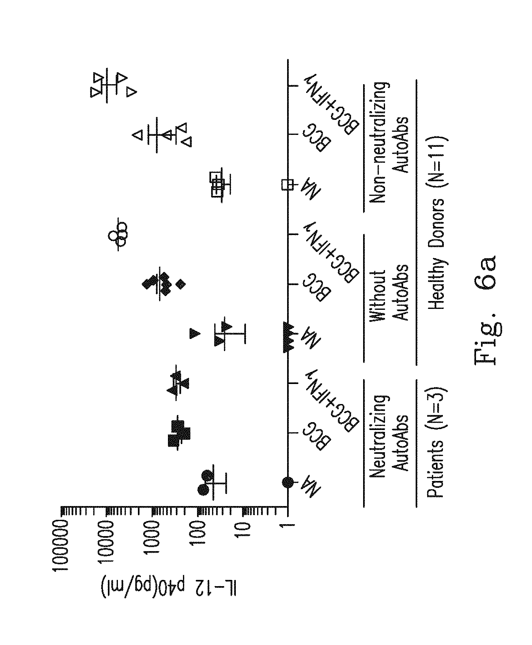
FIGS. 6a-6b illustrate that IL-12 production levels and IFN.sub.r production levels were measured by ELISA;
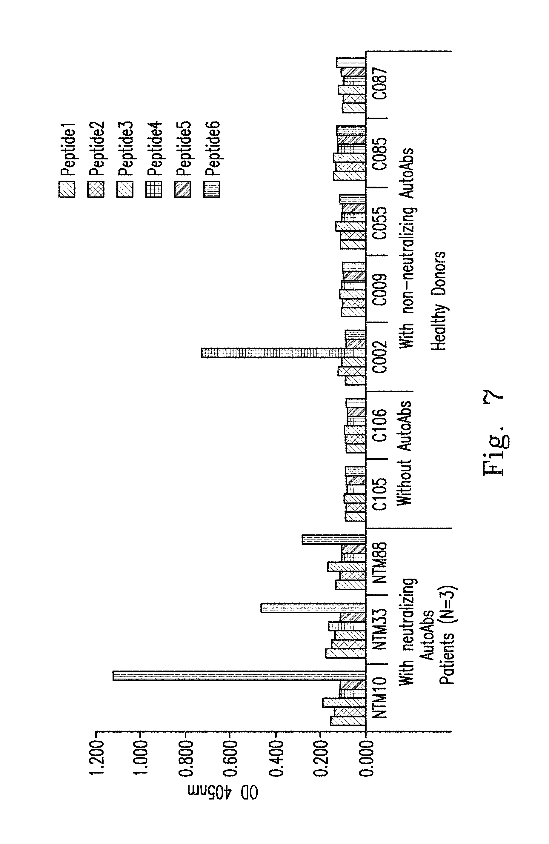
FIG. 7 illustrates the result of epitope mapping;
FIG. 8 illustrates the result of Western blot analysis conducted on various recombinant interferons;
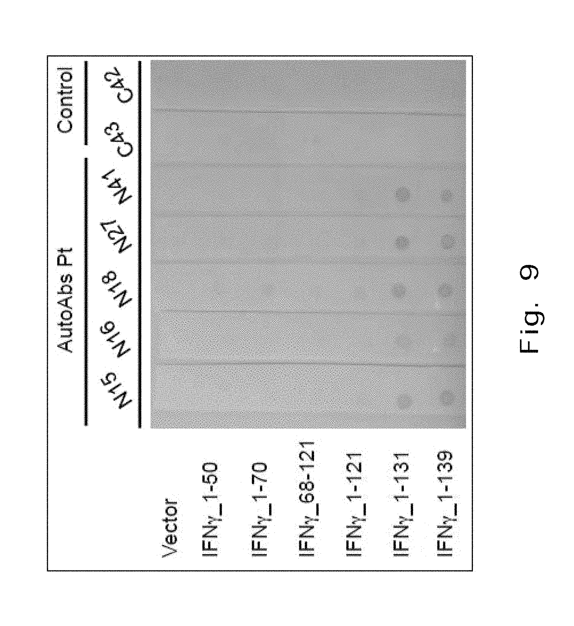
FIG. 9 illustrates the results of probing plasma sampled from IFN.sub.r AutoAbs patients and healthy controls;
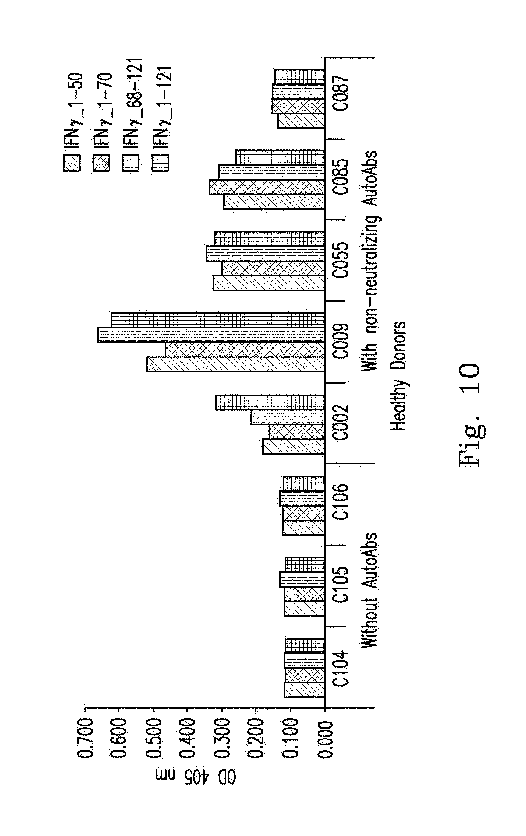
FIG. 10 illustrates the results of protein mapping;
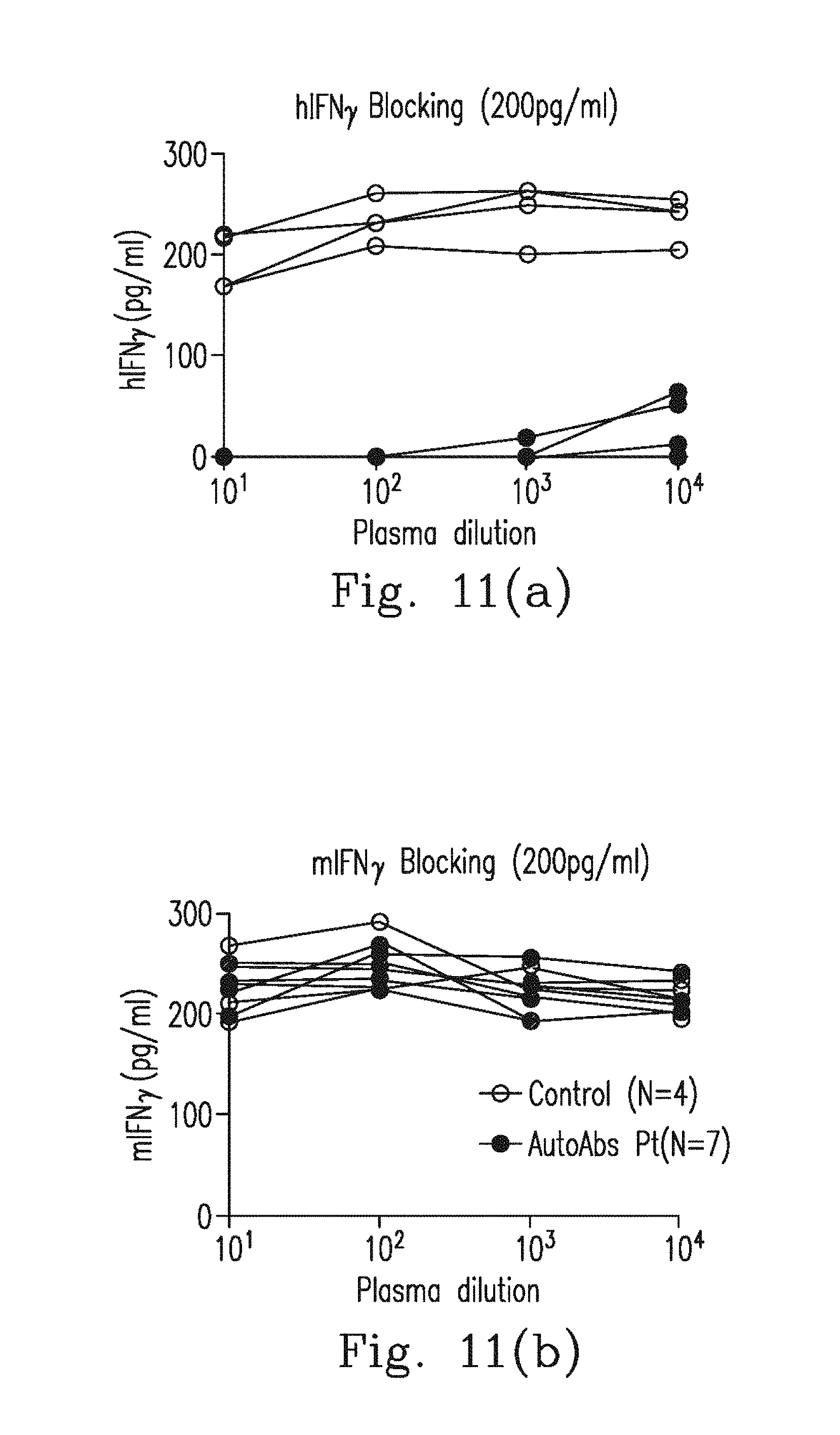
FIGS. 11a-11b illustrate the results of plasma interacting with hIFN.sub.r and mIFN.sub.r;
FIG. 12 illustrates the results of Western blot analysis conducted on different various recombinant Noc2 proteins; and
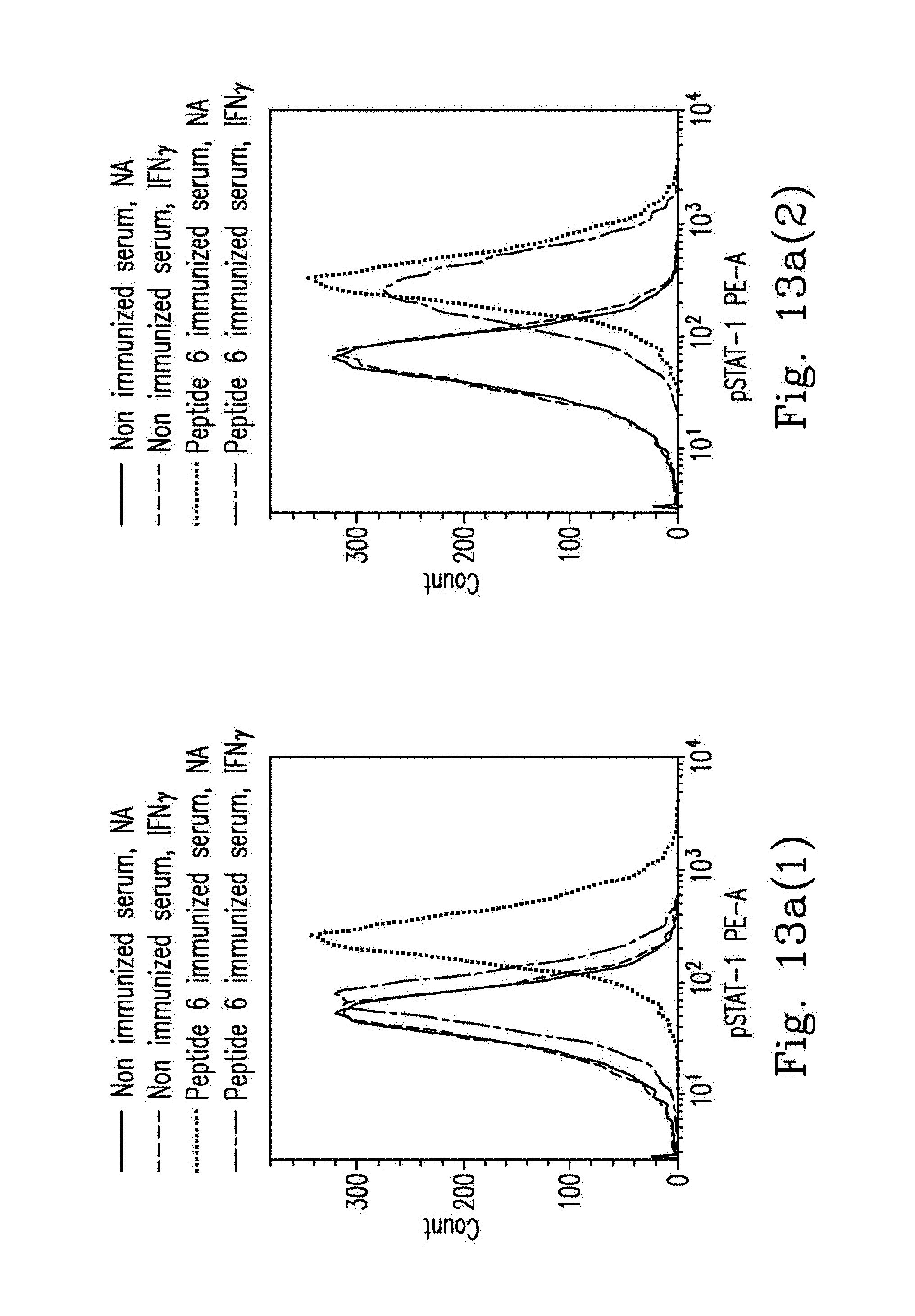
FIGS. 13a-13b illustrate the results of p-STAT-1 expression levels and HLA-DR expression levels.
DETAILED DESCRIPTION OF THE PREFERRED EMBODIMENTS
The present invention will now be described more specifically with reference to the following embodiments. It is to be noted that the following descriptions of preferred embodiments of this invention are presented herein for the purposes of illustration and description only; they are not intended to be exhaustive or to be limited to the precise form disclosed.
[Patients and Definitions]
All participants were adults (age >20 years) and were followed regularly at medical centers in Taiwan. Disseminated mycobacterial infection was diagnosed if patients presented 2 or more noncontiguous places and positive blood or BM cultures. The diagnosis of pulmonary NTM infection was based on the criteria proposed by the American Thoracic Society and Infectious Diseases Society of America. The study was approved by the Institutional Review Board of China Medical University Hospital (DMR98-IRB-261) and Chang Gung Memorial Hospital (103-2861C), and informed written consent was obtained from all of the patients in accordance with the Declaration of Helsinki.
[Reagents]
The inventor used the following monoclonal antibodies: anti-human IFN.sub.r (clone EPR1108, Abcam), anti-V5 tag (clone SV5-Pk1, Abcam), FITC mouse anti-human CD14 (clone M5E2, BD Pharmingen), PE mouse anti-human Human Leukocyte Antigen-antigen D Related (HLA-DR)(clone G46-6, BD Pharmingen) and PE Mouse Anti-Stat1 (pY701) (clone 4a, BD Phosflow).
[Detection and Titration of IFN.sub.r Neutralizing Autoantibodies]
Blood plasma from patients and donors was serially diluted (10.sup.-1 to 10.sup.-6) and incubated with recombinant human IFN.sub.r at a final concentration of 200 pg/mL for 1 hour at 37.degree. C. The IFN.sub.r level was measured with a human IFN.sub.r ELISA kit from BD Biosciences according to the manufacturer's recommendations. All experiments were performed in duplicate.
[Peptide Synthesis]
All peptides were synthesized by Kelowna International Scientific Inc., Taiwan. The peptides were lyophilized, and the purity and mass were assessed by high-performance liquid chromatography and mass spectrometry, respectively.
[Epitope Mapping]
Clear polystyrene 96-well flat bottom plates (Nunc) were coated with 100 .mu.L of peptide solution (5 .mu.g/mL) with b-carbonate buffer (pH 9.6) at 4.degree. C. overnight. After blocking the remaining binding sites with 5% human normal serum albumin (Aventis) in PBS, the plates were incubated at 37.degree. C. for 1 hour. Afterward, the plates were washed 3 times with 0.5% Tween 20 in PBS.
Blood plasma samples were added at a 1:100 dilution, in PBS containing 5% human normal serum albumin. Following incubation at room temperature for 2 hours and being washed 3 times, goat anti-human IgG Fc.sub.r fragment specific conjugated with alkaline phosphate (Jackson ImmunoResearch Laboratories), was added at a 1:5000 dilution, and the plates were kept for 1 hour at room temperature. Finally, after being washed 5 times, 100 .mu.L of p-Nitrophenyl Phosphate (pNPP) was added and the reaction was read at 405 nm by a VICTOR X3 Multilabel Plate Reader (PerkinElmer) after 30 minutes of incubation at 37.degree. C.
[Competition Enzyme-Linked Immunosorbent Assay (ELISA)]
To evaluate the specificity of the reaction, a competition ELISA experiment was performed. Each plasma sample (1:100 dilutions) was pre-incubated with different concentrations (5-30 .mu.g/mL) of the competition peptides overnight at 4.degree. C. Subsequently, all plasma and different concentrations competition peptides mixture samples were examined following the epitope mapping protocol. Peptide 6 was coated with b-carbonate buffer (pH 9.6) at 4.degree. C. overnight. After blocking at 37.degree. C. for 1 hour, all pre-incubated plasma and different concentrations competition peptides mixture samples were added in duplicate. Following 2 hours of room temperature incubation, goat anti-human IgG Fc.sub.r fragment specific conjugated with alkaline phosphate, was added. Finally, 100 .mu.L of pNPP was added and the reaction was read at 405 nm by a VICTOR X3 Multilabel Plate Reader (PerkinElmer) after 30 minutes of incubation at 37.degree. C.
[Prediction of B Cell Epitopes]
The prediction of B cell epitopes was carried out using BepiPred Linear epitope Prediction and Emini Surface Accessibility Prediction. The software takes a single sequence in FASTA format as an input. BepiPred predicts the location of linear B-cell epitopes using a combination of a hidden Markov model and a propensity scale method. In Emini Surface Accessibility Prediction, the calculation was based on surface accessibility scale of a product instead of an addition within the window. The accessibility profile was obtained using a special formula.
[Generation of Recombinant Human IFN.sub.r]
The inventor amplified the IFN.sub.r from base 196 to 624 of the open reading frame (ORF) by polymerase chain reaction (PCR) and cloned it into pMT/BiP/V5-His (Invitrogen) for Drosophila Schneider 2 (S2) cells expression. Recombinant IFN.sub.r was further purified by V5 tagged protein purification kit (MBL). The mutated recombinant IFN.sub.r was performed by QuickChange Site-Directed Mutagenesis kit (Stratagene). The inventor confirmed the sequences and cloning sites of all constructs as well as the size and immunoreactivity of the recombinant IFN.sub.r by Western Blot.
[Generation of Recombinant Aspergillus Noc2]
The inventor amplified the Noc2 from base 1 to 2361 of the ORF by PCR and cloned it into pMT/BiP/V5-His (Invitrogen) for S2 cells expression. Recombinant Noc2 were further purified by a V5 tagged protein purification kit (MBL). Then, the sequences and cloning sites of all constructs were confirmed as well as the size and immunoreactivity of recombinant Noc2 by Western Blot.
[p-STAT1 Intracellular Stain]
A total of 10.sup.6 PBMCs in 200 .mu.L RPMI-1640 with 10% FBS and 1% penicillin/streptomycin was used. Cells were then stimulated with 500 IU IFN.sub.r, IFN.sub.r.sub._1-131 or EE-IFN.sub.r.quadrature. in 160 .mu.L RPMI-1640 pre-incubated with 40 .mu.L of normal plasma or patient plasma for 10 min at room temperature. After 30 minutes of stimulation in a 37.degree. C. incubator, monocytes were identified by FITC-CD14 (BD Pharmingen) surface staining. Fixed by adding 250 .mu.L of FASC lysing solution (BD Pharmingen) and incubated at room temperature for 15 minutes in the dark, followed by two washes with PBS. To permeabilize the cells, 500 .mu.l of ice-cold absolute methanol was added to each tube and incubated for 15 minutes on ice in the dark, followed by two washes with PBS. Next, PE-phospho-STAT1 (pY701) antibody (BD Pharmingen) was added, followed by 30 minutes incubation on ice in the dark. Again, cells were washed with 2 mL of PBS before resuspension in the 500 .mu.L of PBS. Data were collected with a FACSVerse flow cytometer (BD Biosciences) and analyzed using FACSuite software (BD Biosciences).
[Immunoblotting]
Recombinant IFN.sub.r.quadrature. (R&D Systems) and different truncated forms of recombinant IFN.sub.r.quadrature. were subjected to SDS-PAGE using a 10% gel under reducing conditions at 120 V for 2 hours. The proteins were transferred to a PVDF membrane (Invitrogen Life Technologies) at 250 mA for 2 hours. The membrane was blocked in 5% human normal serum albumin (Aventis) in TBS with 0.1% Tween 20 overnight at 4.degree. C. The membrane was incubated with a 1/100 dilution of patient or control blood plasma for 3 hours at room temperature. After being washed three times, the membrane was incubated in a 1/10,000 dilution of mouse anti-human IgG conjugated with horseradish peroxidase (Jackson ImmunoResearch Laboratories) for 1 hour at room temperature. After being washed three times, the membrane was developed with ECL (Merck Millipore).
[HLA-DR Expression]
A total of 10.sup.6 PBMCs in 200 .mu.L RPMI-1640 with 10% FBS and 1% penicillin/streptomycin was used. Cells were then stimulated with 500 IU IFN.sub.r, IFN.sub.r.sub._1-131 or EE-IFN.sub.r in 160 .mu.L RPMI-1640 pre-incubated with 40 .mu.L of normal blood plasma or patient blood plasma for 10 min at room temperature. After 24 hours of stimulation in a 37.degree. C. incubator, monocytes were identified by FITC-CD14 (BD Pharmingen) surface staining. Next, PE-HLA-DR antibody (BD Pharmingen) was added followed by 30 minutes of incubation on ice in the dark. Cells were again washed with 2 ml of PBS before resuspension in 500 .mu.l of PBS. Data were collected with a FACSVerse flow cytometer (BD Biosciences) and analyzed using FACSuite software (BD Biosciences).
Immunization with Noc2 or IFN.sub.r Peptide
The inventor immunized WKY/NcrlNarl rats with 0.25 mg Noc2 or IFN.sub.r.quadrature. peptide (Noc2: CTPKTGKRKRSEQ (SEQ NO: 31); IFN.sub.r: CAAKTGKRKRSQM (SEQ NO: 32)) conjugated with ovalbumin (OVA) and gave a booster dose every two weeks after the initial injection (day 0). After 70 days, the inventor checked the antibody production level by dot blot or ELISA at each injection. All rats were sacrificed ten days after the tenth injection (day 136) and whole blood was collected. After collection of the whole blood, the blood was allowed to clot by leaving it undisturbed at room temperature 15 minutes. Clots were removed by centrifuging at 1500 g for 10 minutes in a refrigerated centrifuge. Sera were aliquoted and stored at -20.degree. C. for further use.
[Experimental Data]
Please refer to FIGS. 1a-1h, which are related to AutoAbs to IFN.sub.r recognized C-terminal region of IFN.sub.r.
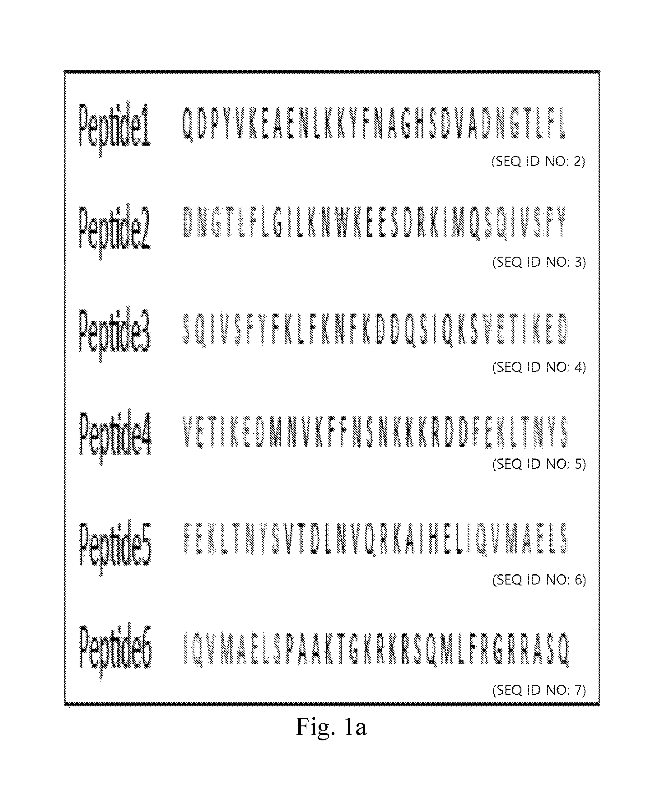
FIG. 1a demonstrates amino acid sequences of human IFN.sub.r 30-mer overlapping synthetic peptides.
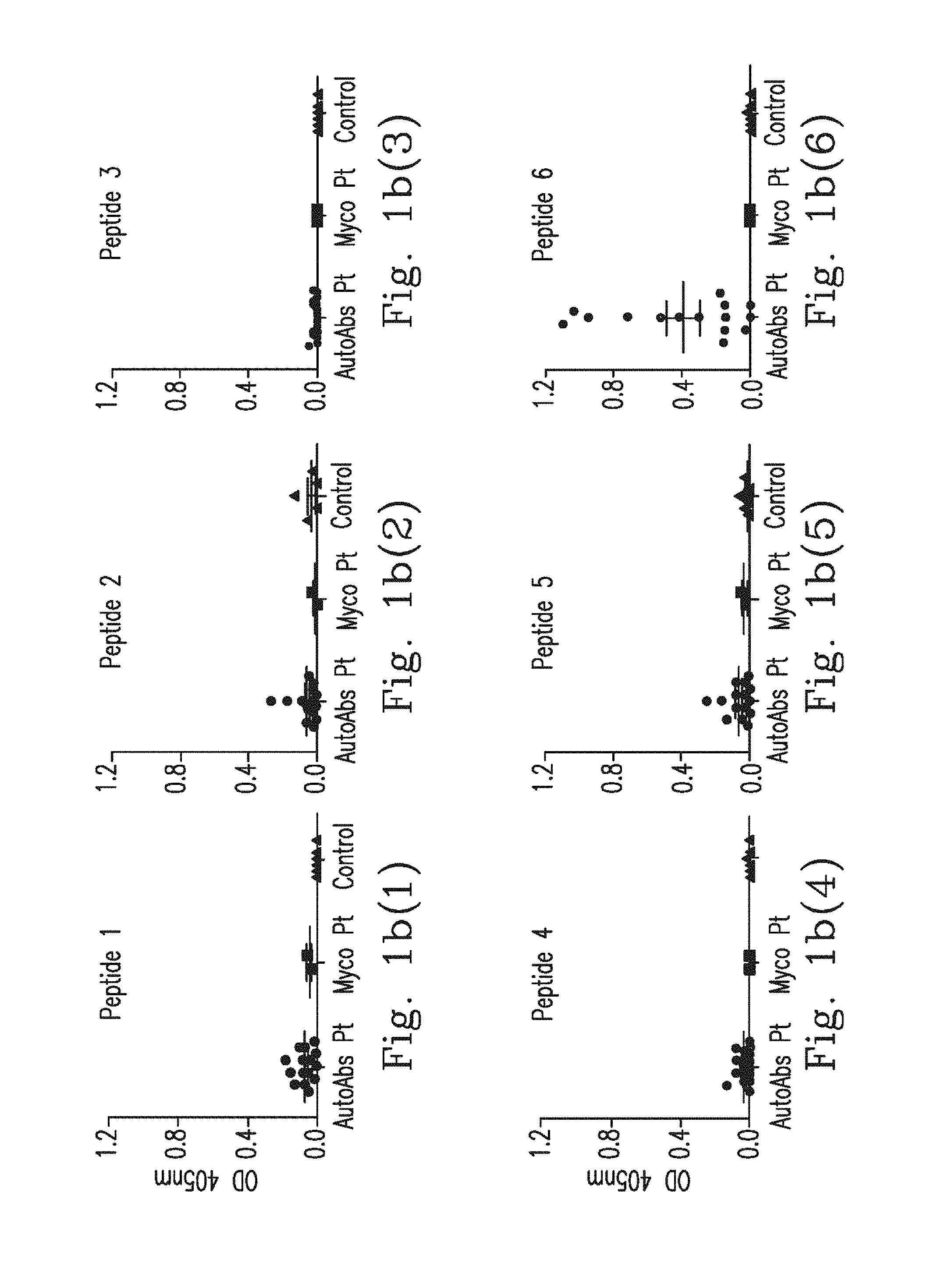
FIG. 1b(1)-FIG. 1b(6) demonstrate epitope mapping for determining serum binding affinity of the synthetic peptide to IFN.sub.r, represented as the mean optical density (OD) at 405 nm; plasma sample from IFN.sub.r AutoAbs patients (n=15), Mycobacteria infected patients (n=2) and healthy controls (n=6).
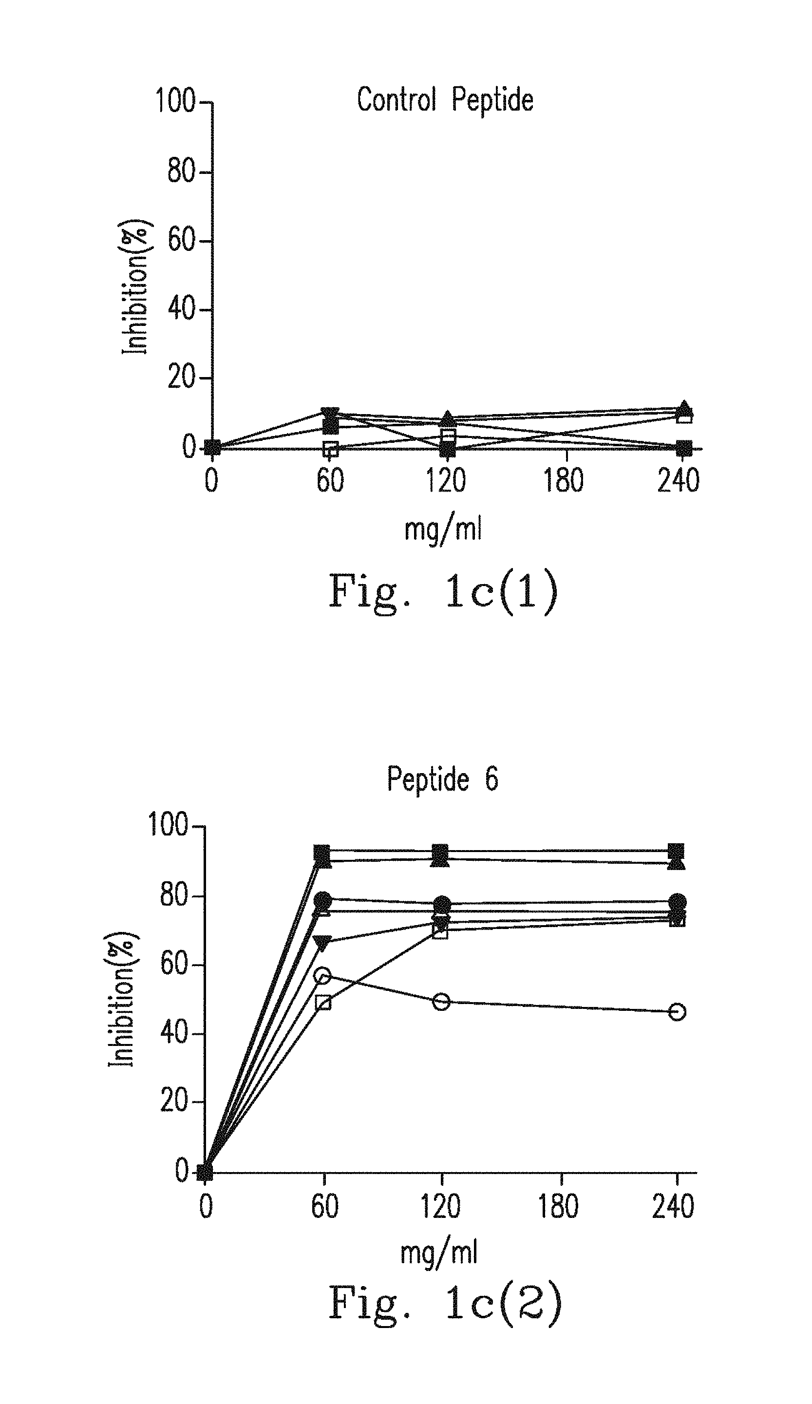
FIG. 1c(1)-FIG. 1c(2) demonstrate the inhibition using ELISA, wherein each plasma sample from IFN.sub.r AutoAbs patients was pre-incubated with different concentrations of a control peptide or peptide 6, and then all plasma dilutions were examined by ELISA for reactivity against peptide 6.
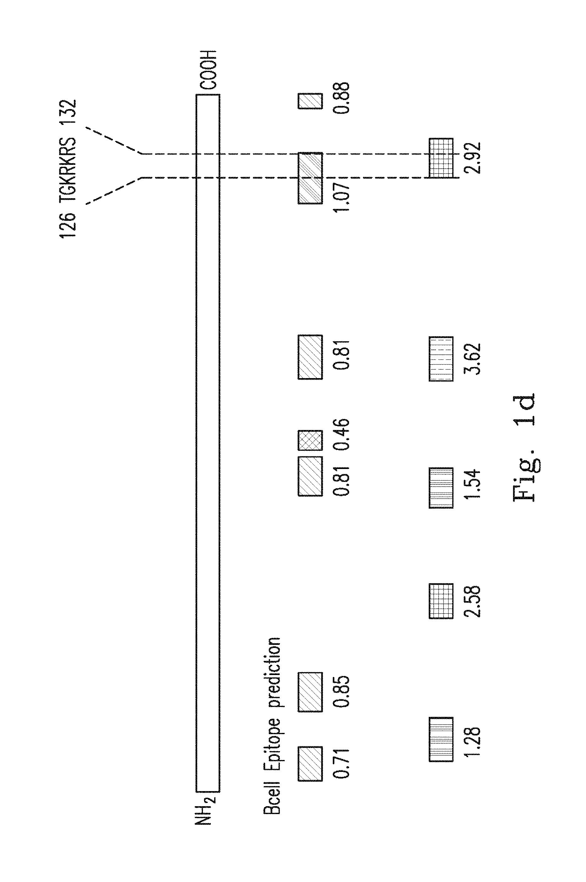
FIG. 1d demonstrates a schematic diagram of IFN.sub.r, and the prediction scores for linear B cell epitopes and surface acceptability regions. Each green bar represents a predicted B cell epitope, and each red bar represents a predicted surface acceptable region. The prediction scores represent the average scores for all amino acids within the region with prediction values above the cut-offs chosen for significance. The bar color intensities are proportional to the prediction scores found.
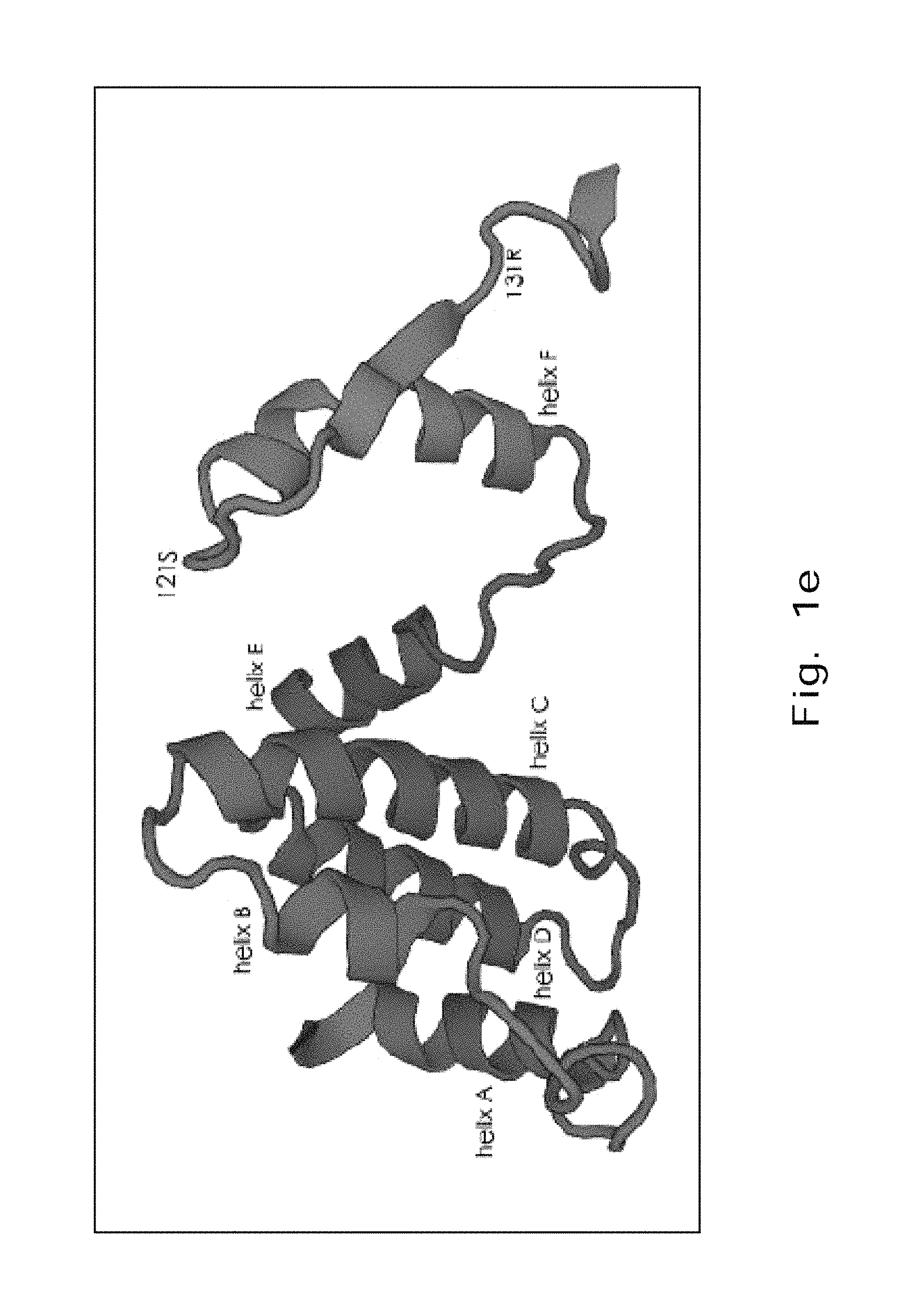
FIG. 1e demonstrates a three-dimensional projection of the structure of IFN.sub.r. The epitope, amino acids 121-131 of IFN.sub.r are located at the C-terminal and extend into the solvent.
FIG. 1f demonstrates a graphical representation of the different length of truncated recombinant IFN.sub.r proteins.
FIG. 1g(1)-FIG. 1g(6) demonstrate protein mapping used to determine plasma binding affinity of the recombinant proteins to IFN.sub.r, represented as the mean optical density (OD) at 405 nm; plasma sample from IFN.sub.r AutoAbs patients (n=16), Mycobacteria infected patients (n=2) and healthy controls (n=3).
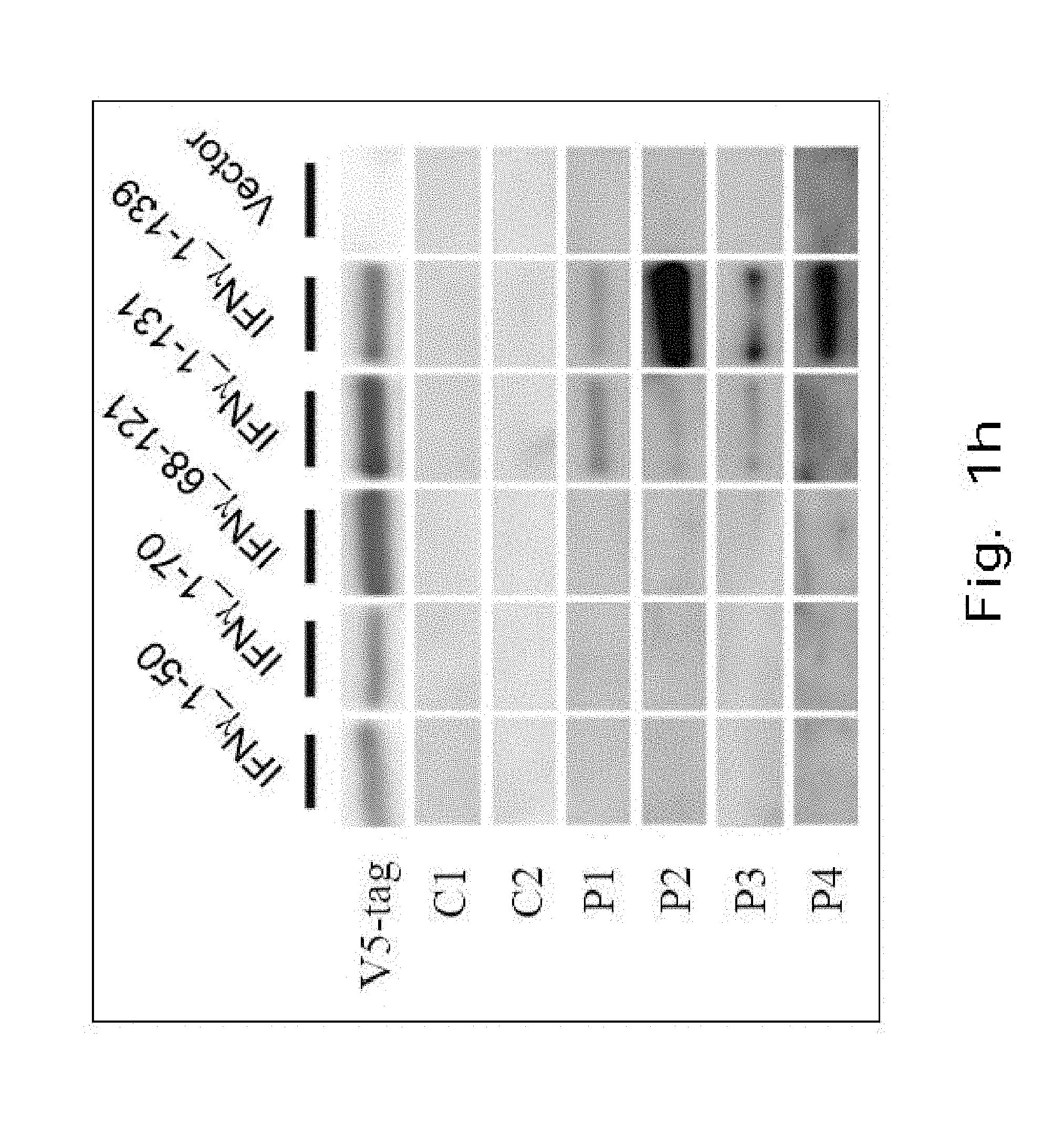
FIG. 1h demonstrates a Western blot showing binding ability of blood plasma from IFN.sub.r AutoAbs patients (n=4, P1-4) and healthy controls (n=2, C1-2) to different truncated forms of IFN.sub.r.
Please refer to Table 1, which demonstrates multiple amino acid sequence alignments of IFN.sub.r in different species and EE-IFN.sub.r (SEQ NO: 15-21). The conserved residues in the other species are boxed. The sequence of EE-IFN.sub.r protein, which has substituted human P.sub.121-127 Ser-Pro-Ala-Ala-Lys-Thr-Gly (SPAAKTG) (SEQ ID NO: 39) with homologous murine Leu-Pro-Glu-Ser-Ser-Leu-Arg (LPESSLR) addressing SEQ NO: 1 are in bold.
TABLE-US-00001 TABLE 1 ##STR00001##
In other embodiments, it is possible to apply homologous substitutes with various peptide lengths to replace the sequence in human P.sub.121-127. For example, a homologous substitute comprises at least one amino acid capable of fulfilling at least one element loss in the epitope. For another example, a mutation procedure is applied in at least one element in the epitope.
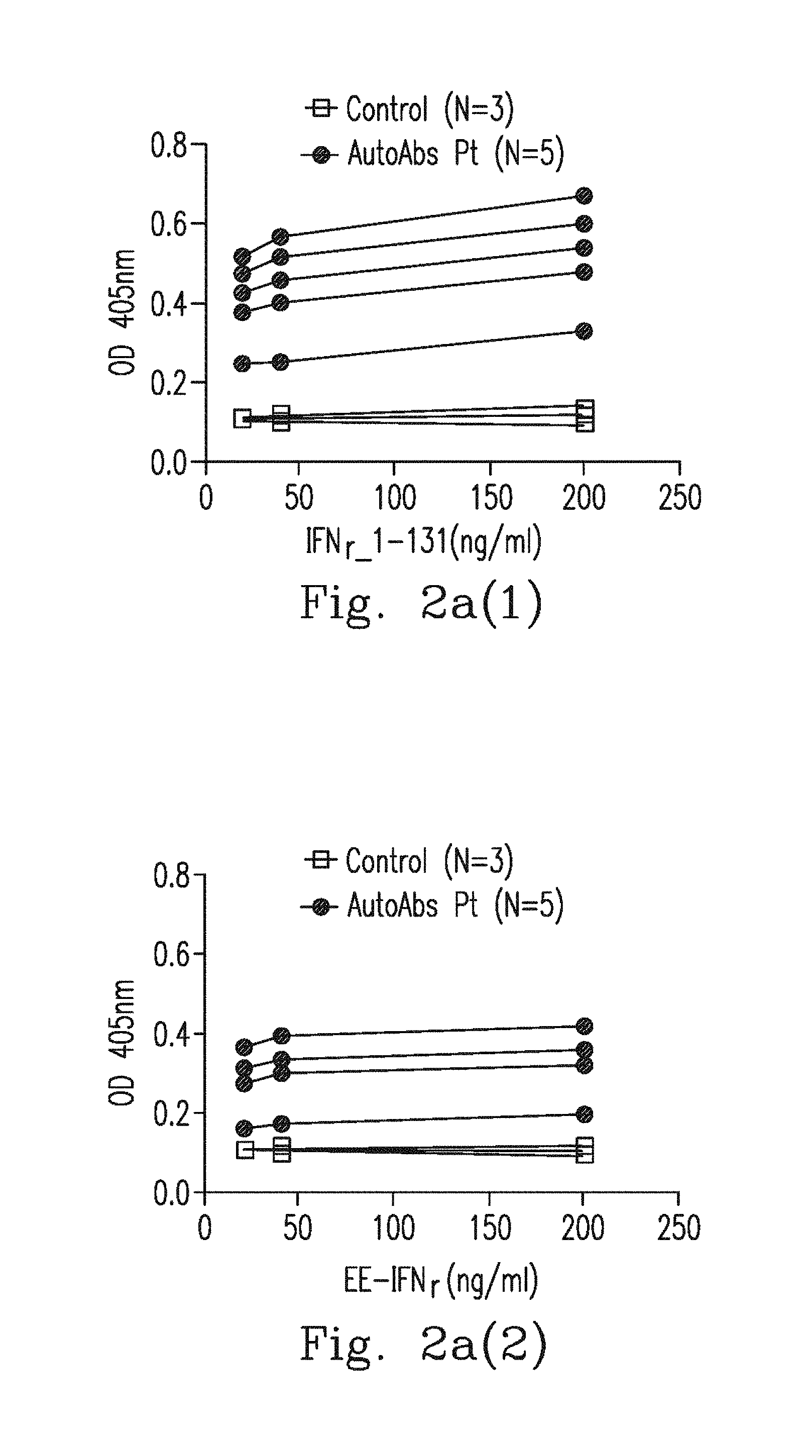
FIG. 2a(1)-FIG. 2a(2) demonstrate the affinity of anti-IFN.sub.r AutoAbs to IFN.sub.r.sub._1-131 and EE-IFN.sub.r.quadrature..quadrature. by ELISA. Anti-IFN.sub.r AutoAbs from 5 patients' blood plasma was bound to IFN.sub.r.sub._1-131 in a dose dependent manner but not to EE-IFN.sub.r represented as the mean optical density (OD) at 405 nm.
FIG. 2b demonstrates a Western blot analysis showing the binding of blood plasma from Anti-IFN.sub.r.quadrature.AutoAbs patients (n=4, P1-4) and healthy controls (n=2, C1-2) to IFN.sub.r1-131 and EE-IFN.sub.r. Plasma from 3 healthy donors had no response to either IFN.sub.r1-131 or EE-IFN.sub.r.
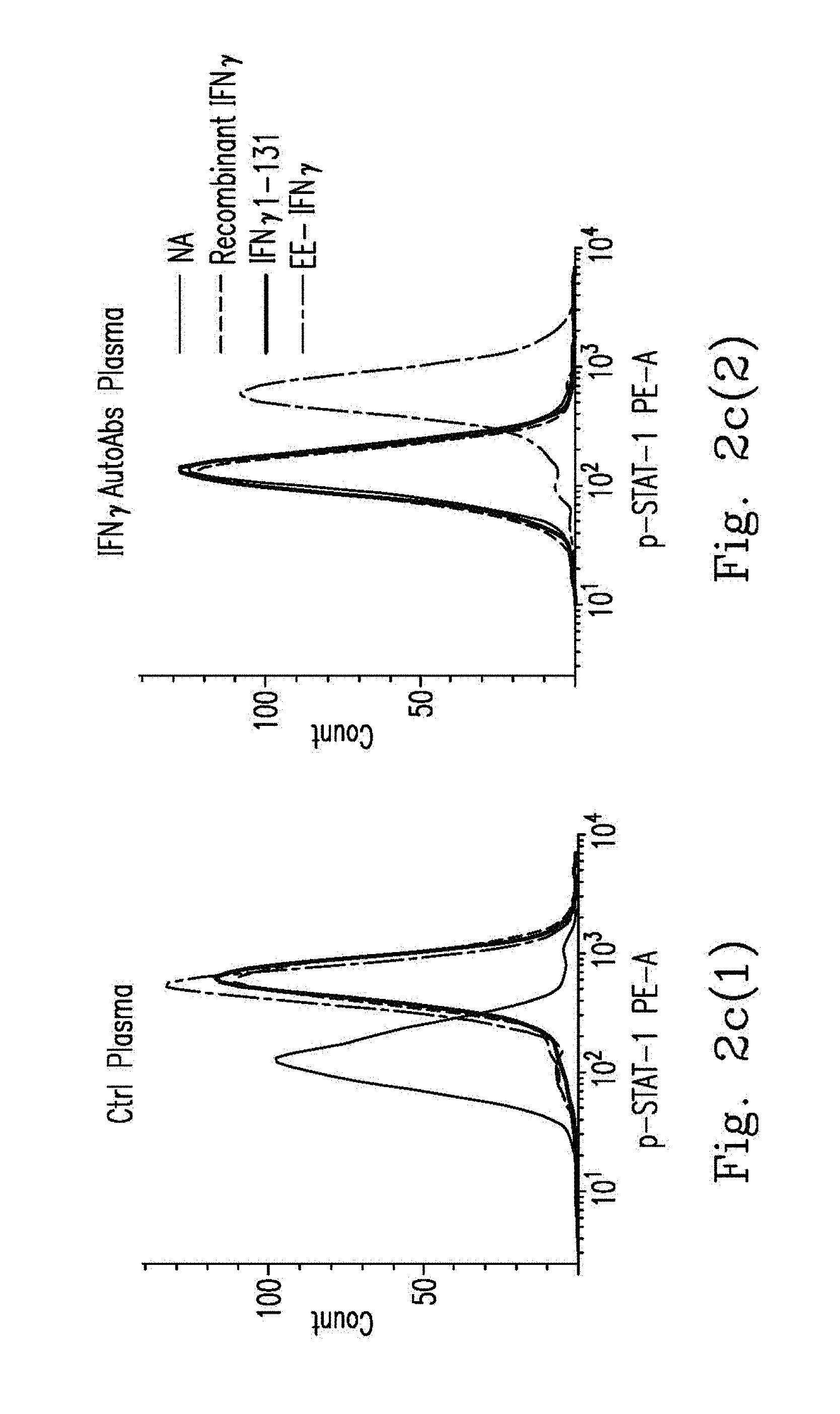
FIGS. 2c-2e demonstrate PBMCs from healthy controls that were pre-incubated with 20% Ctrl plasma (n=6) or anti-IFN.sub.r AutoAbs patients' plasma (n=6) and treated with WT IFN.sub.r, IFN.sub.r.sub._1-131 or EE-IFN.sub.r.quadrature..quadrature. After being gated on the CD14.sup.+, p-STAT-1 levels were determined by flow cytometry with the p-STAT-1 monoclonal antibody. Representative histograms (FIG. 2c(1)-FIG. 2c(2)) and MFI (median fluorescence intensity) (FIG. 2d) are shown.
FIG. 2e demonstrates that PBMCs from healthy controls were pre-incubated with 20% Ctrl plasma (n=4) or anti-IFN.sub.r AutoAbs patients' blood plasma (n=9) and stimulated with BCG BCG plus WT IFN.sub.r, BCG plus IFN.sub.r.sub._1431 or BCG plus EE-IFN.sub.r IL12p40 levels in the supernatant were measured by ELISA.
Table 2 demonstrates homologies in hIFN.sub.r peptides P.sub.125-133 to Aspergillus spp. Noc2 proteins. Amino acid numbers correspond to those of proteins published by The National Center for Biotechnology Information (NCBI). AA is an abbreviation for number of amino acids. And the proteins were sampled from Homo sapiens (SEQ NO: 22), Aspergillus terreus (SEQ NO: 23), Aspergillus fumigatus (SEQ NO: 24), Aspergillus flavus (SEQ NO: 25), Aspergillus nidulans (SEQ NO: 26), Aspergillus niger (SEQ NO: 27), Cryptococcus neoformans (SEQ NO: 28), Mycobacterium intracellulare (SEQ NO: 29) and Clostridium sp. MSTE9 (SEQ NO: 30).
TABLE-US-00002 TABLE 2 Accession Protein Species AA AA Positive code IFN.gamma. P.sub.125-133 Homo sapiens 125 AAKTGKRKRSQML 133 -- GI:56786138 (SEQ ID NO: 22) Ribosome assembly Aspergillus 105 TPKTGKRKRSEQQ 113 9/9 (100%) GI:115397505 protein Noc2 terreus (SEQ ID NO: 23) Ribosome assembly Aspergillus 100 TPKITGKRKRSEEQ 108 8/9 (89%) GI:159131733 protein Noc2 fumigatus (SEQ ID NO: 24) Ribosome assembly Aspergillus 102 TPKTGKRKRTEEQ 110 8/9 (89%) GI:238483989 protein Noc2 flavus (SEQ ID NO: 25) Ribosome assembly Aspergillus 103 SPKIGKRKRSETQ 111 8/9 (89%) GI:67517347 protein Noc2 nidulans (SEQ ID NO: 26) Ribosome assembly Aspergillus 102 TPKIGKRKRSDEQ 110 7/9 (78%) GI:317025642 protein Noc2 niger (SEQ ID NO: 27) Hypothetical Cryptococcus 623 LGSTGKRKRSSMG 631 7/9 (78%) GI:405119493 protein CNAG_05000 neoformans (SEQ ID NO: 28) Hypothetical Mycobacterium 124 NPAGGKRKRSQ-- 132 7/9 (78%) GI:379753578 protein OCO_15660 intracellulare (SEQ ID NO: 29) Diguanylate Clostridium 14 KEQSKKRKRSQMT 22 6/9 (67%) GI:496353461 cyclase sp. MSTE9 (SEQ ID NO: 30)
FIG. 3a(1)-FIG. 3a(2) demonstrate epitope cross reaction mapping used to determine blood plasma binding affinity of the synthetic peptide to Aspergillus Noc2, represented as the mean optical density (OD) at 405 nm; blood plasma sample from IFN.sub.r AutoAbs patients (n=9), Mycobacteria infected patients (n=3) and healthy controls (n=3).
FIG. 3b(1)-FIG. 3b(2) demonstrate inhibition of ELISA, where each plasma sample from IFN.sub.r AutoAbs patients (n=7) was pre-incubated with different concentrations of control peptide or Aspergillus Noc2 peptide, then all plasma dilutions were examined by ELISA for reactivity against peptide 6.
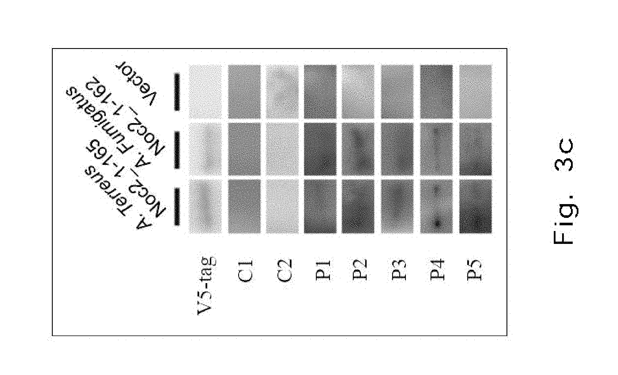
FIG. 3c demonstrates a Western blot analysis showing binding of plasma form Anti-IFN.sub.r AutoAbs patients (n=4, P1-4) and healthy controls (n=2, C1-2) truncated Aspergillus Terreus (Amino Acid: 1-165) and Aspergillus Fumigatus (Amino Acid: 1-162) recombinant Noc2 protein.
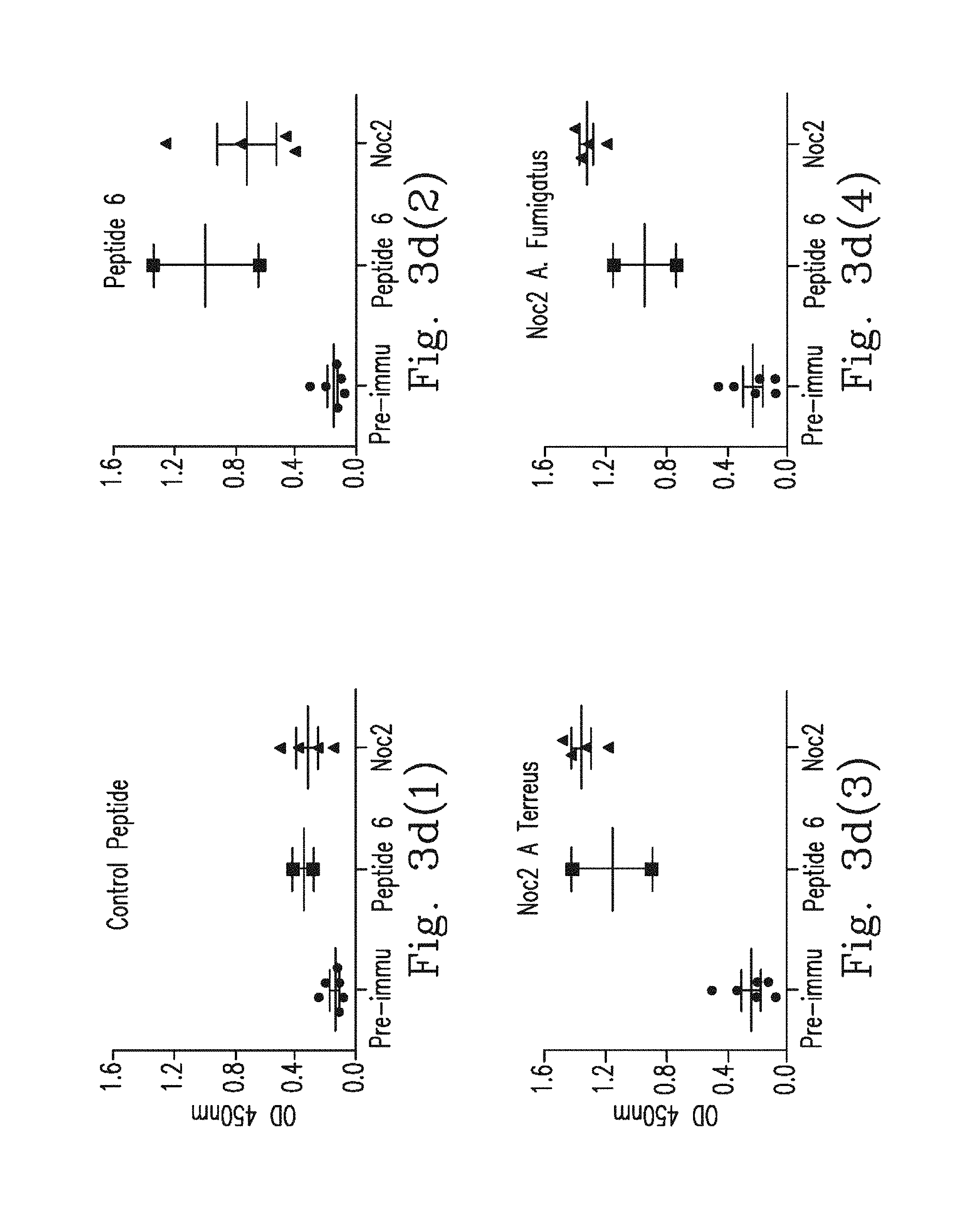
FIG. 3d(1)-FIG. 3d(4) demonstrate immunization with Noc2 peptide induced antibodies to IFN.sub.r. Blood sera from pre-immunized rats (n=6), Noc2 peptide-immunized rats (n=4) and Peptide 6-immunized rats (n=2) were mapped to control peptide, Peptide 6 and Noc2 peptide from Aspergillus terreus or Aspergillus fumigatus. This is represented as the mean optical density (OD) at 405 nm.
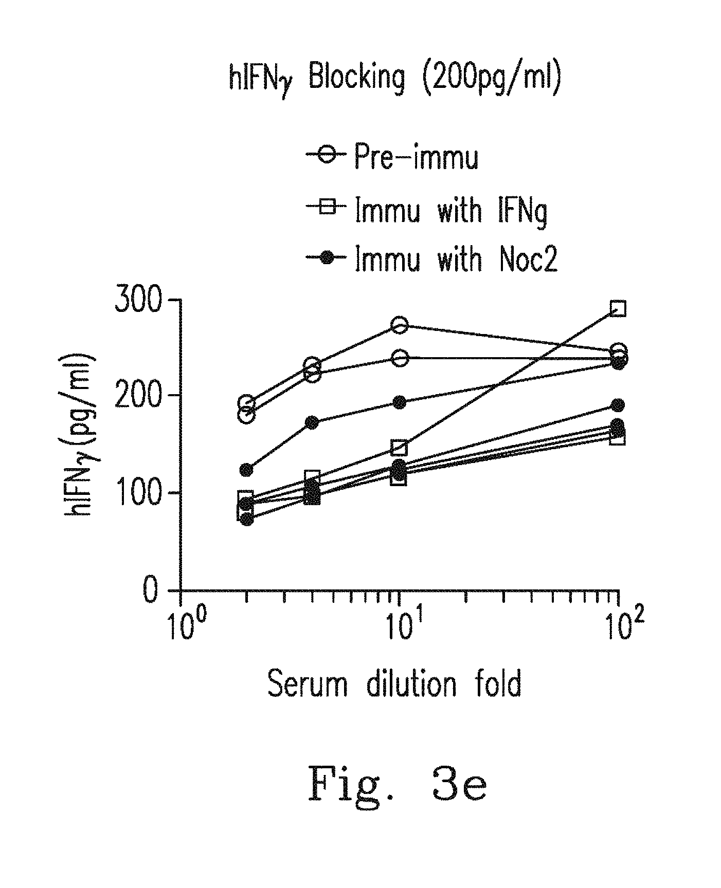
FIG. 3e demonstrates blood sera from pre-immunized rats (n=2), Noc2 peptide-immunized rats (n=4) and Peptide 6-immunized rats (n=2) were serially diluted and incubated with 200 pg/mL of human IFN.sub.r. Sera from pre-immunized rats showed no blocking activities for the detection of hIFN.sub.r. However, blood sera from Noc2 peptide-immunized rats (n=4) and Peptide 6-immunized rats (n=2) at dilutions of up to 1/10.sup.2 inhibited the detection of hIFN.sub.r.
FIGS. 3f-3g demonstrate that THP1 cells were pre-incubated with 50% blood sera from pre-immunized rats (n=2), Noc2 peptide-immunized rats (n=4) or Peptide 6-immunized rats (n=2) and treated with WT IFN.sub.r p-STAT-1 levels were determined by flow cytometry with the p-STAT-1 monoclonal antibody, MFI (median fluorescence intensity) are shown in FIG. 3f HLA-DR expression levels were determined by flow cytometry with the HLA-DR monoclonal antibody, and MFI are shown in FIG. 3g.
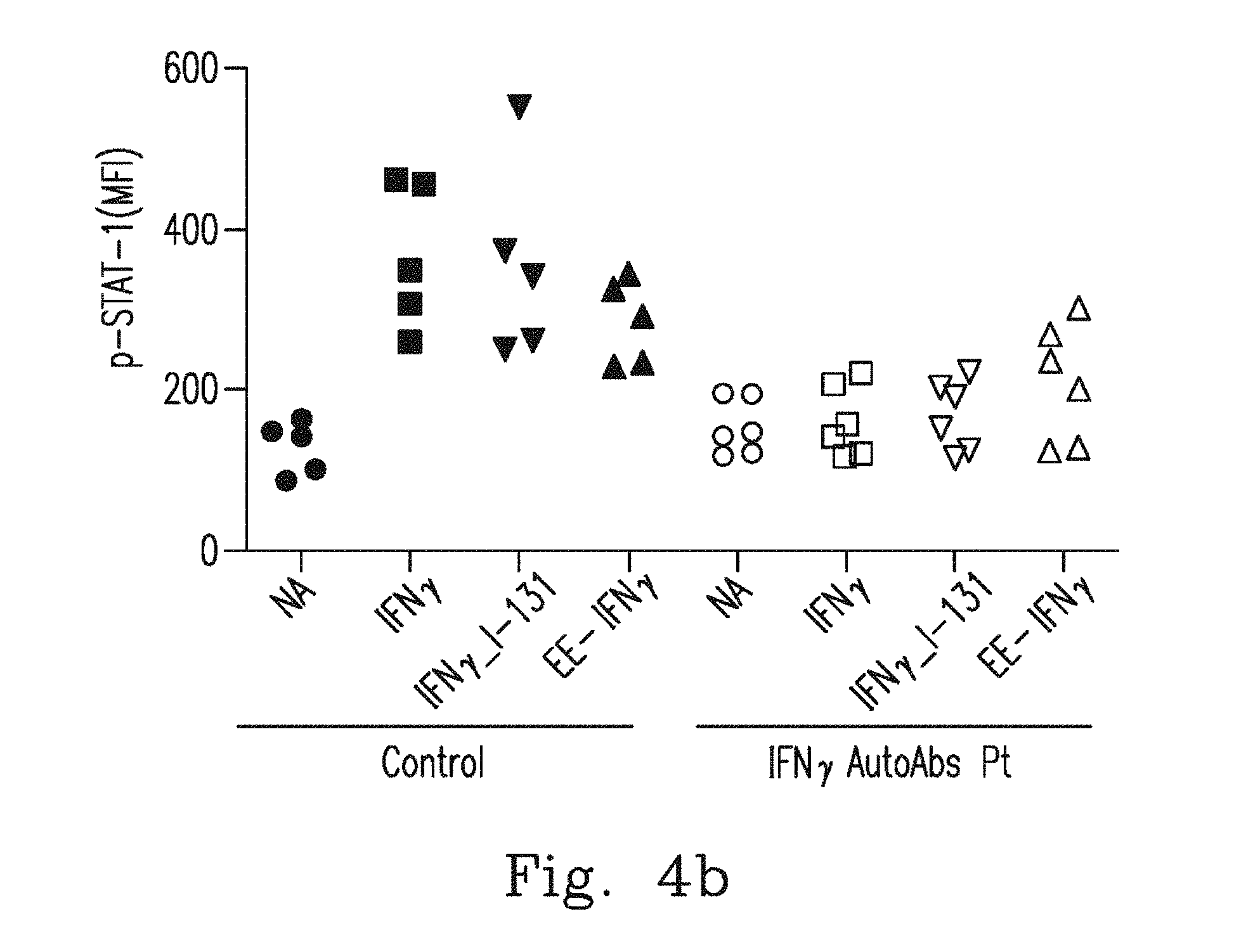
FIGS. 4a-4b demonstrate that PBMCs from healthy controls (n=5) or anti-IFN.sub.r AutoAbs patients (n=6) were pre-incubated with 20% autologous plasma and treated with WT IFN.sub.r, IFN.sub.r1-131 or EE-IFN.sub.r. After being gated on the CD14.sup.+, p-STAT-1 levels were determined by flow cytometry with the p-STAT-1 monoclonal antibody. Representative histograms (FIG. 4a(1)-FIG. 4a(2)) and MFI (median fluorescence intensity) (FIG. 4b) are shown.
FIG. 4c demonstrates that PBMCs from healthy controls (n=3) or anti-IFN.sub.r AutoAbs patients (n=6) were pre-incubated with 20% autologous blood plasma and stimulated with BCG BCG plus WT IFN.sub.r, BCG plus IFN.sub.r.sub._1-131 or BCG plus EE-IFN.sub.r IL12p40 levels in the supernatant were measured by ELISA.
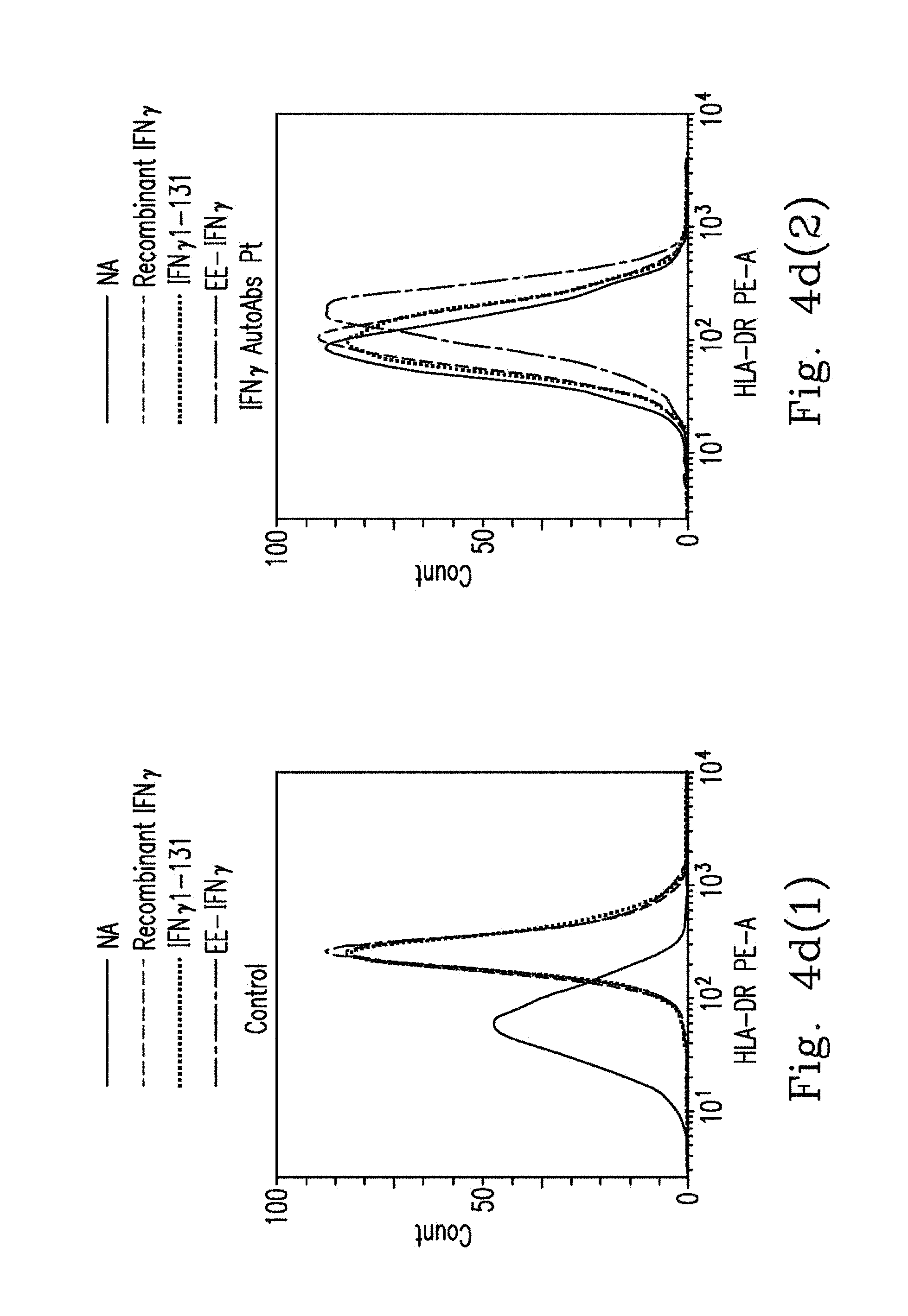
FIGS. 4d-4e demonstrate that PBMCs from healthy controls (n=5) or anti-IFN.sub.r AutoAbs patients (n=4) were pre-incubated with 20% autologous plasma and treated with WT IFN.sub.r, IFN.sub.r.sub._1-131 or EE-IFN.sub.r. After being gated on the CD14.sup.+, HLA-DR expression levels were determined by flow cytometry with the HLA-DR monoclonal antibody. Representative histograms (FIG. 4d(1)-FIG. 4d(2)) and MFI (median fluorescence intensity) (FIG. 4e) are shown.
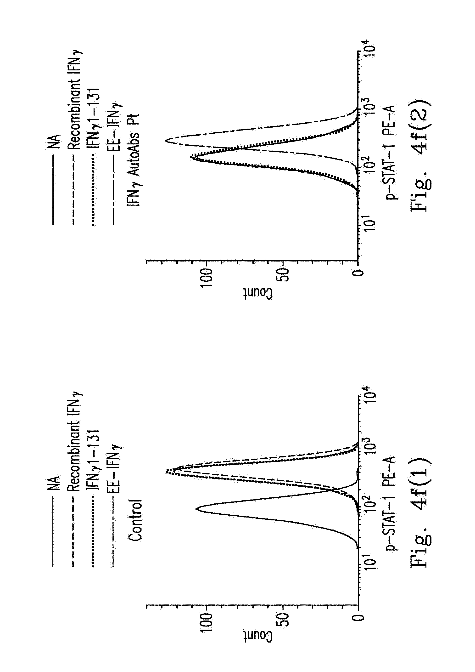
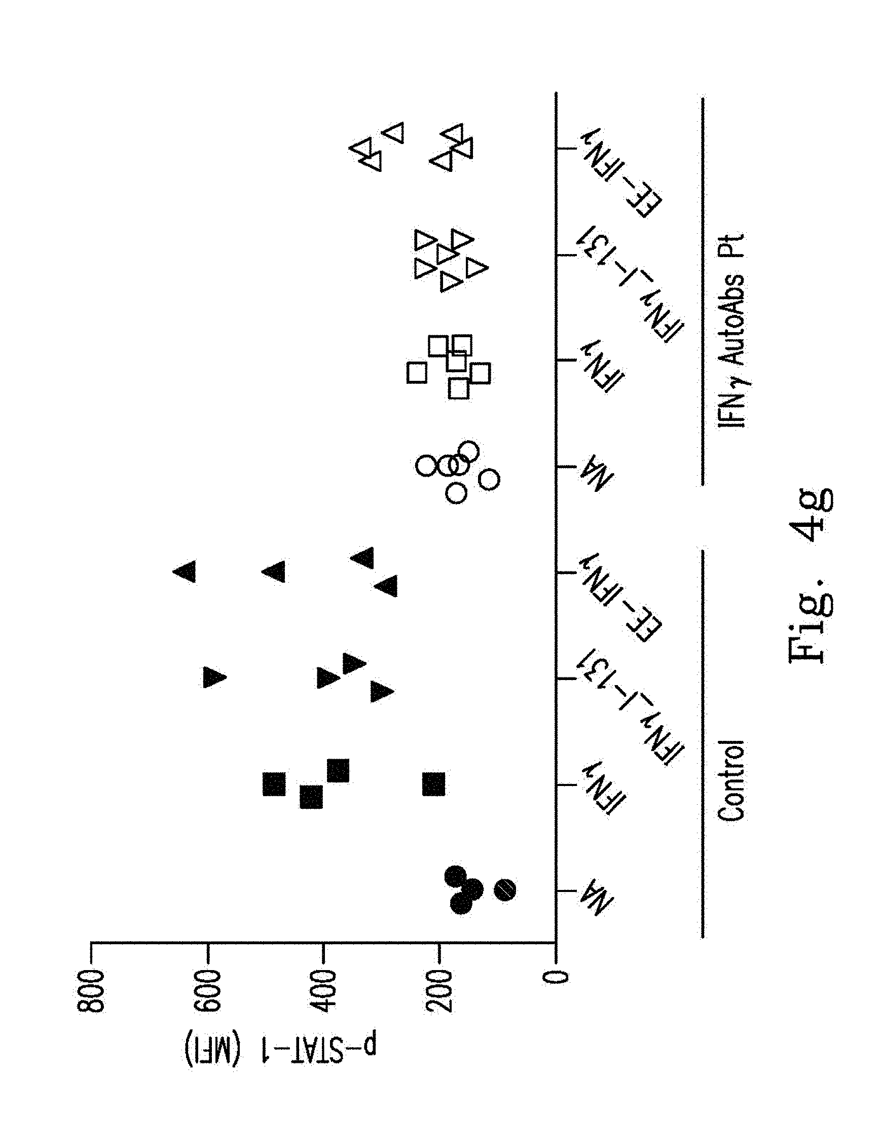
FIGS. 4f-4g demonstrate that whole blood from healthy controls (n=4) or anti-IFN.sub.r AutoAbs patients (n=6) was treated with WT IFN.sub.r, IFN.sub.r.sub._1-131 or EE-IFN.sub.r After being gated on the CD14.sup.+, p-STAT-1 levels were determined by flow cytometry with the p-STAT-1 monoclonal antibody. Representative histograms (FIG. 4f(1)-FIG. 4f(2)) and MFI (median fluorescence intensity) (FIG. 4g) are shown.
FIG. 4h demonstrates that whole blood from healthy controls (n=4) or anti-IFN.sub.r AutoAbs patients (n=5) was stimulated with BCG BCG plus WT IFN.sub.r, BCG plus IFN.sub.r.sub._1-131 or BCG plus EE-IFN.sub.r IL12p40 levels in the supernatant were measured by ELISA.
FIG. 5 demonstrates that direct ELIAS was used to detect AutoAbs against IFN.sub.r. Represented as the mean optical density (OD) at 405 nm; blood plasma samples from IFN.sub.r AutoAbs patients (n=2), and healthy controls (n=65) are shown.
FIG. 6a demonstrates that IL-12 production levels were measured by ELISA after non-activated, BCG activated or BCG plus IFN.sub.r activated in whole blood from three groups individuals were tested. IFN.sub.r AutoAbs patients (n=3), healthy controls without AutoAbs (n=7), and healthy controls with non-neutralizing IFN.sub.r AutoAbs (n=4) are shown.
FIG. 6b demonstrates that IFN.sub.r production levels were measured by ELISA after non-activated, BCG activated or BCG plus IL-12 activated in whole blood from three groups individuals were tested. IFN.sub.r AutoAbs patients (n=3), healthy controls without AutoAbs (n=7), and healthy controls with non-neutralizing IFN.sub.r AutoAbs (n=4) are shown.
FIG. 7 demonstrates epitope mapping used to determine plasma binding affinity of the synthetic peptide to IFN.sub.r, represented as the mean optical density (OD) at 405 nm including plasma samples from IFN.sub.r AutoAbs patients (n=3, P1-3), healthy controls without AutoAbs (n=2, C1-2), and healthy controls with non-neutralizing IFN.sub.r AutoAbs (n=5, N1-5).
FIG. 8 demonstrates a Western blot analysis showing the expression of different forms of recombinant IFN.sub.r by a V5-tag antibody.
FIG. 9 demonstrates blood plasma sampled from IFN.sub.r AutoAbs patients (n=5, P1-5) and healthy controls (n=2, C1-2) were probes for vector, IFN.sub.r.sub._1-50, IFN.sub.r.sub._1-70, IFN.sub.r.sub._68-121, IFN.sub.r.sub._1-121, IFN.sub.r.sub._1-131, and IFN.sub.r.sub._1-139 recombinant proteins.
FIG. 10 demonstrates protein mapping used to determine blood plasma binding affinity of the recombinant proteins to IFN.sub.r, represented as the mean optical density (OD) at 405 nm including blood plasma samples from healthy controls without AutoAbs (n=3, C1-3), and healthy controls with non-neutralizing IFN.sub.r AutoAbs (n=5, N1-5).
FIGS. 11a-11b demonstrate that seven patients' blood plasma was serially diluted and incubated with 200 pg/mL of human IFN.sub.r (hIFN.sub.r) or murine IFN.sub.r (mIFN.sub.r). The blood plasma from healthy donors showed no blocking activities for the detection of hIFN.sub.r or mIFN.sub.r. However, plasma from patients with AutoAbs against IFN.sub.r at dilutions of up to 1/10.sup.4 inhibited the detection of hIFN.sub.r but showed no blocking activity for the detection of mIFN.sub.r.
FIG. 12 demonstrates a Western blot analysis showing the expression of different forms of recombinant Noc2 by V5-tag antibody.
FIGS. 13a-13b demonstrate that THP1 cells were pre-incubated with 50% Sera from pre-immunized rats (n=2), Noc2 peptide-immunized rats (n=4) or Peptide 6-immunized rats (n=2) and treated with WT IFN.gamma. p-STAT-1, and levels were determined by flow cytometry with the p-STAT-1 monoclonal antibody, wherein representative histograms (FIG. 13a(1)-FIG. 13a(2)) are shown. HLA-DR expression levels were determined by flow cytometry with the HLA-DR monoclonal antibody, wherein representative histograms (FIG. 13b(1)-FIG. 13b(2)) are shown.
[Embodiment I] Anti-IFN.sub.r Autoantibodies Recognize a C-Terminal Region in IFN.sub.r Peptide
The inventor tried to identify the epitope recognized by anti-IFN.sub.r AutoAbs isolated from patients by peptide scan: Six 30-mer peptides, overlapping by a 7 or 8 amino acids and covering the entire coding sequence of human IFN.sub.r, were synthesized, and the amino acids sequences of peptides addressing SEQ NOS: 2 to 7 were listed (FIG. 1a). Epitope mapping assay was performed using plasma from three different patient groups: anti-IFN.sub.r AutoAbs patients with disseminated mycobacterial infections (n=15, group-1), patients with mycobacterial infections without anti-IFN.sub.r AutoAbs (n=2, group-2), and healthy controls (n=6, group-3). It was found that only peptide 6 was recognized in the blood plasma isolated from the 12 patients in group-1 (FIG. 1b(1)-FIG. 1b(6)). In contrast, no binding activity to these six peptides was observed in other two patient groups and this observation suggests that recognition of the peptide 6 was not due to a cross-reaction of anti-mycobacterial antibodies. Next, competition ELISA was used to confirm the specificity of the anti-IFN.sub.r AutoAbs to the peptide 6. Blood plasma from group-1 patients was pre-incubated with peptide 6, which led to competition for the binding with coated peptide 6 in a dose dependent manner, and no competition was observed when the control peptides (peptide 1-5) were used as competitors (FIG. 1c(1)-FIG. 1c(2)). These data suggest that the anti-IFN.sub.r AutoAbs from group-1 patients was recognized by one major region in the peptide 6 (P.sub.114-143).
In contrast to neutralizing AutoAbs to IFN.sub.r in patients with mycobacterial infections, non-neutralizing AutoAbs against IFN.sub.r had been reported in healthy individuals. Five donors were found with AutoAbs against IFN.sub.r out of 65 healthy controls by ELISA (FIG. 5). AutoAbs from these five healthy individuals showed a non-neutralizing effect on IFN.sub.r in terms of induction of IL-12 production. In contrast to neutralizing IFN.sub.r AutoAbs from patients, non-neutralizing AutoAbs from healthy donors did not recognize peptide 6 (FIG. 8). These data indicate that the IFN.gamma. AutoAbs from patients with disseminated mycobacterial infections, but not non-neutralizing IFN.gamma. AutoAbs, recognized a major region on peptide 6.
[Embodiment II] Predictions of B-Cell Epitopes Using Computer Modeling
For further confirmation of the precise region recognized by anti-IFN.sub.r AutoAbs, the inventor scanned the primary sequence of IFN.sub.r for possible B-cell linear epitopes and possible surface acceptability in silico. It was found several candidate epitopes addressing SEQ NO: 8-14 in the full length IFN.sub.r (FIG. 1d). Considering the higher prediction scores for screening features and located in the peptide 6, the epitope P.sub.121-131 SPAAKTGKRKR (SEQ ID NO: 33) was predicted in B-cell epitope prediction algorithms and P.sub.126-131 TGKRKR (SEQ ID NO: 37) was predicted in Surface Acceptability prediction algorithms. These results were consistent with peptide epitope mapping data. The critical epitope P.sub.121-131 SPAAKTGKRKR (SEQ ID NO: 33) that we identified is located on an unfolded sequence in the C-terminal region. It extends into the solvent and does not strongly interact with the remainder of the molecules (FIG. 1e).
[Embodiment III] Anti-IFN.sub.r Autoantibodies Recognize a C-Terminal Region in IFN.sub.r Protein
In order to investigate whether anti-IFN.sub.r AutoAbs from group-1 patients recognized the same region in the native IFN.sub.r protein, we generated various truncated IFN.sub.r by Schneider 2 cell expression system (FIG. 1f and FIG. 9). It was found that only full-length IFN.sub.r (IFN.sub.r.sub._1-144) and deletion clone IFN.sub.r.sub._1-131 were recognized by blood plasma isolated from the group-1 patients (FIG. 1g(1)-FIG. 1g(6)). In contrast, blood plasma isolated from the group-2 and group-3 patients did not recognize all the recombinant IFN.sub.r tested. In Western blot and dot blot assays, we also observed similar phenomena that only full-length IFN.sub.r.sub._1-144 (data not shown) and deletion clone IFN.sub.r.sub._1-139 and IFN.sub.r.sub._1-131 were recognized by anti-IFN.sub.r AutoAbs, but not other truncates IFN.sub.r (FIGS. 1h and 10). These data suggest that a major anti-IFN.sub.r.quadrature.AutoAbs-recognized B-cell epitope, P.sub.121-131 (SPAAKTGKRKR) (SEQ NO: 33) was located at the C-terminal of IFN.sub.r.
[Embodiment IV] EE-IFN.gamma.
Inside the epitope region we identified, a.a. 128-131 (KRKR) (SEQ ID NO: 38) (the last 4 resides in SEQ NO: 20) is crucial for the bioactivity of IFN.sub.r.quadrature. and conserved in most species.
Nevertheless, a.a. 121-127 is less conserved among different species (human: SPAAKTG (SEQ ID NO: 39) (the 6.sup.th-12.sup.th resides in SEQ NO: 20); murine: LPESSLR (SEQ NO: 1)) (Table 1).
It had been observed that blood plasma from randomly selected patients with anti-IFN.sub.r AutoAbs did not show blocking activity on the murine IFN.sub.r (FIG. 12), which suggests that SPAAKTG (the 6.sup.th-12.sup.th resides in SEQ NO: 20) is a necessary component for the binding of anti-IFN.sub.r AutoAbs. Previous studies demonstrated that the precise sequence in a.a. 121-127 was not critical for IFN.sub.r functions, and the deletion of some amino acids from the C-terminal of human IFN.sub.r resulted in increasing bioactivity.
Following these observations, the inventor generated an EE-IFN.sub.r protein by substituting the human P.sub.121-127 SPAAKTG sequence (SEQ ID NO: 39) (the 6.sup.th-12.sup.th resides in SEQ NO: 20) with murine LPESSLR sequence (SEQ NO: 1) in the IFN.sub.r.sub._1-131 protein (Table 1).
In other embodiments, EE-IFN.gamma. may be further combined with pharmaceutically acceptable excipients or carriers for clinical use.
The excipient in the present invention also refers to a pharmaceutically acceptable carrier or excipient, or a bio-available carrier or excipient, including a solvent, dispersant, coating, antibacterial or antifungal agent, preservative or slow absorber, which is a proper compound used to prepare a formulation in the prior art. Usually such a carrier or excipient does not have any activity for treatments itself. And the compound disclosed in the present invention cooperating with a pharmaceutically acceptable carrier or excipient is prepared as various formulations, and will not result in adverse drug reactions, allergies or other inappropriate responses after it is administered to animals or humans. Thus the compound in the present invention, cooperating with a pharmaceutically acceptable carrier or excipient, can be used in clinics and humans.
"Effective dose" means a dose which is enough to improve or prevent medical symptoms or biological manifestation. The effective dose may be also stated as a casting dose for use in diagnosis. Unless there is another description in the specification, "active compound" and "pharmaceutically active compound" are substitutes for each other and refer to a pharmaceutical, pharmacological or therapeutic substance as well as other effective material.
Using ELISA, we observed that the affinity of anti-IFN.sub.r AutoAbs to EE-IFN.sub.r was markedly decreased in comparison with wild type (WT) IFN.sub.r.sub._1-131 (FIG. 2a(1)-FIG. 2a(2)). With the aid of Western blot, again, we observed a similar different affinity to anti-IFN.sub.r AutoAbs between EE-IFN.sub.r and WT IFN.sub.r.sub._1-131 (FIG. 2b). These data suggest that the epitope of anti-IFN.sub.r AutoAbs is located in a.a. 121-131 and SPAAKTG was a necessary component for recognition.
[Embodiment V] Anti-IFN.sub.r AutoAbs to Epitope P.sub.121-131 have Neutralizing Activity on IFN.sub.r
The inventor aimed to know if anti-IFN.sub.r AutoAbs to epitope P.sub.121-131 played a critical role in the neutralizing effect in an anti-IFN.sub.r AutoAbs disease. A previous study showed that substitution of the homologous murine sequence between residue 121 and residue 127 resulted in only a small decrease in biological activity. The biological activity of EE-IFN.sub.r was tested by measuring the phosphorylation of signal transducers and activators of transcription 1 (p-STAT1) signaling assay by flow cytometry and interleukin-12 (IL-12) production through ELISA. Up-regulation of the p-STAT1 signaling was observed in the controls' peripheral blood mononuclear cells (PBMCs) that were activated with recombinant IFN.sub.r, WT IFN.sub.r.sub._1-131, or EE-IFN.sub.r in the presence of 20% control blood plasma, which demonstrated that EE-IFN.sub.r had similar bioactivity with WT IFN.sub.r.sub._1-131 in the p-STAT1 signaling (FIGS. 2c and 2d). In pre-incubated with 20% blood plasma with anti-IFN.sub.r AutoAbs from patients, only EE-IFN.sub.r, but not recombinant IFN.sub.r or WT IFN.sub.r.sub._1-131, could induce the phosphorylation of STAT1. These data strongly suggest that the anti-IFN.sub.r AutoAbs had neutralizing effects specific to epitope P.sub.121-131. IFN.sub.r could increase the synthesis and expression of HLA-DR in myeloid cells and other cells types. Therefore, the HLA-DR expression after IFN.sub.r stimulation was measured. Similarly, up-regulation of HLA-DR expression, a process activated by IFN.sub.r, in THP1 cells was observed while stimulated with EE-IFN.sub.r, even in the presence of anti-IFN.sub.r AutoAbs. Similar to STAT1 and HLA-DR signaling, the elevation of IL-12p40 expression couldn't be observed when pre-incubated with 20% patients' blood plasma and stimulated with BCG plus recombinant IFN.sub.r or IFN.sub.r.sub._1-131. In contrast, BCG plus EE-IFN.sub.r did restore IL-12p40 expression in the controls' PBMC pre-incubated with 20% patients' blood plasma (FIG. 2e). These data indicated epitope P.sub.121-131 was the only or major binding site for anti-IFN.sub.r AutoAbs in different patients, in terms of the blocking effect.
[Embodiment VI] IFN.sub.r Epitope P.sub.125-133 is Homologous to Aspergillus Conserved Protein
Recognition of a common epitope and shared HLA haplotypes among the patients with Anti-IFN.sub.r AutoAbs suggest that a common pathogenesis mechanism of molecular mimicry might be involved. To test this possibility, the inventor used P.sub.121-135 a.a. sequence (SPAAKTGKRKRSQML) (SEQ NO: 34) to search for similar peptides in the human and microbe database. No similarity between this region and other human protein was observed; however it was found that the P.sub.125-133 epitopes had 100% positives with amino acids 105-113 of the ribosome assembly protein Noc2 (with eight of the nine amino acids identical) from Aspergillus terreus, and this region in Noc2 was conserved among most Aspergillus spp. available in the database (Table 2).
[Embodiment VII] IFN.sub.r AutoAbs could Cross-React with Aspergillus Noc2
Next, the inventor investigated whether IFN.sub.r AutoAbs from patients could cross-react with Aspergillus Noc2. First, the antigenicity of the synthetic Aspergillus Noc2 peptide (KKDVTPKTGKRKRSEQQKDE (SEQ NO: 35)) was measured, which was evaluated by epitope mapping assay using blood plasma isolated from patients with anti-IFN.sub.r AutoAbs (FIG. 3a(1)-FIG. 3a(2)). This data show that the Aspergillus Noc2 peptide was recognized by blood plasma isolated from patients with anti-IFN.sub.r AutoAbs, but it did not react with blood plasma from mycobacterial infected patients without AutoAbs to IFN.sub.r (FIG. 3a(1)-FIG. 3a(2)). The homogeneity between human IFN.sub.r P.sub.125-133 and Aspergillus Noc2 in the conserved region P.sub.105-113 was further assessed by competition ELISA. Samples isolated from patients with anti-IFN.sub.r AutoAbs were pre-incubated with Aspergillus Noc2 peptide, and then reacted with the peptide 6. The data show that the Aspergillus Noc2 peptide would compete for IFN.sub.r AutoAbs binding with peptide 6 in a dose dependent manner and no competition was observed when the control peptides were used as competitors (FIG. 3b(1)-FIG. 3b(2)). These data suggest that the human IFN.sub.r AutoAbs could cross-react with the Aspergillus Noc2 peptide.
[Embodiment VIII] Autoantibodies to IFN.sub.r Recognized Aspergillus Noc2
To further investigate whether AutoAbs against IFN.sub.r could recognize the Aspergillus Noc2 in the protein level, we generated truncated recombinant Noc2 protein P.sub.1-165 and P.sub.1-162 from Aspergillus Terreus and Aspergillus Fumigatus. A Western blot assay was performed to examine the cross-reaction of anti-IFN.sub.r AutoAbs between human IFN.sub.r and Aspergillus Noc2. Utilizing blood plasma from patients with anti-IFN.sub.r AutoAbs as the primary antibodies, it was found that anti-IFN.sub.r AutoAbs recognize truncated recombinant Noc2 proteins from different Aspergillus species. In contrast, blood plasma from healthy controls could not bind to truncated recombinant Noc2 proteins or WT IFN.sub.r.sub._1-131 protein (FIG. 3c). These data provide evidence that anti-IFN.sub.r AutoAbs from patients could cross-reacts with the Aspergillus Noc2 protein.
[Embodiment IX] Noc2 Immunization Induces Antibodies to IFN.sub.r
In order to investigate the immunogenicity to induce the anti-IFN.sub.r AutoAbs to epitope P.sub.121-131 in vivo, the inventor immunized four rats with synthetic a.a. 103-114 Aspergillus Noc2 peptide conjugated with ovalbumin (OVA). These rats developed anti-Noc2 antibodies that cross-reacted with human IFN.sub.r (FIG. 3d(1)-FIG. 3d(4)). Two rats immunized with synthetic a.a.123-134 human IFN.sub.r peptide conjugated with OVA also developed antibodies to human IFN.sub.r, which could cross-react with Aspergillus Noc2 (FIG. 3d(1)-FIG. 3d(4)). Moreover, it was found that all sera from rats immunized with Noc2 peptide as well as those immunized with IFN.sub.r peptide can block human recombinant IFN.sub.r (FIG. 3e). These data indicate that antibodies to Aspergillus Noc2 could cross-react with human IFN.sub.r in vivo and vice versa.
Next, the inventor checked if these Noc2 antibodies had a neutralizing ability on the IFN.sub.r signaling pathway. Using a p-STAT1 signaling assay by flow cytometry, it was observed that p-STAT1 signaling was up-regulated in THP1 cells while pre-incubated with 50% sera from non-immunized rats and stimulated with recombinant IFN.sub.r. However, decreased up-regulation of p-STAT1 signaling in THP1 cells was observed while pre-incubated with 50% Noc2 or IFN.sub.r immunized rats sera was stimulated with recombinant IFN.sub.r (FIG. 3f). Similar results were also be observed in an HLA-DR expression assay (FIG. 3g). These data indicate that antibodies to Aspergillus Noc2 not only bind to human IFN.sub.r, but also inhibit IFN.sub.r downstream signaling. Thus, rats immunized with Noc2 had a break in the immunological tolerance to IFN.sub.r and anti-IFN.sub.r antibodies were evoked through the process of molecular mimicry.
[Embodiment X] EE-IFN.sub.r Application in Anti-IFN.sub.r AutoAbs Therapy Ex Vivo
The inventor showed that EE-IFN.sub.r could restore IFN.sub.r activated STAT-1 signaling and promote IL-12/HLA-DR expression in control PBMCs/THP1 cells with the presence of anti-IFN.sub.r AutoAbs (FIGS. 2d-2f). Consequently, we investigated the possibility of using EE-IFN.sub.r to restore the IFN.sub.r function in patients with anti-IFN.sub.r AutoAbs ex vivo. Patients' PBMCs were pre-incubated with 10% autologous blood plasma and stimulated with EE-IFN.sub.r or BCG plus EE-IFN.sub.r to measure p-STAT1 signaling or IL-12 production. It was observed that up-regulated p-STAT1 signaling cannot be observed when IFN.sub.r AutoAbs patients' PBMCs are pre-incubated with 10% autologous blood plasma and stimulated with recombinant IFN.sub.r and WT IFN.sub.r.sub._1-131. In contrast, EE-IFN.sub.r could restore p-STAT1 signaling in four of six IFN.sub.r AutoAbs patients' PBMCs pre-incubated with 10% autologous blood plasma (FIGS. 4a-4b). Moreover, BCG plus EE-IFN.sub.r could restore IL-12p40 production in three of six IFN.sub.r AutoAbs patients' PBMCs pre-incubated with 10% autologous blood plasma in contrast to BCG plus recombinant IFN.sub.r or plus WT IFN.sub.r.sub._1-131 (FIG. 4c). The similar phenomena was also be observed in an HLA-DR expression assay. It was found that up-regulated HLA-DR expression levels were not observed when IFN.sub.r AutoAbs patients' PBMCs was pre-incubated with 10% autologous blood plasma and stimulated with recombinant IFN.sub.r and WT IFN.sub.r.sub._1-131. In contrast, EE-IFN.sub.r could restore the HLA-DR expression levels in four of six IFN.sub.r AutoAbs patients' PBMCs that were pre-incubated with 10% autologous plasma (FIGS. 4d-4e).
Next, the inventor examined EE-IFN.sub.r bioactivity to restore IFN.sub.r receptor activation in IFN.sub.r AutoAbs patients' whole blood ex vivo. The p-STAT1 signal was up-regulated in controls' whole blood after being stimulated with recombinant commercial IFN.sub.r, WT IFN.sub.r.sub._1-131 or EE-IFN.sub.r. The up-regulated p-STAT1 signaling was not found when IFN.sub.r AutoAbs patients' whole blood was stimulated with recombinant IFN.sub.r and WT IFN.sub.r.sub._1-131. In contrast, EE-IFN.sub.r could restore p-STAT1 signaling in four of six IFN.sub.r AutoAbs patients' whole blood (FIGS. 4f-4g). Moreover, BCG plus EE-IFN.sub.r could restore IL-12p40 production in three of six IFN.sub.r AutoAbs patients' whole blood in contrast to BCG plus commercial IFN.sub.r or plus WT IFN.sub.r.sub._1-131R (FIG. 4h). Taking these data together, EE-IFN.sub.r could restore bioactivity not only in IFN.sub.r AutoAbs patients' autologous plasma but also in their whole blood ex vivo.
In conclusion, the inventor identified a major epitope in the C-terminal of IFN.sub.r recognized by anti-IFN.sub.r AutoAbs, and these AutoAbs could cross-react with Aspergillus Noc2 protein. The inventor hypothesizes that in the presence of specific HLA class II alleles, neutralizing anti-IFN.sub.r AutoAbs, which recognized a limited epitope P.sub.121-131 in the C-terminal of IFN.sub.r, were triggered by Noc2 protein through the mechanism of molecular mimicry. Moreover, the inventor generated a potential therapeutic EE-IFN.sub.r, which could restore the IFN.sub.r signaling pathway in the presence of patients' blood samples ex vivo. These data suggest that the anti-IFN.sub.r AutoAbs to P.sub.121-131 is the one or the only crucial AutoAbs which causes this disease. These findings provide a new model for the pathogenesis of disseminated mycobacterial infections caused by anti-IFN.sub.r AutoAbs and a new therapeutic strategy for this devastating disease.
Further Embodiments
1. A method for evaluating an efficacy of an isolated recombinant human interferon gamma (hIFN.sub.r) for regulating a peripheral blood mononuclear cell (PBMC), comprising the steps of:
providing the PBMC from a subject with anti-interferon gamma autoantibodies; mixing the isolated recombinant human interferon gamma with the PBMC, wherein the isolated recombinant human interferon gamma contains a homologous substitute; and evaluating the efficacy of the isolated recombinant human interferon gamma according to an expression level of a phosphorylation of signal transducers and activators of transcription 1 (p-STAT1) generated by the PBMC. 2. The method as claimed in Embodiment 1, wherein the isolated recombinant human interferon gamma is produced by the following steps: determining an epitope of a subject suffering from a disseminated mycobacterial infection, the epitope is chosen from at least one residue in a human interferon gamma; and applying a mutation procedure to the at least one residue to activate a receptor of the human interferon gamma which is not neutralized by an autoantibody of the human interferon gamma. 3. The method as claimed in Embodiment 1, wherein the recombinant human interferon gamma is characterized by an epitope of a subject suffering from a disseminated mycobacterial infection; and the homologous substitute substitutes for at least one residue in the epitope. 4. The method as claimed in Embodiment 3, wherein the epitope is located between residue 121 and 127 from a C-terminal of a human interferon gamma. 5. The method as claimed in Embodiment 3, wherein the homologous substitute includes a peptide "Leu-Pro-Glu-Ser-Ser-Leu-Arg" (SEQ NO: 1). 6. The method as claimed in Embodiment 3, wherein the recombinant human interferon gamma further includes one of a pharmaceutically acceptable excipient and a pharmaceutically acceptable carrier. 7. The method as claimed in Embodiment 3, wherein the recombinant human interferon gamma up-regulates the expression level of the p-STAT1 demonstrating a bioactivity of a hIFN.sub.r 8. A method for evaluating an efficacy of an isolated recombinant cytokine for regulating a peripheral blood mononuclear cell (PBMC), comprising the steps of: providing the PBMC from a subject with an anticytokine autoantibody; mixing the isolated recombinant cytokine with the PBMC, wherein the isolated recombinant cytokine contains a homologous substitute; and evaluating the efficacy of the isolated recombinant cytokine according to an expression level of an interleukin-12 (IL-12) generated by the PBMC. 9. The method as claimed in Embodiment 8, wherein the isolated recombinant human interferon gamma is produced by the following steps: determining an epitope of a subject suffering from a disseminated mycobacterial infection, wherein the epitope lacks at least one residue in the amino acid sequence; and substituting the homologous substitute for the at least one residue in the epitope. 10. The method as claimed in Embodiment 9, wherein the epitope is located between residue 121 and 127 from a C-terminal of a human interferon gamma. 11. The method as claimed in Embodiment 9, wherein the homologous substitute includes a peptide "Leu-Pro-Glu-Ser-Ser-Leu-Arg" (SEQ NO: 1). 12. A recombinant protein .quadrature., comprising: a human interferon gamma having a sequence replaced by a peptide of "Leu-Pro-Glu-Ser-Ser-Leu-Arg", wherein the recombinant protein is used to activate a receptor of an interferon gamma (IFN.sub.r) and is free from neutralization by an autoantibody of the interferon gamma of a subject suffering from a disseminated mycobacterial infection. 13. The recombinant protein as claimed in Embodiment 12, wherein the subject is immunized with a human interferon gamma. 14. The recombinant protein as claimed in Embodiment 12, wherein the subject is immunized with Aspergillus Noc2. 15. The recombinant protein as claimed in Embodiment 12, wherein the sequence has a C-terminal and is located between residue 121 and 127 from the C-terminal. 16. The recombinant protein as claimed in Embodiment 15, wherein the recombinant protein is characterized by an expression of a phosphorylation of signal transducers and activators of transcription 1 (p-STAT1). 17. The recombinant protein as claimed in Embodiment 15, wherein the recombinant protein is characterized by an expression of an interleukin-12 (IL-12). 18. The recombinant protein as claimed in Embodiment 15, wherein the recombinant protein is characterized by an expression of Human Leukocyte Antigen-antigen D Related (HLA-DR) expression. 19. The recombinant protein as claimed in Embodiment 15, wherein the recombinant protein further includes one of a pharmaceutically acceptable excipient and a pharmaceutically acceptable carrier. 20. The recombinant protein as claimed in Embodiment 15, wherein the C-terminal of the human interferon gamma is recognized by the autoantibody of the interferon gamma.
While the invention has been described in terms of what is presently considered to be the most practical and preferred embodiments, it is to be understood that the invention needs not be limited to the disclosed embodiments. On the contrary, it is intended to cover various modifications and similar arrangements included within the spirit and scope of the appended claims which are to be accorded with the broadest interpretation so as to encompass all such modifications and similar structures.
SEQUENCE LISTINGS
1
3917PRTMus musculus 1Leu Pro Glu Ser Ser Leu Arg1 5230PRTHomo sapiens 2Gln Asp Pro Tyr Val Lys Glu Ala Glu Asn Leu Lys Lys Tyr Phe Asn1 5 10 15Ala Gly His Ser Asp Val Ala Asp Asn Gly Thr Leu Phe Leu 20 25 30330PRTHomo sapiens 3Asp Asn Gly Thr Leu Phe Leu Gly Ile Leu Lys Asn Trp Lys Glu Glu1 5 10 15Ser Asp Arg Lys Ile Met Gln Ser Gln Ile Val Ser Phe Tyr 20 25 30430PRTHomo sapiens 4Ser Gln Ile Val Ser Phe Tyr Phe Lys Leu Phe Lys Asn Phe Lys Asp1 5 10 15Asp Gln Ser Ile Gln Lys Ser Val Glu Thr Ile Lys Glu Asp 20 25 30530PRTHomo sapiens 5Val Glu Thr Ile Lys Glu Asp Met Asn Val Lys Phe Phe Asn Ser Asn1 5 10 15Lys Lys Lys Arg Asp Asp Phe Glu Lys Leu Thr Asn Tyr Ser 20 25 30630PRTHomo sapiens 6Phe Glu Lys Leu Thr Asn Tyr Ser Val Thr Asp Leu Asn Val Gln Arg1 5 10 15Lys Ala Ile His Glu Leu Ile Gln Val Met Ala Glu Leu Ser 20 25 30730PRTHomo sapiens 7Ile Gln Val Met Ala Glu Leu Ser Pro Ala Ala Lys Thr Gly Lys Arg1 5 10 15Lys Arg Ser Gln Met Leu Phe Arg Gly Arg Arg Ala Ser Gln 20 25 3087PRTHomo sapiens 8Pro Tyr Val Lys Glu Ala Glu1 598PRTHomo sapiens 9Ala Gly His Ser Asp Val Ala Asp1 5107PRTHomo sapiens 10Lys Glu Glu Ser Asp Arg Lys1 5118PRTHomo sapiens 11Lys Asp Asp Gln Ser Ile Gln Lys1 5124PRTHomo sapiens 12Glu Thr Ile Lys1139PRTHomo sapiens 13Asn Lys Lys Lys Arg Asp Asp Phe Glu1 51410PRTHomo sapiens 14Ser Pro Ala Ala Lys Thr Gly Lys Arg Lys1 5 101516PRTBos taurus 15Val Met Asn Asp Leu Ser Pro Lys Ser Asn Leu Arg Lys Arg Lys Arg1 5 10 151616PRTEquus caballus 16Val Met Asn Asp Leu Ser Pro Lys Ala Asn Leu Arg Lys Arg Lys Arg1 5 10 151716PRTSus scrofa 17Val Met Asn Asp Leu Ser Pro Arg Ser Asn Leu Arg Lys Arg Lys Arg1 5 10 151815PRTGallus gallus 18Ile Leu Gln Lys Leu Val Asp Pro Pro Ser Phe Lys Arg Lys Arg1 5 10 151916PRTMus musculus 19Val Val His Gln Leu Leu Pro Glu Ser Ser Leu Arg Lys Arg Lys Arg1 5 10 152016PRTHomo sapiens 20Val Met Ala Glu Leu Ser Pro Ala Ala Lys Thr Gly Lys Arg Lys Arg1 5 10 152116PRTArtificial SequenceEE-IFNr 21Val Met Ala Glu Leu Leu Pro Glu Ser Ser Leu Arg Lys Arg Lys Arg1 5 10 152213PRTHomo sapiens 22Ala Ala Lys Thr Gly Lys Arg Lys Arg Ser Gln Met Leu1 5 102311PRTAspergillus terreus 23Thr Pro Gly Lys Arg Lys Arg Ser Glu Gln Gln1 5 102414PRTAspergillus fumigatus 24Thr Pro Lys Ile Thr Gly Lys Arg Lys Arg Ser Glu Glu Gln1 5 102513PRTAspergillus flavus 25Thr Pro Lys Thr Gly Lys Arg Lys Arg Thr Glu Glu Gln1 5 102613PRTAspergillus nidulans 26Ser Pro Lys Ile Gly Lys Arg Lys Arg Ser Glu Thr Gln1 5 102713PRTAspergillus niger 27Thr Pro Lys Ile Gly Lys Arg Lys Arg Ser Asp Glu Gln1 5 102813PRTCryptococcus neoformans 28Leu Gly Ser Thr Gly Lys Arg Lys Arg Ser Ser Met Gly1 5 102911PRTMycobacterium intracellulare 29Asn Pro Ala Gly Gly Lys Arg Lys Arg Ser Gln1 5 103013PRTClostridium sp. 30Lys Glu Gln Ser Lys Lys Arg Lys Arg Ser Gln Met Thr1 5 103113PRTRattus norvegicus 31Cys Thr Pro Lys Thr Gly Lys Arg Lys Arg Ser Glu Gln1 5 103213PRTRattus norvegicus 32Cys Ala Ala Lys Thr Gly Lys Arg Lys Arg Ser Gln Met1 5 103311PRTHomo sapiens 33Ser Pro Ala Ala Lys Thr Gly Lys Arg Lys Arg1 5 103415PRTHomo sapiens 34Ser Pro Ala Ala Lys Thr Gly Lys Arg Lys Arg Ser Gln Met Leu1 5 10 153520PRTAspergillus terreus 35Lys Lys Asp Val Pro Pro Lys Thr Gly Lys Arg Lys Arg Ser Glu Gln1 5 10 15Gln Lys Asp Glu 20367PRTAspergillus terreus 36Lys Thr Gly Lys Arg Lys Arg1 5376PRTHomo sapiens 37Thr Gly Lys Arg Lys Arg1 5384PRTArtificial SequenceEE-IFNr 38Lys Arg Lys Arg1397PRTHomo sapiens 39Ser Pro Ala Ala Lys Thr Gly1 5
* * * * *
C00001
D00001
D00002
D00003
D00004
D00005
D00006
D00007
D00008
D00009
D00010
D00011
D00012

D00013

D00014

D00015

D00016
D00017
D00018
D00019

D00020

D00021
D00022
D00023

D00024
D00025

D00026
D00027
D00028

D00029

D00030
D00031

D00032
D00033

D00034
D00035
D00036
D00037

D00038
D00039

S00001
XML
uspto.report is an independent third-party trademark research tool that is not affiliated, endorsed, or sponsored by the United States Patent and Trademark Office (USPTO) or any other governmental organization. The information provided by uspto.report is based on publicly available data at the time of writing and is intended for informational purposes only.
While we strive to provide accurate and up-to-date information, we do not guarantee the accuracy, completeness, reliability, or suitability of the information displayed on this site. The use of this site is at your own risk. Any reliance you place on such information is therefore strictly at your own risk.
All official trademark data, including owner information, should be verified by visiting the official USPTO website at www.uspto.gov. This site is not intended to replace professional legal advice and should not be used as a substitute for consulting with a legal professional who is knowledgeable about trademark law.