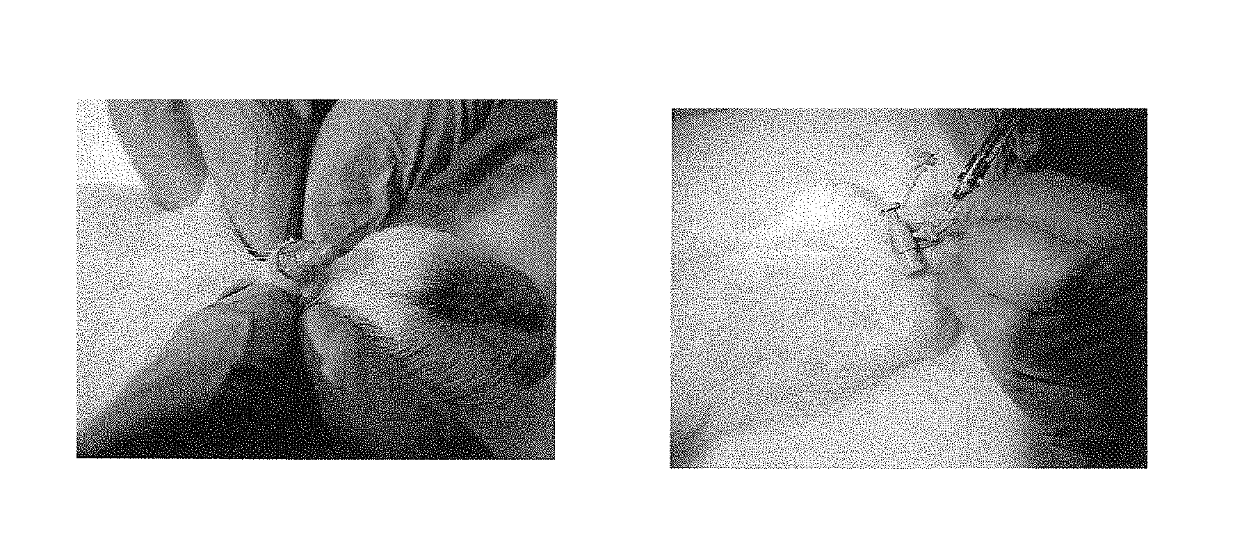
Intraoperative determination of nerve location
Golijanin
U.S. patent number 10,265,419 [Application Number 11/515,419] was granted by the patent office on 2019-04-23 for intraoperative determination of nerve location. This patent grant is currently assigned to Novadaq Technologies ULC. The grantee listed for this patent is Dragan Golijanin. Invention is credited to Dragan Golijanin.


| United States Patent | 10,265,419 |
| Golijanin | April 23, 2019 |
Intraoperative determination of nerve location
Abstract
The invention provides methods for intraoperatively determining the location of nerves by use of fluorescent dyes. The methods are particularly useful for locating the cavernous nerves innervating the penis.
| Inventors: | Golijanin; Dragan (Rochester, NY) | ||||||||||
|---|---|---|---|---|---|---|---|---|---|---|---|
| Applicant: |
|
||||||||||
| Assignee: | Novadaq Technologies ULC
(Burnaby, CA) |
||||||||||
| Family ID: | 37809582 | ||||||||||
| Appl. No.: | 11/515,419 | ||||||||||
| Filed: | September 1, 2006 |
Prior Publication Data
| Document Identifier | Publication Date | |
|---|---|---|
| US 20070122345 A1 | May 31, 2007 | |
Related U.S. Patent Documents
| Application Number | Filing Date | Patent Number | Issue Date | ||
|---|---|---|---|---|---|
| 60713643 | Sep 2, 2005 | ||||
| Current U.S. Class: | 1/1 |
| Current CPC Class: | A61K 49/0034 (20130101) |
| Current International Class: | A61N 5/00 (20060101); A61K 49/00 (20060101) |
| Field of Search: | ;600/312,310,407,431 ;424/9.6 ;604/20 |
References Cited [Referenced By]
U.S. Patent Documents
| 4109647 | August 1978 | Stern et al. |
| 4162405 | July 1979 | Chance et al. |
| 4200801 | April 1980 | Schuresko |
| 4263916 | April 1981 | Brooks et al. |
| 4394199 | July 1983 | Barnhard, IV et al. |
| 4473841 | September 1984 | Murakoshi et al. |
| 4532918 | August 1985 | Wheeler |
| 4541438 | September 1985 | Parker et al. |
| 4556057 | December 1985 | Hiruma et al. |
| 4619249 | October 1986 | Landry |
| 4718417 | January 1988 | Kittrell et al. |
| 4719508 | January 1988 | Sasaki et al. |
| 4768513 | September 1988 | Suzuki |
| 4773097 | September 1988 | Suzaki et al. |
| 4774568 | September 1988 | Matsuo |
| 4786813 | November 1988 | Svanberg et al. |
| 4805597 | February 1989 | Iwakoshi |
| 4815848 | March 1989 | Hadeishi |
| 4821117 | April 1989 | Sekiguchi |
| 4827908 | May 1989 | Matsuo |
| 4852579 | August 1989 | Gilstad et al. |
| 4858001 | August 1989 | Milbank et al. |
| 4860731 | August 1989 | Matsuura |
| 4867137 | September 1989 | Takahashi |
| 4868647 | September 1989 | Uehara et al. |
| 4900934 | February 1990 | Peeters et al. |
| 4930516 | June 1990 | Alfano et al. |
| 4938205 | June 1990 | Nudelman |
| 4957114 | September 1990 | Zeng et al. |
| 4993404 | February 1991 | Lane |
| 4995396 | February 1991 | Inaba et al. |
| 4995398 | February 1991 | Turnidge |
| 4998972 | March 1991 | Chin et al. |
| 5003977 | April 1991 | Suzuki et al. |
| 5042494 | August 1991 | Alfano |
| 5071417 | December 1991 | Sinofsky |
| 5078150 | January 1992 | Hara et al. |
| 5090400 | February 1992 | Saito |
| 5091652 | February 1992 | Mathies et al. |
| 5115137 | May 1992 | Andersson-Engels et al. |
| 5117466 | May 1992 | Buican et al. |
| 5125404 | June 1992 | Kittrell et al. |
| 5131398 | July 1992 | Alfano et al. |
| 5134662 | July 1992 | Bacus et al. |
| 5165079 | November 1992 | Schulz-Hennig |
| 5178616 | January 1993 | Uemiya et al. |
| 5196928 | March 1993 | Karasawa et al. |
| 5214503 | May 1993 | Chiu et al. |
| 5225883 | July 1993 | Carter et al. |
| 5255087 | October 1993 | Nakamura et al. |
| 5279298 | January 1994 | Flower |
| 5318023 | June 1994 | Vari et al. |
| 5318024 | June 1994 | Kittrell et al. |
| 5318869 | June 1994 | Hashimoto et al. |
| 5340592 | August 1994 | Goodrich, Jr. et al. |
| 5361769 | November 1994 | Nilsson |
| 5365057 | November 1994 | Morley et al. |
| 5371355 | December 1994 | Wodecki |
| 5375603 | December 1994 | Feiler |
| 5377676 | January 1995 | Vari et al. |
| 5377686 | January 1995 | O'Rourke et al. |
| 5394199 | February 1995 | Flower |
| 5419323 | May 1995 | Kittrell et al. |
| 5420628 | May 1995 | Poulsen et al. |
| 5421337 | June 1995 | Richards-Kortum et al. |
| 5421339 | June 1995 | Ramanujam et al. |
| 5424841 | June 1995 | Van Gelder et al. |
| 5430476 | July 1995 | Hafele et al. |
| 5437274 | August 1995 | Khoobehi et al. |
| 5438989 | August 1995 | Hochman et al. |
| 5453448 | September 1995 | Narciso, Jr. |
| 5465718 | November 1995 | Hochman et al. |
| 5491343 | February 1996 | Brooker |
| 5496369 | March 1996 | Howard |
| 5507287 | April 1996 | Palcic et al. |
| 5514127 | May 1996 | Shanks |
| 5519534 | May 1996 | Smith et al. |
| 5576013 | November 1996 | Williams et al. |
| 5590660 | January 1997 | MacAulay et al. |
| 5623930 | April 1997 | Wright et al. |
| 5627907 | May 1997 | Gur et al. |
| 5647368 | July 1997 | Zeng et al. |
| 5656498 | August 1997 | Lijima et al. |
| 5662644 | September 1997 | Swor |
| 5664574 | September 1997 | Chance |
| 5673701 | October 1997 | Chance |
| 5689241 | November 1997 | Clarke, Sr. et al. |
| 5699798 | December 1997 | Hochman et al. |
| 5707986 | January 1998 | Miller et al. |
| 5732707 | March 1998 | Widder et al. |
| 5741648 | April 1998 | Hemstreet, III et al. |
| 5743266 | April 1998 | Levene et al. |
| 5756541 | May 1998 | Strong et al. |
| 5785965 | July 1998 | Pratt et al. |
| 5803914 | September 1998 | Ryals et al. |
| 5827190 | October 1998 | Palcic et al. |
| 5845639 | December 1998 | Hochman et al. |
| 5851181 | December 1998 | Talmor |
| 5865754 | February 1999 | Sevick-Muraca et al. |
| 5910510 | June 1999 | Strong et al. |
| 5919616 | July 1999 | Aurelian et al. |
| 5927284 | July 1999 | Borst et al. |
| 5935942 | August 1999 | Zeimer |
| 5951980 | September 1999 | Collen |
| 5956435 | September 1999 | Buzug et al. |
| 5965356 | October 1999 | Aurelian et al. |
| 5999841 | December 1999 | Aoyagi et al. |
| 6008889 | December 1999 | Zeng et al. |
| 6013265 | January 2000 | Aurelian |
| 6021344 | February 2000 | Lui et al. |
| 6032070 | February 2000 | Flock et al. |
| 6054131 | April 2000 | Aurelian |
| 6069689 | May 2000 | Zeng et al. |
| 6074627 | June 2000 | Dean et al. |
| 6081612 | June 2000 | Gutkowicz-Krusin et al. |
| 6093149 | July 2000 | Guracar et al. |
| 6122042 | September 2000 | Wunderman et al. |
| 6140314 | October 2000 | Zeimer |
| 6148227 | November 2000 | Wagnieres et al. |
| 6149671 | November 2000 | Nordquist et al. |
| 6162242 | December 2000 | Peyman |
| 6178340 | January 2001 | Svetliza |
| 6179421 | January 2001 | Pang |
| 6186628 | February 2001 | Van De Velde |
| 6196226 | March 2001 | Hochman et al. |
| 6207168 | March 2001 | Aurelian |
| 6211953 | April 2001 | Niino et al. |
| 6217848 | April 2001 | Achilefu et al. |
| 6223069 | April 2001 | Pfeiffer et al. |
| 6233480 | May 2001 | Hochman et al. |
| 6241672 | June 2001 | Hochman et al. |
| 6246901 | June 2001 | Benaron |
| 6248727 | June 2001 | Zeimer |
| 6263227 | July 2001 | Boggett et al. |
| 6272374 | August 2001 | Flock et al. |
| 6280386 | August 2001 | Alfano et al. |
| 6293911 | September 2001 | Imasizumi et al. |
| 6319273 | November 2001 | Cheen et al. |
| 6331703 | December 2001 | Yarnall et al. |
| 6335429 | January 2002 | Cai et al. |
| 6351663 | February 2002 | Flower et al. |
| 6351667 | February 2002 | Godie |
| 6353750 | March 2002 | Kimura et al. |
| 6399354 | June 2002 | Knipe et al. |
| 6440950 | August 2002 | Zeimer |
| 6443976 | September 2002 | Flower et al. |
| 6447443 | September 2002 | Keogh et al. |
| 6485413 | November 2002 | Boppart et al. |
| 6498945 | December 2002 | Alfheim et al. |
| 6544183 | April 2003 | Thorn Leeson et al. |
| 6566641 | May 2003 | Suda |
| 6603552 | August 2003 | Cline et al. |
| 6621917 | September 2003 | Vilser |
| 6631286 | October 2003 | Pfeiffer et al. |
| 6671540 | December 2003 | Hochman |
| 6757554 | June 2004 | Rubinstein et al. |
| 6804549 | October 2004 | Hayashi |
| 6821946 | November 2004 | Goldspink et al. |
| 6840933 | January 2005 | Pang et al. |
| 6853857 | February 2005 | Pfeiffer et al. |
| 6882366 | April 2005 | Kijima et al. |
| 6899675 | May 2005 | Cline et al. |
| 6915154 | July 2005 | Docherty et al. |
| 6936043 | August 2005 | Peyman |
| 6944493 | September 2005 | Alam et al. |
| 7113817 | September 2006 | Winchester, Jr. et al. |
| 7236815 | June 2007 | Richards-Kortum et al. |
| 7364574 | April 2008 | Flower |
| 7381400 | June 2008 | Woltering |
| 7400753 | July 2008 | Seino et al. |
| 7400755 | July 2008 | West et al. |
| 7482318 | January 2009 | Aurelian et al. |
| 7581191 | August 2009 | Rice et al. |
| 7729750 | June 2010 | Tromberg et al. |
| 7774048 | August 2010 | Nakaoka et al. |
| 7881777 | February 2011 | Docherty et al. |
| 7885438 | February 2011 | Uppaluri et al. |
| 8036437 | October 2011 | Arditi et al. |
| 8073224 | December 2011 | Strobel et al. |
| 8144958 | March 2012 | Nahm et al. |
| 8185176 | May 2012 | Mangat et al. |
| 8194981 | June 2012 | Suzuki |
| 8285353 | October 2012 | Choi et al. |
| 8361775 | January 2013 | Flower |
| 8406860 | March 2013 | Dvorsky et al. |
| 8480579 | July 2013 | Serov et al. |
| 8521260 | August 2013 | Grinvald et al. |
| 8538107 | September 2013 | Rottger |
| 8647605 | February 2014 | Mangat et al. |
| 8725225 | May 2014 | Golijanin et al. |
| 8892190 | November 2014 | Docherty et al. |
| 8929974 | January 2015 | Hauger et al. |
| 8965488 | February 2015 | Dvorsky et al. |
| 9089601 | July 2015 | Golijanin et al. |
| 9129366 | September 2015 | Nahm et al. |
| 9241636 | January 2016 | Koizumi et al. |
| RE45916 | March 2016 | Golijanin et al. |
| 9351644 | May 2016 | Nahm et al. |
| 9357931 | June 2016 | Nahm et al. |
| 9421280 | August 2016 | Mangat et al. |
| 9610021 | April 2017 | Dvorsky et al. |
| 9642532 | May 2017 | Fengler et al. |
| 9816930 | November 2017 | Moriyama et al. |
| 9936887 | April 2018 | Dvorsky et al. |
| 10041042 | August 2018 | Flower |
| 2002/0025541 | February 2002 | Nelson et al. |
| 2002/0038120 | March 2002 | Duhaylongsod et al. |
| 2002/0099279 | July 2002 | Pfeiffer et al. |
| 2002/0099295 | July 2002 | Gil et al. |
| 2002/0146369 | October 2002 | Goldenberg |
| 2002/0181752 | December 2002 | Wallo et al. |
| 2002/0183621 | December 2002 | Pfeiffer et al. |
| 2003/0032885 | February 2003 | Rubinstein et al. |
| 2003/0050543 | March 2003 | Hartmann |
| 2003/0060718 | March 2003 | Alam et al. |
| 2003/0060722 | March 2003 | Pfeiffer et al. |
| 2003/0064025 | April 2003 | Yang et al. |
| 2003/0093064 | May 2003 | Peyman |
| 2003/0093065 | May 2003 | Peyman |
| 2003/0156252 | August 2003 | Morris et al. |
| 2003/0187349 | October 2003 | Kaneko et al. |
| 2003/0232016 | December 2003 | Heinrich |
| 2003/0236458 | December 2003 | Hochman |
| 2004/0066961 | April 2004 | Spreeuwers et al. |
| 2004/0077952 | April 2004 | Rafter et al. |
| 2004/0109231 | June 2004 | Haisch et al. |
| 2004/0156782 | August 2004 | Alam et al. |
| 2004/0162489 | August 2004 | Richards-Kortum et al. |
| 2004/0171827 | September 2004 | Peng et al. |
| 2004/0174495 | September 2004 | Levine |
| 2004/0206364 | October 2004 | Flower |
| 2005/0019744 | January 2005 | Bertuglia |
| 2005/0020891 | January 2005 | Rubinstein et al. |
| 2005/0033145 | February 2005 | Graham et al. |
| 2005/0069525 | March 2005 | Mikael |
| 2005/0089866 | April 2005 | Hinuma et al. |
| 2005/0107380 | May 2005 | Nimmo et al. |
| 2005/0142556 | June 2005 | Hoon et al. |
| 2005/0182321 | August 2005 | Frangioni |
| 2005/0182327 | August 2005 | Petty et al. |
| 2005/0182431 | August 2005 | Hausen et al. |
| 2005/0182434 | August 2005 | Docherty et al. |
| 2005/0187477 | August 2005 | Serov et al. |
| 2005/0197583 | September 2005 | Chance |
| 2005/0254008 | November 2005 | Ferguson et al. |
| 2006/0013768 | January 2006 | Woltering |
| 2006/0079750 | April 2006 | Fauci et al. |
| 2006/0108509 | May 2006 | Frangioni et al. |
| 2006/0118742 | June 2006 | Levenson et al. |
| 2006/0147897 | July 2006 | Grinvald et al. |
| 2006/0239921 | October 2006 | Mangat et al. |
| 2006/0241499 | October 2006 | Irion et al. |
| 2007/0122344 | May 2007 | Golijanin |
| 2007/0203413 | August 2007 | Frangioni |
| 2007/0254276 | November 2007 | Deutsch et al. |
| 2008/0007733 | January 2008 | Marks et al. |
| 2008/0015446 | January 2008 | Mahmood et al. |
| 2008/0025918 | January 2008 | Frangioni et al. |
| 2008/0044073 | February 2008 | Bernhardt et al. |
| 2008/0161744 | July 2008 | Golijanin et al. |
| 2008/0221421 | September 2008 | Choi et al. |
| 2008/0221648 | September 2008 | Flower |
| 2008/0239070 | October 2008 | Westwick et al. |
| 2008/0319309 | December 2008 | Bredno et al. |
| 2009/0005693 | January 2009 | Brauner et al. |
| 2009/0042179 | February 2009 | Peltie et al. |
| 2009/0048516 | February 2009 | Yoshikawa et al. |
| 2009/0054788 | February 2009 | Hauger et al. |
| 2009/0118623 | May 2009 | Serov et al. |
| 2009/0137902 | May 2009 | Frangioni et al. |
| 2009/0252682 | October 2009 | Hillman |
| 2009/0297004 | December 2009 | Baumgart |
| 2010/0022898 | January 2010 | Rubinstein et al. |
| 2010/0036217 | February 2010 | Choi et al. |
| 2010/0061604 | March 2010 | Nahm et al. |
| 2010/0222673 | September 2010 | Mangat et al. |
| 2010/0286529 | November 2010 | Carroll et al. |
| 2011/0001061 | January 2011 | Ishihara |
| 2011/0013002 | January 2011 | Thompson et al. |
| 2011/0063427 | March 2011 | Fengler et al. |
| 2011/0071403 | March 2011 | Sevick-Muraca et al. |
| 2011/0098685 | April 2011 | Flower |
| 2011/0306877 | December 2011 | Dvorsky et al. |
| 2012/0026325 | February 2012 | Bunker et al. |
| 2012/0078093 | March 2012 | Flower |
| 2012/0165662 | June 2012 | Nahm et al. |
| 2012/0271176 | October 2012 | Moghaddam et al. |
| 2013/0230866 | September 2013 | Miyashita et al. |
| 2013/0245456 | September 2013 | Ferguson, Jr. et al. |
| 2013/0286176 | October 2013 | Westwick et al. |
| 2013/0296715 | November 2013 | Lasser et al. |
| 2013/0345560 | December 2013 | Ferguson, Jr. et al. |
| 2014/0099007 | April 2014 | Sarkar et al. |
| 2014/0308656 | October 2014 | Flower |
| 2014/0316262 | October 2014 | Havens |
| 2015/0112192 | April 2015 | Docherty et al. |
| 2015/0112193 | April 2015 | Docherty et al. |
| 2015/0196208 | July 2015 | Dvorsky et al. |
| 2015/0230710 | August 2015 | Nahm et al. |
| 2015/0230715 | August 2015 | Nahm et al. |
| 2016/0038027 | February 2016 | Brzozowski et al. |
| 2016/0041098 | February 2016 | Hirawake et al. |
| 2016/0199515 | July 2016 | Flower |
| 2016/0371834 | December 2016 | Watanabe et al. |
| 2017/0039710 | February 2017 | Minai et al. |
| 2017/0303800 | October 2017 | Flower et al. |
| 2018/0020933 | January 2018 | Dvorsky et al. |
| 2018/0104362 | April 2018 | Golijanin et al. |
| 2018/0120230 | May 2018 | Moriyama et al. |
| 2018/0220907 | August 2018 | Dvorsky et al. |
| 2018/0234603 | August 2018 | Moore et al. |
| 409451 | Aug 2002 | AT | |||
| 2212257 | Aug 1996 | CA | |||
| 2413033 | Mar 2000 | CA | |||
| 2711560 | Jul 2009 | CA | |||
| 1049781 | Mar 1991 | CN | |||
| 1200174 | Nov 1998 | CN | |||
| 1399528 | Feb 2003 | CN | |||
| 101264014 | Sep 2008 | CN | |||
| 3906860 | Sep 1989 | DE | |||
| 19608027 | Sep 1996 | DE | |||
| 10028233 | Jan 2002 | DE | |||
| 10120980 | Nov 2002 | DE | |||
| 69727220 | Dec 2004 | DE | |||
| 102005044531 | Mar 2007 | DE | |||
| 0091805 | Oct 1983 | EP | |||
| 0215772 | Mar 1987 | EP | |||
| 0512965 | Nov 1992 | EP | |||
| 0792618 | Sep 1997 | EP | |||
| 0807402 | Nov 1997 | EP | |||
| 0826335 | Mar 1998 | EP | |||
| 1761171 | Mar 2007 | EP | |||
| 1874181 | Jan 2008 | EP | |||
| 2203831 | Oct 1988 | GB | |||
| S58-222331 | Dec 1983 | JP | |||
| S59-069721 | Apr 1984 | JP | |||
| S59-070903 | Apr 1984 | JP | |||
| H01-236879 | Sep 1989 | JP | |||
| 02-200237 | Aug 1990 | JP | |||
| H03-115958 | May 1991 | JP | |||
| 04-297236 | Oct 1992 | JP | |||
| H05-264232 | Oct 1993 | JP | |||
| H06-007353 | Jan 1994 | JP | |||
| 06-335451 | Dec 1994 | JP | |||
| H07-043303 | Feb 1995 | JP | |||
| 07-065154 | Mar 1995 | JP | |||
| 07-079955 | Mar 1995 | JP | |||
| H07-155285 | Jun 1995 | JP | |||
| H07-155286 | Jun 1995 | JP | |||
| H07-155290 | Jun 1995 | JP | |||
| H07-155291 | Jun 1995 | JP | |||
| H07-155292 | Jun 1995 | JP | |||
| 07-222712 | Aug 1995 | JP | |||
| H07-204156 | Aug 1995 | JP | |||
| H07-222723 | Aug 1995 | JP | |||
| H07-250804 | Oct 1995 | JP | |||
| H07-250812 | Oct 1995 | JP | |||
| 08-024227 | Jan 1996 | JP | |||
| H08-224208 | Sep 1996 | JP | |||
| H08-224209 | Sep 1996 | JP | |||
| H08-224240 | Sep 1996 | JP | |||
| H09-120033 | May 1997 | JP | |||
| H09-305845 | Nov 1997 | JP | |||
| B-3896176 | Dec 1997 | JP | |||
| H09-308609 | Dec 1997 | JP | |||
| H09-309845 | Dec 1997 | JP | |||
| H10-500479 | Jan 1998 | JP | |||
| H10-503480 | Mar 1998 | JP | |||
| H10-085222 | Apr 1998 | JP | |||
| H10-104070 | Apr 1998 | JP | |||
| H10-151104 | Jun 1998 | JP | |||
| H10-506440 | Jun 1998 | JP | |||
| H10-506550 | Jun 1998 | JP | |||
| H10-201700 | Aug 1998 | JP | |||
| H10-201707 | Aug 1998 | JP | |||
| H11-137517 | May 1999 | JP | |||
| H11-155812 | Jun 1999 | JP | |||
| H11-509748 | Aug 1999 | JP | |||
| 2001-198079 | Jul 2001 | JP | |||
| 2002-219129 | Aug 2002 | JP | |||
| 2003-510121 | Mar 2003 | JP | |||
| 2003-144401 | May 2003 | JP | |||
| 2003-329589 | Nov 2003 | JP | |||
| 2004-528917 | Sep 2004 | JP | |||
| 2004-325200 | Nov 2004 | JP | |||
| 2006-503620 | Feb 2006 | JP | |||
| 2006-192280 | Jul 2006 | JP | |||
| 2007-021006 | Feb 2007 | JP | |||
| 2008-525126 | Jul 2008 | JP | |||
| 2008-535600 | Sep 2008 | JP | |||
| 2008-231113 | Oct 2008 | JP | |||
| 2009-095683 | May 2009 | JP | |||
| 2009-291554 | Dec 2009 | JP | |||
| 2010-505582 | Feb 2010 | JP | |||
| 2011-509768 | Mar 2011 | JP | |||
| 2011-519589 | Jul 2011 | JP | |||
| 2011-528918 | Dec 2011 | JP | |||
| 5918532 | May 2016 | JP | |||
| 90-0005434 | Jul 1990 | KR | |||
| 2002-0064287 | Aug 2002 | KR | |||
| 2288633 | Dec 2006 | RU | |||
| WO-1986/02730 | May 1986 | WO | |||
| WO-1990/10219 | Sep 1990 | WO | |||
| WO-1990/12536 | Nov 1990 | WO | |||
| WO 93/25141 | Dec 1993 | WO | |||
| WO-1994/12092 | Jun 1994 | WO | |||
| WO-1995/00171 | Jan 1995 | WO | |||
| WO-1995/26673 | Oct 1995 | WO | |||
| WO-1996/09435 | Mar 1996 | WO | |||
| WO-1996/09792 | Apr 1996 | WO | |||
| WO-1996/18415 | Jun 1996 | WO | |||
| WO-1996/23524 | Aug 1996 | WO | |||
| WO-1996/39925 | Dec 1996 | WO | |||
| WO-1997/08538 | Mar 1997 | WO | |||
| WO-1998/24360 | Jun 1998 | WO | |||
| WO-1998/30144 | Jul 1998 | WO | |||
| WO-1998/46122 | Oct 1998 | WO | |||
| WO-1999/00053 | Jan 1999 | WO | |||
| WO-1999/47940 | Sep 1999 | WO | |||
| WO-1999/53832 | Oct 1999 | WO | |||
| WO-2000/42910 | Jul 2000 | WO | |||
| WO-2000/47107 | Aug 2000 | WO | |||
| WO-2001/08552 | Feb 2001 | WO | |||
| WO-2001/17561 | Mar 2001 | WO | |||
| WO-2001/22870 | Apr 2001 | WO | |||
| WO-2001/39764 | Jun 2001 | WO | |||
| WO-2001/69244 | Sep 2001 | WO | |||
| WO-2001/80734 | Nov 2001 | WO | |||
| WO-2001/82786 | Nov 2001 | WO | |||
| WO-2002/061390 | Aug 2002 | WO | |||
| WO-2003/006658 | Jan 2003 | WO | |||
| WO-2004/006963 | Jan 2004 | WO | |||
| WO-2004/052195 | Jun 2004 | WO | |||
| WO-2005/026319 | Mar 2005 | WO | |||
| WO 2005/034747 | Apr 2005 | WO | |||
| WO-2005/079238 | Sep 2005 | WO | |||
| WO-2006/111836 | Oct 2006 | WO | |||
| WO-2006/111909 | Oct 2006 | WO | |||
| WO-2006/116634 | Nov 2006 | WO | |||
| WO-2006/119349 | Nov 2006 | WO | |||
| WO-2006/121631 | Nov 2006 | WO | |||
| WO-2006/121631 | Nov 2006 | WO | |||
| WO-2006/123742 | Nov 2006 | WO | |||
| WO-2007/028032 | Mar 2007 | WO | |||
| WO-2008/044822 | Apr 2008 | WO | |||
| WO-2008/070269 | Jun 2008 | WO | |||
| WO-2008/070269 | Jun 2008 | WO | |||
| WO-2008/087869 | Jul 2008 | WO | |||
| WO-2009/046985 | Apr 2009 | WO | |||
| WO-2009/046985 | Apr 2009 | WO | |||
| WO-2009/048660 | Apr 2009 | WO | |||
| WO-2009/092162 | Jul 2009 | WO | |||
| WO-2009/127972 | Oct 2009 | WO | |||
| WO-2012/038824 | Mar 2012 | WO | |||
| WO-2012/096878 | Jul 2012 | WO | |||
| WO-2013/190391 | Dec 2013 | WO | |||
| WO-2015/001427 | Jan 2015 | WO | |||
| WO-2013/002350 | Feb 2015 | WO | |||
Other References
|
Dail, W.G., et al., "Multiple vasodilator pathways from the pelvic plexus to the penis of the rat," 1999, International Journal of Impotence Research, vol. 11, pp. 277-285. cited by applicant . De Grand, A.M. and J.V. Frangioni, "An operational near-infrared fluorescence imaging system prototype for large animal surgery," 2003, Technology in Cancer Research & Treatment , vol. 2(6), pp. 1-10. cited by applicant . Frangioni, John V., "In vivo near-infrared fluorescence imaging," 2003, Current Opinion in Chemical Biology,vol. 7, pp. 626-634. cited by applicant . Humblet, V., et al., "High-affinity near-infrared fluorescent small-molecule contrast agents for in vivo imaging of prostate-specific membrane antigen," 2005, Mol. Imaging, vol. 4(4), pp. 448-462. cited by applicant . Kim, S., et al., "Near-infrared fluorescence type II quantum dots for sentinel lymph node mapping," 2004, Nature Biotechnology, vol. 22(1), pp. 93-97. cited by applicant . Liedberg, F., et al., "[Bladder cancer and the sentinel node concept,]" 2003, Aktuelle Urol., vol. 34(2), pp. 115-118. cited by applicant . Liedberg, F., et al., "Intraoperative sentinel node detection improves nodal staging in invasive bladder cancer," 2006, J. Urol., vol. 175, pp. 84-89. cited by applicant . Leissner, J., et al., "Extended radical lymphadenectomy in patients with urothelial bladder cancer: Results of a prospective multicenter study," 2004, J. Urol. vol. 171, pp. 139-144. cited by applicant . Malmstrom, P-U, et al., "RE: Extended radical lymphadenectomy in patients with urothelial bladder cancer: Results of a prospective multicenter study," 2004, J. Urol. vol. 172, p. 386. cited by applicant . Marangos, N., et al., "In vivo visualization of the cochlear nerve and nuclei with fluorescent axonal tracers," 2001, Hearing Research, vol. 162, pp. 48-52. cited by applicant . Nakayama, A., et al., "Functional near-infrared fluorescence imaging for cardiac surgery and targeted gene therapy," 2002, Mol. Imaging, vol. 1(4), pp. 365-377. cited by applicant . Nimura, H., et al., "Infrared ray electronic endoscopy combined with indocyanine green injection for detection of sentinel nodes of patients with gastric cancer," 2004, British Journal of Surgery, vol. 91, pp. 575-579. cited by applicant . Parungo, C.P., et al., "In vivo optical imaging of pleural space drainage to lymph nodes of prognostic significance," 2004, Annas of Surgical Oncology, vol. 11(12), pp. 1085-1092. cited by applicant . Parungo, C.P., et al., "Intraoperative identification of esophageal sentinel lymph nodes with near-infrared fluorescence imaging," 2005, J. Thorac. Cardiovasc. Surg., vol. 129, pp. 844-850. cited by applicant . Schneider, HC Jr., et al., "Fluorescence of testicle. An indication of viability of spermatic cord after torsion," 1975, Urology, vol. 5(1), pp. 133-136. cited by applicant . Sherif, A., et al., "Lymphatic mapping and detection of sentinel nodes in patients with bladder cancer," 2001, J. Urol., vol. 166, pp. 812-815. cited by applicant . Soltesz, E.G., et al., "Intraoperative sentinel lymph node mapping of the lung using near-infrared fluorescent quantum dots," 2005, Ann. Thorac. Surg., vol. 79, pp. 269-277. cited by applicant . Sugi, K. et al., "Comparison of three tracers for detecting sentinel lymph nodes in patients with clinical N0 lung cancer," 2003, Lung Cancer, vol. 39, pp. 37-40. cited by applicant . Uren, Roger F., "Cancer surgery joins the dots," 2004, Nature Biotechnology, vol. 22(1), pp. 38-39. cited by applicant . Author Unkown, "Invitrogen," Material Safety Data Sheet, Jun. 4, 2008, pp. 1-4. cited by applicant . Schmued, et al., "Intracranial Injection of Fluoro-Gold Results in the Degeneration of Local but not Retrogradely Labeled Neurons," Brain Research, 1993, pp. 71-77, vol. 626, Elsevier, Maryland Heights, MO, USA. cited by applicant . Naumann, et al., "Retrograde Tracing of Fluoro-Gold: Different Methods of Tracer Detection at the Ultrastructural Level and Neurodegenerative Changes of Back-Filled Neurons in Long-Term Studies," Journal of Neuroscience Methods, 2000, pp. 11-21, vol. 103, Elsevier, Maryland Heights, MO, USA. cited by applicant . He, "Fluorogold Induces Persistent Neurological Deficits and Circling Behavior in Mice Over-Expressing Human Mutant Tau," Current Neurovascular Research, 2009, pp. 54-61, vol. 6, Bentham Science Publishers Ltd., Oak Park, IL, USA. cited by applicant . Garrett, et. al., "Fluoro-Gold's Toxicity Makes It Inferior to True Blue for Long-Term Studies of Dorsal Root Ganglion Neurons and Motoneurons," Neuroscience Letters, 1991, pp. 137-139, vol. 128, Elsevier, Maryland Heights, MO, USA. cited by applicant . Puigdellivol-Sanchez et al., "On the use of fast blue, fluoro-gold and diamidino yellow for retrograde tracing after peripheral nerve injury: uptake, fading, dye interactions, and toxicity," Journal of Neuroscience Methods, 115:115-127, (2002). cited by applicant . Raabe et al., "Near-Infrared lndocyanine Green Video Angiography: A New Method for lntraoperative Assessment of Vascular Flow," Neurosurgery, 52(1):132-139, (2003). cited by applicant . Ross et al., "Sentinel Node Biopsy in Head and Neck Cancer: Preliminary Results of a Multicenter Trial," Annals of Surgical Oncology, 11(7):690-696, (2004). cited by applicant . Ross et al., "The ability of lymphoscintigraphy to direct sentinel node biopsy in the clinically NO neck for patients with head and neck squamous cell carcinoma," The British Journal of Radiology, 75:950-958, (2002). cited by applicant . Rubben et al., "Infrared Videoangiofluorography of the Skin with lndocyanine Green-Rat Random Cutaneous Flap Model and Results in Man," Microvascular Research, 47:240-251, (1994). cited by applicant . Schmued et al., "In vivo anterograde and retrograde axonal transport of the fluorescent rhodamine-dextran-amine, Fluoro-Ruby, within the CNS," Brain Research, 526: 127-134, (1990). cited by applicant . Shoaib et al., "The Accuracy of Head and Neck Carcinoma Sentinel Lymph Node Biopsy in the Clinically NO Neck," 5th International Conference on Head and Neck Cancer, San Francisco, CA, pp. 2077-2083, (2001). cited by applicant . Stoeckli et al., "Sentinel lymph node evaluation in squamous cell carcinoma of the head and neck," 5th International Conference on Head and Neck Cancer, pp. 221-226, Jul. 29-Aug. 3, 2000. cited by applicant . Tubbs et al., "Anatomic Landmarks for Nerves of the Neck: A Vade mecum for Neurosurgeons," Neurosurgery, 56:0NS256-0NS260, (2005). cited by applicant . Valero-Cabre et al., "Superior Muscle Reinnervation After Autologous Nerve Graft or Poly-L-Lactide- -Caprolactone (PLC) Tube Implantation in Comparison to Silicone Tube Repair," Journal of Neuroscience Research, 63:214-223, (2001). cited by applicant . Jamis-Dow et al., "Small (.ltoreq.3-cm) Renal Masses: Detection with CT versus US and Pathologic Correlation," Radiology, 198(3):785-788, (1996). cited by applicant . Kamolz et al., "lndocyanine green video angiographies help to identify burns requiring operation," Burns, 29:785-791, (2003). cited by applicant . Kobbert et al., "Current concepts in neuroanatomical tracing," Progress in Neurobiology, 62:327-351, (2000). cited by applicant . Lanciego et al., "Multiple axonal tracing: simultaneous detection of three tracers in the same section," Histochem Cell Biol, 110:509-515, (1998). cited by applicant . Lanciego et al., "Multiple neuroanatomical tracing in primates," Brain Research Protocols, 2:323-332, (1998). cited by applicant . Liptay, "Sentinel Node Mapping in Lung Cancer," Annals of Surgical Oncology, 11(3):271S-274S, (2004). cited by applicant . Malmstrom et al., "Early Metastatic Progression of Bladder Carcinoma: Molecular Profile of Primary Tumor and Sentinel Lymph Node," The Journal of Urology, 168:2240-2244, (2002). cited by applicant . Minciacchi et al., "A procedure for the simultaneous visualization of two anterograde and different retrograde fluorescent tracers," Journal of Neuroscience Methods, 38:183-191, (1991). cited by applicant . Motomura et al. "Sentinel Node Biopsy Guided by lndocyanin Green Dye in Breast Cancer Patients," Jpn J Clin Oncol., 29(12):604-607, (1999). cited by applicant . Paques et al., "Axon-Tracing Properties of lndocyanine Green," Arch Ophthalmol, 121:367-370, (2003). cited by applicant . Akintunde et al., "Quadruple labeling of brain-stem neurons: a multiple retrograde fluorescent tracer study of axonal collateralization," Journal of Neuroscience Methods, 45:15-22, (1992). cited by applicant . Butter et al., "Melanoma in children and the use of sentinel lymph node biopsy," Journal of Pediatric Surgery, 40:797-800, (2005). cited by applicant . Dan et al., "1% Lymphazurin vs 10% Fluorescein for Sentinel Node Mapping in Colorectal Tumors," Arch Surg., 139:1180-1184, (2004). cited by applicant . Demos, "Near-infrared autofluorescence imaging for detection of cancer," Journal of Biomedical Optics, 9(3):587-592, (2004). cited by applicant . Dietz et al., "lndocyanine Green, Evidence of Neurotoxicity in Spinal Root Axons," Anesthesiology, 98(2):516-520, (2003). cited by applicant . Dunne et al., "Value of sentinel lymphonodectomy in head and neck cancer patients without evidence of lymphogenic metastatic disease," Auris Nasus Larynx, 28:339-344, (2001). cited by applicant . Fritzsch et al., "Sequential double labeling with different fluorescent dyes coupled to dextran amines as a tool to estimate the accuracy of tracer application and of regeneration," Journal of Neuroscience Methods, 39:9-17, (1991). cited by applicant . Gipponi et al., "New Fields of Application of the Sentinel Lymph Node Biopsy in the Pathologic Staging of Solid Neoplasms: Review of Literature and Surgical Perspectives," Journal of Surgical Oncology, 85:171-179, (2004). cited by applicant . Glover et al., "Fluorescent dextran-amines used as axonal tracers in the nervous system of the chicken embryo," Journal of Neuroscience Methods, 18:243-254, (1986). cited by applicant . Haglund et al., "Enhanced Optical Imaging of Rat Gliomas and Tumor Margins," Neurosurgery, 35(5):930-941, (1994). cited by applicant . Haglund et al., "Enhanced Optical Imaging of Human Gliomas and Tumor Margins," Neurosurgery, 38(2):308-317, (1996). cited by applicant . Angelov et al., "Contralateral trigeminal nerve lesion reduces polyneuronal muscle innervation after facial nerve repair in rats," Eur. J. Neurosci., 11:1369-1378 (1999). cited by applicant . Komurcu et al., "Management strategies for peripheral iatrogenic nerve lesions", Annals of Plastic Surgery, 54 (2):135-139 (2005). cited by applicant . Oddi et al., "Intraoperative Biliary Tree Imaging with Cholyl-Lysyl- Fluorescein: An Experimental Study in the rabbit," Surg. Laproscop. Endosc., 6(3):198-200 (1996). cited by applicant . Ohnishi et al., "Organic Alternatives to Quantum Dots for Intraoperative near-Infrared Fluorescent Lymph Node Mapping," Mol. Imaging, 4(3):172-181 (2005). cited by applicant . Kurihara K. et al., "Nerve Staining with Leucomethylene Blue: An Experimental Study" Plastic and Reconstructive Surgery, 73(6):960-964 (1984). cited by applicant . Nahlieli et al., "Intravital Staining with Methylene Blue as an Aid to Facial Nerve Identification in Parotid Gland Surgery," J. Oral Maxillofac. Surg., 59:355-356 (2001). cited by applicant . U.S. Appl. No. 12/063,349, filed May 10, 2010 in the name of Mangat et al. cited by applicant . U.S. Appl. No. 11/851,312, filed Sep. 6, 2007 in the name of Golijanin et al. cited by applicant . Alander, J.T. et al. (Jan. 1, 2012). "A Review of Indocyanine Green Fluorescent Imaging in Surgery," International Journal of Biomedical Imaging 2012:1-26, article ID 940585. cited by applicant . Alfano et al. (Oct. 1987). "Fluorescence Spectra from Cancerous and Normal Human Breast and Lung Tissues," IEEE Journal of Quantum Electronics QE-23(10):1806-1811. cited by applicant . Alm, A. et al. (Jan. 1, 1973). "Ocular and Optic Nerve Blood Flow at Normal and Increased Intraocular Pressures in Monkeys (Macaca irus): A Study with Radioactively Labelled Microspheres Including Flow Determinations in Brain and Some Other Tissues," Experimental Eye Research 15(1):15-29. cited by applicant . Alonso-Burgos, A. et al. (2006). "Preoperative planning of deep inferior epigastric artery perforator flap reconstruction with multi-slice-CT angiography: imaging findings and initial experience," Journal of Plastic, Reconstructive & Aesthetic Surgery 59:585-593. cited by applicant . Alvarez, F. J. et al. (Apr. 1996). "Behaviour of Isolated Rat and Human Red Blood Cells Upon Hypotonic-Dialysis Encapsulation of Carbonic Anhydrase and Dextran," Biotechnology and Applied Biochemistry 23(2):173-179. cited by applicant . Ancalmo, N. et al. (1997). "Minimally invasive coronary artery bypass surgery: really minimal?" Ann. Thorac. Surg. 64:928-929. cited by applicant . Andersson-Engels, S. et al. (1991). "Fluorescence Characteristics of Atherosclorotic Plaque and Malignant Tumors," in Optical Methods for Tumor Treatment and Early Diagnosis: Mechanisms and Techniques, T. J. Dougherty (Ed.), The Society of Photo-optical Instrumentation Engineers (SPIE) 1426:31-43, fourteen pages. cited by applicant . Andersson-Engels, S. et al. (Mar. 1989). "Tissue Diagnostics Using Laser-Induced Fluorescence," Berichte der Bunsengesellschaft fur physikalische Chemie 93(3):335-342. cited by applicant . Annese, V. et al. (2005). "Erthrocytes-Mediated Delivery of Dexamethasone in Steroid-Dependent IBD Patients--a Pilot Uncontrolled Study," American Journal of Gastroenterology 100:1370-1375. cited by applicant . Argus-50/CA, Inter cellular CA2+ (calcium ion) Image Analysis system (Feb. 1992). "Observation and 2-dimensional analysis of Ca2+ concentration distribution. Fura-2 and Indo-1 compatible. Ca2+ concentrations are calculated from the fluorescence ratio," pp. 1-10. cited by applicant . Awano, T. et al. (Jun. 2010). "Intraoperative EC-IC Bypass Blood Flow Assessment with Indocyanine Green Angiography in Moyamoya and Non-moyamoya Ischemic Stroke," World Neurosurg. 73(6):668-674. cited by applicant . Azuma, R. et al. (2008, presented in part Jun. 2007). "Detection of Skin Perforators by Indocyanine Green Fluorescence Nearly Infrared Angiography," PRS Journal 122(4):1062-1067. cited by applicant . Balacumarswami, L. et al. (Aug. 2004). "Does Off-Pump Total Arterial Grafting Increase the Incidence of Intraoperative Graft Failure?," The Journal of Thoracic and Cardiovascular Surgery 128(2):238-244. cited by applicant . Barton, J.K. et al. (1999) "Simultaneous irradiation and imaging of blood vessels during pulsed laser delivery," Lasers in Surgery and Medicine 24(3):236-243. cited by applicant . Bassingthwaighte, J.B. et al. (Apr. 1974). "Organ Blood Flow, Wash-in, Washout, and Clearance of Nutrients and Metabolites," Mayo Clin. Proc. 49(4):248-255. cited by applicant . Batliwala, H. et al. (Apr. 15, 1995). "Methane-Induced Haemolysis of Human Erythrocytes," Biochemical J. 307(2):433-438. cited by applicant . Bek, T. (1999). "Diabetic Maculopathy Caused by Disturbances in Retinal Vasomotion: A New Hypothesis," Acta Ophthalmologica Scandinavica 77:376-380. cited by applicant . Benson, R.C. et al. (1978). "Fluorescence Properties of Indocyanine Green as Related to Angiography," Phys. Med. Biol. 23(1):159-163. cited by applicant . Black's Medical Dictionary, "Perfusion," 42nd Edition (2009), two pages. cited by applicant . Boer, F.et al. (1994). "Effect of Ventilation on First-Pass Pulmonary Retention of Alfentaril and Sufentanil in Patients Undergoing Coronary Artery Surgery," British Journal Anesthesia 73:458-463. cited by applicant . Boldt, .J. et al. (Feb. 1990). "Lung management during cardiopulmonary bypass: influence on extravascular lung water," Journal of Cardiothoracic Anesthesia 4(1):73-79. cited by applicant . Boldt, J. et al. (1991). "Does the technique of cardiopulmonary bypass affect lung water content?" European Journal of Cardio-Thoracic Surgery 5:22-26. cited by applicant . C2741, Compact High Performance video camera for industrial applications with Built-in contrast enhancement circuit, Jun. 1998, six pgs. cited by applicant . Canada Health. (1997). "Coronary Bypass Surgery and Angioplasty, 1982-1995, Heart Disease and Stroke in Canada," Canada Health, located at <http:/www.hc-sc.gc.ca/hpb>, eighty two pages. cited by applicant . Coffey, J.H. et al. (1984). "Evaluation of Visual Acuity During Laser Photoradiation Therapy of Cancer," Lasers in Surgery and Medicine 4(1):65-71. cited by applicant . Conley, M.P. et al. (Oct. 2004). "Anterograde Transport of Peptide-Conjugated Fluorescent Beads in the Squid Giant Axom Identifies a Zip-Code for Synapse," Biological Bulletin 207(2):164, one page. cited by applicant . Costa, R.A. et al. (Oct. 2001). "Photodynamic Therapy with Indocyanine Green for Occult Subfoveal Choroidal Neovascularization Caused by Age-Related Macular Degeneration," Curr. Eye Res. 23(4):274-275. cited by applicant . Cothren, R.M. et al. (Mar. 1990). "Gastrointestinal Tissue Diagnosis by Laser-Induced Fluorescence Spectroscopy at Endoscopy," Gastrointestinal Endoscopy 36(2):105-111. cited by applicant . Daniels, G. et al. (Apr. 2007). "Towards Universal Red Blood Cell," Nature Biotechnology 25(4):427-428. cited by applicant . De Flora, A. (Sep. 1986). "Encapsulation of Adriamycin in human erythrocytes," Proc. Natl. Acad. Sci., USA 83(18):7029-7033. cited by applicant . Declaration of Brian Wilson dated Aug. 22, 2017 for Inter Partes Review No. IPR2017-01426, twelve pages. cited by applicant . Definition of "Expose," Excerpt of Merriam Webster's Medical Desk Dictionary (1993), four pages. cited by applicant . Definition of "Graft," Excerpt of Stedman's Medical Dictionary for the Health Professions and Nursing; 6th Ed. (2008), three pages. cited by applicant . Deloach, J.R. (ed.) et al. (1985). Red Blood Cells as Carriers for Drugs. A Method for Disseminating Chemotherapeutics, Hormones, Enzymes and Other Therapeutic Agents via the Circulatory System, Karger, Basel, CH, pp. v-vii, (Table of Contents), seven pages. cited by applicant . Deloach, J.R. (Jun. 1983). "Encapsulation of Exogenous Agents in Erythrocytes and the Circulating Survival of Carrier Erythrocytes," Journal of Applied Biochemistry 5(3):149-157. cited by applicant . Desai, N.D. et al. (Oct. 18, 2005, e-published on Sep. 28, 2005) "Improving the Quality of Coronary Bypass Surgery with Intraoperative Angiography," Journal of the American College of Cardiology 46(8):1521-1525. cited by applicant . Detter, C. et al. (Aug. 1, 2007). "Fluorescent Cardiac Imaging: A Novel Intraoperative Method for Quantitative Assessment of Myocardial Perfusion During Graded Coronary Artery Stenosis," Circulation 116(9):1007-1014. cited by applicant . Detter, C. et al. (Jun. 2011). "Near-Infrared Fluorescence Coronary Angiography: A New Noninvasive Technology for Intraoperative Graft Patency Control." The Heart Surgery Forum #2001-6973 5(4):364-369. cited by applicant . Digital CCD Microscopy (date unknown). Chapter 14, pp. 259-282. cited by applicant . Dougherty, T.J. et al. (1990). "Cutaneous Phototoxic Occurrences in Patients Receiving Photofrin," Lasers in Surgery and Medicine 10(5):485-488. cited by applicant . Draijer, M.J. et al. (Jun. 17-19, 2007). "Laser Doppler Perfusion Imaging with a High-Speed CMOS-Camera," in Novel Optical Instrumentation for Biomedical Applications III, C. Depeursinge, ed., Proceedings of SPIE-OSA Biomedical Optics (Optical Society of America, 2007), SPIE-OSA, 6631:0N1-0N7, nine pages. cited by applicant . Ekstrand, M.I. et al. (Feb. 14, 2008). "The Alpha-Herpesviruses: Molecular Pathfinders in Nervous System Circuits," Trends in Molecular Medicine, Elsevier Current Trends 14(3):134-140. cited by applicant . Emery R.W. et al. (Aug. 1996). "Revascularization Using Angioplasty and Minimally Invasive Techniques Documented by Thermal Imaging," The Annals of Thoracic Surgery 62(2):591-593. cited by applicant . Enquist, L.W. et al. (2002). "Directional Spread of an .alpha.-Herpesvirus in the Nervous System," Veterinary Microbiology 86:5-16. cited by applicant . Eren, S. et al. (Dec. 1995). "Assessment of Microcirculation of an Axial Skin Flap Using Indocyanine Green Fluorescence Angiography," Plast. Reconstr. Surg. 96(7):1636-1649. cited by applicant . Falk, T. et al. (Apr. 15, 2001). "A Herpes Simplex Viral Vector Expressing Green Fluorescent Protein can be Used to Visualize Morphological Changes in High-density Neuronal Culture," Electronic Journal of Biotechnology 4(1):34-45. cited by applicant . Flower, R. et al. (Apr.-Jun. 1999). "Effects of free and liposome-encapsulated hemoglobin on choroidal vascular plexus blood flow, using the rabbit eye as a model system," European Journal of Ophthalmology 9 (2):103-114. cited by applicant . Flower, R.W. (1992). "Choroidal Angiography Today and Tomorrow," Retina 12(3):189-190. cited by applicant . Flower, R.W. (Apr. 2000). "Experimental Studies of Indocyanine Green Dye-Enhanced Photocoagulation of Choroidal Neovascularization Feeder Vessels," American Journal of Ophthalmology 129(4):501-512. cited by applicant . Flower, R.W. (Aug. 2002). "Optimizing Treatment of Choroidal Neovascularization Feeder Vessels Associated with Age-Related Macular Degeneration," American Journal of Ophthalmology 134(2):228-239. cited by applicant . Flower, R.W. (Dec. 1973). "Injection Technique for Indocyanine Green and Sodium Fluorescein Dye Angiography of the Eye," Investigative Opthamology 12(12):881-895. cited by applicant . Flower, R.W. (Sep. 1, 1994). "Does Preinjection Binding of Indocyanine Green to Serum Actually Improve Angiograms?," Arch Ophthalmol. 112(9):1137-1139. cited by applicant . Flower, R.W. et al. (Aug. 1977). "Quantification of Indicator Dye Concentration in Ocular Blood Vessels," Exp. Eye Res. 25(2):103-111. cited by applicant . Flower, R.W. et al. (Dec. 1, 2008, e-published Aug. 15, 2008). "Observation of Erythrocyte Dynamics in the Retinal Capillaries and Choriocapillaris Using ICG-Loaded Erythrocyte Ghost Cells," Investigative Ophthalmology, & Visual Science 49(12):5510-5516. cited by applicant . Flower, R.W. et al. (Mar. 26, 2008-Mar. 29, 2008). "Observation of Erythrocyte Dynamics in the Retinal Capillaries and Choriocapillaris Using ICG-Loaded Erythrocyte Ghost Cells," Annual Meeting of the Macula Society, Abstract No. XP002535355, Palm Beach, FL, USA, fourteen pages, (Schedule of the Meeting only). cited by applicant . Forrester et al. (Nov. 1, 2002). "Comparison of Laser Speckle and Laser Doppler Perfusion Imaging: Measurement in Human Skin and Rabbit Articular Tissue," Medical and Biological Engineering and Computing 40(6):687-697. cited by applicant . Frenzel H. et al. (Apr. 18, 2008). "In Vivo Perfusion Analysis of Normal and Dysplastic Ears and its Implication on Total Auricular Reconstruction," Journal of Plastic, Reconstructive and Aesthetic Surgery 61(Supplement1):S21-S28. cited by applicant . Gagnon, A.R. et al. (2006). "Deep and Superficial Inferior Epigastric Artery Perforator Flaps," Cirugia Plastica Ibero-Latinoamericana 32(4):7-13. cited by applicant . Gardner, T.J. (1993). "Coronary Artery Disease and Ventricular Aneurysms," in Surgery, Scientific Principles and Practice, Greenfield, L.J. (ed.) et al., J.B. Lippincott Co., Philadelphia, PA, pp. 1391-1411, twenty three pages. cited by applicant . Geddes, C. D. et al. (2003, e-published on Mar. 20, 2003). "Metal-Enhanced Fluorescence (MEF) Due to Silver Colloids on a Planar Surface: Potential Applications of Indocyanine Green to in Vivo Imaging," Journal of Physical Chemistry A 107(18):3443-3449. cited by applicant . Giunta, R.E. et al. (Jul. 2005). "Prediction of Flap Necrosis with Laser Induced Indocyanine Green Fluorescence in a Rat Model," British Journal of Plastic Surgery 58(5):695-701. cited by applicant . Giunta, R.E. et al. (Jun. 2000). "The Value of Preoperative Doppler Sonography for Planning Free Perforator Flaps," Plastic and Reconstructive Surgery 105(7):2381-2386. cited by applicant . Glossary, Nature, downloaded from the internet <http://www.nature.com/nrg/journal/v4/nI0/glossary/nrgl 183_glossary.html>> HTML on Jun. 30, 2014. cited by applicant . Goldstein, J.A. et al. (Dec. 1998). "Intraoperative Angiography to Assess Graft Patency After Minimally Invasive Coronary Bypass," Ann. Thorac. Surg. 66(6):1978-1982. cited by applicant . Gothoskar A.V. (Mar. 2004). "Resealed Erythrocytes: A Review," Pharmaceutical Technology pp. 140, 142, 144, 146, 148, 150, 152 and 154-158, twelve pages. cited by applicant . Granzow, J.W. et al. (Jul. 2007)."Breast Reconstruction with Perforator Flaps" Plastic and Reconstructive Surgery 120(1):1-12. cited by applicant . Green, H.A. et al. (Jan. 1992). "Burn Depth Estimation Using Indocyanine Green Fluorescence," Arch Dermatol 128(1):43-49. cited by applicant . Hallock, G.G. (Jul. 2003). "Doppler sonography and color duplex imaging for planning a perforator flap," Clinics in Plastic Surgery 30(3):347-357. cited by applicant . Hamamatsu Brochure. (May 1997). Specifications for Real-time Microscope Image Processing System: ARGUS-20 with C2400-75i, four pages. cited by applicant . Hamamatsu. (Date unknown). Microscope Video Camera, for Fluorescent Observation, Easy Fluorescent Image Analysis C2400-731, -751 Series a CCD Camera, seven pages. cited by applicant . Hayashi, J. et al. (Nov. 1993). "Transadventitial Localization of Atheromatous Plaques by Fluorescence Emission Spectrum Analysis of Mono-L Aspartyl-Chlorin e6," Cardiovascular Research 27(11):1943-1947. cited by applicant . Hayata, Y. et al. (Jul. 1982). "Fiberoptic Bronchoscopic Laser Photoradiation for Tumor Localization in Lung Cancer," Chest82(1):10-14. cited by applicant . Herts, B.R. (May 2003). "Imaging for Renal Tumors," Current Opin.. Urol. 13(3):181-186. cited by applicant . Hirano, T. et al. (1989). "Photodynamic Cancer Diagnosis and Treatment System Consisting of Pulse Lasers and an Endoscopic Spectro-Image Analyzer," Laser in Life Sciences 3(2):99-116. cited by applicant . Holm, C. et al. (2002). "Monitoring Free Flaps Using Laser-Induced Fluorescence of Indocyanine Green: A Preliminary Experience," Microsurgery 22(7):278-287. cited by applicant . Holm, C. et al. (Apr. 2003, e-published on Feb. 25, 2003). "Laser-Induced Fluorescence of Indocyanine Green: Plastic Surgical Applications," European Journal of Plastic Surgery 26(1):19-25. cited by applicant . Holm, C. et al. (Dec. 1, 2002). "Intraoperative Evaluation of Skin-Flap Viability Using Laser-Induced Fluorescence of Indocyanine Green," British Journal of Plastic Surgery 55(8):635-644. cited by applicant . Hung, J. et al. (1991). "Autofluorescence of Normal and Malignant Bronchial Tissue," Lasers in Surgery and Medicine 11(2):99-105. cited by applicant . Hyvarinen, L. et al. (1980). "Indocyanine Green Fluorescence Angiography." Acta ophthalmologica 58(4):528-538. cited by applicant . Ikeda, S. (Jul. 1989). "Bronichial Telivision Endoscopy," Chest 96(1):41S-42S. cited by applicant . Jaber, S.F. et al. (Sep. 1998). "Role of Graft Flow Measurement Technique in Anastomotic Quality Assessment in Minimally Invasive CABG," Ann. Thorac. Surg. 66(3):1087-1092. cited by applicant . Jagoe, J.R. et al. (1989). "Quantification of retinal damage during cardiopulmonary bypass," Third International Conference on Image Processing and its Applications (Conf. Publ. No. 307), IEE, pp. 319-323. cited by applicant . Jolion, J. et al. (Aug. 1991). "Robust Clustering with Applications in Computer Vision," IEEE Transactions on Pattern Analysis and Machine Intelligence 13(8):791-802. cited by applicant . Kapadia, C.R. et al. (Jul. 1990). "Laser-Induced Fluorescence Spectroscopy of Human Colonic Mucosa. Detection of Adenomatous Transformation," Gastroenterology 99(1):150-157. cited by applicant . Kato, H. et al. (Jun. 1985). "Early Detection of Lung Cancer by Means of Hematoporphyrin Derivative Fluorescence and Laser Photoradiation," Clinics in Chest Medicine 6(2):237-253. cited by applicant . Kato, H. et al. (Jun. 1990). "Photodynamic Diagnosis in Respiratory Tract Malignancy Using an Excimer Dye Laser System," Journal of Photochemistry and Photobiology, B. Biology 6(1-2):189-196. cited by applicant . Keon, W.J. et al. (Dec. 1979). "Coronary Endarterectomy: An Adjunct to Coronary Artery Bypass Grafting," Surgery 86(6):859-867. cited by applicant . Kiryu, J. et al. (Sep. 1994). "Noninvasive Visualization of the Choriocapillaris and its Dynamic Filling," Investigative Ophthalmology & Visual Science 35(10):3724-3731. cited by applicant . Kitai, T. et al. (Jul. 2005). "Fluorescence Navigation with Indocyanine Green for Detecting Sentinel Lymph Nodes in Breast Cancer," Breast Cancer 12(3):211-215. cited by applicant . Kleszcy ska, H. et al. (Mar. 2005). "Hemolysis of Erythrocytes and Erythrocyte Membrane Fluidity Changes by New Lysosomotropic Compounds," Journal of Fluorescence 15(2):137-141. cited by applicant . Kokaji, K. et al. (Date Unknown). "Intraoperative Quality Assessment by Using Fluorescent Imaging in Off-pump Coronary Artery Bypass Grafting," The Department of Cardiovascular Surgery, University of Keio, Tokyo, Japan, one page, (Abstract only). cited by applicant . Krishnan, K. G. et al. (Apr. 1, 2005). "The Role of Near-Infrared Angiography in the Assessment of Post-Operative Venous Congestion in Random Pattern, Pedicled Island and Free Flaps", British Journal of Plastic Surgery 58(3):330-338. cited by applicant . Kuipers, J.A. et al. (1999). "Recirculatory and Compartmental Pharmacokinetic Modeling of Alfentanil Pigs, the Influence of Cardiac Output," Anesthesiology 90(4):1146-1157. cited by applicant . Kupriyanov, V.V. et al. (Nov. 2004). "Mapping Regional Oxygenation and Flow in Pig Hearts In Vivo Using Near-infrared Spectroscopic Imaging," Journal of Molecular and Cellular Cardiology 37(5):947-957. cited by applicant . Kyo, S. (Date Unknown). "Use of Ultrasound Cardiology during Coronary Artery Bypass Surgery," Heart and Blood Vessel Imaging II, three pages. cited by applicant . Lam, S. et al. (1991). "Mechanism of Detection of Early Lung Cancer by Ratio Fluorometry," Lasers in Life Sciences 4(2):67-73. cited by applicant . Lam, S. et al. (Feb. 1990). "Detection of Early Lung Cancer Using Low Dose Photofrin II," Chest 97(2):333-337. cited by applicant . Lam, S. et al. (Jul. 1, 1990). "Detection of Lung Cancer by Ratio Fluorometry With and Without Photofrin II," Proc. SPIE--Optical Fibers in Medicine V 1201:561-568. cited by applicant . Lam, S. et al. (Nov. 1-4, 1990). "Fluorescence Imaging of Early Lung Cancer," Annual International Conference of the IEEE Engineering in Medicine and Biology Society 12(3):1142-1143. cited by applicant . Lam, S.C. et al. (1993). "Fluorescence Detection," Chapter 20 in Lung Cancer, Roth, J.A. (ed.), et al., Blackwell Scientific Publications Inc., 238 Main Street, Cambridge, Massachusetts, 02142, pp. 325-338, sixteen pages. cited by applicant . Laub, G.W. et al. (Nov. /Dec. 1989). "Experimental Use of Fluorescein for Visualization of Coronary Arteries," Vascular and Endovascular Surgery 23(6):454-457. cited by applicant . Lee, E.T. et al. (Mar. 1997). "A New Method for Assessment of Changes in Retinal Blood Flow," Medical Engineering & Physics 19(2):125-130. cited by applicant . Leithner, "Untersuchung der Sauerstoffkonzentrationsveranderungen in der Mikrozirkulation des Hirnkortex von Ratten bei funktioneller Stimulation mittels Phosphorescence Quenching," [dissertation], Jul. 14, 2003; retrieved from the Internet: <http://edoc.hu-berlin.de/dissertationen/leith ner-ch ristoph-2003-07-14/>, two hundred and eight pages [English Abstract and Machine Translation]. cited by applicant . Lippincott's New Medical Dictionary. "Perfusion," p. 707 (1897), three pages. cited by applicant . Little, J.R. et al. (May 1979). "Superficial Temporal Artery to Middle Cerebral Artery Anastomosis: Intraoperative Evaluation by Fluorescein Angiography and Xenon--133 Clearance," Journal of Neurosurgery 50(5):560-569. cited by applicant . Liu Q. P. et al. (Apr. 2007). "Bacterial Glycosidases for the Production of Universal Red Blood Cells" Nature Biotechnology 25(7):454-464. cited by applicant . Lund, F. et al. (Nov. 1997). "Video Fluorescein Imaging of the Skin: Description of an Overviewing Technique for Functional Evaluation of Regional Cutaneous Blood Evaluation of Regional Cutaneous Perfusion in Occlusive Arterial Disease of the Limbs," Clinical Physiology 17(6):619-633. cited by applicant . Mack, M.J. et al. (Sep. 1998). "Arterial Graft Patency in Coronary Artery Bypass Grafting: What Do We Really Know?," Ann. Thorac. Surg. 66(3):1055-1059. cited by applicant . Magnani, M. et al. (1998). "Erythrocyte Engineering for Drug Delivery and Targeting," Biotechnol. Appl. Biochem. 28:1-6. cited by applicant . Magnani, M. et al. (Jul. 15, 1992). "Targeting Antiretroviral Nucleoside Analogues in Phosphorylated Form to Macrophasges: In Vitro and In Vivo Studies," Proc. Natl. Acad. Sci. USA 89(14):6477-6481. cited by applicant . Martinez-Perez, M. et al. (Sep. 19, 1996). "Unsupervised Segmentation Based on Robust Estimation and Cooccurrence Data," Proceedings of the International Conference on Miage Processing (ICIP) Lausanne 3:943-945. cited by applicant . May, S. (May/Jun. 1995). "Photonic Approaches to Burn Diagnostics," Biophotonics International pp. 44-50. cited by applicant . McKee, T.D. et al. (Mar. 1, 2006). "Degradation of Fibrillar Collagen in a Human Melanoma Xenograft Improves the Efficacy of an Oncolytic Herpes Simplex Virus Vector," Cancer Research 66(5):2509-2513. cited by applicant . Merriam Webster Medline Plus Medical Dictionary. "Perfusion," located at http://www.merriam-webster.com/medlineplus/perfusion, last visited on Apr. 15, 2015, one page. cited by applicant . Mitaka USA, Inc. (2015). "PDE Breast Free Flap Evaluation," located at <http://mitakausa.com/category/pde_education/flaps/>, last visited on Dec. 29, 2015, four pages. cited by applicant . Mitaka USA, Inc. (2015). "PDE-Neo" located at <http://mitakausa.com/pde-neo/>, last visited on Dec. 29, 2015, two pages. cited by applicant . Mohr, F.W. et al. (May 1997). "Thermal Coronary Angiography: A Method for Assessing Graft Patency and Coronary Anatomy in Coronary Bypass Surgery," Ann Thorac. Surgery 63(5):1506-1507. cited by applicant . Montan, S. et al. (Feb. 1, 1985). "Multicolor Imaging and Contrast Enhancement in Cancer-Tumor Localization Using Laser-Induced Fluorescence in Hematoporphyrin-Derivative-Bearing Tissue," Optics Letters 10(2):56-58. cited by applicant . Mothes, H. et al. (Nov. 2004). "Indocyanine-Green Fluorescence Video Angiography Used Clinically to Evaluate Tissue Perfusion in Microsurgery," The Journal of Trauma Injury, Infection, and Critical Care 57(5):1018-1024. cited by applicant . Mullooly, V.M. et al. (1990). "Dihematoporphyrin Ether-Induced Photosensitivity in Laryngeal Papilloma Patients," Lasers in Surgery and Medicine 10(4):349-356. cited by applicant . Murphy (2001). "Digital CCD Microscopy," Chapter 14 in Fundamentals of Light Microscopy and Electronic Imaging, John Wiley and Sons, pp. i-xi and 259-281. cited by applicant . Nakamura, T. et al. (1964). "Use of Novel Dyes, Coomassie Blue and Indocyanine Green in Dye Dilution Method," Tohoka University, Nakamura Internal Department, The Tuberculosis Prevention Society, Tuberculosis Research Laboratory, 17(2):1361-1366, seventeen pages. cited by applicant . Newman et al. (Oct. 31, 2008). "Update on the Application of Laser-Assisted Indocyanine Green Fluorescent Dye Angiography in Microsurgical Breast Reconstruction," American Society of Plastic Surgeons, Plastic Surgery 2008, 2 pages. cited by applicant . Novadaq Technologies Inc. (Jan. 29, 2007). "Novadaq Imaging System Receives FDA Clearance for use During Plastic Reconstructive Surgery," PR Newswire three pages. cited by applicant . Novadaq Technologies Inc. (Jan. 19, 2005). 510(k) Summary--Showing X-Ray Fluoroscopy as Predicate Device, Fluorescent Angiographic System, six pages. cited by applicant . Ogata, F. et al. (Jun. 2007). "Novel Lymphography Using Indocyanine Green Dye for Near-Infrared Fluorescence Labeling," Annals of Plastic Surgery 58(6):652-655. cited by applicant . Ooyama, M. (Oct. 12-15, 1994). The 8th Congress of International YAG Laser Symposium, The 15th Annual Meeting of Japan Society for Laser Medicine, Sun Royal Hotel, Japan, eight pages. cited by applicant . Ott, P. (1998). "Hepatic Elimination of Indocyanine Green with Special Reference to Distribution Kinetics and the Influence of Plasma Protein Binding," Pharmacology & Toxicology 83(Supp. II):5-48. cited by applicant . Oxford Concise Medical Dictionary. "Perfusion," p. 571 (1980), three pages. cited by applicant . Pagni, S. et al. (Jun. 1997). "Anastomotic Complications in Minimally Invasive Coronary Bypass Grafting," Ann. Thorac. Surg. 63(6 Suppl):S64-S67. cited by applicant . Palcic et al. (1991). "Lung Imaging Fluorescence Endoscope: A Device for Detection of Occult Lung Cancer," Medical Design and Material, thirteen pages. cited by applicant . Palcic, B. et al. (1990). "Development of a Lung Imaging Fluorescence Endoscope," Annual International Conference of the IEEE Engineering in Medicine and Biology Society 12(1):0196-0197. cited by applicant . Palcic, B. et al. (Aug. 1, 1990). "The Importance of Image Quality for Computing Texture Features in Biomedical Specimens," Proc. SPIE 1205:155-162. cited by applicant . Palcic, B. et al. (Jun. 1, 1991). "Lung Imaging Fluorescence Endoscope: Development and Experimental Prototype," Proc. SPIE 1448:113-117. cited by applicant . Palcic, B. et al. (Mar. 1991). "Detection and Localization of Early Lung Cancer by Imaging Techniques," Chest 99(3):742-743. cited by applicant . Pandharlpande, P.V. et al. (Mar. 2005). "Perfusion Imaging of the Liver: Current Challenges and Future Goals," Radiology 234(3):661-673. cited by applicant . Peak, M.J. et al. (1986). "DNA-to-Protein Crosslinks and Backbone Breaks Caused by FAR-and Near-Ultraviolet and Visible Light Radiations in Mammalian Cells," in Mechanism of DNA Damage and Repair, Implications for Carcinogenesis and Risk Assessment, SIMIC, M.G. (ed.) et al., Plenum Press, 233 Spring Street, New York, N.Y. 10013, pp. 193-202. cited by applicant . Peiretti et al. (2005). "Human erythrocyte-ghost-mediated choroidal angiography and photocoagulation." Database Biosis [online] Biosciences Information Service, Philadelphia, PA, US, XP002725023, Database accession No. Prev200600056121 (abstract), three pages. cited by applicant . Peiretti, E. et al. (May 2005). "Human Erythrocyte-Ghost-Mediated Choroidal Angiography and Photocoagulation," Investigative Ophthalmology & Visual Science, ARVO Annual Meeting Abstract 46(13):4282, located at <http://iovs.arvojournals.org/article.aspx?articleid=2403707>, last visited on Oct. 7, 2016, two pages. cited by applicant . Perez, M.T. et al. (Sep. 2002). "In Vivo Studies on Mouse Erythrocytes Linked to Transferrin," IUBMB Life 54(3):115-121. cited by applicant . Pfister, A.J. et al. (Dec. 1992). "Coronary Artery Bypass Without Cardiopulmonary Bypass," Ann. Thorac. Surg. 54(6):1085-1092, (Discussion by S.R. Gundry). cited by applicant . Phillips, R.P. et al. (1991). "Quantification of Diabetic Maculopathy by Digital Imaging of the Fundus," Eye 5(1):130-137. cited by applicant . Piermarocchi, S. et al. (Apr. 2002). "Photodynamic Therapy Increases the Eligibility for Feeder Vessel Treatment of Choroidal Neovascularization Caused by Age-Related Macular Degeneration," American Journal of Ophthalmology 133(4):572-575. cited by applicant . Profio, A.E. et al. (Jul.-Aug. 1984). "Fluorometer for Endoscopic Diagnosis of Tumors," Medical Physics 11(4):516-520. cited by applicant . Profio, A.E. et al. (Jun. 1, 1991). "Endoscopic Fluorescence Detection of Early Lung Cancer," Proc. SPIE 1426:44-46. cited by applicant . Profio, A.E. et al. (Nov./Dec. 1979). "Laser Fluorescence Bronchoscope for Localization of Occult Lung Tumors," Medical Physics 6:523-525. cited by applicant . Profio, A.E. et al. (Sep.-Oct. 1986). "Digital Background Subtraction for Fluorescence Imaging," Medical Physics 13(5):717-721. cited by applicant . Pyner, S. et al. (Nov. 2001). "Tracing Functionally Identified Neurones in a Multisynaptic Pathway in the Hamster and Rat Using Herpes Simplex Virus Expressing Green Fluorescent Protein," Experimental Physiology 86(6):695-702. cited by applicant . Raabe et al. (2009, e-published on Nov. 12, 2008). "Laser Doppler Imaging for Intraoperative Human Brain Mapping", NeuroImage 44:1284-1289. cited by applicant . Rava, R.P. et al. (Jun. 1, 1991). "Early Detection of Dysplasia in Colon and Bladder Tissue Using Laser-Induced Fluorescence," Proc. SPIE 1426:68-78. cited by applicant . Razum, N. et al. (Nov. 1987). "Skin Photosensitivity: Duration and Intensity Following Intravenous Hematoporphyrin Derivatives, HpD and DHE," Photochemistry and Photobiology 46(5):925-928. cited by applicant . Report on Observation by C2400-75i and ARGUS20 Under Low illumination conditions, Jan. 17, 2008. cited by applicant . Request for Invalidation mailed on Jun. 29, 2007 for Japanese Patent No. JP-3881550, filed by Hamamatsu Photonics, Inc. (with English Translation). cited by applicant . Reuthebuch, O et al. (Feb. 2004). "Novadaq SPY: Intraoperative Quality Assessment in Off Pump Coronary Artery Bypass Grafting," Chest 125(2):418-424. cited by applicant . Reuthebuch, O.T. et al. (May 2003). "Graft Occlusion After Deployment of the Symmetry Bypass System," Ann. Thorac. Surg. 75(5):1626-1629. cited by applicant . Richards-Kortum, R. et al. (Jun. 1991). "Spectroscopic Diagnosis of Colonic Dysplasia: Spectroscopic Analysis," Biochemistry and Photobiology 53(6):777-786. cited by applicant . Roberts, W.W. et al. (Dec. 1997). "Laparoscopic Infrared Imaging," Surg. Endoscopy 11(12):1221-1223. cited by applicant . Rodnenkov, O.V. et al. (May 2005). "Erythrocyte Membrane Fluidity and Haemoglobin Haemoporphyrin Conformation: Features Revealed in Patients with Heart Failure," Pathophysiology 11(4):209-213. cited by applicant . Ropars, C. (ed.) et al. (1987). Red Blood Cells as Carriers for Drugs. Potential therapeutic Applications. Pergamon Press, Oxford, New York, pp. v-vii, (Table of Contens only), four pages. cited by applicant . Ross, G.L. et al. (Dec. 2002). "The Ability of Lymphoscintigraphy to Direct Sentinel Node Biopsy in the Clinically N0 Neck for Patients with Head and Neck Squamous Cell Carcinoma," The British Journal of Radiology 75(900):950-958. cited by applicant . Rossi, L. et al. (2001). "Erthrocyte-Mediated Delivery of Dexamethasone in Patients with Chronic Obstructive Pulmonary Disease," Biotechnol. Appl. Biochem. 33:85-89. cited by applicant . Rossi, L. et al. (1999). "Heterodimer-Loaded Erthrocytes as Bioreactors for Slow Delivery of the Antiviral Drug Azidothymidine and the Antimycobacterial Drug Ethambutol," Aids Research and Human Retroviruses 15(4):345-353. cited by applicant . Rossi, L. et al. (2004). "Low Doses of Dexamethasone Constantly Delivered by Autologous Erythrocytes Slow the Progression of Lung Disease in Cystic Fibrosis Patients," Blood Cells, Molecules, and Diseases 33:57-63. cited by applicant . Rozen, W.M. et al. (Jan. 2008). "Preoperative Imaging for DIEA Perforator Flaps: A Comparative Study of Computed Tomographic Angiography and Doppler Ultrasound," Plastic and Reconstructive Surgery 121(1):9-16. cited by applicant . Rubens, F.D. et al. (2002). "A New and Simplified Method for Coronary and Graft Imaging During CABG," The Heart Surgery Forum 5(2):141-144. cited by applicant . Sakatani, K. et al. (Nov. 1997). "Noninvasive Optical Imaging of the Subarchnoid Space and Cerebrospinal Fluid Pathways Based on Near Infrared Fluorescence," J. Neurosurg. 87(5):738-745. cited by applicant . Salmon, E.D. et al. (Oct. 1994). "High Resolution Multimode Digital Imaging System for Mitosis Studies in Vivo and in Vitro," Biol. Bull 187(2):231-232. cited by applicant . Sato, et al., (1991). "Development of a Visualization Method for the Microcirculation of Deep Viscera Using an Infrared Intravital Microscope System," Research on ME Devices and ME Technology, five pages, (with English translation). cited by applicant . Satpathy G.R. et al. (Oct. 2004) "Loading Red Blood Cells with Trehalose: A Step Towards Biostabilization," Cryobiology 49(2):123-136. cited by applicant . Schaff, H.V. et al. (Oct. 15, 1996). "Minimal Thoracotomy for Coronary Artery Bypass: Value of Immediate Postprocedure Graft Angiography," Supplement to Circulation 94(8):I-51, (Abstract No. 0289), two pages. cited by applicant . Schellingerhout, D. et al. (Oct. 2000). "Quantitation of HSV Mass Distribution in a Rodent Brain Tumor Model," Gene Therapy 7(19):1648-1655. cited by applicant . Seeman, P. (Jan. 1, 1967). "Transient Holes in the Erythrocyte Membrane During Hypotonic Hemolysis and Stable Holes in the Membrane After Lysis by Saponin and Lysolecithin," Journal of Cell Biology 32(1):55-70. cited by applicant . Sekijima, M. et al. (Sep. 2004). "An Intraoperative Fluorescent Imaging System in Organ Transplantation," Transplantation Proceedings 36(7):2188-2190. cited by applicant . Serov, A. et al. (Mar. 1, 2002). "Laser Doppler Perfusion Imaging with a Complimentary Metal Oxide Semiconductor Image Sensor," Optics Letters 27(5):300-302. cited by applicant . Serov, A.N. et al. (Sep. 23, 2003). "Quasi-Parallel Laser Doppler Perfusion Imaging Using a CMOS Image Sensor," Proc. SPIE 5067:73-84. cited by applicant . Sezgin, M. et al. (Jan. 2004). "Survey Over Image Thresholding Techniques and Quantitative Performance Evaluation," Journal of Electronic Imaging 13(1):146-165. cited by applicant . Sheth, S.A. et al. (Apr. 22, 2004)"Linear and Nonlinear Relationships between Neuronal Activity, Oxygen Metabolism, and Hemodynamic Responses," Neuron 42(2):347-355. cited by applicant . Siemers, B.M. et al. (Nov. 2001). "The Acoustic Advantage of Hunting at Low Heights Above Water: Behavioual Experiments on the European `Trawling` Bats Myotis Capaccinii, M Dasycneme and M. Daubentonii," J. Eperimental Biol. 204(Pt. 22):3843-3854. cited by applicant . Skalidis, E.I. et al. (Nov. 16, 2004). "Regional Coronary Flow and Contractile Reserve in Patients with Idiopathic Dilated Cardiomyopathy," Journal of the American College of Cardiology 44(10):2027-2032. cited by applicant . Slakter, J.S. et al. (Jun. 1995). "Indocyanine-Green Angiography," Current Opinion in Ophthalmology 6(III):25-32. cited by applicant . Smith, G.A. et al. (Mar. 13, 2001). "Herpesviruses Use Bidirectional Fast-Axonal Transport to Spread in Sensory Neurons," Proceedings of the National Academy of Sciences of the United States of America 98(6):3466-3470. cited by applicant . Sony Corporation. The Sony U-Matic Videocassette Recorder, VO-9800, ten pages. cited by applicant . Staurenghi, G. et al. (Dec. 2001)."Combining Photodynamic Therapy and Feeder Vessel Photocoagulation: A Pilot Study," Seminars in Ophthalmology 16(4):233-236. cited by applicant . Stern, M.D. (Mar. 6, 1975). "In Vivo Evaluation of Microcirculation by Coherent Light Scattering," Nature 254(5495):56-58. cited by applicant . Still, J. et al. (Mar. 1999). "Evaluation of the Circulation of Reconstructive Flaps Using Laser-Induced Fluorescence of Indocyanine Green," Ann. Plast. Surg. 42(3):266-274. cited by applicant . Still, J.M. et al. (Jun. 2001). "Diagnosis of Burn Depth Using Laser-Induced Indocyanine Green Fluorescence: A Preliminary Clinical Trial," Burns 27(4):364-371. cited by applicant . Subramanian, V.A. et al. (Oct. 15, 1995). "Minimally Invasive Coronary Bypass Surgery: A Multi-Center Report of Preliminary Clinical Experience," Supplement to Circulation 92(8):I-645, (Abstract No. 3093), two pages. cited by applicant . Sugimoto, K. et al. (Jun. 2008, e-published on Mar. 19, 2008). "Simultaneous Tracking of Capsid, Tegument, and Envelope Protein Localization in Living Cells Infected With Triply Fluorescent Herpes Simplex Virus 1," Journal of Virology 82(11):5198-5211. cited by applicant . Suma, H. et al. (2000). "Coronary Artery Bypass Grafting Without Cardiopulmonary Bypass in 200 Patients," J. Cardiol. 36(2):85-90, (English Abstract only). cited by applicant . Summary of Invention Submitted to EPO, "Development of Novadaq SPY.TM. Cardiac Imaging Invention," five pages. cited by applicant . Taggart, D.P. et al. (Mar. 2003). "Preliminary Experiences with a Novel Intraoperative Fluorescence Imaging Technique to Evaluate the Patency of Bypass Grafts in Total Arterial Revascularization," Ann Thorac Surg. 75(3):870-873. cited by applicant . Taichman, G.C. et al. (Jun. 1987). "The Use of Cardio-Green for Intraoperative Visualization of the Coronary Circulation: Evaluation of Myocardial Toxicity," Texas Heart Institute Journal 14(2):133-138. cited by applicant . Takahashi, M. et al. (Sep. 2004). "SPY: An Innovative Intra-Operative Imaging System to Evaluate Graft Patency During Off-Pump Coronary Artery Bypass Grafting," Interactive Cardio Vascular and Thoracic Surgery 3(3):479-483. cited by applicant . Takayama, T. et al. (Apr. 1992). "Intraoperative Coronary Angiography Using Fluorescein Basic Studies and Clinical Application," Vascular and Endovascular Surgery 26(3):193-199. cited by applicant . Takayama, T. et al. (Jan. 1991). "Intraoperative Coronary Angiography Using Fluorescein" The Annals of Thoracic Surgery 51(1):140-143. cited by applicant . Tanaka, E. et al. (Jul. 2009). "Real-time Assessment of Cardiac Perfusion, Coronary Angiography, and Acute Intravascular Thrombi Using Dual-channel Near-infrared Fluorescence Imaging," The Journal of Thoracic and Cardiovascular Surgery 138(1):133-140. cited by applicant . Tang, G.C. et al. (1989). "Spectroscopic Differences between Human Cancer and Normal Lung and Breast Tissues," Lasers in Surgery and Medicine 9(3):290-295. cited by applicant . Taylor, K.M. (Apr. 1998). "Brain Damage During Cardiopulmonary Bypass," Annals of Thoracic Surgery 65(4):S20-S26. cited by applicant . The American Heritage Medical Dictionary. "Perfuse." p. 401 (2008), three pages. cited by applicant . Thelwall, P.E. et al. (Oct. 2002). "Human Erythrocyte Ghosts: Exploring the Origins of Multiexponential Water Diffusion in a Model Biological Tissue with Magnetic Resonance," Magnetic Resonance in Medicine 48(4):649-657. cited by applicant . Torok, B. et al. (May 1996). "Simultaneous Digital Indocyanine Green and Fluorescein Angiography," Klinische Monatsblatter Fur Augenheilkunde 208(5):333-336, (Abstract only), two pages. cited by applicant . Tsutsumi, D. et al. "Moisture Detection of road surface using infrared camera," Reports of the Hokkaido Industrial Research Institute (No. 297), Issued on Nov. 30, 1998, two pages. cited by applicant . Unno, N. et al. (Feb. 2008, e-published on Oct. 26, 2007). "Indocyanine Green Fluorescence Angiography for intraoperative assessment of Blood flow: A Feasibility Study," Eur J Vasc Endovasc Surg. 35(2):205-207. cited by applicant . Van Son, J.A.M. et al. (Nov. 1997). "Thermal Coronary Angiography for Intraoperative Testing of Coronary Patency in Congenital Heart Defects," Ann Thorac Surg. 64(5):1499-1500. cited by applicant . Verbeek, X. et al. (2001). "High-Resolution Functional Imaging With Ultrasound Contrast Agents Based on RF Processing in an In Vivo Kidney Experiment", Ultrasound in Med. & Biol. 27(2):223-233. cited by applicant . Wachi, A. et al. (Apr. 1995). "Characteristics of Cerebrospinal Fluid Circulation in Infants as Detected With MR Velocity Imaging," Child's Nerv Syst 11(4):227-230. cited by applicant . Wagnieres, G.A. et al. (Jul. 1, 1990). "Photodetection of Early Cancer by Laser Induced Fluorescence of a Tumor-Selective Dye: Apparatus Design and Realization," Proc. SPIE 1203:43-52. cited by applicant . Weinbeer, M. et al. (Nov. 25, 2013). "Behavioral Flexibility of the Trawling Long-Legged Bat, Macrophyllum Macrophyllum (Phyllostomidae)," Frontiers in Physiology 4(Article 342):1-11. cited by applicant . What is Perfusion? A Summary of Different Typed of Perfusion. (Sep. 1, 2004). Located at, <http://www.perfusion.com/cgi-bin/absolutenm/templates/articledisplay.- asp?articleid=1548#.Vo8HvO2FPGj>, last visited on Jan. 7, 2016, two pages. cited by applicant . Wise, R.G. et al. (Nov. 2005). "Simultaneous Measurement of Blood and Myocardial Velocity in the Rat Heart by Phase Contrast MRI Using Sparse q-Space Sampling" Journal of Magnetic Resonance Imaging 22(5):614-627. cited by applicant . Woitzik, J. et al. (Apr. 2005). "Intraoperative Control of Extracranial-Intracranial Bypass Patency by Near-Infrared Indocyanine Green Videoangiography," J. Neurosurg. 102(4):692-698. cited by applicant . Wollert, H.G. et al. (Dec. 1989). "Intraoperative Visualization of Coronary Artery Fistula Using Medical Dye," The Thoracic and Cardiovascular Surg. 46(6):382-383. cited by applicant . Wu, C. et al. (Apr. 15, 2005). "cGMP (Guanosine 3',5'-Cyclic Monophosphate) Transport Across Human Erythrocyte Membranes," Biochemical Pharmacology 69(8):1257-1262. cited by applicant . Yada, T. et al. (May 1993). "In Vivo Observation of Subendocardial Microvessels of the Beating Porcine Heart Using a Needle-Probe Videomicroscope with a CCD Camera," Circulation Research 72(5):939-946. cited by applicant . Yamaguchi, S. et al. (Apr. 2005). "Evaluation of Skin Perfusion After Nipple-Sparing Mastectomy by Indocyanine Green Dye" Journal of Saitama Medical University, Japan, 32(2):45-50, (with English Abstract). cited by applicant . Yoneya, S. et al. (Jun. 1998). "Binding Properties of Indocyanine Green in Human Blood," IOVS 39(7):1286-1290. cited by applicant . Yoneya, S. et al. (Sep. 1993). "Improved Visualization of the Choroidal Circulation with Indocyanine Green Angiography," Arch Opthalmol. 111(9):1165-1166. cited by applicant . Young. I.T. et al. (1993). "Depth of Focus in Microscopy," SCIA '93, Proc. of the 8th Scandinavian Conference on Image Analysis, Tromso, Norway, pp. 493-498, six pages. cited by applicant . Australian Examination Report No. 1 dated Jun. 26, 2018 for Australian Patent Application No. 2014408488, filed on Mar. 31, 2017, nine pages. cited by applicant . Canadian Notice of Allowance dated Jan. 4, 2018 for Canadian Application No. 2,750,760, filed on Jul. 25, 2008, one page. cited by applicant . Canadian Notice of Allowance dated Sep. 27, 2017 for Canadian Application No. 2,811,847, filed on Mar. 20, 2013, one page. cited by applicant . Canadian Office Action dated Feb. 13, 2018 for CA Application No. 2,963,450 filed on Apr. 3, 2017, three pages. cited by applicant . Canadian Office Action dated Feb. 27, 2017 for Canadian Application No. 2,750,760, filed on Jul. 25, 2011, three pages. cited by applicant . Canadian Office Action dated Feb. 28, 2018 for CA Application No. 2,963,987 filed on Mar. 27, 2017, five pages. cited by applicant . Canadian Office Action dated Jan. 19, 2017 for Canadian Application No. 2,914,778 filed on Dec. 8, 2015, four pages. cited by applicant . Canadian Office Action dated Mar. 16, 2016 for CA Application No. 2,750,760 filed on Jan. 23, 2009, five pages. cited by applicant . Canadian Office Action dated Nov. 28, 2017 for Canadian Application No. 2,914,778 filed on Dec. 8, 2015, six pages. cited by applicant . Canadian Office Action dated Oct. 25, 2016 for Canadian Patent Application No. 2,811,847, filed on Sep. 20, 2011, three pages. cited by applicant . Canadian Office Action dated Sep. 30, 2015 for CA Application No. 2,811,847, filed on Sep. 20, 2011, four pages. cited by applicant . Chinese Fifth Office Action dated Dec. 19, 2017 for Chinese Application No. 201180057244.8 filed on Sep. 20, 2011, eleven pages. cited by applicant . Chinese First Office Action dated Apr. 6, 2017 for Chinese Application No. 201510214021.8, filed on May 14, 2009, fifteen pages. cited by applicant . Chinese Fourth Office Action dated Mar. 13, 2017 for Chinese Application No. 201180057244.8 filed on Sep. 20, 2011, twenty pages. cited by applicant . Chinese Office Action dated Jul. 3, 2012, issued in counterpart Chinese Application No. 200980123414.0, eight pages. cited by applicant . Chinese Office Action dated May 23, 2013, issued in counterpart Chinese Application No. 200980123414.0, nineteen pages. cited by applicant . Chinese Office Action dated Nov. 12, 2015 for Chinese Patent Application No. 201180057244.8, filed on Sep. 20, 2010, five pages. cited by applicant . Chinese Second Office Action dated Feb. 8, 2018 for Chinese Application No. 201510214021.8, filed on May 14, 2009, seventeen pages. cited by applicant . Chinese Third Office Action dated Aug. 8, 2016 for Chinese Application No. 201180057244.8 filed on Sep. 20, 2011, eighteen pages. cited by applicant . European Decision in Opposition Proceeding Revoking (Jun. 10, 2010). European Patent No. 1 143 852, thirty pages. cited by applicant . European Decision of European Patent Office Technical Board of Appeal Revoking Counterpart Patent No. 1143852, dated Oct. 23, 2013. cited by applicant . European Decision to Grant dated Apr. 21, 2017 for EP Application No. 09732993.2, filed on Nov. 8, 2010, two pages. cited by applicant . European Decision to Grant dated Mar. 15, 2018 for EP Application No. 09739980.2 filed on Nov. 30, 2010, two pages. cited by applicant . European Extended Search Report dated Apr. 28, 2014 for EP Application No. 09 732 993.2, filed on Apr. 14, 2008, eight pages. cited by applicant . European Extended Search Report dated Feb. 22, 2012 for EP Application No. 09 704 642.9, filed on Jan. 25, 2008, fifteen pages. cited by applicant . European Extended Search Report dated Jan. 28, 2014 for EP Application No. 11 826 475.3, filed on Sep. 20, 2010, six pages. cited by applicant . European Extended Search Report dated Jun. 6, 2018 for EP Application No. 18166591.0 filed on Apr. 10, 2018, six pages. cited by applicant . European Extended Search Report dated May 23, 2018 for EP Application No. 14903635.2 filed on May 2, 2017, nine pages. cited by applicant . European Extended Search Report dated Oct. 14, 2015 for EP Application No. 13806313.6 filed on Jun. 20, 2013, nine pages. cited by applicant . European Extended Search Report dated Sep. 16, 2016 for EP Application No. 16183434.6 filed on Aug. 9, 2016, ten pages. cited by applicant . European Notice of Allowance dated Oct. 21, 2015 for EP Application No. 11 826 475.3, filed on Sep. 20, 2010, eight pages. cited by applicant . European Notice of Allowance dated Oct. 29, 2015 for EP Application No. 09 704 642.9, filed on Jan. 25, 2008, two pages. cited by applicant . European Office Action--Communication Pursuant to Article 94(3) EPC dated Apr. 6, 2018 for EP Application No. 15188378.2 filed on Oct. 5, 2015, four pages. cited by applicant . European Office Action--Communication Pursuant to Article 94(3) EPC dated Aug. 31, 2017 for EP Application No. 09739980.2 filed on Nov. 30, 2010, four pages. cited by applicant . European Office Action--Communication Pursuant to Article 94(3) EPC dated Mar. 9, 2016 for European Patent Application No. 09739980.2 filed May 1, 2009, five pages. cited by applicant . European Office Action--Communication Pursuant to Article 94(3) dated May 27, 2016 for EP Application No. 15160177.0 filed on Aug. 11, 2000, five pages. cited by applicant . European Office Action--Communication Pursuant to Article 94(3) dated Sep. 21, 2017 for European Application No. 16163909.1 filed on Apr. 5, 2016, three pages. cited by applicant . European Office Action--Communication Pursuant to Rules 70(2) and 70a(2) EPC dated May 15, 2014 in EP Application No. 09732993.2, one page. cited by applicant . European Office Action--Communication Pursuant to Rules 70(2) and 70a(2) EPC dated Nov. 14, 2016 in EP Application No. 16163909.1, two pages. cited by applicant . European Office Action--Communication Under Rule 71(3) EPC (Intention to Grant) dated Dec. 1, 2017 for European patent Application No. 09739980.2, filed on Nov. 30, 2010, seven pages. cited by applicant . European Office Action--Communication under Rule 71(3) EPC (Intention to Grant) dated Nov. 21, 2017 for European Patent Application No. 15160177.0, filed on Aug. 11, 2000, seven pages. cited by applicant . European Office Action dated Mar. 27, 2015 for EP Application No. 09 732 993.2, filed on Apr. 14, 2008, six pages. cited by applicant . European Opposition of European Patent No. EP1143852 lodged by Hamamatsu Photonics, Inc., Jul. 30, 2008. cited by applicant . European Partial Search Report dated Dec. 16, 2010 for European Application No. 10186218.3 filed on Aug. 11, 2000, seven pages. cited by applicant . European Partial Search Report dated Jan. 10, 2018 for EP Application No. 17171383.7 filed on May 16, 2017, eleven pages. cited by applicant . European Partial Search Report dated Jun. 11, 2014 for European Application No. 13178642.8, filed on May 1, 2009, five pages. cited by applicant . European Partial Search Report dated Jun. 28, 2016 for European Application No. 16163909.1 filed on Apr. 5, 2016, six pages. cited by applicant . European Summons to attend Oral Proceedings pursuant to Rule 115(1) EPC issued on Apr. 25, 2016 for European Patent Application No. 09732993.2, filed on Apr. 14, 2009, five pages. cited by applicant . European Summons to attend Oral Proceedings pursuant to Rule 115(1) EPC mailed on Dec. 16, 2016 for European patent application No. 15160177.0, filed on Aug. 11, 2000, seven pages. cited by applicant . European Supplemental Search Report dated Jul. 6, 2004 for EP Application No. 00955472.6 filed on Aug. 11, 2000, five pages. cited by applicant . Indian Examination Report dated Jan. 16, 2018 for Indian Application No. 2993/DELNP/2011, filed on Apr. 25, 2011, eleven pages. cited by applicant . Indian Examination Report dated Jul. 28, 2017 for Indian Application No. 1983/MUMNP/2007, filed on Nov. 27, 2007, seven pages. cited by applicant . Indian Examination Report dated Sep. 22, 2016 for Indian Application No. 7566/DELNP/2010, filed on Oct. 27, 2010, nine pages. cited by applicant . International Preliminary Examination Report completed on Jul. 1, 2001 for PCT/US00/22088, filed on Aug. 11, 2000, three pages. cited by applicant . International Preliminary Report on Patentability dated Apr. 4, 2017 for PCT Application No. PCT/CA2015/050973 filed on Sep. 28, 2015, six pages. cited by applicant . International Search Report and the Written Opinion of the International Searching Authority, or the Declaration dated Apr. 3, 2008 for PCT/US07/77892, filed on Sep. 7, 2007, ten pages. cited by applicant . International Search Report and Written Opinion dated Jul. 29, 2009 for PCT/US2009/043975 filed on May 14, 2009, eleven pages. cited by applicant . International Search Report and Written Opinion dated Oct. 24, 2017 for PCT Application No. PCT/CA2017/050564 filed on May 10, 2017, fourteen pages. cited by applicant . International Search Report dated Dec. 3, 2009 for PCT Patent Application No. PCT/IB2009/005700, filed on Apr. 14, 2009, three pages. cited by applicant . International Search Report dated Dec. 3, 2015 for PCT Application No. PCT/CA2015/050973 filed on Sep. 28, 2015, three pages. cited by applicant . International Search Report dated Feb. 1, 2012 for PCT Patent Application No. PCT/IB2011/002381, filed on Sep. 20, 2011, five pages. cited by applicant . International Search Report dated Jan. 22, 2014 for PCT Application No. PCT/IB2013/001934, filed on Jun. 20, 2013, four pages. cited by applicant . International Search Report dated Jul. 4, 2008 for PCT Patent Application No. PCT/US2007/080847, filed on Oct. 9, 2007, three pages. cited by applicant . International Search Report dated Jun. 2, 2009 for PCT Application No. PCT/EP2008/008547, filed on Oct. 9, 2008, five pages. cited by applicant . International Search Report dated Jun. 8, 2009 for PCT Patent Application No. PCT/CA2009/000073, filed on Jan. 23, 2009, three pages. cited by applicant . International Search Report dated Oct. 18, 2000 for PCT Application No. PCT/US2000/22088, filed on Aug. 11, 2000, one page. cited by applicant . International Search Report dated Sep. 11, 2009 for Application No. PCT/US2009/042606 filed on May 1, 2009, five pages. cited by applicant . Invitation to Pay Additional Fees and, Where Applicable, Protest Fee dated Jul. 4, 2017 for PCT/CA2017/050564, filed on May 10, 2017, two pages. cited by applicant . Japanese Final Office Action dated Feb. 5, 2018 for Japanese Patent Application No. 2016-014503, filed on Jan. 28, 2016, six pages. cited by applicant . Japanese First Office Action dated Feb. 1, 2016 for Japanese Patent Application No. 2015-517876 filed Jun. 20, 2013, eight pages. cited by applicant . Japanese First Office Action dated Jul. 28, 2017 for Japanese Patent Application No. 2016-203798 filed Oct. 17, 2016, four pages. cited by applicant . Japanese Notice of Allowance dated Jun. 8, 2018 for Japanese Patent Application No. 2016-203798 filed Oct. 17, 2016, six pages. cited by applicant . Japanese Notice of Allowance dated Sep. 16, 2016 for Japanese Patient Application No. 2015-517876 filed on Jun. 20, 2013, six pages. cited by applicant . Japanese Notice of Allowance dated Sep. 25, 2017 for Japanese Patent Application No. 2013-529729, filed on Mar. 21, 2013, six pages. cited by applicant . Japanese Office Action dated Apr. 1, 2016 for Japanese Patent Application No. 2013-529729, filed on Mar. 21, 2013, seven pages. cited by applicant . Japanese Office Action dated Jul. 30, 2013, issued in counterpart Japanese Application No. 2011-504574 filed on Apr. 14, 2009, six pages. cited by applicant . Japanese Office Action dated Mar. 3, 2017 for Japanese Patent Application No. 2016-014503, filed on Jan. 28, 2016, ten pages. cited by applicant . Japanese Office Action dated Mar. 19, 2018 for Japanese Application No. 2017-518785 filed on Apr. 7, 2017, eight pages. cited by applicant . Japanese Office Action dated Mar. 31, 2017 for Japanese Patent Application No. 2013-529729, filed on Mar. 21, 2013, eleven pages. cited by applicant . Japanese Office Action dated May 7, 2018 for Japanese Patent Application No. 2017-516925 filed on Mar. 28, 2017, four pages. cited by applicant . Japanese Office Action dated Sep. 14, 2015 for Japanese Patent Application No. 2011-504574, filed on Apr. 14, 2009, three pages. cited by applicant . Korean Notice of Allowance dated Apr. 27, 2017 for Korean Patent Application No. 10-2016-7007994, filed on Mar. 25, 2016, three pages. cited by applicant . Korean Notice of Allowance dated Apr. 29, 2016 for Korean Patent Application No. 10-2010-7024977, filed on Apr. 14, 2009, three pages. cited by applicant . Korean Office Action dated Apr. 17, 2018 for Korean Patent Application No. 10-2017-7012463, filed on May 8, 2017, six pages. cited by applicant . Korean Patent Office Action dated Jun. 25, 2014 in Korean Patent Application No. 10-2013-7035027, filed on May 14, 2009, fifteen pages. cited by applicant . Korean Office Action dated Nov. 30, 2015 for Korean Patent Application No. 10-2010-7024977, filed on Apr. 14, 2009, two pages. cited by applicant . Mexican Office Action dated May 30, 2013, issued in counterpart Mexican Application No. MX/a/2010/011249. cited by applicant . Novadaq Technologies Inc.'s Preliminary Response filed on Aug. 23, 2017 to Petition for Inter Partes Review of U.S. Pat. No. 8,892,190, sixty one pages. cited by applicant . Petition for Inter Partes Review of U.S. Pat. No. 8,892,190 (May 11, 2017), filed by Visionsense Corp., fifty four pages. cited by applicant . Russian Decision on Grant dated Jul. 29, 2013, issued in counterpart Russian Application No. 2011111078.14, five pages. cited by applicant . Russian Office Action dated Mar. 29, 2013, issued in counterpart Russian Application No. 2011111078.14, three pages. cited by applicant . Translation of Decision of Japanese Patent Office Trial Board revoking Counterpart Patent No. 3,881,550, twenty six pages. cited by applicant . U.S. Final Office Action dated Apr. 2, 2013 for U.S. Appl. No. 13/419,368, filed Mar. 13, 2012, five pages. cited by applicant . U.S. Final Office Action dated Apr. 4, 2017 for U.S. Appl. No. 12/933,477, filed Sep. 20, 2010, twelve pages. cited by applicant . U.S. Final Office Action dated Apr. 10, 2008 for U.S. Appl. No. 11/106,154, filed Apr. 14, 2005, six pages. cited by applicant . U.S. Final Office Action dated Apr. 12, 2017 for U.S. Appl. No. 14/543,429, filed Nov. 17, 2014, nine pages. cited by applicant . U.S. Final Office Action dated Apr. 20, 2016 for U.S. Appl. No. 14/543,429, filed Nov. 17, 2014, seven pages. cited by applicant . U.S. Final Office Action dated Aug. 10, 2012 for U.S. Appl. No. 11/912,877, filed Aug. 13, 2008, ten pages. cited by applicant . U.S. Final Office Action dated Dec. 4, 2014 for U.S. Appl. No. 12/776,835, filed May 10, 2010, thirteen pages. cited by applicant . U.S. Final Office Action dated Dec. 31, 2015 for U.S. Appl. No. 14/177,050 filed Feb. 10, 2014, eighteen pages. cited by applicant . U.S. Final Office Action dated Feb. 1, 2013 for U.S. Appl. No. 12/776,835, filed May 10, 2010, thirteen pages. cited by applicant . U.S. Final Office Action dated Feb. 4, 2015 for U.S. Appl. No. 13/314,418, filed Dec. 8, 2011, six pages. cited by applicant . U.S. Final Office Action dated Feb. 13, 2015 for U.S. Appl. No. 14/543,429, filed Nov. 17, 2014, six pages. cited by applicant . U.S. Final Office Action dated Feb. 18, 2010 for U.S. Appl. No. 11/106,154, filed Apr. 14, 2005, six pages. cited by applicant . U.S. Final Office Action dated Jul. 9, 2015 for U.S. Appl. No. 14/543,356, filed Nov. 17, 2014, eight pages. cited by applicant . U.S. Final Office Action dated Jul. 21, 2016 for U.S. Appl. No. 14/543,356, filed Nov. 17, 2014, seven pages. cited by applicant . U.S. Final Office Action dated Jun. 1, 2015 for U.S. Appl. No. 14/543,429, filed Nov. 17, 2014, nine pages. cited by applicant . U.S. Final Office Action dated Jun. 13, 2014 for U.S. Appl. No. 12/776,835, filed May 10, 2010, thirteen pages. cited by applicant . U.S. Final Office Action dated Jun. 25, 2014 for U.S. Appl. No. 12/933,477, filed Sep. 20, 2010, fifteen pages. cited by applicant . U.S. Final Office Action dated Mar. 20, 2017 for U.S. Appl. No. 14/177,050, filed Feb. 10, 2014, twenty three pages. cited by applicant . U.S. Final Office Action dated Mar. 28, 2013 for U.S. Appl. No. 12/063,349, filed May 12, 2010, twenty pages. cited by applicant . U.S. Final Office Action dated May 29, 2013 for U.S. Appl. No. 12/933,477, filed Sep. 20, 2010, twelve pages. cited by applicant . U.S. Final Office Action dated Nov. 6, 2013 for U.S. Appl. No. 13/419,368, filed Mar. 13, 2012, five pages. cited by applicant . U.S. Final Office Action dated Oct. 7, 2011 for U.S. Appl. No. 11/851,312, filed Sep. 6, 2007, ten pages. cited by applicant . U.S. Final Office Action dated Sep. 13, 2011 for U.S. Appl. No. 11/106,154, filed Apr. 14, 2005, five pages. cited by applicant . U.S. Final Office Action dated Sep. 17, 2015 for U.S. Appl. No. 12/933,477, filed Sep. 20, 2010, six pages. cited by applicant . U.S. Final Office Action dated Sep. 23, 2004 for U.S. Appl. No. 09/744,034, filed Apr. 27, 2001, seven pages. cited by applicant . U.S. Final Office Action dated Sep. 29, 2016 for U.S. Appl. No. 13/922,996, filed Jun. 20, 2013, fourteen pages. cited by applicant . U.S. Non-Final Office Action dated Apr. 1, 2015 for U.S. Appl. No. 12/933,477, filed Sep. 20, 2010, fourteen pages. cited by applicant . U.S. Non-Final Office Action dated Apr. 26, 2012 for U.S. Appl. No. 12/776,835, filed May 10, 2010, nine pages. cited by applicant . U.S. Non-Final Office Action dated Apr. 28, 2010 for U.S. Appl. No. 11/946,672, filed Nov. 28, 2007, nine pages. cited by applicant . U.S. Non-Final Office Action dated Aug. 10, 2016 for U.S. Appl. No. 14/177,050, filed Feb. 10, 2014, twenty pages. cited by applicant . U.S. Non-Final Office Action dated Aug. 29, 2014 for U.S. Appl. No. 12/063,349, filed May 12, 2010, nineteen pages. cited by applicant . U.S. Non-Final Office Action dated Dec. 16, 2016 for U.S. Appl. No. 14/868,369, filed Sep. 28, 2015, seven pages. cited by applicant . U.S. Non-Final Office Action dated Dec. 20, 2013 for U.S. Appl. No. 12/933,477, filed Sep. 20, 2010, thirteen pages. cited by applicant . U.S. Non-Final Office Action dated Dec. 30, 2010 for U.S. Appl. No. 11/106,154, filed Apr. 14, 2005, six pages. cited by applicant . U.S. Non-Final Office Action dated Feb. 1, 2011 for U.S. Appl. No. 11/851,312, filed Sep. 6, 2007, seven pages. cited by applicant . U.S. Non-Final Office Action dated Feb. 5, 2016 for U.S. Appl. No. 14/543,356, filed Nov. 17, 2014, seven pages. cited by applicant . U.S. Non-Final Office Action dated Jan. 8, 2018 for U.S. Appl. No. 15/077,677, filed Mar. 22, 2016, nine pages. cited by applicant . U.S. Non-Final Office Action dated Jan. 9, 2009 for U.S. Appl. No. 11/106,154, filed Apr. 14, 2005, six pages. cited by applicant . U.S. Non-Final Office Action dated Jan. 22, 2014 for U.S. Appl. No. 11/851,312, filed Sep. 6, 2007, ten pages. cited by applicant . U.S. Non-Final Office Action dated Jan. 27, 2012 for U.S. Appl. No. 11/912,877, filed Aug. 13, 2008, eleven pages. cited by applicant . U.S. Non-Final Office Action dated Jan. 31, 2018 for U.S. Appl. No. 15/799,727 filed Oct. 31, 2017, seven pages. cited by applicant . U.S. Non-Final Office Action dated Jul. 2, 2015 for U.S. Appl. No. 14/177,050, filed Feb. 10, 2014, nineteen pages. cited by applicant . U.S. Non-Final Office Action dated Jul. 8, 2014 for U.S. Appl. No. 13/314,418, filed Dec. 8, 2011, seven pages. cited by applicant . U.S. Non-Final Office Action dated Jul. 22, 2015 for U.S. Appl. No. 13/314,418, filed Dec. 8, 2011, six pages. cited by applicant . U.S. Non-Final Office Action dated Jun. 28, 2012 for U.S. Appl. No. 12/063,349, filed May 12, 2010, seventeen pages. cited by applicant . U.S. Non-Final Office Action dated Mar. 6, 2007 for U.S. Appl. No. 11/106,154, filed Apr. 14, 2005, eight pages. cited by applicant . U.S. Non-Final Office Action dated Mar. 10, 2004 for U.S. Appl. No. 09/744,034, filed Apr. 27, 2001, seven pages. cited by applicant . U.S. Non-Final Office Action dated Mar. 13, 2015 for U.S. Appl. No. 14/543,356, filed Nov. 17, 2014, eight pages. cited by applicant . U.S. Non-Final Office Action dated Mar. 22, 2018 for U.S. Appl. No. 15/610,102, filed May 31, 2017, eleven pages. cited by applicant . U.S. Non-Final Office Action dated May 6, 2015 for U.S. Appl. No. 12/063,349, filed May 12, 2010, seventeen pages. cited by applicant . U.S. Non-Final Office Action dated May 21, 2015 for U.S. Appl. No. 13/922,996, filed Jun. 20, 2013, fourteen pages. cited by applicant . U.S. Non-Final Office Action dated Nov. 9, 2015 for U.S. Appl. No. 14/177,045, filed Feb. 10, 2014, seven pages. cited by applicant . U.S. Non-Final Office Action dated Nov. 18, 2016 for U.S. Appl. No. 12/933,477, filed Sep. 20, 2010, six pages. cited by applicant . U.S. Non-Final Office Action dated Nov. 27, 2015 for U.S. Appl. No. 14/543,429, filed Nov. 17, 2014, six pages. cited by applicant . U.S. Non-Final Office Action dated Oct. 3, 2013 for U.S. Appl. No. 12/776,835, filed May 10, 2010, twelve pages. cited by applicant . U.S. Non-Final Office Action dated Oct. 12, 2016 for U.S. Appl. No. 14/543,429, filed Nov. 17, 2014, nine pages. cited by applicant . U.S. Non-Final Office Action dated Oct. 13, 2017 for U.S. Appl. No. 13/922,996, filed Jun. 20, 2013, seventeen pages. cited by applicant . U.S. Non-Final Office Action dated Oct. 26, 2017 for U.S. Appl. No. 14/543,429, filed Nov. 17, 2014, nine pages. cited by applicant . U.S. Non-Final Office Action dated Oct. 28, 2016 for U.S. Appl. No. 14/543,356, filed Nov. 17, 2014, eight pages. cited by applicant . U.S. Non-Final Office Action dated Sep. 5, 2012 for U.S. Appl. No. 12/933,477, filed Sep. 20, 2010, seven pages. cited by applicant . U.S. Non-Final Office Action dated Sep. 15, 2010 for U.S. Appl. No. 11/106,154, filed Apr. 14, 2005, six pages. cited by applicant . U.S. Non-Final Office Action dated Sep. 27, 2017 for U.S. Appl. No. 14/177,050, filed Feb. 10, 2014, twenty two pages. cited by applicant . U.S. Notice of Allowance dated Apr. 17, 2014 for U.S. Appl. No. 13/419,368, filed Mar. 13, 2012, five pages. cited by applicant . U.S. Notice of Allowance dated Aug. 7, 2014 for U.S. Appl. No. 13/850,063, filed Mar. 25, 2013, nine pages. cited by applicant . U.S. Notice of Allowance dated Dec. 2, 2016 for U.S. Appl. No. 14/598,832, filed Jan. 16, 2015, seven pages. cited by applicant . U.S. Notice of Allowance dated Dec. 6, 2017 for U.S. Appl. No. 15/476,290, filed Mar. 31, 2017, nine pages. cited by applicant . U.S. Notice of Allowance dated Jul. 12, 2017 for U.S. Appl. No. 14/868,369, filed Sep. 28, 2015, nine pages. cited by applicant . U.S. Notice of Allowance dated Jul. 13, 2016 for U.S. Appl. No. 14/598,832, filed Jan. 16, 2015, seven pages. cited by applicant . U.S. Notice of Allowance dated Mar. 7, 2005 for U.S. Appl. No. 09/744,034, filed Apr. 27, 2001, five pages. cited by applicant . U.S. Notice of Allowance dated Mar. 15, 2016 for U.S. Appl. No. 14/598,832, filed Jan. 16, 2015, seven pages. cited by applicant . U.S. Notice of Allowance dated Mar. 29, 2018 for U.S. Appl. No. 14/177,050, filed Feb. 10, 2014, ten pages. cited by applicant . U.S. Notice of Allowance dated May 26, 2016 for U.S. Appl. No. 14/177,045, filed Feb. 10, 2014, eight pages. cited by applicant . U.S. Notice of Allowance dated Nov. 25, 2015 for U.S. Appl. No. 14/598,832, filed Jan. 16, 2015, seven pages. cited by applicant . U.S. Notice of Allowance dated Nov. 30, 2010 for U.S. Appl. No. 11/946,672, filed Nov. 28, 2007, six pages. cited by applicant . U.S. Notice of Allowance dated Oct. 4, 2013 for U.S. Appl. No. 11/912,877, filed Aug. 13, 2008, nine pages. cited by applicant . U.S. Notice of Allowance dated Oct. 6, 2014 for U.S. Appl. No. 13/419,368, filed Mar. 13, 2012, five pages. cited by applicant . U.S. Notice of Allowance dated Oct. 16, 2014 for U.S. Appl. No. 13/850,063, filed Mar. 25, 2013, eight pages. cited by applicant . U.S. Notice of Allowance dated Oct. 18, 2012 for U.S. Appl. No. 12/841,659, filed Jul. 22, 2010, seven pages. cited by applicant . U.S. Notice of Allowance dated Sep. 6, 2018 for U.S. Appl. No. 12/776,835, filed May 10, 2010, five pages. cited by applicant . U.S. Notice of Allowance dated Sep. 6, 2018 for U.S. Appl. No. 13/922,996, filed Jun. 20, 2013, nine pages. cited by applicant . U.S. Notice of Allowance dated Sep. 11, 2018 for U.S. Appl. No. 15/799,727, filed Oct. 31, 2017, eight pages. cited by applicant . U.S. Restriction Requirement dated Jun. 26, 2017 for U.S. Appl. No. 15/077,677, filed Mar. 22, 2016, seven pages. cited by applicant . Written Opinion of the International Searching Authority dated Jul. 4, 2008 for PCT Patent Application No. PCT/US2007/080847, filed on Oct. 9, 2007, six pages. cited by applicant . Written Opinion of the International Searching Authority dated Jun. 2, 2009 for PCT Patent Application No. PCT/EP2008/008547, filed on Oct. 9, 2008, eleven pages. cited by applicant . Written Opinion of the International Searching Authority dated Dec. 3, 2009 for PCT Patent Application No. PCT/IB2009/005700, filed on Apr. 14, 2009, six pages. cited by applicant . Written Opinion of the International Searching Authority dated Dec. 3, 2015 for PCT Patent Application No. PCT/CA2015/050973 filed on Sep. 28, 2015, five pages. cited by applicant . Written Opinion of the International Searching Authority dated Feb. 1, 2012 for PCT Patent Application No. PCT/IB2011/002381, filed on Sep. 20, 2011, four pages. cited by applicant . Written Opinion of the International Searching Authority dated Jan. 22, 2014 for PCT Patent Application No. PCT/IB2013/001934, filed on Jun. 20, 2013, six pages. cited by applicant . Written Opinion of the International Searching Authority dated Jun. 8, 2009 for PCT Patent Application No. PCT/CA2009/000073, filed on Jan. 23, 2009, four pages. cited by applicant. |
Primary Examiner: Nguyen; Hien
Attorney, Agent or Firm: Morrison & Foerster LLP
Parent Case Text
CROSS-REFERENCES TO RELATED APPLICATIONS
This application claims priority from and the benefit of U.S. Provisional Application 60/713,643, filed Sep. 2, 2005. The contents of which are hereby incorporated by reference.
Claims
What is claimed is:
1. A method of determining the location of a cavernous nerve in a subject during a surgical operation, comprising: a) between 1 hour and 30 hours prior to said surgical operation, injecting indocyanine green (ICG) into a cavernous body of a penis or a clitoris or an epidural space, wherein the ICG is taken up by or proceeds along the path of said cavernous nerve; b) exposing said cavernous nerve to illumination comprising an excitation wavelength of ICG, thereby causing said ICG in or along the nerve or portion thereof to fluoresce; and c) detecting the fluorescence of said ICG, thereby determining the location of said nerve during said surgical operation.
2. The method of claim 1, wherein said injection into said cavernous body is into a crus of said cavernous body.
3. The method of claim 1, wherein said determination of the location of said nerve is used to avoid transecting said nerve.
4. The method of claim 1, wherein said surgical operation is a radical prostatectomy.
5. The method of claim 1, wherein said surgical operation is a radical hysterectomy.
6. The method of claim 1, wherein said nerve or said portion of said nerve is visualized on a image display, thereby permitting determination of the location of said nerve or portion of said nerve.
7. The method of claim 1, wherein said exposing of said nerve to excitation wavelength is by a laparoscopic instrument.
8. The method of claim 1, wherein said dye is a dye which fluoresces when exposed to near infrared light.
9. The method of claim 1, wherein the subject is a human.
10. The method of claim 1, wherein said ICG is administered between about 18 hours and about 24 hours before said surgical operation.
11. The method of claim 1, wherein said ICG is administered between about 6 hours and about 24 hours before said surgical operation.
12. The method of claim 3, wherein the cavernous nerve is the small cavernous nerve.
13. The method of claim 3, wherein the cavernous nerve is the large cavernous nerve.
Description
STATEMENT AS TO RIGHTS TO INVENTIONS MADE UNDER FEDERALLY SPONSORED RESEARCH AND DEVELOPMENT
Not Applicable
REFERENCE TO A "SEQUENCE LISTING," A TABLE, OR A COMPUTER PROGRAM LISTING APPENDIX SUBMITTED ON A COMPACT DISK.
Not Applicable
BACKGROUND OF THE INVENTION
A variety of medical techniques suitable for imaging biological tissues and organs are known. These include traditional x-rays, ultra-sound, magnetic resonance imaging, and computerized tomography.
A variety of dyes useful for medical imaging have been described, including radio opaque dyes, fluorescent dyes, and calorimetric dyes (see e.g., U.S. Pat. Nos. 5,699,798; 5,279,298; 6,351,663). Imaging techniques and systems using fluorescent dyes have been described for the heart and eye (see, e.g., U.S. Pat. No. 5,279,298). Some dyes can serve both an imaging function and a therapeutic function (see, e.g. U. S. Pat. No. 6,840,933). Some specific neuronal imaging agents have been used to visualize tissue in the central nervous system. Tracer uptake and transport has been demonstrated in different studies using various routes of administration including antegrade, retrograde and combined routes (Jones et al. 1978, Annu Rev Neurosci.; 1:215; Rosina A., 1982, Neurosci Lett. 33(3):217; Illing RB, et al., 1985, Neuroscience 14(2):455; Sloniewski P, et al., 1985, Neurosci Lett. 60(2):189; and Schmued et al., 1986, Brain Res. 377(1):147). After appropriate time for endo/pinocytosis, perineural lymphatic and axonal transport, which generally measures 0.5-2 mm per hour, tracers were visually detected using ultraviolet or visible light (Bentivoglio et al., 1980, Neurosci Lett. 18(1):19; Minciacchi D et al., 1991, J Neurosci Methods. 38(2-3):183). Non-toxic tracers such as Indocyanine Green, Fast Blue, and Fluorogold, have been used in mammals without evidence of neuronal toxicity several months after the treatment (Thielert et al., 1993, J Comp Neurol. 337(1):113; Yeterian et al., 1994, Exp Brain Res. 99(3):383; vogt Weisenhorn et al., 1995, J Comp Neurol. 362(2):233). Marangos et al. labeled the auditory nerve using Fluorogold and Fast Blue in rats and monkeys by suctioning out perilymph and filling the cochlea with neuronal tracers to identify the nerve and cochlear brain stem nucleus for the positioning of electrodes for an auditory neuroprosthesis (Marangos N, et al., 2001, Hear Res. 162(1-2):48).
The prostate is an accessory sex gland in men. It is about the size of a walnut, and surrounds the neck of the bladder and the urethra, the tube that carries urine from the bladder. It is partly muscular and partly glandular, with ducts opening into the prostatic portion of the urethra. It is made up of three lobes: a center lobe with one lobe on each side. The prostate gland secretes a slightly alkaline fluid that forms part of the seminal fluid.
Prostate cancer is the most common type of cancer (excluding skin cancer) among American men. It is found most often in men aged 50 and over, with an especially high prevalence rate among African Americans. In men, it is second only to lung cancer as a cause of cancer-related death. The American Cancer Society has estimated that 220,900 new cases of prostate cancer will be diagnosed annually and that 28,900 men annually will die of the disease (Cancer Facts and Figures, American Cancer Society, 2003). Treatment options include hormonal therapy aimed at lowering testosterone levels, radiation therapy, chemo therapy and surgery.
Surgical removal of the entire prostate gland is called radical prostatectomy ("RP"). The aim of radical prostatectomy is removal of early-stage prostate cancer, one that has not yet spread locally or to distant organs. Radical prostatectomy complications include incontinence and impotence. Most men experience urinary incontinence after surgery. Many continue to have intermittent problems with dribbling caused by coughing or exertion. Damage to nerves which innervate both the prostate and the penis plays a significant part in these unwanted side effects. Approximately 40 to 60% of men undergoing RP are impotent due to injury to the cavernous nerves during the surgery.
Topographically, cavernous nerves are part of the neurovascular bundle, which travels at the posterolateral border of the prostate, outside the prostatic capsule and on the anterolateral surface of the rectum. McNeal described large superior and small inferior pedicles innervating the base and the apex of the prostate respectively (McNeal J E., 1988, Am J Surg Pathol.; 12(8):619.). After reaching the apex at the 5 and 7-o'clock positions, nerves travel posterolaterally to the urethra. At the level of membranous urethra they divide into more superficial branches to the sphincter muscle and finally at the level of the hilum of the penis, together with the arteries, the nerves pierce the cavernous bodies and innervate erectile tissue diffusely (Lue et al., 1983, J Urol.; 130(6):1237; Breza et al., 1989, J Urol. 141(2):437).
The risk of impotence may be reduced by avoiding cutting or stretching bundles of nerves and blood vessels that run along the surface of the prostate gland and are needed for an erection. Successful nerve sparing surgery, however, is often difficult to achieve because of the difficulty in distinguishing between the prostate tissue, in particular the cancerous prostate tissue, and the innervating nerve tissue. Appropriate mapping of the nerves can also lead to better understanding of cavernous nerves topography and penile accessory innervation. Accordingly, a need exists for improved methods of imaging peripheral nerves, such as the nerves which innervate the prostate. The present invention fills these and other needs.
BRIEF SUMMARY OF THE INVENTION
The invention provides methods of determining the location of a nerve or portion of a nerve of interest in a subject during a surgical operation. The methods comprise, prior to the surgical operation, administering to an organ or area of the subject innervated by the nerve or portion of a nerve of interest a dye which fluoresces at an emission wavelength when the dye is contacted with an excitation wavelength, whereby the dye is taken up by or proceeds along the path of the nerve; exposing the nerve or portion of nerve during said operation to illumination comprising said excitation wavelength, thereby causing the fluorescent dye in or along the nerve or portion thereof to fluoresce; and detecting the fluorescence of the dye, thereby determining the location of said nerve or portion of nerve during said surgical operation. In some embodiments, the dye is injected into a cavernous body of the penis. In some embodiments, the injection into the cavernous body is into a crus of said cavernous body. In some embodiments, said dye is injected into a cavernous body of the clitoris, into the vaginal wall, or into both. In some embodiments, the dye is injected by epidural injection. In some embodiments, the nerve is transected during the surgical operation, creating two ends, and the detection of fluorescence of step is used to determine the location of the two ends. In some embodiments, the determination of the location of the two ends of the transected nerve is used to guide grafting of nerve tissue between the ends or to reconnect them. In some embodiments, the determination of the location of the nerve is used to avoid transecting said nerve. In some embodiments, the surgical operation is a radical prostatectomy. In some embodiments, the surgical operation is a radical hysterectomy. In some embodiments, the nerve is a cavernous nerve. In some embodiments, the nerve or aid portion of the nerve is visualized on a image display, thereby permitting determination of the location of the nerve or portion of the nerve. In some embodiments, the exposing of the nerve or portion of nerve to excitation wavelength is by a laparoscopic instrument. In some embodiments, the dye is a dye which fluoresces when exposed to near infrared light. In some embodiments, the dye is a tricarbocyanine dye or an analog thereof. In some embodiments, the tricarbocyanine dye is indocyanine green. In some embodiments, the subject is a human. In some embodiments, the dye is administered between 1 hour and 30 hours before the surgical operation. In some embodiments, the dye is administered between about 18 hours and about 24 hours before the surgical operation. In some embodiments, the dye is administered between about 6 hours and about 24 hours before the surgical operation. In some embodiments, the nerve is the small cavernous nerve. In some embodiments, the nerve is the large cavernous nerve.
BRIEF DESCRIPTION OF THE DRAWINGS
FIGS. 1a-d. FIG. 1a shows the exposing of the penis of a rat. FIGS. 1b and 1c show the injection of ICG into the crura of the left and right cavernous bodies, respectively. FIG. 1d shows the exposed penile crura and cavernous nerves under near infrared (NIRF) illumination following injection of ICG. Arrows show the location of the cavernous nerves.
FIG. 2. FIG. 2 shows a cavernous nerve under NIRF illumination following injection of ICG into the crura of the cavernous bodies. The white arrow points to the nerve.
FIG. 3. FIG. 3 shows a hook electrode (arrow) hooked around a cavernous nerve under NIRF illumination, after ICG injection into the crura of the cavernous bodies.
FIG. 4. FIG. 4 shows NIRF illumination of a cavernous nerve (arrow) from an animal injected with ICG as described above.
FIGS. 5a and 5b. FIGS. 5a and 5b are photographs of a cavernous nerve excised from an animal whose penile crura were injected with ICG. FIG. 5a shows the nerve under infrared and LED illumination. FIG. 5b shows the same nerve under NIRF alone.
DETAILED DESCRIPTION OF THE INVENTION
As set forth in the Background, some dyes and other agents have been used to image nerve cells in the eye and nerves connecting the ear to the central nervous system. Surprisingly, dyes can also be used to image (that is, to permit visualization) of nerves elsewhere in the body and can be used either to reduce the chance that nerves will inadvertently be transected during surgical procedures or to help guide neural grafts when unintended transactions occur or when they cannot otherwise be avoided.
The original surgical treatment for prostate cancer was radical perineal prostatectomy, which was followed, in the 1940's, by radical retropubic prostatectomy. Both forms of surgery not only typically led to incontinence and to impotence, but also resulted in extensive bleeding, which made it difficult to detect and preserve associated structures. The severity of the side effects from these surgical procedures made radiation therapy for prostate cancer a popular alternative once it was developed in the 1960's. The development of less bloody surgical techniques in the 1970's, however, resulted in the preservation of more structure and less inadvertent damage to blood vessels and to nerves. In the early 1980's, Drs. Patrick Walsh and Pieter Donker were able for the first time to determine that the nerves for the penis did not go through the prostate. In 1982, Dr. Walsh showed it was possible to remove the prostate without severing the nerves innervating the penis. This so-called "nerve sparing" surgery, medically termed "anatomic radical prostatectomy," properly done, permits retention of potency in many patients. Nonetheless, surgeons still sever the nerves in some patients, leading to loss of potency in those patients. Thus, visualizing the nerves during surgery is highly desirable.
In particular, the methods of the invention provide for the visualization of cavernous nerve cells during radical prostatectomy ("RP") and other surgical procedures. The recovery of erectile function is a significant concern for patients undergoing treatment for localized prostate cancer. As noted in the Background, impotence is a common side effect of RP resulting from damage to the cavernous nerves that innervate the penis. Damage to the nerve tissue surrounding the prostate usually results from the surgeon's inability to distinguish between prostate tissue and nerve tissue. The present invention increases the ability of the surgeon to identify and avoid the cavernous nerves as they traverse the area surrounding the prostate and other abdominal organs and thus to reduce or to prevent damage to those nerves tissue during the course of surgery.
The methods are useful not only to reduce the inadvertent transection of these nerves during prostate surgery, but also to aid in grafting the ends of the nerves if they are transected. In the event of transection, genitofemoral or sural nerve grafts can be applied directly to the ends to facilitate sprouting of regenerative neural fibers. In this case, the light visible from the fluorescence of the ends of transected nerves provides a target to guide the anastomosis of the nerves by the nerve graft. The methods can further be used to identify the cavernous nerves in patients undergoing radical pelvic surgeries, such as low anterior resection or abdominal perineal resection.
In the methods of the invention, a fluorescent dye that can be taken up by nerve cells and that has low toxicity is injected into the corpora cavernosa or one corpus cavernosum of the penis (as persons of skill are aware, "corpora cavernosa" is the plural form and "corpus cavernosum" is the singular form. Thus, the practitioner may choose to inject the dye into one corpus cavernosum or into both, depending on whether the practitioner wishes to be able to image one or both of the subject's cavernous nerves.) For convenience, the corpora cavernosa will sometimes be referred to herein as the "cavernous bodies."
The injections may be made into the cavernous bodies as they run parallel to each other along the length of the penis, or at the base or root of the penis into the crura, which is the area where the two corpora cavernosa diverge within the subject's body and anchor to bone. (If the intent is to visualize just one of the cavernous nerves, the injection will be made into the appropriate crus of penis, "crus" being the singular form of "crura.") In studies underlying the present invention, injections into the cavernous bodies closer to the glans of the penis, and therefore more distal from the body, resulted in less favorable visualization of the nerves than did injections into the crura. Accordingly, injection into the crura is preferred.
In humans, the injections are easily made by palpating the pubic bone and making the injection about 1 cm distal (along the penis, away from the body) from the pubic bone. This location is the same as that at which patients are taught to self inject erectile dysfunction drugs to cause erections, so it is well known and relatively easy to find even for the layperson.
Following injection, the dye is transported along the nerves supplying the organ, permitting visualization of the nerve and determination of their location. Without wishing to be bound by theory, it is surmised that at least some of the flow of the dye is in lymphatic channels around the nerve rather than in the nerve itself. This belief arises because, in studies underlying the invention, it was noted both that the periphery of the nerve fluoresced more strongly than the middle of the nerve that the speed at which the dye moved along the nerve seemed faster than might be expected from retrograde transport alone.
Since there are two sets of cavernous nerves, which run on the left and right sides of the prostate, the surgeon will generally choose to inject the dye into both the left and the right cavernous bodies of the penis so that both sets of nerves can be visualized. The dye is then transported along by the cavernous nerves. Helpfully, accessory nerves may also be visualized by this procedure. Since these nerves can also play some role in achieving erections, the visualization of these nerves and consequent ability to avoid transecting them, or to reconnect them if they are transected, further improves the prospect that the patient will not be impotent following the operation.
In some instances, which are expected to be relatively uncommon, the surgeon may wish to visualize only one cavernous nerve. In these instances, the surgeon may inject dye only into the cavernous body innervated by the cavernous nerve whose visualization is desired. It is contemplated, however, that the surgeon will usually want the ability to visualize and thereby locate, the nerves around both sides of the prostate, in part because visualizing the transected ends will permit the nerves to be bridged (interpositioned) with genitofemoral or sural nerve. Additionally, in some prostate cancers, it is difficult for the surgeon to determine where the prostate tissue ends. In such cases, visualization of the nerves through the methods of the invention will assist the surgeon determine the margin between prostate and non-prostate tissue. Accordingly, injection of dye into both cavernous bodies is preferred.
Imaging of the nerves can also be made by injecting the dye by an epidural injection in the area of S2-S4 of the spinal cord level to allow antegrade flow of the dye along the nerve. Tens of thousands of epidural injections are made every year, for example, to women undergoing childbirth, and techniques for making such injections, and for positioning them at desired levels are well known in the art. It is expected that dye injected by epidural injection, will undergo transport along the nerves from the spine towards the penis, and will permit visualization of the nerves in a manner like that seen from injections of dye into the cavernous bodies.
In recent years, it has also been recognized that women undergoing gynecological operations, such as hysterectomies, can suffer damage to nerves controlling vaginal erectile tissue in a manner similar to that sustained by men under radical prostatectomy. Nerve damage in the course of radical hysterectomies can also result in post-operative bladder dysfunction due to damage to the pelvic autonomic nerves during surgical resection, as well as other pelvic morbidity. This recognition has led to attempts to develop nerve-sparing techniques for gynecological procedures. See, e.g., Possover, Gynecol Oncol. 2003; 90(2):245-7; Ito and Saito, Eur J Surg Oncol. (2004), 30(10):1137-40; Butler-Manuel et al., (2000), Cancer, 89(4):834-841; Trimbos et al., Int J Gynecol Cancer (2001), 11:180-186.
Based on the results seen in visualizing the cavernous nerves in male animals, it is expected that the same procedures can be used to visualize the nerves innervating homologous structures in women, thereby improving the ability to visualize and locate the nerves during gynecological operations, such as simple and radical hysterectomies. The clitoris contains a glans, shaft, and crura containing cavernous bodies homologous to the structures of the penis. The dye can be injected into the cavernous bodies in the clitoris in a like manner to the injections described above with regard to the penis. Specifically, the dye is injected into the clitoris or into the anterior wall of the vagina just below the clitoris or, preferably, both. Alternatively, or in addition, the dye is administered as an epidural injection at the S2-S4 spinal cord level, at the start of the parasympathetic outflow. It is expected that the improved ability to visualize the nerves will help the surgeon avoid transecting the nerves or, if the nerves are transected, will help the surgeon identify the cut ends and assist in bridging the gap with a nerve graft or, where possible, reattaching the cut ends.
Based on the results seen in visualizing the cavernous nerves following injection of dye into the tissue of the organ that they innervate, it is believed that injection of dye into the tissue of other organs will result in the uptake of dye by nerves innervating those organs and will result in the ability to image those nerves. It is not expected, however, that this method will be useful in imaging the brain.
It is understood that fluorescent dyes have a particular excitation wavelength which causes the dye to fluoresce and emit light of a particular emission wavelength. As persons of skill are aware, there is a considerable literature on the characteristics of different dyes, including their excitation wavelength and emission wavelength.
The methods described herein are suitable for use in mammals. Examples of mammals for which the techniques can be used include, but are not limited to, non-human primates, dogs, cats, sheep, cows, pigs, horses, mice, rats, rabbits, and guinea pigs. The methods are particularly useful in visualizing nerves in humans, and particularly the cavernous nerves in humans.
Penile Anatomy
It is assumed that urologic surgeons and other persons of skill are well familiar with the anatomy of the penis, the prostate, and the surrounding areas, and that no detailed discussion is needed here. For purposes of the present discussion, it is noted that the penis can be thought of as comprising three cylinders. Two, the corpora cavernosa, are disposed on either side of the penis, and make up the bulk of the penis. The third, the corpus spongiosum, which contains the urethra, is disposed in the middle of the penis, in a cleft between the undersides of the corpora cavernosa. A "deep artery" runs down the center of each corpus cavernosum and provides blood to sinusoidal spaces in the respective corpus. During erection, the deep artery is expanded and the sinusoidal spaces are swollen, while the emissary veins that drain blood from the corpora cavernosa are compressed. The sinusoidal spaces within each corpus cavernosum communicate, permitting a common intracavernosal pressure and a common penile rigidity.
Preganglionic parasympathetic neurons originate from the sacral spinal nucleus at levels S2-S4. The axons travel from the anterior sacral roots and end as cavernous nerves giving innervation to the cavernous bodies. Stimulation of pelvic and cavernous nerves has been shown to result in an erection in animals as well as in humans. Complete loss of erection is observed after bilateral cavernous nerve resection. The sympathetic contribution to the cavernous bodies originates at the T11-L2 level of the spinal cord, and travels through the prevertebral pathway, consisting of lumbar splanchnic nerves, hypogastric nerves, pelvic plexus and cavernous nerves and through the paravertebral chain, leading to the pelvic nerves, pelvic plexus, cavernous and pudendal nerves.
Stimulation of the paravertebral sympathetic chain results in detumescence. Topographically, cavernous nerves are part of the neurovascular bundle, which travels at the posterolateral border of the prostate, outside the prostatic capsule and on the anterolateral surface of the rectum. Large superior and small inferior pedicles innervate the base and the apex of the prostate, respectively. After reaching the apex at the 5 and 7 o'clock positions, nerves travel posterolaterally to the urethra. At the level of membranous urethra, they divide into more superficial branches to the sphincter muscle and finally at the level of the hilum of the penis, together with the arteries, the nerves pierce the cavernous bodies and innervate erectile tissue diffusely. A description of the prostatic plexus and associated nerves, including the cavernous nerves, can be found on-line by entering "http://" followed by "education.yahoo.com/reference/gray/subjects/subject?id=220".
Instrumentation
For convenience, the following discussion will refer to instrumentation optimized for use with the exemplar dye ICG. Persons of skill are, of course, aware of the excitation and emission frequencies of other fluorescent dyes and can adjust the device as needed for use with respect to other dyes as desired.
Conveniently, the device used for visualization of the nerves in the area of interest comprises both a laser and a camera. For use with ICG, the laser preferably consists of a laser diode providing a maximum of 3W output at 806 nm. For other dyes, the laser diode is selected to provide a light with a wavelength at an excitation frequency appropriate for the dye selected. For convenience of reference, the discussion below refers to the exemplar dye ICG. Persons of skill will recognize that the other dyes mentioned herein as suitable for use in the inventive methods and procedures could be substituted for ICG, with the light source selected or adjusted to provide illumination optimized for the excitation frequency suitable for the particular dye chosen and the device for capturing the light emitted by the dye being selected or adjusted to be able to receive light of the appropriate frequency.
The laser output is decollimated (i.e. optics are used to spread out the laser light from a tight beam) to provide even illumination over a field of view, for example, 7.6 cm by 7.6 cm at a working distance of 30 cm. The unit typically contains a charge-coupled device ("CCD") video camera sensitive into the near infrared spectrum and, for use with ICG, is equipped with an 815 nm edge filter. An articulated arm, connected to a wheeled base, supports the laser/camera device. This allows the imaging head to be moved into close proximity to the surgical table and for vertical movement of the head to attain the correct focal distance above the area of interest. The imaging head and extension arm that protrudes over the surgical field is typically covered with an optically transparent sterile drape. The laser can be activated by means of a computer command or by foot pedal. Such laser/camera devices are commercially available. Laser/camera devices suitable for intra-operative imaging are commercially available. In a preferred embodiment, the laser/camera device is a SPY.RTM. Intra-operative Imaging System (Novadaq Technologies, Inc., Mississauga, Ontario, Canada).
For visualizing the cavernous nerves, the ICG is administered by injection into one of the corpus cavernosum of interest, preferably into the crus, permitting the dye to be taken up by the nerve serving that corpus cavernosum, and transported by retrograde transport back towards the pelvic plexus. Generally, the practitioner will want to visualize both cavernous nerves and will inject the dye into both cavernous bodies. Following an interval sufficient for the dye to be transported throughout the area in which the nerve is to be visualized, a 806 nm excitation light causes the dye to fluoresce, emitting light at 830 nm. The emitted light can then be imaged directly or, preferably, is captured using an imaging system. Typically, the capture system is a charge-coupled device (CCD) camera or CMOS (complementary symmetry metal oxide semiconductor) image sensor, which feeds the image to a monitor so that the surgeon can visualize the fluorescence of the dye in the nerves in the area of interest in real time. Optionally, the camera is also attached to a computer and the image is saved, which not only permits documentation of the nerve's location and path in the area of interest, but also can be used for training urologic surgeons, nurses, and other medical staff. Typically, the time required for positioning the device is 2 minutes, while the total time that the nerve or nerves are illuminated with laser light is 30 seconds.
The methods described herein are suitable for use in mammals. Examples of suitable mammals include, but are not limited to, humans, non-human primates, dogs, cats, sheep, cows, pigs, horses, mice, rats, rabbits, and guinea pigs. Use in humans is primates, and particularly in humans, is preferred.
Dyes for imaging
As persons of skill are aware, fluorescent dyes have a particular excitation wavelength which causes the dye to fluoresce and emit light of a particular emission wavelength. Persons of skill will appreciate that a considerable literature is available in the art on the characteristics of different dyes, including their excitation wavelength and emission wavelength. This literature is well known, and will not be set forth in detail herein.
The dye is imaged by exciting it with a light that has an excitation wavelength appropriate for the particular dye used. Persons of skill are aware that a variety of dyes exist, and that each dye has an excitation wavelength and an emission wavelength. Some dyes, for example, fluoresce under ultraviolet ("UV") illumination while others fluoresce under incandescent illumination. There is a large literature on the use of fluorescent dyes and probes in biological assays, such as Dewey, T. G., Ed., Biophysical and Biochemical Aspects of Fluorescence Spectroscopy, Plenum Publishing (1991), Guilbault, G. G., Ed., Practical Fluorescence, Second Edition, Marcel Dekker (1990), Lakowicz, J. R., Ed., Topics in Fluorescence Spectroscopy: Techniques (Volume 1, 1991); Principles (Volume 2, 1991); Biochemical Applications (Volume 3, 1992); Probe Design and Chemical Sensing (Volume 4, 1994); Nonlinear and Two-Photon Induced Fluorescence (Volume 5, 1997); Protein Fluorescence (Volume 6, 2000); DNA Technology (Volume 7, 2003); Plenum Publishing, and Lakowicz, J. R., Principles of Fluorescence Spectroscopy, Second Edition, Plenum Publishing (1999). Invitrogen Inc. (Carlsbad, Calif.) makes a comprehensive handbook on fluorescence and its use in biological assays available on its website. The reference is entitled "The Handbook--A Guide to Fluorescent Probes and Labeling Technologies" and is currently in its tenth edition. The Handbook can be found on-line by entering "http://H" followed by "probes.invitrogen.com/handbook/".
Fluorescent dyes suitable for use in the methods of the invention are non-toxic dyes which fluoresce when exposed to radiant energy, e.g. light. Preferably, the dyes are near infrared fluorochromes, or "NIRF" that emit light in the near infra red spectrum. In some embodiments, the dye is a tricarbocyanine dye, and in particularly preferred embodiments, is indocyanine green ("ICG"). ICG is commercially available from, for example, Akom, Inc. (Buffalo Grove, Ill.), which sells it under the name IC-GREEN.TM.. In other embodiments the dye is selected from fluorescein isothiocyanate, rhodamine, phycoerythrin, phycocyanin, allophycocyanin, o-phthaldehyde, fluorescamine, Rose Bengal, trypan blue, and fluoro-gold. The dyes may be mixed or combined. In some embodiments, dye analogs may be used. A "dye analog" is a dye that has been chemically modified, but still retains its ability to fluoresce when exposed to radiant energy of an appropriate wavelength. ICG, Fast Blue and Fluorogold have all been used in mammals with low evidence of neuronal toxicity and are preferred.
ICG is particularly preferred both because it has low toxicity and because it has been approved by the Food and Drug Administration for several diagnostic purposes in humans. Its absorption (excitation) and emission peaks (805 and 835 nm, respectively) lie within the "optical window" of tissue, where absorption due to endogenous chromophores is low. Near infrared light can therefore penetrate tissue to a depth of several millimeters to a few centimeters. After intravenous injection, ICG is bound within 1 to 2 seconds, mainly to globulins (1-lipoproteins), and remains intravascular, with normal vascular permeability. ICG is not metabolized in the body and is excreted exclusively by the liver, with a plasma half-life of 3 to 4 minutes. It is not reabsorbed from the intestine and does not undergo enterohepatic recirculation. Commercially available ICG contains iodine and should not be administered to persons with a history of reaction to iodine. The recommended dose for ICG video angiography is 0.2 to 0.5 mg/kg; the maximum daily dose should not exceed 5 mg/kg.
For intraoperatively visualizing the cavernous nerves, the surgical field, or the portion of the surgical field in which imaging is desired, is illuminated with a light of the excitation wavelength or wavelengths suitable for the dye or dyes used. Since the nerves are quite thin (accounting in part for the difficulty in discerning them with the unaided eye), ambient light may need to be dimmed to permit the fluorescence to be seen. Observation will typically also require magnification. Where the excitation wavelength is outside of the visible range (where, for example, the excitation wavelength is in the ultraviolet or near infrared range), the light source may be designed to permit switching or "toggling" between the excitation wavelength and visible light. This permits the practitioner to note the position of the nerves using the fluorescent property in relation to the rest of the surgical field and surrounding (but non-fluorescent) structures.
In some embodiments, an instrument having an optical configuration similar to a fluorescence microscope may be used, in which a dichroic mirror is used to split the paths of the illumination (the excitation light). The excitation light reflects off the surface of the dichroic mirror into the objective, while the fluorescence emission passes through the dichroic mirror to the eyepiece or is converted into a signal to be presented on a screen. The instrument may further have an excitation filter or an emission filter, or both, to select the wavelengths appropriate for each function. Conveniently, the filters are interference filters, which block transmission of frequencies out of their bandpass.
The dye is typically administered by an injection into one or both of the corpora cavemosa. Typically, the dye will be administered some hours preoperatively, to permit the dye to be taken up by the nerves and transported throughout the area of interest prior to commencing the radical prostatectomy or other surgical operation.
Conveniently, the dye may be administered in the patient's room. Typically, the dye is administered sufficiently before the intended surgery to permit the nerves to take up the dye and to transport it over the length of their axons, but not so long before the surgery that the dye has been transported in large part to the cell body. Preferably, the dye is administered more than 2 hours, but less than 48 hours, before the intended surgery. More preferably, the dye is administered at least about 5 hours but not more than 40 hours before the intended surgery. In some embodiments, the dye is administered at least about 10 hours but not more than 36 hours before the intended surgery. In other embodiments, the dye is administered at least about 14 hours but not more than 30 hours before the intended surgery. In preferred embodiments, the dye is administered between about 16 hours to 26 hours before the intended surgery. In still preferred embodiments, the dye is administered between about 18 hours to about 24 hours before the intended surgery, with "about" meaning an hour on either side.
The maximum daily dosage of ICG for adults is 2 mg/kg. There is no data available describing the signs, symptoms, or laboratory findings accompanying an overdose of ICG. The LD.sub.50 after IV administration ranges between 60 and 80 mg/kg in mice, 50 and 70 mg/kg in rats, and 50 to 80 mg/kg in rabbits.
EXAMPLES
Example 1
Intraoperative video angiography is performed with a laser-fluorescence imaging device (Novadaq Technologies, Inc., Mississauga, Ontario, Canada) consisting of a near infrared (NIR) laser light source and a NIR-sensitive digital camcorder. For measurements, the unit is positioned 30 to 40 cm from the area of interest. ICG, dissolved in water, is then injected as a bolus. The NIR light emitted by the laser light source induces ICG fluorescence. The fluorescence is recorded by a digital video camera, with optical filtering to block ambient and laser light so that, when desired, only ICG fluorescence is captured. Images can be observed on screen in real time (25 images/sec). The images can be reviewed and stored on the digital video camera or transferred to a computer or to storage media.
Example 2
The rat cavernous nerve model is well-recognized as a model for radical retropubic prostatectomy-associated neurogenic erectile dysfunction and has distinctive advantages over the other animal models. The rat cavernous nerves are single neural bundles that run alongside the prostate on either side. They are easily identified and are ideal neural bundles to evaluate the ability of neuro tracer to highlight pelvic nerves. Further, electrical stimulation is easily accomplished and yields reliable and reproducible results. Additionally, neurophysiological studies are feasible, and animal purchase, housing, and maintenance costs are low. The medical literature describes the successful use of Sprague-Dawley rats for the assessment of erectile dysfunction after cavernous nerve injury.
The animals are divided into three groups: Group I, placebo injection; group II intracavernous ICG injection; group III Fluorogold injection. Groups I-III are subdivided into 3 sub-groups, according to the timing for intraoperative evaluation of axonal fluorescent staining in cavernous nerves. Retrograde injection of placebo (distilled water) or fluorochromes, ICG or Fluorogold, is administered by intra-penile, sub-albugineal injection of 25 ul of ICG diluted in 100 .mu.l of water for injection, per cavernous body.
Sprague-Dawley rats, 60 to 100 days old, weighing 275-325 grams are used. All animals are anesthetized using intraperitoneal injection of Ketamine/Xylazine (40-80 mg/kg and 5/10 mg/kg, respectively). No pre-anesthetic medications are used. When appropriate depth of anesthesia is reached, positioning of the animal takes place. All animals are fastened to a padded rack in the supine position using gauze knots to fix all four extremities to the rack equipped with heating device. Depth of anesthesia, regularity of respirations, and heart beat palpation are repeatedly checked. A pulse oximeter is used to monitor the animal.
Surgery/Procedure starts after appropriate preparation of surgical field by Povidone-Iodine scrub, 70% Isopropyl Alcohol and Povidone-lodine solution. The surgical field includes the genital area, lower abdomen and perineum. The penis is squeezed out from prepuce, then stretched using finger grip at the glans and, when maximally stretched, a clamp used for atraumatic clamping in neurosurgical operation on brain aneurysms is placed at the root of the penis. This allows blood to pool inside cavernous bodies, an erection, and easier application of a 27 Gauge butterfly needle to sub-albugineal space bilaterally. Adequate placement is assured when blood is easily aspirated. 0.5 mg/kg of ICG and 1 mg/kg of Fluorogold diluted in distilled water to total volume of 50 .mu.l is injected, 25 .mu.l per cavernous body. A placebo group is injected using 25 .mu.l of distilled water per cavernous body. Animals in all groups have an exploratory laparotomy through midline lower incision, with intraoperative identification of cavernous nerves. Midline incisions are made from umbilicus to pubis. Bowels and testicles, after their release from scrotal attachments, are packed back to upper abdomen. For better visualization of the pelvic structures, surgical loops with 3.8.times. magnification are used. In all animals, pelvic ganglion and cavernous nerves are identified bilaterally. The pressure at the level of penile base is held for 15 minutes to allow better penetration of injected fluorochromes into neural endings in cavernous bodies. After release of the clamps, the 27 Gauge needles are removed, but a slight pressure by fingers at the injection sites is maintained for 3-5 minutes to prevent extravasation. Buprenorphine (0.01-0.05 mg/kg) is administered intraoperatively and then as needed to control pain.
Post-procedurally, optimal recovery is routinely performed in all animals: animals are kept warm using warming packs, pads and lamps, the animals are placed on a paper towel, and are rotated from side to side every 15 minutes until they are able to maintain sternal recumbence. 3-4 ml of Lactated Ringer solution, warmed to 37.degree. C., is administered subcutaneously, unilaterally in the flank region. This prevents postoperative dehydration of the animal. Animals are attended at all times during postoperative recovery. The animals are then returned to cages, and hydration is assessed on a daily basis with desired volume of 60-80 ml/kg/day, which translates to approximately 30 ml of fluids per 300 g animal, per day. Similarly, a 300 gram rat which is estimated to be 10% dehydrated would need to have 300 g .times.0.10=30 ml of fluids replaced per day. Analgesia is used for all animals for postoperative pain. Animals are checked for signs of pain every 6 hours during first 24 hours post-surgery and then every 12 hours until sacrifice surgery. Signs of acute pain are guarding (protecting the painful area), vocalization, especially when the animal moves or the painful area is touched, licking, biting, scratching or shaking a particular area, restlessness, such as pacing and repeatedly lying down and getting up again (stereotypic-like behavior), lack of mobility as seen with joint, colic or gut pain or an unusual gait posture during movement, failure to groom, causing an unkempt appearance (rats accumulate red porphyrin around the eyes when they fail to groom properly), abnormal resting postures in which the animal appears to be sleeping or is hunched up and cannot get comfortable, failure to show normal patterns of inquisitiveness or alertness, loss of appetite or reduction in water consumption, and changes in behavior or signs of aggression.
To perform neurostimulation and harvesting of cavernous nerves and cavernous bodies, the rats undergo a second surgery (non-survival) and postmortem harvesting at chosen timepoints after fluorochrome injection.
Anesthesia identical to the first procedure is administered and then appropriate skin preparation for surgery is performed. The same level of incision is used in second surgery/harvesting procedure. The bladder and prostate are exposed as in the first surgery. The penis is denuded of skin and the prepuce circumcised. Cavernous nerves, located lateral to urethra and prostate bilaterally, are identified using the SPY.RTM. Imaging System (Novadaq Technologies, Inc. Toronto, Canada) and isolated for electrode placement. A stainless-steel bipolar electrode with parallel hooks (1 mm apart) is placed around a cavernous nerve and positioned by micromanipulator. The electrode cable is attached to a Grass S48 stimulator (Quincy, Mass.), with stimulation parameters 16 Hz, 5 msec duration, 0.5 to 4 volts. To monitor the intracavernous pressure (ICP), a heparinized (100 units per 1 ml NS) 24 gauge angiocath (Insyte-N, Becton Dickinson Vascular Access, Sandy, Utah), attached to polyethylene-50 tubing, is inserted in the rat's right cavernous body. A cannula inserted to the right cavernous body is connected to a pressure transducer and an amplifier unit (Harvard Apparatus, Holliston, Mass). The amplifier is connected to a data acquisition module (DI-190, Dataq Instruments, Akron, Ohio). The data is recorded on a computer with Windaq/Lite recording software (Dataq). Similarly, Central Arterial Pressure (CAP) is measured after appropriate placement of silicone tube into common carotid artery identified just lateral to the trachea. This allows measurement of the cavernous/central arterial pressure ratio and accurate assessment of erection in animal after cavernous nerve stimulation. When ICP measurements are completed, the animal is euthanized by performing elongation of abdominal incision with opening of the chest and use of large vascular clip to ligate aorta and vena cava at the exit/entrance from/to the heart. Death of the animal is confirmed using pulse oximeter and observation of complete absence of heart contractions. Both cavernous nerves are harvested in full length after appropriate exposure of the pelvic structures bilaterally, close to the pubis. Nerves are transected and an excised segment is fixed in 1.5 ml, 5% formalin, and contralateral segment is frozen in liquid nitrogen for further analysis.
Already denuded cavernous bodies are transected close to their roots and divided tangentially. Identical fixation and storage are used for cavernous bodies as for cavernous nerves. At the end of non-survival surgery cavernous nerves are harvested as well as cavernous bodies using NS and 10% Formaldehyde adequately marked tubes. Half of the specimen is placed into Formalin Aldehyde and half into NS. These are sent for pathological assessment of the structures for presence of fluorochrome in cavernous bodies and presence of axonal tracer/fluorochrome in cavernous nerves. Electronic microscopy is used to assess subcellular integrity of the neural and myovascular structures.
Example 3
To determine whether crural injections of dye would also result in the ability to image the cavernous nerves, 0.22 ml 1 f 0.25 mg/ml of ICG was injected subalbugineally into both crura of 22 rats. The area was examined for retrograde axonal transport at different time points, using a NIRF intraoperative imaging system (SPY.RTM., Novadaq Technologies Inc., Mississauga, Ontario, Canada) that illuminates tissues with a 805 nm laser and captures images with a camera sensitive to infrared fluorescence. Near infrared microscopy and conventional histology were used to confirm the macroscopically identified fluorescent structures.
Intracrural injection of ICG permitted identification of the nerves at 6, 8, 12, 18, 24 and 36 hours post-operatively. (These were specific time points at which examinations were made. It can be assumed that the nerves would have also fluoresced and therefore permitted identification of the nerves at times, such as 20 hours and 30 hours, between those at which the nerves were examined.) Although other penile structures fluoresced for extended periods, fluorescence of the cavernous nerves was not detectable at longer post-injection periods (e.g., 30 days). The highest intensity was achieved at 18 and at 24 hours post-injection. NIRF and hematoxylin and eosin (H&E) staining were used to confirm that the fluorescence observed macroscopically coincided with the cavernous nerves.
Example 4
The cavernous nerves of rats were imaged using ICG following the protocol set forth in the preceding Examples. FIGS. 1a, b, and c show the exposing of the penis and injection of ICG into the crura of the cavernous bodies. FIG. 1d shows the penile crura and cavernous nerves under near infrared (NIRF) illumination following injection of ICG.
FIG. 2 shows a cavernous nerve under NIRF. The white arrow points to the nerve. In a series of studies, nerves were identified in situ at 6, 8, 12, 18, 24, and 36 hours post ICG injection. The highest fluorescent intensity was noted at 18 and at 24 hours post injection. The fluorescent nerves were then excised. NIRF and hematoxylin and eosin (H&E) staining were used to confirm that the fluorescence observed macroscopically coincided with the cavernous nerves. FIG. 3 shows a hook electrode hooked around a cavernous nerve under NIRF, after ICG injection as described above. FIG. 4 shows NIRF illumination of a cavernous nerve (arrow) from an animal injected with ICG as described above.
FIGS. 5a and 5b are photographs of a cavernous nerve excised from an animal injected with ICG as described above. FIG. 5a shows the nerve under infrared and LED illumination. FIG. 5b shows the same nerve under NIRF alone. The surgeon can alternate at will between visualizing the nerve under normal illumination (with or without infrared illumination) and by fluorescence induced by NIRF illumination.
It is understood that the examples and embodiments described herein are for illustrative purposes only and that various modifications or changes in light thereof will be suggested to persons skilled in the art and are to be included within the spirit and purview of this application and scope of the appended claims. All publications, patents, and patent applications cited herein are hereby incorporated by reference in their entirety for all purposes.
* * * * *
References
-
hc-sc.gc.ca/hpb
-
nature.com/nrg/journal/v4/nI0/glossary/nrgl183_glossary.html
-
edoc.hu-berlin.de/dissertationen/leithner-christoph-2003-07-14
-
merriam-webster.com/medlineplus/perfusion
-
mitakausa.com/category/pde_education/flaps
-
-
iovs.arvojournals.org/article.aspx?articleid=2403707
-
perfusion.com/cgi-bin/absolutenm/templates/articledisplay.asp?articleid=1548#.Vo8HvO2FPGj
D00000
D00001
D00002
D00003
D00004
D00005
XML
uspto.report is an independent third-party trademark research tool that is not affiliated, endorsed, or sponsored by the United States Patent and Trademark Office (USPTO) or any other governmental organization. The information provided by uspto.report is based on publicly available data at the time of writing and is intended for informational purposes only.
While we strive to provide accurate and up-to-date information, we do not guarantee the accuracy, completeness, reliability, or suitability of the information displayed on this site. The use of this site is at your own risk. Any reliance you place on such information is therefore strictly at your own risk.
All official trademark data, including owner information, should be verified by visiting the official USPTO website at www.uspto.gov. This site is not intended to replace professional legal advice and should not be used as a substitute for consulting with a legal professional who is knowledgeable about trademark law.