Methods and compositions for the treatment of proliferative disorders
Lu , et al.
U.S. patent number 10,265,288 [Application Number 14/334,052] was granted by the patent office on 2019-04-23 for methods and compositions for the treatment of proliferative disorders. This patent grant is currently assigned to Beth Israel Deaconess Medical Center, Inc.. The grantee listed for this patent is Beth Israel Deaconess Medical Center, Inc.. Invention is credited to Kun Ping Lu, Shuo Wei, Xiao Zhen Zhou.


View All Diagrams
| United States Patent | 10,265,288 |
| Lu , et al. | April 23, 2019 |
| **Please see images for: ( Certificate of Correction ) ** |
Methods and compositions for the treatment of proliferative disorders
Abstract
The invention features methods of treating a proliferative disorder characterized by elevated Pin1 marker levels and/or reduced Pin1 Ser71 phosphorylation in a subject by administering a retinoic acid compound. Additionally, the invention features methods of treating proliferative disorders (e.g., proliferative disorders characterized by elevated Pin1 marker levels) by administering a retinoic acid compound in combination with another anti-proliferative compound. Finally, the invention also features methods including high-throughput screens for discovering and validating Pin1 inhibitors.
| Inventors: | Lu; Kun Ping (Newton, MA), Wei; Shuo (Chestnut Hill, MA), Zhou; Xiao Zhen (Newton, MA) | ||||||||||
|---|---|---|---|---|---|---|---|---|---|---|---|
| Applicant: |
|
||||||||||
| Assignee: | Beth Israel Deaconess Medical
Center, Inc. (Boston, MA) |
||||||||||
| Family ID: | 46831088 | ||||||||||
| Appl. No.: | 14/334,052 | ||||||||||
| Filed: | July 17, 2014 |
Prior Publication Data
| Document Identifier | Publication Date | |
|---|---|---|
| US 20150044278 A1 | Feb 12, 2015 | |
Related U.S. Patent Documents
| Application Number | Filing Date | Patent Number | Issue Date | ||
|---|---|---|---|---|---|
| 14004759 | |||||
| PCT/US2012/029077 | Mar 14, 2012 | ||||
| 61452357 | Mar 14, 2011 | ||||
| Current U.S. Class: | 1/1 |
| Current CPC Class: | A61K 31/203 (20130101); A61K 31/232 (20130101); A61K 31/7064 (20130101); A61K 31/337 (20130101); A61K 31/704 (20130101); A61K 45/06 (20130101); A61K 31/4418 (20130101); A61K 31/11 (20130101); A61K 31/475 (20130101); A61K 31/436 (20130101); A61K 31/203 (20130101); A61K 2300/00 (20130101); A61K 31/4418 (20130101); A61K 2300/00 (20130101); A61K 31/704 (20130101); A61K 2300/00 (20130101); A61K 31/232 (20130101); A61K 2300/00 (20130101); A61K 31/11 (20130101); A61K 2300/00 (20130101); A61K 31/436 (20130101); A61K 2300/00 (20130101); A61K 31/7064 (20130101); A61K 2300/00 (20130101); A61K 31/337 (20130101); A61K 2300/00 (20130101); A61K 31/475 (20130101); A61K 2300/00 (20130101) |
| Current International Class: | A61K 39/395 (20060101); A61K 31/232 (20060101); A61K 31/11 (20060101); A61K 31/436 (20060101); A61K 45/06 (20060101); A61K 31/203 (20060101); A61K 31/337 (20060101); A61K 31/7064 (20060101); A61K 31/704 (20060101); A61K 31/475 (20060101); A61K 31/4418 (20060101) |
References Cited [Referenced By]
U.S. Patent Documents
| 5952467 | September 1999 | Hunter et al. |
| 5972697 | October 1999 | Hunter et al. |
| 6462173 | October 2002 | Lu et al. |
| 6495376 | December 2002 | Lu et al. |
| 6596848 | July 2003 | Hunter et al. |
| 6764698 | July 2004 | Byun et al. |
| 7125677 | October 2006 | Hunter et al. |
| 7125955 | October 2006 | Hunter et al. |
| 7148003 | December 2006 | Hunter et al. |
| 7161060 | January 2007 | Duff et al. |
| 7164012 | January 2007 | Hunter et al. |
| 7175830 | February 2007 | Collins et al. |
| 8129131 | March 2012 | Lu |
| 8258099 | September 2012 | Lu et al. |
| 2002/0025521 | February 2002 | Lu et al. |
| 2002/0106348 | August 2002 | Huang et al. |
| 2004/0176912 | September 2004 | Sowadski et al. |
| 2005/0159485 | July 2005 | Jost-Price et al. |
| 2005/0239095 | October 2005 | Lu et al. |
| 2006/0018899 | January 2006 | Kao et al. |
| 2006/0074222 | April 2006 | Lu et al. |
| 2008/0118505 | May 2008 | Tedder |
| 2008/0214470 | September 2008 | Lu et al. |
| 2008/0248043 | October 2008 | Babcook et al. |
| 2009/0258352 | October 2009 | Lu et al. |
| 2009/0318391 | December 2009 | Ben-Sasson |
| 2010/0010084 | January 2010 | Yu |
| 2010/0278832 | November 2010 | Kamogawa et al. |
| 2011/0104756 | May 2011 | Rodriguez et al. |
| 2012/0183560 | July 2012 | Akassoglou |
| 2013/0028900 | January 2013 | Lu et al. |
| 2014/0086909 | March 2014 | Lu et al. |
| 2014/0219957 | August 2014 | Lu et al. |
| 2014/0242100 | August 2014 | Lu et al. |
| WO-97/17986 | May 1997 | WO | |||
| WO-99/09969 | Mar 1999 | WO | |||
| WO-02/065091 | Aug 2002 | WO | |||
| WO-02/092765 | Nov 2002 | WO | |||
| WO-2004/016751 | Feb 2004 | WO | |||
| WO-2004/101745 | Nov 2004 | WO | |||
| WO-2005/027727 | Mar 2005 | WO | |||
| WO-2005/105058 | Nov 2005 | WO | |||
| WO-2009/003096 | Dec 2008 | WO | |||
| WO-2009/146218 | Dec 2009 | WO | |||
| WO-2012/125724 | Sep 2012 | WO | |||
| WO-2013/185055 | Dec 2013 | WO | |||
| WO-2014/152157 | Sep 2014 | WO | |||
| WO-2016/011268 | Jan 2016 | WO | |||
Other References
|
Wulf et al., "Pin 1 is overexpressed in breast cancer and cooperates with Ras signaling in increasing the transcriptional acivity of c-Jun towards cyclin D1," The EMBO Journal vol. 20, No. 13, (2001). cited by examiner . Hu et al., "Nanoparticle-assisted combination therapies for effective cancer treatment," Therapeutic Delivery (2010) 1(2), 323-334. cited by examiner . Kim et al., "Controlled Release of All-Trans-Retinoic Acid from PEGylated Gelatin nanoparticles by Enzymatic Degradation," Biotechnol. Bioprocess. Eng. 1999, 4, 215-218. cited by examiner . Gianni et al., "Inhibition of the Peptidyl-Prolyl-Isomerase Pin1 Enhances the Responses of Acute Myeloid Leukemia Cells to Retinoic Acid via Stabilization of RAR.alpha. and PML-RAR.alpha.," Cancer Res 2009; 69:(3), Feb. 1, 2009. cited by examiner . Bao et al., "Prevalent overexpression of prolyl isomerase Pin1 in human cancers," Am J Pathol. 164(5):1727-37 (2004). cited by applicant . Bartkova et al., "Cyclin D1 protein expression and function in human breast cancer," Int J Cancer. 57(3):353-61 (1994). cited by applicant . Gillet et al., "Amplification and overexpression of cyclin D1 in breast cancer detected by immunohistochemical staining," Cancer Res. 54(7):1812-7 (1994). cited by applicant . Arrieta et al., "Randomized phase II trial of All-trans-retinoic acid with chemotherapy based on paclitaxel and cisplatin as first-line treatment in patients with advanced non-small-cell lung cancer," J Clin Oncol. 28(21):3463-71 (2010). cited by applicant . Budd et al., "Phase I/II trial of all-trans retinoic acid and tamoxifen in patients with advanced breast cancer," Clin Cancer Res. 4(3):635-42 (1998). cited by applicant . Connolly et al., "Molecular pathways: current role and future directions of the retinoic acid pathway in cancer prevention and treatment," Clin Cancer Res. 19(7):1651-9 (2013). cited by applicant . Decensi et al., "Randomized double-blind 2 .times. 2 trial of low-dose tamoxifen and fenretinide for breast cancer prevention in high-risk premenopausal women," J Clin Oncol. 27(23):3749-56 (2009). cited by applicant . Ramlau et al., "Randomized phase III trial comparing bexarotene (L1069-49)/cisplatin/vinorelbine with cisplatin/vinorelbine in chemotherapy-naive patients with advanced or metastatic non-small-cell lung cancer: SPIRIT I," J Clin Oncol. 26(11):1886-92 (2008). cited by applicant . International Search Report and Written Opinion for International Application No. PCT/US15/40774, dated Oct. 23, 2015 (13 pages). cited by applicant . International Preliminary Report on Patentability for International Application No. PCT/US15/040774, dated Jan. 17, 2017 (6 pages). cited by applicant . International Preliminary Report on Patentability for International Application No. PCT/US12/029077, dated Sep. 17, 2013 (6 pages). cited by applicant . Lu et al., "Prolyl isomerase Pin1 in cancer," Cell Res. 24(9):1033-49 (2014). cited by applicant . Lu et al., "The prolyl isomerase PIN1: a pivotal new twist in phosphorylation signalling and disease," Nat Rev Mol Cell Biol. 8(11):904-16 (2007). cited by applicant . Wei et al., "Active Pin1 is a key target of all-trans retinoic acid in acute promyelocytic leukemia and breast cancer," Nat Med. 21(5):457-66 (2015). cited by applicant . Zhou et al., "The isomerase PIN1 controls numerous cancer-driving pathways and is a unique drug target," Nat Rev Cancer. 16(7):463-78 (2016). (16 pages). cited by applicant . Liao et al., "Chemical or genetic Pin1 inhibition exerts potent anticancer activity against hepatocellular carcinoma by blocking multiple cancer-driving pathways," Sci Rep. 7:43639; DOI: 10.1038/srep43639 (2017) (11 pages). cited by applicant . U.S. Appl. No. 61/968,862, Lu et al. cited by applicant . Esnault et al., "Pin1 modulates the type 1 immune response," PLoS One. 2(2):e226 (2007). cited by applicant . Gianni et al., "Inhibition of the peptidyl-prolyl-isomerase Pin1 enhances the responses of acute myeloid leukemia cells to retinoic acid via stabilization of RARalpha and PML-RARalpha," Cancer Res. 69(3):1016-26 (2009). cited by applicant . International Search Report and Written Opinion for International Application No. PCT/US14/027017, dated Oct. 28, 2014 (19 pages). cited by applicant . International Search Report and Written Opinion for International Application No. PCT/US2012/029077, dated Jul. 18, 2012 (8 pages). cited by applicant . International Search Report for International Application No. PCT/US2012/039850, dated Oct. 3, 2012 (3 pages). cited by applicant . Jeong et al., "Novel role of Pin1 induction in type II collagen-mediated rheumatoid arthritis," J Immunol. 183(10):6689-97 (2009). cited by applicant . Lam et al., "Prolyl isomerase Pin1 is highly expressed in Her2-positive breast cancer and regulates erbB2 protein stability," Mol Cancer 7(91):1-12 (2008). cited by applicant . Nakamura, et al. "Proline isomer-specific antibodies reveal the early pathogenic tau conformation in Alzheimer's disease" Cell. 149(1):232-44 (2012). cited by applicant . Office Action for U.S. Appl. No. 14/334,052, dated Nov. 20, 2014 (21 pages). cited by applicant . Written Opinion of the International Searching Authority for International Application No. PCT/US2012/039850, dated Oct. 3, 2012 (5 pages). cited by applicant. |
Primary Examiner: Barsky; Jared
Attorney, Agent or Firm: Clark & Elbing LLP
Government Interests
STATEMENT AS TO FEDERALLY SPONSORED RESEARCH
This invention was made with government support under grants GM058556, AG017870, and CA122434 awarded by NIH. The government has certain rights in the invention.
Parent Case Text
CROSS-REFERENCE TO RELATED APPLICATIONS
This application is a continuation of U.S. application Ser. No. 14/004,759, filed Nov. 25, 2013, which is the U.S. National Stage of International Application No. PCT/US2012/029077, filed Mar. 14, 2012, which claims benefit of U.S. Provisional Application No. 61/452,357, filed Mar. 14, 2011.
Claims
What is claimed is:
1. A method of treating a cancer or leukemia in a subject with the cancer or leukemia, respectively, said method comprising: (a) performing an assay to identify the subject as having an elevated level of Pin1 in the cells of the cancer relative to non-cancerous cells of the same tissue type: and (b) administering to said subject a pharmaceutical composition comprising: (i) an effective amount of a retinoic acid compound, and (ii) one or more additional therapeutic agents, wherein the retinoic acid compound is administered to the subject as the sole Pin1 inhibitor and in an amount that reduces Pin1 activity in the cells of the cancer or leukemia by 30% or more relative to a subject not administered the retinoic acid compound, and wherein said retinoic acid compound, but not said one or more additional therapeutic agents, is formulated for controlled or extended release.
2. The method of claim 1, wherein the rate of release of said retinoic acid compound exceeds the rate of metabolism of said retinoic acid compound.
3. The method of claim 1, wherein said retinoic acid compound is released at period intervals.
4. The method of claim 1, wherein said pharmaceutical composition is formulated as a single unit tablet, multiple unit tablet, capsule, oil solution, suspension, emulsion, microcapsule, microsphere, nanoparticle, or patch.
5. The method of claim 1, wherein said pharmaceutical composition comprises one or more liposomes comprising said retinoic acid compound.
6. The method of claim 1, wherein said one or more additional therapeutic agents comprises one or more anti-proliferative agents.
7. The method of claim 1, wherein said pharmaceutical composition is formulated such that the release of said retinoic acid compound and said one or more additional therapeutic agents is simultaneous.
8. The method of claim 1, wherein said pharmaceutical composition is formulated such that said retinoic acid compound is released prior to said one or more additional therapeutic agents.
9. The method of claim 1, wherein said pharmaceutical composition is formulated such that said retinoic acid compound is released after said one or more additional therapeutic agents.
10. The method of claim 1, wherein said retinoic acid compound is all-trans retinoic acid.
11. The method of claim 1, wherein said cancer or leukemia is selected from the group consisting of: acute leukemia, acute lymphocytic leukemia, acute myelocytic leukemia, acute myeloblastic leukemia, acute promyelocytic leukemia, acute myelomonocytic leukemia, acute monocytic leukemia, acute erythroleukemia, chronic leukemia, chronic myelocytic leukemia, chronic lymphocytic leukemi), Hodgkin's disease, non-Hodgkin's disease, fibrosarcoma, myxosarcoma, liposarcoma, chondrosarcoma, osteogenic sarcoma, chordoma, angiosarcoma, endotheliosarcoma, lymphangiosarcoma, lymphangioendotheliosarcoma, synovioma, mesothelioma, Ewing's tumor, leiomyosarcoma, rhabdomyosarcoma, colon carcinoma, pancreatic cancer, breast cancer, ovarian cancer, prostate cancer, squamous cell carcinoma, basal cell carcinoma, adenocarcinoma, sweat gland carcinoma, sebaceous gland carcinoma, papillary carcinoma, papillary adenocarcinomas, cystadenocarcinoma, medullary carcinoma, bronchogenic carcinoma, renal cell carcinoma, hepatoma, bile duct carcinoma, choriocarcinoma, seminoma, embryonal carcinoma, Wilm's tumor, cervical cancer, uterine cancer, testicular cancer, lung carcinoma, small cell lung carcinoma, bladder carcinoma, epithelial carcinoma, glioma, astrocytoma, medulloblastoma, craniopharyngioma, ependymoma, pinealoma, hemangioblastoma, acoustic neuroma, oligodenroglioma, schwannoma, meningioma, melanoma, neuroblastoma, and retinoblastoma.
12. The method of claim 11, wherein said cancer or leukemia is breast cancer.
13. The method of claim 1, wherein said pharmaceutical composition and said one or more therapeutic agents are administered separately or in a single formulation.
14. A method of treating a cancer or leukemia in a subject with the cancer or leukemia, respectively, said method comprising: (a) performing an assay to identify the subject as having an elevated level of Pin1 in the cells of the cancer relative to non-cancerous cells of the same tissue type: and administering to said subject a pharmaceutical composition comprising: (i) an effective amount of a retinoic acid compound, and (ii) a second therapeutic agent selected from the group consisting of a microtubule inhibitor, a topoisomerase inhibitor, a platin, an alkylating agent, an anti-metabolite, an AKT antagonist, a cyclin D1 antagonist, a HER2 antagonist, an NF-kB antagonist, a Plk antagonist, a Raf-1 antagonist, a Stat3 antagonist, and an ISIS-STAT antagonist; wherein the retinoic acid compound is administered to the subject as the sole Pin1 inhibitor and in an amount that reduces Pin1 activity in the cells of the cancer or leukemia by 30% or more relative to a subject not administered the retinoic acid compound, and wherein said pharmaceutical composition is formulated for controlled or extended release.
15. A method of treating a cancer or leukemia in a subject with the cancer or leukemia, respectively, said method comprising: (a) performing an assay to identify the subject as having an elevated level of Pin1 in the cells of the cancer relative to non-cancerous cells of the same tissue type: and (b) administering to said subject having elevated levels of Pin1 activity a pharmaceutical composition comprising: (i) an effective amount of a retinoic acid compound, and (ii) a second therapeutic agent selected from the group consisting of MK-2206, ON 013105, Herceptin, RTA 402, B12536, Sorafenib, ISIS-STAT3Rx, paclitaxel, gemcitabine, doxorubicin, vinblastine, etoposide, 5-fluorouracil, carboplatin, altretamine, aminoglutethimide, amsacrine, anastrozole, azacitidine, bleomycin, busulfan, carmustine, chlorambucil, 2-chlorodeoxyadenosine, cisplatin, colchicine, cyclophosphamide, cytarabine, cytoxan, dacarbazine, dactinomycin, daunorubicin, docetaxel, estramustine phosphate, floxuridine, fludarabine, gentuzumab, hexamethylmelamine, hydroxyurea, ifosfamide, imatinib, interferon, irinotecan, lomustine, mechlorethamine, melphalen, 6-mercaptopurine, methotrexate, mitomycin, mitotane, mitoxantrone, pentostatin, procarbazine, rituximab, streptozocin, tamoxifen, temozolomide, teniposide, 6-thioguanine, topotecan, trastuzumab, vincristine, vindesine, and vinorelbine; wherein the retinoic acid compound is administered as the sole Pin1 inhibitor and in an amount that reduces Pin1 activity in the cells of the cancer or leukemia by 30% or more relative to a subject not administered the retinoic acid compound, and wherein said pharmaceutical composition is formulated for controlled or extended release.
16. The method of claim 1, wherein said retinoic acid is administered in an amount sufficient to reduce the Pin1 activity in the cells of said cancer by 50% or more relative to a subject not administered said retinoic acid compound.
17. The method of claim 16, wherein said retinoic acid is administered in an amount sufficient to reduce the Pin1 activity in the cells of said cancer by 90% or more relative to a subject not administered said retinoic acid compound.
18. The method of claim 14, wherein said retinoic acid compound is administered in an amount sufficient to reduce the Pin1 activity in the cells of said cancer by 50% or more relative to a subject not administered said retinoic acid compound.
19. The method of claim 15, wherein said retinoic acid compound is administered in an amount sufficient to reduce the Pin1 activity in the cells of said cancer by 50% or more relative to a subject not administered said retinoic acid compound.
Description
FIELD OF THE INVENTION
In general, this invention relates to the treatment of proliferative disorders (e.g., proliferative disorders characterized by elevated Pin1 marker levels) with retinoic acid compounds.
BACKGROUND OF THE INVENTION
The increased number of cancer cases reported in the United States, and, indeed, around the world, is a major concern. Currently there are only a handful of detection and treatment methods available for some specific types of cancer, and these provide no absolute guarantee of success. In order to be most effective, these treatments require not only an early detection of the malignancy, but a reliable assessment of the severity of the malignancy.
It is apparent that the complex process of tumor development and growth must involve multiple gene products. It is therefore important to define the role of specific genes involved in tumor development and growth and identify those genes and gene products that can serve as targets for the diagnosis, prevention, and treatment of cancers.
In the realm of cancer therapy, it often happens that a therapeutic agent that is initially effective for a given patient becomes, over time, ineffective or less effective for that patient. The very same therapeutic agent may continue to be effective over a long period of time for a different patient. Further, a therapeutic agent that is effective, at least initially, for some patients can be completely ineffective or even harmful for other patients. Accordingly, it would be useful to identify genes and/or gene products that represent prognostic genes with respect to a given therapeutic agent or class of therapeutic agents. It then may be possible to determine which patients will benefit from particular therapeutic regimen and, importantly, determine when, if ever, the therapeutic regime begins to lose its effectiveness for a given patient. The ability to make such predictions would make it possible to discontinue a therapeutic regime that has lost its effectiveness well before its loss of effectiveness becomes apparent by conventional measures.
Recent advances in the understanding of molecular mechanisms of oncogenesis have led to exciting new drugs that target specific molecular pathways. These drugs have transformed cancer treatments, especially for those caused by some specific oncogenic events, such as Herceptin for breast cancer, caused by HER2/Neu, and Gleevec for chronic myelogenous leukemia caused by Bcr-Abl. However, it has been increasingly evident that, in many individual tumors, there are a large number of mutated genes that disrupt multiple interactive and/or redundant pathways. Thus, intervening in a single pathway may not be effective. Furthermore, cancer resistance to molecularly targeted drugs can develop through secondary target mutation or compensatory activation of alternative pathways, so-called "oncogenic switching." Thus, a major challenge remains how to simultaneously inhibit multiple oncogenic pathways either using a combination of multiple drugs, with each acting on a specific pathway, or using a single drug that concurrently blocks multiple pathways. The results disclosed herein suggest that Pin1 inhibitors might have a major impact on treating cancers, especially aggressive and/or drug-resistant cancers.
We and others have shown that Pin1 is prevalently overexpressed in human cancers and that high Pin1 marker levels correlate with poor clinical outcome in many cancers. In contrast, the Pin1 polymorphism that reduces Pin1 expression is associated with reduced cancer risk in humans. Significantly, Pin1 activates at least 19 oncogenes/growth enhancers, including .beta.-catenin, cyclin D1, NF-.kappa.B, c-Jun, c-fos, AKT, A1B1, HER2/Neu, MCl-1, Notch, Raf-1, Stat3, c-Myb, Hbx, Tax, and v-rel, and also inactivates at least 12 tumor suppressors/growth inhibitors, including PML, SMRT, FOXOs, RAR.alpha., and Smad (FIGS. 1A and 1B). Whereas Pin1 overexpression causes cell transformation and tumorigenesis, Pin1 knockdown inhibits cancer cell growth in cell cultures and mice. Pin1-null mice are highly resistant to tumorigenesis induced either by oncogenes such as activated Ras or HER2/Neu, or tumor suppressors such as p53. Thus, Pin1 inhibitors might have the desired property to suppress numerous oncogenic pathways simultaneously for treating cancers, especially those aggressive and/or drug-resistant cancers.
SUMMARY OF THE INVENTION
In one aspect, the invention provides a method of treating a proliferative disease in a subject by administering a retinoic acid compound (e.g., a deuterated compound) to the subject in an amount sufficient to treat the subject, wherein the subject is determined to have elevated levels of a Pin1 marker (e.g., Ser71 phosphorylation) prior to the administration.
In another aspect, the invention features a method of treating a proliferative disease in a subject by determining Pin1 marker levels (e.g., reduced Ser71 phosphorylation) in a sample (e.g., tumor samples, blood, urine, biopsies, lymph, saliva, phlegm, and pus) from the subject and administering a retinoic acid compound to the subject if sample is determined to have elevated Pin1 marker levels.
In any of the foregoing aspects, the method can also include the administration of a second anti-proliferative compound (e.g., at a low dosage) or anti-cancer compound (e.g., an anti-angiogenic compound). The second compound can be administered separately, or in a single formulation with, the retinoic acid compound. The second anti-proliferative compound can be, e.g., MK-2206, ON 013105, RTA 402, BI 2536, Sorafenib, ISIS-STAT3Rx, a microtubule inhibitor, a topoisomerase inhibitor, a platin, an alkylating agent, an anti-metabolite, paclitaxel, gemcitabine, doxorubicin, vinblastine, etoposide, 5-fluorouracil, carboplatin, altretamine, aminoglutethimide, amsacrine, anastrozole, azacitidine, bleomycin, busulfan, carmustine, chlorambucil, 2-chlorodeoxyadenosine, cisplatin, colchicine, cyclophosphamide, cytarabine, cytoxan, dacarbazine, dactinomycin, daunorubicin, docetaxel, estramustine phosphate, floxuridine, fludarabine, gentuzumab, hexamethylmelamine, hydroxyurea, ifosfamide, imatinib, interferon, irinotecan, lomustine, mechlorethamine, melphalen, 6-mercaptopurine, methotrexate, mitomycin, mitotane, mitoxantrone, pentostatin, procarbazine, rituximab, streptozocin, tamoxifen, temozolomide, teniposide, 6-thioguanine, topotecan, trastuzumab, vincristine, vindesine, and/or vinorelbine. Additionally, or alternatively, any of the foregoing methods can include determining Pin1 marker levels in the sample after the administration of a retinoic acid compound.
In any of the foregoing aspects, the retinoic acid compound may selected from 13-cis-retinoic acid, all-trans-retinoic acid, retinol, retinyl acetate, retinal, AC-55649, acitretin or any of the compounds listed in FIG. 2A, FIG. 2B, FIG. 10A, FIG. 10D, and Table 1.
The elevated Pin1 marker level of any of the foregoing methods can be due to, e.g., an inherited trait or a somatic mutation.
The proliferative disorder of any of the foregoing methods can be, e.g., leukemias, polycythemia vera, lymphomas, Waldenstrom's macroglobulinemia, heavy chain disease, and solid tumors. Specifically, the proliferative disorder can be e.g., acute leukemia, acute lymphocytic leukemia, acute myelocytic leukemia, acute myeloblastic leukemia, acute promyelocytic leukemia, acute myelomonocytic leukemia, acute monocytic leukemia, acute erythroleukemia, chronic leukemia, chronic myelocytic leukemia, chronic lymphocytic leukemi), Hodgkin's disease, non-Hodgkin's disease, fibrosarcoma, myxosarcoma, liposarcoma, chondrosarcoma, osteogenic sarcoma, chordoma, angiosarcoma, endotheliosarcoma, lymphangiosarcoma, lymphangioendotheliosarcoma, synovioma, mesothelioma, Ewing's tumor, leiomyosarcoma, rhabdomyosarcoma, colon carcinoma, pancreatic cancer, breast cancer, ovarian cancer, prostate cancer, squamous cell carcinoma, basal cell carcinoma, adenocarcinoma, sweat gland carcinoma, sebaceous gland carcinoma, papillary carcinoma, papillary adenocarcinomas, cystadenocarcinoma, medullary carcinoma, bronchogenic carcinoma, renal cell carcinoma, hepatoma, bile duct carcinoma, choriocarcinoma, seminoma, embryonal carcinoma, Wilm's tumor, cervical cancer, uterine cancer, testicular cancer, lung carcinoma, small cell lung carcinoma, bladder carcinoma, epithelial carcinoma, glioma, astrocytoma, medulloblastoma, craniopharyngioma, ependymoma, pinealoma, hemangioblastoma, acoustic neuroma, oligodenroglioma, schwannoma, meningioma, melanoma, neuroblastoma, and retinoblastoma.
The invention also features a method for identifying a Pin1 ligand including: (i) incubating Pin1 protein with a fluorescently labeled probe (e.g., HF488-FP or FITC-FP labeled peptide probes), forming a Pin1-probe complex; (ii) adding a test compound to the incubation; and (iii) determining whether any substantial portion of the probe is displaced from the Pin1-probe complex by the test compound, such displacement indicating that the test compound is a Pin1 ligand. The invention also features a method for identifying a Pin1 modulating compound including: (i) incubating human-derived cancer cells (e.g., breast cancer cells) that have Neu/Erb2 gene amplification; (ii) applying a test compound to the cell; and (iii) determining whether Neu/Erb2 overexpression is reduced, wherein reduction of Neu/Erb2 overexpression indicates that the test compound is a Pin1 modulating compound.
By the term "proliferative disorder" is meant a disorder characterized by inappropriate accumulation of a cell population in a tissue (e.g., by abnormal cell growth). This inappropriate accumulation may be the result of a genetic or epigenetic variation that occurs in one or more cells of the cell population. This genetic or epigenetic variation causes the cells of the cell population to grow faster, die slower, or differentiate slower than the surrounding, normal tissue. The cell population includes cells of hematopoietic, epithelial, endothelial, or solid tissue origin.
As used herein, the term "abnormal cell growth" is intended to include cell growth which is undesirable or inappropriate. Abnormal cell growth also includes proliferation which is undesirable or inappropriate (e.g., unregulated cell proliferation or undesirably rapid cell proliferation). Abnormal cell growth can be benign and result in benign masses of tissue or cells, or benign tumors. Many art-recognized conditions are associated with such benign masses or benign tumors including diabetic retinopathy, retrolental fibrioplasia, neovascular glaucoma, psoriasis, angiofibromas, rheumatoid arthritis, hemangiomas, and Karposi's sarcoma. Abnormal cell growth can also be malignant and result in malignancies, malignant masses of tissue or cells, or malignant tumors. Many art-recognized conditions and disorders are associated with malignancies, malignant masses, and malignant tumors including cancer and carcinoma.
As used herein, the term "tumor" is intended to encompass both in vitro and in vivo tumors that form in any organ of the body. Tumors may be associated with benign abnormal cell growth (e.g., benign tumors) or malignant cell growth (e.g., malignant tumors). The tumors which are described herein are preferably sensitive to the Pin1 inhibitors of the present invention. Examples of the types of tumors intended to be encompassed by the present invention include those tumors associated with breast cancer, skin cancer, bone cancer, prostate cancer, liver cancer, lung cancer, brain cancer, cancer of the larynx, gallbladder, pancreas, rectum, parathyroid, thyroid, adrenal, neural tissue, head and neck, colon, stomach, bronchi, kidneys.
As used herein, the term "Pin1 marker" refers to a marker which is capable of being indicative of Pin1 activity levels in a sample of the invention. Pin1 markers include nucleic acid molecules (e.g., mRNA, DNA) which corresponds to some or all of a Pin1 gene, peptide sequences (e.g., amino acid sequences) which correspond to some or all of a Pin1 protein, nucleic acid sequences which are homologous to Pin1 gene sequences, peptide sequences which are homologous to Pin1 peptide sequences, antibodies to Pin1 protein, substrates of Pin1 protein, binding partners of Pin1 protein, and activity of Pin1.
By "elevated levels of a Pin1 marker" is meant a level of Pin1 marker that is altered thereby indicating elevated Pin1 activity. "Elevated levels of a Pin1 marker" include levels at least 5%, 6%, 7%, 8%, 9%, 10%, 15%, 20%, 25%, 30%, 40%, 50%, 60%, 70%, 80%, 90%, 100%, 200%, 500%, 1000%, or greater than, or 5%, 6%, 7%, 8%, 9%, 10%, 15%, 20%, 25%, 30%, 40%, 50%, 60%, 70%, 80%, 90%, 100% less than the marker levels measured in a normal, disease fee subject or tissue.
By the term "retinoic acid compound" is meant a compound that is either (a) the diterpene retinoic acid, or a derivative thereof, or (b) a compound having the structure R.sup.1--Ar.sup.1-L.sup.1Ar.sup.2-L.sup.2-C(.dbd.O)R.sup.3 (Formula (I)). Exemplary retinoic acid compounds described herein (including derivatives thereof) include the compounds identified in FIGS. 2A, 2B, 10A, 10D, and Table 1. The term "diterpene retinoic acid" encompasses any stereoisomer of retinoic acid (e.g., the retinoic acid may be in the all-trans configuration (ATRA) or one or more of the double bonds may be in the cis configuration (e.g., 13-cis retinoic acid (13cRA)). Derivatives of the diterpene retinoic acid include reduced forms such as retinal, retinol, and retinyl acetate. In Formula (I), each of Ar.sup.1 and Ar.sup.2 is, independently, optionally substituted aryl or an optionally substituted heteroaryl; R.sup.1 is H, an optionally substituted alkyl group, an optionally substituted alkenyl group, or an optionally substituted alkynyl group; each of L.sup.1 and L.sup.2 is selected, independently from a covalent bond, an optionally substituted C.sub.1-10 alkylene, an optionally substituted C.sub.2-10 alkenylene (e.g., --CH.dbd.CH--, --COCH.dbd.CH--, --CH.dbd.CHCO--, a dienyl group, or a trienyl group), optionally substituted C.sub.2-10 alkynylene (e.g., --C.ident.C--), or --(CHR.sup.4).sub.nCONR.sup.5--, --NR.sup.5CO--, where n is 0 or 1, R.sup.4 is H or OH, and R.sup.5 is H or optionally substituted alkyl; and R.sup.3 is H, OR.sup.4 or N(R.sup.4).sup.2, where each R.sup.4 is selected, independently, from H, optionally substituted alkyl, or optionally substituted heteroalkyl.
Any of the chemical groups, functional groups, or substituents described herein may be deuterated if the chemical group, functional group, or substituent has --H. As used herein, when a particular position in a compound of this invention is designated as being "deuterated" or "having deuterium," it is understood that the position contains deuterium or includes deuterium (the element deuterium is represented by the letter "D" in chemical structures and formulas and indicated with a lower case "d" in chemical names, according to the Boughton system). When any of the position is deuterated, it is understood that the abundance of deuterium at that position is substantially greater than the natural abundance of deuterium, which is 0.015%. In certain embodiments, a composition of the invention has a minimum isotopic enrichment factor of at least 5 (0.075% deuterium incorporation), e.g., at least 10 (0.15% deuterium incorporation). In other embodiments, a composition has an isotopic enrichment factor of at least 50 (0.75% deuterium incorporation), at least 500 (7.5% deuterium incorporation), at least 2000 (30% deuterium incorporation), at least 3000 (45% deuterium incorporation), at least 4000 (60% deuterium incorporation), at least 4500 (67.5% deuterium incorporation), at least 5000 (75% deuterium incorporation), at least 5500 (82.5% deuterium incorporation), at least 6000 (90% deuterium incorporation), or at least 6600 (99% deuterium incorporation).
As used herein, the term "isotopic enrichment factor" refers to the ratio of the isotopic abundance of a composition to the natural abundance of the specified isotope. For example, deuterium has a natural abundance of 0.015%. A compound with, for example, 45% deuterium incorporation at a specified position, has an isotopic enrichment factor of 3000 at that site relative to the natural abundance of deuterium.
TABLE-US-00001 TABLE 1 List of compounds structurally similar to retinoic acid CID IUPAC Other names 444795 (2E,4E,6E,8E)-3,7-dimethyl-9-(2,6,6-trimethylcyclohexen-1- Retinoic acid; yl)nona-2,4,6,8-tetraenoic acid tretinoin; Vitamin A acid 25145416 (2Z,4E,6Z,8Z)-3,7-dimethyl-9-(2,6,6-trimethylcyclohexen-1- yl)nona-2,4,6,8-tetraenoic acid 23275881 (2Z,4Z,6E,8Z)-3,7-dimethyl-9-(2,6,6-trimethylcyclohexen-1- yl)nona-2,4,6,8-tetraenoic acid 12358678 (2E,4E,6E,8Z)-3,7-dimethyl-9-(2,6,6-trimethylcyclohexen-1- CHEMBL- 44478; yl)nona-2,4,6,8-tetraenoic acid CHEBI:168407; AC- 540 10881132 (2Z,4Z,6E,8E)-3,7-dimethyl-9-(2,6,6-trimethylcyclohexen-1- yl)nona-2,4,6,8-tetraenoic acid 10638113 (2E,4E,6E,8E)-3,7-dimethyl-9-(2,6,6-trimethylcyclohexen-1- yl)nona-2,4,6,8-tetraenoic acid 9861147 (2E,4Z,6Z,8E)-3,7-dimethyl-9-(2,6,6-trimethylcyclohexen-1- yl)nona-2,4,6,8-tetraenoic acid 9796370 (2E,4Z,6E,8E)-3,7-dimethyl-9-(2,6,6-trimethylcyclohexen-1- 1tyr; (11Z)-retinoic yl)nona-2,4,6,8-tetraenoic acid acid; 11-cis-Retinoic acid 6603983 (2E,4Z,6E,8Z)-3,7-dimethyl-9-(2,6,6-trimethylcyclohexen-1- Tocris-- 0695; Lopac- yl)nona-2,4,6,8-tetraenoic acid R-2625; Lopac-R- 3255 6419708 (2Z,4E,6Z,8E)-3,7-dimethyl-9-(2,6,6-trimethylcyclohexen-1- 9,13-di- -cis-RA; yl)nona-2,4,6,8-tetraenoic acid 9,13-Di-cis-retinoic acid; 9-cis,13-cis- Retinoic acid 5282379 (2Z,4E,6E,8E)-3,7-dimethyl-9-(2,6,6-trimethylcyclohexen-1- Isotret- inoin; 13-cis- yl)nona-2,4,6,8-tetraenoic acid Retinoic acid; Accutan 449171 (2E,4E,6Z,8E)-3,7-dimethyl-9-(2,6,6-trimethylcyclohexen-1- Alitreti- noin; Panretin; yl)nona-2,4,6,8-tetraenoic acid 9-CIS-RETINOIC ACID 5538 3,7-dimethyl-9-(2,6,6-trimethylcyclohexen-1-yl)nona-2,4,6,8- Spectrum- _001676; tetraenoic acid SpecPlus_000696; AC1L1KKH 54305566 2,4-dideuterio-7-methyl-3-(trideuteriomethyl)-9-(2,6,6- trimethylcyclohexen-1-yl)nona-2,4,6,8-tetraenoic acid 54305565 9-[3,3-dideuterio-6,6-dimethyl-2-(trideuteriomethyl)cyclohexen-1-- yl]-3,7-dimethylnona-2,4,6,8-tetraenoic acid 10566385 (2E,4E,6Z,8E)-7-methyl-3-(trideuteriomethyl)-9-(2,6,6- trimethylcyclohexen-1-yl)nona-2,4,6,8-tetraenoic acid 10518761 (2E,4E,6Z,8E)-7-methyl-9-(2,6,6-trimethylcyclohexen-1-yl)-3- (tritritiomethyl)nona-2,4,6,8-tetraenoic acid 10470200 (2E,4Z,6Z,8E)-4,5-dideuterio-3,7-dimethyl-9-(2,6,6- trimethylcyclohexen-1-yl)nona-2,4,6,8-tetraenoic acid 10425032 (2E,4E,6Z,8E)-4,5-dideuterio-3,7-dimethyl-9-(2,6,6- trimethylcyclohexen-1-yl)nona-2,4,6,8-tetraenoic acid 10357701 (2E,4E,6Z,8E)-3,7-dimethyl-9-(2,6,6-trimethyl-4,5- ditritiocyclohexen-1-yl)nona-2,4,6,8-tetraenoic acid 10267048 (2E,4E,6Z,8E)-3,7-dimethyl-9-(2,6,6-trimethylcyclohexen-1-yl)- 4,5-ditritionona-2,4,6,8-tetraenoic acid 10086398 (2Z,4Z,6Z,8E)-3,7-dimethyl-9-(2,6,6-trimethylcyclohexen-1-yl)- 4,5-ditritionona-2,4,6,8-tetraenoic acid 10086397 (2E,4E,6Z,8E)-3,7-dimethyl-9-(2,6,6-trimethyl-3,4- ditritiocyclohexen-1-yl)nona-2,4,6,8-tetraenoic acid 10063649 (2E,4E,6Z,8E)-9-[2,6-dimethyl-6-(trideuteriomethyl)cyclohexen- 1-yl]-3,7-dimethylnona-2,4,6,8-tetraenoic acid 10040620 (2E,4E,6Z,8E)-9-(4,5-dideuterio-2,6,6-trimethylcyclohexen-1-yl)- 3,7-dimethylnona-2,4,6,8-tetraenoic acid 10017935 (2Z,4E,6E,8E)-3,7-dimethyl-9-(2,6,6-trimethylcyclohexen-1-yl)- 4,5-ditritionona-2,4,6,8-tetraenoic acid 10017822 (2E,4E,6Z,8E)-9-(3,4-dideuterio-2,6,6-trimethylcyclohexen-1-yl)- 3,7-dimethylnona-2,4,6,8-tetraenoic acid 9995220 (2E,4Z,6Z,8E)-3,7-dimethyl-9-(2,6,6-trimethylcyclohexen-1-yl)- 4,5-ditritionona-2,4,6,8-tetraenoic acid 9972327 (2Z,4Z,6E,8E)-3,7-dimethyl-9-(2,6,6-trimethylcyclohexen-1-yl)- 4,5-ditritionona-2,4,6,8-tetraenoic acid 9972326 (2E,4Z,6E,8E)-3,7-dimethyl-9-(2,6,6-trimethylcyclohexen-1-yl)- 4,5-ditritionona-2,4,6,8-tetraenoic acid 9839397 (2E,4E,6Z,8E)-3,7-dimethyl-9-(2,6,6-trimethylcyclohexen-1-yl)-5- tritionona-2,4,6,8-tetraenoic acid 6913160 (2Z,4E,6Z,8E)-3,7-dimethyl-9-(2,6,6-trimethylcyclohexen-1-yl)-5- R- etinoic-11-t acid; tritionona-2,4,6,8-tetraenoic acid AC1OC7MJ; all- trans-(11-3H)- Retinoic acid 6913136 (2Z,4E,6Z,8E)-3,7-dimethyl-9-(2,6,6-trimethylcyclohexen-1-yl)- AC1- OC7KP; 4,5-ditritionona-2,4,6,8-tetraenoic acid Retinoic-11,12-t2 acid; 11,12-3H- Retinoic acid 6913131 (2Z,4E,6Z,8E)-3,7-dimethyl-9-(2,6,6-trimethylcyclohexen-1-yl)- AC1- OC7KA; 5,6-ditritionona-2,4,6,8-tetraenoic acid Retinoic-10,11-t2 acid; all-trans-(10,11- 3H2)-Retinoic acid 6439661 (2Z,4E,6Z,8E)-3,7-dimethyl-9-(2,6,6-trimethylcyclohexen-1- yl)nona-2,4,6,8-tetraenoic acid 134262 3,7-dimethyl-9-(2,6,6-trimethylcyclohexen-1-yl)nona-2,4,6,8- SHGAZH- PCJJPHSC- tetraenoic acid SPLUINJESA-N; FDEFF7D13961B76 6CC9FE8A740623243 56684147 (2E,6Z,8E)-3,7-dimethyl-9-(2,6,6-trimethylcyclohexen-1-yl)nona- 2,6,8-trienoic acid 54219808 3,6,7-trimethyl-9-(2,6,6-trimethylcyclohexen-1-yl)nona-2,4,6,8- tetraenoic acid 53936974 3,7-dimethyl-9-(2,6,6-trimethylcyclohexen-1-yl)nona-2,6,8- trienoic acid 53740187 3,7-dimethyl-9-(2,6,6-trimethylcyclohexen-1-yl)nona-2,4,6- trienoic acid 44725022 (Z)-3-[(E)-2-(2,6,6-trimethylcyclohexen-1-yl)ethenyl]hept-2-enoic- AC1Q2V68; (2Z)-3- acid [(E)-2-(2,6,6- trimethylcyclohex-1- en-1-yl)ethenyl]hept- 2-enoic acid 21590819 (2Z,4E,8E)-3-methyl-7-methylidene-9-(2,6,6- CHEMBL182393 trimethylcyclohexen-1-yl)nona-2,4,8-trienoic acid 11738545 (2E,4E,6E,8E)-3,7-dimethyl-9-(2,6,6-trimethylcyclohexen-1- yl)deca-2,4,6,8-tetraenoic acid 10518336 (2E,4E,8E)-3-methyl-7-methylidene-9-(2,6,6- CHEMBL426963 trimethylcyclohexen-1-yl)nona-2,4,8-trienoic acid 10380944 (2E,4E,6Z,8E)-3-ethyl-7-methyl-9-(2,6,6-trimethylcyclohexen-1- yl)nona-2,4,6,8-tetraenoic acid 10335106 (2E,4E,6E)-3,7-dimethyl-9-(2,6,6-trimethylcyclohexen-1-yl)nona- C- HEMBL487208 2,4,6-trienoic acid 10286439 (2E,4E,6Z,8E)-7-ethyl-3-methyl-9-(2,6,6-trimethylcyclohexen-1- yl)nona-2,4,6,8-tetraenoic acid 10149682 (2E,4E,6Z,8E)-3,6,7-trimethyl-9-(2,6,6-trimethylcyclohexen-1- yl)nona-2,4,6,8-tetraenoic acid 10041353 (2E,4E,6E,8E)-3-ethyl-7-methyl-9-(2,6,6-trimethylcyclohexen-1- yl)nona-2,4,6,8-tetraenoic acid 6439749 (2E,4E,6E,8E)-9-(2-ethyl-6,6-dimethylcyclohexen-1-yl)-3,7- SRI 2712-24; 2,4,6,8- dimethylnona-2,4,6,8-tetraenoic acid Nonatetracenoic acid, 5496917 (2E,4Z,6E)-3,7-dimethyl-9-(2,6,6-trimethylcyclohexen-1-yl)nona- AC- 1NUZ8L 2,4,6-trienoic acid 5326825 (2Z,4Z,6E)-3,7-dimethyl-9-(2,6,6-trimethylcyclohexen-1-yl)nona- AC- 1NS159 2,4,6-trienoic acid 4136524 3-[2-(2,6,6-trimethylcyclohexen-1-yl)ethenyl]hept-2-enoic acid AC1N4YDA 135317 9-(2-ethyl-6,6-dimethylcyclohexen-1-yl)-3,7-dimethylnona- 2,4,6,8-tetraenoic acid 54525370 13-(2,6,6-trimethylcyclohexen-1-yl)trideca-2,4,6,8,10,12- hexaenoic acid 54472611 4,7-dimethyl-9-(2,6,6-trimethylcyclohexen-1-yl)nona-2,4,6,8- tetraenoic acid 54398880 3-methyl-5-[2-[2-(2,6,6-trimethylcyclohexen-1- yl)ethenyl]cyclopenten-1-yl]penta-2,4-dienoic acid 54044750 11-(2,6,6-trimethylcyclohexen-1-yl)undeca-2,4,6,8,10-pentaenoic acid 53876852 3,7-dimethyl-9-(2,4,6,6-tetramethylcyclohexen-1-yl)nona-2,4,6,8- tetraenoic acid 53790569 9-(2,6,6-trimethylcyclohexen-1-yl)nona-2,4,6,8-tetraenoic acid 53743104 5,9-dimethyl-11-(2,6,6-trimethylcyclohexen-1-yl)undeca- 2,4,6,8,10-pentaenoic acid 44579060 (2E,4E,6Z,8E)-9-(2-butyl-6,6-dimethylcyclohexen-1-yl)-3,7- CHEMBL- 518436 dimethylnona-2,4,6,8-tetraenoic acid 44393163 (2Z,4E,8E)-7-methylidene-9-(2,6,6-trimethylcyclohexen-1- yl)nona-2,4,8-trienoic acid 25141345 (2E,4E,6E,8E)-9-(2-butyl-6,6-dimethylcyclohexen-1-yl)-3,7- dimethylnona-2,4,6,8-tetraenoic acid 19609253 (2E,4E)-3-methyl-5-[2-[(E)-2-(2,6,6-trimethylcyclohexen-1- yl)ethenyl]cyclopenten-1-yl]penta-2,4-dienoic acid 14731990 (2E,4E,6E,8E)-7-methyl-9-(2,6,6-trimethylcyclohexen-1-yl)nona- 2,4,6,8-tetraenoic acid 11141121 (2E,4E,6E,8E)-4,7-dimethyl-9-(2,6,6-trimethylcyclohexen-1- yl)nona-2,4,6,8-tetraenoic acid 10712359 (2E,4E,6Z)-3-methyl-7-[(E)-2-(2,6,6-trimethylcyclohexen-1- yl)ethenyl]undeca-2,4,6-trienoic acid 10474100 (2E,4E,6E,8E,10E,12E)-3,7,11-trimethyl-13-(2,6,6- trimethylcyclohexen-1-yl)trideca-2,4,6,8,10,12-hexanoic acid 10426543 (E,4E)-3-methyl-4-[3-[(E)-2-(2,6,6-trimethylcyclohexen-1- yl)ethenyl]cyclohex-2-en-1-ylidene]but-2-enoic acid 10358907 (Z,4E)-3-methyl-4-[(4E)-3-methyl-4-[(2,6,6-trimethylcyclohexen-1-- yl)methylidene]cyclohexa-2,5-dien-1-yl)methylidene) 10314319 (2E,4E,6E,8E,10E)-5,9-dimethyl-11-(2,6,6-trimethylcyclohexen- CHE- MBL225948 1-yl)undeca-2,4,6,8,10-pentaenoic acid 10286753 (2E,4E,6Z,8E)-7-tert-butyl-3-methyl-9-(2,6,6- trimethylcyclohexen-1-yl)nona-2,4,6,8-tetraenoic acid 10266931 (2E,4E,6Z)-3-methyl-7-[(E)-2-(2,6,6-trimethylcyclohexen-1- CHEMBL- 507779 yl)ethenyl]deca-2,4,6-trienoic acid 10125803 (2E,4E,6Z)-3-methyl-7-[(E)-2-(2,6,6-trimethylcyclohexen-1- yl)ethenyl]deca-2,4,6-trienoic acid 10087786 (Z,4E)-3-methyl-4-[3-[(E)-2-(2,6,6-trimethylcyclohexen-1- yl)ethenyl]cyclohex-2-en-1-ylidene]but-2-enoic acid 10015486 (2E,4E,6E)-5-methyl-7-(2,6,6-trimethylcyclohexen-1-yl)hepta- 2,4,6-trienoic acid 9929074 (2E,4E,6E,8E)-3,7-dimethyl-9-(2,6,6-trimethylcyclohexen-1- yl)nona-2,4,6,8-tetraenoic acid 9860303 (2E,4E,6E,8E)-9-(2,6,6-trimethylcyclohexen-1-yl)nona-2,4,6,8- tetraenoic acid 5355027 (2E,4E)-3-methyl-5-(2,6,6-trimethylcyclohexen-1-yl)penta-2,4- C15 acid; dienoic acid AC1NS6O9; NSC23978 167095 3-methyl-5-(2,6,6-trimethylcyclohexen-1-yl)penta-2,4-dienoic acid AC1L4ZB4 56606832 3,7-dimethyl-9-(9,9,11-trimethylspiro[2.5]oct-10-en-10-yl)nona- 2,4,6,8-tetraenoic acid 54548815 3,7,11,11-tetramethyldodeca-2,4-dienoic acid 54515105 7-methyl-3-[2-(2,6,6-trimethylcyclohexen-1-yl)ethenyl]nona-2,5- Y- LWKTERFWUXE dienoic acid BW-UHFFFAOYSA- N; 005B26AC36D10A0 C9DB5EF006864943F 54358950 3-methyl-5-[2-[2-(2,6,6-trimethylcyclohexen-1- yl)ethenyl]cyclohepten-1-yl]penta-2,4-dienoic acid 54353726 3,7,11,11-tetramethyltrideca-2,4-dienoic acid 54193713 3-methyl-5-[2-[2-(2,6,6-trimethylcyclohexen-1- yl)ethenyl]cycloocten-1-yl]penta-2,4-dienoic acid 53946778 2,3,7-trimethyl-9-(2,6,6-trimethylcyclohexen-1-yl)nona-2,4,6,8- tetraenoic acid 53944823 9-(6,6-dimethylcyclohexen-1-yl)-3,7-dimethylnona-2,4,6,8- JAIGDKS- XLVOFM tetraenoic acid H-UHFFFAOYSA- N; F42136BEED6C5A3 745B9BA23356D7830 53921377 3-methyl-5-[2-[2-(2,6,6-trimethylcyclohexen-1- yl)ethenyl]cyclohexen-1-yl]penta-2,4-dienoic acid 44579100 (2E,4E,6Z,8E)-9-[6,6-dimethyl-2-(2-methylpropyl)cyclohexen-1- CHE- MBL476773 yl]-3,7-dimethylnona-2,4,6,8-tetraenoic acid 44579056 (2E,4E,6E,8E)-9-[6,6-dimethyl-2-(2-methylpropyl)cyclohexen-1- CHE- MBL476348 yl]-3,7-dimethylnona-2,4,6,8-tetraenoic acid 44314230 (2Z,5E)-7-methyl-3-[(E)-2-(2,6,6-trimethylcyclohexen-1- CHEMBL755- 48; yl)ethenyl]nona-2,5-dienoic acid CHEBI:220121 25011742 (2E,8E)-3,7-dimethyl-9-(2,6,6-trimethylcyclohexen-1-yl)nona-2,8- dienoic acid 22646220 (2E,4E,6E,8E)-2,3-dimethyl-9-(2,6,6-trimethylcyclohexen-1- yl)nona-2,4,6,8-tetraenoic acid 20830941 (2E,4E,6E,8E)-2,3-dimethyl-9-(2,6,6-trimethylcyclohexen-1- yl)nona-2,4,6,8-tetraenoic acid 19609240 (2E,4E)-3-methyl-5-[(1Z)-2-[(E)-2-(2,6,6-trimethylcyclohexen-1-
yl)ethenyl]cycloocten-1-yl]penta-2,4-dienoic acid 18977383 (2E,4E,6E,8E)-3,7-dimethyl-9-(2,5,6,6-tetramethylcyclohexen-1- yl)nona-2,4,6,8-tetraenoic acid 15125883 (2Z,4E,6E,8E)-2,3,7-trimethyl-9-(2,6,6-trimethylcyclohexen-1- yl)nona-2,4,6,8-tetraenoic acid 15125882 (2E,4E,6E,8E)-2,3,7-trimethyl-9-(2,6,6-trimethylcyclohexen-1- CHE- MBL153895; yl)nona-2,4,6,8-tetraenoic acid 14-methyl-all-trans- retinoic acid; LMPR01090034 11266097 (2Z,4E,8E)-3-methyl-9-(2,6,6-trimethylcyclohexen-1-yl)nona- 2,4,8-trien-6-ynoic acid 11000660 (2E,4E,6Z,8E)-9-(6,6-dimethylcyclohexen-1-yl)-3,7- dimethylnona-2,4,6,8-tetraenoic acid 10733921 (2E,4E,6Z)-7-(8,8-dimethyl-4,5,6,7-tetrahydro-3H-naphthalen-2- yl)-3-methylocta-2,4,6-trienoic acid 10636975 (2E,4E,6E,8E)-9-(6,6-dimethylcyclohexen-1-yl)-3,7- dimethylnona-2,4,6,8-tetraenoic acid 10591236 (2E,4E,6Z)-7-(4a,8-dimethyl-4,5,6,7-tetrahydro-3H-naphthalen-2- yl)-3-methylocta-2,4,6-trienoic acid 10404132 (Z,4E)-3-methyl-4-[(4E)-3-methyl-4-[(2,6,6-trimethylcyclohexen-1-- yl)methylidene]cyclohex-2-en-1-ylidene]but-2-enoic acid 10314318 (E,4E)-3-methyl-4-[(4E)-3-methyl-4-[(2,6,6-trimethylcyclohexen-1-- yl)methylidene]cyclohex-2-en-1-ylidene]but-2-enoic acid 10215224 (2E,4E,6Z,8E)-3-methyl-7-propan-2-yl-9-(2,6,6- trimethylcyclohexen-1-yl)nona-2,4,6,8-tetraenoic acid 10193246 (2E,4E)-3-methyl-6-[1-[(E)-2-(2,6,6-trimethylcyclohexen-1- yl)ethenyl]cyclopropyl]hexa-2,4-dienoic acid 9841547 (2E,4E)-3-methyl-5-[2-[(E)-2-(2,6,6-trimethylcyclohexen-1- yl)ethenyl]cyclohepten-1-yl]penta-2,4-dienoic acid 9830767 (2Z,4E,6Z,8E)-9-(6,6-dimethylcyclohexen-1-yl)-3,7- dimethylnona-2,4,6,8-tetraenoic acid 9819335 (2E,4E)-3-methyl-5-[2-[(E)-2-(2,6,6-trimethylcyclohexen-1- Ro 25-6603; 173792- yl)ethenyl]cyclohexen-1-yl]penta-2,4-dienoic acid 73-9 56667667 (2E,4E,6Z,8E)-3,7-dimethyl-9-(6-methyl-3-prop-1-en-2- CHEMBL45599- 3; ylcyclohexen-1-yl)nona-2,4,6,8-tetraenoic acid CHEMBL455994 54758572 (2Z,4E,6Z,8E)-3,7-dimethyl-9-(2,6,6-trimethylcyclohexen-1- 9-cis-- Retinoate; yl)nona-2,4,6,8-tetraenoate CPD-13549 54426679 2,7-dimethyl-9-(2,6,6-trimethylcyclohexen-1-yl)nona-2,4,6,8- tetraenoic acid 54325149 6-chloro-3,7-dimethyl-9-(2,6,6-trimethylcyclohexen-1-yl)nona- 2,4,6,8-tetraenoic acid 53702687 6-iodo-3,7-dimethyl-9-(2,6,6-trimethylcyclohexen-1-yl)nona- 2,4,6,8-tetraenoic acid 29986894 (2E,4Z,6E,8E)-3,7-dimethyl-9-(2,6,6-trimethylcyclohexen-1- ZINC22- 066351 yl)nona-2,4,6,8-tetraenoate 29927144 (2E,4E,6E,8Z)-3,7-dimethyl-9-(2,6,6-trimethylcyclohexen-1- ZINC21- 992287 yl)nona-2,4,6,8-tetraenoate 24916820 (2E,4E,6E)-3,7-dimethyl-9-(2,6,6-trimethylcyclohexen-1-yl)nona- 2- g78 2,4,6-trienoate 24771817 3,7-dimethyl-9-(2,6,6-trimethylcyclohexen-1-yl)nona-2,4,6,8- CHEB- I:15036 tetraenoate 21917290 (2E,4E,6E,8E)-9-(5-tert-butyl-2,6,6-trimethylcyclohexen-1-yl)- 3,7-dimethylnona-2,4,6,8-tetraenoic acid 19609245 (2E,4E,6E,8E)-6-chloro-3,7-dimethyl-9-(2,6,6- trimethylcyclohexen-1-yl)nona-2,4,6,8-tetraenoic acid 19609224 (2E,4E,6E,8E)-6-iodo-3,7-dimethyl-9-(2,6,6-trimethylcyclohexen- 1-yl)nona-2,4,6,8-tetraenoic acid 10924150 (2E,4E,6Z,8E)-9-(2,6-dimethylcyclohexen-1-yl)-3,7- dimethylnona-2,4,6,8-tetraenoic acid 10613228 (2E,4E,6E,8E)-9-(2,6-dimethylcyclohexen-1-yl)-3,7- dimethylnona-2,4,6,8-tetraenoic acid 10469989 (2E,6Z,8E)-3,7-dimethyl-9-(2,6,6-trimethylcyclohexen-1-yl)nona- 2,6,8-trien-4-ynoic acid 10334998 (2E,4E)-3-methyl-5-[2-[(E)-2-(2,6,6-trimethylcyclohexen-1- yl)ethenyl]cyclopropyl]penta-2,4-dienoic acid 9904356 (2Z,4E,6Z)-3,7-dimethyl-9-(2,6,6-trimethylcyclohexen-1-yl)nona- 2,4,6-trien-8-ynoic acid 7364357 (2E,4E,6Z,8E)-3,7-dimethyl-9-(2,6,6-trimethylcyclohexen-1- AC1OKKW- 8; yl)nona-2,4,6,8-tetraenoate ZINC12661824; 7048538 (2Z,4E,6E,8E)-3,7-dimethyl-9-(2,6,6-trimethylcyclohexen-1- 13-cis-- retinoate; yl)nona-2,4,6,8-tetraenoate ZINC03792789 6440565 2E,4E,6E)-3,7-dimethyl-9-(2,6,6-trimethylcyclohexen-1-yl)nona- 7,8- -Dehydroretinoic 2,4,6-trien-8-ynoic acid acid; 7,8- Didehydroretinoic acid 6419707 (2E,4E,6E,8E)-3,7-dimethyl-9-(2,6,6-trimethylcyclohexen-1- Retinoa- te; all-trans- yl)nona-2,4,6,8-tetraenoate Retinoate; Tretinoine 5771658 (Z)-3-(2,6,6-trimethylcyclohexen-1-yl)prop-2-enoic acid NSC-202789; AC1NY9IQ; NCGC00014560 5383969 (E)-3-(2,6,6-trimethylcyclohexen-1-yl)prop-2-enoic acid NSC202789; NSC- 20278 5353358 (2Z,4E)-3-methyl-6-(2,7,7-trimethyl-3-methylidene-1,4,5,6- AC1NS43- Q tetrahydroinden-2-yl)hexa-2,4-dienoic acid 5289278 (2E,4E)-3-methyl-6-[(2R)-2,7,7-trimethyl-3-methylidene-1,4,5,6- NS- C202789; 3- tetrahydroinden-2-yl]hexa-2,4-dienoic acid (2,6,6-trimethyl-1- cyclohexen-1- yl)acrylic acid; AC1L77HZ 305742 3-(2,6,6-trimethylcyclohexen-1-yl)prop-2-enoic acid NSC202789; 3- (2,6,6-trimethyl-1- cyclohexen-1- yl)acrylic acid; AC1L77HZ 1851 3-methyl-6-(2,7,7-trimethyl-3-methylidene-1,4,5,6- AC1L1CDO tetrahydroinden-2-yl)hexa-2,4-dienoic acid 54399542 6-bromo-3,7-dimethyl-9-(2,6,6-trimethylcyclohexen-1-yl)nona- 2,4,6,8-tetraenoic acid 54233476 3,7-dimethyl-5-oxo-9-(2,6,6-trimethylcyclohexen-1-yl)nona-2,6,8- trienoic acid 54033110 2,5,9-trimethyl-11-(2,6,6-trimethylcyclohexen-1-yl)undeca- 2,4,6,8,10-pentaenoic acid 53936708 3-methyl-5-[2-[2-(2,6,6-trimethylcyclohexen-1- yl)ethynyl]cyclopenten-1-yl]penta-2,4-dienoic acid 44314320 (2Z,4E)-3-methyl-5-[2-[(E)-2-(3,3,6,6-tetramethylcyclohexen-1- CH- EMBL73973; yl)ethenyl]cyclopropyl]penta-2,4-dienoic acid CHEBI:220303 44314319 (2E,4E)-3-methyl-5-[2-[(E)-2-(3,3,6,6-tetramethylcyclohexen-1- CH- EMBL74331; yl)ethenyl]cyclopropyl]penta-2,4-dienoic acid CHEBI:220301 22373193 (2E,4E)-3-methyl-5-[2-[2-(2,6,6-trimethylcyclohexen-1- yl)ethynyl]cyclopenten-1-yl]penta-2,4-dienoic acid 21145248 (2Z,4E,6E,8E)-3,7-dimethyl-9-(2,6,6-trimethylcyclohexen-1- yl)nona-2,4,6,8-tetraenoic acid 20151571 (2E,4E,6E,8E)-3,7-dimethyl-9-(2,6,6-trimethylcyclohexen-1- yl)nona-2,4,6,8-tetraenoic acid 19609231 (2E,4E,6E,8E)-6-bromo-3,7-dimethyl-9-(2,6,6- trimethylcyclohexen-1-yl)nona-2,4,6,8-tetraenoic acid 16727824 (2E,4E,6Z,8E)-3,7-dimethyl-9-(2,6,6-trimethylcyclohexen-1- All-tr- ans-Retinoic yl)nona-2,4,6,8-tetraenoic acid acid & 9-cis-Retinoic Acid 11015604 (2E,4E,6E,8E,10E,12E,14E,16E)-2,6,11,15-tetramethyl-17- (2,6,6-trimethylcyclohexen-1-yl)-3-tritioheptadeca-2,4,6,8- trimethylcyclyhexen-1-yl)-3-tritioheptadeca-2,4,6,8,10,12,14,16- octaenoic acid 10406618 (2E,4Z,6E,8E,10E,12E)-2,7,11-trimethyl-13-(2,6,6- trimethylcyclohexen-1-yl)trideca-2,4,6,8,10,12-hexanoic acid 9976193 (2E,4E,6E,8E,10E,12E)-2,7,11-trimethyl-13-(2,6,6- trimethylcyclohexen-1-yl)trideca-2,4,6,8,10,12-hexanoic acid 9843074 (2E,4E,6E)-3-methyl-7-(4,4,7,7-tetramethyl-2-pentyl-1,3,5,6- tetrahydroinden-2-yl)hepta-2,4,6-trienoic acid 6439881 (2Z,4E,6Z,8E)-9-(3,3-difluoro-2,6,6-trimethylcyclohexen-1-yl)- DFR- A; 4,4- 3,7-dimethylnona-2,4,6,8-tetraenoic acid Difluororetinoic acid; AC1O5SM 6436320 (2E,4E,6Z,8E,10E,12E,14E,16E)-2,6,11,15-tetramethyl-17-(2,6,6- AC1- O5LFK; beta- trimethylcyclohexen-1-yl)heptadeca-2,4,6,8,10,12,14,16-octaenoic apo-8'-C- arotenoic acid acid; 8'-Apo-beta,psi- carotenoic acid 5387557 (2Z)-2-[5-(2,6,6-trimethylcyclohexen-1-yl)-3-[(E)-2-(2,6,6- NSC624- 510; trimethylcyclohexen-1-yl)ethenyl]cyclohexanoic acid AC1NTSHG; AC1Q5T6Y 5366642 (2E,4E,6E,8E)-9-(3,3-difluoro-2,6,6-trimethylcyclohexen-1-yl)- 4,4- -Difluororetinoic 3,7-dimethylnona-2,4,6,8-tetraenoic acid acid; AC1NSNWF; 4,4-Difluororetinoic acid (all-trans) 361473 2-[5-(2,6,6-trimethylcyclohexen-1-yl)-3-[2-(2,6,6- AC1L7IQC; trimethylcyclohexen-1-yl)ethenyl]cyclohex-2-en-1-yl)heptadeca- NCI60_0074- 32; 2-[5- 2,4,6,8,10,12,14,16-octaenoic acid (2,6,6- trimethylcyclohexen- 1-yl)-3-[2-(2,6,6- trimethylcyclohexen- 1- yl)ethenyl]cyclohex- 2-en-1-ylidene]acetic acid 146218 9-(3,3-difluoro-2,6,6-trimethylcyclohexen-1-yl)-3,7-dimethylnona- 2,4,6,8-tetraenoic acid 56660872 (2E,4E,6Z,8E)-3,7-dimethyl-9-(2-methyl-5-prop-1-en-2- CHEMBL45764- 5; ylcyclohexen-1-yl)nona-2,4,6,8-tetraenoic acid CHEMBL513434 54587023 (2E,4E,6Z,8E)-3,7-dimethyl-9-[(3S,6R)-3-methyl-6-prop-1-en-2- CHE- MBL1773351 ylcyclohexen-1-yl]nona-2,4,6,8-tetraenoic acid 54586043 (2E,4E,6Z)-3-methyl-7-[(3R,6S)-3-methyl-6-propan-2- CHEMBL1773361- ylcyclohexen-1-yl]octa-2,4,6-trienoic acid 54310202 7-ethyl-3,11-dimethyltrideca-2,4-dienoic acid 54177995 8-(3-ethyl-2-propan-2-ylcyclohex-2-en-1-ylidene)-3,7- OZUTXDDSOLQ- KN dimethylocta-2,4,6-trienoic acid K-UHFFFAOYSA- N; 982DADEA9DC557 9A132BDF2AD7FA 647A 54012267 3,8,12-trimethyltrideca-2,4-dienoic acid 53787191 3,8,13-trimethyltetradeca-2,4-dienoic acid 53743194 4-methyl-6-(2,6,6-trimethylcyclohexen-1-yl)hex-2-enoic acid 53710521 3,7,13-trimethyltetradeca-2,4-dienoic acid 53707670 3,7-dimethyl-8-(3-methyl-2-propan-2-ylcyclohex-2-en-1- BYHSFJNWVL- BCI ylidene)octa-2,4,6-trienoic acid M-UHFFFAOYSA- N; 14B10A34153F37A6 6327788679FAC42F 53666154 3,7,11-trimethyltrideca-2,4-dienoic acid 53438161 3,7,11-trimethyltetradeca-2,4-dienoic acid 53427754 7,7-dimethylicosa-2,4-dienoic acid 52952998 (2E,4E,6Z,8E)-3,7-dimethyl-9-[(3R,6S)-3-methyl-6-prop-1-en-2- CHE- MBL1773352 ylcyclohexen-1-yl]nona-2,4,6,8-tetraenoic acid 44631433 (2Z,4E)-3-methyl-5-(2,2,4-trimethylcyclohex-3-en-1-yl)penta-2,4- - FZFFLFPGBIXCKI- dienoic acid STRRHFTISA- 44291210 (2Z,4Z,6Z,8E)-8-(3-ethyl-2-propan-2-ylcyclohex-2-en-1-ylidene)- C- HEMBL43954 3,7-dimethylocta-2,4,6-trienoic acid 44290946 (2E,4Z,6Z,8E)-8-(3-ethyl-2-propan-2-ylcyclohex-2-en-1-ylidene)- C- HEMBL43833; 3,7-dimethylocta-2,4,6-trienoic acid CHEBI:167938 24845989 sodium (2Z,4E,6Z,8E)-3,7-dimethyl-9-(2,6,6- LS-143475 trimethylcyclohexen-1-yl)nona-2,4,6,8-tetraenoate 23670222 potassium (2E,4E,6E,8E)-3,7-dimethyl-9-(2,6,6- trimethylcyclohexen-1-yl)nona-2,4,6,8-tetraenoate 23665641 sodium (2E,4E,6E,8E)-3,7-dimethyl-9-(2,6,6- Sodium retinoate; trimethylcyclohexen-1-yl)nona-2,4,6,8-tetraenoate Retinoic acid, sodium salt; Vitamin A acid sodium sal 23265304 (2E,4E)-3-methyl-5-(2,2,4-trimethylcyclohex-3-en-1-yl)penta-2,4- dienoic acid 21437585 (2E,4E)-3,8,12-trimethyltrideca-2,4-dienoic acid 21437539 (2E,4E)-3,8,13-trimethyltetradeca-2,4-dienoic acid 21437504 (2E,4E)-3,7,13-trimethyltetradeca-2,4-dienoic acid 21158960 (2E,4E)-7,7-dimethylicosa-2,4-dienoic acid 20270951 (6E,8E)-2,7-dimethyl-9-(2,6,6-trimethylcyclohexen-1-yl)nona- 2,3,6,8-tetraenoic acid 19609232 (2E,4E)-3-methyl-5-[2-[(E)-2-(2,6,6-trimethylcyclohexen-1- yl)ethenyl]cyclohexen-1-yl]penta-2,4-dienoic acid 11130378 (2E,4E,6E,8E)-3,7-dimethyl-9-(2,6,6-trimethylcyclohex-2-en-1- yl)nona-2,4,6,8-tetraenoic acid 11066537 (2Z,4E,6E,8E)-3,7-dimethyl-9-(2,6,6-trimethylcyclohex-2-en-1- yl)nona-2,4,6,8-tetraenoic acid 10470917 (2Z,4E,6Z,8E)-8-(3-ethyl-2-propan-2-ylcyclohex-2-en-1-ylidene)- 3,7-dimethylocta-2,4,6-trienoic acid 10402558 (2Z,4E,6E,8E)-3,7-dimethyl-8-(3-methyl-2-propan-2-ylcyclohex- 2-en-1-ylidene)octa-2,4,6-trienoic acid 10357464 (2E,4E,6Z,8E)-3,7-dimethyl-8-(3-methyl-2-propan-2-ylcyclohex-
2-en-1-ylidene)octa-2,4,6-trienoic acid 10086191 (2E,4E,6E,8E)-3,7-dimethyl-8-(3-methyl-2-propan-2-ylcyclohex- CHE- MBL333032; 2-en-1-ylidene)octa-2,4,6-trienoic acid CHEBI:299410 10086189 (2Z,4E,6Z,8E)-3,7-dimethyl-8-(3-methyl-2-propan-2-ylcyclohex- 2-en-1-ylidene)octa-2,4,6-trienoic acid 9972952 (2Z,4E,6E,8E)-8-(3-ethyl-2-propan-2-ylcyclohex-2-en-1-ylidene)- CH- EMBL44582; 3,7-dimethylocta-2,4,6-trienoic acid CHEBI:168408 9972949 (2E,4E,6Z,8E)-8-(3-ethyl-2-propan-2-ylcyclohex-2-en-1-ylidene)- 3,7-dimethylocta-2,4,6-trienoic acid 9883342 (2E,4E,6E,8E)-8-(3-ethyl-2-propan-2-ylcyclohex-2-en-1-ylidene)- CH- EMBL46398; 3,7-dimethylocta-2,4,6-trienoic acid CHEBI:168441 5372326 (E)-3-methyl-5-(2,6,6-trimethylcyclohexen-1-yl)pent-2-enoic acid AC1NSY3I; 2- Pentenoic acid, 3- methyl-5-(2,6,6- trimethyl-1- cyclohexenyl); (E)-3- methyl-5-(2,6,6- trimethylcyclohexen- 1-yl)pent-2-enoic acid 445560 (2E,4E,6Z,8E)-3,7-dimethyl-9-(2,6,6-trimethylcyclohex-2-en-1- AC1L9- I79 yl)nona-2,4,6,8-tetraenoic acid 56667221 (2E,4E,6Z,8E)-3,7-dimethyl-9-(3-methyl-6-propan-2- CHEMBL508378 ylcyclohexen-1-yl)nona-2,4,6,8-tetraenoic acid 54585066 (2E,4E,6Z,8E)-3,7-dimethyl-9-[(1S,4R,5R)-4,6,6-trimethyl-3- CHEMB- L1773358 bicyclo[3.1.1]hept-2-enyl]nona-2,4,6,8-tetraenoic acid 54585064 (2E,4E,6Z,8E)-3,7-dimethyl-9-[(3R)-3-methyl-6-propan-2- CHEMBL177- 3355 ylidenecyclohexen-1-yl]nona-2,4,6,8-tetraenoic acid 54582176 (2E,4E,6Z,8E)-3,7-dimethyl-9-[(3S)-3-methyl-6-propan-2- CHEMBL177- 3354 ylidenecyclohexen-1-yl]nona-2,4,6,8-tetraenoic acid 54581148 (2E,4E,6Z,8E)-3,7-dimethyl-9-[(1R,2R,5S)-2-methyl-5-propan-2- CHE- MBL1773360 yl-3-bicyclo[3.1.0]hex-3-enyl]nona-2,4,6, 8-tetraenoic acid 54542310 3,4,4-trimethyltetradec-2-enoic acid 54521054 3,4,4-trimethyloctadec-2-enoic acid 54518673 3,7-dimethyl-9-(2,6,6-trimethyl-5-oxocyclohexen-1-yl)nona- 2,4,6,8-tetraenoic acid 54348687 3,7,10,11-tetramethyldodeca-2,4-dienoic acid 54325421 3,4,4-trimethylheptadec-2-enoic acid 54316493 3,4,4-trimethylpentadec-2-enoic acid 54305044 2-ethyl-3,7-dimethyl-9-(2,6,6-trimethylcyclohexen-1-yl)nona- 2,4,6,8-tetraenoic acid 54265680 3,7,11,15-tetramethylhexadeca-2,4-dienoic acid 54194359 3,7-dimethyl-9-(2,6,6-trimethyl-4-oxocyclohexen-1-yl)nona- 2,4,6,8-tetraenoic acid 54170467 3,7,11,15-tetramethylhexadeca-2,4,6,14-tetraenoic acid 54167172 3,4,4-trimethylhexadec-2-enoic acid 54105865 3,7,7,11,11-pentamethyldodec-2-enoic acid 54064253 2-ethyl-5,9-dimethyl-3-(2,6,6-trimethylcyclohexen-1-yl)undeca- 2,4,6,8,10-pentaenoic acid 53961371 3,7,11-trimethyldodeca-2,4,11-trienoic acid 53936602 9-[5-(2-cyclohexylethyl)-2,6,6-trimethylcyclohexen-1-yl]-3,7- dimethylnona-2,4,6,8-tetraenoic acid 53825233 3,7,11,15,19-pentamethylicosa-2,4,6,10,18-pentaenoic acid 53801569 3-methyl-5-[2-[2-(2,6,6-trimethylcyclohexen-1- yl)ethynyl]cyclohepten-1-yl]penta-2,4-dienoic acid 53725805 3,7-dimethyldodeca-2,4-dienoic acid 53700416 3,7,11,15-tetramethylhexadeca-2,4,6-trienoic acid 52953080 (2E,4E,6Z,8E)-3,7-dimethyl-9-[(3S,6R)-3-methyl-6-propan-2- CHEMBL- 1773353 ylcyclohexen-1-yl]nona-2,4,6,8-tetraenoic acid 52952997 (2E,4E,6Z,8E)-3,7-dimethyl-9-[(1R,4S,5S)-4,6,6-trimethyl-3- CHEMB- L1773357 bicyclo[3.1.1]hept-2-enyl]nona-2,4,6,8-tetraenoic acid 52921782 (2E,5R,10E,12E)-3,5,15-trimethyl-7-methylidenehexadeca- LMFA01020- 367; 2,10,12-trienoic acid 16:3(2E,10E,12E)(3Me, 5Me[R],7My,15Me) 46178652 (2E,4E)-5-[(1R)-2,2-dimethyl-6-methylidenecyclohexyl]-3- methylpenta-2,4-dienoic acid 44579059 (2E,4E,6Z,8E)-3,7-dimethyl-9-(2,2,6-trimethylcyclohexyl)nona- CHE- MBL451158 2,4,6,8-tetraenoic acid 25147656 (2E,4E,6Z,8E)-3,7-dimethyl-9-[(3R,6S)-3-methyl-6-propan-2- CHEMBL- 508378 ylcyclohexen-1-yl]nona-2,4,6,8-tetraenoic acid 22168242 (2E,4E,6E,10E)-3,7,11,15,19-pentamethylicosa-2,4,6,10,18- pentaenoic acid 22168239 (2E,4E,6E)-3,7,11,15-tetramethylhexadeca-2,4,6-trienoic acid 22168234 (2E,4E,6E)-3,7,11,15-tetramethylhexadeca-2,4,6,14-tetraenoic acid 21764469 (2E,4E)-3-methyl-5-[(1R)-2,6,6-trimethylcyclohex-2-en-1- yl]penta-2,4-dienoic acid 21650797 acetyl (2E,4E,6E,8E)-3,7-dimethyl-9-(2,6,6-trimethylcyclohexen- 1-yl)nona-2,4,6,8-tetraeneperoxoate 21525820 (2E,4E)-7,11,11-trimethyldodeca-2,4-dienoic acid 21525806 (2E,4E)-3,7-dimethyldodeca-2,4-dienoic acid 21291068 (E)-3,4,4-trimethylhexadec-2-enoic acid 21291063 (E)-3,4,4-trimethyltetradec-2-enoic acid 21291060 (E)-3,4,4-trimethylpentadec-2-enoic acid 21291047 (E)-3,4,4-trimethylheptadec-2-enoic acid 21291045 (E)-3,4,4-trimethyloctadec-2-enoic acid 20830940 (2E,4E,6Z,8E)-3,7-dimethyl-9-(2,5,6,6-tetramethylcyclohexen-1- yl)nona-2,4,6,8-tetraenoate 20306860 (2E,4E)-3,7,11-trimethyldodeca-2,4,11-trienoic acid 20027300 azanium (2E,4E,6E,8E)-3,7-dimethyl-9-(2,6,6- trimethylcyclohexen-1-yl)nona-2,4,6,8-tetraenoate 19609235 (2E,4E)-2-iodo-3-methyl-5-(2,6,6-trimethylcyclohexen-1-yl)penta- 2,4-dienoic acid 19606927 (2E,4E,6E,8E)-3,7-dimethyl-9-(2,6,6-trimethyl-4-oxocyclohexen- 1-yl)nona-2,4,6,8-tetraenoic acid 18977382 (2E,4E,6E,8E)-3,7-dimethyl-9-(2,5,6,6-tetramethylcyclohexen-1- yl)nona-2,4,6,8-tetraenoate 16061319 (2Z,4E,6Z,8E)-7-(hydroxymethyl)-3-methyl-9-(2,6,6- 19-Hydroxy-13-- cis- trimethylcyclohexen-1-yl)nona-2,4,6,8-tetraenoic acid retinoic acid; LMPR01090029 16061318 (2E,4E,6Z,8E)-7-(hydroxymethyl)-3-methyl-9-(2,6,6- 19-Hydroxy-all- -trans- trimethylcyclohexen-1-yl)nona-2,4,6,8-tetraenoic acid retinoic acid; LMPR01090028 15125888 (2E,4E,6E,8E)-2-ethyl-3,7-dimethyl-9-(2,6,6-trimethylcyclohexen- - CHEMBL154239 1-yl)nona-2,4,6,8-tetraenoic acid 11747707 (2E,4E,6Z,8E)-3,7-dimethyl-9-(6-methylcyclohexen-1-yl)nona- 2,4,6,8-tetraenoic acid 11602784 (2E,4E)-3-methyl-5-[2-[2-(2,6,6-trimethylcyclohexen-1- yl)ethynyl]cyclohepten-1-yl]penta-2,4-dienoic acid 10516342 (2E,4E,6E,8E)-3,7-dimethyl-9-(6-methylcyclohexen-1-yl)nona- 2,4,6,8-tetraenoic acid 10354668 (Z,4E)-4-(3-ethyl-2-propan-2-ylcyclohex-2-en-1-ylidene)-3- methylbut-2-enoic acid 10053647 (2Z,4Z,6E,8E,10E,12E,14E,16E,18E,20E,22E,24E)- 2,6,10,14,19,23-hexamethyl-25-(2,6,6-trimethylcyclohexen-1- yl)pentacosa-2,4,6,8,10, 12,14,16,18,20,22,24-dodecaenoic acid 9995780 (2Z,4E,6E,8E)-3,7-dimethyl-9-(2,6,6-trimethyl-5-oxocyclohexen- Oxo- -13-cis-retinoate; 1-yl)nona-2,4,6,8-tetraenoic acid 4-keto-13-cis- retinoate 9949957 (2E,4E,6E,8E)-3,7-dimethyl-8-[3-(2-methylpropyl)-2-propan-2- ylcyclohex-2-en-1-ylidene]octa-2,4,6-trienoic acid 9948768 (2E,4E,6Z,8E)-3,7-dimethyl-9-(2,6,6-trimethyl-5-oxocyclohexen- 1-yl)nona-2,4,6,8-tetraenoic acid 9829386 (2E,4Z,6E,8E,10E,12E,14E,16E,18E,20E,22E,24E)- 2,6,10,14,19,23-hexamethyl-25-(2,6,6-trimethylcyclohexen-1- yl)pentacosa-2,4,6,8,10, 12,14,16,18,20,22,24-dodecaenoic acid 6477090 (2Z,4Z,6Z,8E,10Z,12Z,14E,16Z,18Z,20E,22Z,24E)- AC1O53P5; 3',4'- 2,6,10,14,19,23-hexamethyl-25-(2,6,6-trimethylcyclohexen-1- Didehydro-,.p- si.- yl)pentacosa-2,4,6,8,10, caroten-16'-oic acid 12,14,16,18,20,22,24-dodecaenoic acid 6439734 (2Z,4E,6Z,8E)-3,7-dimethyl-9-(2,2,6-trimethylcyclohexyl)nona- 7,8-- Dihydroretinoic 2,4,6,8-tetraenoic acid acid 6437018 (2Z,4E)-3,7,11-trimethyldodeca-2,4-dienoic acid AC1O5MUO; EINECS 258-354-9 6437016 (2E,4E)-3,7,11-trimethyldodeca-2,4-dienoic acid AC1O5MUI; CHEMBL37590 5476505 (2E,4E)-3-methyl-5-(2,6,6-trimethylcyclohex-2-en-1-yl)penta-2,4- A- C1O5MUI; dienoic acid CHEMBL37590 5460164 (2E,4E,6E,8E)-3,7-dimethyl-9-(2,2,6-trimethylcyclohexyl)nona- Reti- nyl ester; all- 2,4,6,8-tetraenoic acid trans-Retinyl ester 5281248 (2E,4E,6E,8E,10E,12E,14E,16E,18E,20E,22E,24E)- NSC635690; 2,6,10,14,19,23-hexamethyl-25-(2,6,6-trimethylcyclohexanoic Torularhodin;- acid AC1NQY9 637039 2E,4E,6E,8E,10E,12E,14E,16E,18E,20E)-2,6,10,15,19- Neurosporaxanthi- n; pentamethyl-21-(2,6,6-trimethylcyclohexen-1-yl)hexanoic acid all-trans- Neurosporaxanthin 428485 3-methyl-5-(2,6,6-trimethylcyclohex-2-en-1-yl)penta-2,4-dienoic AC1- L8LML; 3- acid methyl-5-(2,6,6- trimethylcyclohex-2- en-1-yl)penta-2,4- dienoic acid 103723 3,7,11-trimethyldodeca-2,4-dienoic acid 94165 2,6,10,14,19,23-hexamethyl-25-(2,6,6-trimethylcyclohexen-1- AC1L3RN8- ; yl)pentacosa-2,4,6,8,10,12,14,16,18,20,22,24-dodecanenoic acid NCI60_011910 56661049 (2E,4E,6Z,8E)-3,7-dimethyl-9-(4,4,6,6-tetramethyl-2- CHEMBL455992- bicyclo[3.1.1]hept-2-enyl)nona-2,4,6,8-tetraenoic acid 54581147 (2E,4E,6Z,8E)-9-[(1S,5R)-6,6-dimethyl-4-bicyclo[3.1.1]hept-3- CHE- MBL1773359 enyl]-3,7-dimethylnona-2,4,6,8-tetraenoic acid 54478024 3,4,4-trimethylnon-2-enoic acid 54476971 3,4,4-trimethylundec-2-enoic acid 54287870 3-formyl-7-methyl-9-(2,6,6-trimethylcyclohexen-1-yl)nona- RVKZSGI- KOAAYJJ- 2,4,6,8-tetraenoic acid UHFFFAOYSA-N; 293564D2B64FAC5 F524A1B691CBF7C 6B 54116397 3,7-dimethyl-2-propan-2-yl-9-(2,6,6-trimethylcyclohexen-1- NKQIYD- SGIYJXSA- yl)nona-2,4,6,8-tetraenoic acid UHFFFAOYSA-N; 5597749F477D668D 55E163C44DA1F3EB 54073647 3,4,4-trimethyldec-2-enoic acid 53995964 3-methyl-5-[2-[2-(2,6,6-trimethylcyclohexen-1- yl)ethenyl]cyclohexyl]penta-2,4-dienoic acid 53919798 3,4,4-trimethyldodec-2-enoic acid 53889922 3,7-dimethyl-9-(2,4,4,6,6-pentamethyl-3-oxocyclohexen-1- yl)nona-2,4,6,8-tetraenoic acid 53887460 4-(hydroxymethyl)-3,7-dimethyl-9-(2,6,6-trimethylcyclohexen-1- yl)nona-2,4,6,8-tetraenoic acid 53854796 3-methyl-6-(3,3,7,7-tetramethyl-3a,4,5,6-tetrahydroinden-2- ylidene)hexa-2,4-dienoic acid 53754609 2-ethyl-5,9-dimethyl-11-(2,6,6-trimethylcyclohexen-1-yl)undeca- 2,4,6,8,10-pentaenoic acid 50925583 (2E,4E,6E,8E)-9-[(1R,2R,4aS,8aR)-1,6-dimethyl-2-propyl- 4a,5,8,8a-tetrahydro-2H-naphthalen-1-yl]-8-methylnona-2,4,6,8- tetraenoic acid 45039634 (2E,4E,6E,8E)-9-[6,6-dimethyl-3-oxo-2- (trideuteriomethyl)cyclohexen-1-yl]-3,7-dimethylnona-2,4,6,8- tetraenoic acid 21291081 (E)-3,4,4-trimethyldec-2-enoic acid 21291044 (E)-3,4,4-trimethyldodec-2-enoic acid 21291042 (E)-3,4,4-trimethylnon-2-enoic acid 21291032 (E)-3,4,4-trimethylundec-2-enoic acid 19384872 (E)-4-[(2E,4E,6E,8E)-3,7-dimethyl-9-(2,6,6-trimethylcyclohexen- 1-yl)nona-2,4,6,8-tetraenoyl]oxy-4-oxobut-2-enoic acid 16061321 (2Z,4E,6Z,8E)-7-formyl-3-methyl-9-(2,6,6-trimethylcyclohexen- 19-- Oxo-9-cis-retinoic 1-yl)nona-2,4,6,8-tetraenoic acid acid; LMPR01090031 16061320 (2E,4E,6Z,8E)-7-formyl-3-methyl-9-(2,6,6-trimethylcyclohexen- 19-- Oxo-all-trans- 1-yl)nona-2,4,6,8-tetraenoic acid retinoic acid; LMPR01090030 15125894 (2E,4E,6E,8E)-3,7-dimethyl-2-propan-2-yl-9-(2,6,6- CHEMBL153894 trimethylcyclohexen-1-yl)nona-2,4,6,8-tetraenoic acid 10043037 (2E,4E,6E,8E)-3,7-dimethyl-9-(2,4,4,6,6-pentamethyl-3- CHEMBL1030- 68 oxocyclohexen-1-yl)nona-2,4,6,8-tetraenoic acid 9972939 (2E,4E,6Z,8E)-3,7-dimethyl-9-(2,6,6-trimethyl-3-oxocyclohexen- 1-yl)nona-2,4,6,8-tetraenoic acid 9906064 (2E,4E)-3-methyl-5-[(1R)-2-[(E)-2-(2,6,6-trimethylcyclohexen-1- yl)ethenyl]cyclohexyl]penta-2,4-dienoic acid 9902057 (2Z,4E,6Z,8E)-4-(hydroxymethyl)-3,7-dimethyl-9-(2,6,6- trimethylcyclohexen-1-yl)nona-2,4,6,8-tetraenoic acid 6437087 (2Z,4E,6E,8E)-3,7-dimethyl-9-(2,6,6-trimethyl-3-oxocyclohexen- Oxo- retinoic acid; 4- 1-yl)nona-2,4,6,8-tetraenoic acid Oxo-isotretinoin 6437063 (2E,4E,6E,8E)-3,7-dimethyl-9-(2,6,6-trimethyl-3-oxocyclohexen- 4-O- xoretinoic acid;
1-yl)nona-2,4,6,8-tetraenoic acid 4-Ketoretinoic acid 447276 (2E,4E,6E,8E)-3,7-dimethyl-9-(2,6,6-trimethylcyclohexa-1,3-dien- Vi- tamin A2 acid; 3,4- 1-yl)nona-2,4,6,8-tetraenoic acid Didehydroretinoic acid 104857 3,7-dimethyl-9-(2,6,6-trimethyl-3-oxocyclohexen-1-yl)nona- 2,4,6,8-tetraenoic acid
The term "aryl," as used herein, represents a mono- or bicyclic C.sub.6-C.sub.14 group with [4n+2].pi. electrons in conjugation and where n is 1, 2, or 3. Aryl groups also include ring systems where the ring system having [4n+2].pi. it electrons is fused to a non-aromatic cycloalkyl or a non-aromatic heterocyclyl. Phenyl is an aryl group where n is 1. Aryl groups may be unsubstituted or substituted with, e.g., 1, 2, 3, or 4 substituent groups as defined herein. Still other exemplary aryl groups include, but are not limited to, naphthyl, 1,2-dihydronaphthyl, 1,2,3,4-tetrahydronaphthyl, fluorenyl, indanyl, and indenyl.
The term "cycloalkyl," as used herein, represents a monovalent saturated or unsaturated non-aromatic cyclic hydrocarbon group from three to ten carbons, unless otherwise specified, and is exemplified by cyclopropyl, cyclobutyl, cyclopentyl, cyclohexyl, cycloheptyl, bicyclo[2.2.1.]heptyl, and the like. In some embodiments, the cycloalkyl is a polycyclic (e.g., adamantyl). Cycloalkyl groups may be unsubstituted or substituted with, e.g., 1, 2, 3, or 4 substituent groups as defined herein.
The term "heteroaryl," as used herein, represents an aromatic (i.e., containing 4n+2 pi electrons within the ring system) 5- or 6-membered ring containing one, two, three, or four heteroatoms independently selected from the group consisting of nitrogen, oxygen, and sulfur, as well as bicyclic, tricyclic, and tetracyclic groups in which any of the aromatic ring is fused to one, two, or three heterocyclic or carbocyclic rings (e.g., an aryl ring). Exemplary heteroaryls include, but are not limited to, furan, thiophene, pyrrole, thiadiazole (e.g., 1,2,3-thiadiazole or 1,2,4-thiadiazole), oxadiazole (e.g., 1,2,3-oxadiazole or 1,2,5-oxadiazole), oxazole, isoxazole, isothiazole, pyrazole, thiazole, triazole (e.g., 1,2,4-triazole or 1,2,3-triazole), pyridine, pyrimidine, pyrazine, pyrazine, triazine (e.g., 1,2,3-triazine 1,2,4-triazine, or 1,3,5-triazine), 1,2,4,5-tetrazine, indolyl, quinolinyl, isoquinolinyl, benzimidazolyl, benzothiazolyl, and benzoxazolyl. Heteroaryls may be unsubstituted or substituted with, e.g., 1, 2, 3, or 4 substituents groups as defined herein.
The term "heterocyclyl," as used herein represents a non-aromatic 5-, 6- or 7-membered ring, unless otherwise specified, containing one, two, three, or four heteroatoms independently selected from the group consisting of nitrogen, oxygen, and sulfur. Heterocyclyl groups may be unsubstituted or substituted with, e.g., 1, 2, 3, or 4 substituent groups as defined herein.
Where a group is substituted, the group may be substituted with 1, 2, 3, 4, 5, or 6 substituent groups. Optional substituent groups include, but are not limited to: C.sub.1-6 alkyl, C.sub.2-6 alkenyl, C.sub.2-6 alkynyl, aryl, heteroaryl, cycloalkyl, heterocyclyl, halogen (--F, --Cl, --Br, or --I), azido(--N.sub.3), nitro (--NO.sub.2), cyano (--CN), acyloxy(--OC(.dbd.O)R'), acyl (--C(.dbd.O)R'), alkoxy (--OR'), amido (--NR'C(.dbd.O)R'' or --C(.dbd.O)NRR'), amino (--NRR'), carboxylic acid (--CO.sub.2H), carboxylic ester (--CO.sub.2R'), carbamoyl (--OC(.dbd.O)NR'R'' or --NRC(.dbd.O)OR'), hydroxy (--OH), oxo (.dbd.O), isocyano (--NC), sulfonate (--S(.dbd.O).sub.2OR), sulfonamide (--S(.dbd.O).sub.2NRR' or --NRS(.dbd.O).sub.2R'), or sulfonyl (--S(.dbd.O).sub.2R), where each R or R' is selected, independently, from H, C.sub.1-6 alkyl, C.sub.2-6 alkenyl, C.sub.2-6 alkynyl, aryl, or heteroaryl. In some embodiments, the substituent groups themselves may be further substituted with, for example, 1, 2, 3, 4, 5, or 6 substituents as defined herein. For example, a C.sub.1-6 alkyl, aryl, or heteroaryl group may be further substituted with 1, 2, 3, 4, 5, or 6 substituents as described herein.
The retinoic acid compounds of the invention inhibit Pin1 activity (e.g., as determined by the fluorescence polarization-based displacement assay or PPIase assay as describe herein). This inhibition can be, e.g., greater than 20%, 30%, 40%, 50%, 60%, 70%, 80%, 90%, 95%, 99%, or greater.
The language "anti-proliferative compound" is intended to include chemical reagents which inhibit the growth of proliferating cells or tissues wherein the growth of such cells or tissues is undesirable. Chemotherapeutic agents are well known in the art (as well as described herein), and are typically used to treat neoplastic diseases, tumors, and cancers.
"Treatment," as used herein, is defined as the application or administration of a therapeutic agent (e.g., a retinoic acid compound) to a patient, or application or administration of a therapeutic agent to an isolated tissue or cell line from a patient, who has a disease, a symptom of disease or a predisposition toward a disease, with the purpose to cure, heal, alleviate, relieve, alter, remedy, ameliorate, improve or affect the disease, the symptoms of disease or the predisposition toward disease, or to slow the progression of the disease.
As used herein, the terms "sample" and "biological sample" include samples obtained from a mammal or a subject containing Pin1 which can be used within the methods described herein, e.g., tissues, cells and biological fluids isolated from a subject, as well as tissues, cells and fluids present within a subject. Typical samples from a subject include tissue samples, tumor samples, blood, urine, biopsies, lymph, saliva, phlegm, pus, and the like.
By a "low dosage" or "low concentration" is meant at least 5% less (e.g., at least 10%, 20%, 50%, 80%, 90%, or even 95%) than the lowest standard recommended dosage or lowest standard recommended concentration of a particular compound formulated for a given route of administration for treatment of any human disease or condition. For example, a low dosage of an anti-proliferative compound formulated for oral administration will differ from a low dosage of an anti-proliferative compound formulated for intravenous administration.
BRIEF DESCRIPTION OF THE DRAWINGS
FIG. 1A is a schematic showing relationship between Pin1 and the activity of the indicated genes.
FIG. 1B is a schematic showing the role of Pin1 in the indicated signal transduction pathways.
FIG. 2A is a schematic of the chemical structure of 13-cis-retinoic acid and all-trans retinoic acid.
FIG. 2B is a schematic showing additional retinoic acid compounds.
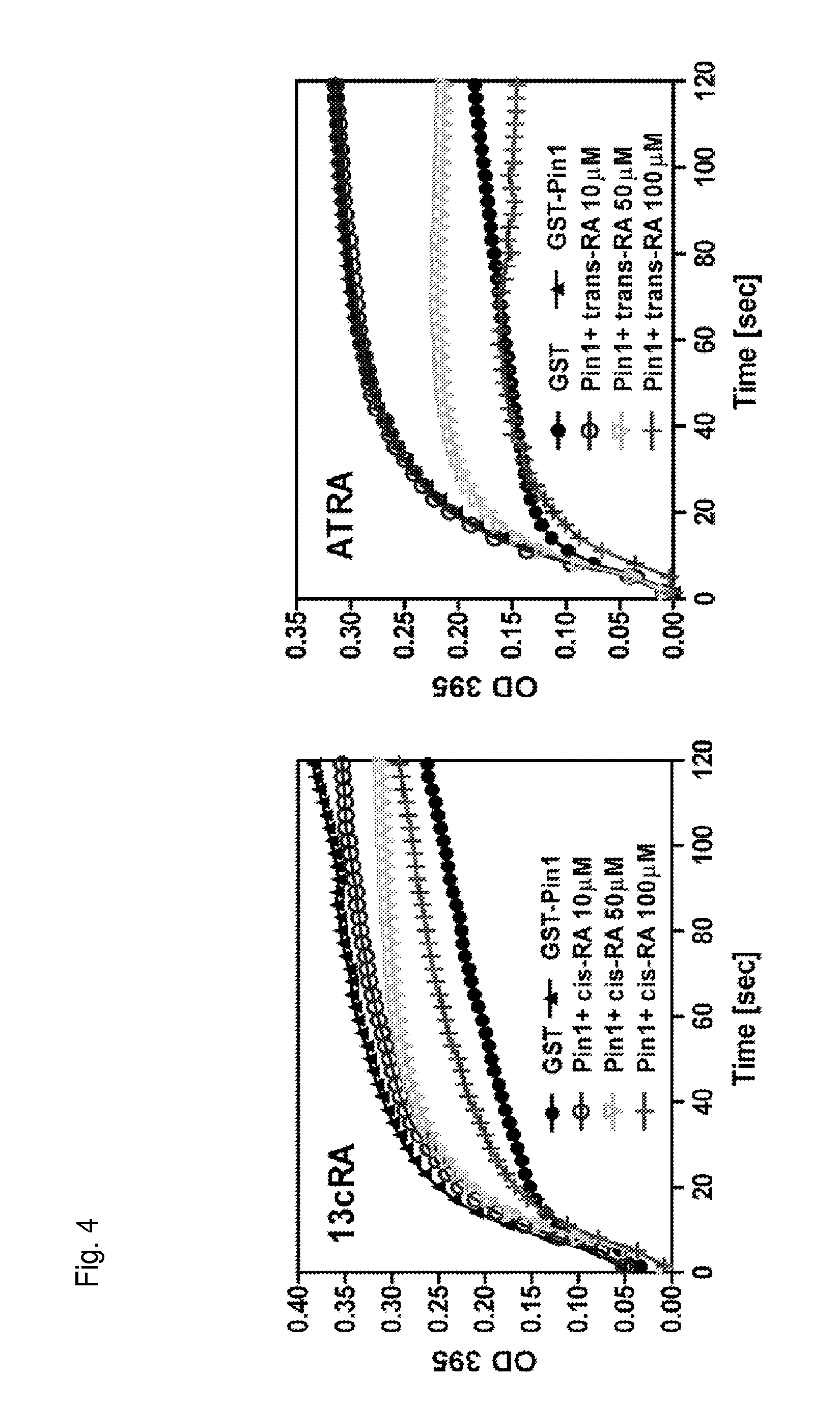
FIG. 3 is a pair of graphs showing the ability of 13-cis-retinoic acid and all-trans retinoic acid at different concentrations to compete with a fluorescence-labeled Pin1 peptide inhibitor probe BIKL-488 for binding Pin1 after an incubation of the indicated amount of time.
FIG. 4 is a pair of graphs showing inhibition of Pin1 by cis-retinoic acid and trans-retinoic acid at the indicated concentrations.
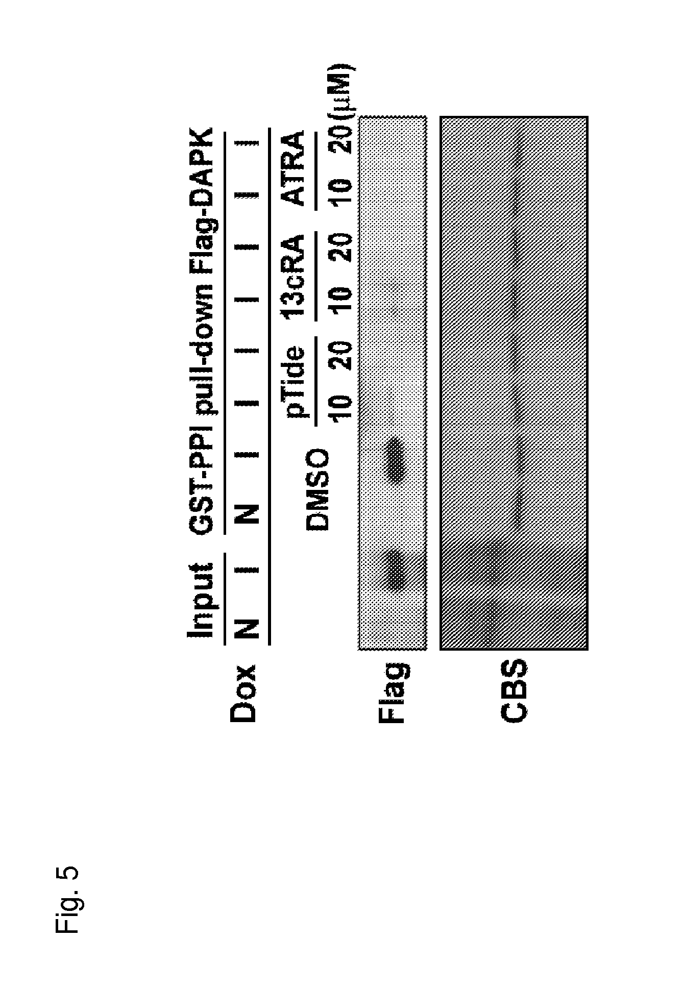
FIG. 5 is a photograph indicating both cis and trans retinoic acids can bind to Pin1 and compete away DAPK, which binds to the Pin1 catalytic domain.
FIG. 6A is a graph showing specific residues in the Pin1 active site that are important for interacting with the detecting probe BIKL-488.
FIG. 6B is a graph showing specific residues in the Pin1 active site that are important for interacting with retinoic acid.
FIG. 7A is a pair of graphs showing cell viability of the indicated cell lines as a function of concentration of cis-retinoic acid or trans-retinoic acid.
FIG. 7B is a series Western blots showing expression of the indicated protein in cells treated with of either cis-retinoic acid or trans-retinoic acid in the indicated cell lines.
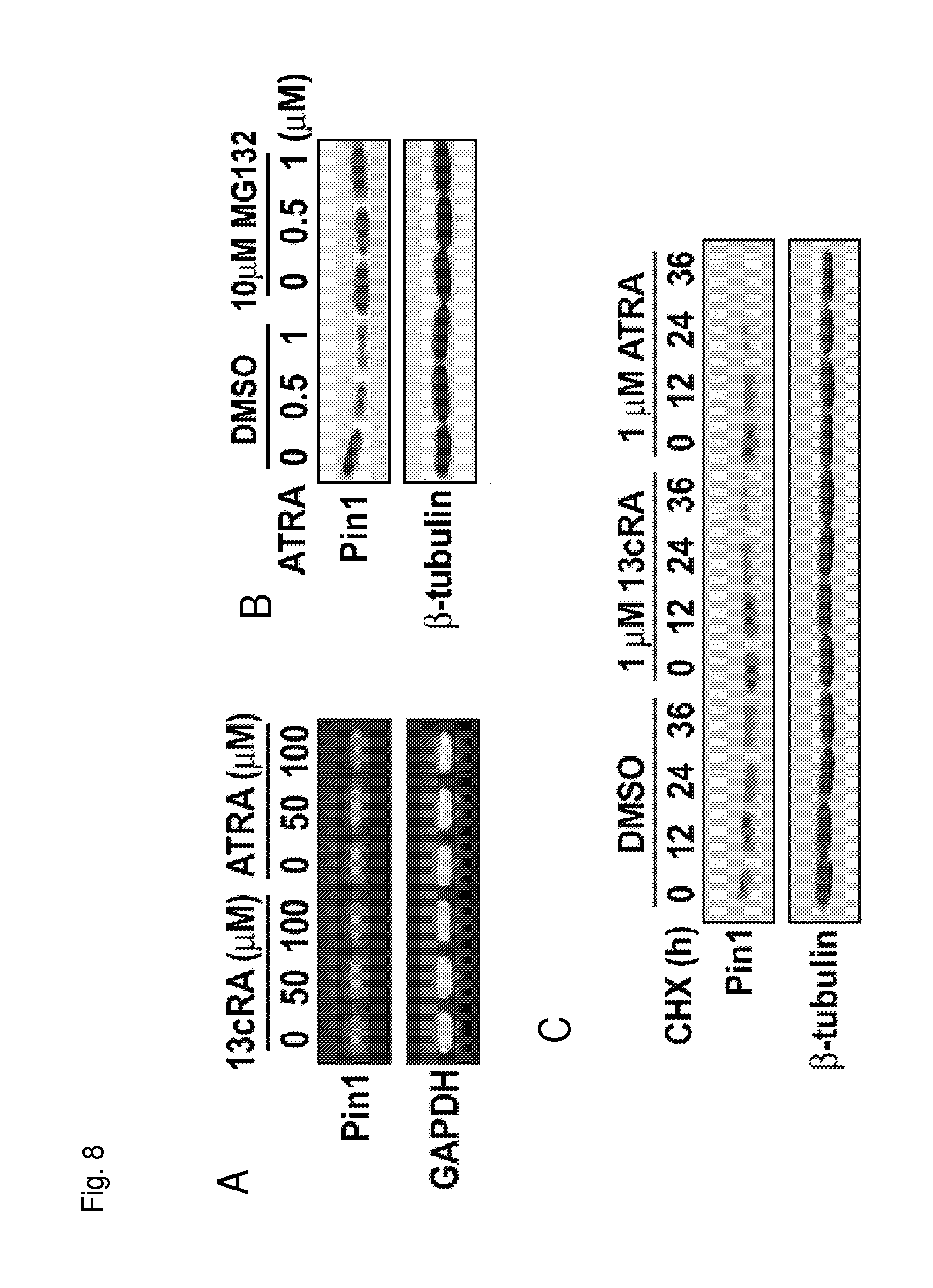
FIG. 8A is a photograph indicating the amount of Pin1 or GAPDH mRNA produced in the presence of the indicated compound as measured in an RT-PCR experiment.
FIG. 8B is a Western blot showing Pin1 and tubulin protein levels in cells treated with the indicated compounds.
FIG. 8C is a Western blot showing Pin1 and tubulin protein levels in cells treated with cycloheximide and the indicated compound.
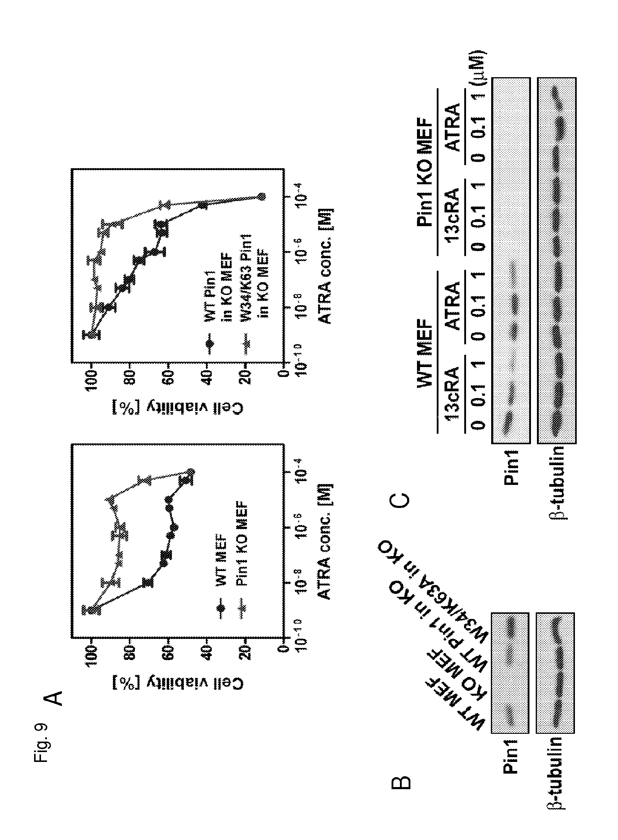
FIG. 9A is a pair of graphs showing cell viability as a function of the concentration of all trans-retinoic acid in the indicated cell types.
FIG. 9B is a Western blot showing Pin1 protein levels in the indicated cell types.
FIG. 9C is Western blot showing Pin1 protein levels in the indicated cell types treated with either 13cRA or ATRA.
FIG. 10A is a series of schematics showing the indicated retinoic acid compounds and .beta.-carotene.
FIG. 10B is a graph showing the concentration of free particles of Pin1 as a function of concentration of the indicated compounds.
FIG. 10C is a table summarizing the results of FIG. 10B.
FIG. 10D is a series of schematics showing compounds that modulate the retinoic acid receptor.
FIG. 11A is a schematic of the chemical structure of all-trans retinoic acid and acitretin.
FIG. 11B is a graph showing the concentration of free particles of Pin1 as a function of concentration of the indicated compounds.
FIG. 11C is a Western blot showing Pin1 and tubulin protein levels in cells treated with the different concentrations of the indicated compound.
FIG. 11D is a graph showing cell viability as a function of the concentration of the indicated compounds.
FIG. 12 is a graph showing cell number as a function of the concentration of the indicated compound or combination of compounds. For data points corresponding to the combination of compounds, the concentration value corresponds to the concentration of each individual compound in the combination.
FIG. 13A is a Western blot showing Pin, Her2, and actin protein levels in human breast cancer cells treated with the siRNA to inhibit Pin1.
FIG. 13B is a graph showing cell number as a function of inhibiting Pin1 in cells by siRNA.
DETAILED DESCRIPTION OF THE INVENTION
In general, the invention features methods of treating a proliferative disorder characterized by elevated Pin1 marker levels in a subject by administering a retinoic acid compound. Additionally, the invention features methods of treating proliferative disorders (e.g., proliferative disorders characterized by elevated Pin1 marker levels) by administering a retinoic acid compound in combination with one or more additional anti-proliferative compounds or other anti-cancer therapies.
Inhibitors of Pin1 (e.g., retinoic acid compounds) are useful for treating proliferative disorders (e.g., disorders characterized by increased Pin1 activity). Furthermore, because Pin1 acts in several different oncogenic pathways, Pin1 inhibition would be expected to behave synergistically with many anti-proliferative compounds.
I. Pin1
Phosphorylation on serine/threonine-proline motifs restrains cis/trans prolyl isomerization, and also creates a binding site for the essential protein Pin1. Pin1 binds and regulates the activity of a defined subset of phosphoproteins, as well as participating in the timing of mitotic progression. Both structural and functional analyses have indicated that Pin1 contains a phosphoserine/threonine-binding module that binds phosphoproteins, and a catalytic activity that specifically isomerizes the phosphorylated phosphoserine/threonine-proline. Both of these Pin1 activities are essential for Pin1 to carry out its function in vivo.
Pin1 is overexpressed in human cancer samples and the levels of Pin1 are correlated with the aggressiveness of tumors. We have found that a potent anticancer reagent with an unknown mechanism potently and reversibly inhibits Pin1 isomerase activity. Moreover, inhibition of Pin1 by various approaches, including the Pin1 inhibitor, Pin1 antisense polynucleotides, or genetic depletion, kills human and yeast dividing cells by inducing premature mitotic entry and apoptosis. Thus, upon phosphorylation, Pin1 latches onto phosphoproteins and twists the peptide bond next to the proline, which regulates the function of phosphoproteins and participates in controlling the timing of mitotic progression.
Pin1 is highly conserved and contains a protein-interacting module, called WW domain, and a catalytically active peptidyl-prolyl isomerase (PPIase). Pin1 is structurally and functionally distinct from members of two other well-characterized families of PPIases, the cyclophilins and the FKBPs. PPIases are ubiquitous enzymes that catalyze the typically slow prolyl isomerization of proteins, allowing relaxation of local energetically unfavorable conformational states. Phosphorylation on Ser/Thr residues immediately preceding Pro not only alters the prolyl isomerization rate, but also creates a binding site for the WW domain of Pin1. The WW domain acts a novel phosphoserine-binding module targeting Pin1 to a highly conserved subset of phosphoproteins. Furthermore, Pin1 displays a unique phosphorylation-dependent PPIase that specifically isomerizes phosphorylated Ser/Thr-Pro bonds and regulates the function of phosphoproteins.
Taken together, these results indicate that the Pin1 subfamily of enzymes is a diagnostic and therapeutic target for diseases characterized by uncontrolled cell proliferation, primarily malignancies.
II. Measurement of PIN1 Marker Levels
The present invention pertains to the treatment of proliferative diseases identified as coinciding with elevated Pin1 marker levels with retinoic acid compounds. In some aspects, the invention features the determination of Pin1 marker levels in a subject; where retinoic acid is administered in subjects where Pin1 marker levels are determined to be elevated. In other aspects, the invention can also feature the measurement of Pin1 marker levels subsequent to the administration of retinoic acid compounds in order to evaluate the progress of therapy in treating the proliferative disorder.
Accordingly, one aspect of the present invention relates to diagnostic assays for measuring levels of Pin1 marker, as well as Pin1 activity, in the context of a biological sample (e.g., tumor samples, blood, urine, biopsies, lymph, saliva, phlegm, and pus) to thereby determine whether an individual is a candidate for treatment with a retinoic acid compound. The invention features both treatment of subjects exhibiting symptoms of a proliferative disorder and individuals at risk for developing a proliferative disorder.
Diagnostic Assays
An exemplary method for detecting the presence or absence of Pin1 protein or nucleic acid in a biological sample involves obtaining a biological sample from a test subject and contacting the biological sample with a compound or an agent capable of detecting Pin1 protein or a nucleic acid (e.g., mRNA, genomic DNA) that encodes Pin1 protein such that the presence of Pin1 protein or nucleic acid is detected in the biological sample. A preferred agent for detecting Pin1 mRNA or genomic DNA is a labeled nucleic acid probe capable of hybridizing to Pin1 mRNA or DNA. The nucleic acid probe can be, for example, a Pin1 nucleic acid or a corresponding nucleic acid such as an oligonucleotide of at least 15, 30, 50, 100, 250 or 500 nucleotides in length which is capable of specifically hybridizing under stringent conditions to Pin1 mRNA or genomic DNA. Other suitable probes for use in the diagnostic assays of the invention are described herein.
A preferred agent for detecting Pin1 marker is an antibody capable of binding to Pin1 protein, preferably an antibody with a detectable label. Antibodies can be polyclonal, or more preferably, monoclonal. An intact antibody, or a fragment thereof (e.g., Fab or F(ab')2) can be used. The term "labeled," with regard to the probe or antibody, is intended to encompass direct labeling of the probe or antibody by coupling (i.e., physically linking) a detectable substance to the probe or antibody, as well as indirect labeling of the probe or antibody by reactivity with another reagent that is directly labeled. Examples of indirect labeling include detection of a primary antibody using a fluorescently labeled secondary antibody and end-labeling of a DNA probe with biotin such that it can be detected with fluorescently labeled streptavidin.
With respect to antibody-based detection techniques, one of skill in the art can raise anti-Pin1 antibodies against an appropriate immunogen, such as isolated and/or recombinant Pin1 or a portion or fragment thereof (including synthetic molecules, such as synthetic peptides) using no more than routine experimentation. Synthetic peptides can be designed and used to immunize animals, such as rabbits and mice, for antibody production. The nucleic and amino acid sequence of Pin1 is known (Hunter et al., WO 97/17986 (1997); Hunter et al., U.S. Pat. Nos. 5,952,467 and 5,972,697, the teachings of all of which are hereby incorporated by reference in their entirety) and can be used to design nucleic acid constructs for producing proteins for immunization or in nucleic acid detection methods or for the synthesis of peptides for immunization.
Conditions for incubating an antibody with a test sample can vary depending upon the tissue or cellular type. Incubation conditions can depend on the format employed in the assay, the detection methods employed, and the type and nature of the antibody used in the assay. One skilled in the art will recognize that any one of the commonly available immunological assay formats (such as radioimmunoassays, enzyme-linked immunosorbent assays, diffusion based Ouchterlony, or rocket immunofluorescent assays) can readily be adapted to employ the antibodies of the present invention. Examples of such assays can be found in Chard, "An Introduction to Radioimmunoassay and Related Techniques," Elsevier Science Publishers, Amsterdam, The Netherlands (1986); Bullock et al., "Techniques in Immunocytochemistry," Academic Press, Orlando, Fla. Vol. 1 (1982), Vol. 2 (1983), Vol. 3 (1985); Tijssen, "Practice and Theory of enzyme Immunoassays: Laboratory Techniques in Biochemistry and Molecular Biology," is Elsevier Science Publishers, Amsterdam, The Netherlands (1985).
The detection method of the invention can be used to detect Pin1 mRNA, protein, or genomic DNA in a biological sample in vitro as well as in vivo. For example, in vitro techniques for detection of Pin1 mRNA include Northern hybridizations and in situ hybridizations. In vitro techniques for detection of Pin1 protein include enzyme linked immunosorbent assays (ELISAs), Western blots, immunoprecipitations, immunofluorescence, or quantitative sequencing reactions. In vitro techniques for detection of Pin1 genomic DNA include Southern hybridizations. The detection of genomic mutations in Pin1 (or other genes that effect Pin1 marker levels) can be used to identify inherited or somatic mutations. Furthermore, in vivo techniques for detection of Pin1 protein include introducing into a subject a labeled anti-Pin1 antibody. For example, the antibody can be labeled with a radioactive marker whose presence and location in a subject can be detected by standard imaging techniques.
In another embodiment, the biological sample contains protein molecules from the test subject. Alternatively, the biological sample can contain mRNA molecules from the test subject or genomic DNA molecules from the test subject. A preferred biological sample is a serum sample isolated by conventional means from a subject.
In another embodiment, the methods further involve obtaining a control biological sample from a control subject, contacting the control sample with a compound or agent capable of detecting Pin1 marker such that the presence of Pin1 marker is detected in the biological sample, and comparing the presence of Pin1 marker in the control sample with the presence of Pin1 marker in the test sample.
The immunological assay test samples of the present invention may include cells, protein or membrane extracts of cells, blood or biological fluids such as ascites fluid or brain fluid (e.g., cerebrospinal fluid). The test sample used in the above-described method is based on the assay format, nature of the detection method and the tissues, cells or extracts used as the sample to be assayed. Methods for preparing protein extracts or membrane extracts of cells are well known in the art and can be readily be adapted in order to obtain a sample which is capable with the system utilized. The invention also encompasses kits for detecting the presence of Pin1 in a biological sample. For example, the kit can comprise a labeled compound or agent capable of detecting Pin1 protein or mRNA in a biological sample; means for determining the amount of Pin1 in the sample; and means for comparing the amount of Pin1 in the sample with a standard. The compound or agent can be packaged in a suitable container. The kit can further comprise instructions for using the kit to detect Pin1 protein or nucleic acid.
Pin1 marker levels can also be measured in an assay designed to evaluate a panel of target genes, e.g., a microarray or multiplex sequencing reaction. In the embodiments of the invention described herein, well known biomolecular methods such as northern blot analysis, RNase protection assays, southern blot analysis, western blot analysis, in situ hybridization, immunocytochemical procedures of tissue sections or cellular spreads, and nucleic acid amplification reactions (e.g., polymerase chain reactions) may be used interchangeably. One of skill in the art would be capable of performing these well-established protocols for the methods of the invention. (See, for example, Ausubel, et al., "Current Protocols in Molecular Biology," John Wiley & Sons, NY, N.Y. (1999)).
Diagnostic assays can be carried out in, e.g., subjects diagnosed or at risk of a proliferative disorder. Such disorders include, without limitation, leukemias (e.g., acute leukemia, acute lymphocytic leukemia, acute myelocytic leukemia, acute myeloblastic leukemia, acute promyelocytic leukemia, acute myelomonocytic leukemia, acute monocytic leukemia, acute erythroleukemia, chronic leukemia, chronic myelocytic leukemia, chronic lymphocytic leukemia), polycythemia vera, lymphoma (Hodgkin's disease, non-Hodgkin's disease), Waldenstrom's macroglobulinemia, heavy chain disease, and solid tumors such as sarcomas and carcinomas (e.g., fibrosarcoma, myxosarcoma, liposarcoma, chondrosarcoma, osteogenic sarcoma, chordoma, angiosarcoma, endotheliosarcoma, lymphangiosarcoma, lymphangioendotheliosarcoma, synovioma, mesothelioma, Ewing's tumor, leiomyosarcoma, rhabdomyosarcoma, colon carcinoma, pancreatic cancer, breast cancer, ovarian cancer, prostate cancer, squamous cell carcinoma, basal cell carcinoma, adenocarcinoma, sweat gland carcinoma, sebaceous gland carcinoma, papillary carcinoma, papillary adenocarcinomas, cystadenocarcinoma, medullary carcinoma, bronchogenic carcinoma, renal cell carcinoma, hepatoma, bile duct carcinoma, choriocarcinoma, seminoma, embryonal carcinoma, Wilm's tumor, cervical cancer, uterine cancer, testicular cancer, lung carcinoma, small cell lung carcinoma, bladder carcinoma, epithelial carcinoma, glioma, astrocytoma, medulloblastoma, craniopharyngioma, ependymoma, pinealoma, hemangioblastoma, acoustic neuroma, oligodenroglioma, schwannoma, meningioma, melanoma, neuroblastoma, and retinoblastoma).
Prognostic Assays
The diagnostic methods described herein can furthermore be utilized to identify subjects having or at risk of developing a disease or disorder associated with aberrant Pin1 expression or activity. For example, the assays described herein, such as the preceding diagnostic assays or the following assays, can be utilized to identify a subject having or at risk of developing a disorder associated with Pin1 marker (e.g., a proliferative disorder). Thus, the present invention provides a method for identifying a disease or disorder associated with aberrant Pin1 expression or activity in which a test sample is obtained from a subject and Pin1 protein or nucleic acid (e.g., mRNA, genomic DNA) is detected, wherein the presence of Pin1 protein or nucleic acid is diagnostic for a subject having or at risk of developing a Pin1-associated disorder and are, therefore, susceptible to treatment with a retinoic acid compound.
Furthermore, the present invention provides methods for determining whether a subject can be effectively treated with a retinoic acid compound for a disorder associated with aberrant Pin1 expression or activity in which a test sample is obtained and Pin1 protein or nucleic acid expression or activity is detected (e.g., wherein the abundance of Pin1 protein or nucleic acid expression or activity is diagnostic for a subject that can be administered the agent to treat a disorder Pin1-associated disorder).
In one embodiment, the present invention provides methods for determining Pin1 post-translational modifications. For example, phosphorylation of Pin1 on Ser71 in the catalytic active site by the tumor suppressor DAPK1 completely inhibits Pin1 catalytic activity and cell function to promote oncogenesis. More importantly, phosphorylation of Pin1 on Ser71 in the catalytic active site also prevents retinoic acid compounds from binding to Pin1 active site and induce Pin1 degradation and to inhibit Pin1 function. Therefore, by detecting reduced Ser71 phosphorylation using phospho-specific Pin1 antibodies that we have generated can be a method to select patients for RA treatments and to explain some patients may not respond to RA. Because aberrantly proliferating cells exhibit reduced Ser71 phosphorylation, these cells are more sensitive to RA treatments compared to normal cells.
The methods of the invention can also be used to detect genetic alterations in a Pin1 gene, thereby determining if a subject with the altered gene is at risk for a disorder associated with the Pin1 gene and, consequently, a candidate for retinoic acid therapy. In preferred embodiments, the methods include detecting, in a sample of cells from the subject, the presence or absence of a genetic alteration characterized by at least one of an alteration affecting the integrity of a gene encoding a Pin1-protein, or the mis-expression of the Pin1 gene. For example, such genetic alterations can be detected by ascertaining the existence of at least one of 1) a deletion of one or more nucleotides from a Pin1 gene; 2) an addition of one or more nucleotides to a Pin1 gene; 3) a substitution of one or more nucleotides of a Pin1 gene, 4) a chromosomal rearrangement of a Pin1 gene; 5) an alteration in the level of a messenger RNA transcript of a Pin1 gene, 6) aberrant modification of a Pin1 gene, such as of the methylation pattern of the genomic DNA, 7) the presence of a non-wild type splicing pattern of a messenger RNA transcript of a Pin1 gene, 8) a non-wild type level of a Pin1-protein, 9) allelic loss of a Pin1 gene, and 10) inappropriate post-translational modification of a Pin1-protein. As described herein, there are a large number of assay techniques known in the art which can be used for detecting alterations in a Pin1 gene. A preferred biological sample is a tissue or serum sample isolated by conventional means from a subject, e.g., a cardiac tissue sample.
In certain embodiments, detection of the alteration involves the use of a probe/primer in a polymerase chain reaction (PCR) (see, e.g., U.S. Pat. Nos. 4,683,195 and 4,683,202), such as anchor PCR or RACE PCR, or, alternatively, in a ligation chain reaction (LCR) (see, e.g., Landegran et al. (1988) Science 241:1077-1080; and Nakazawa et al. (1994) Proc. Natl. Acad. Sci. USA 91:360-364), the latter of which can be particularly useful for detecting point mutations in the Pin1-gene (see Abravaya et al. (1995) Nucleic Acids Res 0.23:675-682). This method can include the steps of collecting a sample from a patient, isolating nucleic acid (e.g., genomic, mRNA or both) from the sample, contacting the nucleic acid sample with one or more primers which specifically hybridize to a Pin1 gene under conditions such that hybridization and amplification of the Pin1-gene (if present) occurs, and detecting the presence or absence of an amplification product, or detecting the size of the amplification product and comparing the length to a control sample. It is anticipated that PCR and/or LCR may be desirable to use as a preliminary amplification step in conjunction with any of the techniques used for detecting mutations described herein.
Alternative amplification methods include: self sustained sequence replication (Guatelli, J. C. et al., (1990) Proc. Natl. Acad. Sci. USA 87:1874-1878), transcriptional amplification system (Kwoh, D. Y. et al, (1989) Proc. Natl. Acad. Sci. USA 86:1173-1177), Q-Beta Replicase (Lizardi, P. M. et al. (1988) Bio-Technology 6:1197), or any other nucleic acid amplification method, followed by the detection of the amplified molecules using techniques well known to those of skill in the art. These detection schemes are especially useful for the detection of nucleic acid molecules if such molecules are present in very low numbers.
In an alternative embodiment, mutations in a Pin1 gene from a sample cell can be identified by alterations in restriction enzyme cleavage patterns. For example, sample and control DNA is isolated, amplified (optionally), digested with one or more restriction endonucleases, and fragment length sizes are determined by gel electrophoresis and compared. Differences in fragment length sizes between sample and control DNA indicates mutations in the sample DNA. Moreover, the use of sequence specific ribozymes (see, for example, U.S. Pat. No. 5,498,531) can be used to score for the presence of specific mutations by development or loss of a ribozyme cleavage site.
In other embodiments, genetic mutations in Pin1 can be identified by hybridizing a sample and control nucleic acids, e.g., DNA or RNA, to high density arrays containing hundreds or thousands of oligonucleotides probes (Cronin, M. T. et al. (1996) Human Mutation 7: 244-255; Kozal, M. J. et al. (1996) Nature Medicine 2: 753-759). For example, genetic mutations in Pin1 can be identified in two dimensional arrays containing light-generated DNA probes as described in Cronin, M. T. et al. supra. Briefly, a first hybridization array of probes can be used to scan through long stretches of DNA in a sample and control to identify base changes between the sequences by making linear arrays of sequential overlapping probes. This step allows the identification of point mutations. This step is followed by a second hybridization array that allows the characterization of specific mutations by using smaller, specialized probe arrays complementary to all variants or mutations detected. Each mutation array is composed of parallel probe sets, one complementary to the wild-type gene and the other complementary to the mutant gene.
In yet another embodiment, any of a variety of sequencing reactions known in the art can be used to directly sequence the Pin1 gene and detect mutations by comparing the sequence of the sample Pin1 with the corresponding wild-type (control) sequence. Examples of sequencing reactions include those based on techniques developed by Maxam and Gilbert ((1977) Proc. Natl. Acad. Sci. USA 74:560) or Sanger ((1977) Proc. Natl. Acad. Sci. USA 74:5463). It is also contemplated that any of a variety of automated sequencing procedures can be utilized when performing the diagnostic assays ((1995) Biotechniques 19:448), including sequencing by mass spectrometry (see, e.g., PCT International Publication No. WO 94/16101; Cohen et al. (1996) Adv. Chromatogr. 36:127-162; and Griffin et al. (1993) Appl. Biochem. Biotechnol. 38:147-159).
Other methods for detecting mutations in the Pin1 gene include methods in which protection from cleavage agents is used to detect mismatched bases in RNA/RNA or RNA/DNA heteroduplexes (Myers et al. (1985) Science 230:1242). In general, the art technique of "mismatch cleavage" starts by providing heteroduplexes formed by hybridizing (labeled) RNA or DNA containing the wild-type Pin1 sequence with potentially mutant RNA or DNA obtained from a tissue sample. The double-stranded duplexes are treated with an agent which cleaves single-stranded regions of the duplex such as which will exist due to basepair mismatches between the control and sample strands. For instance, RNA/DNA duplexes can be treated with RNase and DNA/DNA hybrids treated with Si nuclease to enzymatically digesting the mismatched regions. In other embodiments, either DNA/DNA or RNA/DNA duplexes can be treated with hydroxylamine or osmium tetroxide and with piperidine in order to digest mismatched regions. After digestion of the mismatched regions, the resulting material is then separated by size on denaturing polyacrylamide gels to determine the site of mutation. See, for example, Cotton et al. (1988) Proc. Nat Acad Sci USA 85:4397; Saleeba et al. (1992) Methods Enzymol. 217:286-295. In a preferred embodiment, the control DNA or RNA can be labeled for detection.
In still another embodiment, the mismatch cleavage reaction employs one or more proteins that recognize mismatched base pairs in double-stranded DNA (so called "DNA mismatch repair" enzymes) in defined systems for detecting and mapping point mutations in Pin1 cDNAs obtained from samples of cells. For example, the mutY enzyme of E. coli cleaves A at G/A mismatches and the thymidine DNA glycosylase from HeLa cells cleaves Tat G/T mismatches (Hsu et al. (1994) Carcinogenesis 15:1657-1662). According to an exemplary embodiment, a probe based on a Pin1 sequence, e.g., a wild-type Pin1 sequence, is hybridized to a cDNA or other DNA product from a test cell(s). The duplex is treated with a DNA mismatch repair enzyme, and the cleavage products, if any, can be detected from electrophoresis protocols or the like. See, for example, U.S. Pat. No. 5,459,039.
In other embodiments, alterations in electrophoretic mobility will be used to identify mutations in Pin1 genes. For example, single strand conformation polymorphism (SSCP) may be used to detect differences in electrophoretic mobility between mutant and wild type nucleic acids (Orita et al. (1989) Proc Natl. Acad. Sci. USA: 86:2766, see also Cotton (1993) Mutat Res 285:125-144; and Hayashi (1992) Genet Anal Tech Appl 9:73-79). Single-stranded DNA fragments of sample and control Pin1 nucleic acids will be denatured and allowed to renature. The secondary structure of single-stranded nucleic acids varies according to sequence, the resulting alteration in electrophoretic mobility enables the detection of even a single base change. The DNA fragments may be labeled or detected with labeled probes. The sensitivity of the assay may be enhanced by using RNA (rather than DNA), in which the secondary structure is more sensitive to a change in sequence. In a preferred embodiment, the subject method utilizes heteroduplex analysis to separate double stranded heteroduplex molecules on the basis of changes in electrophoretic mobility (Keen et al. (1991) Trends Genet. 7:5).
In yet another embodiment the movement of mutant or wild-type fragments in polyacrylamide gels containing a gradient of denaturant is assayed using denaturing gradient gel electrophoresis (DGGE) (Myers et al. (1985) Nature 313:495). When DGGE is used as the method of analysis, DNA will be modified to insure that it does not completely denature, for example by adding a GC clamp of approximately 40 bp of high-melting GC-rich DNA by PCR. In a further embodiment, a temperature gradient is used in place of a denaturing gradient to identify differences in the mobility of control and sample DNA (Rosenbaum and Reissner (1987) Biophys Chem 265:12753).
Examples of other techniques for detecting point mutations include, but are not limited to, selective oligonucleotide hybridization, selective amplification, or selective primer extension. For example, oligonucleotide primers may be prepared in which the known mutation is placed centrally and then hybridized to target DNA under conditions which permit hybridization only if a perfect match is found (Saiki et al. (1986) Nature 324:163); Saiki et al. (1989) Proc. Natl Acad. Sci USA 86:6230). Such allele specific oligonucleotides are hybridized to PCR amplified target DNA or a number of different mutations when the oligonucleotides are attached to the hybridizing membrane and hybridized with labeled target DNA.
Alternatively, allele specific amplification technology which depends on selective PCR amplification may be used in conjunction with the instant invention. Oligonucleotides used as primers for specific amplification may carry the mutation of interest in the center of the molecule (so that amplification depends on differential hybridization) (Gibbs et al. (1989) Nucleic Acids Res. 17:2437-2448) or at the extreme 3' end of one primer where, under appropriate conditions, mismatch can prevent, or reduce polymerase extension (Prossner et al. (1993) Tibtech 11:238). In addition it may be desirable to introduce a novel restriction site in the region of the mutation to create cleavage-based detection (Gasparini et al. (1992) Mol. Cell Probes 6:1). It is anticipated that in certain embodiments amplification may also be performed using Taq ligase for amplification (Barany (1991) Proc. Natl. Acad. Sci USA 88:189). In such cases, ligation will occur only if there is a perfect match at the 3' end of the 5' sequence making it possible to detect the presence of a known mutation at a specific site by looking for the presence or absence of amplification.
The methods described herein may be performed, for example, by utilizing pre-packaged diagnostic kits comprising at least one probe nucleic acid or antibody reagent described herein, which may be conveniently used, e.g., in clinical settings to diagnose patients exhibiting symptoms or family history of a disease or illness involving a Pin1 gene.
Furthermore, any cell type or tissue in which Pin1 is expressed may be utilized in the prognostic assays described herein.
As with the diagnostic assay described above, prognostic assays of Pin1 activity can be included as part of a panel of target genes.
Additional methods of detecting Pin1 activity and diagnosing Pin1 related disorders are disclosed in U.S. Patent Application Publication Nos.: 2009/0258352, 2008/0214470, 2006/0074222, 2005/0239095, US2002/0025521, U.S. Pat. No. 6,495,376, and PCT Application Publication No. WO02/065091, each of which is hereby incorporated by reference in its entirety.
The present invention also features methods and compositions to diagnose, treat and monitor the progression of a disorder described herein (e.g., a cellular proliferation disorder) by detection and measurement of, for example, Pin1 substrates (or any fragments or derivatives thereof) containing a phosphorylated Ser/Thr-Pro motif in a cis or trans conformation, as described in U.S. Patent Provisional Application No. 61/255,431, which is hereby incorporated by reference in its entirety. The methods can include measurement of absolute levels of the Pin1 substrate (examples of which are listed in Tables 2, 3 and 4) in a cis or trans conformation as compared to a normal reference, using conformation specific antibodies. For example, a serum level or level in a biopsy of a Pin1 substrate in the cis or trans conformation that is less than 5 ng/ml, 4 ng/ml, 3 ng/ml, 2 ng/ml, or less than 1 ng/ml serum or a biopsy is considered to be predictive of a good outcome in a patient diagnosed with a disorder (e.g., a disorder associated with a deregulation of Pin1 activity). A serum level of the substrate in the cis or trans conformation that is greater than 5 ng/ml, 10 ng/ml, 20 ng/ml, 30 ng/ml, 40 ng/ml, or 50 ng/ml is considered diagnostic of a poor outcome in a subject already diagnosed with a disorder, e.g., associated with a deregulation of Pin1 activity.
For diagnoses based on relative levels of substrate in a particular conformation (e.g., a Pin1 substrate in the cis or trans conformation), a subject with a disorder (e.g., a disorder associated with a deregulation of PPIase activity) will show an alteration (e.g., an increase of 10%, 20%, 30%, 40%, 50%, 60%, 70%, 80%, 90%, or more) in the amount of the substrate in, for example, the cis conformation. A normal reference sample can be, for example, a prior sample taken from the same subject prior to the development of the disorder or of symptoms suggestive of the disorder, a sample from a subject not having the disorder, a sample from a subject not having symptoms of the disorder, or a sample of a purified reference polypeptide in a given conformation at a known normal concentration (i.e., not indicative of the disorder).
Standard methods may be used to measure levels of the substrate in any bodily fluid, including, but not limited to, urine, blood, serum, plasma, saliva, amniotic fluid, or cerebrospinal fluid. Such methods include immunoassay, ELISA, Western blotting, and quantitative enzyme immunoassay techniques.
For diagnostic purposes, the conformation-specific antibodies may be labeled. Labeling of the antibody is intended to encompass direct labeling of the antibody by coupling (e.g., physically linking) a detectable substance to the antibody, as well as indirect labeling the antibody by reacting the antibody with another reagent that is directly labeled. For example, the antibody can be labeled with a radioactive or fluorescent marker whose presence and location in a subject can be detected by standard imaging techniques.
The diagnostic methods described herein can be used individually or in combination with any other diagnostic method described herein for a more accurate diagnosis of the presence or severity of a disorder (e.g., a cellular proliferation disorder or a neurological disorder). Examples of additional methods for diagnosing such disorders include, e.g., examining a subject's health history, immunohistochemical staining of tissues, computed tomography (CT) scans, or culture growths.
Monitoring the Effects of Retinoic Acid Treatment, and Disease Progression
In one embodiment, the present invention features a method for monitoring the effectiveness of treatment of a subject with a retinoic acid compound comprising the steps of (i) obtaining a pre-administration sample from a subject prior to administration of the compound; (ii) detecting the level of expression or activity of a Pin1 protein, Pin1 phosphorylation on Ser71, mRNA, or genomic DNA in the pre-administration sample; (iii) obtaining one or more post-administration samples from the subject; (iv) detecting the level of expression or activity of the Pin1 protein, mRNA, or genomic DNA in the post-administration samples; (v) comparing the level of expression or activity of the Pin1 protein, mRNA, or genomic DNA in the pre-administration sample with the Pin1 protein, mRNA, or genomic DNA in the post administration sample or samples; and (vi) altering the administration of the retinoic acid compound to the subject accordingly. According to such an embodiment, Pin1 expression, phosphorylation or activity may be used as an indicator of the effectiveness of the retinoic acid compound, even in the absence of an observable phenotypic response.
In another embodiment, the diagnostic methods described herein can also be used to measure the levels of, for example, polypeptides (e.g., Pin1 substrates listed in Tables 2, 3 and 4) with pSer/Thr-Pro motifs in the cis or trans conformation using conformation specific antibodies, The methods can include repeated measurements, using conformation specific antibodies, for diagnosing the disorder and monitoring the treatment or management of the disorder. In order to monitor the progression of the disorder in a subject, subject samples can be obtained at several time points and conformation specific antibodies can be used to monitor the levels of cis and trans isomers of Pin1 substrates (listed in Tables 2, 3 and 4). For example, the diagnostic methods can be used to monitor subjects during chemotherapy (e.g., therapy with a retinoic acid compound or other agent described herein). In this example, serum samples from a subject can be obtained before treatment with a chemotherapeutic agent, again during treatment with a chemotherapeutic agent, and again after treatment with a chemotherapeutic agent. In this example, the level of Pin1 substrate with a pSer/Thr-Pro motif in the cis conformation in a subject is closely monitored using the conformation-specific antibodies of the invention and, if the level of Pin1 substrate with a pSer/Thr-Pro motif in the cis conformation begins to increase during therapy, the therapeutic regimen for treatment of the disorder can be modified as determined by the clinician (e.g., the dosage of the therapy may be changed or a different therapeutic may be administered). The monitoring methods of the invention may also be used, for example, in assessing the efficacy of a particular drug or therapy in a subject, determining dosages, or in assessing progression, status, or stage of the infection.
TABLE-US-00002 TABLE 2 General Pin1 Substrates Substrate (GenBank Functional Consequence Accession Targeting of PPIase Activity of Number) Site(s) Pin1 Upon Substrate G2/M and Mitotic Regulation NIMA -- Regulation of mitotic (P11837) function RAB4 -- -- (NP_004569) CDC25 pThr48/67-Pro Dephosphorylation and (AAA58417) regulation of activity WEE1 pT186-P Inhibition of WEE1 (NP_003381) activity PLK1 -- -- (P53350) MYT1 -- -- (NP_004194) CDC27 -- -- (AAH11656) CENP-F -- -- (P49454) Incenp -- -- (NP_064623) RPB1 pSer5-Pro Regulation of CTD (CAA65619) dephosphorylation NHERF-1 pSer279/301-P Dephosphorylation (AAA80218) KRMP1 pT-1604-P Regulation of mitotic (NP_057279) function CK2 Multiple pSer/Thr- Inhibition of kinase (NP_808227) Pro sites activity TopoII.alpha. -- Inhibition or induction (NP_001058) of phosphorylation DAB2 -- Dephosphorylation (NP_001334) p54nrb Multiple pSer/Thr- -- (CAA72157) Pro sites Sil Multiple pSer/Thr- Regulation of function (CAC14001) Pro sites EMI1 pS10-P Stabilization (NP_036309) G1/S Regulation Cyclin D1 pT286-P Stabilization and nuclear (NP_444284) localization Ki67 pT234-P -- c-Myc pT58-P Dephosphorylation and (CAA46984) destabilization Cyclin E pS384-P Destabilization (P24864) Growth and Oncogenic Signaling c-Jun pS63/73-P Transactivation (AAH06175) B-catenin pS246-P Stabilization, protein (P35222) interaction, and transactivation Cf-2 -- Destabilization (NP_034298) NF-.kappa.B pT254-P Stabilization, protein (AAH33210) interaction, and transactivation RAF1 Multiple pSer/Thr- Dephosphorylation and (AAA60247) Pro sites prolonging activation c-Fos Multiple pSer/Thr- Transactivation (CAA24756) Pro sites RAR.alpha. pS77-P Stabilization and (NP_001019980) transactivation AIB1/SRC-3 -- Transactivation and destabilization HBx pS41-P Stabilization and (NP_110380) potentiation STAT3 pS727-P Transactivation (NP_998827) DNA Damage, Oxidative Stress Response, and Apoptosis p53 Multiple pSer/Thr- Stabilization and (BAC16799) Pro sites transactivation Bcl-2 pS70/87-P -- (NP_000648) p73 Multiple pSer/Thr- Stabilization and (CAA72221) Pro sites transactivation BimEL pS65-P Stabilization (AAC39593) p66.sup.Shc -- Mitochondrial import (AAH14158) CHE1 -- Destabilization (P06276) Neuronal Survival and Degeneration Tau pT231-P Dephosphorylation and (NP_058519) pT212-P protein interaction APP pT668-P Promotes non-amyloidogenic (P05067) APP processing and reduces A.beta. production APP fragment pT668-P Increases A.beta. production from C99 APP fragment Synphilin-1 pS211/215-P Protein interaction (AAD30362) Gephyrin pS188/194/200-P Protein interaction (CAC81240) MCL1 pT163-P Stabilization (CAI15504) Immune Response and Asthma NFAT -- (NP_666017) AUF1 -- Protein interaction (NP_112738) IRF3 pS339-P Destabilization (AAH71721) BTK pS21/115-P Destabilization (CAI42359) Others SIN2-RPD3 -- Reduces histone deacetylases hSpt5 -- (NP_001124297)
TABLE-US-00003 TABLE 3A Pin1 targets where Pin1 prevents protein from degradation PIN1 PATHWAYS/ PROTEIN BINDING KINASE/ DISEASES/ NAME PIN1 FUNCTION MOTIF PHOSPHATASE MECHANISM 1 p53 1. Stabilization; Increase Ser33, Ser315, Chk2 Genotoxic stress, p53 promoter binding Thr181; Pro82 DNA damage activity Trophoblast 2. Pin1 in formation of invasiveness HMWC and stabilizes p53 2 Cyclin D1 Stabilization; Thr286 Block Socs-1 Cell localization and ub-mediated proliferation/ transcription proteolysis cancers 3 Tau 1. Dephosphorylated at Thr231 Tauopathy Thr231 2. Pin1 knockdown or KO increased WT tau protein stability in vitro 4 .beta.-Catenin Stability; localization Ser246 Could stabilize Cell and transactivation .beta.-catenin proliferation/cancers by inhibiting GSK-3.beta. dependent degradation 5 c-Jun Transcriptional activity Ser63/Ser73 JNK Breast cancer; AML 6 p65/NF-kB Nuclear translocation; Thr254 Cytokines; Hepatocyte stability NF-kappaB activation 7 p73 Stabilizing, Ser412, Thr442 c-Abl and p300 Genotoxic stress transcriptional activity and Thr482 8 Synphilin-1 Facilitates Lewy Body Ser211 and casein kinase II Parkinson disease formation; stabilizes Ser215 alpha-synuclein 9 c-Fos Transcriptional C-terminal ERK activation 10 Sil No impact on Sil Unknown spindle checkpoint 11 p54nrb Thr412, Thr430 Cdk1 and Thr452 12 Bruton Mediates Btk Ser21 an Ser115 Tyrosine kinase tyrosine degradation kinase (Btk) 13 AUF1 Regulates GM-CSF Ser83 mRNA; AUF1, AU rich element-binding protein 14 BIM.sub.EL Stabilize BIM.sub.EL and Ser65 JIP3, MKK7 and JNK Neuronal apoptosis induce apoptosis 15 Mcl-1 1. Pin1 inhibits Mcl-1 Thr163; Thr92 JNK3 induces Mcl-1 Oligodendrocyte (Myeloid cell ubiquitination degradation by apoptosis; Mcl-1 leukemia counting the mediated sequence-1) 2. Stabilizes Mcl-1 protective binding chemoresistance; of Pin1 breast cancer Erk phosphorylates Thr92 and Thr163 16 HBx; Pin1 overexpression Ser41 Pin1 binds HBx and Hepatocarcinogenesis Hepatitis B increased the protein enhance virus stability of HBx hepatocarcinogenesis encoded in HBV-infected protein X hepatocytes 17 Origin Prevents degradation of APC; Topo II Mitotisis; chromosome recognition ORC1 by inactivating segregation and for complex, mitotic APC complex reprogramming subunit 1 replicons (ORC1) 18 Bcl-2 1. Induce changes in the Possibly mediated by bioactivity of Bcl-2; cdc2 2. Prevents dephosphorylation of Bcl-2 19 Erb2 Stabilize ErbB2 Ubiquitinated ErbB2 pathway; Her2-positive breast erbB2 ubiquitin mediated cancer degradation 20 PPAR.gamma. Prevents the Ser84 Ras mediated kinase Macrophages mediated polyubiquitination of atherosclerosis PPAR.gamma. through ubiquitin-proteosome pathway 21 Cep55 Increased Cep55 Ser425, Ser428 Cdk1, Plk1 Mitosis and stability cytokinesis 22 Spt23 Ess1 stabilizes Spt23 Ser654 Unsaturated fatty-acid synthesis 23 p27 Pin1 protects p27 from Thr187 Cdk2 Cell cycle; Cancer degradation through polyubiquitination mechanism. 24 Akt Regulates Akt protein Thr92, Thr450 Oncogenesis stability 25 HTLV Tax Increased Tax protein Ser160 Pathogenesis of Human protein expression; inhibits Tax T-cell leukemia virus protein degradation type 1 (HTLV-1) related diseases 26 Nanog Leads to Nanog Ser52, Ser65 Stem cells pluripotency; stability cell renewal 27 Viral Stabilized phospho- Ser57 JNK HIV-1 cDNA Integrase HIV-1 integrase integration and infection
TABLE-US-00004 TABLE 3B Pin1 targets where Pin1 enhances degradation of phosphorylated proteins PATHWAYS/ PROTEIN PIN1 BINDING KINASE/ DISEASES/ NAME PIN1 FUNCTION MOTIF PHOSPHATASE MECHANISM 1 c-Myc Enhances c-Myc Thr58 Tumorgenesis degradation 2 Cyclin E Depletion of Pin1 Ser384 Cyclin E-Cdk2 complex Cell cycle, genomic upregulates cellular level instability and of cyclin E tumorigenesis 3 SRC-3/AIB1 Enhances SRC-3 N/A Steroid receptor degradation 4 RAR.alpha. Induces its degradation Ser77 Retinoic acid receptor; 5 IRF3 Promote its degradation Ser339 Host antiviral responses via the ubiquitin- during virus infection proteasome pathway. 6 Che-1 Conformational changes Thr144 p53 transcription; DNA induced by Pin1 are damage apoptotic requested for Che- pathway 1/HDM2 interaction 7 Pim-1 protein Binding of Pin1 leads to a N/A PP2A Elevated in lymphomas kinase decrease in the protein leukemias and prostate level of Pim-1 cancer through c-Myc pathway 8 Promyelocytic Binds to phosphorylated C-terminal of Breast cancer; hydrogen leukemia C terminus of PML and PML (Ser403, peroxide-induced death; protein enhances PML Ser505, Ser518 cell proliferation (PML) degradation and Ser527) 9 FOXO A novel negative FOXO Oxidative stress regulator, interconnecting FOXO phosphorylation and monoubiquitination in response to cellular stress to regulate p27 10 Silencing Pin1 destabilizes SMRT Ser1241, Her2/Neu/ErbB2 Human cancer mediator for Thr1445, Ser1469 receptor; Cdk2 retinoic acid and thyroid hormone receptor (SMRT) 11 TRF-1 Pin1 inhibition resulted in Thr149 Cdk Cancer; ageing decrease TRF1 degradation; 12 G protein- Promotes GRK2 Ser670 CDK2-cyclinA Cell cycle progression; coupled degradation p53 response and the receptor induction of apoptosis kinase 2 (GRK2) 13 SF-1 Pin1 promotes SF-1 Ser203 CDK7 Gonadotropin beta- ubiquitination and subunit gene transcription degradation 14 Sulfotransferase Pin1 destabilizes Thr8, Thr11 ERK1 and PP2A Metabolism of 4A1 SULT4A1 endogenous and (SULT4A1) exogenous compounds 15 Smad2/ Reduced Smad2/3 protein Thr179, Ser204, Smurf2 with Smads and TGF-.beta. signaling Smad3 levels Ser208, Ser213 enhanced Smad (Smad3 linker ubiquitination domain) 16 MEF2C Pin1 decreases MEF2C Ser98/Ser110 Muscle terminal stability differentiation
TABLE-US-00005 TABLE 3C Pin1 targets where Pin1 regulates target phosphorylation/dephosphorylation/other modifications PATHWAYS/ PROTEIN PIN1 BINDING KINASE/ DISEASES/ NAME PIN1 FUNCTION MOTIF PHOSPHATASE MECHANISM 1 Tau Enhance Thr231 Neuronal differentiation; dephosphorylation at stress induced; Thr231 Alzheimer disease 2 Raf-1 Dephosphorylation by N.A. Ras/MAP Kinase PP2A 3 Type-1 Cycling phosphorylation Thr72 CDK1-cyclin B Entry and exit in mitotic protein in mitotic phosphatase Inhibitor-2 (I- 2) 4 TGF-.beta.1 Decay, accumulation and Chronic asthma mRNA translation of TGF-.beta.1 mRNA in Eosinophils 5 K-homology parathyroid hormone Ser181 splicing (PTH) mRNA stability regulator protein (KSRP)
TABLE-US-00006 TABLE 4 Pin1-targets for promoting tumorigenesis Activation/ Substrates Function Binding Sites Effects Inactivation AIB1/SRC3 Transactivator -- Activity + Akt Protein kinase pThr.sup.92/450-Pro Stability + Bax Apoptosis pThr.sup.167-Pro Activity - Bcl-2 Antiapoptic protein -- Stability - Btk Tyrosine kinase pSer.sup.21/115-Pro Stability - .beta.-catenin Transcription factor pSer.sup.246-Pro Localization, + stability C/EBP Transcription factor -- Activity - Cyclin D1 Transcription factor pThr.sup.286-Pro Localization, + stability Daxx Apoptosis pSer.sup.178-Pro Stability - FAK Tyrosine kinase pSer.sup.910-Pro Activity + c-Fos Transcription factor -- Activity + FOXO4 Transcription factor -- Localization, - activity GRK2 G protein receptor pSer.sup.670-Pro Stability - Hbx Transactivator pSer.sup.41-Pro Activity, + stability c-Jun Transcription factor pSer.sup.63/73-Pro Activity, + stability Mcl-1 Apoptosis pThr.sup.92/163-Pro Stability + c-Myb Transactivator pSer.sup.528-Pro Activity + Neu Growth factor receptor -- Stability + NF-.kappa.B Transcription factor pThr.sup.254-Pro Localization, + stability Notch1 Growth factor -- Activity + p70S6K Ribosomal S6 kinases -- Activity + p53 Transcription factor -- Activity, -* stability Plk1 Mitotic kinase -- Binding + activity PML Transcription factor pSer.sup.403/505/518/527-Pro Stability - Raf-1 Protein kinase -- Activity + RAR.alpha. Transcriptional regulator pSer.sup.77-Pro Stability - V-Rel Transcription factor pThr.sup.254-Pro Localization, + stability Smad Transactivator -- Stability - SMRT Transcriptional pSer.sup.1241/1469, Stability - co-repressor Thr.sup.1445-Pro Stat3 Transcription factor pSer.sup.727-Pro Activity + Tax Viral oncoprotein pSer.sup.160-Pro Activity, + stability Pin2/TRF1 Telomere regulation pThr.sup.149-Pro Stability -
III. Methods of Treatment
The present invention provides for both prophylactic and therapeutic methods of treating a subject at risk of (or susceptible to) a proliferative disorder (e.g., a disorder associated with increased Pin1 expression or activity) with a retinoic compound.
Certain embodiments of the invention feature formulation of a retinoic acid compound for, e.g., controlled or extended release. Many strategies can be pursued to obtain controlled and/or extended release in which the rate of release outweighs the rate of metabolism of the therapeutic compound. For example, controlled release can be obtained by the appropriate selection of formulation parameters and ingredients (e.g., appropriate controlled release compositions and coatings). Examples include single or multiple unit tablet or capsule compositions, oil solutions, suspensions, emulsions, microcapsules, microspheres, nanoparticles, patches, and liposomes. The release mechanism can be controlled such that the retinoic acid compound is released at period intervals, the release could be simultaneous, or a delayed release of one of the agents of the combination can be affected, when the early release of one particular agent is preferred over the other.
Certain embodiments of the invention feature a deuterated retinoic acid compound that is made by replacing some or all hydrogen with deuterium using state of the art techniques (e.g., as described herein and at www.concertpharma.com).
Prophylactic Methods
In one aspect, the invention provides a method for preventing a proliferative disorder in a subject by administering to the subject a retinoic acid compound. Subjects at risk for a disease which is caused or contributed to by aberrant Pin1 expression or activity can be identified by, for example, any or a combination of diagnostic or prognostic assays as described herein. Administration of a retinoic acid compound can occur prior to the manifestation of symptoms characteristic of the Pin1 aberrancy, such that a disease or disorder is prevented or, alternatively, delayed in its progression.
Combination Therapies
Anti-proliferative and other anti-cancer compounds (e.g., anti-angiogenic compounds) are useful for treating proliferative disorders in combination with the retinoic acid compounds of the invention.
Anti-proliferative agents that can be used in combination with a retinoic acid compound include, without limitation, microtubule inhibitors, topoisomerase inhibitors, platins, alkylating agents, and anti-metabolites. Particular anty-proliferative agents that are useful in the methods and compositions of the invention include, without limitation, paclitaxel, gemcitabine, doxorubicin, vinblastine, etoposide, 5-fluorouracil, carboplatin, altretamine, aminoglutethimide, amsacrine, anastrozole, azacitidine, bleomycin, busulfan, carmustine, chlorambucil, 2-chlorodeoxyadenosine, cisplatin, colchicine, cyclophosphamide, cytarabine, cytoxan, dacarbazine, dactinomycin, daunorubicin, docetaxel, estramustine phosphate, floxuridine, fludarabine, gentuzumab, hexamethylmelamine, hydroxyurea, ifosfamide, imatinib, interferon, irinotecan, lomustine, mechlorethamine, melphalen, 6-mercaptopurine, methotrexate, mitomycin, mitotane, mitoxantrone, pentostatin, procarbazine, rituximab, streptozocin, tamoxifen, temozolomide, teniposide, 6-thioguanine, topotecan, trastuzumab, vincristine, vindesine, and vinorelbine. The ability of a compound to inhibit the growth of a neoplasm can be assessed using known animal models.
Compounds which are known to interact with other proteins implicated in Pin1 signaling pathways can also be useful in combination with a retinoic acid compound (see, e.g., the targets and compounds in Table 5).
TABLE-US-00007 TABLE 5 Target Target Class Representative Antagonist AKT Kinase MK-2206 Cyclin D1 Cyclin ON 013105 HER2/Neu (ErbB-2) Kinase Herceptin NF-.quadrature.F Transcription Factor RTA 402 Plk Kinase BI 2536 Raf-1 Kinase Sorafenib Stat3 Transcription Factor ISIS-STAT3Rx .quadrature.SIS-STAT Adhesion Nucleic Acid-Based Rx in Enzon Program at Santaris
Such compounds can act synergistically with a retinoic acid compound. Additionally, co-administration with a retinoic acid compound may result in the efficacy of the anti-proliferative compound at lower (and thus safer) doses (e.g., at least 5% less (e.g., at least 10%, 20%, 50%, 80%, 90%, or even 95%) less than when the anti-proliferative compound is administered alone.
Therapy according to the invention may be performed alone or in conjunction with another therapy and may be provided at home, the doctor's office, a clinic, a hospital's outpatient department, or a hospital. Treatment optionally begins at a hospital so that the doctor can observe the therapy's effects closely and make any adjustments that are needed, or it may begin on an outpatient basis. The duration of the therapy depends on the type of disease or disorder being treated, the age and condition of the patient, the stage and type of the patient's disease, and how the patient responds to the treatment. Additionally, a person having a greater risk of developing an proliferative disease may receive treatment to inhibit or delay the onset of symptoms.
Routes of administration for the various embodiments include, but are not limited to, topical, transdermal, nasal, and systemic administration (such as, intravenous, intramuscular, subcutaneous, inhalation, rectal, buccal, vaginal, intraperitoneal, intraarticular, ophthalmic, otic, or oral administration). As used herein, "systemic administration" refers to all nondermal routes of administration, and specifically excludes topical and transdermal routes of administration.
In combination therapy (e.g., a retinoic acid compound with a second anti-proliferative agent), the dosage and frequency of administration of each component of the combination can be controlled independently. For example, one compound may be administered three times per day, while the second compound may be administered once per day. Alternatively, one compound may be administered earlier and the second compound may be administered later. Combination therapy may be given in on-and-off cycles that include rest periods so that the patient's body has a chance to recover from any as yet unforeseen side effects. The compounds may also be formulated together such that one administration delivers both compounds.
Each compound of the combination may be formulated in a variety of ways that are known in the art. For example, the first and second agents may be formulated together or separately. Desirably, the first and second agents are formulated together for the simultaneous or near simultaneous administration of the agents. Such co-formulated compositions can include the two drugs together in the same pill, ointment, cream, foam, capsule, liquid, etc. It is to be understood that, when referring to the formulation of combinations of the invention, the formulation technology employed is also useful for the formulation of the individual agents of the combination, as well as other combinations of the invention. By using different formulation strategies for different agents, the pharmacokinetic profiles for each agent can be suitably matched.
The individually or separately formulated agents can be packaged together as a kit. Non-limiting examples include kits that contain, e.g., two pills, a pill and a powder, a suppository and a liquid in a vial, two topical creams, ointments, foams etc. The kit can include optional components that aid in the administration of the unit dose to patients, such as vials for reconstituting powder forms, syringes for injection, customized IV delivery systems, inhalers, etc. Additionally, the unit dose kit can contain instructions for preparation and administration of the compositions. The kit may be manufactured as a single use unit dose for one patient, multiple uses for a particular patient (at a constant dose or in which the individual compounds may vary in potency as therapy progresses); or the kit may contain multiple doses suitable for administration to multiple patients ("bulk packaging"). The kit components may be assembled in cartons, blister packs, bottles, tubes, and the like.
IV. Experimental Results
We have identified a series of nanomolar inhibitors that binds only to the catalytically active PPIase domain of Pin1. Upon electroporation or microinjection, these Pin1 peptide inhibitors block HeLa division, and this block is rescued by co-injected Pin1, indicating that the compounds are highly specific and potent. Using the above Pin1 peptide inhibitors, we have developed a high-throughput screen for identifying Pin1 inhibitors using a single-step fluorescence polarization-based displacement assay (FP-HTS). The FP-HTS detects molecules that compete for the substrate binding to the catalytic active site, measures ligand binding under equilibrium conditions, and does not suffer from product inhibition. The HF488 fluorescent probes for the FP assay contain only four residue core structure of Bth-L-phos.Thr-Pip-Nal (pTide), with a Kd of 258 nM for Pin1, was synthesized by Anaspec. We performed FP-HTS in a 384-well plate format with full length Pin1 and produced robust FP, resulting in a 6-7 fold increase in polarization degree values, using a Synergy II plate reader. This novel FP-HTS showed robust and reproducible performance. The assay can tolerate up to 10% DMSO. The Z' is around 0.70 and is consistent for day-to-day performance. The coefficient of variation is in the range of 4-5%. More importantly, we have shown that both pTide, the unlabeled Pin1 peptide as the positive control in this project, and juglone, the Pin1 small molecule inhibitor, displaced HF488 probe from Pin1. Although it is difficult to determine the Kd for the covalent and irreversible inhibitor juglone, the Kd for pTide was .about.250 nM, similar to that derived from PPIase-based assays.
We used the FP-HTS with a 5 nM probe and 200 nM Pin1 to conduct a pilot screen on a selected set of chemical libraries. We obtained the resulting potential positive hits and grouped them into 3 classes according to the Z-score, which is folds of standard deviation below the mean. The top strong chemical is the clinically used drug 13-cis-retinoic acid (cis-RA) (FIG. 2A). cis-RA is particularly attractive for the following reasons. 1) cis-RA is not listed in promiscuous inhibitor databases 2) all-trans retinoic acid (trans-RA), the pairing compound of cis-RA, is currently used as oral prescription for patients with acute promyelocytic leukemia (AML); 3) both cis-RA and trans-RAs are used as a medication in the phase II/III clinical trial for breast cancer, and more because of its ability to kill rapidly dividing cells; and 4) although it has been reported that RAs target on retinoic acid receptor (RAR), the exact mechanism of the anticancer action is unknown. As a result, we examined whether the anti-cancer effect of RAs depends on RAR in the breast cancer cells. RAR.alpha. knockdown can only partially rescue-cis-RA-mediated cell death, indicating that RAs may have unidentified "off-target effect." To confirm that RAs indeed target Pin1, we examined cis-RA and trans-RA in the FP assay, and, surprisingly, found that trans-RA displayed even more prominent Pin1 inhibition than cis-RA dose and that cis-RA would eventually catch up with trans-RA in the long-term incubation with Pin1, likely due to resonance-mediated cis-trans conversion (FIG. 3). In the PPIase assay, Pin1 activity was blocked by either cis- or trans-RA in a dose-dependent manner (FIG. 4). These data confirm that the interaction between RAs and Pin1 is specific and not due to aggregation. Furthermore, both trans and cis RA blocked the association between Pin1 and DAPK1 in a dose-dependent manner, with trans being more potent (FIG. 5). These results indicate that RAs binds to Pin1 C-terminal catalytic domain because DAPK1 is known to bind this domain (Lee et al., 2011 Mol Cell in press). To determine which amino acid residue in the Pin1 catalytic domain that are important for retinoic acid binding, point mutations of Pin1 including K63A, S67E, R68/69A, H59A or S71E completely or significantly abolished trans-RA binding to Pin1 (FIGS. 6A and B). Together, these data indicate that RAs inhibit Pin1 by occupying its catalytic PPIase pocket in the C-terminus and that phosphorylation of Ser67 or Ser71 inhibits RA binding to Pin1.
To further discern the causal relationship between the anti-proliferative effect of RAs on Pin1, cell viability of three breast cancer cell lines was tested with different dosages of cis- or trans-RA, of which SKBR3 and T47D exhibited preferential sensitivity to RAs with an IC50 in the nano-molar range, while the normal cell line MCF10A remained unaffected (FIG. 7A). This discrepancy between cell lines was correlated with the RAs' ability to suppress Pin1 expression. Pin1 level was decreased by treatment of RAs in drugs-responsive SKBR3 and T47D, but not in drugs-irresponsive normal cells, MCF10A (FIG. 7B), in which Pin1 target protein, cyclin D1, was served as biomarkers of in vivo Pin1 activity. Moreover, RAs did not alter Pin1 mRNA level, but did reduce Pin1 protein stability, suggesting that RAs interact with Pin1, result in Pin1 degradation, and subsequently leads to anti-proliferation of breast cancer cells (FIGS. 8A-C). To further confirm this premise, wild-type mouse embryonic fibroblast (WT MEF) and Pin1 knockout MEF (Pin1 KO MEF) were used to test trans-RA-mediated cell viability (FIGS. 9A-C). As expected, Pin1 KO MEF was more resistant to trans-RA than WT MEF due to lack of drug target. In addition, Pin1 KO MEF stably expressing WT Pin1, but not W34/K63A Pin1 mutant, enabled cells to re-sensitize trans-RA. These results indicate that RAs-mediated cell death is at least in part dependent on Pin1.
As trans-RA is selected as the current lead compound, it is important to identify the essential moiety of RAs on Pin1 interaction for further lead optimization. Many commercially available retinoids were tested for Pin1 inhibition. Only those with end carboxylic group maintained Pin1 inhibition in a cell culture model, which are cis-RA, trans-RA, AC-55649, and acitretin (FIGS. 10A, 10B, 11A, and 11B). Other RA compounds that are metabolized to contain an end carboxylic group are also likely to be useful in treating proliferative disorders (FIG. 10D). In addition, compounds that modulate the retinoic acid receptor (FIGS. 10D and 11A and compounds listed in Table 1) are also likely to be useful in treating proliferative disorders.
In addition, we have developed Cell-based assays to screen and validate Pin1 inhibitor hits. We have shown 1) that Pin1 is highly expressed in HER2-positive human breast cancer tissues; 2) that Pin1 inhibition almost completely suppresses HER2 overexpression on cell surface in human HER2+ breast cancer cell lines such as AU565 and SKBR3 cells; 3) that Pin1 inhibition greatly increases the sensitivity of HER2+ breast cancer cells to the mTOR inhibitor, but not to the HER2 inhibitor, suggesting that Pin1 might act on Her2 to regulate cell growth; 4) that Pin1 acts on Neu and multiple substrates in Neu-mediated oncogenic pathway; and 5) that Pin1 knockout in mice inhibits breast cancer development induced by activated Her2. Therefore, Pin1 is essential for maintaining HER2 overexpression and growth of human HER2+ breast cancer cells. Given that HER2 expression on cell surface and cell growth are readily assayed on 384-well format, we can test the ability of the hits to repress HER2 overexpression and cell growth of HER2_AU565 and SKBR3 cells, which will be treated with Pin1 prodrugs or hits, and then immunostained with Alexa 488-anti-HER2 monoclonal antibody (BioLegend), followed by automated microscopy.
We have further shown that combinations of therapeutic compounds including retinoic acid compounds are useful for treating cancer, e.g., cancer characterized by elevated Pin1 activity. FIG. 12 shows results obtained from the treatment of breast cancer cells overexpressing Pin1 with ATRA or Doxorubicin or their combination, followed by counting cancer cell numbers. The results show that ATRA and Doxorubicin combination dramatically increases anticancer potency and reduce the dose of each drug to inhibit cancer cell growth. Therefore, ATRA can drastically reduce dose and toxicity of Doxorubicin and other chemotherapeutic drugs.
We have also shown that Pin1 inhibition, using siRNA, dramatically reduces Neu/Erb2 overexpression and cell proliferation of human breast cancer cells that have Neu/Erb2 gene amplification (FIGS. 13A and 13B). This provides us with a method to identify a Pin1 modulatory compound by applying a test compound to human-derived cancer cells that have Neu/Erb2 gene amplification, and determining the effect of the test compound on Neu/Erb2 overexpression and cell proliferation.
Other Embodiments
All publications, patents, and patent applications mentioned in this specification are herein incorporated by reference to the same extent as if each independent publication or patent application was specifically and individually indicated to be incorporated by reference.
While the invention has been described in connection with specific embodiments thereof, it will be understood that it is capable of further modifications and this application is intended to cover any variations, uses, or adaptations of the invention following, in general, the principles of the invention and including such departures from the present disclosure that come within known or customary practice within the art to which the invention pertains and may be applied to the essential features hereinbefore set forth, and follows in the scope of the claims.
Other embodiments are within the claims.
* * * * *
References
D00001
D00002
D00003
D00004
D00005
D00006
D00007
D00008
D00009
D00010
D00011
D00012
D00013
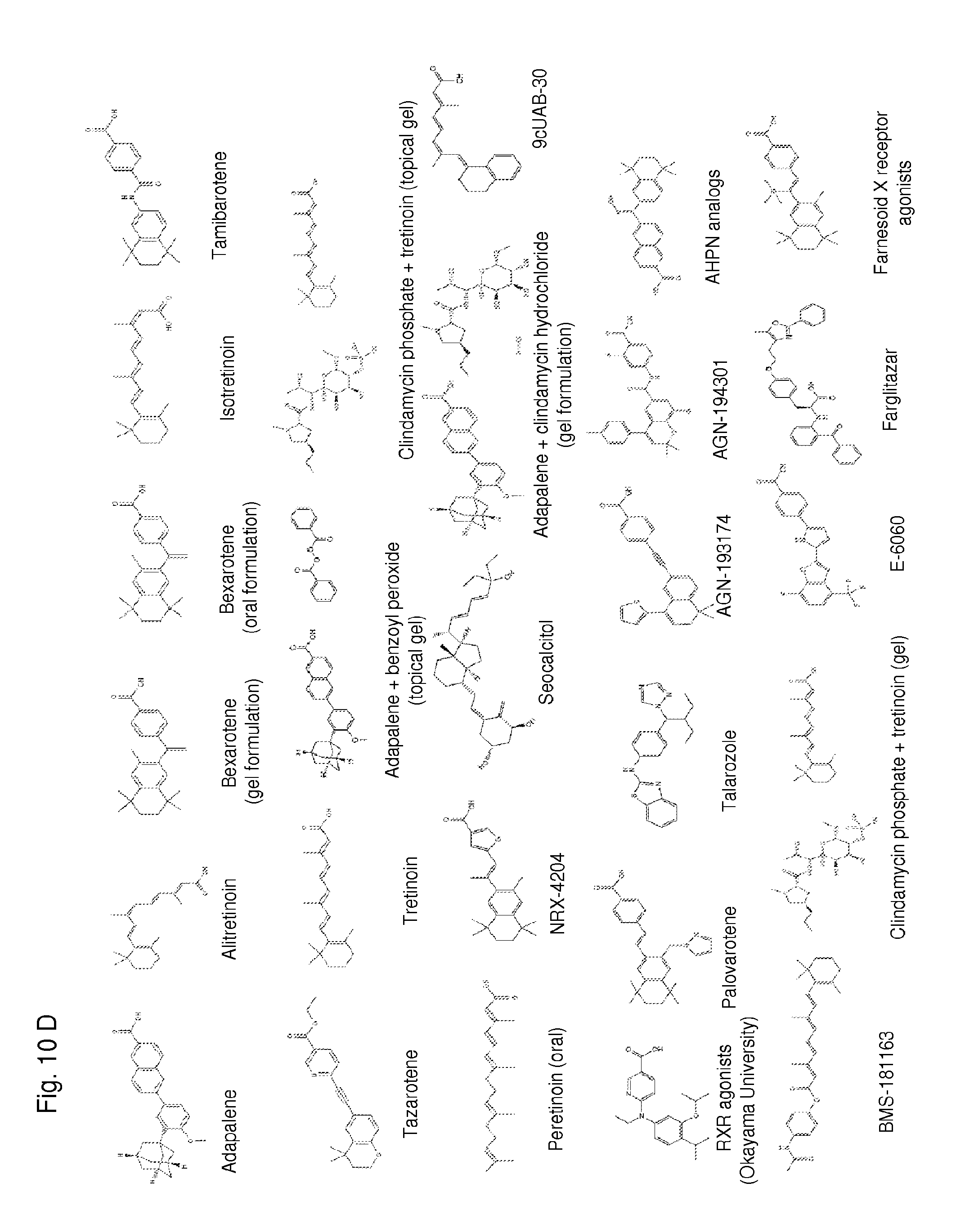
D00014
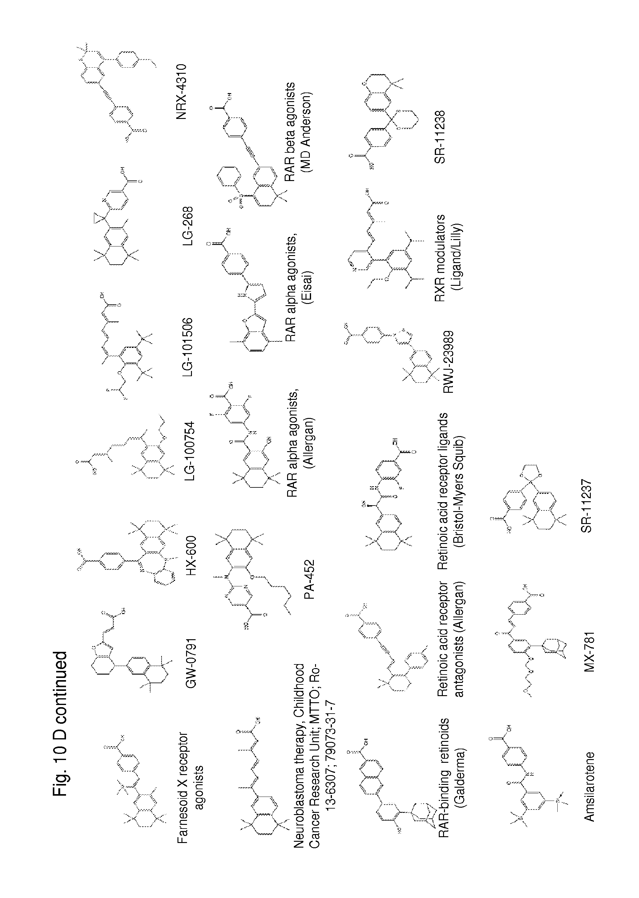
D00015
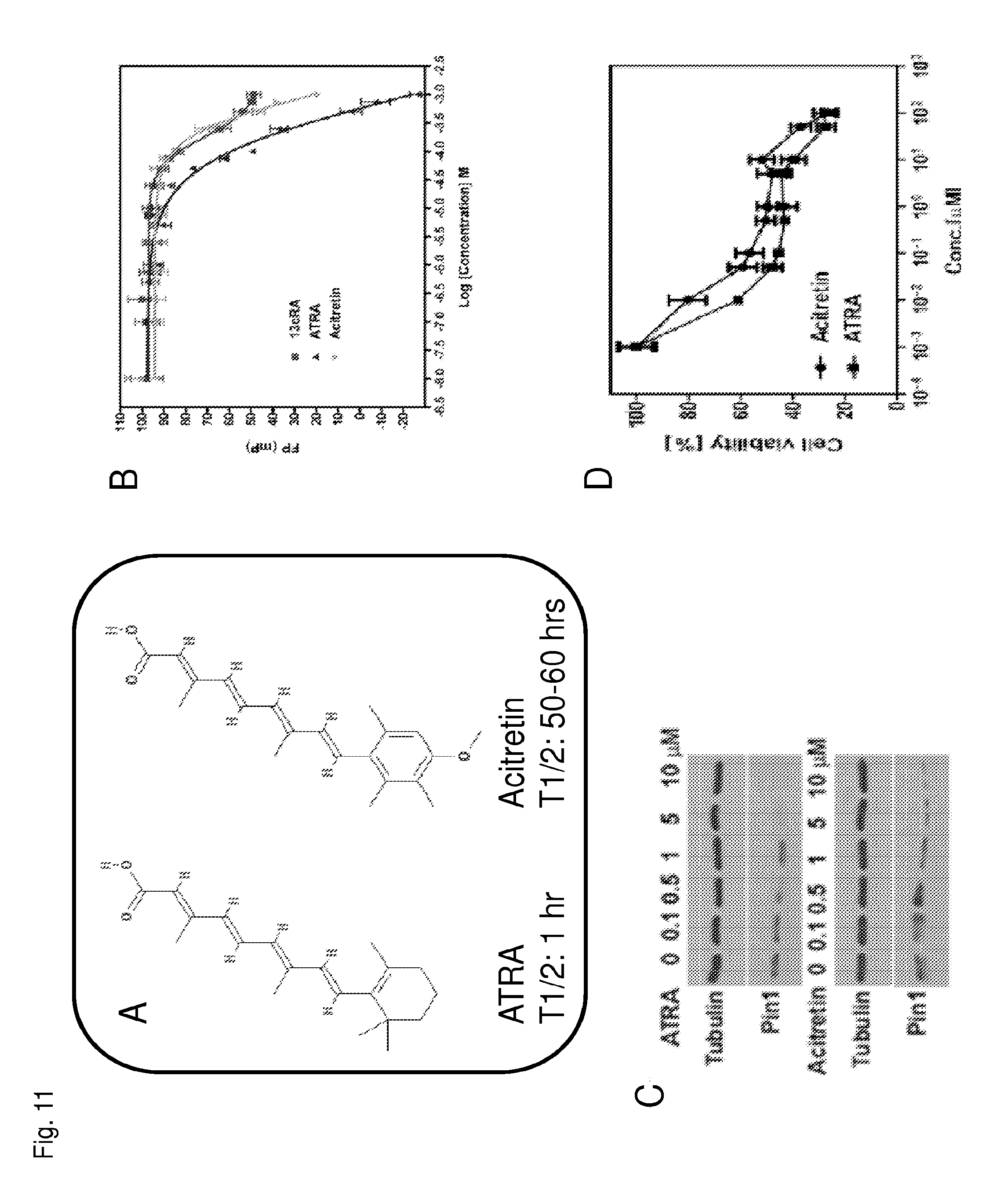
D00016
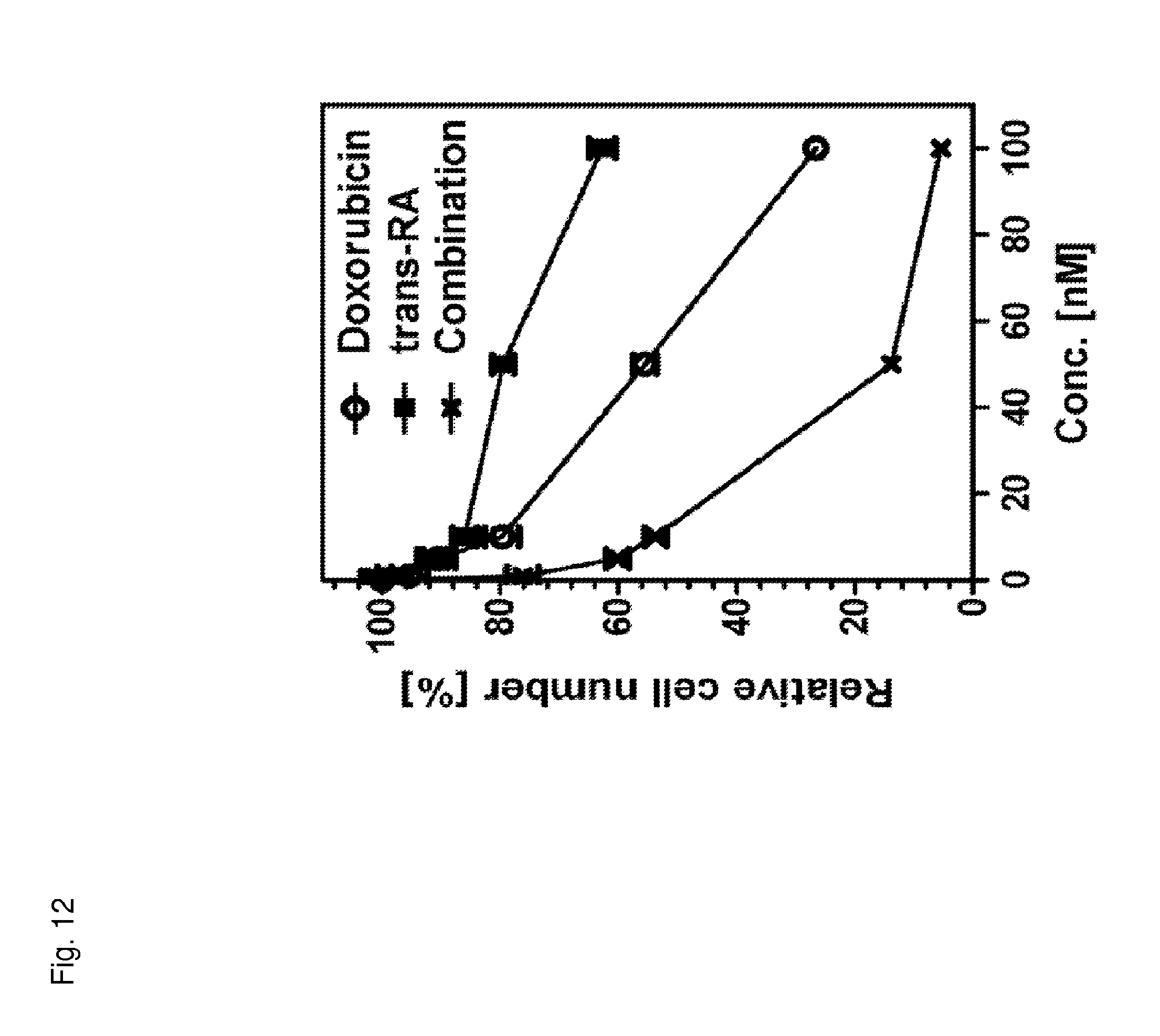
D00017

XML
uspto.report is an independent third-party trademark research tool that is not affiliated, endorsed, or sponsored by the United States Patent and Trademark Office (USPTO) or any other governmental organization. The information provided by uspto.report is based on publicly available data at the time of writing and is intended for informational purposes only.
While we strive to provide accurate and up-to-date information, we do not guarantee the accuracy, completeness, reliability, or suitability of the information displayed on this site. The use of this site is at your own risk. Any reliance you place on such information is therefore strictly at your own risk.
All official trademark data, including owner information, should be verified by visiting the official USPTO website at www.uspto.gov. This site is not intended to replace professional legal advice and should not be used as a substitute for consulting with a legal professional who is knowledgeable about trademark law.