Selective stimulation to modulate the sympathetic nervous system
Kramer , et al.
U.S. patent number 10,232,180 [Application Number 15/861,592] was granted by the patent office on 2019-03-19 for selective stimulation to modulate the sympathetic nervous system. This patent grant is currently assigned to The Board of Trustees of the Leland Stanford Junior University, St. Jude Medical Luxembourg Holdings SMI S.A.R.L. ("SJM LUX SMI"). The grantee listed for this patent is The Board of Trustees of the Leland Stanford Junior University, St. Jude Medical Luxembourg Holdings SMI S.A.R.L. ("SJM LUX SMI"). Invention is credited to Mir A. Imran, Daniel H. Kim, Jeffery M. Kramer.


| United States Patent | 10,232,180 |
| Kramer , et al. | March 19, 2019 |
Selective stimulation to modulate the sympathetic nervous system
Abstract
Systems, methods and devices are provided for the targeted treatment of a variety of medical conditions by directly neuromodulating a target anatomy associated with the condition while minimizing or excluding undesired neuromodulation of other anatomies. Typically, the target anatomy includes one or more dorsal root ganglia, dorsal roots, dorsal root entry zones, or portions thereof. Such target stimulation areas are utilized due in part to their effect on the sympathetic nervous system.
| Inventors: | Kramer; Jeffery M. (San Francisco, CA), Kim; Daniel H. (Houston, TX), Imran; Mir A. (Los Altos, CA) | ||||||||||
|---|---|---|---|---|---|---|---|---|---|---|---|
| Applicant: |
|
||||||||||
| Assignee: | The Board of Trustees of the Leland
Stanford Junior University (Stanford, CA) St. Jude Medical Luxembourg Holdings SMI S.A.R.L. ("SJM LUX SMI") (Luxembourg, LU) |
||||||||||
| Family ID: | 47068549 | ||||||||||
| Appl. No.: | 15/861,592 | ||||||||||
| Filed: | January 3, 2018 |
Prior Publication Data
| Document Identifier | Publication Date | |
|---|---|---|
| US 20180126166 A1 | May 10, 2018 | |
Related U.S. Patent Documents
| Application Number | Filing Date | Patent Number | Issue Date | ||
|---|---|---|---|---|---|
| 15346587 | Nov 8, 2016 | ||||
| 14954740 | Nov 8, 2016 | 9486633 | |||
| 13458697 | Apr 27, 2012 | ||||
| 12369706 | Jul 24, 2012 | 8229565 | |||
| 11222516 | Mar 10, 2009 | 7502651 | |||
| 61480958 | Apr 29, 2011 | ||||
| 60608357 | Sep 8, 2004 | ||||
| Current U.S. Class: | 1/1 |
| Current CPC Class: | A61N 1/36135 (20130101); A61N 1/36071 (20130101); A61N 1/36114 (20130101); A61N 1/36057 (20130101); A61N 1/0551 (20130101) |
| Current International Class: | A61N 1/05 (20060101); A61N 1/36 (20060101) |
References Cited [Referenced By]
U.S. Patent Documents
| 525891 | September 1894 | Fricke |
| 3724467 | April 1973 | Avery et al. |
| 3845770 | November 1974 | Theeuwes et al. |
| 3916899 | November 1975 | Theeuwes et al. |
| 4141367 | February 1979 | Ferreira |
| 4232679 | November 1980 | Schulman |
| 4298003 | November 1981 | Theeuwes et al. |
| 4313448 | February 1982 | Stokes |
| 4374527 | February 1983 | Iversen |
| 4479491 | October 1984 | Martin |
| 4549556 | October 1985 | Tarjan et al. |
| 4573481 | March 1986 | Bullara |
| 4577642 | March 1986 | Stokes |
| 4590946 | May 1986 | Loeb |
| 4607639 | August 1986 | Tanagho et al. |
| 4739764 | April 1988 | Lue et al. |
| 4786155 | November 1988 | Fantone et al. |
| 4803988 | February 1989 | Thomson |
| 4920979 | May 1990 | Bullara |
| 4940065 | July 1990 | Tanagho et al. |
| 4950270 | August 1990 | Bowman et al. |
| 4976711 | December 1990 | Parins et al. |
| 5135525 | August 1992 | Biscoping et al. |
| 5270099 | December 1993 | Kamiyama et al. |
| 5299569 | April 1994 | Wernicke et al. |
| 5344438 | September 1994 | Testerman et al. |
| 5358514 | October 1994 | Schulman et al. |
| 5370644 | December 1994 | Langberg |
| 5411537 | May 1995 | Munshi et al. |
| 5411540 | May 1995 | Edell et al. |
| 5417719 | May 1995 | Hull et al. |
| 5419763 | May 1995 | Hilderbrand |
| 5458626 | October 1995 | Krause |
| 5489294 | February 1996 | McVenes et al. |
| 5505201 | April 1996 | Grill et al. |
| 5514175 | May 1996 | Kim et al. |
| 5584835 | December 1996 | Greenfield |
| 5634462 | June 1997 | Tyler et al. |
| 5643330 | July 1997 | Holsheimer et al. |
| 5702429 | December 1997 | King |
| 5711316 | January 1998 | Elsberry et al. |
| 5713922 | February 1998 | King |
| 5733322 | March 1998 | Starkebaum |
| 5741319 | April 1998 | Woloszko et al. |
| 5755750 | May 1998 | Petruska et al. |
| 5776170 | July 1998 | MacDonald et al. |
| 5807339 | September 1998 | Bostrom et al. |
| 5824021 | October 1998 | Rise |
| 5865843 | February 1999 | Baudino |
| 5871531 | February 1999 | Struble |
| 5885290 | March 1999 | Guerrero et al. |
| 5938690 | August 1999 | Law et al. |
| 5941906 | August 1999 | Barreras et al. |
| 5948007 | September 1999 | Starkebaum et al. |
| 5957965 | September 1999 | Moumane et al. |
| 5983141 | November 1999 | Sluijter et al. |
| 5984896 | November 1999 | Boyd |
| 6002964 | December 1999 | Feler et al. |
| 6044297 | March 2000 | Sheldon et al. |
| 6045532 | April 2000 | Eggers et al. |
| 6051017 | April 2000 | Loeb et al. |
| 6104957 | August 2000 | Alo et al. |
| 6120467 | September 2000 | Schallhorn |
| 6161048 | December 2000 | Sluijter et al. |
| 6175764 | January 2001 | Loeb et al. |
| 6181965 | January 2001 | Loeb et al. |
| 6185455 | February 2001 | Loeb et al. |
| 6205359 | March 2001 | Boveja |
| 6208902 | March 2001 | Boveja |
| 6214016 | April 2001 | Williams et al. |
| 6259952 | July 2001 | Sluijter et al. |
| 6298256 | October 2001 | Meyer |
| 6314325 | November 2001 | Fitz |
| 6319241 | November 2001 | King et al. |
| 6349233 | February 2002 | Adams |
| 6353762 | March 2002 | Baudino et al. |
| 6356786 | March 2002 | Rezai et al. |
| 6360750 | March 2002 | Gerber et al. |
| 6366814 | April 2002 | Boveja et al. |
| 6393325 | May 2002 | Mann et al. |
| 6413255 | July 2002 | Stern |
| 6425887 | July 2002 | McGuckin et al. |
| 6438423 | August 2002 | Rezai et al. |
| 6440090 | August 2002 | Schallhorn |
| 6466821 | October 2002 | Pianca et al. |
| 6493588 | December 2002 | Malaney et al. |
| 6510347 | January 2003 | Borkan |
| 6512958 | January 2003 | Swoyer et al. |
| 6516227 | February 2003 | Meadows et al. |
| 6517542 | February 2003 | Papay et al. |
| 6522926 | February 2003 | Kieval et al. |
| 6535767 | March 2003 | Kronberg |
| 6582441 | June 2003 | He et al. |
| 6587725 | July 2003 | Durand et al. |
| 6605094 | August 2003 | Mann et al. |
| 6606521 | August 2003 | Paspa et al. |
| 6611715 | August 2003 | Boveja |
| 6625496 | September 2003 | Ollivier |
| 6638276 | October 2003 | Sharkey et al. |
| 6658302 | December 2003 | Kuzma et al. |
| 6714822 | March 2004 | King et al. |
| 6748276 | June 2004 | Diagnault, Jr. et al. |
| 6754539 | June 2004 | Erickson et al. |
| 6788975 | September 2004 | Whitehurst et al. |
| 6792318 | September 2004 | Chitre et al. |
| 6832115 | December 2004 | Borkan |
| 6835194 | December 2004 | Johnson et al. |
| 6839588 | January 2005 | Rudy |
| 6849075 | February 2005 | Bertolero et al. |
| 6862479 | March 2005 | Whitehurst et al. |
| 6871099 | March 2005 | Whitehurst et al. |
| 6873342 | March 2005 | Perry et al. |
| 6889094 | May 2005 | Kuzma et al. |
| 6901287 | May 2005 | Davis et al. |
| 6902547 | June 2005 | Aves et al. |
| 6909917 | June 2005 | Woods et al. |
| 6928320 | August 2005 | King |
| 6971391 | December 2005 | Wang et al. |
| 6978180 | December 2005 | Tadlock |
| 7047082 | May 2006 | Schrom et al. |
| 7096070 | August 2006 | Jenkins et al. |
| 7127287 | October 2006 | Duncan et al. |
| 7181289 | February 2007 | Pflueger et al. |
| 7333857 | February 2008 | Campbell |
| 7337005 | February 2008 | Kim et al. |
| 7337006 | February 2008 | Kim et al. |
| 7447546 | November 2008 | Kim et al. |
| 7450993 | November 2008 | Kim et al. |
| 7502651 | March 2009 | Kim et al. |
| 7580753 | August 2009 | Kim et al. |
| 8082039 | December 2011 | Kim et al. |
| 8229565 | July 2012 | Kim et al. |
| 8380318 | February 2013 | Kishawi et al. |
| 8518092 | August 2013 | Burdulis |
| 8712546 | April 2014 | Kim et al. |
| 8983624 | March 2015 | Imran |
| 9044592 | June 2015 | Imran et al. |
| 9056197 | June 2015 | Imran et al. |
| 9205259 | December 2015 | Kim et al. |
| 9205260 | December 2015 | Kim et al. |
| 9205261 | December 2015 | Kim et al. |
| 9486633 | November 2016 | Kramer et al. |
| 2001/0003799 | June 2001 | Boveja |
| 2001/0006967 | July 2001 | Crain et al. |
| 2002/0064841 | May 2002 | Klemic et al. |
| 2002/0077543 | June 2002 | Grzeszczuk et al. |
| 2002/0077684 | June 2002 | Clemens et al. |
| 2002/0087113 | July 2002 | Hartlaub |
| 2002/0099430 | July 2002 | Verness |
| 2002/0116030 | August 2002 | Rezai |
| 2002/0128694 | September 2002 | Holsheimer |
| 2002/0147486 | October 2002 | Soukup et al. |
| 2002/0198527 | December 2002 | Muckter |
| 2003/0018367 | January 2003 | Dilorenzo |
| 2003/0023241 | January 2003 | Drewry et al. |
| 2003/0045919 | March 2003 | Swoyer et al. |
| 2003/0069569 | April 2003 | Burdette et al. |
| 2003/0078633 | April 2003 | Firlik et al. |
| 2003/0088301 | May 2003 | King |
| 2003/0100933 | May 2003 | Ayal et al. |
| 2003/0114905 | June 2003 | Kuzma |
| 2003/0130577 | July 2003 | Purdy et al. |
| 2003/0144657 | July 2003 | Bowe et al. |
| 2003/0144709 | July 2003 | Zabara et al. |
| 2003/0181958 | September 2003 | Dobak, III |
| 2003/0187490 | October 2003 | Gliner |
| 2003/0195602 | October 2003 | Boling |
| 2003/0220677 | November 2003 | Doan et al. |
| 2004/0015202 | January 2004 | Chandler et al. |
| 2004/0019359 | January 2004 | Worley et al. |
| 2004/0019369 | January 2004 | Duncan et al. |
| 2004/0059404 | March 2004 | Bjorklund et al. |
| 2004/0111118 | June 2004 | Hill et al. |
| 2004/0116977 | June 2004 | Finch et al. |
| 2004/0122360 | June 2004 | Waldhauser et al. |
| 2004/0122477 | June 2004 | Whitehurst et al. |
| 2004/0122497 | June 2004 | Zhang et al. |
| 2004/0122498 | June 2004 | Zhang et al. |
| 2004/0147992 | July 2004 | Bluger et al. |
| 2004/0210290 | October 2004 | Omar-Pasha |
| 2004/0215286 | October 2004 | Stypulkowski |
| 2004/0230273 | November 2004 | Cates et al. |
| 2004/0230280 | November 2004 | Cates et al. |
| 2004/0243210 | December 2004 | Morgan et al. |
| 2005/0027338 | February 2005 | Hill |
| 2005/0033295 | February 2005 | Wisnewski |
| 2005/0033393 | February 2005 | Daglow |
| 2005/0038489 | February 2005 | Grill |
| 2005/0070982 | March 2005 | Heruth et al. |
| 2005/0080325 | April 2005 | Erickson |
| 2005/0090885 | April 2005 | Harris et al. |
| 2005/0096718 | May 2005 | Gerber et al. |
| 2005/0149154 | July 2005 | Cohen et al. |
| 2005/0154437 | July 2005 | Williams |
| 2005/0159799 | July 2005 | Daglow et al. |
| 2005/0203599 | September 2005 | Garabedian et al. |
| 2005/0222647 | October 2005 | Wahlstrand et al. |
| 2005/0251237 | November 2005 | Kuzma et al. |
| 2006/0004364 | January 2006 | Green et al. |
| 2006/0009820 | January 2006 | Royle |
| 2006/0041295 | February 2006 | Osypka |
| 2006/0052836 | March 2006 | Kim et al. |
| 2006/0052856 | March 2006 | Kim et al. |
| 2006/0064150 | March 2006 | Heist et al. |
| 2006/0089609 | April 2006 | Bleich et al. |
| 2006/0089696 | April 2006 | Olsen et al. |
| 2006/0094976 | May 2006 | Bleich |
| 2006/0095088 | May 2006 | DeRidder |
| 2006/0155344 | July 2006 | Rezai et al. |
| 2006/0161235 | July 2006 | King |
| 2006/0167525 | July 2006 | King |
| 2006/0195169 | August 2006 | Gross et al. |
| 2006/0200121 | September 2006 | Mowery |
| 2006/0206118 | September 2006 | Kim et al. |
| 2006/0241716 | October 2006 | Finch et al. |
| 2006/0247750 | November 2006 | Seifert et al. |
| 2007/0043400 | February 2007 | Donders et al. |
| 2007/0060954 | March 2007 | Cameron et al. |
| 2007/0123954 | May 2007 | Gielen et al. |
| 2007/0179579 | August 2007 | Feler et al. |
| 2007/0213671 | September 2007 | Hiatt |
| 2007/0255366 | November 2007 | Gerber et al. |
| 2007/0270928 | November 2007 | Erlebacher |
| 2007/0276319 | November 2007 | Betts |
| 2008/0009927 | January 2008 | Vilims |
| 2008/0033431 | February 2008 | Jung et al. |
| 2008/0039916 | February 2008 | Colliou et al. |
| 2008/0103572 | May 2008 | Gerber |
| 2008/0103579 | May 2008 | Gerber |
| 2008/0103580 | May 2008 | Gerber |
| 2008/0119711 | May 2008 | Nikumb et al. |
| 2008/0140152 | June 2008 | Imran et al. |
| 2008/0140153 | June 2008 | Burdulis |
| 2008/0147156 | June 2008 | Imran |
| 2008/0154349 | June 2008 | Rossing et al. |
| 2008/0183221 | July 2008 | Burdulis |
| 2008/0188916 | August 2008 | Jones et al. |
| 2009/0204173 | August 2009 | Fang et al. |
| 2009/0248095 | October 2009 | Schleicher et al. |
| 2009/0270960 | October 2009 | Zhao et al. |
| 2009/0299444 | December 2009 | Boling |
| 2010/0121408 | May 2010 | Imran et al. |
| 2010/0179562 | July 2010 | Linker et al. |
| 2010/0191307 | July 2010 | Fang et al. |
| 2010/0292769 | November 2010 | Brounstein et al. |
| 2011/0184486 | July 2011 | De Ridder |
| 2011/0276056 | November 2011 | Grigsby et al. |
| 2012/0158094 | June 2012 | Kramer et al. |
| 2012/0277839 | November 2012 | Kramer et al. |
| 2012/0283800 | November 2012 | Perryman et al. |
| 2012/0310140 | December 2012 | Kramer et al. |
| 2012/0330384 | December 2012 | Perryman et al. |
| 2013/0066400 | March 2013 | Perryman et al. |
| 2013/0079849 | March 2013 | Perryman et al. |
| 2013/0144359 | June 2013 | Kishawi et al. |
| 2013/0345783 | December 2013 | Burdulis |
| 2014/0031837 | January 2014 | Perryman et al. |
| 2014/0200625 | July 2014 | Kim et al. |
| 2014/0343624 | November 2014 | Kramer |
| 2015/0151126 | June 2015 | Kishawi et al. |
| 2015/0165193 | June 2015 | Imran |
| 2015/0251004 | September 2015 | Imran et al. |
| 2015/0258338 | September 2015 | Kishawi et al. |
| 2015/0343206 | December 2015 | Burdulis |
| 2016/0250468 | September 2016 | Kim et al. |
| 2016/0250469 | September 2016 | Kim et al. |
| 2017/0095666 | April 2017 | Kim et al. |
| 2017/0128725 | May 2017 | Kim et al. |
| 2017/0274212 | September 2017 | Kramer et al. |
| 2401143 | Oct 2000 | CN | |||
| 101594907 | Dec 2009 | CN | |||
| 101678204 | Mar 2010 | CN | |||
| 0779080 | Jun 1997 | EP | |||
| 1304135 | Apr 2003 | EP | |||
| 2756864 | Jul 2014 | EP | |||
| 03041191 | Jun 1991 | JP | |||
| 06218064 | Aug 1994 | JP | |||
| 08500996 | Feb 1996 | JP | |||
| 8080353 | Mar 1996 | JP | |||
| 10243954 | Sep 1998 | JP | |||
| 2004512105 | Apr 2004 | JP | |||
| 2006523215 | Oct 2004 | JP | |||
| 2005516697 | Jun 2005 | JP | |||
| 2006508768 | Mar 2006 | JP | |||
| 2008526299 | Jul 2008 | JP | |||
| 2009539425 | Nov 2009 | JP | |||
| 2009539426 | Nov 2009 | JP | |||
| WO02/096512 | Dec 2002 | WO | |||
| WO03/018113 | Mar 2003 | WO | |||
| WO03/043690 | May 2003 | WO | |||
| WO03/063692 | Aug 2003 | WO | |||
| WO03/066154 | Aug 2003 | WO | |||
| WO03/084433 | Oct 2003 | WO | |||
| WO03/090599 | Nov 2003 | WO | |||
| WO2005/092432 | Oct 2005 | WO | |||
| WO2006/033039 | Mar 2006 | WO | |||
| WO2006/084635 | Aug 2006 | WO | |||
| WO2009/134350 | Nov 2009 | WO | |||
| WO2013/019757 | Feb 2013 | WO | |||
| WO2013/025632 | Feb 2013 | WO | |||
Other References
|
Abdulla et al.; Axotomy- and autotomy-induced changes in the excitability of rat dorsal root ganglion neurons; J Neurophysiol; 85(2); pp. 630-643; Feb. 2001. cited by applicant . Advanced Neuromodulation Systems, Inc. (ANSI) Research Briefing dated Aug. 20, 2004 by Stephens Inc. Investment Bankers pp. 1-4. cited by applicant . Advanced Neuromodulation Systems, Inc. (ANSI) Research Bulletin dated Jul. 2, 2004 by Stephens Inc. Investment Bankers pp. 1-7. cited by applicant . Advanced Neuromodulation Systems, Inc. (ANSI) Research Bulletin dated Jul. 27, 2004 by Stephens Inc. Investment Bankers pp. 1-9. cited by applicant . Advanced Neuromodulation Systems, Inc. Equity Research dated Jan. 16, 2003 by Pacific Growth Equities pp. 1-8. cited by applicant . Alaeddini; Angina pectoris: practice essentials, background, pathophysiology; 4 pages; Jan. 4, 2016 retrieved from the internet; (http://emedicine.medscape.com/article/150215-overview). cited by applicant . Alo, Kenneth M. New Trends in Neuromodulation for the Management of Neuropathic Pain. Neurosurgery. 50 (4): 690-703. Apr. 2002. cited by applicant . Aoki, Yasuchika et al. Distribution and Immunocytochemical Characterization of Dorsal Root Ganglion Neurons Innervating the Lumbar Intervertebral Disc in Rats: A Review. Life Sciences. 74 (21): 2627-2642. Apr. 2004. cited by applicant . Askar, Zahid, et al. Scott Wiring for Direct Repair of Lumbar Spondylolysis. Spine . . . 28 (4): 354-357. Feb. 2003. cited by applicant . Baba, Hiroshi et al. Peripheral Inflammation Facilitates A? Fiber-Mediated Synaptic Input to the Substantia Gelatinosa of the Adult Rat Spinal Cord. The Journal of Neuroscience. 19 (2): 859-867. Jan. 1999. cited by applicant . Bajwa, Zahid H. et al. Herpetic Neuralgia: Use of Combination Therapy for Pain Relief in Acute and Chronic Herpes Zoster. Geriatrics. 56 (12): 18-24. Dec. 2001. cited by applicant . Barendse, G.A. et al. Randomized Controlled Trial of Percutaneous Intradiscal Radiofrequency Thermocoagulation for Chronic Discogenic Back Pain: Lack of Effect From a 90-Second 70 C Lesion. Spine. 26 (3): 287-92. (Abstract Only). Feb. 1, 2001. cited by applicant . Barlocher, C.B. et al. Kryorhizotomy: An Alternative Technique for Lumbar Medial Branch Rhizotomy in Lumbar Facet Syndrome. J Neurosurg. 98 (1): 14-20. (Abstract Only). Jan. 2003. cited by applicant . Blau, A. et al. Characterization and Optimization of Microelectrode Arrays for In Vivo Nerve Signal Recording and Stimulation. Biosens Bioelectron. 12 (9-10): 883-92. (Abstract Only). Nov. 1997. cited by applicant . Boston Scientific a Neuromodulation Primer dated Jun. 9, 2004 in Medical Supplies and Devices, published by Susquehanna Financial Group, LLLP pp. 1-17. cited by applicant . Brammah, T.B. et al. . Syringomyelia as a Complication of Spinal Arachnoiditis. Spine. 19 (22): 2603-5. (Abstract Only). Nov. 15, 1994. cited by applicant . Braverman D.L. et al. Using Gabapentin to Treat Failed Back Surgery Syndrome Caused by Epidural Fibrosis: A Report of 2 Cases. Arch Phys Med Rehabil. 82 (5): 691-3. (Abstract Only). May 2001. cited by applicant . Burton et al.; The organization of the seventh lumbar spinal ganglion of the cat; J Comp Neurol.; 149(2); pp. 215-232; May 15, 1973. cited by applicant . Carlton, Susan M. et al. Tonic Control of Peripheral Cutaneous Nociceptors by Somatostatin Receptors. Journal of Neuroscience. 21 (11): 4042-4049. Jun. 1, 2001. cited by applicant . Chaplan, S.R. et al. Quantitative Assessment of Tactile Allodynia in the Rat Paw. Journal of Neuroscience Methods. 53 (1): 55-63. Jul. 1994. cited by applicant . Cho, J. Percutaneo Radiofrequency Lumbar Facet Rhizotomy in Mechanical Low Back Pain Syndrome. Stereotact Funct Neurosurg. 68 (1-4): 212-7. (Abstract Only). Jul. 3, 1998. cited by applicant . Cipolla--The Cerebral Circulation,Chap. 3--Perivascular Innervation ; Morgan & Claypool Life Sciences; San Rafael, Ca.; 1(1):pp. 3; Jan. 2009. cited by applicant . Clark, Robert K. "Anatomy and physiology: understanding the human body"; Jones & Bartlett Publishers; Sudbury, MA; ISBN 0-7637-4816-6; Chapter 12; pp. 213-215; Feb. 28, 2005. cited by applicant . Crampon, M.-A. et al. Nerve Cuff Electrode With Shape Memory Alloy Armature: Design and Fabrication. Bio-Medical Materials and Engineering. 12 (4): 397-410. Jan. 2002. cited by applicant . Cuoco, Jr., Frank A. et al. Measurement of External Pressures Generated by Nerve Cuff Electrodes. IEEE Transactions on Rehabilitation Engineering. 8 (1): 35-41. Mar. 2000. cited by applicant . Cyberonics, Inc. Equity Research dated Jan. 16, 2003 by Pacific Growth Equities pp. 1-14. cited by applicant . Denny, N.M. et al. Evaluation of an Insulated Tuohy Needle System for the Placement of Interscalene Brachial Plex Catheters. Anaesthesia. 58 (6): 554-7. (Abstract Only). Jun. 2003. cited by applicant . Dorsal Root Ganglion; www.biology-online.org/dDorsal_root_ganglion; downloaded Nov. 5, 2013; 4 pgs. cited by applicant . Dreyfuss, Paul et al. Efficacy and Validity of Radiofrequency Neurotomy for Chronic Lumbar Zygapophysial Joint Pain. Spine. 25 (10): 1270-1277. May 15, 2000. cited by applicant . Dubuisson, D. Treatment of Occipital Neuralgia by Partial Posterior Rhizotomy at C1-3. J Neurosurg. 82 (4): 581-6. (Abstract Only). Apr. 1995. cited by applicant . Eschenfelder, Sebastian et al. Dorsal Root Section Elicits Signs of Neuropathic Pain Rather than Reversing Them in Rats With L5 Spinal Nerve Injury. Pain. 87 (2): 213-219. Aug. 2000. cited by applicant . Firth, Ava et al. Development of a Scale to Evaluate Postoperative Pain in Dogs. J Am Vet Med Assoc. 214 (5): 651-659. Mar. 1, 1999. cited by applicant . Garcia Cosamalon, P.J. et al. Dorsal Percutaneo Radiofrequency Rhizotomy Guided With CT Scan in Intercostal Neuralgias. Technical note. Acta Neurochir (Wien). 109(3-4): 140-1. Sep. 1, 1991. cited by applicant . Giorgi, C. et al. Surgical Treatment of Glossopharyngeal Neuralgia and Pain From Cancer of the Nasopharynx. A 20-Year Experience. J Neurosurg. 61 (5): 952-5. (Abs. Only). Nov. 1984. cited by applicant . Gocer, A.I. et al. Percutaneous Radiofrequency Rhizotomy of Lumbar Spinal Facets the Results of 46 cases. Neurosurg Rev. 20 (2): 114-6. (Abstract Only). Jun. 1, 1997. cited by applicant . Haller, H. et al. Treatment of Chronic Neuropathic Pain After Traumatic Central Cervical Cord Lesion with Gabapentin. Journal of Neural Transmission. 110 (9): 977-981. Sep. 2003. cited by applicant . Herron, L.D. Selective Nerve Root Block in Patient Selection for Lumbar Surgery: Surgical Results. J Spinal Disord. 2 (2): 75-9. (Abstract Only). Jun. 1989. cited by applicant . Higuchi, Yoshinori, et al. Exposure of the Dorsal Root Ganglion in Rats to Pulsed Radiofrequency Currents Activates Dorsal Horn Lamina I and II Neurons. Neurosurgery. 50 (4): 850-856. Apr. 2002. cited by applicant . Holsheimer, J. et al. Effects of Electrode Geometry and Combination on Nerve Fibre Selectivity in Spinal Cord Stimulation. Medical & Biological Engineering & Computing. 33 (5): 676-682. Sep. 1995. cited by applicant . Horsch, S. et al. Epidural spinal cord stimulation in the treatment of severe peripheral arterial occlusive disease; Annals of Vascular Surgery; 8(5): 468-74. Sep. 1994. cited by applicant . Igarashi, T. et al. Lysis of Adhesions and Epidural Injection of Steroid/Local Anaesthetic During Epiduroscopy Potentially Alleviate Low Back and Leg Pain in Elderly Patients With Lumbar Spinal Stenosis. British Journal of Anaesthesia. 93 (2): 181-7.Aug. 2004. cited by applicant . Julius, David et al. Molecular Mechanisms of Nociception. Nature. 413 (6852): 203-210. Sep. 13, 2001. cited by applicant . Kanpolat, Yucel et al. Percutaneo Controlled Radiofrequency Trigeminal Rhizotomy for the Treatment of Idiopathic Trigeminal Neuralgia: 25-Year Experience with 1600 Patients. Neurosurgery. 48 (3): 524-534. Mar. 2001. cited by applicant . Kapadia, N.P. et al. Gabapentin for Chronic Pain in Spinal Cord Injury: A Case Report. Arch Phys Med Rehabil. 81 (10): 1439-41. (Abstract Only). Oct. 2000. cited by applicant . Kapoor, Vibhu et al. Refractory Occipital Neuralgia: Preoperative Assessment With CT-Guided Nerve Block Prior to Dorsal Cervical Rhizotomy. American Journal of Neuroradiology. 24 (10): 2105-10. Nov.-Dec. 2003. cited by applicant . Karai, Laszlo et al. Deletion of Vanilloid Receptor 1-Expressing Primary Afferent Neurons for Pain Control. Journal of Clinical Investigation. 113 (9): 1344-1352. May 2004. cited by applicant . Kline, David G. et al. Management and Results of Sciatic Nerve Injuries: a 24-Year Experience. Journal of Neurosurgery. 89 (1): 13-23. Jul. 1998. cited by applicant . Kobayashi, Shigeru et al. Pathology of Lumbar Nerve Root Compression Part 1: Intraradicular Inflammatory Changes Induced by Mechanical Compression. Journal of Orthopaedic Research. 22 (1): 170-179. Jan. 2004. cited by applicant . Kobayashi, Shigeru et al. Pathology of Lumbar Nerve Root Compression Part 2: Morphological and Immunohistochemical Changes of Dorsal Root Ganglion. Journal of Orthopaedic Research. 22 (1): 180-188. Jan. 2004. cited by applicant . Kocsis et al.; NR2B receptors are involved in the mediation of spinal segmental reflex potentials but not in the cumulative motoneuronal depolarization in vitro; Brain Research Bulletin, Elsevier Science Ltd.; vol. 64; No. 2; pp. 133-138; Aug. 30, 2004. cited by applicant . Koszewski, W. et al. [The DREZ Lesion as an Effective Treatment for Chronic Hypothetically Post-Herpetic Neuropathic Pain. Case Report and Review of Literature]. Neurol Neurochir Pol. 37 (4): 943-53. (Abstract Only). Dec. 2003. cited by applicant . Lawrence, Stephen M. et al. Long-Term Biocompatibility of Implanted Polymer-Based Intrafascicular Electrodes. Journal of Biomedical Materials Research. Article first publ. online: 63 (5): 501-506. Jul. 31, 2002. cited by applicant . Lee, In-Seop et al. Characterization of Iridium Film as a Stimulating Neural Electrode. Biomaterials. 23 (11): 2375-2380. Jun. 2002. cited by applicant . Lew, Henry L. et al. Preganglionic Approach to Transforaminal Epidural Steroid Injections. Am. J. Phys. Med. Rehabil. 83 (5): 378. May 2004. cited by applicant . Lopez et al.; Excitatory and inhibitory effects of serotonin on spinal nociceptive reflexes . . . ; (Database Biosis Biosciences information service, Philadelphia, PA, US, XP002567533, accession No. PREV200100573757); Abstract; 2001. cited by applicant . Ma et al.; Enhanced excitability of dissociated primary sensory neurons after chronic compression of the dorsal root ganglion in the rat; Pain; 113(1-2); pp. 106-112; Jan. 2005. cited by applicant . Maher, C.O. et al. Lateral Exit-Zone Stenosis and Lumbar Radiculopathy. J Neurosurg. 90 (1 Suppl): 52-8. Jan. 1999. (Abstract Only). cited by applicant . Mailley, Sophie et al. Thin Film Platinum Cuff Electrodes for Neurostimulation: In Vitro Approach of Safe Neurostimulation Parameters. Bioelectrochemistry. 63(1-20: 359-364. Jun. 2004. cited by applicant . Masini, Michelle et al. Activated Pyrolytic Carbon Tip Pacing Leads: An Alternative to Steroid-Eluting Pacing Leads? PACE. 19(11 Pt 2): 1832-1835. Nov. 1996. cited by applicant . Mayfield Clinic for Brain & Spine; printed from http://www.mayfieldclinic.com/PE-AnatSpine.htm (last updated Jan. 2013); 7 pages. cited by applicant . medicinenet.com; Definition of Lateral; printed from http://www.medterms.com/script/main/art.asp?articlekey=6226 (on Jun. 4, 2014); 3 pages. cited by applicant . Medtronic, Inc. Equity Research dated Dec. 18, 2002 by Pacific Growth Equities pp. 1-20. cited by applicant . Medtronic. Analysis of Sales/Earnings-F1Q05: Many Gives and Takes in the Quarter dated Aug. 20, 2004 by Morgan Stanley pp. 1-25. cited by applicant . Methods of Placement of Neurostimulation Lead, Infusion, Catheter, and/or Sensor via Peripheral Vasculature. From IP.com PriorArtDatabase--Apr. 10, 2003--#000012136 http://www.priorartdatabase.com/IPCOM/000012136. cited by applicant . Bernstein et al. A Prospective Clinical Evaluation of a Rechargeable IPG: An Interim Analysis of Sustainability of Treatment; (Presentation Abstract): North American Neuromodulation Society; Abs. No. 2010-A-132-NANS; p. 126; Las Vegas, NV.; Dec. 2-5, 2010. cited by applicant . Modern Ideas: The Gate Control Theory of Chronic Pain. Spine-Health.com: Your Comprehensive Resource for Back Pain. http://www.spine-health.com/topics/cd/pain/chronic_pain_theories/chronic_- pain_theory02.html (accessed Feb. 24, 2006); 2 pages. cited by applicant . Mond, Harry G. et al. Implantable Transveno Pacing Leads: The Shape of Things to Come. PACE. 27: 887-893. Jun. 2004. cited by applicant . Monti, Enrico. Peripheral Nerve Stimulation: A Percutaneous Minimally Invasive Approach. Neuromodulation. 7 (3): 193. Jul. 2004. (Abstract Only). cited by applicant . Myles et al.; Effects of different methods of peripheral nerve repair on the number and distribution of muscle afferent neurons in rat dorsal root ganglion; J Neurosurg; 77(3); pp. 457-462; Sep. 1992. cited by applicant . Nannini et al.; Muscle recruitment with intrafascicular electrodes; IEEE Trans on Biomedical Engineering; vol. 38; No. 8; pp. 769-776; Aug. 1991. cited by applicant . Naples, Gregory G. A Spiral Nerve Cuff Electrode for Peripheral Nerve Stimulation. IEEE Transactions on Biomedical Engineering. 35 (11): 905-916. Nov. 1988. cited by applicant . Narozny, Martin et al. Therapeutic Efficacy of Selective Nerve Root Blocks in the Treatment of Lumbar Radicular Leg Pain. Swiss Med Wkly. 131(5-6): 75-80. Feb. 2001. cited by applicant . Nashold, Blaine S. et al. Peripheral Nerve Stimulation for Pain Relief Using a Multicontact Electrode System. Technical note. Journal of Neurosurgery. 51 (6): 872-873. Dec. 1979. cited by applicant . Nashold, Blaine S. et al. Long-Term Pain Control by Direct Peripheral-Nerve Stimulation. The Journal of Bone and Joint Surgery. 64 (1): 1-10. Jan. 1982. cited by applicant . Neumann, Simona et al. Regeneration of Sensory Axons Within the Injured Spinal Cord Induced by Intraganglionic cAMP Elevation. Neuron. 34 (6): 885-93. Jun. 13, 2002. cited by applicant . Nielson, K.D. et al. Peripheral Nerve Injury From Implantation of Chronic Stimulating Electrodes for Pain Control. Surg Neurol. 5 (1): 51-3. (Abstract Only).Jan. 1976. cited by applicant . North, Richard B. et al. Dorsal Root Ganglionectomy for Failed Back Surgery Syndrome: A 5-Year Follow-Up Study. J Neurosurg. 74(2): 236-242. Feb. 1991. cited by applicant . North, Richard B. et al. Chapter 123: Current Concepts in the Neurosurgical Management of Persistent Pain (pp. 1634-1637). Operative Neurosurgical Techniques 4th Edition (Henry H. Schmidek et al. eds.). Philadelphia: W.B. Saunders Company. Publ. date: Aug. 18, 2000. cited by applicant . Nygaard, Oystein P. et al. The Function of Sensory Nerve Fibers in Lumbar Radiculopathy: Use of Quantitative Sensory Testing in the Exploration of Different Populations of Nerve Fibers and Dermatomes. Spine. 23 (3): 348-352. Feb. 1, 1998. cited by applicant . Obata, K. et al. Activation of Extracellular Signal-Regulated Protein Kinase in the Dorsal Root Ganglion Following Inflammation Near the Nerve Cell Body. Neuroscience. 126 (4): 1011-1021. Accepted Apr. 22, 2004. cited by applicant . Obata, Koichi, et al. Expression of Neurotrophic Factors in the Dorsal Root Ganglion in a Rat Model of Lumbar Disc Herniation. Pain. 99 (1-2): 121-132. Sep. 2002. cited by applicant . Olby, Natasha J. et al. Development of a Functional Scoring System in Dogs With Acute Spinal Cord Injuries. Am J Vet Res. 62(10): 1624-1628. Oct. 2001. cited by applicant . Parlier-Cuau, Caroline et al. Symptomatic Lumbar Facet Joint Synovial Cysts: Clinical Assessment of Facet Joint Steroid Injection After 1 and 6 Months and Long-Term Follow-Up in 30 Patients. Radiology. 210 (2): 509-513. Feb. 1999. cited by applicant . Pedrolli, C. et al. [Dorsolumbar Arachnoid Cysts. A Case Report]. Recenti Prog Med. 81 (11): 699-701. Nov. 1990. (Abstract Only). cited by applicant . The Peripheral Nervous System; http://cnx.org/content/m44751/latest; downloaded Nov. 5, 2013; 7 pgs. cited by applicant . Prats-Galino et al.; Representations of hindlimb digits in rat dorsal root ganglia; J Comp Neurol; 408(1); pp. 137-145; May 24, 1999. cited by applicant . Rodriguez, Francisco J. et al. Polyimide Cuff Electrodes for Peripheral Nerve Stimulation. Journal of Neuroscience Methods. 98 (2): 105-118. Jun. 1, 2000. cited by applicant . Rokugo, Tomoyuki et al. A Histochemical Study of Substance P in the Rat Spinal Cord: Effect of Transcutaneo Electrical Nerve Stimulation. J Nippon Med Sch. 69 (5): 428-433. Oct. 2002. cited by applicant . Romero, E. et al. Neural Morphological Effects of Long-Term Implantation of the Self-Sizing Spiral Cuff Nerve Electrode. Medical & Biological Engineering & Computing. 39 (1): 90-100. Jan. 2001. cited by applicant . Rongstad, K. et al. Popliteal Sciatic Nerve Block for Postoperative Analgesia. Foot Ankle Int. 17 (7): 378-82. Jul. 1996. (Abstract Only). cited by applicant . Ruetten, S. et al. Endoscopic Surgery of the Lumbar Epidural Space (Epiduroscopy): Results of Therapeutic Intervention in 93 Patients. Minim Invasive Neurosurg. 46 (1): 1-4. Feb. 2003. (Abstract Only). cited by applicant . Sairyo, K. et al. A New Endoscopic Technique to Decompress Lumbar Nerve Roots Affected by Spondylolysis. Technical Note. J Neurosurg. 98(3): 290-3. Apr. 2003. (Abstract Only). cited by applicant . Salame, K. et al. Surgical Treatment of Spasticity by Selective Posterior Rhizotomy 30 Years Experience. Isr Med Assoc J. 5 (8): 543-6. Aug. 2003. (Abstract Only). cited by applicant . Saris, S.C. et al. Sacrococcygeal Rhizotomy for Perineal Pain. Neurosurgery. 19 (5): 789-93. Nov. 1986. (Abstract Only). cited by applicant . Sauvage, P.J. et al. Intraspinal Synovial Cysts of the Lumbar Spine: Imaging Findings and Treatment; Journal of Radiologle; 81(1); pp. 33-38; Jan. 2000. cited by applicant . Schwartzman, Robert J. et al. Neuropathic Central Pain: Epidemiology, Etiology, and Treatment Options. Arch Neurol. 58 (10): 1547-1550. Oct. 2001. cited by applicant . Sedan, R. et al. Therapeutic Electrical Neurostimulation. French Language Society of Neurosurgery--28th Annual Congress--Athens, May 29-30, 1978. Neurochirurgie. 24: 3-& Suppl. 1 (in French with English Summary pp. 121-125.). cited by applicant . Sheth, Rishi N. et al. Mechanical Hyperalgesia After an L5 Ventral Rhizotomy or an L5 Ganglionectomy in the Rat. Pain. 96: 63-72. Mar. 2002. cited by applicant . Siddall, Philip J. et al. Persistent Pain as a Disease Entity: Implications for Clinical Management. Anesth Analg. 99: 510-20. Aug. 2004. cited by applicant . Silvers, H.R. Lumbar Percutaneo Facet Rhizotomy. Spine. 15 (1): 36-40. Jan. 1990. (Abstract Only). cited by applicant . Slappendel, R. et al. The efficacy of Radiofrequency Lesioning of the Cervical Spinal Dorsal Root Ganglion in a Double Blinded Randomized Study: No difference Between 40 Degrees C and 67 Degrees C Treatments. Pain. 73 (2): 159-63. Nov. 1997. (Abstract Only). cited by applicant . Sluijter, Menno E. et al. The Effects of Pulsed Radiofrequency Fields Applied to the Dorsal Root Ganglion--A Preliminary Report. The Pain Clinic. 11 (2): 109-117. Jan. 1, 1998. cited by applicant . Smith, H.P. et al. Radiofrequency Neurolysis in a Clinical Model: Neuropathological Correlation. J Neurosurg. 55 (2): 246-53. Aug. 1981. (Abstract Only). cited by applicant . Spaic, M. et al. Drez Surgery on Con Medullaris (After Failed Implantation of Vascular Omental Graft) for Treating Chronic Pain ; Acta Neurochir(Wein). 141(12): 1309-1312. Dec. 19, 1999. cited by applicant . Spaic, M. et al. Microsurgical DREZotomy for Pain of Spinal Cord and Cauda Equina Injury Origin: Clinical Characteristics of Pain and Implications for Surgery in a Series of 26 Patients. Acta Neurochir (Wien). 144 (5): 453-462. May 2002. cited by applicant . Stanton-Hicks, M. et al. Stimulation of the Central and Peripheral Nervo System for the Control of Pain. Journal of Clinical Neurophysiology. 14 (1): 46-62. Jan. 1997. cited by applicant . Steinbok, P. et al. Complications After Selective Posterior Rhizotomy for Spasticity in Children With Cerebral Palsy. Pediatr Neurosurg. 28 (6): 300-13. Jun. 1998. (Abstract Only). cited by applicant . Stolker, Robert J. et al. The Treatment of Chronic Thoracic Segmental Pain by Radiofrequency Percutaneo Partial Rhizotomy. J Neurosurg. 80(6): 986-992. Jun. 1994. cited by applicant . Strait, T.A. et al. Intraspinal Extradural Sensory Rhizotomy in Patients With Failure of Lumbar Disc Surgery. J Neurosurg. 54(2): 193-6. Feb. 1981. (Abstract Only). cited by applicant . Taha, J.M. et al. Long-Term Results of Radiofrequency Rhizotomy in the Treatment of Cluster Headache. Headache. 35 (4): 193-6. Apr. 1995. (Abstract Only). cited by applicant . Taub, Arthur et al. Dorsal Root Ganglionectomy for Intractable Monoradicular Sciatica: A Series of 61 Patients. Stereotact Funct Neurosurg. 65 (1-4): 106-110. Oct. 16, 1995. cited by applicant . Truijen et al.; Parasympathetic control of blood flow to the activated human brain; Exp Physiol; 95(10):980-981; Oct. 2010. cited by applicant . Uematsu, Sumio. Chapter 106: Percutaneos Electrothermocoagulation of Spinal Nerve Trunk, Ganglion, and Rootlets (pp. 1207-1221). Operative Neurosurgical Techniques, Indications, Methods and Results 2nd edition. (Henry H. Schmidek et al. eds.); 1988. cited by applicant . Van Zundert, Jan et al. Pulsed Radiofrequency in Chronic Pain Management: Looking for the Best Use of Electrical Current. World Institute of Pain. 5 (2): 74-76. Jun. 2005. cited by applicant . Van De Kraats, Everine B. et al. Noninvasive Magnetic Resonance to Three-Dimensional Rotational X-Ray Registration of Vertebral Bodies for Image-Guided Spine Surgery. Spine. 29 (3): 293-297. Feb. 2004. cited by applicant . Van Kleef, M. et al. Effects and Side Effects of a Percutaneo Thermal Lesion of the Dorsal Root Ganglion in Patients with Cervical Pain Syndrome. Pain. 52 (1): 49-53. Jan. 1993. cited by applicant . Van Kleef, M. et al. Radiofrequency Lesion Adjacent to the Dorsal Root Ganglion for Cervicobrachial Pain: A Prospective Double Blind Randomized Study. Neurosurgery. 38 (6): 1127-31. Jun. 1996. cited by applicant . Van Kleef, Maarten et al. Chapter 160: Radiofrequency Lesions in the Treatment of Pain of Spinal Origin (pp. 1585-1599). Textbook of Stereotactic and Functional Neurosurgery 1st Edition. (Philip L. Gildenberg et al. eds.). New York: McGraw-Hill. 1998. cited by applicant . Van Zundert, J. et al. Pulsed and Continuous Radiofrequency Current Adjacent to the Cervical Dorsal Root Ganglion of the Rat Induces Late Cellular Activity in the Dorsal Horn. Anesthesiology. 102 (1): 125-31. Jan. 2005. cited by applicant . Vaughan, R. Percutaneous Radiofrequency Gangliotomy in the Treatment of Trigeminal Neuralgia and Other Facial Pain. Aust N Z J Surg. 45 (2): 203-7. May 1975. (Abstract Only). cited by applicant . Viton, J.-M. et al. Short-Term Assessment of Periradicular Corticosteroid Injections in Lumbar Radiculopathy Associated With Disc Pathology. Neuroradiology. 40 (1): 59-62. Jan. 1998. cited by applicant . Viton, J.M. et al. Short-Term Evaluation of Periradicular Corticosteroid Injections in the Treatment of Lumbar Radiculopathy Associated With Disc Disease. Rev Rhum Engl Ed. 65 (3): 195-200. Mar. 1998. (Abstract Only). cited by applicant . Wagner, A.L. et al. Selective Nerve Root Blocks. Tech Vasc Interv Radiol. 5 (4): 194-200. Dec. 2002. (Abstract Only). cited by applicant . Waxman et al.; Sodium channels, excitability of primary sensory neurons, and the molecular basis of pain; Muscle Nerve; 22(9); pp. 1177-1187; Sep. 1999. cited by applicant . Weiner, Richard L. The Future of Peripheral Nerve Neurostimulation. Neurological Research. 22 (3): 299-304. Apr. 2000. cited by applicant . Weiner, Richard L. Peripheral Nerve Neurostimulation. Neurosurgery Clinics of North America. 14 (3): 401-408. Jul. 2003. cited by applicant . Weinstein, James et al. The Pain of Discography. Spine. 13(12):1344-8. Dec. 1988. cited by applicant . Wedley et al. Handbook of Clinical Techniques in the Management of Chronic Pain. Taylor & Francis; pp. 17-19. Nov. 27, 1996. cited by applicant . Wessels et al.; A rostrocaudal somatotopic organization in the brachial dorsal root ganglia of neonatal rats; Clin Neurol Neurosurg; 95 Suppl; pp. S3-11; Dec. 31, 1993. cited by applicant . Wessels et al.; Evidence for a rostrocaudal organization in dorsal root ganglia during development as demonstrated by intra-uterine WGA-HRP injections into the hindlimb of rat fetuses; Brain Res Dev Brain Res; 54(2); pp. 273-281; Jul. 1, 1990. cited by applicant . Wessels et al.; Somatotopic organization in the sensory innervation of the rat hindlimb during development . . . ; Eur J Morphol; 28(2-4); pp. 394-403; Dec. 1989. cited by applicant . Wessels et al.; The rostrocaudal organization in the dorsal root ganglia of the rat: a consequence of plexus formation?; Anat Embryol (Berl); 190(1); pp. 1-11; Jul. 1994. cited by applicant . Wetzel, F. Todd et al. Extradural Sensory Rhizotomy in the Management of Chronic Lumbar Radiculopathy: A Minimum 2-Year Follow-up Study. Spine. 22 (19): 2283-2291. Oct. 1, 1997. cited by applicant . Wetzel, F.T. Chronic Benign Cervical Pain Syndromes: Surgical Considerations. Spine. 17 (10 Suppl): S367-74. Oct. 1992. (Abstract Only). cited by applicant . Wetzel, F.T. et al. The Treatment of Chronic Extremity Pain in Failed Lumbar Surgery. The Role of Lumbar Sympathectomy. Spine. 17 (12): 2367-8. Dec. 1992. (Abstract Only). cited by applicant . White, P.F. et al. The Use of a Continuous Popliteal Sciatic Nerve Block After Surgery Involving the Foot and Ankle: Does It Improve the Quality of Recovery? Anesth Analg. 97 (5): 1303-9. Nov. 2003. (Abstract Only). cited by applicant . Whitworth, Louis Anthony et al. Application of Spinal Ablative Techniques for the Treatment of Benign Chronic Painful Conditions. Spine. 27 (22): 2607-2612. Nov. 15, 2002. cited by applicant . Wilkinson, H. A. et al. Sensory Ganglionectomy: Theory, Technical Aspects, and Clinical Experience. J Neurosurg. 95(1): 61-6. Jul. 2001. (Abstract Only). cited by applicant . Wong, C.B. et al. Clinical Outcomes of Revision Lumbar Spinal Surgery: 124 Patient With a Minimum of Two Years of Follow-Up. Chang Gung Med J. 25 (3): 175-82. Mar. 2002. (Abstract Only). cited by applicant . Wright, Robert E. et al. Neurostimulation of the L2 Dorsal Root Ganglion for Intractable Disc Pain: Description of a Novel Technique. Presented at the IFESS 1998; 1 page, 1998. cited by applicant . Wu, Gang et al. Early Onset of Spontaneous Activity in Uninjured C-Fiber Nociceptors After Injury to Neighboring Nerve Fibers. Journal of Neuroscience. 21 (8): RC140. Apr. 15, 2001. cited by applicant . Yamashita, Toshihiko et al. A Quantitative Analysis of Sensory Function in Lumbar Radiculopathy Using Current Perception Threshold Testing. Spine. 27 (14): 1567-1570. Jul. 15, 2002. cited by applicant . Yoshida, Hirotoshi et al. Lumbar Nerve Root Compression Caused by Lumbar Intraspinal Gas: Report of Three Cases. Spine. Feb. 1, 1997, vol. 22 (3): 348-351. cited by applicant . Young, R.F. Chapter 161: Dorsal Rhizotomy and Dorsal Root Ganglionectomy (pp. 3442-3451). Neurological Surgery 4th Edition. (Julian R. Youmans ed.). Philadelphia: W.B. Saunders Company. Jan. 15, 1996. cited by applicant . Imran; U.S. Appl. No. 14/814,343 entitled "Grouped leads for spinal stimulation," filed Jul. 30, 2015. cited by applicant. |
Primary Examiner: Stice; Paula J
Attorney, Agent or Firm: Shay Glenn LLP
Parent Case Text
CROSS-REFERENCES TO RELATED APPLICATIONS
This application is a continuation of U.S. patent application Ser. No. 15/346,587, entitled "SELECTIVE STIMULATION TO MODULATE THE SYMPATHETIC NERVOUS SYSTEM," filed on Nov. 8, 2016, U.S. Patent Application Publication No. US 2017/0274212 A1, now abandoned, which is a continuation of U.S. patent application Ser. No. 14/954,740, entitled "SELECTIVE STIMULATION TO MODULATE THE SYMPATHETIC NERVOUS SYSTEM," filed on Nov. 30, 2015, now U.S. Pat. No. 9,486,633, which is a continuation of U.S. patent application Ser. No. 13/458,697, entitled "SELECTIVE STIMULATION TO MODULATE THE SYMPATHETIC NERVOUS SYSTEM," filed on Apr. 27, 2012, now abandoned, which claims priority under 35 U.S.C. 119(e) to U.S. Provisional Patent Application No. 61/480,958, entitled "SELECTIVE STIMULATION OF DORSAL ROOT GANGLION TO MODULATE THE SYMPATHETIC NERVOUS SYSTEM," filed on Apr. 29, 2011, which is incorporated by reference in its entirety. U.S. patent application Ser. No. 13/458,697 is also a continuation-in-part of U.S. patent application Ser. No. 12/369,706, entitled "METHODS OF STIMULATING A DORSAL ROOT GANGLION," filed on Feb. 11, 2009, now U.S. Pat. No. 8,229,565, which is a divisional of U.S. patent application Ser. No. 11/222,516, entitled "METHODS FOR STIMULATING A DORSAL ROOT GANGLION," filed on Sep. 7, 2005, now U.S. Pat. No. 7,502,651, which claims priority under 35 U.S.C. 119(e) to U.S. Provisional Patent Application No. 60/608,357, entitled "NEUROSTIMULATION SYSTEMS AND METHODS," filed on Sep. 8, 2004, all of which are incorporated herein by reference in their entirety.
Claims
What is claimed is:
1. A method of treating a condition or disorder in a patient necessitating weight control, comprising: implanting a first stimulation lead with at least one electrode positioned to selectively stimulate a dorsal root ganglion (DRG) in the patient; implanting a second stimulation lead with at least one electrode positioned to stimulate neural tissue of a sympathetic chain of the patient that is downstream from the DRG wherein the neural tissue of the sympathetic chain of the patient is selected from the group consisting of: a celiac ganglion, an interior mesenteric ganglion, and a superior mesenteric ganglion; and applying electrical stimulation, from at least one implantable pulse generator and using the first and second stimulation leads, to the DRG and to the neural tissue of the sympathetic chain to selectively modulate gastric motility and hunger in the patient and to modulate hormonal regulation to treat the condition or disorder of the patient necessitating weight control.
2. The method of treating a condition or disorder in a patient necessitating weight control according to claim 1 wherein after performing the implanting a first stimulation lead step the first stimulation lead is positioned to selectively stimulate a dorsal root ganglion within a thoracic vertebral level of a spine of the patient.
3. The method of treating a condition or disorder in a patient necessitating weight control according to claim 2 wherein after performing the implanting a second stimulation lead step the second stimulation lead is positioned to stimulate neural tissue of the sympathetic chain of the patient comprising a ganglion of the sympathetic chain within a thoracic vertebral level of a spine of the patient.
4. The method of treating a condition or disorder in a patient necessitating weight control according to claim 3 wherein the ganglion of the sympathetic chain is one of a celiac ganglion and a superior mesenteric ganglion.
5. The method of treating a condition or disorder in a patient necessitating weight control according to claim 1 wherein after performing the implanting a first stimulation lead step the first stimulation lead is positioned to selectively stimulate a dorsal root ganglion within a lumbar vertebral level of a spine of the patient.
6. The method of treating a condition or disorder in a patient necessitating weight control according to claim 5 wherein after performing the implanting a second stimulation lead step the second stimulation lead is positioned to stimulate neural tissue of the sympathetic chain of the patient comprising a ganglion of the sympathetic chain within a lumbar vertebral level of a spine of the patient.
7. The method of treating a condition or disorder in a patient necessitating weight control according to claim 6 wherein the ganglion of the sympathetic chain is an interior mesenteric ganglion.
8. The method of treating a condition or disorder in a patient necessitating weight control according to claim 1 wherein after performing the implanting a first stimulation lead step the first stimulation lead is positioned to selectively stimulate a dorsal root ganglion on a first thoracic vertebral level of a spine of the patient; and implanting a third stimulation lead with at least one electrode positioned to selectively stimulate a dorsal root ganglion (DRG) in the patient on a second thoracic vertebral level of a spine of the patient.
9. The method of treating a condition or disorder in a patient necessitating weight control according to claim 1 wherein after performing the implanting a first stimulation lead step the first stimulation lead is positioned to selectively stimulate a dorsal root ganglion on a first lumbar vertebral level of a spine of the patient; and implanting a third stimulation lead with at least one electrode positioned to selectively stimulate a dorsal root ganglion (DRG) in the patient on a second lumbar vertebral level of a spine of the patient.
10. The method of treating a condition or disorder in a patient necessitating weight control according to claim 1 wherein after performing the implanting a first stimulation lead step the first stimulation lead is positioned to selectively stimulate a dorsal root ganglion on a first side of a thoracic vertebral level of a spine of the patient; and implanting a third stimulation lead with at least one electrode positioned to selectively stimulate a dorsal root ganglion (DRG) in the patient on a second side of the thoracic vertebral level of a spine of the patient.
11. The method of treating a condition or disorder in a patient necessitating weight control according to claim 1 wherein after performing the implanting a first stimulation lead step the first stimulation lead is positioned to selectively stimulate a dorsal root ganglion on a first side of a lumbar vertebral level of a spine of the patient; and implanting a third stimulation lead with at least one electrode positioned to selectively stimulate a dorsal root ganglion (DRG) in the patient on a second side of the lumbar vertebral level of a spine of the patient.
12. The method of treating a condition or disorder in a patient necessitating weight control according to claim 1 wherein after performing the implanting a first stimulation lead and implanting the second stimulation lead steps the first stimulation lead and the second stimulation lead are implanted on the same side of the spine of the patient.
13. The method of treating a condition or disorder in a patient necessitating weight control according to claim 12 wherein after performing the implanting a first stimulation lead and implanting the second stimulation lead steps the first stimulation lead and the second stimulation lead are implanted on the same spinal level of the spine of the patient.
14. The method of treating a condition or disorder in a patient necessitating weight control according to claim 12 wherein after performing the implanting a first stimulation lead and implanting the second stimulation lead steps the first stimulation lead and the second stimulation lead are implanted on different spinal levels of the spine of the patient.
15. The method of treating a condition or disorder in a patient necessitating weight control according to claim 1 wherein after performing the implanting a first stimulation lead and implanting the second stimulation lead steps the first stimulation lead and the second stimulation lead are implanted on different sides of the spine of the patient.
16. The method of treating a condition or disorder in a patient necessitating weight control according to claim 15 wherein after performing the implanting a first stimulation lead and implanting the second stimulation lead steps the first stimulation lead and the second stimulation lead are implanted on the same spinal level of the spine of the patient.
17. The method of treating a condition or disorder in a patient necessitating weight control according to claim 15 wherein after performing the implanting a first stimulation lead and implanting the second stimulation lead steps the first stimulation lead and the second stimulation lead are implanted on different spinal levels of the spine of the patient.
Description
STATEMENT AS TO RIGHTS TO INVENTIONS MADE UNDER FEDERALLY SPONSORED RESEARCH AND DEVELOPMENT
NOT APPLICABLE
REFERENCE TO A "SEQUENCE LISTING," A TABLE, OR A COMPUTER PROGRAM LISTING APPENDIX SUBMITTED ON A COMPACT DISK
NOT APPLICABLE
BACKGROUND
A variety of diseases and medical conditions plague the population causing pain, dysfunction, distress, social problems, and ultimately death. These may be caused by external factors, such as infectious disease, or caused by internal dysfunctions, such as autoimmune diseases. Such conditions usually affect people not only physically but also emotionally.
Consequently, a vast array of medical treatments and therapies have been generated in an attempt to prevent, improve, palliatively treat or cure these medical conditions. Examples of such treatments have included the development of drugs, medical devices, gene therapy, hormone therapy, biotherapy, virotherapy, bacteriophage therapy, ozonotherapy, hydrotherapy, neuromodulation, phototherapy, and radiation, to name a few.
However, many of these treatments cause adverse effects in addition to or in place of the intended therapeutic effect. Common adverse effects include alteration in body weight, change in enzyme levels, loss of function, development of pain, or pathological changes detected at the microscopic, macroscopic or physiological level, to name a few. The severity of adverse effects can range from nausea to death.
Therefore, there remains a need for the further development of devices, systems and methods of treating various medical conditions while reducing or eliminating adverse effects. Such devices, systems and methods should be targeted with minimal deleterious effects on unaffected body regions. At least some of these objectives will be met by the present invention.
SUMMARY OF THE DISCLOSURE
The present invention provides targeted treatment of a variety of medical conditions by directly neuromodulating a target anatomy associated with the condition while minimizing or excluding undesired neuromodulation of other anatomies. In preferred embodiments, the target anatomy includes one or more dorsal root ganglia, dorsal roots, dorsal root entry zones, or portions thereof. Such target stimulation areas are utilized due in part to their effect on the sympathetic nervous system. In particular, many of these target anatomies house sensory fibers that are isolated from motor fibers. Sensory fibers are involved in a variety of reflexes and feed-forward physiologic processes that control the sympathetic nervous system and these reflexes and processes can be utilized in the treatment of various disorders. In addition, in some embodiments, such targeted neuromodulation reduces or eliminates undesired side effects, such as painful tingling or unwanted movements caused by direct stimulation of motor nerves, such as within the ventral root. Further, such targeted therapy minimizes or eliminates global activation or inactivation of the sympathetic nervous system and the complications that arise from such activation or inactivation.
In a first aspect of the present invention, a method is provided of modulating a neural pathway in the sympathetic nervous system. In some embodiments, the method comprises positioning at least one electrode of a lead in close proximity to a dorsal root ganglion upstream of at least one ganglion of the sympathetic nerve chain, and providing energy to the at least one electrode so as to neuromodulate the dorsal root ganglion in a manner that influences a condition associated with the at least one ganglion of the sympathetic nerve chain while excluding neuromodulation of an associated ventral root.
In some embodiments, neuromodulating a dorsal root ganglion comprises neuromodulating a dorsal root ganglion in a manner that influences functional activation of a bodily system associated with the at least one ganglion along the sympathetic nerve chain. In other embodiments, the neuromodulating a dorsal root ganglion comprises neuromodulating a dorsal root ganglion in a manner that influences functional activation of an organ associated with the at least one ganglion along the sympathetic nerve chain.
In some embodiments, neuromodulating a dorsal root ganglion comprises neuromodulating a dorsal root ganglion in a manner that influences functional inhibition of a bodily system associated with the at least one ganglion along the sympathetic nerve chain. Further, in some embodiments, neuromodulating a dorsal root ganglion comprises neuromodulating a dorsal root ganglion in a manner that influences functional inhibition of an organ associated with the at least one ganglion along the sympathetic nerve chain.
In some embodiments, neuromodulating a dorsal root ganglion comprises neuromodulating a dorsal root ganglion in a manner that lessens vascular resistance of a blood vessel associated with the at least one ganglion along the sympathetic nerve chain. In other embodiments, neuromodulating a dorsal root ganglion comprises neuromodulating a dorsal root ganglion in a manner that improves vascular perfusion to an ischemic body region or tissue.
It may be appreciated that in some embodiments the condition comprises an ischemic disorder, diabetes, peripheral vascular disease, stroke, erectile dysfunction, a sympathetically maintained or mediate pain condition, Raynaud's disease, heart disease, angina pectoris, vascular disease, a skin ulceration, a wound healing disorder, asthma, hypertension, an immune system disorder or a renal disorder, but is not so limited. It may also be appreciated that in some embodiments the at least one ganglion of the sympathetic nerve chain is a cervical ganglion, a thoracic ganglion or a lumbar ganglion.
In some embodiments, the positioning step comprises positioning the at least one electrode on the dorsal root ganglion epinurium.
In other embodiments, the method further comprises directly applying stimulation to the at least one ganglion along the sympathetic nerve chain. In some instances, the directly applying stimulation step for the at least one ganglion along the sympathetic nerve chain is performed using an electrode exposed to the at least one ganglion along the sympathetic nerve chain.
In a second aspect of the present invention, another method is provided of modulating a portion of a neural pathway in the sympathetic nervous system. In some embodiments, the method includes positioning at least one electrode of a lead in close proximity to a target dorsal root ganglion associated with the portion of the neural pathway, and energizing the at least one electrode so that the portion of the neural pathway is altered and energy provided by the at least one electrode dissipates within the target dorsal root ganglion while excluding an associated ventral root.
In some embodiments, the energy provided by the at least one electrode selectively stimulates a soma and/or one of the ascending or descending axons within the target dorsal root ganglion which activates a premotor neuron. In some instances, the activation of the premotor neuron acts upon a sympathetic motor neuron causing inhibition of the release of norephinephrine by the sympathetic motor neuron. In some instances, the activation of the premotor neuron acts upon a sympathetic motor neuron causing inhibition of vascular resistance in a blood vessel influenced by the sympathetic motor neuron.
In some embodiments, altering of the portion of the neural pathway increases perfusion to a region of the body associated with the portion of the neural pathway. In some instances, the region of the body comprises a brain. In other instances, the region of the body comprises an ischemic limb. In some embodiments, altering of the portion of the neural pathway increases perfusion to a portion of a peripheral vascular system affected by a peripheral vascular disease. And in some embodiments, altering of the portion of the neural pathway alleviates sympathetically mediated pain or sympathetically maintained pain.
Other objects and advantages of the present invention will become apparent from the detailed description to follow, together with the accompanying drawings.
BRIEF DESCRIPTION OF THE DRAWINGS
FIG. 1 illustrates an embodiment of an implantable stimulation system.
FIG. 2 illustrates example placement of the leads of the embodiment of FIG. 1 within a patient anatomy.
FIG. 3 illustrates an example cross-sectional view of an individual spinal level showing a lead positioned on, near or about a target dorsal root ganglion.
FIG. 4 illustrates a lead positioned near a dorsal root ganglion so as to influence the sympathetic nervous system in the treatment of a condition or disorder.
FIG. 5 is a schematic illustration of a portion the sympathetic nervous system.
FIG. 6 is an illustration of a portion of sympathetic nervous system neuromodulated by an embodiment of the present invention.
FIG. 7 is an illustration of embodiments of the present invention implanted for the direct stimulation of a single sympathetic nerve ganglion and a single dorsal root ganglion on the same spinal level.
DETAILED DESCRIPTION
The sympathetic system is responsible for mobilizing the body's responses under stressful situations, also known as the `flight or fight` response. The sympathetic system acts on many different organs of the body including the eyes (contraction and dilation of the pupils), heart (increase in heart rate, blood flow, blood pressure), lungs (dilation of bronchioles), digestive system (inhibiting movement of food), kidney (increase secretion of rennin), and penis (promote ejaculation). The sympathetic system is also active at a basal level on these and many organs so as to maintain a state of homeostasis in the body.
Given the unique role of the sympathetic system in the body and the ability of the sympathetic system to affect a wide array of internal organs, the sympathetic system may be utilized to treat a variety of conditions throughout the body. Such conditions include, but are not limited to, ischemic disorders, diabetes, peripheral vascular disease, stroke, erectile dysfunction, sympathetically maintained or mediate pain conditions, Raynaud's disease, heart disease, angina pectoris, vascular disease, skin ulcerations, wound healing disorders, asthma, hypertension, immune system disorders, and renal disorders, to name a few.
Many of these conditions involve ischemia or impaired blood flow to a particular region of the body. Although such impairment of blood flow is caused by a myriad of factors depending on the condition suffered by the patient, increase in blood flow to these areas can assist in treating these conditions and can be achieved by affecting the sympathetic nervous system.
Blood flow and pressure is continuously regulated by nerves. At specific locations in the walls of blood vessels, including the aortic arch and carotid sinus, blood pressure is sensed based on the amount of stretch in the walls. When blood pressure increases for any reason, nerve signals are sent to the blood pressure regulating centers located in the brainstem and suprabulbar regions. In response to the nerve signals, the blood pressure regulating centers send out nerve signals that slow the heart and dilate the blood vessels resulting in lowering of the blood pressure back toward its normal basal level. The basal level can be considered vascular tone. In general, vascular tone refers to the degree of constriction experienced by a blood vessel relative to its maximally dilated state. All arterial and venous vessels under basal conditions exhibit some degree of smooth muscle contraction that determines the diameter, and hence tone, of the vessel. Basal vascular tone differs among organs. Those organs having a large vasodilatory capacity (e.g., myocardium, skeletal muscle, skin, splanchnic circulation) have high vascular tone, whereas organs having relatively low vasodilatory capacity (e.g., cerebral and renal circulations) have low vascular tone.
Vascular tone is determined by many different competing vasoconstrictor and vasodilator influences acting on the blood vessel. These influences can be separated into extrinsic factors that originate from outside of the organ or tissue in which the blood vessel is located, and intrinsic factors that originate from the vessel itself or the surrounding tissue. The primary function of extrinsic factors is to regulate arterial blood pressure by altering systemic vascular resistance, whereas intrinsic mechanisms are important for local blood flow regulation within an organ. Vascular tone at any given time is determined by the balance of competing vasoconstrictor and vasodilator influences.
In general, activation of extrinsic factors and control mechanisms can either increase or decrease vascular tone (i.e., cause vasoconstriction). In one such example, increasing sympathetic nerve activity can increase vascular tone, thus causing an increase in vasoconstriction. Therefore, inhibition of the sympathetic nervous system causes arterial vasodilation and improved blood flow to areas that suffer from restricted blood flow. Thus, treatment of a condition involving ischemia or impaired blood flow to a particular region of the body may be treated by inhibition of portions of the sympathetic nervous system. However, it may be appreciated that in some instances, treatment of a condition (including conditions involving ischemia or impaired blood flow) may be treated by activation of portions of the sympathetic nervous system. The present invention provides for such types of treatment, in addition to other utilizations of the sympathetic nervous system to treat a variety of conditions.
The present invention provides for targeted treatment of such conditions with minimal deleterious side effects, such as undesired motor responses, undesired stimulation of unaffected body regions, global activation or inactivation of the sympathetic nervous system and the complications that arise from such activation or inactivation. This is achieved by directly neuromodulating a target anatomy associated with the condition while minimizing or excluding undesired neuromodulation of other anatomies. In most embodiments, neuromodulation comprises stimulation, however it may be appreciated that neuromodulation may include a variety of forms of altering or modulating nerve activity by delivering electrical or pharmaceutical agents directly to a target area. For illustrative purposes, descriptions herein will be provided in terms of stimulation and stimulation parameters, however, it may be appreciated that such descriptions are not so limited and may include any form of neuromodulation and neuromodulation parameters.
Typically, the systems and devices are used to neuromodulate portions of neural tissue of the central nervous system, wherein the central nervous system includes the spinal cord and the pairs of nerves along the spinal cord which are known as spinal nerves. The spinal nerves include both dorsal and ventral roots which fuse to create a mixed nerve which is part of the peripheral nervous system. At least one dorsal root ganglion (DRG) is disposed along each dorsal root prior to the point of mixing. Thus, the neural tissue of the central nervous system is considered to include the dorsal root ganglions and exclude the portion of the nervous system beyond the dorsal root ganglions, such as the mixed nerves of the peripheral nervous system. Typically, the systems and devices of the present invention are used to selectively stimulate one or more dorsal root ganglia, while minimizing or excluding undesired stimulation of other tissues, such as surrounding or nearby tissues, ventral root and portions of the anatomy associated with body regions which are not targeted for treatment. In other embodiments, dorsal roots, dorsal root entry zones, or portions are targeted for stimulation. It may be appreciated that stimulation of other tissues are contemplated.
The target stimulation areas of the present invention, particularly the dorsal root ganglia, are utilized due in part to their effect on the sympathetic nervous system. It is in these areas that sensory fibers are isolated from motor fibers. Sensory fibers are involved in a variety of reflexes and feed-forward physiologic processes that control the sympathetic nervous system and these reflexes and processes can be utilized in the treatment of various disorders. Thus, by stimulating sensory fibers in these areas, fundamental reflexes and processes can be affected to lessen the symptoms of a variety of disorders. In addition, such targeted stimulation reduces undesired side effects, such as painful tingling or unwanted movements caused by direct stimulation of motor nerves, such as within the ventral root.
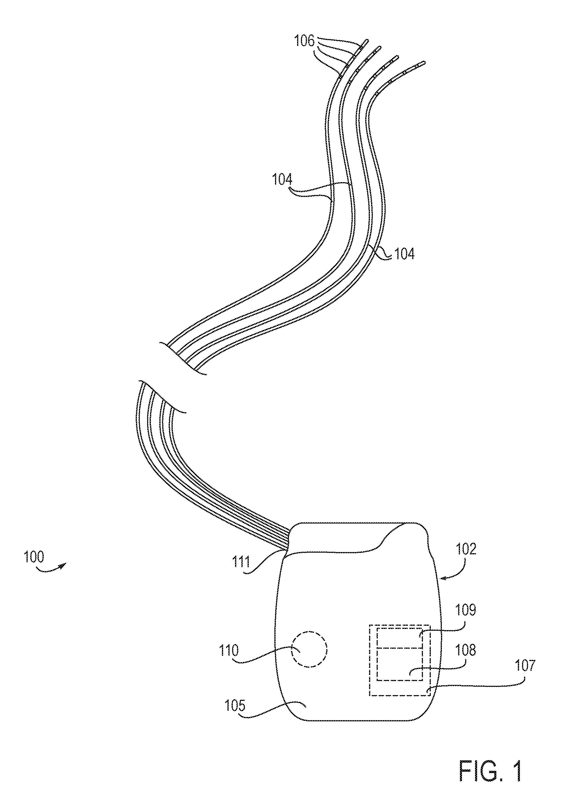
The present invention utilizes such reflex arcs and feed-forward processes to treat patients presenting with one or more disorders. FIG. 1 illustrates an embodiment of an implantable stimulation system 100 for treatment of such patients. The system 100 includes an implantable pulse generator (IPG) 102 and at least one lead 104 connectable thereto. In preferred embodiments, the system 100 includes four leads 104, as shown, however any number of leads 104 may be used including one, two, three, four, five, six, seven, eight, up to 58 or more. Each lead 104 includes at least one electrode 106. In preferred embodiments, each lead 104 includes four electrodes 106, as shown, however any number of electrodes 106 may be used including one, two, three, four five, six, seven, eight, nine, ten, eleven, twelve, thirteen, fourteen, fifteen, sixteen or more. Each electrode can be configured as off, anode or cathode. In some embodiments, even though each lead and electrode are independently configurable, at any given time the software ensures only one lead is stimulating at any time. In other embodiments, more than one lead is stimulating at any time, or stimulation by the leads is staggered or overlapping.
Referring again to FIG. 1, the IPG 102 includes electronic circuitry 107 as well as a power supply 110, e.g., a battery, such as a rechargeable or non-rechargeable battery, so that once programmed and turned on, the IPG 102 can operate independently of external hardware. In some embodiments, the electronic circuitry 107 includes a processor 109 and programmable stimulation information in memory 108.
The implantable stimulation system 100 can be used to stimulate a variety of anatomical locations within a patient's body. In preferred embodiments, the system 100 is used to stimulate one or more dorsal roots, particularly one or more dorsal root ganglions. FIG. 2 illustrates example placement of the leads 104 of the embodiment of FIG. 1 within the patient anatomy. In this example, each lead 104 is individually advanced within the spinal column S in an antegrade direction. Each lead 104 has a distal end which is guidable toward a target DRG and positionable so that its electrodes 106 are in proximity to the target DRG. Specifically, each lead 104 is positionable so that its electrodes 106 are able to selectively stimulate the DRG, either due to position, electrode configuration, electrode shape, electric field shape, stimulation signal parameters or a combination of these. FIG. 2 illustrates the stimulation of four DRGs, each DRG stimulated by one lead 104. These four DRGs are located on three levels, wherein two DRGs are stimulated on the same level. It may be appreciated that any number of DRGs and any combination of DRGs may be stimulated with the stimulation system 100 of the present invention. It may also be appreciated that more than one lead 104 may be positioned so as to stimulate an individual DRG and one lead 104 may be positioned so as to stimulate more than one DRG.
It may be appreciated that selective stimulation or neuromodulation concepts described herein may be applied in a number of different configurations. Unilateral (on or in root ganglion(s) on one level on one side of the spinal cord), bi-lateral (on or in two root ganglions on the same level on opposite sides of the spinal cord), unilevel (one or more root ganglion on the same level) or multi-level (at least one root ganglion is stimulated on each of two or more levels) or combinations of the above including stimulation of a portion of the sympathetic nervous system and one or more dorsal root ganglia associated with the neural activity or transmission of that portion of the sympathetic nervous system. As such, embodiments of the present invention may be used to create a wide variety of stimulation control schemes, individually or overlapping, to create and provide zones of treatment.
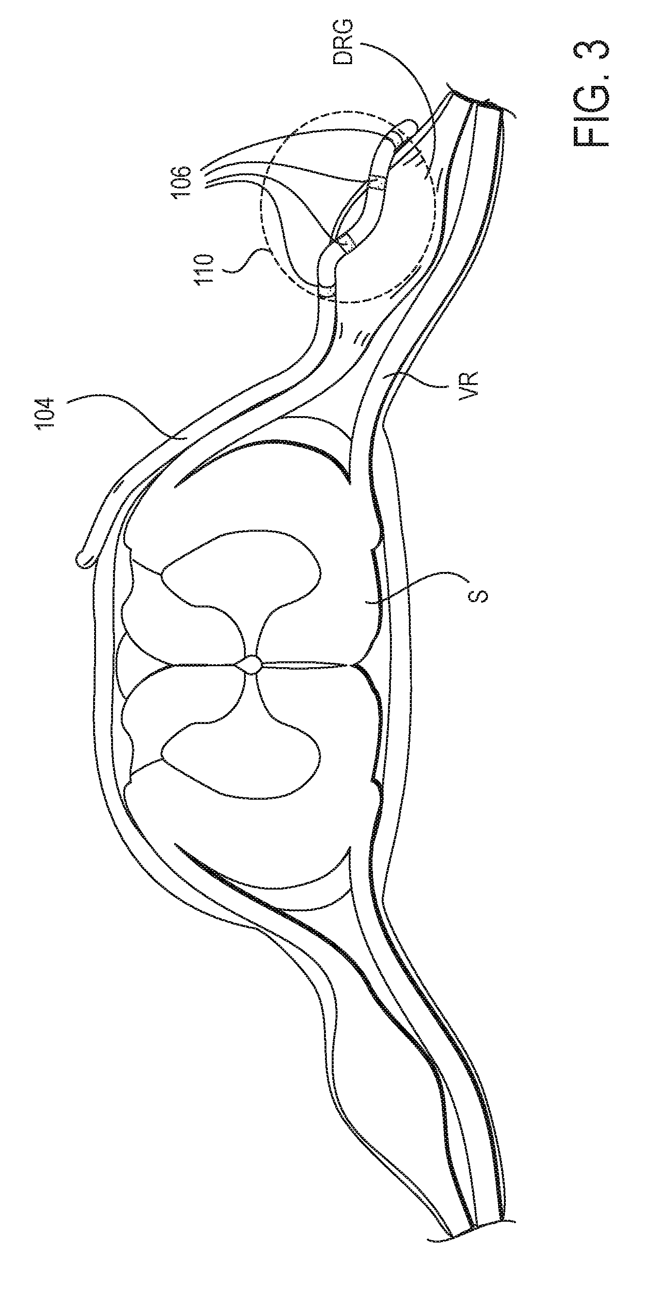
FIG. 3 illustrates an example cross-sectional view of an individual spinal level showing a lead 104 of the stimulation system 100 positioned on a target DRG. In this example, the lead 104 is advanced within the epidural space along the spinal cord S to the appropriate spinal level wherein the lead 104 is advanced laterally toward the target DRG. In some instances, the lead 104 is advanced through or partially through a foramen. At least one, some or all of the electrodes 106 are positioned on, near, about or in proximity to the DRG. In preferred embodiments, the lead 104 is positioned so that the electrodes 106 are disposed along a surface of the DRG opposite to the ventral root VR, as illustrated in FIG. 3. It may be appreciated that the surface of the DRG opposite the ventral root VR may be diametrically opposed to portions of the ventral root VR but is not so limited. Such a surface may reside along a variety of areas of the DRG which are separated from the ventral root VR by a distance.
In some instances, such electrodes 106 may provide a stimulation region indicated by dashed line 110, wherein the DRG receives stimulation energy within the stimulation region and the ventral root VR does not as it is outside of the stimulation region. Thus, such placement of the lead 104 may assist in reducing any possible stimulation of the ventral root VR due to distance. However, it may be appreciated that the electrodes 106 may be positioned in a variety of locations in relation to the DRG and may selectively stimulate the DRG due to factors other than or in addition to distance, such as due to stimulation profile shape and stimulation signal parameters, to name a few. It may also be appreciated that the target DRG may be approached by other methods, such as a retrograde epidural approach. Likewise, the DRG may be approached from outside of the spinal column wherein the lead 104 is advanced extraforaminally, from a outside a foramen toward the spinal column, optionally passing through or partially through a foramen and is implanted so that at least some of the electrodes 106 are positioned on, about or in proximity to the DRG.
In order to position the lead 104 in such close proximity to the DRG, the lead 104 is appropriately sized and configured to maneuver through the anatomy. In some embodiments, such maneuvering includes atraumatic epidural advancement along the spinal cord S, through a sharp curve toward a DRG, and optionally through a foramen wherein the distal end of the lead 104 is configured to then reside in close proximity to a small target such as the DRG. Consequently, the lead 104 is significantly smaller and more easily maneuverable than conventional spinal cord stimulator leads. Example leads and delivery systems for delivering the leads to a target such as the DRG are provided in U.S. patent application Ser. No. 12/687,737, entitled "Stimulation Leads, Delivery Systems and Methods of Use", incorporated herein by reference for all purposes.
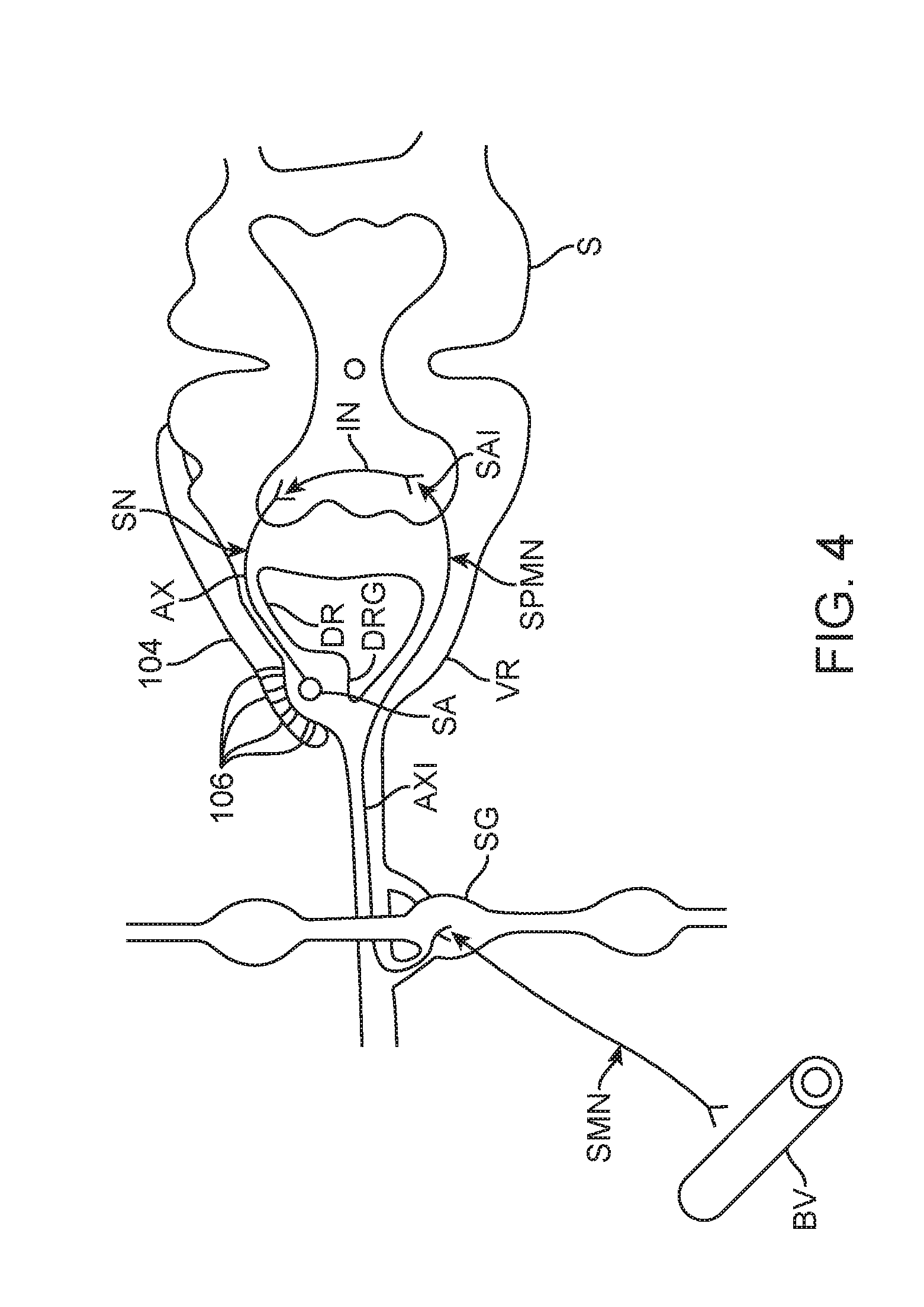
FIG. 4 illustrates the lead 104 positioned near a DRG so as to influence the sympathetic nervous system in the treatment of a condition or disorder. In this schematic illustration, a sensory neuron SN is shown having a soma SA disposed within the DRG and an axon AX which extends through the dorsal root DR to the dorsal horn of the spinal cord S. The sensory neuron SN connects with an interconnector neuron IN within the spinal cord S which connects with sympathetic premotor neuron SPMN. The sympathetic premotor neuron SPMN includes a soma SA1 disposed within the ventral horn of the spinal cord S and an axon AX1 which extends through the ventral root VR and enervates a sympathetic ganglion SG. Here, the sympathetic premotor neuron SPMN synapses with a sympathetic motor neuron SMN that ultimately affects a blood vessel BV and alters vascular resistance. The sympathetic motor neuron SMN releases norepinephrine, a neurotransmitter. Norepinephrine increases vascular resistance or blood pressure by increasing vascular tone through a-adrenergic receptor activation. It may be appreciated that in other embodiments, the sympathetic motor neuron may release or co-release other transmitters.
As mentioned previously, treatment of a condition involving ischemia or impaired blood flow to a particular region of the body may be treated by inhibition of the sympathetic nervous system. Referring again to FIG. 4, at least one, some or all of the electrodes 106 are positioned on, about or in proximity to the target DRG. In some embodiments, the involved sensory neuron SN, particularly its soma SA within the target DRG, is selectively stimulated by energy provided by at least one of the electrodes 106. Such stimulation is transmitted through the interneuron IN to the sympathetic premotor neuron SPMN which acts upon a sympathetic motor neuron SMN via the associated sympathetic ganglion SG. This inhibits release of norepinephrine by the sympathetic motor neuron SMN which in turn lessens vascular resistance and improves blood flow to the areas that had suffered from restricted blood flow.
In some embodiments, selective stimulation of the involved sensory neuron SN is achieved with the choice of the size of the electrode(s), the shape of the electrode(s), the position of the electrode(s), the stimulation signal, pattern or algorithm, or any combination of these. Such selective stimulation stimulates the targeted neural tissue while excluding untargeted tissue, such as surrounding or nearby tissue. In some embodiments, the stimulation energy is delivered to the targeted neural tissue so that the energy dissipates or attenuates beyond the targeted tissue or region to a level insufficient to stimulate modulate or influence such untargeted tissue. In particular, selective stimulation of tissues, such as the dorsal root, DRG, or portions thereof, exclude stimulation of the ventral root wherein the stimulation signal has an energy below an energy threshold for stimulating a ventral root associated with the target dorsal root while the lead is so positioned. Examples of methods and devices to achieve such selective stimulation of the dorsal root and/or DRG are provided in U.S. Pat. No. 9,056,197, entitled "Selective Stimulation Systems and Signal Parameters for Medical Conditions", incorporated herein by reference for all purposes. It may be appreciated that indiscriminant stimulation of the ventral root, such as from an electrode which emits stimulation energy which directly stimulates the ventral root, typically causes unpleasant sensations for the patient, such as tingling, buzzing or undesired motions or movements. Therefore, it is desired to stimulate sympathetic premotor neurons via synapses in the spinal cord rather than directly via the ventral root.
As mentioned previously, given the unique role of the sympathetic system in the body and the ability of the sympathetic system to affect a wide array of internal organs, the sympathetic system may be utilized to treat a variety of conditions throughout the body. In particular, a condition involving ischemia or impaired blood flow to a particular region of the body may be treated by inhibition or activation of the sympathetic nervous system. Some of these conditions will be described in more detail below. However, it may be appreciated that other disorders and conditions may also be treated with the devices, systems and methods of the present invention.
Diabetes
Diabetes is a metabolism disorder in which the quantity of glucose in the blood is too elevated (hyperglycemia). This is because the body either does not produce enough insulin, produces no insulin, or has cells that do not respond properly to the insulin the pancreas produces. Since insulin makes it possible for cells to take in glucose, this metabolic disorder results in too much glucose building up in the blood.
Elevated blood sugar levels cause a variety of health problems and complications for diabetic patients. A very common complication is foot problems, including nerve damage or peripheral neuropathy that results in loss of feeling or pain and burning sensations in the feet and legs. Once nerve damage progresses, it triggers loss of motor control and abnormal gait and can result in ulcers and amputations. The major cause of such nerve damage is loss of circulation. High blood sugars damage both large and small blood vessels that carry oxygen and nutrients to the nerves. If there is not enough blood being sent to the nerves, the nerves are damaged wherein electrical signals can no longer pass or pass at a slower speed. Good messaging in nerves also depends on an outer protective coating called myelin. This electrical insulator is also vulnerable to damage from high blood sugars. Preventing such foot problems in diabetes begins by preventing the loss of circulation that will result in serious nerve damage.
Diabetic patients are also twice as likely to have a heart attack or stroke. This is because diabetes worsens atherosclerosis, a condition in which arteries narrow. High blood sugar levels have two effects on cells lining blood vessels. First, it increases the production of free radicals, highly reactive molecules that damage sensitive cell components like DNA, causing premature cell death (apoptosis). Second, it reduces the availability of nitric oxide (NO), which would otherwise enable blood vessels to relax and blood flow to increase. In patients without diabetes, fast blood flow triggers a cascade which leads to dilation of blood vessels and reduced inflammation. The diabetic patient does not have the benefit of such triggering due to the reduction in blood flow, which in turn worsens the condition.
Therefore the diabetic patient may be beneficially treated by increasing blood flow to areas of the body by stimulating associated dorsal root ganglions as described above. In particular, such increase in blood flow may reduce the incidence of nerve damage, heart attack and stroke in those suffering from diabetes.
Peripheral Vascular Disease
Peripheral vascular disease (PVD), refers to the obstruction of large arteries in the periphery of the vascular system. PVD causes either acute or chronic ischemia (lack of blood supply). PVD also includes a subset of diseases classified as microvascular diseases resulting from episodal narrowing of the arteries (Raynaud's phenomenon), or widening thereof (erythromelalgia). For the patient, PVD can manifest as claudication (pain, weakness, numbness, or cramping in muscles due to decreased blood flow), sores, wounds, or ulcers that heal slowly or not at all, noticeable changes in skin color (blueness or paleness) or temperature (coolness) when compared to the other limbs, or diminished hair and nail growth on affected limb and digits, to name a few. Individuals with PVD may require amputation and can have an elevated risk for cardiovascular events and eventual death of a cardiac or cerebrovascular etiology. Thus, patients suffering from peripheral vascular disease may be beneficially treated by increasing blood flow to portions of the peripheral vascular system by stimulating associated dorsal root ganglions as described above.
Limb Ischemia
Limb ischemia is an obstruction of the arteries that seriously decreases blood flow to the extremities (hands, feet and legs) and has progressed to the point of severe pain and even skin ulcers or sores. Limb ischemia is often present in people suffering from severe cases of peripheral vascular disease. However, there are a variety of risk factors for developing the disease, including age, smoking, diabetes, obesity, sedentary lifestyle, high cholesterol, high blood pressure, and family history of atherosclerosis or claudication. Thus, patients suffering from limb ischemia for any reason may be beneficially treated by increasing blood flow to the limb by stimulating associated dorsal root ganglions as described above.
Myocardial Ischemia
Myocardial Ischemia develops when coronary blood flow becomes inadequate to meet myocardial oxygen demand. In some instances, myocardial ischemia results from abnormal constriction or deficient relaxation of coronary microcirculation (ie, resistance vessels). Coronary spasm can also reduce coronary flow reserve significantly by causing dynamic stenosis of coronary arteries. Myocardial ischemia causes myocardial cells to switch from aerobic to anaerobic metabolism, with a progressive impairment of metabolic, mechanical, and electrical functions. Angina pectoris, often described as severe chest pain, is a common clinical manifestation of myocardial ischemia. It is caused by chemical and mechanical stimulation of sensory afferent nerve endings in the coronary vessels and myocardium. These nerve fibers extend from the first to fourth thoracic spinal nerves, ascending via the spinal cord to the thalamus, and from there to the cerebral cortex.
The heart and coronary arteries are innervated by sympathetic afferent fibers that have their cell bodies concentrated in the dorsal root ganglia of the T2 to T6 spinal segments but can extend as far as the C8 to T9 segments. Dorsal root ganglion cells have axons that enter the tract of Lissauer and terminate in the same segment, or the axons can ascend and descend a few segments before terminating in the spinal gray matter. Patients suffering from myocardial ischemia may be beneficially treated by increasing blood flow in the coronary vascular system by stimulating associated dorsal root ganglions as described above. Likewise, patients presenting with angina pectoris may be beneficially treated for pain symptoms by stimulating associated dorsal root ganglions as described above.
Stroke
Initial treatment for a stroke varies depending on whether it is an ischemic stroke (caused by a blood clot) or a hemorrhagic stroke (caused by bleeding in the brain). For an ischemic stroke, initial treatment focuses on restoring blood flow. Permanent damage from a stroke often occurs within the first few hours so swift restoration of blood flow will lessen damage that will occur. Current treatments include a clot-dissolving medicine called tissue plasminogen activator (t-PA), which can increase chances of survival and recovery. In addition, the patient may receive aspirin or aspirin combined with another antiplatelet medicine. However, aspirin is not recommended within 24 hours of treatment with t-PA. Other medicines may be given to control blood sugar levels, fever, and seizures. Patients suffering from an ischemic stroke may be beneficially treated by quickly restoring blood flow to the brain by stimulating associated dorsal root ganglions as described above.
Erectile Dysfunction
Erectile dysfunction is a sexual dysfunction characterized by the inability to develop or maintain an erection of the penis. A penile erection is the hydraulic effect of blood entering and being retained in sponge-like bodies within the penis. Thus, there are a variety of circulatory causes of erectile dysfunction. The most common circulatory causes are cardiovascular disease and diabetes. By treating these circulatory maladies with the devices, systems and methods described herein, erectile dysfunction may be prevented or treated.
Sympathetically Mediated Pain
Sympathetically mediated pain and sympathetically maintained pain refers to pain signals that are transmitted to the brain from the sympathetic nervous system, the part of the nervous system controlling `involuntary` functions of the body such as heart rate, sweating, constriction of blood vessels, and digestion. In certain abnormal situations the pain signals from the sympathetic nervous system become constant and severe, even though there is no obvious cause of pain. The mechanism by which this happens is complex and not fully understood.
Sympathetic pain usually has a severe, burning characteristic and often begins in the hand or foot. The affected area is very hypersensitive to even the lightest touch. Pink or bluish discoloration of the involved area may occur because of abnormal circulation, and abnormal sweating may also be noticed. There are a number of diagnostic phrases used by physicians when discussing sympathetic pain syndromes. In the past the most commonly used phrase was Reflex Sympathetic Dystrophy, or RSD. Other terms used to describe the condition include causalgia and sympathetically mediated pain. Recently, "Chronic Regional Pain Syndrome" or CRPS has become commonly used. Such sympathetic pain can also be treated by selective stimulation of one or more dorsal root ganglions since sympathetic afferents can travel through the DRG. In some embodiments, a negative feedback loop on efferent sympathetic activity is created. And, in other embodiments, a stellate ganglion blockade is used in treating certain pain conditions.
Thus, blood vessels are just one of many targets that can be influenced by affecting the sympathetic nervous system via selective stimulation of one or more dorsal root ganglions. A variety of other end organs may also be influenced by selective stimulation of one or more dorsal root ganglions to treat medical conditions associated with these end organs. For example, the lungs may be influenced in the treatment patients suffering from constriction of air passages. There are a variety of circumstances and conditions that cause the bronchi of the lungs to become narrow, or constrict, making it difficult to breathe. Bronchoconstriction, or the narrowing of the airways, is typically caused by the muscles surrounding the lungs becoming tight. A build-up of excess mucous as well as inflammation can also cause constriction. The constriction results in coughing, wheezing and shortness of breath. There are several conditions that cause this; such conditions include but are not limited to: Chronic lung disease (CLD), Emphysema, Exercise-Induced bronchoconstriction, Allergen-induced bronchoconstriction, and Asthma. In some embodiments, bronchodilation, the process by which the bronchi (tubes in the lungs made of connective tissue and muscle) are dilated, or opened, is achieved by selective stimulation of one or more dorsal root ganglions.
It is known that bronchodilation can occur as part of the body's natural response. When the sympathetic nervous system is activated in what is commonly known as the "fight or flight" response, the hormones and neurotransmitters of adrenaline (also called epinephrine) and noradrenaline (also called norepinephrine) are released. This response can be naturally triggered by physical or mental stress. And, aspects of this natural response can be harnessed to treat patients suffering from bronchoconstriction. In particular, one or more dorsal root ganglia associated with portions of the sympathetic nervous system involved in bronchodilation are selectively stimulated using the devices, systems and method described and referenced herein. Such selective stimulation leads to desired bronchodilation in treatment of the medical condition suffered by the patient.
As mentioned, a variety of end organs may also be influenced by selective stimulation of one or more dorsal root ganglions to treat medical conditions associated with these end organs. FIG. 5 is a schematic illustration of a portion of the sympathetic nervous system and associated target organs and tissues that can be influenced. As shown, each sympathetic ganglion SG along the sympathetic chain is associated with a spinal level, in particular, a dorsal root ganglion on a spinal level. And, one or more sympathetic ganglions SG are associated with a particular organ, system or tissue, such as the heart, liver or stomach, to name a few. It may be appreciated that stimulation of one or more dorsal root ganglions may alternatively or additionally influence other ganglions, such as mesenteric ganglions, celiac ganglions, stellate ganglions and cervical ganglions, to name a few. These ganglions in turn affect particular organs, systems or tissues.
FIG. 6 illustrates how embodiments of the present invention may be advantageously utilized for neurostimulation of the sympathetic chain using direct stimulation of an associated DRG. This aspect of the present invention takes advantage of the anatomical placement of the DRG relative to the sympathetic chain in conjunction with gate control theory to direct DRG stimulation for control of the sympathetic system. Thus, selective neurostimulation techniques of the present invention may be advantageously employed to, for example, provide and/or augment therapeutic tools in regards to weight control, hormonal regulation, vascular perfusion, etc. Additional alternative embodiments include the use of specific stimulation to provide organ system autonomic modulation. Through implantation of stimulation electrodes and systems of the present invention to stimulate the appropriate DRG upstream of the associated portion(s) of the sympathetic chain, the associated system may be controlled, modulated or influenced utilizing the electrical and/or pharmacological agent stimulation techniques described herein. Thus, there is provided a method of modulating a neural pathway in the sympathetic nervous system by stimulating a spinal dorsal root ganglion upstream of at least one ganglion of the sympathetic nerve chain to influence a condition associated with the at least one ganglion of the sympathetic nerve chain.
In one specific example, by stimulating the DRG 40 associated with spinal level 13.3, as shown in FIG. 6, the portion of the sympathetic chain associated with hormonal regulation may be altered, modified, influenced or controlled. Similarly, by stimulating the DRG 40' associated with spinal level 13.2 and/or the DRG 40'' associated with level 13.1, the portion of the sympathetic chain associated with the gastrointestinal tract, or urinary incontinence (i.e., urinary bladder, urethra, prostate, etc.) may be altered, modified, influenced or controlled.
Optionally or additionally, the direct stimulation techniques described herein may be used to directly stimulate individual nerve ganglion of the sympathetic nervous system, such as, for example, the celiac ganglion, superior mesenteric ganglion, inferior mesenteric ganglion and others listed in FIGS. 5, 6 or known to those of ordinary skill. It is to be appreciated that the stimulation systems, pulse generators, leads, electrodes, and/or microelectrodes and other components are modified and sized as needed to allow for direct stimulation of the ganglion including implanting into the ganglion or within adjacent nerve sheaths leading to the ganglion. FIG. 7 illustrates an embodiment of a combined direct stimulation of a DRG 38 with microelectrode 115 as well as a suitably sized microelectrode 115 implanted in a sympathetic nerve root ganglion 63. The electrodes in FIG. 7 may stimulate independently or in a coordinated fashion to achieve the desired clinical outcome or other desired result. Similar to the discussion above for electrode placement in, on or about the DRG, electrode placement for the sympathetic chain may also be unilateral, bilateral, on adjacent portions of the chain or separate portions of the chain as needed.
One aspect of the present invention is a method of modulating a neural pathway in the sympathetic nervous system including stimulating a spinal dorsal root ganglion upstream of at least one ganglion of the sympathetic nerve chain to influence a condition associated with the at least one ganglion of the sympathetic nerve chain. In one specific embodiment, stimulating a spinal dorsal root ganglion comprises stimulating a spinal dorsal root ganglion upstream of at least one ganglion of the sympathetic nerve chain to influence functional activation of a bodily system associated with the at least one ganglion along the sympathetic nerve chain, to influence functional activation of an organ associated with the at least one ganglion along the sympathetic nerve chain, or to influence functional inhibition of a bodily system associated with the at least one ganglion along the sympathetic nerve chain. In specific embodiments, the ganglion of the sympathetic nerve chain is a cervical ganglion, a thoracic ganglion, a lumbar ganglion or a sacral ganglion.
It may be appreciated that embodiments of the present invention may be used in conjunction with other neurostimulation techniques by combining an upstream stimulation using specific DRG stimulation of the present invention with another stimulation acting downstream of the DRG stimulation. As used herein, downstream and upstream refer to pathways closer to the brain (i.e., upstream) or further from the brain (i.e., downstream). For example, several stimulation techniques are described by Rezai in US Patent Publication 2002/0116030 and U.S. Pat. No. 6,438,423 and by Dobak in publication 2003/0181958, all of which are incorporated herein by reference. In specific aspects, embodiments of the present invention may be used to provide electrical and combinational (i.e., with a pharmacological agent) stimulation of the sympathetic nerve chain as described by Rezai alone (i.e., using the appropriate DRG stimulation or implanting directly into a nerve root ganglion.). Alternatively or additionally, embodiments of the present invention provide specific, direct stimulation of one or more DRG and are used in combination with the stimulation techniques described by Rezai (i.e., conventional stimulation of the sympathetic chain using one or more of Rezai's techniques).
Referring back to FIG. 1, in some embodiments, the implantable pulse generator (IPG) 102 comprises circuitry which initiates or modifies the electrical stimulation in response to one or more sensors. Example sensors include, among others, blood glucose sensors, blood pressure sensors, blood flow sensors (including Doppler and other techniques), heart rate sensors, blood oxygen sensors, temperature sensors, accelerometers, strain gauges, electrocardiograms, brain wave detectors (electroencephalograms, other interiorly and exteriorly measured composite neuronal activity), electrical devices which measure electrical activity in muscles and/or nerves, or other devices capable of measuring physiological parameters indicative of symptoms of the disorder under treatment.
In some embodiments, the one or more sensors sense the status of one or more symptoms of the disorder. Such status information is utilized to modify the electrical stimulation to a level which is appropriate to improve status of the disorder in real time. This modification of electrical stimulation may be particularly beneficial in the treatment of conditions which have a time dependency, such as stroke.
In some embodiments, the sensor detects one or more of the following functions or aspect of the body: carbon dioxide pressure in a target tissue, action potential conduction (such as in a target nerve), body movement, balance, motor activity including muscle tone, heart rate, blood pressure, capillary pressure, venous pressure, arterial pressure, blood flow, circulation (including blood and lymphatic), perfusion, electrocardiogram, oxygenation (including blood oxygenation levels, oxygen saturation levels, oxygen consumption, oxygen pressure), concentration of certain biological molecules/substances in the body (such as, for example, glucose, liver enzymes, electrolytes, hormones, creatinine, medications, concentration of various cells, platelets, or bacteria), pH levels, chemical production, neurotransmitter levels, electrolyte levels in the circulation/tissue, nitrogen pressure, respiratory function, chest wall expansion, diaphragmatic movement, cognitive activity, electroencephalogram, flushing, galvanic skin responses, perspiration, body temperature regulation, response to external stimulation, pain, speech, temperature, visual activity, intra-bladder pressure, and water pressure.
In some embodiments, the sensor is positioned so as to measure sympathetic nerve outflow. In such embodiments, the sensor may be positioned on or near the sympathetic chain.
In some embodiments, the implantable pulse generator (IPG) 102 comprises circuitry which initiates or modifies the electrical stimulation in response to a timer or clock. Thus, stimulation may be reduced or eliminated during times in which it is determined that the patient desires reduced or no treatment. Such periods of reduced usage may extend the life of the power supply 110.
Although the foregoing invention has been described in some detail by way of illustration and example, for purposes of clarity of understanding, it will be obvious that various alternatives, modifications, and equivalents may be used and the above description should not be taken as limiting in scope of the invention which is defined by the appended claims.
INCORPORATION BY REFERENCE
All publications and patent applications mentioned in this specification are herein incorporated by reference to the same extent as if each individual publication or patent application was specifically and individually indicated to be incorporated by reference.
* * * * *
References
-
emedicine.medscape.com/article/150215-overview
-
biology-online.org/dDorsal_root_ganglion
-
mayfieldclinic.com/PE-AnatSpine.htm
-
medterms.com/script/main/art.asp?articlekey=6226
-
priorartdatabase.com/IPCOM/000012136
-
spine-health.com/topics/cd/pain/chronic_pain_theories/chronic_pain_theory02.html
-
cnx.org/content/m44751/latest
D00000
D00001
D00002
D00003
D00004
D00005
D00006
D00007
XML
uspto.report is an independent third-party trademark research tool that is not affiliated, endorsed, or sponsored by the United States Patent and Trademark Office (USPTO) or any other governmental organization. The information provided by uspto.report is based on publicly available data at the time of writing and is intended for informational purposes only.
While we strive to provide accurate and up-to-date information, we do not guarantee the accuracy, completeness, reliability, or suitability of the information displayed on this site. The use of this site is at your own risk. Any reliance you place on such information is therefore strictly at your own risk.
All official trademark data, including owner information, should be verified by visiting the official USPTO website at www.uspto.gov. This site is not intended to replace professional legal advice and should not be used as a substitute for consulting with a legal professional who is knowledgeable about trademark law.