Antimicrobial peptides derived from hepatitis B virus core protein arginine-rich domain
Shih , et al. Feb
U.S. patent number 10,214,564 [Application Number 14/766,359] was granted by the patent office on 2019-02-26 for antimicrobial peptides derived from hepatitis b virus core protein arginine-rich domain. This patent grant is currently assigned to Academia Sinica. The grantee listed for this patent is Academia Sinica. Invention is credited to Heng-Li Chen, Chiaho Shih, Pei-Yi Su.


| United States Patent | 10,214,564 |
| Shih , et al. | February 26, 2019 |
Antimicrobial peptides derived from hepatitis B virus core protein arginine-rich domain
Abstract
A pharmaceutical composition for use in killing and/or inhibiting the growth and/or proliferation of a microorganism in a subject in need thereof, or for treating a subject afflicted with a microbial infection is disclosed. The composition comprises: (a) an effective amount of an isolated peptide, wherein the peptide comprises the arginine-rich carboxy-terminal region of hepatitis B virus core protein (HBc) and exhibits an antimicrobial activity; and (b) a pharmaceutically acceptable carrier. The peptide exhibits an activity against Gram-negative bacteria, Gram-positive bacteria, and/or fungi.
| Inventors: | Shih; Chiaho (Taipei, TW), Chen; Heng-Li (Taipei, TW), Su; Pei-Yi (Taipei, TW) | ||||||||||
|---|---|---|---|---|---|---|---|---|---|---|---|
| Applicant: |
|
||||||||||
| Assignee: | Academia Sinica (Taipei,
TW) |
||||||||||
| Family ID: | 51300113 | ||||||||||
| Appl. No.: | 14/766,359 | ||||||||||
| Filed: | February 5, 2014 | ||||||||||
| PCT Filed: | February 05, 2014 | ||||||||||
| PCT No.: | PCT/US2014/014938 | ||||||||||
| 371(c)(1),(2),(4) Date: | August 06, 2015 | ||||||||||
| PCT Pub. No.: | WO2014/124047 | ||||||||||
| PCT Pub. Date: | August 14, 2014 |
Prior Publication Data
| Document Identifier | Publication Date | |
|---|---|---|
| US 20160002300 A1 | Jan 7, 2016 | |
Related U.S. Patent Documents
| Application Number | Filing Date | Patent Number | Issue Date | ||
|---|---|---|---|---|---|
| 61761650 | Feb 6, 2013 | ||||
| Current U.S. Class: | 1/1 |
| Current CPC Class: | A61K 39/292 (20130101); A61P 31/04 (20180101); C12N 7/00 (20130101); C07K 14/005 (20130101); A61K 39/12 (20130101); C12N 2730/10134 (20130101); C12N 2730/10133 (20130101); A61K 38/00 (20130101) |
| Current International Class: | C07K 14/005 (20060101); C12N 7/00 (20060101); A61K 39/29 (20060101); A61K 39/12 (20060101); A61K 38/00 (20060101) |
References Cited [Referenced By]
U.S. Patent Documents
| 6060595 | May 2000 | Scaglioni |
| 7244706 | July 2007 | Mann |
| WO-2004/050883 | Jun 2004 | WO | |||
Other References
|
Isidro-Llobet et al. ("Amino-Acid Protecting Groups"; Chem. Rev. 2009; 109, 2455-2504). cited by examiner . Brandenburg et al. ("Antimicrobial Peptides: Multifunctional Drugs for Different Applications" Polymers 2012, 4, 539-560). cited by examiner . Fee et al. ("Protein PEGylation: An overview of chemistry and process considerations" EPR; Issue 1, 2010). cited by examiner . Miura et al. ("Basic peptide protamine exerts antimicrobial activity against periodontopathic bacteria"; J. biomedical Science and Engineering, 2010,3, 1069-1072). cited by examiner . Li et al "Nuclear Export and Import of Human Hepatitis B Virus Capsid Protein and Particles" PLOS Pathogens vol. 6, pp. 1-17, 2010. cited by applicant . Chen et al "Identification of a Novel Antimicrobial Peptide from Human Hepatitis B Virus Core Protein Arginine-Rich Domain (ARD)" Pathogens vol. 9, pp. 1-16. cited by applicant . Chen et al "Improvement of In Vivo Antimicrobial Activity of HBcARD Peptides by D-Arginine Replacement" Applied Microbiology and Biotechnology vol. 100, pp. 9125-9132, 2016. cited by applicant . Chu et al "Nucleic Acid Chaperone Activity Associated with the Arginine-Rich Domain of Human Hepatitis B Virus Core Protein" Journal of Virology vol. 88, pp. 2530-2543, 2014. cited by applicant . Jung et al "C-Terminal Substitution of HBV Core Proteins with Those From DHBV Reveals That Arginine-Rich .sup.167RRRSQSPRR.sup.175 Domain is Critical for HBV Replication" One vol. 7, pp. 1-14, 2012. cited by applicant . Protective Groups for Peptide Synthesis [online], Peptide Guide 2012 [retrieved on May 25, 2014]. Retrieved from the internet: http://peptideguide.com/protecting-groups-spps.html, p. 1; p. 1, paragraphs 2-3. cited by applicant. |
Primary Examiner: McDonald; Jennifer Pitrak
Assistant Examiner: Martinez; Tara L
Attorney, Agent or Firm: Cesari and McKenna, LLP
Parent Case Text
CROSS REFERENCE TO RELATED APPLICATION
This is the US national stage of International Patent Application No. PCT/US2014/014938, filed on Feb. 5, 2014, which claims priority to U.S. Provisional Application No. 61,761.650, filed on Jun. 2, 2013, the content of which is hereby incorporated by reference herein in its entirety.
Claims
What is claimed is:
1. A pharmaceutical composition comprising: (a) an effective amount of an isolated peptide, wherein the peptide consists of a protecting group and the amino acid sequence of SEQ ID NO:1, and exhibits an anti-bacterial or anti-fungal activity; and (b) a pharmaceutically acceptable carrier.
2. The composition of claim 1, wherein the peptide is characterized by having an activity against Gram-negative bacteria, Gram-positive bacteria, and/or fungi.
3. The composition of claim 1, wherein the peptide exhibits an activity against colistin-resistant A. baumannii.
4. The composition of claim 1, wherein the composition is formulated for topical, aerosol, oral, systemic intravenous, intraperitoneal, ocular, rectal, or inhalation administration.
5. A method of killing or inhibiting proliferation of a microorganism or treating a microbial infection in a subject, comprising: administering to a subject in need thereof a pharmaceutical composition that contains (a) an effective amount of an isolated peptide and (b) a pharmaceutically acceptable carrier, the microorganism being a bacterium or fungus and the microbial infection being a bacterial or fungal infection, wherein the peptide comprises the amino acid sequence of SEQ ID NO: 1.
6. The method of claim 5, wherein the peptide consists of the sequence of SEQ ID NO: 1.
7. The method of claim 5, wherein the peptide consists of the sequence of SEQ ID NO: 1 and a protecting group.
Description
FIELD OF THE INVENTION
The present invention relates to antimicrobial peptides.
BACKGROUND OF THE INVENTION
The increase of drug-resistant pathogens caused by the extensive use of traditional antibiotics is a serious concern worldwide. There is an urgent need to develop more effective treatment to overcome the drug-resistance problem. Antimicrobial peptides (AMP) are a new class of antibiotics with a new mode of action and remarkable therapeutic effects. In general, they contain 10-50 amino acids, with an overall positive charge and an amphipathic structure. It is well known that most AMPs can directly bind to bacteria membrane and kill them by disrupting membrane or targeting intracellular components. Most importantly, they are effective to antibiotics-resistant pathogens. This unique feature has encouraged the development of AMPs as novel antibiotics in the last few decades.
Prior to the present invention, no literature has reported that peptides derived from hepatitis B virus core protein (HBc) possess antimicrobial activities. Hepatitis B virus core protein (21 KDa) is essential for viral replication. It contains a capsid assembly domain at N-terminus (residue 1 to 149) and an arginine-rich domain (ARD) at C-terminus (residues 150 to 183) (Birnbaum et al. (1990) J. Virol 64: 3319-3330; Nassal M (1992) Virol 66: 4107-4116). ARD contains 16 arginines separated into four arginine-rich clusters (ARD I, II, IV) and has a function of binding to nucleic acids. When it binds to HBV pre-genomic RNA or palyanions, HBc can assemble into a stable capsid. In addition, ARD contains important signals for nuclear export and import of HBc core protein and particles. It was unexpectedly discovered that the growth of E. coli expressing HBc1-183 was much slower than that of E. coli expressing HBc1-149 (unpublished results).
SUMMARY OF THE INVENTION
In one aspect, the invention relates to a pharmaceutical composition comprising: (a) an effective amount of an isolated peptide, wherein the peptide comprises the arginine-rich carboxy-terminal region of hepatitis B virus core protein (HBc) and exhibits an antimicrobial activity; and (b) a Pharmaceutically acceptable carrier.
In another aspect, the invention relates to a pharmaceutical composition comprising: (a) an effective amount of an isolated peptide comprising a fragment of HBc, the fragment comprising more than one arginine-rich domain (ARD) selected from the group consisting of (i), (ii) and (iii) as follows: (i) HBc ARD I-IV, (ii) HBc ARD I-III; or (iii) HBc ARD II-IV; wherein the peptide exhibits an antimicrobial activity; and (b) a pharmaceutically acceptable carrier.
In another aspect, the invention relates to a pharmaceutical composition comprising an effective amount of an isolated peptide, the peptide comprising an arginine-rich sequence derived from the C-terminal region of hepatitis B virus core protein (HBc), wherein the peptide is characterized by having an antimicrobial activity.
Further in another aspect, the invention relates to a pharmaceutical composition as aforementioned for use in killing and/or inhibiting the growth and/or proliferation of a microorganism by causing the composition as aforementioned to be in contact with the microorganism.
Yet in another aspect, the invention relates to a pharmaceutical composition as aforementioned for use in killing and/or inhibiting the growth and/or proliferation of a microorganism in a subject in need thereof, or for treating a subject afflicted with a microbial infection. The subject is afflicted with Staphylococcus aureus or K. pneumoniae infection.
These and other aspects will become apparent from the following description of the preferred embodiment taken conjunction with the following drawings, although variations and modifications therein may be affected without departing from the spirit and scope of the novel concepts of the disclosure.
The accompanying drawings illustrate one or more embodiments of the invention and, together with the written description, serve to explain the principles of the invention. Wherever possible, the same reference numbers are used throughout the drawings to refer to the same or like elements of an embodiment.
BRIEF DESCRIPTION OF THE DRAWINGS
FIG. 1 shows amino acid sequences of various HBc ARD peptides tested for bactericidal activity. The lower panel presents various phosphorylated peptides and an R-to-A mutant peptide with a total of four Arg-to-Ala substitutions in ARD-III and ARD-IV of HBc147-183.
FIG. 2 shows killing kinetics of HBc147-183 against P. aeruginosa, K. pneumoniae, E. coli and S. aureus. Bacteria were treated with HBc147-183 (1.times.MBC). The viability of bacteria was measured at indicated time points. Samples were measured in triplicates.
FIG. 3 shows localization of FITC-HBc147-183 peptide on bacteria. Approximate 10.sup.7 CFU of P. aeruginosa ATCC9027, ATCC27853 (A and B), K. pneumoniae ATCC13884 (C), E. coli ATCC25922 (D), and S. aureus ATCC19636, ATCC25923 and ATCC 29213 (E, F and G) were incubated with HBc147-183 (0.5.times.MBC) for 1 hour. The bacteria were washed, fixed and stained with DAPI (blue). Images were taken using confocal microscopy.
FIG. 4 shows possible bactericidal mechanisms of HBc147-183. (A) SYTOX Green uptake of P. aeruginosa, K. pneumoniae, E. coli, and S. aureus by HBc147-184. Measurements of the fluorescence were recorded every minute. (B) Dose-dependent curves of membrane permeabilization of P. aeruginosa by HBc147-183 and HBc153-176 at 0.5, 1 and 2 .mu.M. Two .mu.M melittin was used as a positive control. Samples were measured in triplicates. (C) DNA-binding activity of HBc147-183. HBc147-183 was mixed with pSUPER plasmid DNA at indicated N/P ratio for 30 minutes. The mobility of DNA was determined by a gel retardation assay.
FIG. 5 shows dose response effects of LPS and LPS antibody on the bactericidal activity of HBc147-183. LPS from P. aeruginosa and E. coli, and LPS antibody were mixed with P. aeruginosa and HBc147-183 (1.times.MBC) for 3 hours. The bacteria were then plated on MH agar for the measurement of viability. Samples were measured in triplicates.
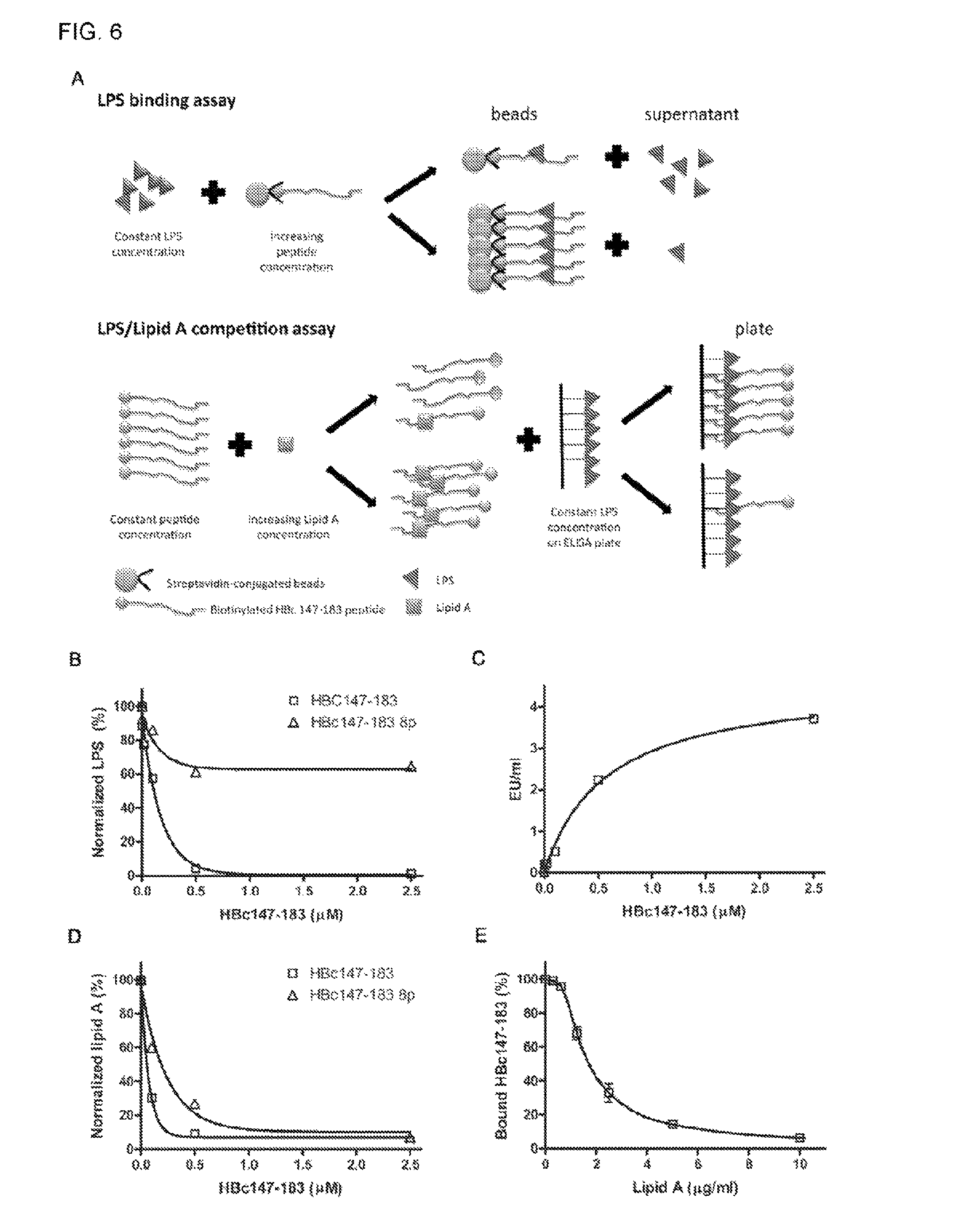
FIG. 6 shows the ARD peptide HBc147-183 being capable of binding to LPS and Lipid A in several different in vitro binding assays. Samples were measured in triplicates in each assay. (A) The cartoon illustrates the in vitro assays of peptide-LPS and peptide-Lipid A binding as well as LPS/Lipid A competition. (B) Constant amount of LPS was incubated with increasing concentrations of biotinylated ARD HBc147-183 peptide on the streptavidine-conjugated beads (0, 0.004, 0.02, 0.1, 0.5 and 2.5 .mu.M), Unbound LPS in the supernatant was measured with the LAL ELISA assay. The EU values were normalized with a control without peptide treatment. HBc147-183 8p (containing 8 phosphorylated amino acids) was also included as a control peptide due to its poor binding with LPS. (C) Beads-bound LPS was released into the supernatant by overnight digestion with trypsin agarose. Free LPS in the supernatant was analyzed with the LAL ELISA assay. Released LPS in the supernatant appeared to be in proportion to the amount of ARD peptide HBc147-183 on the beads. (D) Constant amount of Lipid A was incubated with increasing concentrations of HBc147-183 and HBc147-183 8P, respectively. The supernatant was also detected with LAL ELISA reagent. The result here is consistent with the notion that Lipid A can bind to HBc147-183 directly. (E) LPS/Lipid A competition assay. Constant amount of LPS (1 .mu.g) was coated on each well on the ELISA plate, and then incubated with a reaction mixture containing constant amount of 10 nM HBc147-183 and increasing concentrations of Lipid A. The gradual increase of Lipid A reduced the amount of plate-bound ARD peptide HBc147-183 in a dose dependent manner.
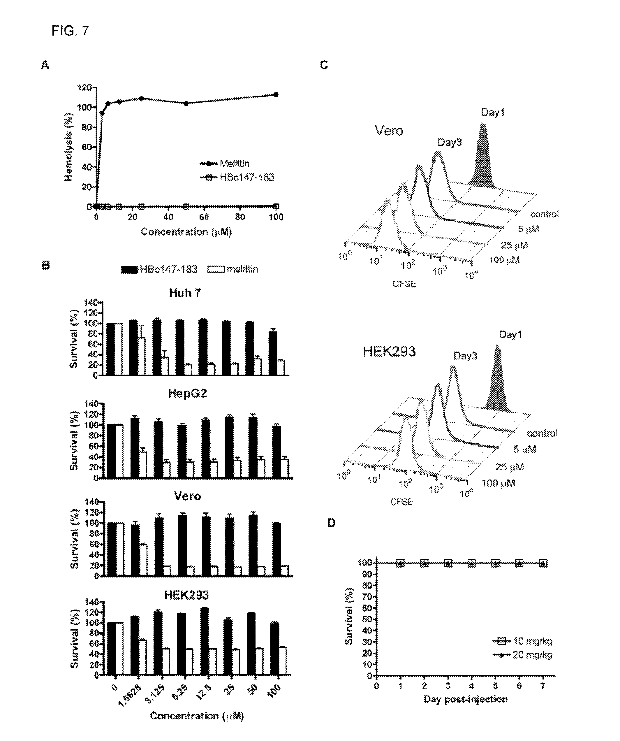
FIG. 7 shows cytotoxicity assays of ARD peptide HBc147-183. (A) Hemolytic activities of HBc147-183 and melittin were measured with 10% human red blood cells (RBC). Compared to melittin, HBc147-183 showed no hemolytic activity. (B) Huh7, HepG2, Vero and HEK293 cells were incubated with varying concentrations (0 to 100 .mu.M) of HBc147-183 and melittin for 1 hour at 37.degree. C. The effects on cell viability were determined by MTT assay. Melittin was used as a positive control. HBc147-183 showed no detectable effect on cell viability, while melittin exhibited strong toxicity. (C) Kidney cells, Vero and HEK293 were stained with CFSE and seeded at day 0. At day 1, cells were incubated with varying concentrations (0 to 100 .mu.M) of HBc147-183 for 1 hour. Cell proliferation at day 1 and day 3 were determined by flow cytometry. Similar to the mock control experiment, no significant effect on Vero and HEK293 cells was detected. Samples assayed in FIGS. 7A-C were measured in triplicates. (D) (D) In vivo toxicity of ARD peptide HBc147-183 was determined using three-week old male ICR mice. The mice were injected intraperitoneally with peptide (10 and 20 mg/kg of body weight). All mice were alive after 7 days.
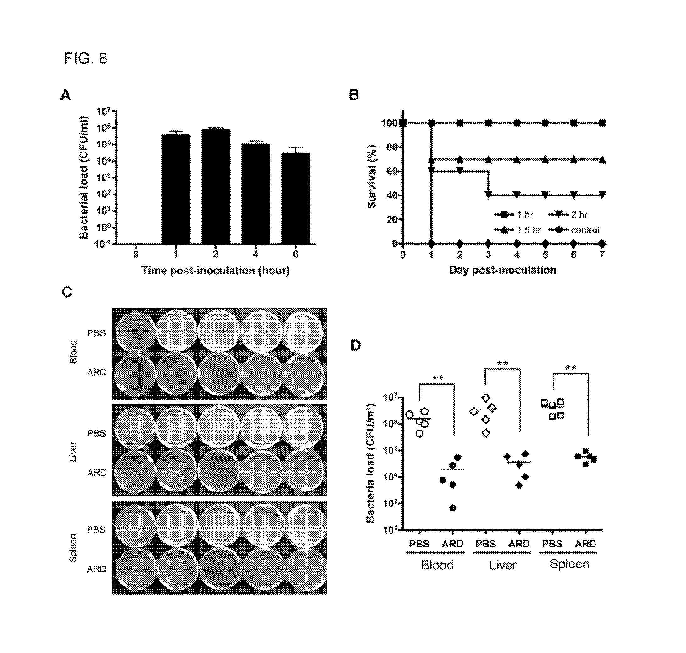
FIG. 8 shows in vivo studies of the protection activity of ARD peptide HBc147-183 against S. aureus. (A) Three-week old male ICR mice were challenged with a lethal dose of S. aureus ATCC 19636 and then divided into five separate groups for five different time points. At each indicated time point (n=5), blood samples were collected, diluted and plated on BHI agar. The number of bacteria was counted the following day. A maximal bacterial load in the blood was observed at 2 h post-inoculation. The data were shown in mean 6 SD. (B) ICR mice inoculated with a lethal dose of S. aureus as described above were treated by intraperitoneal injection with ARD peptide (10 mg/kg) at 1, 1.5 or 2h post-inoculation, respectively. Each group contained 10 mice. All mice (100%) treated with the PBS control died at day 1, while treatment of ARD peptide at 1, 1.5 or 2 h post-inoculation protected the mice with survival rates of 100%, 70% and 40% after 7 days, respectively. (C) As described above, ICR mice were i.p. inoculated with S. aureus, followed by i.p. injection with PBS (n=5) or 10 mg/kg ARD peptide (n=5) at 1 h post-inoculation. At 4 h post-inoculation, blood, liver and spleen were collected. Liver and spleen samples were homogenized, diluted and, together with blood samples, plated on BHI agar. The number of bacteria was counted the following day. In comparison to mice treated with PBS, treatment of ARD peptide effectively reduced the bacterial load in blood, liver and spleen. (D) Quantitative comparison of bacterial loads in blood, liver and spleen samples of mice treated with PBS (open circle, diamond and square) versus ARD peptide HBc147-183 (solid circle, diamond and square). The line indicated the mean of bacterial load. **P,0.01 (Mann-Whitney U test) for PBS and ARD peptide HBc147-183.
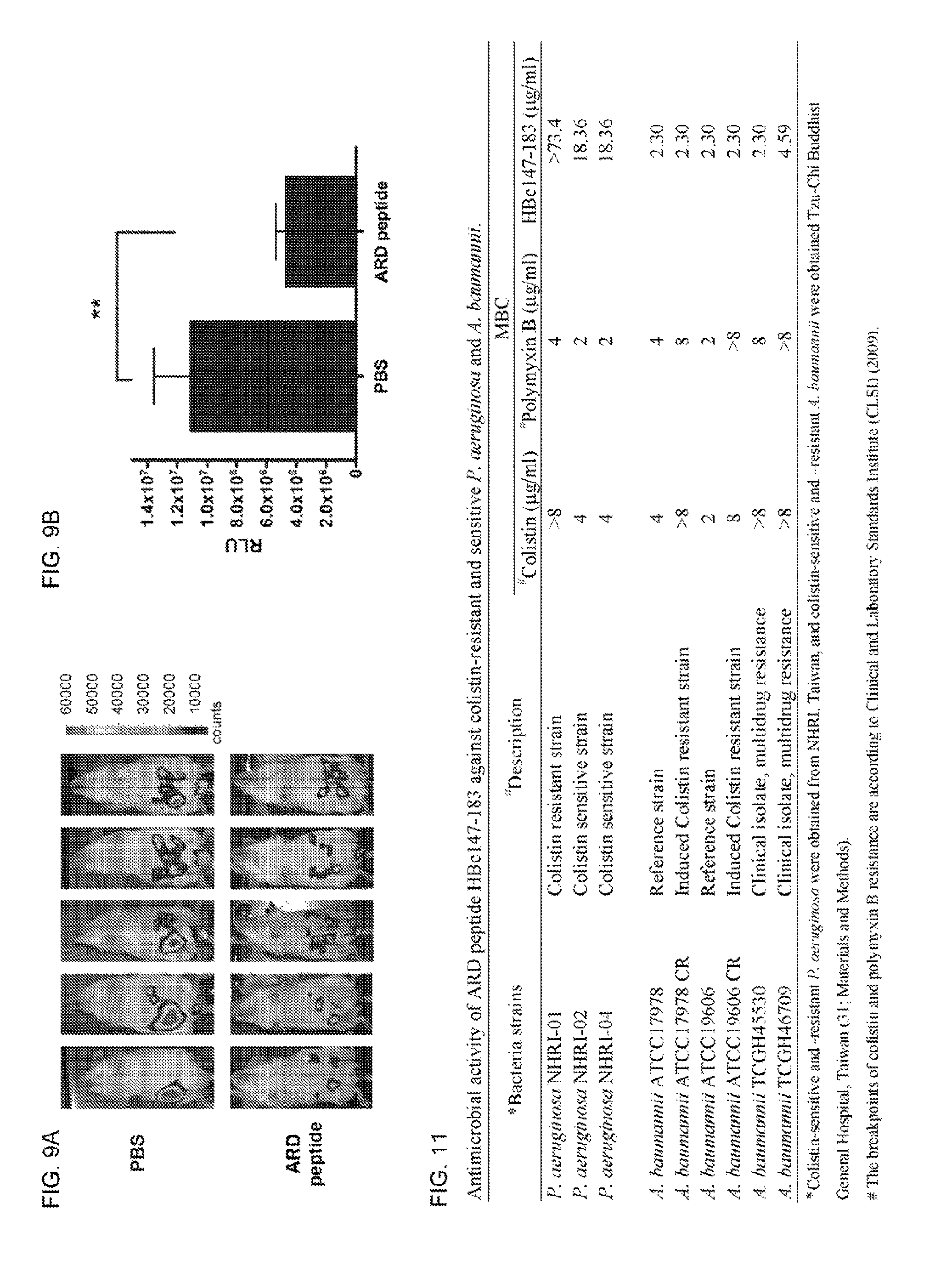
FIG. 9 shows an IVES analysis of in vivo antimicrobial activity of ARD peptide against K. pneumoniae. (A) K. pneumoniae-infected mice were treated with either PBS (n=5) or 10 mg/kg ARD peptide (n=5) at 1 hour post-inoculation. Four hours post-inoculation, mice were anesthetized and imaged. Bacterial load was displayed in the photographic image with an overlay of bioluminescence. False color imaging represents intense luminescence in red, moderate luminescence in green and low luminescence in blue and purple. (B) Total flux was quantified by IVIS imaging software. **P,0.01 (Mann-Whitney U test) for PBS and ARD peptide HBc147-183.
FIG. 10 is a table showing antimicrobial activity of HBC ARD peptides.
FIG. 11 is a table showing antimicrobial activity of ARD peptide HBc147-183 against colistin-resistant and sensitive P. aeruginosa and A. baumannii.
FIG. 12 shows sequence alignments of human hepatitis B virus (HBV) core protein (HBc) arginine rich domain (ARD) HBc ARD domain is highly conserved among different serotypes isolated from patients of different geographic areas.
FIGS. 13A-B show sequence alignments of HBc ARD domains of hepadnaviruses from primate, rodent and avian origins. HBc ARD sequences are highly conserved among human, wooly monkey, ground squirrel, woodchuck, and bat. The arginine (positive charge) clustering subdomains at HBc ARD are designated as ARD-I, ARD-II, ARD-III, and ARD-IV. FIG. 13B further shows that there are also four clustering positive charge amino acids in the core protein C-terminus of duck, heron, parrot. Ross's goose and snow goose hepatitis B virus. Despite the sequence divergence and evolutionary distance between the primate, rodent, and avian hepadnaviruses, it is contemplated that these positive charge-rich domain at the C-terminus of core protein from rodent (A) and avian (B) hepadnaviruses could also contain antimicrobial activities (cf Table 1).
DETAILED DESCRIPTION OF THE INVENTION
The present invention is more particularly described in the following examples that are intended as illustrative only since numerous modifications and variations therein will be apparent to those skilled in the art. Various embodiments of the invention are now described in detail. Referring to the drawings, like numbers indicate like components throughout the views. As used in the description herein and throughout the claims that follow, the meaning of "a", "an", and "the" includes plural reference unless the context clearly dictates otherwise. Also, as used in the description herein and throughout the claims that follow, the meaning of "in" includes "in" and "on" unless the context clearly dictates otherwise. Moreover, titles or subtitles may be used in the specification for the convenience of a reader, which shall have no influence on the scope of the present invention. Additionally, some terms used in this specification are more specifically defined below.
Definitions
The terms used in this specification generally have their ordinary meanings in the art, within the context of the invention, and in the specific context where each term is used. Certain terms that are used to describe the invention are discussed below, or elsewhere in the specification, to provide additional guidance to the practitioner regarding the description of the invention. For convenience, certain terms may be highlighted, for example using italics and/or quotation marks. The use of highlighting has no influence on the scope and meaning of a term; the scope and meaning of a term is the same, in the same context, whether or not it is highlighted. It will be appreciated that same thing can be said in more than one way. Consequently, alternative language and synonyms may be used for any one or more of the terms discussed herein, nor is any special significance to be placed upon whether or not a term is elaborated or discussed herein. Synonyms for certain terms are provided. A recital of one or more synonyms does not exclude the use of other synonyms. The use of examples anywhere in this specification including examples of any terms discussed herein is illustrative only, and in no way limits the scope and meaning of the invention or of any exemplified term. Likewise, the invention is not limited to various embodiments given in this specification.
Unless otherwise defined, all technical and scientific terms used herein have the same meaning as commonly understood by one of ordinary skill in the art to which this invention pertains. In the case of conflict, the present document, including definitions will control.
As used herein, "around", "about" or "approximately" shall generally mean within 20 percent, preferably within 10 percent, and more preferably within 5 percent of a given value or range. Numerical quantities given herein are approximate, meaning that the term "around", "about" or "approximately" can be inferred if not expressly stated.
Antimicrobial activity refers to the activity to kill or inhibit the growth of microorganisms such as bacteria, fungi and/or protozoans.
As used herein, the term "the arginine-rich carboxy-terminal region of hepatitis B virus core protein (HBc)" refers to a highly conserved arginine-rich C-terminal region of HBc (FIGS. 12 and 13A-B) and is characterized by having an antimicrobial activity. It is contemplated that the arginine-rich carboxy-terminal regions from avian and/or rodent hepadnaviruses share the same antimicrobial activities. The C-terminal end of HBc contains four arginine-rich clusters designated as ARD I-IV. Each arginine-rich cluster or domain (ARD) contains 2 or more arginine residues in continuity or in close proximity (e.g., being separated by one or two different amino acid residues). In one embodiment, each ARD may contain 2,3 or 4 continuous arginine residues.
As used herein, "an amino acid sequence derived from HBc" refers to "an amino acid sequence originates from hepatitis B virus core protein and possess an antimicrobial activity". It may be a fragment of HBc, with or without a modification, which contains ARD and possess an antimicrobial activity. A fragment of HBc with a modification includes, but not limited to PEGylation at either N- or C-terminus.
The term "two clusters" and "two repeats" are interchangeable. The term "two clusters of SPRRRR" means "2 repeats of SPRRRR", or "2 SPRRRR".
The term "amphipathic structure" refers to a molecule having hydrophobic an(hydrophilic regions.
The HBV is divided into four major serotypes (adr, adw, ayr, ayw) based on antigenic epitopes present on its envelope proteins. The term "serotype" or "serovar" refers to distinct variations within a species of bacteria or viruses or among immune cells of different individuals.
The term "protecting group" refers to a functional group that is attached to a therapeutic protein or peptide to prolong its circulatory time. A protecting group includes, but not limited to, a polyethylene glycol (PEG). PEGylation can also provide water solubility to hydrophobic proteins or peptides.
The term "treating" or "treatment" refers to administration of an effective amount of a therapeutic agent to a subject in need thereof with the purpose of cure, alleviate, relieve, remedy, ameliorate, or prevent the disease, the symptoms of it, or the predisposition towards it. Such a subject can be identified by a health care professional based on results from any suitable diagnostic method.
"An effective amount" refers to the amount of an active agent that is required to confer a therapeutic effect on the treated subject. Effective doses will vary, as recognized by those skilled in the art, depending on routes of administration, excipient usage, and the possibility of co-usage with other therapeutic treatment.
The "Guidance for Industry and Reviewers Estimating the Safe Starting Dose in Clinical Trials for Therapeutics in Adult Healthy Volunteers" published by the U.S. Department of Health and Human Services Food and Drug Administration discloses "a human equivalent dose" may be obtained by calculations from the following formula: HED=animal dose in mg/kg.times.(animal weight in kg/human weight in kg).sup.0.33.
For example for i.p. administration, if a mouse (20 gram BW) dose is 10 mg/kg, then an human dose may be calculated as 10 mg/kg.times.(0.02/patient's body weight).sup.0.33. A human equivalent effective dose, however, may vary, depending on other factors such as the route of administration.
It was an unexpected discovery and unsolved mystery that the growth of E. coli expressing HBc1-183 was very poor and much slower than that of E. coli expressing HBc1-149. it appeared that it was HBc 150-183 that somehow retarded the growth of E. coli, and reduced HBc 1-183 protein yield dramatically Here, we disclose the in vitro antimicrobial activities of HBc147-183 against a wide variety of bacteria, including multidrug resistant (MDR) and colistin (polymyxin E)-resistant A baumannii. The antimicrobial peptides from HBV core protein (HBc) arginine-rich domain (ARD) are mainly composed of SPRRR repeats and are effective against both Gram-positive and Gram-negative bacteria, as well as fungi. Using a peritoneal sepsis mouse model, it was demonstrated further that ARD peptides can effectively protect all the mice challenged with a lethal dose of Staphylococcus aureus. Treatment of ARD peptides also caused significant reduction of bacterial load of S. aureus and K. pneumoniae in infected mice. Potential mechanisms for the bactericidal activity were investigated. The ARD peptides appeared to be capable of direct binding to the Lipid A moiety of lipopolysaccharide (LPS) in several different binding assays. In summary, with high antimicrobial activity and very low toxicity against human cells and animal models, these HBc ARD peptides may have a therapeutic potential in the future (Chen et al. "Identification of a Novel Antimicrobial Peptides from Human Hepatitis B Virus Core Protein Arginine-Rich Domain (ARD)" PLoS Pathog 9(6) e1003425, which is incorporated herein by reference in its entirety).
In one aspect, the invention relates to a pharmaceutical composition comprising: (a) an effective amount of an isolated peptide, wherein the peptide comprises the arginine-rich carboxy-terminal region of hepatitis B virus core protein (HBc) and exhibits an antimicrobial activity; and (b) a pharmaceutically acceptable carrier.
In another aspect, the invention relates to a pharmaceutical composition comprising: (a) an effective amount of an isolated peptide comprising a fragment of HBc, the fragment comprising more than one arginine-rich domain (ARD) selected from the group consisting of (i), (ii) and (iii) as follows: (i) HBc ARD I-IV; (ii) HBc ARD I-III; or (iii) HBc ARD II-IV; wherein the peptide exhibits an antimicrobial activity; and (b) a pharmaceutically acceptable carrier.
The HBc may be selected from the group consisting of a mammalian HBc and an avian HBc. A mammalian HBc includes, but not limited to, human hepatitis B core protein (HBc), woolly monkey HBc, ground squirrel HBc, woodchuck HBc, and bat HBc. An avian HBc includes, but not limited to, duck, heron, parrot, Ross's goose, and snow goose.
In another aspect, the invention relates to a pharmaceutical composition comprising an effective amount of an isolated peptide, the peptide comprising an arginine-rich sequence derived from the C-terminal region of hepatitis B virus core protein (HBc), wherein the peptide is characterized by having an antimicrobial activity.
Further in another aspect, the invention relates to a pharmaceutical composition as aforementioned for use in killing and/or inhibiting the growth and/or proliferation of a microorganism by causing the composition to be in contact with the microorganism. The microorganism may be present in a subject.
Yet in another aspect, the invention relates to a pharmceutical composition as aforementioned for use in killing and/or inhibiting the growth and/or proliferation of a microorganism in a subject in need thereof, or for treating a subject afflicted with a microbial infection. The subject May be afflicted with Staphylococcus aureus or K. pneumoniae infection.
The antimicrobial peptide according to the invention contains few or no hydrophobic amino acids, and thus has no amphipathic structure.
In one embodiment of the invention, the peptide comprises the amino acid sequence of Ser Pro Arg Arg Arg Arg (SPRRRR; SEQ ID NO: 13) or Arg Arg Arg Ser (RRRS; SEQ ID NO: 14).
Alternatively, the peptide may comprise two clusters of SPRRRR (SEQ ID NO: 13), or three clusters of SPRRR (SEQ ID NO: 15). The peptide may comprise RRRS (SEQ ID NO: 14).
In another embodiment of the invention, the peptide comprises at least 2 clusters of Pro Arg (PR) located upstream to the sequence RRRS (SEQ ID NO: 14). The at least 2 clusters of PR may be immediately adjacent to, or nearby the RRRS sequence with a few residues apart, such as 7, 8, 9, 10, 11, 12, 13, 14, 15, 16, 17 or 18 amino acid residues apart.
The peptide has no RRGGRRRR sequence (SEQ ID NO: 17) at the C-terminus thereof. The peptide may comprise a protecting group.
In another embodiment of the invention, the peptide has a cysteine (C) at the C-terminus thereof.
In another embodiment of the invention, the peptide has at least 19 amino acids but no more than 37 amino acids in length.
In another embodiment of the invention, the peptide is characterized by having an activity against Gram-negative bacteria, Grain-positive bacteria, and/or fungi.
In another embodiment of the invention, the peptide comprises more than one HBc arginine-rich domain selected from the group consisting of (i), (ii) and (iii) as follows: (i) HBc ARD I, II, III, and IV; (ii) HBc ARD I, II and III; or (iii) HBc ARD II, III, and IV.
The peptide comprises 3 or 4 ARD. In another embodiment of the invention, the peptide exhibits an activity against colistin-resistant A. baumannii.
In another embodiment of the invention, the peptide is free of the sequence of SQSRESQC (SEQ ID NO: 16) at the C-terminus thereof and is characterized by having an activity against Gram-negative bacteria.
Alternatively, the peptide may comprise HBc ARD II-IV but without HBc ARD I and exhibits an activity against P. aeruginosa, or the peptide may comprise HBc ARD I-III but without HBc ARD IV and exhibits an activity against K. pneumonia.
In another embodiment of the invention, the peptide exhibits the following characteristics:
i) a reduced antimicrobial activity provided that the Ser in one of the two SPRRRR (SEQ ID NO: 13) clusters is phosphotylated; and/or
ii) a loss of antimicrobial activity provided that the Ser in each SPRRR (SEQ ID NO. 15) cluster is phosphorylated.
The peptide exhibits bactericidal activity and has no cytotoxicity to red blood cells, kidney cells, and/or liver cells
In another embodiment of the invention, the aforementioned peptide comprises: i) Ser or Pro amino acid residues downstream to the ARD IV at the C-terminal portion of the peptide. The peptide may further comprise: ii) Ser and/or Pro amino acid residues between each ARD (i.e., between ARD I and II, between ARD II and III, and between ARD III and IV.
In another embodiment of the invention, the Ser residue in the amino acid sequence of the peptide is not phosphorylated.
The composition may be formulated for topical, aerosol, oral, systemic intravenous, ocular, or rectal administration, or for inhalation administration.
In another embodiment of the invention, the peptide comprises the amino acid sequence selected from the group consisting of SEQ ID NOs: 1-10, and any serotype thereof.
The amount of the peptide in the composition is effective in killing and/or inhibiting the growth and/or proliferation of Gram-negative bacteria, Gram-positive bacteria, and/or fungi.
EXAMPLES
Without intent to limit the scope of the invention, exemplary instruments, apparatus, methods and their related results according to the embodiments of the present invention are given below. Note that titles or subtitles may be used in the examples for convenience of a reader, which in no way should limit the scope of the invention. Moreover, certain theories are proposed and disclosed herein; however, in no way they, whether they are right or wrong, should limit the scope of the invention so long as the invention is practiced according to the invention without regard for any particular theory or scheme of action.
Materials and Methods
All animal experiments were conducted under protocols approved by Academia Sinica institutional Animal Care & Utilization Committee (ASIACUC permit number 12-02-322). Research was conducted in compliance with the principles stated in the Guide for the Care and Use of Laboratory Animals, National Research Council, 1996.
Bacterial Isolates
The antimicrobial activities of HBc ARD peptides were tested on numerous bacterial strains from ATCC, including Pseudomonas aeruginosa Migula strain (ATCC 27853, ampicillin-resistant), Pseudomonas aeruginosa Migula strain (ATCC 9027, ampicillin-resistant), Klebsiella pneumoniae strain (ATCC 17593), Escherichia coli strain (ATCC 25922), Staphylococcus aureus subsp. strain (ATCC 25923, methicillin-resistant), Staphylococcus aureus subsp. strain (ATCC29213, methicillin-resistant), Staphylococcus aureus subsp. strain (ATCC 19636, methicillinresistant), and Candida albicans strain (ATCC 10231).
Clinical isolates of Pseudomonas aeruginosa (NHRI-01, NHRI-02 AND NHRI-04) were obtained through the program of Taiwan Surveillance of Antimicrobial Resistance, National Health Research Institutes, Taiwan, Acinetobacter baumannii (ATCC 17989, ATCC 17978 CR, ATCC19606, ATCC 19606 CR, TCGH 45530 AND TCGH 46709) were obtained from Tzu-Chi Buddhist General Hospital in Taiwan, and clinical isolates (TCGH 1 45530 AND TCGH 46709) were identified using the Vitek system (Biomerieux Vitek, Inc., MO, USA). A. baumannii is defined as multidrug-resistant when the organism is resistant to piperacillin, piperacillin-tazobactam, ampicillin/sulbactam, imipenem, ceftazidime, gentamicin, amikacin, tetracycline, chloramphenicol, ciprofloxacin, and cotrimoxazole. Susceptibility to colistin was determined using the broth-dilution method, in accordance with the guidelines of the Clinical and Laboratory Standards Institute.
Antimicrobial Activity
All peptides were purchased from Yao-Hong Biotechnology Inc. (Taipei, Taiwan). Vendors provided data of peptide characterizations, including HPLC and Mass. Antimicrobial activity was determined as described with some modifications as detailed below. Bacteria were grown overnight in Mueller-Hinton broth (Difeo) at 37.degree. C., and during the mid-logarithmic phase, bacteria were diluted to 10.sup.6 CFU (colony formation unit)/ml in phosphate buffer (10 mM sodium phosphate and 50 mM sodium chloride, pH 7.2). Peptides were serially diluted in the same buffer. Fifty microliter (.mu.l) of bacteria was mixed with fifty .mu.l of peptides at varying concentrations followed by incubation at 37.degree. C. for 3 hours without shaking. At the end of incubation, bacteria were placed on Mueller-Hinton broth agar plates, and allowed growth at 37.degree. C. overnight for measurement of minimal bactericidal concentration (MBC). The lowest peptide concentration on the agar plate, which displayed no bacterial growth (zero colony), was defined as MBC. All peptides were tested in triplicate.
For measurement of killing kinetics, bacteria and peptides were prepared as described above. Fifty .mu.l of bacteria were mixed with fifty .mu.l of peptides at the concentrations corresponding to MBC and were incubated at 37.degree. C. At the indicated time, bacteria were serially dilated and placed on Mueller-Hinton broth agar plates for viability measurement.
Confocal Fluorescence Microscopy
The localization of peptide was monitored by confocal fluorescence microscopy. Bacteria were grown to mid-logarithmic phase and collected by centrifugation. Approximate 10.sup.7 CFU were resuspended in a phosphate buffer containing FITC-labeled HBc147-183 at a concentration corresponding to 0.5.times.MBC. Following incubation for 1 hour at 37.degree. C., cells were washed, fixed, and immobilized on poly-L-lysine coated glass slides. ProLong Gold antifade reagent with DAPI (Invitrogen) was added to the slides prior to mounting. Localization of labeled-peptide was observed using an Olympus Ultraview confocal microscopy equipped with a 100.times. oil immersion lens.
SYTOX Green Uptake
Briefly, bacteria (10.sup.7 CFU) were prepared and mixed with 1 .mu.M SYTOX Green (Invitrogen) for 15 minutes in the dark. After the addition of peptides to the final concentrations corresponding to their respective MBC, fluorescence intensity was measured at 37.degree. C. using wavelengths 485 nm and 520 nm filters for excitation and emission. Melittin (Sigma), a major toxin of bee venom, was used as a positive control to provide maximal permeabilization.
Gel Retardation Assay
The proportion between amino nitrogen (NH.sub.3.sup.+) of HBc147-183 and phosphate (PO.sub.4.sup.-) of DNA was defined as N/P ratio. Briefly, HBc147-183 was incubated with pSUPER plasmid DNA at different N/P ratio (0, 0.2, 0.4, 0.6, 0.8, 1, 2, 3 and 4) for 30 minutes at 37.degree. C. The mobility of pSUPER plasmid DNA was analyzed by electrophoresis on 1% agarose gel.
In Vitro Binding Assay between ARD Peptides and LPS/LipidA
Several kinds of peptide-LPS or peptide-Lipid A binding assays were performed as follows:
1) Streptavidine-conjugated beads (Dynabeads My one Streptavidin T1, Invitrogen) were blocked by P. aeruginosa LPS (Sigma) at 37.degree. C for 1.5 hour. After washing with PBST (PBS, pH 7.4 containing 0.1% (w/v) Tween-20), aliquots containing 250 pmol streptavidine-conjugated beads were incubated with a reaction mixture overnight at 4.degree. C. The reaction mixture was prepared by mixing increasing amounts of biotinylated peptide HBc147-183 (0, 0.004, 0.02 0.1, 0.5 and 2.5 .mu.M) and 5 .mu.g/ml P. aeruginosa LPS (Sigma) or 200 .mu.g/ml E. coli lipid A (Sigma), at 37.degree. C. for 3 hour. After incubation overnight at 4.degree. C., the reduction of LPS (or Lipid A) in the supernatants were measured by the Limulus Amebocyte Lysate (LAL) test (Charles River Endosafe) with an ELISA reader (Molecular Devices). The amount (EU/ml) of LPS was calculated according to the standard curve prepared with Endosafe Control Srandard Endotoxin.
2) To directly measure the increasing amounts of LPS hound to the increasing amounts of peptide HBc147-183 on the peptide-coated beads, the beads were then washed with PBST three times and incubated with 100 .mu.l of PBS containing 0.15 units of trypsin agarose (Sigma) for overnight digestion at 37.degree. C. After trypsinization, the trypsin-released LPS in the supernatants were collected and measured by the Limulus Amebocyte Lysate (LAL) test. Similar results were obtained by another LPS testing method: Endosafe-PST Cartridges (Charles River Laboratories).
3) To perform the LPS/Lipid A competition assay, one .mu.g of LPS was coated on High Binding ELISA plates (Corning) overnight at 4.degree. C. The LPS-coated plates were washed by PBST and then blocked with PBST containing 5% BSA for 1 hour at 37.degree. C. After washing, HBc147-183 (10 nM), mixed with varying concentrations of E. coli Lipid A (0 to 10 .mu.g/ml), were added into each well and incubated for 1 hour at 37.degree. C. Plate-bound HBc147-183 was measured by streptavidin conjugated with HRP (1:10000 dilution) for 1 hour at 37.degree. C. TMB substrates were added into each well for color development. The absorption was measured at 450 nm with a reference wavelength at 655 nm.
Hemolytic Activity
The hemolytic activities of peptides were determined by hemolysis against human red blood cells (hRBCs). Human blood was obtained in EDTA-containing tube and was centrifuged at 450 g for 10 min. The pellet was washed three times with PBS buffer, and a solution of 10% hRBCs was prepared. hRBCs solution was mixed with serial dilutions of peptides in PBS buffer, and the reaction mixtures were incubated for 1 h at 37.degree. C. After centrifugation at 450 g for 10 min, the percentage of hemolysis was determined by measuring the absorbance at the wavelength of 405 nm of the supernatant. Blank and 100% hemolysis were determined in PBS buffer and in the presence of 1% Triton X-100, respectively.
Cytotoxicity
Cytotoxicity was measured in HepG2, Huh7, HEK293, and Vero cells by MTT assay. Cells were seeded at 10.sup.4 cells/well in a 96-well plate and serial dilutions of peptides were added into each well. PBS was used as a negative control and melittin was used as a positive control. After 1 hour of incubation, the medium were replaced by a fresh medium containing 10% MTT solution (PROMEGA.TM.), and the plate was incubated for 4 hours in 5% CO.sub.2 at 37.degree. C., The absorbance at the wavelength a 595 nm was measured by an ELISA reader (BIO-RAD.TM. model 680).
CFSE Cell Proliferation Assay
To set up CFSE cell proliferation assay, 293 cells (human kidney origin) and Vero cells (monkey kidney origin) were resuspended in PBS to a final concentration of 10.sup.6 cells/ml before incubation with 10 .mu.M CFSE dye (CELLTRACE.TM. CFSE cell proliferation kit, INVITROGEN.TM.) at 37.degree. C. for 10 min. To quench the staining, ice-old culture media were then added and incubated on ice for 5 min. Labeled cells were then pelleted and washed three times with a fresh medium containing 10% FBS before seeding into six well plates at a density of 3.3.times.10.sup.5cells/well. After 20 h, the medium was removed and incubated with a fresh medium containing 5, 25 and 100 .mu.M HBc 147-183 for one hour (FITC-labeled ARD peptide had been largely internalized in 10 minutes after the addition of ARD peptides to the medium of HepG2 cells). Forty-eight hours later, cells were harvested and analyzed by flow cytometry (FACSCanto, BD Bioscience).
In Vivo Animal Studies
Three-week old male ICR mice (19 to 21 g) were purchased from BioLASCO (Taiwan) Overnight culture of bacteria in BHI broth (Difco) was subcultured in fresh BRI broth to log phase. Inoculums were diluted in BHI broth to indicated densities. To test the acute toxicity of ARD peptide in vivo, ICR male mice were inoculated intraperitoncally (i.p.) with 10 and 20 mg/kg HBc147-183 in PBS, respectively. Each group contained 5 mice. After peptide injection, the number of dead mice was recorded daily for 7 days post-injection. To test the antimicrobial activity of the ARD peptide in vivo, all mice were inoculated i.p. with Staphylococcus aureus ATCC 19636 (4.times.10.sup.6 CFU/mouse) in BHI broth. Peptide HBc147-183 (10 mg/kg) was administered i.p. at 1, 1.5 and 2 hours post-inoculation. PBS (10 ml/kg) control was administered at 1 hour post-inoculation. Each group contained 10 mice. Mortality was monitored daily for 7 days post-inoculation. In a separate experiment to measure the bacterial load, mice were inoculated i.p. with Staphylococcus aureus ATCC 19636 (10.sup.6 CFU/mouse) in BHI broth. All mice were administered at 1 hour post-inoculation with peptide HBc147-183 (10 mg/kg) or PBS (10 ml/kg) control, and sacrificed at 4 hours post-inoculation. Blood samples (200 .mu.l) were mixed with 100 mM EDTA (10 .mu.l) and were diluted 20-fold in PBS (Ca.sup.2+ and Mg.sup.2+ free). Liver and spleen samples (0.1 g) were homogenized in sterile PBS (500 .mu.l). Samples were diluted approximately 100-fold and plated on BHI agar for scoring the colony numbers.
To test the in vivo antimicrobial activity of the ARD peptide against Gram-negative bacteria, mice were inoculated with Klebsiella pneumoniae Xen39 (10.sup.7 cfu/mouse) (Caliper LifeSciences), an engineered strain containing a modified Photorhabdus luminescens huxABCDE operon. One hour post-inoculation, mice received either 10 ml/kg PBS (n=5) or 10 mg/kg ARD peptide (n=5), respectively. In vivo imaging was carried out at 4 hours post-inoculation. The mice were anesthetized first before transferring to the IVIS imaging system (IVIS spectrum), and luminescence was measured with an exposure time of 1 minutes or less. The image system measured the number of photons and translated the data to false color images that depicted the region of strong luminescence with red, moderate luminescence with yellow and green, and mild luminescence with blue. Decreasing bioluminescence indicated reduction of bacteria. The images were overlay of photographic images and bioluminescence using a computor-generated color scale. Total flux (RLU) of region of interest (ROI) was quantified by the IVIS imaging software.
Results
In Vitro Anti Microbial Activity of HBc Peptides
As shown in FIGS. 1 and 10, HBc147-183 displayed a broad-spectrum activity against Gram-negative bacteria (P. aeruginosa, K. pneumoniae and E. coli), Gram-positive bacteria (S. aureus), and fungi (C. albicans). Among these tested strains, P. aeruginosa and K. pneumonia were the most sensitive to this peptide. The MBCs of HBc147-183 were lower than 4 .mu.M for P. aeruginosa and K. pneumonia, and around 4 .mu.M for E. coli, and S. aurens, C. albicans was the least sensitive to this peptide (MBC.about.8 .mu.M).
To further map the active sequences of the antimicrobial activity, various peptides (FIG 1) in different length were synthesized and tested as before. Peptide HBc147-175, with the deletion of the last eight amino acids at the C-terminus, maintained strong activity against Gram-negative bacteria, albeit it lost the activity against S. aureus and C. albicans. We detected no activity against all of the tested bacteria and fungi from peptides ARD I-II (HBc147-159) or ARD III-IV (HBc164-176 and HBc162-175). In contrast, all peptides containing ARD II-IV (HBc153-176, HBc157-176, HBc153-175, 1 HBc155-175, and HBc157-175) and ARD I-III (HBc147-167) exhibited strong activity against P. aeruginosa and K. pneumonia, respectively, albeit they were weak against E. coli (FIG. 10). Therefore, peptide ARD II-IV and ARD I-III appeared to be necessary and sufficient for the bactericidal activity against P. aeruginosa and K. pneumonia.
Positive Charge of ARD Peptides is Critical to the Bactericidal Activity.
Phophorylation studies on serine residues S155, S162, S170, S176 and S181 revealed that serine phosphorylation in general weakened the potency of antimicrobial activity. It was found that all HBc peptides, once phosphorylated, lost their activities against C. albicans (FIG. 11). For bacteria, the phosphorylation on S181 showed no effects, whereas phosphorylations on S155, S162, S170, and S176 reduced the antimicrobial activity. The MBCs dropped to 8 .mu.M for HBc155p and HBc176p, and 32 .mu.M for HBc162p and HBc170p, respectively. When S155, S162 and S170 were simultaneously phosphorylated (HBc155p162p170p), the antimicrobial activity was completely lost (>32 .mu.M). The results suggested that, except for S181, serine phosphorylation is generally detrimental to the antimicrobial activity of HBc ARD peptide. To confirm the importance of arginine residues for bactericidal activity, we synthesized and tested peptide HBc147-183-III-IV AA, which has two R-to-A substitution mutations in each of ARD III and ARD IV. Similar to phosphorylated HBc ARD peptides, the MBC of HBc147-183-III-IV AA was significantly increased compared to HBc147-183. The result indicated that arginine residues are required for the antimicrobial activity.
Drug Resistance
The antimicrobial activity of HBc147-183 against colistin-resistant P. aeruginosa and A. baumannii was tested. As shown in FIG. 11, while HBc147-183 killed colistin-sensitive P. aeruginosa at 4 .mu.M. colistin-resistant P. aeruginosa were cross-resistant to HBc147-183 (MBC>16 .mu.M). In contrast to P. aeruginosa, the MBCs of HBc147-183 against colistin-sensitive and colistin-resistant A. baumannii are in a similar range of 0.5-1 .mu.M. This result indicates that, for colistin-resistant A. baumannii, there is no cross-resistance to our ARD peptide HBc147-183.
Killing Kinetics
Time course of bacterial viability was determined after the tested bacteria (P. aeruginosa, K. pneumonia, E. coli and S. aureus) were treated with HBc147-183 at the concentrations corresponding to the MBC (FIG. 2). The results showed that P. aeruginosa was immediately killed within 20 minutes upon the addition of HBc147-183 (2 .mu.M). Although K. pneumonia and E. coli were members of Gram-negative bacteria, they were killed by 4 .mu.M HBc147-183 in 180 minutes. For S. aureus, complete killing by 4 .mu.M HBc147-183 was observed in 120 minutes.
Localization and Mechanism of HBc147-183
P. aeruginosa, E. coli and S. aureus were treated with FITC-labeled HBc147-183 corresponding to 0.5.times.MBC, and the localization of HBc147-183 was visualized using confocal fluorescence microscopy (FIG. 3). The results showed that, upon peptide treatment, P. aeruginosa, K. pneumonia and E. coli appeared as hollow rods with fluorescence dearly defined bacteria surface, suggesting that HBc147-183 was accumulated on the membrane (FIGS. 3A-D). To understand better the effect of HBc peptides on the membranes, SYTOX Green uptake assay was performed A significant degree of membrane permeabilization was induced on P. aeruginosa upon the addition of 2 .mu.M HBc147-183 (FIG. 4A). Although it was also accumulated on the membrane of K. pneumonia and E. coli, 4 .mu.M HBc147-183 was not able to induce membrane permeabilization as observed on S. aureus. Consistent with the bactericidal activity of HBc147-183 against P. aeruginosa, HBc153-176 caused same membrane permeabilization within 10 minutes in a dose-dependent manner (FIG. 4B). This indicated that the bactericidal effect of HBc peptides on P. aeruginosa is directly through the membrane permeabilization with a fast kinetics similar to that of killing kinetics (FIG. 2). On the other hand, HBc147-183 was found to penetrate through the membrane of S. aurens and localized in the cytoplasm (FIGS. 3E-G). To investigate the potential interaction between HBc 147-183 and DNA, HBc147-183 was mixed with pSUPER plasmid DNA at different N/P ratio (Materials and Methods) and analyzed by gel electrophoresis (FIG. 4C). The results showed that the mobility of DNA was decreased when the ratio of peptide/DNA increased and the plasmid DNA was completely retarded at the ratio of 1, suggesting that HBc147-183 has a strong binding activity to plasmid DNA. Overall, it suggests that the bactericidal mechanisms of HBc147-183 on Gram-positive and Gram-negative bacteria may be completely different.
Direct Binding of HBc147-183 to LPS
To determine whether LPS of Gram-negative bacteria could serve as a potential target of HBc147-183, LPS (0.05 to 50 .mu.g/ml) from either P. aeruginosa or E. coli (Sigma) were incubated with both P. aeruginosa and 2 .mu.M HBc147-183 for three hours, respectively. The results showed that the bactercidal activity of HBc147-183was significantly reduced by addition of either LPS at the concentration of 50 .mu.g/ml. (FIG. 5). In addition, HBc147-183 preferentially bound to the LPS from P. aeruginosa, rather than that from E. coli. However, the addition of anti-LPS antibody (Genetex Co.) cannot sufficiently neutralize the bactericidal activity of HBc147-183 (FIG. 5). It suggests that HBc147-183 could bind to not only LPS but also other target molecules on the membrane. Alternative, HBc147-183 and the anti-LPS polyclonal antibody used here could bind predominantly to two different epitopes on the LPS.
As shown in FIG. 6A, the potential interaction between HBc147-183 and LPS (or Lipid A moiety in vitro was investigated using several different binding assays. In FIG. 6B, when increasing amount of HBc147-183 was bound to the strepavidine-conjugated Dynabeads and allowed incubation with a constant amount of LPS, gradually increasing amount of LPS appeared to be depleted from the supernatant HBc147-183 8p, which has eight ser/thr phosphorylations, was used in parallel as a control peptide. Similar results were obtained by another LPS testing method: Endosafe-PTS Cartridges (Charles River Laboratories). In FIG. 6C, the beads-captured LPS were dissociated from the beads by trypsin agarose digestion of the ARD peptide HBc147-183. The amount of released LPS was measured by the LAL test (Materials and Methods). LPS contains mainly the polysaccharide and Lipid A moieties. To determine whether the ARD peptide can bind to Lipid A directly, we tested in FIG. 6D the binding between Lipid A and the ARD peptide in a manner similar to FIG. 6B. As expected, increasing amounts of HBc147-183 on the beads led to decreasing amounts of Lipid A remaining in the supernatant. The inverse correlation between the ARD peptide on the beads and the Lipid A in the supernatant (FIG. 6D) is strikingly similar to what was observed previously between the ARD peptide HBc147-183 on the beads and LPS in the supernatant (FIG. 6B). To directly demonstrate that the ARD peptide can bind to the Lipid A moiety of LPS, we performed a competition experiment between Lipid A and LPS (FIG. 6E). The LPS-coated ELISA plates were incubated with constant amount of HBc147-183 (10 .mu.M), which was premixed with varying concentrations of E. coli Lipid A (0 to 10 .mu.g/ml). After extensive washing, plate-bound (i.e., LPS-bound) biotinylated peptide HBc147-183 was measured by streptavidin conjugated with HRP, followed by adding TMB substrates and color development. The binding of HBc147-183 to LPS was significantly decreased by the increasing concentrations of Lipid A (FIG. 6E). The result here lends support for the notion that Lipid A moiety of LPS can serve as a direct target for ARD peptide HBc147-183
Cytocoxicity
To determine the cytotoxicity of HBc peptides, we measured the hemolytic activity of HBc147-183. Compared to the melittin control, no detectable hemolysis by HBc147-183 was observed after one hour of incubation (FIG. 7A). In addition, MTT assay was performed to determine the cytotoxicity of HBc147-183 to human hepatoma (Huh 7 and HepG2 cells) and kidney cells (Vero and HEK293 cells). The viability of cells treated with melittin at low dose (3.125 .mu.M) was significantly decreased. In contrast, HBc147-183 caused only a low level of cytotoxicity at the concentration of 100 .mu.M (FIG. 7B). The CFSE cell proliferation assay was also performed to determine the effect of HBc147-183 on the proliferation of Vero and HEK293 kidney cells. In comparison to day 1, CFSE intensity of cells treated with HBc147-183 (5, 25 and 100 .mu.M) decreased to the same level as the mock control on day 3 (FIG. 7C), suggesting that ARD peptide HBc147-183 has no significant effect on cell proliferation.
Animal Model
To conduct the experimental infection with bacteria, we i.p. inoculated mice with Staphylococcus aureus ATCC 19636 (4.times.10.sup.6 cfu/mouse). Bacterial load in blood at 1, 2, 4 and 6 hours post-inoculation was determined As shown in FIG. 8A, bacteria rapidly transferred to the blood compartment from peritoneal cavity. Within 2 hours, the number of bacteria in the blood achieved the maximum (10.sup.6 cfu/ml). Thereafter, the number of bacteria in the blood gradually decreased spontaneously. To distinguish the ARD peptide-mediated from the spontaneous clearance, we therefore tested the in vivo protection activity of the ARD peptide within 2 hours post-inoculation. Briefly, mice were i.p. inoculated with Staphylococcus aureus ATCC 19636 and received a single dose of 10 ml/kg PBS or a single dose of 10 mg/kg ARD peptide at 1, 1.5, 2 hours post-inoculation, respectively. Mice (n=10) treated with PBS died within 24 hours post-inoculation (FIG. 8B). In contrast, administration of ARD peptide (10 mg/kg) at 1 hour post-inoculation can effectively protect all mice (n=10) from death at day 7. When we administered ARD peptide at 1.5 (n=10) and 2 (n=10) hours post-inoculation, survival rates were decreased to 70% and 40%, respectively Instead of using death as a surrogate indicator of the antimicrobial activity of ARD peptide, we also determined directly the in vivo effect of ARD peptide on bacterial load of infected mice (FIG. 8C). Mice were inoculated with Staphylococcus aeurus as before and treated with 10 ml/kg PBS (n=5) or 10 mg/kg ARD peptide (n=5) at 1 hour post-inoculation. Four hours post-inoculation, bacterial load in blood, liver and spleen samples of control mice were in the range of 10.sup.6 cfu/ml (FIGS. 8C and 8D).
Administration of ARD peptide significantly reduced the bacterial load (.about.10.sup.4 cfu/ml) by 100-fold in blood, liver and spleen than the PBS control mice (P<0.01). In addition to Staphylococcus aureus, we also examined the in vivo antimicrobial activity of ARD peptide on K. pneumoniae using an IVIS imaging system. Similar to the change in bacterial load of S. aureus, bioluminescence of mice inoculated with K. pneumonae Xen39 peaked at 2 hour post-inoculation (data not shown). We then treated K. pneumoniae Xen39-infected mice with PBS or ARD peptide at 1 hour post-inoculation, respectively. The results showed that the bioluminescence of ARD peptide-treated mice was very weak, whereas PBS control showed a more extensive bioluminescence (FIG. 9A). There was a significant difference in the overall RLU values of mice treated with PBS versus ARD peptide (P<0.01) (FIG. 9B). Taken together, the results indicated that HBc147-183 exhibited significant antimicrobial activity in vivo. Table 1 shows comparison of minimal bactericidal concentrations of human HBc ARD and woodchuck HBc ARD against Gram-negative and Gram-positive bacteria.
TABLE-US-00001 TABLE 1 Minimal bactericidal concentration (.mu.M) Bacteria strain hHBc ARD.sup.1 wHBc ARD.sup.2 P. aeruginosa ATCC 9027 2 2 P. aeruginosa ATCC 27853 2-4 2 K. pneumoniae ATCC 13884 2 2 E. coli ATCC 25922 4 4 S. aureus ATCC 19636 4 8 .sup.1Human hepatitis B virus core protein arginine-rich domain (ARD)(TVVRRRGRSPRRRTPSPRRRRSQSPRRRRSQSRESQC; SEQ ID NO: 1) .sup.2Woodchuck hepatitis B virus core protein ARD (TVIRRRGGARASRSPRRRTPSPRRRRSQSPRRRRSQSPSANC; SEQ ID NO: 4)
The invention relates to a novel antimicrobial peptide (HBc147-183) isolated from the C-terminal domain of HBc. The computer program based on the antimicrobial peptide database (Wang et al. (2004) Nucleic Acids Res 32: D590-592) predicted unfavorably that HBc147-183 could serve as an antibacterial peptide, due to its very low content of hydrophobic amino acids. Contrary to the computer prediction, surprisingly, HBc147-183 exhibited a broad-spectrum antimicrobial activity. While colistin-resistant P. aeruginosa exhibited cross-resistance to ARD peptide HBc1 47-183, we found a strong activity of hbc147-183 (MBC=0.5-1 .mu.m) against all tested colistin-resistant A. baumannii. Our ARD peptide could bind to Lipid A of E. coli and LPS of P. aeruginosa (FIG. 6). While only the full-length HBc147-183 (ARD I-IV) was effective against the tested gram-positive bacteria, S. aureus, ARD I-IV (HBc153-176) and ARD I-III (HBc147-167), in a less than full-length context, exhibited strong activity against gram-negative P. aeruginosa and K. pneumoniae, respectively (but not E. coli).
Comparisons with Other AMPs
It is surprising that the ARD domain of HBc protein (HBc147-183) exhibits novel and broad spectrum antimicrobial activity. This peptide shares some degree of similarity with several antimicrobial peptides in literature, such as protamine (PRRRRSSSRPVRRRRRPRVSRRRRRRGGRRRR; SEQ ID NO: 11) and drosocin (GKPRPYSPRPTSHPRPIRV; SEQ ID NO: 12). A radial diffusion assay showed that a single arginine-rich domain (RRRR) is sufficient for antimicrobial activity, especially against gram-negative bacteria (Lesmes et al. (2009) Peptides 30: 2150-2160). Unlike protamine, the arginine-rich domain of HBc147-183, such as ARD I-II and ARD III-IV, were not sufficient for the antimicrobial activity. In addition, sequence alignment by anitimicrobial peptide database revealed that HBc153-176 shares 44% amino acid sequence homology with Drosocin, which is a proline-rich peptide isolated from Drosophila. However, except for P. aeruginosa, Drosocin is predominately active against most Gram-negative bacteria. Drosocin kills bacteria via an apparently non-membranolytic mechanism. The HBc ARD is a novel peptide with a broad spectrum bactericidal activity quite distinct from other known arginine-rich antimicrobial peptides. Protamine contains RRGGRRRR (SEQ ID NO: 17), while HBc147-183 contains SQSRESQC (SEQ ID NO: 16) at the C-terminus of HBc.
Bactericidal Mechanisms
The results showed the membrane localization of HBc147-183 on Gram-negative bacteria (FIG. 3) and the neutralization activity of LPS from either P. aeruginosa or E. coli (FIG. 5). It suggests that HBc147-183 could have a strong binding activity to LPS. The results revealed a direct binding of HBc147-183 to LPS and Lipid A (FIGS. 6B, 6C and 6D). Furthermore, Lipid A moiety of LPS was shown to be one major direct target of HBc147-183 (FIG. 6E). However, incubation of LPS antibody with P. aeruginosa and HBc147-183 failed to neutralize the bactericidal activity of HBc ARD peptide (FIG. 5). One interpretation for this negative result is that HBc147-183 could bind not only LPS but also some other molecules on the bacterial membrane. We found a better neutralization effect of the LPS from P. aeruginosa than that from E. coli (FIG. 5). The preference of binding by HBc147-183 for the LPS of P. aeruginosa is correlated with its stronger bactericidal activity against P. aeruginosa.
The mode of action of HBc147-183 on P. aeruginosa could be related to membrane permeabilization based on the fast killing kinetics (FIG. 2) and its membrane localization (FIGS. 3A and 3B). This speculation is also supported by the results of SYTOX Green uptake experiment (FIG. 4A). Like P. aeruginosa. HBc147-183 was also accumulated on the membrane of K. pneumonia and E. coli. However, the killing kinetics and SYTOX Green uptake experiments of K. pneumonia and E. coli did not support for a mechanism of membrane permeabilization. It remains to be further investigated how bacteria can e killed by the ARD peptides using a mechanism other than membrane permeabilization.
In the case of Gram-positive bacteria, we found that HBc147-183 was not accumulated on the membrane (FIG. 3). Instead, it can enter the cytoplasm of S. aureus without any apparent development of membrane permeabilization (FIG. 4A). In addition to LPS, HBc147-183 can also bind strongly to plasmid DNA (FIG. 4C). Taken together, the bactericidal mechanism of HBc147-183 against Gram-positive bacteria appeared to be more similar to Buforin II, which was reported to kill bacteria by binding to DNA and RNA after penetrating bacterial membrane.
Although HBc147-183 can penetrate through the cell membrane of Huh 7 and HepG2 cells (data not shown), we observed no significant cytotoxic effect on human hepatoma cells Huh 7 and HepG2, and kidney cells Vero and HEK293, even at a high peptide concentration (100 .mu.M) by MTT assay (FIG. 7B) and proliferation assay (FIG. 7C). Taken together with the results from the hemolytic assay (FIG. 7A), HBc147-183 appears to be much safer relative to melittin in cell culture. The animal model study showed no apparent in vivo toxicity of ARD peptide at 20 mg/kg dose in the ICR mice by i.p. injection (FIG. 7D). At as low as 10 mg/kg level, treatment of ARD peptide can protect mice from death (FIG. 8B). In contrast, all mice receiving the PBS control were dead soon after bacterial inoculation. In addition to the sepsis survival model, treatment of ARD peptide (10 mg/kg) also resulted in a significant reduction of bacterial load of S. aureus and K. pneumoniae, whereas PBS control mice showed high levels of bacterial load (FIGS. 8C-D and FIG. 9). The results demonstrated the in vivo antimicrobial potency of HBc ARD peptide.
SEQUENCE LISTINGS
1
33137PRTHuman Hepatitis B virus 1Thr Val Val Arg Arg Arg Gly Arg Ser Pro Arg Arg Arg Thr Pro Ser 1 5 10 15 Pro Arg Arg Arg Arg Ser Gln Ser Pro Arg Arg Arg Arg Ser Gln Ser 20 25 30 Arg Glu Ser Gln Cys 35 236PRTWoolly monkey Hepatitis B virus 2Thr Val Val Arg Arg Arg Arg Pro Ser Gly Arg Arg Thr Pro Ser Pro 1 5 10 15 Arg Arg Arg Arg Ser Gln Ser Pro Arg Arg Arg Arg Ser Gln Ser Pro 20 25 30 Ala Ser Ser Cys 35 341PRTGround squirrel Hepatitis B virus 3Thr Val Ile Arg Arg Arg Gly Ser Ala Arg Val Val Arg Ser Pro Arg 1 5 10 15 Arg Arg Thr Pro Ser Pro Arg Arg Arg Arg Ser Gln Ser Pro Arg Arg 20 25 30 Arg Pro Gln Ser Pro Ala Ser Asn Cys 35 40 442PRTWoodchuck hepatitis B virus 4Thr Val Ile Arg Arg Arg Gly Gly Ala Arg Ala Ser Arg Ser Pro Arg 1 5 10 15 Arg Arg Thr Pro Ser Pro Arg Arg Arg Arg Ser Gln Ser Pro Arg Arg 20 25 30 Arg Arg Ser Gln Ser Pro Ser Ala Asn Cys 35 40 543PRTBat Hepatitis B virus 5Thr Ile Val Arg Arg Arg Gly Gly Ser Arg Ala Thr Arg Ser Pro Arg 1 5 10 15 Arg Arg Thr Pro Ser Pro Arg Arg Arg Arg Ser Gln Ser Pro Arg Arg 20 25 30 Arg Arg Ser Gln Ser Pro Ala Ser Ser Asn Cys 35 40 636PRTDuck hepatitis B virus 6Arg Lys Pro Arg Gly Leu Glu Pro Arg Arg Arg Lys Val Lys Thr Thr 1 5 10 15 Val Val Tyr Gly Arg Arg Arg Ser Lys Ser Arg Glu Arg Arg Ala Pro 20 25 30 Thr Pro Gln Arg 35 736PRTHeron hepatitis B virus 7Arg Lys Pro Arg Gly Leu Glu Pro Arg Arg Arg Lys Val Lys Thr Thr 1 5 10 15 Val Val Tyr Gly Arg Arg Arg Ser Lys Ser Arg Gly Arg Arg Ser Ser 20 25 30 Pro Ser Gln Arg 35 836PRTParrot Hepatitis B virus 8Arg Lys Pro Arg Gly Leu Glu Pro Arg Arg Arg Lys Val Lys Thr Thr 1 5 10 15 Val Val Tyr Gly Arg Arg Arg Ser Lys Ser Arg Glu Arg Ser Ser Ser 20 25 30 Ser Pro Gln Arg 35 936PRTRoss's goose Hepatitis B virus 9Arg Lys Pro Arg Gly Leu Glu Pro Arg Arg Arg Lys Val Lys Thr Thr 1 5 10 15 Val Val Tyr Gly Arg Arg Arg Ser Lys Ser Arg Glu Arg Arg Ala Pro 20 25 30 Thr Pro Gln Arg 35 1036PRTSnow goose Hepatitis B virus 10Arg Lys Pro Arg Gly Leu Glu Pro Arg Arg Arg Lys Val Lys Thr Thr 1 5 10 15 Val Val Tyr Gly Arg Arg Arg Ser Lys Ser Arg Glu Arg Arg Ala Ser 20 25 30 Ser Pro Gln Arg 35 1132PRTOncorhynchus mykiss 11Pro Arg Arg Arg Arg Ser Ser Ser Arg Pro Val Arg Arg Arg Arg Arg 1 5 10 15 Pro Arg Val Ser Arg Arg Arg Arg Arg Arg Gly Gly Arg Arg Arg Arg 20 25 30 1219PRTDrosophila melanogaster 12Gly Lys Pro Arg Pro Tyr Ser Pro Arg Pro Thr Ser His Pro Arg Pro 1 5 10 15 Ile Arg Val 136PRTHepatitis B virus 13Ser Pro Arg Arg Arg Arg 1 5 144PRTHepatitis B virus 14Arg Arg Arg Ser 1 155PRTHepatitis B virus 15Ser Pro Arg Arg Arg 1 5 168PRTHepatitis B virus 16Ser Gln Ser Arg Glu Ser Gln Cys 1 5 178PRTOncorhynchus mykiss 17Arg Arg Gly Gly Arg Arg Arg Arg 1 5 1837PRTHuman Hepatitis B virus 18Thr Val Val Arg Arg Arg Gly Arg Ser Pro Arg Arg Arg Thr Pro Ser 1 5 10 15 Pro Arg Arg Arg Arg Ser Lys Ser Pro Arg Arg Arg Arg Ser Gln Ser 20 25 30 Arg Glu Ser Gln Cys 35 1937PRTHuman Hepatitis B virus 19Thr Val Val Arg Arg Arg Gly Arg Ser Pro Arg Arg Arg Thr Pro Ser 1 5 10 15 Pro Arg Arg Arg Arg Ser Gln Ser Pro Arg Arg Arg Arg Ser Gln Ser 20 25 30 Arg Glu Ser Gln Cys 35 2039PRTHuman Hepatitis B virus 20Thr Val Val Arg Arg Arg Asp Arg Gly Arg Ser Pro Arg Arg Arg Thr 1 5 10 15 Pro Ser Pro Arg Arg Arg Arg Ser Gln Ser Pro Arg Arg Arg Arg Ser 20 25 30 Gln Ser Arg Glu Ser Gln Cys 35 2137PRTHuman Hepatitis B virus 21Thr Val Val Arg Arg Arg Gly Arg Ser Pro Arg Arg Arg Thr Pro Ser 1 5 10 15 Pro Arg Arg Arg Arg Ser Gln Ser Pro Arg Arg Arg Arg Ser Gln Ser 20 25 30 Arg Glu Ser Gln Cys 35 2237PRTHuman Hepatitis B virus 22Thr Val Val Arg Arg Arg Gly Arg Ser Pro Arg Arg Arg Thr Pro Ser 1 5 10 15 Pro Arg Arg Arg Arg Ser Gln Ser Pro Arg Arg Arg Arg Ser Gln Ser 20 25 30 Arg Glu Ser Gln Cys 35 2339PRTHuman Hepatitis B virus 23Thr Val Val Arg Arg Arg Asp Arg Gly Arg Ser Pro Arg Arg Arg Thr 1 5 10 15 Pro Ser Pro Arg Arg Arg Arg Ser Gln Ser Pro Arg Arg Arg Arg Ser 20 25 30 Gln Ser Arg Glu Ser Gln Cys 35 2437PRTHuman Hepatitis B virus 24Thr Val Val Arg Arg Arg Gly Arg Ser Pro Arg Arg Arg Thr Pro Ser 1 5 10 15 Pro Arg Arg Arg Arg Ser Gln Ser Pro Arg Arg Arg Arg Ser Gln Ser 20 25 30 Arg Glu Ser Gln Cys 35 2537PRTHuman Hepatitis B virus 25Ala Val Val Arg Arg Arg Gly Arg Ser Pro Arg Arg Arg Thr Pro Ser 1 5 10 15 Pro Arg Arg Arg Arg Ser Gln Ser Pro Arg Arg Arg Arg Ser Gln Ser 20 25 30 Arg Gly Ser Gln Cys 35 2639PRTHuman Hepatitis B virus 26Thr Val Val Arg Arg Arg Asp Arg Gly Arg Ser Pro Arg Arg Arg Thr 1 5 10 15 Pro Ser Pro Arg Arg Arg Arg Ser Gln Ser Pro Arg Arg Arg Arg Ser 20 25 30 Gln Ser Arg Glu Ser Gln Cys 35 2737PRTHuman Hepatitis B virus 27Thr Val Val Arg Arg Arg Gly Arg Ser Pro Arg Arg Arg Thr Pro Ser 1 5 10 15 Pro Arg Arg Arg Arg Ser Gln Ser Pro Arg Arg Arg Arg Ser Gln Ser 20 25 30 Arg Glu Ser Gln Cys 35 2837PRTHuman Hepatitis B virus 28Thr Val Val Arg Gln Arg Gly Arg Thr Ile Arg Arg Arg Thr Pro Ser 1 5 10 15 Pro Arg Arg Arg Arg Ser Gln Ser Pro Arg Arg Arg Arg Ser Gln Ser 20 25 30 Arg Glu Ser Gln Cys 35 2937PRTHuman Hepatitis B virus 29Thr Val Val Arg Arg Arg Gly Arg Ser Pro Arg Arg Arg Thr Pro Ser 1 5 10 15 Pro Arg Arg Arg Arg Ser Gln Ser Pro Arg Arg Arg Arg Ser Gln Ser 20 25 30 Arg Glu Ser Gln Cys 35 3037PRTHuman Hepatitis B virus 30Thr Val Val Arg Arg Arg Gly Arg Ser Pro Arg Arg Arg Thr Pro Ser 1 5 10 15 Pro Arg Arg Arg Arg Ser Gln Ser Pro Arg Arg Arg Arg Ser Gln Ser 20 25 30 Arg Glu Ser Gln Cys 35 3137PRTHuman Hepatitis B virus 31Thr Val Val Arg Arg Arg Gly Arg Ser Pro Arg Arg Arg Thr Pro Ser 1 5 10 15 Pro Arg Arg Arg Arg Ser Gln Ser Pro Arg Arg Arg Arg Ser Gln Ser 20 25 30 Arg Glu Ser Gln Cys 35 3237PRTHuman Hepatitis B virus 32Thr Val Val Arg Arg Arg Gly Arg Ser Pro Arg Arg Arg Thr Pro Ser 1 5 10 15 Pro Arg Arg Arg Arg Ser Gln Ser Pro Arg Arg Arg Arg Ser Gln Ser 20 25 30 Arg Glu Ser Gln Cys 35 3337PRTHuman Hepatitis B virus 33Thr Val Val Arg Gly Arg Gly Arg Ser Ser Arg Arg Arg Thr Pro Ser 1 5 10 15 Pro Arg Arg Arg Arg Ser Gln Ser Pro Arg Arg Arg Arg Ser Gln Ser 20 25 30 Arg Glu Ser Gln Cys 35
* * * * *
References
D00001
D00002
D00003
D00004
D00005
D00006
D00007
D00008
S00001
XML
uspto.report is an independent third-party trademark research tool that is not affiliated, endorsed, or sponsored by the United States Patent and Trademark Office (USPTO) or any other governmental organization. The information provided by uspto.report is based on publicly available data at the time of writing and is intended for informational purposes only.
While we strive to provide accurate and up-to-date information, we do not guarantee the accuracy, completeness, reliability, or suitability of the information displayed on this site. The use of this site is at your own risk. Any reliance you place on such information is therefore strictly at your own risk.
All official trademark data, including owner information, should be verified by visiting the official USPTO website at www.uspto.gov. This site is not intended to replace professional legal advice and should not be used as a substitute for consulting with a legal professional who is knowledgeable about trademark law.