Synergistic combination of a phenolic-rich maple syrup extract and an antibiotic
Tufenkji , et al. Feb
U.S. patent number 10,206,906 [Application Number 15/159,980] was granted by the patent office on 2019-02-19 for synergistic combination of a phenolic-rich maple syrup extract and an antibiotic. This patent grant is currently assigned to THE ROYAL INSTITUTION FOR THE ADVANCEMENT OF LEARNING/MCGILL UNIVERSITY. The grantee listed for this patent is THE ROYAL INSTITUTION FOR THE ADVANCEMENT OF LEARNING/MCGILL UNIVERSITY. Invention is credited to Vimal Maisuria, Nathalie Tufenkji.


| United States Patent | 10,206,906 |
| Tufenkji , et al. | February 19, 2019 |
| **Please see images for: ( Certificate of Correction ) ** |
Synergistic combination of a phenolic-rich maple syrup extract and an antibiotic
Abstract
It is provided an anti-microbial composition comprising a phenolic-rich extract such as a phenolic-rich maple syrup extract (PRMSE) and at least one antibiotic and a method of treating a bacterial infection comprising administering to a subject in need thereof a phenolic-rich extract and at least one antibiotic.
| Inventors: | Tufenkji; Nathalie (Laval, CA), Maisuria; Vimal (Montreal, CA) | ||||||||||
|---|---|---|---|---|---|---|---|---|---|---|---|
| Applicant: |
|
||||||||||
| Assignee: | THE ROYAL INSTITUTION FOR THE
ADVANCEMENT OF LEARNING/MCGILL UNIVERSITY (Montreal,
CA) |
||||||||||
| Family ID: | 57324993 | ||||||||||
| Appl. No.: | 15/159,980 | ||||||||||
| Filed: | May 20, 2016 |
Prior Publication Data
| Document Identifier | Publication Date | |
|---|---|---|
| US 20160339071 A1 | Nov 24, 2016 | |
Related U.S. Patent Documents
| Application Number | Filing Date | Patent Number | Issue Date | ||
|---|---|---|---|---|---|
| 62165243 | May 22, 2015 | ||||
| Current U.S. Class: | 1/1 |
| Current CPC Class: | A61K 31/43 (20130101); A61K 36/20 (20130101); A61K 31/496 (20130101); A61K 31/43 (20130101); A61K 2300/00 (20130101); A61K 31/496 (20130101); A61K 2300/00 (20130101); Y02A 50/401 (20180101); Y02A 50/475 (20180101); Y02A 50/471 (20180101); Y02A 50/30 (20180101); Y02A 50/478 (20180101); Y02A 50/402 (20180101); Y02A 50/481 (20180101); Y02A 50/473 (20180101) |
| Current International Class: | A61K 36/00 (20060101); A61K 36/20 (20060101); A61K 31/496 (20060101); A61K 31/43 (20060101) |
| 2658916 | Sep 2010 | CA | |||
Other References
|
Bharti, Combination Studies of Oreganum Vulgare Extract Fractions and Volatile Oil along with Ciprofloxacin and Fluconazole against Common Fish Pathogens. Advanced pharmaceutical bulletin, (2013) vol. 3, No. 1, pp. 239-246 (Year: 2013). cited by examiner . Li et al, Effects of maple (Acer) plant part extracts on proliferation, apoptosis and cell cycle arrest of human tumorigenic and non-tumorigenic colon cells. Phytotherapy research : PTR, (Jul. 2012) vol. 26, No. 7, pp. 995-1002 (Year: 2012). cited by examiner . Gurtowska et al, Ciprofloxacin criteria in antimicrobial prophylaxis and bladder cancer recurrence. Medical Science Monitor, (2010) vol. 16, No. 10, pp. RA218-RA223 (Year: 2010). cited by examiner. |
Primary Examiner: Mi; Qiuwen
Attorney, Agent or Firm: Norton Rose Fulbright Canada
Parent Case Text
CROSS-REFERENCE TO RELATED APPLICATIONS
This application claims priority from U.S. Provisional Application Ser. No. 62/165,243, filed May 22, 2015, the entire content of which is incorporated herein by reference.
Claims
What is claimed is:
1. An anti-microbial composition with synergistic effect comprising a phenolic-rich maple syrup extract and at least one antibiotic.
2. The composition of claim 1, wherein the at least one antibiotic is a fluoroquinolone antibiotic, a .beta.-lactam antibiotic, aminoglycoside, polyketides, glycopeptides, benzenoids, macrolides, ansamycins, sulfonamides, chloramphenicol, oxazolidinones, carboxylic acids antibiotic, organic phosphonic acids antibiotic, quinolones and their derivatives.
3. The composition of claim 1, wherein the at least one antibiotic is at least one of ciprofloxacin, carbenicillin, ampicillin, penicillin, kanamycin, gentamycin, tetracycline, levofloxacin, trimethoprim, sulfamethoxazole, norfloxacin, nitrofurantoin, fosfomycin, azithromycin, minocycline, am ikacin, cephalosporin, erythromycin, daptomycin, vancomycin and their derivatives.
4. The composition of claim 1, wherein the phenolic-rich maple syrup extract is from Acer nigrum, Acer lanum, Acer acuminatum, Acer albopurpurascens, Acer argutum, Acer barbinerve, Acer buergerianum, Acer caesium, Acer campbellii, Acer campestre, Acer capillipes, Acer cappadocicum, Acer carpinifolium, Acer caudatifolium, Acer caudatum, Acer cinnamomifolium, Acer circinaturn, Acer cissifolium, Acer crassum, Acer crataegifolium, Acer davidii, Acer decandrum, Acer diabolicum, Acer distylum, Acer divergens, Acer erianthum, Acer erythranthum, Acer fabri, Acer garrettii, Acer glabrum, Acer grandidentatum, Acer griseum, Acer heldreichii, Acer henryi, Acer hyrcanum, Acer ibericum, Acer japonicum, Acer kungshanense, Acer kweilinense, Acer laevigatum, Acer laurinum, Acer lobelii, Acer lucidum, Acer macrophyllum, Acer mandshuricum, Acer maximowiczianum, Acer miaoshanicum, Acer micranthum, Acer miyabei, Acer mono, Acer mono.times., Acer truncatum, Acer monspessulanum, Acer negundo, Acer ningpoense, Acer nipponicum, Acer oblongum, Acer obtusifolium, Acer oliverianum, Acer opalus, Acer palmatum, Acer paxii, Acer pectinatum, Acer pensylvanicum, Acer pentaphyllum, Acer pentapomicum, Acer pictum, Acer pilosum, Acer platanoides, Acer poliophyllum, Acer pseudoplatanus, Acer pseudosieboldianum, Acer pubinerve, Acer pycnanthum, Acer rubrum, Acer rufinerve, Acer saccharinum, Acer saccharum, Acer sempervirens, Acer shirasawanum, Acer sieboldianum, Acer sinopurpurescens, Acer spicatum, Acer stachyophyllum, Acer sterculiaceum, Acer takesimense, Acer tataricum, Acer tegmentosum, Acer tenuifolium, Acer tetramerum, Acer trautvetteri, Acer triflorum, Acer truncatum, Acer tschonoskii, Acer turcomanicum, Acer ukurunduense, Acer velutinum, Acer wardii, Acer.times.peronai, or Acer.times.pseudoheldreichii.
5. The composition of claim 1, wherein said composition reduces biofilm formation of a bacterial strain.
6. The composition of claim 1, wherein the anti-microbial composition is against Gram-negative or Gram-positive bacteria.
7. The composition of claim 1, wherein the anti-microbial composition is against a bacterial strain of Escherichia coli, Proteus mirabilis, Pseudomonas aeruginosa, Salmonella species, Enterobacter cloacae, Burkholderia cepacia, Chromobacterium violaceum, Klebsiella pneumoniae, Helicobacter pylori, Acinetobacter baumannii, Vibrio cholera, Mycoplasma pneumoniae, Neisseria gonorrhoeae, Legionella pneumophila, Serratia species, Shigella species, Rickettsia rickettsia, Chlamydia pneumoniae, Mycobacterium tuberculosis, Yersinia species, Moraxella catarrhalis or Proteobacteria pathogens.
8. The composition of claim 1, wherein the extract comprises a catechol, a catechaldehyde, a gallic acid, a syringaldehyde, a vanillin, and/or a hydroxybenzoic acid.
9. The composition of claim 1, wherein said composition represses the expression of a gene associated with multiple drug resistance, motility, virulence determinants, adhesion and/or biofilm formation.
10. The composition of claim 9, wherein said gene associated with multiple drug resistance is emrA, acre, marC, acrA, oprM, mexA or mexX.
11. The composition of claim 9, wherein said gene associated with motility is fliC, flhD, motB, fimH, fimA, papA2, flaA, fleQ.
12. The composition of claim 9, wherein said gene associated with virulence determinants is chuA, cysJ, plcH, phzS or pvdA.
13. The composition of claim 9, wherein said gene associated with adhesion is fimH, fimA, papA2, atfB, cupA1 or pelA.
14. The composition of claim 9, wherein said gene associated with biofilm formation is uvrY, ureD or lasB.
15. A method of treating a bacterial infection comprising administering to a subject in need thereof the anti-microbial composition of claim 1.
Description
TECHNICAL FIELD
It is provided an anti-microbial composition comprising a phenolic-rich extract and at least one antibiotic.
BACKGROUND
Gram-negative and Gram-positive bacteria colonize surfaces in health care settings, on indwelling medical devices, and even on live tissue, leading to infections that are often treated with antibiotic therapy. However, two major factors complicate the effectiveness of antibiotic treatments, namely (i) the rising number of antibiotic-resistant bacteria, and (ii) the formation of biofilms. These complications lead to increased patient morbidity, increased costs of treatment, and higher rates of hospitalization.
Antibiotic resistance is the inevitable evolutionary survival mechanism of bacteria which is aggravated by the over-use of antibiotics in the medical and farming industries. Bacterial biofilms are structured, surface-associated microbial communities, protected by a self-produced matrix of extracellular polymeric substances, and are the most common mode of bacterial growth. Formation of biofilms complicates the treatment of infections because bacteria in biofilm mode are generally very persistent, requiring considerably higher doses of antibiotics for treatment compared to planktonic bacteria. High antibiotic doses disturb the body's microbiome, putting the patient's health at risk, as well as increasing the potential for development of antibiotic-resistant strains.
In view of the reduced effectiveness of current therapies and a declining repertoire of clinically useful drugs, there is a need to be provided with molecules endowed with antimicrobial and/or anti-biofilm properties.
SUMMARY
It is provided anti-microbial composition comprising a phenolic-rich extract and at least one antibiotic.
In an embodiment, the phenolic-rich extract is a phenolic-rich maple syrup extract (PRMSE).
In a further embodiment, the at least one antibiotic is a fluoroquinolone antibiotic, a .beta.-lactam antibiotic, aminoglycoside, polyketides, glycopeptides, benzenoids, macrolides, ansamycins, sulfonamides, chloramphenicol, oxazolidinones, carboxylic acids antibiotic, organic phosphonic acids antibiotic, quinolones and their derivatives.
In another embodiment, the at least one antibiotic is at least one of ciprofloxacin, carbenicillin, ampicillin, penicillin, kanamycin, gentamycin, tetracycline, levofloxacin, trimethoprim, sulfamethoxazole, norfloxacin, nitrofurantoin, fosfomycin, azithromycin, minocycline, am ikacin, cephalosporin, erythromycin, daptomycin, vancomycin and their derivatives.
In an additional embodiment, the PRMSE is from Acer nigrum, Acer lanum, Acer acuminatum, Acer albopurpurascens, Acer argutum, Acer barbinerve, Acer buergerianum, Acer caesium, Acer campbellii, Acer campestre, Acer capillipes, Acer cappadocicum, Acer carpinifolium, Acer caudatifolium, Acer caudatum, Acer cinnamomifolium, Acer circinaturn, Acer cissifolium, Acer crassum, Acer crataegifolium, Acer davidii, Acer decandrum, Acer diabolicum, Acer distylum, Acer divergens, Acer erianthum, Acer erythranthum, Acer fabri, Acer garrettii, Acer glabrum, Acer grandidentatum, Acer griseum, Acer heldreichii, Acer henryi, Acer hyrcanum, Acer ibericum, Acer japonicum, Acer kungshanense, Acer kweilinense, Acer laevigatum, Acer laurinum, Acer lobelii, Acer lucidum, Acer macrophyllum, Acer mandshuricum, Acer maximowiczianum, Acer miaoshanicum, Acer micranthum, Acer miyabei, Acer mono, Acer mono.times., Acer truncatum, Acer monspessulanum, Acer negundo, Acer ningpoense, Acer nipponicum, Acer oblongum, Acer obtusifolium, Acer oliverianum, Acer opalus, Acer palmatum, Acer paxii, Acer pectinatum, Acer pensylvanicum, Acer pentaphyllum, Acer pentapomicum, Acer pictum, Acer pilosum, Acer platanoides, Acer poliophyllum, Acer pseudoplatanus, Acer pseudosieboldianum, Acer pubinerve, Acer pycnanthum, Acer rubrum, Acer rufinerve, Acer saccharinum, Acer saccharum, Acer sempervirens, Acer shirasawanum, Acer sieboldianum, Acer sinopurpurescens, Acer spicatum, Acer stachyophyllum, Acer sterculiaceum, Acer takesimense, Acer tataricum, Acer tegmentosum, Acer tenuifolium, Acer tetramerum, Acer trautvetteri, Acer triflorum, Acer truncatum, Acer tschonoskii, Acer turcomanicum, Acer ukurunduense, Acer velutinum, Acer wardii, Acer.times.peronai, or Acer.times.pseudoheldreichii.
In an embodiment, the composition reduces biofilm formation of a bacterial strain.
In a further embodiment, the anti-microbial composition is against Gram-negative or Gram-positive bacteria.
In another embodiment, the anti-microbial composition is against a bacterial strain of Escherichia coli, Proteus mirabilis, Pseudomonas aeruginosa, Salmonella species, Enterobacter cloacae, Burkholderia cepacia, Chromobacterium violaceum, Klebsiella pneumoniae, Helicobacter pylori, Acinetobacter baumannii, Vibrio cholera, Mycoplasma pneumoniae, Neisseria gonorrhoeae, Legionella pneumophila, Serratia species, Shigella species, Rickettsia rickettsia, Chlamydia pneumoniae, Mycobacterium tuberculosis, Yersinia species, Moraxella catarrhalis or Proteobacteria pathogens.
In another embodiment, the extract comprises a catechol a catechaldehyde, a gallic acid, a syringaldehyde, a vanillin, and/or a hydroxybenzoic acid.
In an additional embodiment, the extract inhibits the efflux pump of a bacterium.
In an embodiment, the composition represses the expression of a gene associated with multiple drug resistance, motility, virulence determinants, adhesion and/or biofilm formation.
In another embodiment, the gene associated with multiple drug resistance is emrA, acrB, marC, acrA, oprM, mexA or mexX; the gene associated with motility is fliC, flhD, motB, fimH, fimA, papA2, flaA, fleQ; the gene associated with virulence determinants is chuA, cysJ, plcH, phzS or pvdA; the gene associated with adhesion is fimH, fimA, papA2, atfB, cupA1 or pelA; and the gene associated with biofilm formation is uvrY, ureD or lasB.
It is also provided a method of treating a bacterial infection comprising administering to a subject in need thereof a phenolic-rich extract as defined herein and at least one antibiotic.
In an embodiment, the subject is a human, an animal, a Galleria mellonella or a Drosophila melanogaster.
In a further embodiment, the composition described herein is used for treating an urinary tract infection, lung infection, kidney infection, gastrointestinal infection, wound infection, acute sinusitis, skin and skin structure infections, bone and joint infections, lyme disease, typhus fever, rocky mountain spotted fever, rickettsialpox, or tuberculosis.
BRIEF DESCRIPTION OF THE DRAWINGS
Reference will now be made to the accompanying drawings.
FIG. 1 illustrates HPLC-UV chromatograms of polyphenolic-rich maple syrup extract (PRMSE). Compounds were identified based on the peaks obtained from pure phenolic compounds in separate runs. The numbers on the peaks correspond to the retention time of pure phenolic compounds (GA: gallic acid).
FIG. 2 illustrates the effect of PRMSE, with and without ciprofloxacin, on biofilm formation of (FIG. 2A) E. coli CFT073, (FIG. 2B) P. mirabilis HI4320, (FIG. 2C) P. aeruginosa PAO1 and (FIG. 2D) P. aeruginosa PA14. The graph presents normalized biofilm levels (OD.sub.570/OD.sub.600) versus different sub-inhibitory concentrations of ciprofloxacin for each strain grown in LB medium (Control) or in LB medium amended with sub-inhibitory concentrations of PRMSE (3.13, 6.25 and 12.5 mg mL.sup.-1).
FIG. 3 illustrates the effect of catechol, with and without ciprofloxacin, on biofilm formation of (FIG. 3A) E. coli CFT073, (FIG. 3B) P. mirabilis HI4320, (FIG. 3C) P. aeruginosa PAO1 and (FIG. 3D) P. aeruginosa PA14. The graph shows the normalized biofilm level (OD.sub.570 nm/OD.sub.600 nm) versus different sub-inhibitory concentrations of ciprofloxacin for each strain grown in LB medium (Control) or in LB medium amended with sub-inhibitory concentrations (0.31, 0.63 and 1.25 mg mL.sup.-1) of catechol.
FIG. 4 illustrates the effect of PRMSE, with and without ciprofloxacin, on eradication of monoculture biofilms on silicone discs of (FIG. 4A) E. coli CFT073, (FIG. 4B) P. mirabilis HI4320, (FIG. 4C) P. aeruginosa PAO1 and (D) P. aeruginosa PA14. The graph presents recovered bacterial cells from biofilm on silicone discs versus different sub-inhibitory concentrations of ciprofloxacin for each strain exposed to PBS (Control) or in PBS amended with sub-inhibitory concentrations of PRMSE (3.13, 625 and 12.5 mg mL.sup.-1).
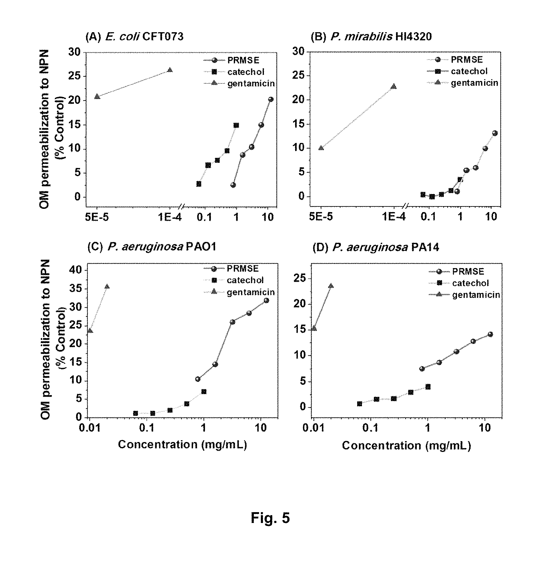
FIG. 5 illustrates PRMSE, catechol, and gentamicin-mediated NPN uptake in (FIG. 5A) E. coli CFT073, (FIG. 5B) P. mirabilis HI4320, (FIG. 5C) P. aeruginosa PAO1 and (FIG. 5D) P. aeruginosa PA14. Bacterial cells were pretreated with 1 mM N-ethylmaleimide (NEM) in 5 mM HEPES buffer (pH 7.2) and incubated at room temperature in the presence or absence (control) of various sub-MICs of PRMSE, catechol, and gentamicin. Enhanced uptake of NPN was determined by an increase in fluorescence (ex/em: 350 nm/420 nm) caused by partitioning of NPN into the hydrophobic interior of the outer membrane.
FIG. 6 illustrates the effect of PRMSE on expression of virulence genes for (FIG. 6A) E. coli CFT073, (FIG. 6B) P. mirabilis HI4320, (FIG. 6C) P. aeruginosa PAO1, and (FIG. 6D) P. aeruginosa PA14. Error bars are the standard deviation of values obtained from three replicates. All gene expressions were normalized to the corresponding housekeeping gene (gapA of E. coli CFT073, rpoA of P. mirabilis HI4320, rpoD of P. aeruginosa PAO1 and PA14) and then related to the normalized expression level of the same gene in the control.
FIG. 7 illustrates growth curves in M9 minimal medium for (FIG. 7A) E. coli CFT073, (FIG. 7B) P. mirabilis HI4320, (FIG. 7C) P. aeruginosa PAO1 and (FIG. 7D) P. aeruginosa PA14, in the presence of PRMSE. Bacterial growth (OD.sub.600) was monitored at 37.degree. C. for 16 h in M9 minimal medium. Error bars represent the standard deviation of values obtained from four replicates of two independent experiments performed on different days. Viable cell population was monitored at the same culture conditions and determined by serial dilution in PBS and plating on LB agar plates. Abbreviations: PRMSE..times., MS polyphenolic extract at .times. mg mL.sup.-1 (e.g., PRMSE 25 indicates polyphenolic-rich MS extract at 25 mg mL.sup.-1).
FIG. 8 illustrates G. mellonella larvae survival over time when injected with P. aeruginosa PA14, PRMSE or ciprofloxacin (Cip). The larvae were injected separately with bacteria (1-100 CFU per larva), PRMSE (12.5 mg/mL) or ciprofloxacin (0.03 .mu.g/mL) and monitored for their survival at 28.degree. C. Each data set is representative of experiments with five larvae per condition in triplicate.
FIG. 9 illustrates the in vivo efficacy of PRMSE or ciprofloxacin (Cip) mono- and combination (comb) therapies analyzed for the protection of G. mellonella larvae from PA14 infection. Larvae were infected with bacteria (10.+-.3 CFU per larva) and after 2 h injected with PRMSE (12.5 mg/mL) or ciprofloxacin (0.03 .mu.g/mL), alone and in combination, and monitored for their survival at 28.degree. C. Synergy between PRMSE and ciprofloxacin to protect G. mellonella larvae against PA14 infection is shown with arrows. Each data set represents one experiment with five larvae per condition in triplicate, the experiment repeated twice (*P<0.05, Mantel-cox test, n=6).
FIG. 10 illustrates the in vitro analysis of hemolymph recovered from G. mellonella larvae treated with PRMSE or ciprofloxacin (Cip), alone and in combination. Larvae were injected with PRMSE (mg/mL) or Cip (.mu.g/mL) alone and in combination, and hemolymph recovered 3 h after treatment were spotted on Pseudomonas isolation agar plates previously inoculated with PA14 cells to produce a lawn of confluent growth. The plate was observed after 24 h of incubation at 37.degree. C. The experiment was performed with three larvae per each condition in triplicate. S-control: saline control.
FIG. 11 illustrates the in vivo anti-virulence synergy of PRMSE (6.25 mg mL.sup.-1) with ciprofloxacin (0.015 .mu.g mL.sup.-1) for the protection of D. melanogaster against P. aeruginosa PA14 infection. Mortality was scored daily for 14 days. Synergy between PRMSE and ciprofloxacin to protect D. melanogaster flies against PA14 infection is shown with arrows. Results represent measurements from experiments performed with duplicates, twice (*p<0.01, Log-rank Mantel-cox test).
FIG. 12 illustrates the survival of P. aeruginosa PA14 on sucrose filters during fly feeding assays. Total viable bacterial counts per filter were determined every second day for filters containing bacterial cells on sucrose agar. PA14 cells on filter were untreated or treated with PRMSE (6.25 mg mL-1) or ciprofloxacin (0.015 .mu.g mL-1) alone and in combination. Average data represent data from experiment performed in triplicate and error bars represent S.D.
FIG. 13 illustrates the inhibition of virulence determinants of P. aeruginosa PAO1 and PA14 strains in the absence or presence of different PRMSE concentrations (mg/mL). In FIG. 13A and FIG. 13B, LasA: staphylolytic protease and AprA: alkaline protease. Results are expressed as means and standard deviations (SD) of triplicate assays (*p<0.05). In FIG. 13C, total proteolytic activity is determined on skim milk agar plate.
FIG. 14 illustrates the interactions of ciprofloxacin and different phenolic formulations for the growth inhibition of E. coli CFT073 and P. aeruginosa PA14. FIC index .ltoreq.0.5 indicates synergistic interaction.
DETAILED DESCRIPTION
In accordance with the present disclosure, there is provided anti-microbial composition comprising a phenolic-rich extract and at least one antibiotic.
As encompassed herein the phenolic-rich extract is preferably a phenolic-rich maple syrup extract (PRMSE).
Many plants synthesize aromatic substances, most of which are phenols or their oxygen-substituted derivatives. These phenolic compounds are believed to be promising candidates as complementary therapeutics since they can modify bacterial behavior by affecting bacterial motility, surface adhesion, biofilm formation, quorum sensing, and production of virulence determinants. Traditional medicinal approaches owe their significance to the bioactive components that have their origin in plant sources and many are associated with routine dietary habits.
Syrup obtained by concentrating the sap from certain maple species of North American maple trees (i.e., the sugar maple, such as for example Acer saccharum Marsh, and the red maple, A. rubrum L.), contains a vast number of natural and process-derived phytochemicals, the majority of which are phenolic compounds. Phenolic-Rich Maple Syrup Extracts (PRMSE) are obtained by extracting the phenolic compounds of maple syrup with organic solvents. These extracts have been reported to exhibit anti-proliferative effects against a panel of human tumor cell lines.
Encompassed herein is a maple tree of a species such as Acer nigrum, Acer lanum, Acer acuminatum, Acer albopurpurascens, Acer argutum, Acer barbinerve, Acer buergerianum, Acer caesium, Acer campbellii, Acer campestre, Acer capillipes, Acer cappadocicum, Acer carpinifolium, Acer caudatifolium, Acer caudatum, Acer cinnamomifolium, Acer circinaturn, Acer cissifolium, Acer crassum, Acer crataegifolium, Acer davidii, Acer decandrum, Acer diabolicum, Acer distylum, Acer divergens, Acer erianthum, Acer erythranthum, Acer fabri, Acer garrettii, Acer glabrum, Acer grandidentatum, Acer griseum, Acer heldreichii, Acer henryi, Acer hyrcanum, Acer ibericum, Acer japonicum, Acer kungshanense, Acer kweilinense, Acer laevigatum, Acer laurinum, Acer lobelii, Acer lucidum, Acer macrophyllum, Acer mandshuricum, Acer maximowiczianum, Acer miaoshanicum, Acer micranthum, Acer miyabei, Acer mono, Acer mono.times., Acer truncatum, Acer monspessulanum, Acer negundo, Acer ningpoense, Acer nipponicum, Acer oblongum, Acer obtusifolium, Acer oliverianum, Acer opalus, Acer palmatum, Acer paxii, Acer pectinatum, Acer pensylvanicum, Acer pentaphyllum, Acer pentapomicum, Acer pictum, Acer pilosum, Acer platanoides, Acer poliophyllum, Acer pseudoplatanus, Acer pseudosieboldianum, Acer pubinerve, Acer pycnanthum, Acer rubrum, Acer rufinerve, Acer saccharinum, Acer saccharum, Acer sempervirens, Acer shirasawanum, Acer sieboldianum, Acer sinopurpurescens, Acer spicatum, Acer stachyophyllum, Acer sterculiaceum, Acer takesimense, Acer tataricum, Acer tegmentosum, Acer tenuifolium, Acer tetramerum, Acer trautvetteri, Acer triflorum, Acer truncatum, Acer tschonoskii, Acer turcomanicum, Acer ukurunduense, Acer velutinum, Acer wardii, Acer.times. peronai, or Acer.times. pseudoheldreichii.
Herein, it is provided the anti-bacterial efficacy of phenolic-rich maple syrup extract (PRMSE) in treating clinical and multiple drug-resistant pathogenic bacterial strains. Experiments disclosed herein involve combinations of phenolic rich maple syrup extract and at least one antibiotic (ciprofloxacin or carbenicillin) at different concentrations analyzed for growth inhibition of each pathogenic bacterial strain (Escherichia coli CFT073, Proteus mirabilis HI4320, Pseudomonas aeruginosa PAO1 and P. aeruginosa PA14).
The extract described herein can be effective composition against a bacterial strain of Escherichia coli, Proteus mirabilis, Pseudomonas aeruginosa, Salmonella species, Enterobacter cloacae, Burkholderia cepacia, Chromobacterium violaceum, Klebsiella pneumoniae, Helicobacter pylori, Acinetobacter baumannii, Vibrio cholera, Mycoplasma pneumoniae, Neisseria gonorrhoeae, Legionella pneumophila, Serratia species, Shigella species, Rickettsia rickettsia, Chlamydia pneumoniae, Mycobacterium tuberculosis, Yersinia species, Moraxella catarrhalis or Proteobacteria pathogens.
Another set of experiments demonstrating anti-biofilm activity of PRMSE against pathogenic bacterial strains is disclosed. The anti-bacterial properties of PRMSE increase antibiotic susceptibility of each pathogenic bacterial strain at sub-inhibitory concentrations. This extract also significantly reduced biofilm formation and eradication of monoculture biofilm of each pathogenic bacterial strain from the surface of silicone discs at sub-lethal concentrations. Transcriptome analysis revealed that PRMSE significantly repressed the expression of multiple drug resistance genes, motility and attachment-related genes, and virulence-associated genes of pathogenic clinical strains. Two key components of PRMSE, namely, gallic acid and catechol, were found to increase outer-membrane permeability, and PRMSE effectively inhibited efflux pumps involved in multidrug resistance in pathogenic bacteria.
As encompassed herein, the antibiotic used in combination with the extract described herein can be a fluoroquinolone antibiotic, a .beta.-lactam antibiotic, am inoglycoside, polyketides, glycopeptides, benzenoids, macrolides, ansamycins, sulfonamides, chloramphenicol, oxazolidinones, carboxylic acids antibiotic, organic phosphonic acids antibiotic, quinolones and their derivatives.
Alternatively, the antibiotic encompassed herein can be antibiotic ciprofloxacin, carbenicillin, ampicillin, penicillin, kanamycin, gentamycin, tetracycline, levofloxacin, trimethoprim, sulfamethoxazole, norfloxacin, nitrofurantoin, fosfomycin, azithromycin, minocycline, am ikacin, cephalosporin, erythromycin, daptomycin, vancomycin and their derivatives.
HPLC chromatograms (FIG. 1) show the presence of six predominant phenolic compounds in PRMSE: gallic acid, 1,2-dihydroxybenzene (catechol), 3,4-dihydroxybenzaldehyde (catechaldehyde), syringaldehyde, vanillin and 3-hydroxybenzoic acid. Based on their bioactive properties reported in the literature, the following four compounds were selected, purchased in pure form and screened along with PRMSE for growth inhibition and synergy with ciprofloxacin: gallic acid, catechol, catechaldehyde, and syringaldehyde.
MICs for PRMSE and ciprofloxacin against the bacterial strains E. coli CFT073, P. mirabilis HI4320, P. aeruginosa PAO1 and P. aeruginosa PA14 are shown in Table 1.
TABLE-US-00001 TABLE 1 Synergistic interactions of ciprofloxacin and PRMSE for growth inhibition Bacterial strains P. P. P. E. coli mirabilis aeruginosa aeruginosa Interactions CFT073 HI4320 PAO1 PA14 MIC of ciprofloxacin 0.008 0.016 1.0 0.06 (.mu.g mL.sup.-1) FIC.sup.a of ciprofloxacin 0.25 0.02 0.03 0.03 MIC of PRMSE 25 50 50 50 (mg mL.sup.-1) FIC.sup.a of PRMSE 0.13 0.13 0.06 0.06 FICI.sup.b 0.38 0.14 0.1 0.1 Synergistic? Yes Yes Yes Yes .sup.aFIC: Fractional Inhibitory Concentration, FIC = MIC of compound 1 in the combination, divided by the MIC of compound 1 alone .sup.bFICI: Fractional Inhibitory Concentration Index, FICI = FIC.sub.compound 1 + FIC.sub.compound 2
To investigate the presence of synergistic interaction between ciprofloxacin (a fluoroquinolone, which has biofilm penetration properties) and PRMSE, a checkerboard microdilution analysis was performed. The corresponding functional inhibitory concentration index (FICI) values were <0.5 for all tested strains (Table 1), demonstrating a strong synergistic effect between PRMSE and ciprofloxacin. Because P. aeruginosa is known to be partially resistant to carbenicillin (a .beta.-lactam antibiotic), the effect of PRMSE on the susceptibility of the P. aeruginosa strains towards carbenicillin was also investigated. The MIC and FICI values in Table 2 show that PRMSE acted in synergy with carbenicillin against the growth of the two P. aeruginosa strains.
TABLE-US-00002 TABLE 2 Synergistic interactions of carbenicillin and PRMSE for growth inhibition P. aeruginosa strains Interactions PAO1 PA14 MIC of carbenicillin (.mu.g mL.sup.-1) 64 128 FIC of carbenicillin 0.13 0.063 MIC of PRMSE (mg mL.sup.-1) 50 50 FIC of PRMSE 0.03 0.063 FICI 0.16 0.13 Synergistic? Yes Yes
The MICs for the pure phenolic compounds are presented in Table 3.
TABLE-US-00003 TABLE 3 Interaction of phenolic constituents of PRMSE with ciprofloxacin Interactions FIC of Bacterial MIC FIC.sup.a of com- Strains Constituents (mg mL.sup.-1) ciprofloxacin pound FICI E. coli gallic acid 5 1 1 2 CFT073 catechol 1.25 0.25 0.25 0.5* catechaldehyde 1.25 1 1 2 syringaldehyde 5 0.5 0.25 0.75 P. mirabilis gallic acid 5 1 1 2 HI4320 catechol 1.25 0.25 0.25 0.5* catechaldehyde 1.25 0.5 1 1.5 syringaldehyde 5 0.5 0.5 1 P. gallic acid 5 1 1 2 aeruginosa catechol 2.5 0.25 0.25 0.5* PAO1 catechaldehyde 1.25 0.5 0.51 1.01 syringaldehyde 5 1 1 2 P. gallic acid 5 1 1 2 aeruginosa catechol 2.5 0.25 0.25 0.5* PA14 catechaldehyde 1.25 1 1 2 syringaldehyde 5 0.5 0.25 0.75 *Values represent synergistic interaction .sup.aFIC: Fractional Inhibitory Concentration, FIC = MIC of compound 1 in the combination, divided by the MIC of compound 1 alone .sup.bFICI: Fractional Inhibitory Concentration Index, FICI = FIC.sub.compound 1 + FIC.sub.compound 2.
Amongst the four selected phenolic constituents of PRMSE, catechol (at 1.25 mg mL.sup.-1 for both E. coli CFT073 and P. mirabilis HI4320, and at 2.5 mg mL.sup.-1 for P. aeruginosa PAO1 and PA14) and catechaldehyde (at 1.25 mg mL.sup.-1 for all four strains) were found to clearly inhibit growth at lower concentrations. Furthermore, interaction of these four phenolic compounds with ciprofloxacin was examined using a checkerboard microdilution assay. The results presented in Table 3 show that the combination of catechol and ciprofloxacin was the only synergistic combination (<0.5 FICI) for all investigated strains.
To investigate possible synergistic antimicrobial action among the four phenolic constituents of PRMSE, different combinations of gallic acid, catechol, catechaldehyde and syringaldehyde were tested using the checkerboard microdilution assay; the corresponding FICIs are presented in Table 4.
TABLE-US-00004 TABLE 4 Interaction of individual phenolic constituents of PRMSE. FIC.sup.a FICI.sup.b Bacterial Phenolic gallic gallic strains constituents acid catechol catechaldehyde acid catechol catechalde- hyde E. coli catechol 0.13 NA.sup.c -- 0.25* NA -- CFT073 catechaldehyde 0.25 0.25 NA 0.5* 0.5* NA syringaldehyde 0.5 0.25 0.25 0.75 0.31* 0.31* P. mirabilis catechol 0.13 NA -- 0.37* NA -- HI4320 catechaldehyde 0.13 0.25 NA 0.37* 0.5* NA syringaldehyde 0.5 0.25 0.25 1 0.75 0.75 P. aeruginosa catechol 0.25 NA -- 0.5* NA -- PAO1 catechaldehyde 0.13 0.25 NA 0.37* 0.37* NA syringaldehyde 0.5 0.25 0.25 1 0.5* 0.5* P. aeruginosa catechol 0.25 NA -- 0.37* NA -- PA14 catechaldehyde 0.25 0.25 NA 0.5* 0.5* NA syringaldehyde 0.13 0.5 0.5 0.63 1 1 *Values represent synergistic interaction .sup.aFIC: Fractional Inhibitory Concentration, FIC = MIC of compound 1 in the combination, divided by the MIC of compound 1 alone. .sup.bFICI: Fractional Inhibitory Concentration Index, FICI = FIC.sub.compound 1 + FIC.sub.compound 2 .sup.cNA: not applicable
The most potent synergy on growth inhibition was observed for gallic acid-catechol and gallic acid-catechaldehyde pairs, resulting in FICIs of 0.25-0.50, whereas all combinations of gallic acid with syringaldehyde led to FICIs ranging between 0.63-1.0 (Table 4), indicating no synergistic interaction. Interestingly, catechol exhibited strong synergy for growth inhibition with catechaldehyde against all chosen strains and with syringaldehyde against E. coli CFT073 and P. aeruginosa PAO1, as confirmed by FICIs (Table 4), suggesting that catechol is a potent component of PRMSE, mainly responsible for its antimicrobial activity.
To investigate the potential of PRMSE for biofilm inhibition, biofilms were developed in polystyrene microtitre plates in the presence of different combinations of PRMSE, with and without ciprofloxacin at sub-lethal concentrations. These sub-lethal concentrations of PRMSE 3.13 and 6.25 mg mL.sup.-1 for E. coli CFT073; 6.25 and 12.5 mg mL.sup.-1 for P. mirabilis HI4320, P. aeruginosa PAO1 and PA14) and ciprofloxacin (0.0005-0.004 .mu.g mL.sup.-1 for E. coli CFT073, 0.001-0.008 .mu.g mL.sup.-1 for P. mirabilis HI4320, 0.00625-0.5 .mu.g mL.sup.-1 for P. aeruginosa PAO1 and 0.004-0.03 .mu.g mL.sup.-1 for P. aeruginosa PA14) were chosen for biofilm studies, based on results obtained in the MIC assay. Anti-biofilm activity of PRMSE is presented in FIG. 2; the amount of sessile biomass (OD.sub.570) was normalized to the level of planktonic growth (OD.sub.600) to minimize bias from possible differences in growth levels on biofilm quantification. PRMSE alone showed significant inhibition of monoculture biofilm formation of all tested strains (p<0.05). PRMSE in combination with ciprofloxacin (at sub-lethal concentrations) had significant inhibitory effects on biofilm formation (p<0.05) for E. coli CFT073 (.about.70% inhibition at 6.25 mg mL.sup.-1), P. mirabilis HI4320 (70% inhibition at 12.5 mg mL.sup.-1), P. aeruginosa PAO1 (83% inhibition at 12.5 mg mL.sup.-1) and P. aeruginosa PA14 (.about.54% inhibition at 12.5 mg mL.sup.-1), in a dose-dependent manner. Since catechol was the only compound that exhibited synergy in antimicrobial activity with ciprofloxacin (Table 3), only the combination of catechol and ciprofloxacin was analyzed in this assay.
Catechol with and without ciprofloxacin (both at sub-lethal concentrations) manifested significant inhibitory effects (p<0.05) on biofilm formation for all four tested strains (FIG. 3).
To analyze the applicability of PRMSE to eradicate biofilm from biomaterial surfaces, an in vitro biofilm assay was performed using silicone discs. The biofilm eradication performance of PRMSE in combination with ciprofloxacin is presented in FIG. 4; viable cell recoveries from the silicone discs after exposure to different combinations of PRMSE and ciprofloxacin were evaluated. PRMSE alone did not show significant eradication of monoculture biofilm on silicone discs for any of the tested strains, while PRMSE in combination with sub-lethal concentration of ciprofloxacin had significant biofilm eradication effect with synergistic interaction at higher concentration of ciprofloxacin (p<0.05). These results are correlated with the above-mentioned anti-biofilm activity of PRMSE.
In an effort to elucidate the mechanism(s) for the observed synergistic interactions, the change in bacterial outer membrane permeability was quantified using 1-N-phenylnapthylamine (NPN) as an indicator. The results show that both PRMSE and catechol, at sub-lethal concentrations, increased membrane permeability in all tested strains (Table 5, FIG. 5).
TABLE-US-00005 TABLE 5 Biological effects of PRMSE and catechol Phenolic Pure Positive controls.sup.e Bacterial Untreated extract compound gentamicin strains Effects based on bioassays cells PRMSE catechol (.mu.g mL.sup.-1) CCCP CTAB E. coli Minimum permeabilization -- 1.6 0.06 0.006 NA.sup.f NA CFT073 concentration [mg mL.sup.-1] (NPN assay) .sup.a % Reduction in efflux pump 36.9 22.7 34.3 NA 15.8 NA activity .sup.b % cells with uncompromised 100 88.7 68 NA NA 0 membrane (BacLight assay).sup.c Bacterial colony forming ability (.DELTA. 0 0.8 2.3 NA NA 5.7 log CFU mL.sup.-1).sup.d P. mirabilis Minimum permeabilization -- 1.6 1 0.013 NA NA HI4320 concentration [mg mL.sup.-1] % Reduction in efflux pump 35.9 23.4 33.9 NA 14.5 NA activity % cells with uncompromised 100 72.6 50.3 NA NA 0 membrane Bacterial colony forming ability (.DELTA. 0 1.2 3.2 NA NA 4.5 log CFU mL.sup.-1) P. aeruginosa Minimum permeabilization -- 0.8 0.5 1 NA NA PAO1 concentration [mg mL.sup.-1] % Reduction in efflux pump 37.6 22.3 35.8 NA 19.3 NA activity % cells with uncompromised 100 87.9 80.4 NA NA 0 membrane Bacterial colony forming ability (.DELTA. 0 0.9 1.5 NA NA 5.1 log CFU mL.sup.-1) P. aeruginosa Minimum permeabilization -- 0.8 0.5 1 NA NA PA14 concentration [mg mL.sup.-1] % Reduction in efflux pump 30.9 19 29.9 NA 17 NA activity % cells with uncompromised 100 89.1 78.6 NA NA 0 membrane Bacterial colony forming ability (.DELTA. 0 0.9 1.9 NA NA 4.9 log CFU mL.sup.-1) .sup.a Assay was performed at sub-MIC of extract and pure compound compared to reference antibiotic (gentamicin at above and below MICs), these concentrations led to maximal increase in NPN (1-N-phenylnapthylamine) uptake based on fluorescence intensity recorded. .sup.b The ratio of green to red fluorescence was normalized to the untreated control and expressed as a percentage of the control. Cells were treated at 4 .times. MIC of pure compound and extract for 10 min. .sup.cReduction in fluorescence intensity as a percentage of that at the first time point of recording. Cells were treated overnight at 0.25 times the MIC of pure compound and extract. .sup.dBacterial colonies were counted from LB agar plate, and the log decrease in CFU/mL compared to the untreated control was calculated. .sup.eCCCP: carbonyl cyanide m-chlorophenylhydrazone (100 .mu.M), CTAB: cetyltrimethyl-ammonium bromide (10 .mu.M). .sup.fNA, not applicable.
These effects were similar to the known outer membrane permeabilizing effect of gentamicin (FIG. 5).
Bacterial cell membrane damage was further examined using the BacLight assay. As shown in Table 5, catechol and PRMSE exhibited moderate membrane disruptive effects. The cells exposed to PRMSE or catechol were also tested for their ability to form colonies on solid agar medium. Catechol reduced the number of CFUs compared to the negative control (.gtoreq.1.5 log reduction in CFU mL-1 for E. coli CFT073, P. mirabilis HI4320 and P. aeruginosa PA14) during the 10 min exposure period, whereas PRMSE had a weak effect on the colony forming ability of chosen strains. The CFU measurement correlates well with the BacLight results; however, because bacterial membrane damage is not necessarily a lethal event, the severity of the effect reflected by each assay is different.
The effect of PRMSE and catechol on inhibition of bacterial drug resistance efflux pump was investigated using the ethidium bromide (EtBr) efflux pump assay. PRMSE exhibited significant inhibitory effect on efflux pump for all bacterial strains with values comparable to the positive control CCCP, which is an established proton motive force modulator (Table 5). There was weak reduction in fluorescence intensity for cells treated with PRMSE indicating accumulation of EtBr in the cell as a result of efflux pump inhibition. Cells treated with catechol also exhibited reduction in EtBr efflux, but to a lesser extent than cells treated with PRMSE (Table 5).
To explore the genetic basis for the synergy in antimicrobial activity observed between PRMSE and antibiotics, as well as the effect of PRMSE on bacterial biofilms, transcriptional analysis was performed using qRT-PCR to observe the differential expression of genes associated with bacterial motility, virulence, drug resistance, adhesion and biofilm formation for each of the four bacterial strains. The results in FIG. 6 indicate that PRMSE, at sub-lethal concentrations, repressed the expression of genes associated with multiple drug resistance (emrA, acre and marC in CFT073; acrA and marC in HI4320; oprM, mexA and mexX in PAO1 and PA14), motility (fliC, flhD, motB, fimH, fimA, papA2 in CFT073; flaA, flhD in HI4320; fliC, fleQ in PAO1 and PA14), virulence determinants (chuA in CFT073; cysJ in HI4320; plcH, phzS and pvdA in PAO1 and PA14), adhesion (fimH, fimA, papA2 in CFT073; atfB in HI4320; cupA1, pelA in PAO1 and PA14) and biofilm formation (uvrY in CFT073; ureD in HI4320; lasB in PAO1 and PA14). Transcriptional analysis confirms the trends observed with the biofilm assay (i.e., biofilm inhibition in the presence of PRMSE correlates with down-regulation of biofilm associated genes) and antibiotic synergy tests (i.e., down-regulation of multiple drug resistance genes correlates with increased antibiotic susceptibility in the presence of PRMSE).
PRMSE was used at sub-lethal concentrations for this experiment and does not affect bacterial growth of all four strains (FIG. 7). This is important because conditions that affect growth can inhibit gene expression. Based on these results, it can be concluded that the observed differences in gene expression were due to the presence of PRMSE and not due to growth inhibition by this extract.
Galleria mellonella larvae have emerged as an ideal host model due to a number of characteristics, such as a high degree of functional and structural homology to the innate immune systems of mammals, and possess characteristics to understand virulence mechanisms of human pathogens in vivo (Cook and McArthur, 2013, Virulence, 4:350-353). P. aeruginosa is an opportunistic human pathogen that displays an extraordinarily broad host range to infect vertebrates, non-vertebrate insects, and plants (Apidianakis and Rahme, 2009 Nature Protocol, 4:1285-1284). To determine the effective larvae killing dose of P. aeruginosa PA14, the survival of G. mellonella larvae injected into the hemolymph (blood-like fluid) with different cell densities of P. aeruginosa PA14 was observed. As shown in FIG. 8, a single PA14 bacterium decreased G. mellonella larvae survival within 96 h post-infection period. Optimal killing of G. mellonella larvae for use in the anti-infection assay was observed with a dose of 10 CFU/larva, as this led to effective larvae killing of >60% at 24 h post-infection (FIG. 8). This condition was used for further studies with PRMSE.
The individual effects of PRMSE and ciprofloxacin on the survival of G. mellonella larvae after injection was analyzed. PRMSE and ciprofloxacin, at sub-MICs of 12.5 mg/mL and 0.03 .mu.g/mL, respectively, had no killing effect on G. mellonella larvae within 72 h post-infection period (FIG. 8). These observations show that PRMSE and ciprofloxacin at sub-MICs are safe for G. mellonella larvae.
To explore the efficacy of PRMSE and ciprofloxacin to limit P. aeruginosa infection in vivo, PRMSE and ciprofloxacin were injected in G. mellonella larvae as mono- and combination therapies at sub-MICs. G. mellonella larvae were infected by injection with a lethal dose of PA14 strain (10.+-.3 cells from exponential cultures) and incubated at 28.degree. C. for 2 h. These infected G. mellonella larvae were injected a second time with or without PRMSE (12.5 mg/mL), with or without the antibiotic ciprofloxacin (0.03 .mu.g/mL) at 2 h post-infection period. As shown in FIG. 9, the median survival of G. mellonella larvae infected with only P. aeruginosa PA14 was 36 h (without PRMSE or ciprofloxacin), which is significantly lower (X.sup.2=4.9, df=1, p<0.05) than that measured for the combination therapy of PRMSE and ciprofloxacin (84 h), based on the comparison of survival curves. Monotherapy of ciprofloxacin at sub-MIC fails to protect G. mellonella larvae against P. aeruginosa PA14 infection, but monotherapy of PRMSE shows moderate protection to G. mellonella larvae against PA14 infection with median survival of 60 h. Overall, these results show that PRMSE protects G. mellonella larvae from P. aeruginosa infection. They also clearly demonstrate that PRMSE acts in synergy with ciprofloxacin to protect G. mellonella larvae from P. aeruginosa infection.
The difference in the treated or untreated P. aeruginosa PA14 cells present in the hemolymph (blood-like fluid) of G. mellonella larvae after the injection may be due to modified survival of the bacteria in the hemolymph during incubation. Another possibility is that PRMSE induces the immune response or transformations of antimicrobial compounds into toxic materials in the larval hemolymph during incubation. To address these possibilities, an additional set of larvae were injected with 5 .mu.L saline solution with or without PRMSE, with or without ciprofloxacin (at selected concentrations in ng per larva). The larval hemolymph was recovered at 3 h post-injection and spotted on the freshly inoculated cells of PA14 to analyze growth inhibition by recovered hemolymph. As shown in FIG. 10, at the end of incubation period, recovered larval hemolymph turned to brown colored spots on agar plate, possibly due to coagulation or clotting of hemolymph proteins, but no inhibition zone in the lawn of PA14 colonies was observed. Thus, larval hemolymph recovered from larvae treated with or without PRMSE, with or without ciprofloxacin, had no effect on P. aeruginosa PA14 growth in vitro, further confirming that the observed effect of PRMSE on pathogenicity of PA14 strain is due to inhibition of virulence rather than a growth-inhibitory effect.
The Drosophila melanogaster fly has emerged as an another ideal host model for an understanding of P. aeruginosa virulence mechanisms and immune response in vivo because of the high degree of homology between vertebrates and fly innate immune systems (Apidianakis and Rahme, 2009 Nature Protocol 4:1285-1284). Similar to the in vivo assay with G. mellonella larvae, the individual effects of PRMSE and ciprofloxacin on the survival of D. melanogaster flies infected with P. aeruginosa PA14 after feeding them with extract or antibiotic were analyzed. PRMSE and ciprofloxacin, at sub-MICs of 6.25 mg/mL and 0.015 .mu.g/mL, respectively, had no killing effect on D. melanogaster flies within 264 h post-infection period (FIG. 11). These observations show that PRMSE and ciprofloxacin individually at sub-MICs are safe for D. melanogaster flies.
To explore the synergistic efficacy of PRMSE and ciprofloxacin to limit P. aeruginosa infection in vivo, D. melanogaster flies were fed with PRMSE and ciprofloxacin as mono- and combination therapies at sub-MICs in the presence of P. aeruginosa PA14. D. melanogaster male flies were starved of food and water for 5-6 h before the infection with PA14, without and with mono- and combination therapies. As shown in FIG. 11, the median survival of D. melanogaster male flies infected with only P. aeruginosa PA14 was 108 h (without PRMSE or ciprofloxacin), but 216 h with the combination therapy of PRMSE and ciprofloxacin, which is significantly (X.sup.2=7.9, df=1, p<0.01) higher based on the comparison of survival curves. Monotherapy of PRMSE or ciprofloxacin at sub-MIC fails to protect D. melanogaster against P. aeruginosa PA14 infection. This result indicates that synergy between PRMSE and ciprofloxacin protects D. melanogaster against P. aeruginosa PA14 infection.
The difference in the treated or untreated PA14 strain's ability to kill D. melanogaster in this feeding assay may have been due to modified survival of the bacteria on the filter papers used for exposure during incubation. To address this possibility, the survival of PA14 on the paper discs without and with PRMSE and ciprofloxacin as mono- and combination therapies under the same conditions as the fly feeding assay was analyzed. There was no significant difference in culturability of the PA14 cells on the filter paper discs in the absence and presence of mono- and combination therapies during incubation of six days (FIG. 12), indicating that an alteration in survival ability of bacteria could not account for the observed differences in fly killing.
The effect of PRMSE on virulence determinant enzymes of PAO1 and PA14 was investigated at different concentrations. At the selected concentrations, bacterial growth was not inhibited. As shown in FIGS. 13A and 13B, treatment with PRMSE significantly (P<0.05) inhibited the staphylolytic (LasA) and alkaline proteolytic (AprA) activities of P. aeruginosa PAO1 and PA14. Total proteolytic activities of P. aeruginosa PAO1 and PA14 treated without and with PRMSE were analyzed on skim milk agar plate. There was no halo formation around the wells loaded with supernatant of sample with 12.5 mg/mL PRMSE compared to the untreated control sample, indicating inhibition of total proteolytic activity. Treatment with PRMSE inhibited total proteolytic activity of PAO1 and PA14 strains (FIG. 13C). This result indicates that PRMSE inhibits activities of virulence determinant enzymes of P. aeruginosa.
A checkerboard assay was performed using different combinations of the pure phenolic compounds identified in PRMSE, i.e., gallic acid, catechol, catechaldehyde, 3-hydroxybenzoic acid, vanillin and syringaldehyde. Interaction was analyzed with ciprofloxacin against E. coli CFT073 and P. aeruginosa PA14. Formulation-1 represents relative concentrations of phenolics as determined in PRMSE by HPLC (Table 6). Additional different formulations were prepared as variations of Formulation-1 and assessed for synergistic interaction. As shown in FIG. 14, the corresponding FICI values of Formulation-13 were <0.5, indicating synergistic interaction with ciprofloxacin against both CFT073 and PA14. The FICIvalues for Formulations 1-11 were between >0.5 and .ltoreq.4 indicating no interaction or indifference. Formulation-12 shows synergy with ciprofloxacin against PA14 (0.5 FIC index), but not against CFT073 (>0.5 FIC index).
TABLE-US-00006 TABLE 6 Concentrations (mg/mL) of phenolic compounds in different formulations for checkerboard assay Phenolic Formulation # compounds 1 2 3 4 5 6 7 8 9 10 11 12 13 gallic acid 8.52 4.26 4.26 4.26 0.65 0.65 0.30 4.26 2.13 0.65 0.65 1.3 0.6- 5 catechol 0.30 0.30 0.65 0.30 0.65 0.65 0.30 0.30 0.65 0.65 0.65 0.65 0.65 catechaldehyde 0.02 0.02 0.02 0.04 0.04 0.30 0.65 0.02 0.04 0.04 0.12 0.65- 0.65 3-hydroxybenzoic 0.06 0.06 0.06 0.06 0.06 0.06 0.06 0.06 0.12 0.12 0.12 0.- 12 0.12 acid vanillin 0.002 0.002 0.002 0.002 0.002 0.002 0.002 0.020 0.04 0.04 0.04 0.- 04 0.04 syringaldehyde 0.001 0.001 0.001 0.001 0.30 0.002 0.002 0.010 0.02 0.02 0.- 02 0.02 0.02
In summary, the antimicrobial, anti-biofilm, and anti-virulence effects of a cocktail of phenolic compounds extracted from maple syrup (PRMSE) against a range of pathogenic bacteria is disclosed. PRMSE potentiate antibiotic susceptibility in both planktonic and biofilm modes of growth, which could be partially due to its ability to permeabilize the bacterial membrane, inhibit multidrug resistance efflux pumps, and downregulate genes associated with multidrug resistance. Amongst the four tested phenolic constituents of PRMSE, results indicate that catechol plays a key role in the synergistic activity of PRMSE with ciprofloxacin.
Phenolic extracts of maple syrup have been reported to exhibit antioxidant and anticancer activity linked with the presence of diverse phytochemical constituents, including phenylpropanoid. Of the 51 known metabolites in maple syrup (from A. saccharum), four compounds were investigated in more detail. Combination of catechol with ciprofloxacin, gallic acid, catechaldehyde or syringaldehyde, and the combination of gallic acid with catechaldehyde, exhibited a synergistic antimicrobial effect against all chosen strains in vitro (Tables 3 and 4). Catechol has been previously reported to interact synergistically with other phenolic compounds in terms of antioxidant capacity. Moreover, catechol is made synthetically and can be readily available for potential use as a disinfectant.
PRMSE and catechol exhibit anti-biofilm activity against the four pathogenic strains examined. Genes associated with biofilm formation (uvrY, fimH, fimA and papA2 of E. coli, ureD, ureA and atfB of P. mirabilis, and lasB and cupA1 of P. aeruginosa) were downregulated upon supplementation with PRMSE. In addition, PRMSE and catechol synergized with ciprofloxacin at sub-lethal concentrations to further inhibit the formation of biofilms (FIGS. 2 and 3). Ciprofloxacin has good penetration properties in the biofilm matrix, nevertheless, resistance has been reported in P. aeruginosa and E. coli biofilms. This suggests that the mechanism of resistance of these biofilms to ciprofloxacin goes beyond poor drug penetration and may be due to the presence of persister or resistant cells. Moreover, PRMSE exhibits synergy with ciprofloxacin for eradication of monoculture biofilm from silicone substrates, showing the applicability of PRMSE as a biofilm eradication strategy along with antibiotics.
Further, PRMSE, at sub-lethal concentrations, can increase the uptake of NPN into the cell outer membrane, indicating its ability to permeabilize the membrane (Table 5). Results provided herein indicate that outer membrane permeabilization by PRMSE is achieved without altering the cell membrane integrity.
Multidrug efflux pumps of Gram-negative bacteria that traverse both the outer and inner membranes make a key contribution to intrinsic antimicrobial resistance. PRMSE showed significant inhibition of the EtBr transport across the cell envelope, which indicates a decreased activity of bacterial multidrug resistance efflux pumps (Table 5) and reduction in expression of genes associated with multi-drug resistance (FIG. 6). Active efflux involves multi-drug resistance efflux pump assemblies in the bacterial cell membranes that transport structurally unrelated compounds, including different classes of antibiotics, antiseptics, and cationic dyes, such as ethidium bromide (EtBr) and acriflavin. Thus, the observed inhibition of efflux pumps is of interest, as it results in (i) increased intracellular drug concentration, (ii) restoration of drug activity against resistant strains, (iii) minimization of further development of resistance, and (iv) reduced biofilm formation.
Accordingly, PRMSE increases the potency of conventional antibiotics against planktonic and biofilm cells of four pathogenic bacterial strains through a strong synergistic effect. Membrane permeabilization and efflux pump inactivation contribute to this antibacterial and anti-biofilm synergy. In addition, catechol is the major contributor to the anti-biofilm and antibacterial synergy effects. It is thus provided the combined use of PRMSE (or its active component catechol) with antibiotics to target bacterial biofilms. By combining antibiotics with PRMSE that exhibit synergistic interaction, it is possible to decrease the dosage of antibiotics used to eradicate bacterial biofilms.
A synergistic combination of antibiotic with catechol inhibited the growth of tested clinical strains in vitro. Furthermore, combinations of catechol with selected phenolic components of maple syrup were highly synergistic against the growth of clinical strains. Accordingly, the combination of phenolics from PRMSE and an antibiotic is an effective anti-bacterial therapy, and imparts prospective biological property to maple syrup.
It is further provided that PRMSE alone or ciprofloxacin alone at sub-MICs are safe for Galleria mellonella larvae and Drosophila melanogaster flies. Combination therapy of PRMSE combined with ciprofloxacin shows synergy to protect G. mellonella larvae and D. melanogaster flies from P. aeruginosa PA14 infection.
Monotherapy of ciprofloxacin at sub-MIC fails to protect G. mellonella larvae and D. melanogaster flies against P. aeruginosa PA14 infection, but monotherapy of PRMSE at sub-MIC shows moderate protection to G. mellonella larvae against P. aeruginosa PA14 infection.
The effect of PRMSE on pathogenicity of P. aeruginosa PA14 strain during in vivo infection is due to inhibition of virulence rather than a growth-inhibitory effect.
The mechanisms of action protecting in vivo models against P. aeruginosa infection are that PRMSE (i) inhibits activities of virulence determinant enzymes of P. aeruginosa, (ii) inhibits multidrug efflux pump activity, (iii) increases cell membrane permeability to antibiotics, (iv) downregulates genes associated with virulence, drug resistance, biofilm formation, motility and adhesion. Accordingly, different combinations of the pure phenolic compounds previously identified in PRMSE do not show synergy with ciprofloxacin against E. coli CFT073 and P. aeruginosa PA14. Only very specific combinations of the pure compounds act similarly to the PRMSE.
Accordingly, it is provided a method of treating a bacterial infection comprising administering to a subject in need thereof a phenolic-rich extract as disclosed herein and at least one antibiotic. The subject can be for example, a human, an animal, a Galleria mellonella or a Drosophila melanogaster.
It is thus encompassed that by treating bacterial infections, the composition described herein can treat an urinary tract infection, lung infection, kidney infection, gastrointestinal infection, wound infection, acute sinusitis, skin and skin structure infections, bone and joint infections, lyme disease, typhus fever, rocky mountain spotted fever, rickettsialpox, or tuberculosis.
The present disclosure will be more readily understood by referring to the following examples which are given to illustrate the invention rather than to limit its scope.
Example I
Bacterial Strains and Preparation of PRMSE
The following organisms were used: E. coli strain CFT073 (ATCC 700928), P. mirabilis HI4320, P. aeruginosa PAO1 (ATCC 15692) and P. aeruginosa PA14 (UCBPP-PA14). Pure stock cultures are maintained at -80.degree. C. in 30% (v/v) frozen glycerol solution. Starter cultures are prepared by streaking frozen cultures onto LB agar [LB broth: tryptone 10 g L.sup.-1, yeast extract 5 g L.sup.-1and NaCl 5 g L.sup.-1, supplemented with 1.5 w/v % agar (Fisher Scientific, ON, Canada)]. After overnight incubation at 37.degree. C., a single colony was inoculated into 10 mL of LB broth and the culture was incubated at 37.degree. C. on an orbital shaker at 200 rpm for time lengths specific to each experiment. LB broth was used for bacterial culture in all experiments unless otherwise specified.
All maple syrup samples (grade D, amber, production year 2013; 66.degree. Brix syrup) were stored at -20.degree. C. until extraction, according to a protocol described by Gonzales et al. (2012, J. Funct. Foods, 4: 185-196). Briefly, maple syrup is enriched for phenolic content by using Amberlite XAD-16 (Sigma-Aldrich Canada) resin column chromatography (C4919, bed volume of 245 mL, Sigma-Aldrich Canada). The column is subsequently extracted with methanol (Sigma-Aldrich Canada). This extract is first subjected to solvent removal under vacuum and then solubilized in 0.3% v/v DMSO to yield PRMSE. Extracts are stored at -20.degree. C., thawed at room temperature before each experiment, and filter-sterilized using 0.22 .mu.m PVDF membrane filters (EMD Millipore Millex.TM., Fisher Scientific, ON, Canada) before use. PRMSE prepared in this manner is reported to not contain any natural sugar (sucrose, glucose or fructose).
The relative level of phenolic compounds in PRMSE is estimated using HPLC, as reported in the literature (Sadiki an Martin, 2013, Food Anal. Methods, 6: 737-744). Samples are analyzed using Agilent Technologies 1200 Series analytical liquid chromatographic system, consisting of binary pumps LC-20AB, degasser DGU-20A5, column oven CTO-20 AC, auto sampler SIL-20 AC and UV SPD-M20A detector. Phenolic compounds are separated on a Zorbax SB-C18 column (4.6.times.150 mm, 5 .mu.m; Agilent) at 30.degree. C. Trifluoroacetic acid at 0.2% (phase A) and methanol (phase B) are used as eluents. The elution gradient starts with 2% phase B, increased to 50% phase B at 35 min, 80% phase B at 43 min, and held at 80% for 2 min. The injection volume is 10 .mu.L and the flow rate is 0.5 mL min.sup.-1. Data are collected and evaluated by the Analyst 1.4.2 Software. The analytes are identified by comparing retention times and UV spectra, recorded in the range of 200-400 nm, with the individual chromatogram of each phenolic standard. The following pure phenolic compounds can be used as standards: gallic acid, 1,2-dihydroxybenzene (catechol), 3,4-dihydroxybenzaldehyde (catechaldehyde), syringaldehyde, vanillin and 3-hydroxybenzoic acid.
Example II
Determination of Minimum Inhibitory Concentration (MIC)
MICs are determined by preparing two-fold serial dilutions of PRMSE, pure phenolic compounds, and antibiotics in Mueller Hinton broth adjusted with Ca.sup.2+ and Mg.sup.2+ (MHB-II, Oxoid.TM., Fisher Scientific, ON, Canada). A range of concentration of antibiotics, ciprofloxacin (0.0003-0.25 .mu.g mL.sup.-1) and carbenicillin (0.5-512 .mu.g mL.sup.-1), are chosen due to their known potency against all four bacterial strains. Dilutions are prepared in flat bottom, 96 well microtitre plates (Falcon.TM., Corning.TM., Fisher Scientific, ON, Canada). Each well of a microtitre plate is then inoculated with the desired bacterial strain (grown in MHB-II and diluted to 106 CFU mL.sup.-1) and the plate is incubated at 37.degree. C. for 18 h under static conditions. Bacterial growth is assessed by (i) monitoring the optical density of the cell suspension in each well at 600 nm (OD.sub.600 nm), and (ii) the resazurin microtitre plate assay. In the resazurin microtitre plate assay, each well of a microtitre plate is supplemented with 20 .mu.M resazurin, incubated in the dark for 20 min at room temperature, followed by fluorescence measurements at ex/em 570/590 nm using a TECAN Infinite M200 Pro microplate reader (Tecan Group Ltd., Switzerland). The lowest concentration of a compound able to prevent increase in OD.sub.600 nm and resazurin fluorescence intensity is recorded as the MIC for that compound.
Example III
Checkerboard Microdilution Assay
The checkerboard microdilution assay is used for evaluation of in vitro antimicrobial synergy between two compounds (i.e., antibiotic/PRMSE, antibiotic/pure phenolic compound, and pure phenolic compounds with each other). Two-fold serial dilutions are prepared in MHB-II for each of the two compounds under study. The serial dilutions are then loaded into 96 well plates to achieve combinations having different concentrations of each of the two compounds. Each well is subsequently inoculated with 10.sup.6 CFU mL.sup.-1 of the desired bacterial strain and incubated at 37.degree. C. for 18 h under static conditions. The Fractional Inhibitory Concentration Index (FICI) for each combination is calculated by using the following formula: FIC.sub.component 1=MIC.sub.component 1, in combination/MIC.sub.component 1,alone FICI=FIC.sub.component 1+FIC.sub.component 2
The FICIs are interpreted as follows: FICI of .ltoreq.1.5 (synergy); 0.5<FICI .ltoreq.1 (no interaction/indifference); FICI of >4 (antagonism).
Example IV
Biofilm Assays
Biofilm formation is quantified using the standard microtitre plate model. Briefly, overnight cultures (LB broth, 37.degree. C., 200 rpm) are diluted 1:100 (v/v) into fresh LB broth (with or without PRMSE or catechol), to 106 CFU mL.sup.-1. Aliquots (100 .mu.L) of these cultures are transferred into the wells of polystyrene, flat bottom, non-treated 96 well plates (Falcon.TM., Corning.TM.), in triplicate. For all assays, biofilms are allowed to develop for 16 h at 37.degree. C. under static conditions, after which OD.sub.600 values are recorded, the spent broth is decanted from the wells, and the wells are gently rinsed three times with DI water. The washed biofilm is stained with crystal violet (CV). For CV stain assay, 100 .mu.L of 0.1% (w/v) CV is loaded in each well and the plates are incubated for 15 min under static conditions at room temperature. The wells are subsequently rinsed with DI water to remove excess dye and the CV adsorbed to the biomass in each well is solubilized in 100 .mu.L of absolute ethanol for 10 min. The solubilized CV is then quantified (as OD.sub.570) using a microplate reader. Control experiments are performed with cell-free broth to adjust for the background signal.
In vitro assessment of PRMSE for eradication of pre-formed biofilm on low-surface energy silicone surfaces (a model biomaterial) is performed using the method described in Rosenblatt et al. (2013, Antimicrob. Agents Chemother., 57: 3555-3560) with some modification. Silicone discs (1-cm diameter) are placed into wells of a 24-well plate and incubated overnight at 37.degree. C. with 0.5 mL human plasma (P9523, Sigma-Aldrich Canada). After incubation, the plasma solution is removed and replaced with 1 mL of .about.106 CFU mL.sup.-1 inoculum of the challenge organism in Mueller-Hinton broth II (MHB-II, Oxoid.TM., Fisher Scientific, ON, Canada). These plates are incubated further for 24 h at 37.degree. C., after which the discs are washed by gentle shaking at 50 rpm for 30 min in 10 mM sterile phosphate buffer saline solution (PBS, pH 7.0, containing 0.85% NaCl) to remove non-adherent cells. After washing, discs are placed in a fresh 24-well plate containing 1 mL PRMSE solution in each well at different concentrations with and without ciprofloxacin (different concentrations) and incubated at 37.degree. C. for 2 h. The discs are then removed and placed in sterile Falcon.TM. tubes (centrifuge tube 15 mL, Fisher Scientific, ON, Canada) containing 3 mL of 10 mM sterile PBS solution and sonicated in bath sonicator (60 Hz and 150 W) for 10 min to disrupt any remaining biofilm. The bacterial cell concentration in the resulting suspension is quantified using standard plate counts. An independently prepared bacterial suspension is subjected to the same sonication conditions to account for any damage to the cells as a result of this treatment.
Example V
RNA Extraction, cDNA Synthesis, and Comparative qRT-PCR
Bacterial cells are grown to an OD.sub.600 0.5-0.8 (16 h, 37.degree. C., 150 rpm) in LB broth with or without different concentrations of PRMSE. Total RNA is extracted using a Direct Zol kit (Zymo Research). RNA concentration is quantified by measuring the absorbance of the sample at 260 and 280 nm, and 300 ng of RNA is used for cDNA synthesis using the high capacity cDNA reverse transcription kit (Applied Biosystems.RTM., Life Technologies Inc., Canada).
Expression of target genes is quantified using quantitative real time PCR (qRT-PCR), using the synthesized cDNA. qRT-PCR is performed with an ABI Prism 7900 HT thermal cycler (Applied Biosystems) using Power SYBR.RTM. GREEN PCR master mix (Applied Biosystems.RTM., Life Technologies Inc., Canada). Conditions for qRT-PCR are as follows: 50.degree. C. for 2 min, initial denaturation at 95.degree. C. for 10 min, and 45 cycles of 15 s at 95.degree. C. and 1 min at 60.degree. C. Results are analyzed with SDS software, version 2.2 (Applied Biosystems). Data are normalized to the endogenous reference gene of respective strains. The threshold cycle method (2.sup.-.DELTA..DELTA.CT) is used to analyze changes in gene expression in a given sample relative to the control (cells grown under the same conditions without PRMSE). For each sample of cells, qRT-PCR is performed in triplicate and the entire experiment is repeated twice with RNA samples extracted from independent cultures. Oligonucleotide primers are designed (Table 6) using Primer3Plus based on the published genome sequences of CFT073, HI4320, PAO1 and PA14. Moreover, the reported oligonucleotide primers sequences used to amplify the gene of interest are listed in Table 7.
TABLE-US-00007 TABLE 7 Primer sequences of the indicated genes used for quantitative RT-PCR. Oligonucleotide sequence (5'-3') SEQ SEQ ID ID Organisms Genes Forward NO: Reverse NO: E. coli gapA* AAGTTGGTGTTGACGTTGTCGCTG 1 ATAACCACTTTCTTCGCACCAGCGG 2 E. coli fimA ACTCTGGCAATCGTTGTTCTGTCG 3 ATCAACAGAGCCTGCATCAACTGC 4 E. coli papA2 ACGGGTGAAATTTGATGGAGCCAC 5 AATTCGCAACTGCTGAGAAGGCAC 6 E. coli flhD TCCGCTATGTTTCGTCTCGGCATA 7 ACCAGTTGATTGGTTTCTGCCAGC 8 E. coli fliC ACAGCCTCTCGCTGATCACTCAAA 9 GCGCTGTTAATACGCAAGCCAGAA 10 E. coli acrB CTGATCATCGTGGTCGGCATGGC 11 CCAGTCCTTCAAGGAAACGAACGC 12 E. coli motB GCGTTACGTCCACATCTCAA 13 ATGTCGCGCATATAGGGTTC 14 E. coli uvrY GCCAGTTTGTCTGAACGTGA 15 CTGTTCACCGTTTTCGGACT 16 E. coli marC GTCGGAAGAGCTGGAAGATG 17 GAACTCTGACGCACTGTGGA 18 E. coli emrA ACAGGTAGCGCGTTCTCACT 19 AGCGTGGATAAACCGATACG 20 E. coli chuA AGCAAACAACCTGGCTATGG 21 CTCTTTATCGAAGGCGTTGC 22 E. coli fimH GGAACCATTCAGGCAGTGAT 23 CGGTTTTACAGGCGAATGAC 24 P. mirabilis rpoA* GCAAATCTGGCATTGGCCCTGTTA 25 TAGGGCGCTCATCTTCTTCCGAAT 26- P. mirabilis MD AAGGCTTCCGCAATGTTTAGAC 27 GTTGCAAATCATCCACTCTGGA 28 P. mirabilis flaA TGCTGGTGCAACTTCATACG 29 TTTGTCAGCACCTTCCAGTG 30 P. mirabilis ureA GGGGTGCCAGAGATGATAAA 31 CCGGGGATCATGTTATTACC 32 P. mirabilis ureD CCTTACGCACATGCCCTATT 33 CTTGTGCAACCGTCAATGTC 34 P. mirabilis atfB ATTTAGCTGCAGCCGACAGT 35 GAGTCTGTGCGCCATAATCA 36 P. mirabilis marC ATCTCGGCCACAGTGGTATC 37 AATAAAGCGGGGAGATCAGC 38 P. mirabilis acrA GCTGAAATTGCTCGCCTAAC 39 GCAACAGCTTGAGCGTACTG 40 P. mirabilis cysJ CAATGCACGTCGTTTAGCTG 41 TTCCCCCTGTGTTGAGGTAA 42 P. rpoD* GCCGAGATCAAGGAAATCAA GTGTACTTCTTGGCGATGGAA aeruginosa P. lasB AAGCCATCACCGAAGTCAAG 45 CGGATCACCAGTTCCACTTT 46 aeruginosa P. plcH TGACTTCGCTGTTCGACTTC 47 TGGGCTCGTAGGACCAGTAT 48 aeruginosa P. phzS CGTCGGCATCAATATCCAG 49 ATCGAGTACTGCGGATAGGC 50 aeruginosa P. pvdA GTTCCACCACAGCCAGTACC 51 CTGTCGTTGAGGTCGATGAA 52 aeruginosa P. fliC AACTTCGACGTAACCGTTGG 53 TGGTCAGTACACCCTTGTCG 54 aeruginosa P. mexA CGACCAGGCCGTGAGCAAGCAGC 55 GGAGACCTTCGCCGCGTTGTCGC 56 aeruginosa P. mexX TGAAGGCGGCCCTGGACATCAGC 57 GATCTGCTCGACGCGGGTCAGCG 58 aeruginosa P. oprM TCAACCTGCGCTACACCA 59 GCTACCGTCCTCCAGCTTC 60 aeruginosa P. cupA1 GCGGCAAACACTATCACATTC 61 AACAGGGTGGTGAAATGCTC 62 aeruginosa P. fleQ GATCAGCTGACCTGCAACAG 63 GCAGGTACTCGTCCCAACTG 64 aeruginosa *Housekeeping genes (Endogenous control)
Example VI
Membrane Permeabilization and Membrane Integrity Assays
The outer membrane permeabilization activities of PRMSE and catechol are determined by the 1-N-phenylnapthylamine (NPN, Sigma-Aldrich Canada) assay as described in Falla et al., 1996, J. Biol. Chem., 271: 19298-19303) with some modifications. Briefly, overnight bacterial cultures are diluted 1:1 in MHB-II medium to a final volume of 10 mL, with or without sub-MIC supplementation of PRMSE, catechol or gentamycin (positive control), and grown to an OD.sub.600 of 0.5-0.6 (37.degree. C., 200 rpm). The cells are harvested, washed with 5 mM HEPES buffer (pH 7.2), and resuspended in the same volume (10 mL) of 5 mM HEPES buffer (pH 7.2) containing 1 mM N-ethylmaleimide (NEM, Sigma-Aldrich Canada). Aliquots (1 mL) are mixed with NPN to a final concentration of 10 .mu.M (in cell suspension) and fluorescence is measured using the microplate reader (ex/em 350/420 nm).
The BacLight kit (L-13152, Invitrogen.TM., Life Technologies Inc., Canada) is used to assess cell membrane damage. Overnight bacterial cultures are diluted 1:40 in fresh MHBII broth to a final volume of 5 mL, grown to an OD.sub.600 of 0.5-0.6, washed with filter-sterilized 10 mM phosphate buffered saline (PBS, pH 7.0) and resuspended in 1/10 of the original volume. The washed cells are then diluted 1:20 v/v into PRMSE or catechol at 4.times.MIC or 0.3% v/v DMSO (control). Cultures are incubated at room temperature (21.+-.2.degree. C.) on a tube rocker for 10 min. At the end of the incubation period, an aliquot is taken for CFU counts and the remaining suspension is washed with 10 mM PBS and resuspended to an OD.sub.600) of 0.3. An aliquot (100 .mu.L) of each bacterial suspension is removed and added to a 96-well, black, clear-bottom plate (Corning.TM., Fisher Scientific, ON, Canada) along with an equal volume of the BacLight.TM. reagent (2.times. stock solution, L13152, Invitrogen.TM., Life Technologies Inc., Canada) and the plates are incubated for 10 min at room temperature in the dark. At the end of the incubation period, fluorescence intensity is recorded for both kit components, SYTO-9 (ex/em 485/530 nm) and propidium iodide (ex/em 485/645 nm), using the microplate reader. Fluorescence readings from samples are normalized to the values obtained from untreated control to determine the ratio of membrane compromised cells to cells with intact membrane. Cetyltrimethylammonium bromide (CTAB, Sigma-Aldrich Canada), a cationic detergent that is known to cause membrane damage, is used at concentration of 10 .mu.M as a positive control for membrane disruption.
Example VII
Ethidium Bromide (EtBr) Efflux Assay
To assess the effect of PRMSE and catechol on the inhibition of the proton motive force-driven multidrug efflux pump, an ethidium bromide (EtBr) efflux assay was performed using the method described in Kaatz et al. (2000, Antimicrob. Agents Chemother., 44: 1404-1406). An overnight grown culture of each strain is diluted 1:100 using MHB-II broth to a final volume of 10 mL and grown to an OD.sub.600 of 0.8-1.0 (at 37.degree. C., 150 rpm). Cells are loaded in polystyrene microcentrifuge tubes (2 mL) and mixed with 5 .mu.M EtBr and PRMSE or catechol at 25% of their MIC, or 100 .mu.M of the proton conductor, carbonyl cyanide m-chlorophenylhydrazone (CCCP, Sigma-Aldrich Canada), as positive control. Replica tubes that do not receive PRMSE, catechol or proton conductor served as negative controls. The tubes are incubated for 1 h (37.degree. C., 150 rpm). The inoculum is then adjusted to 0.4 OD600 with MHB-II broth containing 5 .mu.M EtBr and 2 mL aliquots of this mixture are pelleted (5000.times.g, 10 min at 4.degree. C.). The pellets are incubated on ice immediately, resuspended in 1 mL of MHB-II, and aliquoted (200 .mu.L) into a polystyrene 96 well black, clear-bottom plate (Corning.TM., Fisher Scientific, ON, Canada). EtBr efflux from the cells is monitored at room temperature using the microplate reader (ex/em 530/600 nm). Readings are taken at 5 min intervals for 1 h to monitor efflux pump activity. The background fluorescence of the medium is subtracted from all measurements and the assay is repeated independently in triplicate.
Example VIII
Galleria mellonella Larvae (Wax Moth) Infection Assay
G. mellonella infection assay is performed as described previously with some modification (Jander et al., 2000, J Bacteriol., 182: 3843-3845). Briefly, PA14 culture grown overnight in TSB broth is diluted 1:100 in the same growth medium and further grown to OD.sub.600 nm of 1.27 at 37.degree. C. under 100 rpm shaking. Freshly grown culture is centrifuged, and pellets are washed and resuspended in 10 mM MgSO.sub.4 to an OD600 of 0.1. To prepare the lethal dose of P. aeruginosa PA14 culture serial 10-fold dilutions are made in 10 mM MgSO.sub.4 supplemented with 1 mg/mL of ampicillin (adjusted to about 1-10.sup.3 bacterial cells in 5 .mu.L of suspension) containing or not containing PRMSE, without and with ciprofloxacin at sub-MICs. Ampicillin is used to prevent infection by bacterial contaminants on the surface of the larvae at a final concentration of approximately 5 ng per larva during survival phase. G. mellonella larvae are injected with 5 .mu.L aliquot of each treatment solution using prewashed Hamilton syringe. Five larvae are injected per each treatment and the assay is performed in triplicate. PRMSE and ciprofloxacin at sub-MICs are assessed individually to monitor adverse effect on larvae survival. To analyze the effect of PRMSE on G. mellonella survival post-PA14 infection, an additional set of larvae are first injected with bacteria at their proleg, and after 2 h they are injected at second proleg with 5 .mu.L saline solution containing or not PRMSE without and with ciprofloxacin (at selected concentrations in ng per larva). All injected larvae are incubated in Petri plates at 28.degree. C. under the dark condition, and the number of dead larvae is scored daily after infection. A larva is considered dead when it displays no vital signs in response to touch. A mock inoculation using prewashed Hamilton syringe is also performed in each experiment to monitor the killing due to physical injury or infection by pathogenic contaminants. To rule out any growth-inhibitory effect of PRMSE on PA14 cells due to induction of the immune response, production of antimicrobial peptides or transformation of the extract into a toxic compound(s) in the larval hemolymph, an additional set of larvae are injected with 5 .mu.L of PRMSE without and with ciprofloxacin (sub-MICs) or saline solution as a control. The larval hemolymph is recovered at 3 h after the treatment and 5 .mu.L aliquot of each larval hemolymph sample is spotted on the Pseudomonas isolation agar (BD-Difco) plates previously inoculated with PA14 cells to produce a lawn of confluent growth. The appearance of growth inhibition halos is verified after 24 h of incubation at 37.degree. C.
Example IX
Drosophila melanogaster (Fruit Fly) Infection Assay
In this infection assay, PRMSE was analyzed for its anti-infective activity. As well, antibiotic ciprofloxacin is analyzed in combination with PRMSE for its synergistic anti-infective interactions. Fruit flies (D. melanogaster) are infected orally in fly feeding assay as before (Lutter et al., 2008, Infect Immun., 76: 1877-1888), with some modifications. Briefly, flies are anesthetized under a gentle stream of carbon dioxide. Male flies (3- to 5-days-old) are starved of food and water for 5-6 h and separated into vials (10 per vial) containing 5 ml of 5% sucrose agar (sterile) without and with phenolic extract without and with antibiotic at sub-MICs and 2.3-cm filter paper disks (sterile) containing freshly grown bacterial culture suspension. To achieve this freshly grown culture, an overnight P. aeruginosa PA14 culture is inoculated in 6 mL TSB culture and incubated at 37.degree. C. and 100 rpm until OD.sub.600=3.0. This culture is centrifuged at 12,000.times.g for 1 min and the resulting pellet resuspended in 150 .mu.L of sterile 5% sucrose, without and with sub-MICs of phenolic extract or antibiotic. All filters are soaked appropriately with 150 .mu.L of culture suspension prepared in sterile 5% sucrose without and with sub-MICs of PRMSE or antibiotic, and placed over 5% sucrose agar using sterile forceps, in each feeding vial prior to transferring flies into the vial. Separate feeding vials with soaked filter disks with 150 .mu.L of 5% sucrose without PA14 cells, PRMSE or antibiotic, on 5% sucrose agar are used as negative controls for each experiment. Post-infection mortality of flies is monitored daily for 14 days, with each treatment tested twice in duplicate.
The difference in the treated or untreated PA14 strains' ability to kill D. melanogaster in this feeding assay may have been due to modified survival of the bacteria on the filter papers used for exposure during incubation. To address this possibility, the survival of PA14 on the paper discs without and with sub-MICs of PRMSE or antibiotic under the same conditions as the fly feeding assay is analyzed. Enumeration of viable bacteria on filters during the infection period is performed using separate test vials with different treatments inoculated with P. aeruginosa PA14 and inoculated control sucrose vials that are sampled on alternative days, up to 6 days during infection period. Briefly, filters from the test vials are removed in a sterile environment, placed in 50 mL polypropylene tube containing 5 mL of LB broth and vortexed for 30 s. This LB medium containing the sampled bacterial cells is serially diluted in phosphate buffer saline solution (pH 7.4), and 30 .mu.L is plated onto Pseudomonas isolation agar (BD-Difco). Colonies are enumerated after the incubation at 37.degree. C. for 24 h.
Example X
Enzyme Inhibition Assays
The filter-sterilized culture supernatant samples are prepared from late stationary phase cultures of P. aeruginosa PAO1 and PA14 grown without and with PRMSE at different concentrations. For assessment of LasA staphylolytic activity, 5 mL of Staphylococcus aureus ATCC 25923 overnight cultures are boiled for 15 min, and 100 .mu.L are mixed with 300 .mu.L of filtered culture supernatants of PAO1 and PA14. The OD.sub.600 is measured after 2 h of incubation at 37.degree. C. and 100 rpm. To analyze alkaline protease (AprA) activity, filter-sterilized culture supernatant samples (200 .mu.L) of PAO1 and PA14 are vortexed with 25 mg of Hide-Remazol Brilliant Blue R powder (Sigma-Aldrich) in 800 .mu.L of 20 mM Tris-HCl buffer (pH 8.0) containing 1 mM CaCl.sub.2. The tube is then incubated at 37.degree. C. at 150 rpm for 1 h. The insoluble hide azure blue is removed by centrifugation at 10,000.times.g for 4 min at 4.degree. C. and the absorption of the supernatant is measured at 595 nm. All enzyme assay experiments are carried out in triplicate. Total proteolytic activity is assessed using LB agar plates containing 2% (w/v) skim milk powder.
While the present disclosure has been described in connection with specific embodiments thereof, it will be understood that it is capable of further modifications and this application is intended to cover any variations, uses, or adaptations, including such departures from the present disclosure as come within known or customary practice within the art and as may be applied to the essential features hereinbefore set forth, and as follows in the scope of the appended claims.
SEQUENCE LISTINGS
1
64124DNAArtificial SequenceForward primer gapA 1aagttggtgt tgacgttgtc gctg 24225DNAArtificial SequenceReverse primer gapA 2ataaccactt tcttcgcacc agcgg 25324DNAArtificial SequenceForward primer filmA 3actctggcaa tcgttgttct gtcg 24424DNAArtificial SequenceReverse primer firmA 4atcaacagag cctgcatcaa ctgc 24524DNAArtificial SequenceForward primer papA2 5acgggtgaaa tttgatggag ccac 24624DNAArtificial SequenceReverse primer papA2 6aattcgcaac tgctgagaag gcac 24724DNAArtificial SequenceForward primer flhD 7tccgctatgt ttcgtctcgg cata 24824DNAArtificial SequenceReverse primer flhD 8accagttgat tggtttctgc cagc 24924DNAArtificial SequenceForward primer fliC 9acagcctctc gctgatcact caaa 241024DNAArtificial SequenceReverse primer fliC 10gcgctgttaa tacgcaagcc agaa 241123DNAArtificial SequenceForward primer acrB 11ctgatcatcg tggtcggcat ggc 231224DNAArtificial SequenceReverse primer acrB 12ccagtccttc aaggaaacga acgc 241320DNAArtificial SequenceForward primer motB 13gcgttacgtc cacatctcaa 201420DNAArtificial SequenceReverse primer motB 14atgtcgcgca tatagggttc 201520DNAArtificial SequenceForward primer uvrY 15gccagtttgt ctgaacgtga 201620DNAArtificial SequenceReverse primer uvrY 16ctgttcaccg ttttcggact 201720DNAArtificial SequenceForward primer marC 17gtcggaagag ctggaagatg 201820DNAArtificial SequenceReverse primer marC 18gaactctgac gcactgtgga 201920DNAArtificial SequenceForward primer emrA 19acaggtagcg cgttctcact 202020DNAArtificial SequenceReverse primer emrA 20agcgtggata aaccgatacg 202120DNAArtificial SequenceForward primer chuA 21agcaaacaac ctggctatgg 202220DNAArtificial SequenceReverse primer chuA 22ctctttatcg aaggcgttgc 202320DNAArtificial SequenceForward primer fimH 23ggaaccattc aggcagtgat 202420DNAArtificial SequenceReverse primer fimH 24cggttttaca ggcgaatgac 202524DNAArtificial SequenceForward primer rpoA 25gcaaatctgg cattggccct gtta 242624DNAArtificial SequenceReverse primer rpoA 26tagggcgctc atcttcttcc gaat 242722DNAArtificial SequenceForward primer flhD 27aaggcttccg caatgtttag ac 222822DNAArtificial SequenceReverse primer flhD 28gttgcaaatc atccactctg ga 222920DNAArtificial SequenceForward primer flaA 29tgctggtgca acttcatacg 203020DNAArtificial SequenceReverse primer flaA 30tttgtcagca ccttccagtg 203120DNAArtificial SequenceForward primer ureA 31ggggtgccag agatgataaa 203220DNAArtificial SequenceReverse primer ureA 32ccggggatca tgttattacc 203320DNAArtificial SequenceForward primer ureD 33ccttacgcac atgccctatt 203420DNAArtificial SequenceReverse primer ureD 34cttgtgcaac cgtcaatgtc 203520DNAArtificial SequenceForward primer atfB 35atttagctgc agccgacagt 203620DNAArtificial SequenceReverse primer atfB 36gagtctgtgc gccataatca 203720DNAArtificial SequenceForward primer marC 37atctcggcca cagtggtatc 203820DNAArtificial SequenceReverse primer marC 38aataaagcgg ggagatcagc 203920DNAArtificial SequenceForward primer acrA 39gctgaaattg ctcgcctaac 204020DNAArtificial SequenceReverse primer acrA 40gcaacagctt gagcgtactg 204120DNAArtificial SequenceForward primer cysJ 41caatgcacgt cgtttagctg 204220DNAArtificial SequenceReverse primer cysJ 42ttccccctgt gttgaggtaa 204320DNAArtificial SequenceForward primer rpoD 43gccgagatca aggaaatcaa 204421DNAArtificial SequenceReverse primer rpoD 44gtgtacttct tggcgatgga a 214520DNAArtificial SequenceForward primer lasB 45aagccatcac cgaagtcaag 204620DNAArtificial SequenceReverse primer lasB 46cggatcacca gttccacttt 204720DNAArtificial SequenceForward primer plcH 47tgacttcgct gttcgacttc 204820DNAArtificial SequenceReverse primer plcH 48tgggctcgta ggaccagtat 204919DNAArtificial SequenceForward primer phzS 49cgtcggcatc aatatccag 195020DNAArtificial SequenceReverse primer phzS 50atcgagtact gcggataggc 205120DNAArtificial SequenceForward primer pvdA 51gttccaccac agccagtacc 205220DNAArtificial SequenceReverse primer pvdA 52ctgtcgttga ggtcgatgaa 205320DNAArtificial SequenceForward primer fliC 53aacttcgacg taaccgttgg 205420DNAArtificial SequenceReverse primer fliC 54tggtcagtac acccttgtcg 205523DNAArtificial SequenceForward primer mexA 55cgaccaggcc gtgagcaagc agc 235623DNAArtificial SequenceReverse primer mexA 56ggagaccttc gccgcgttgt cgc 235723DNAArtificial SequenceForward primer mexX 57tgaaggcggc cctggacatc agc 235823DNAArtificial SequenceReverse primer mexX 58gatctgctcg acgcgggtca gcg 235918DNAArtificial SequenceForward primer oprM 59tcaacctgcg ctacacca 186019DNAArtificial SequenceReverse primer oprM 60gctaccgtcc tccagcttc 196121DNAArtificial SequenceForward primer cupA1 61gcggcaaaca ctatcacatt c 216220DNAArtificial SequenceReverse primer cupA1 62aacagggtgg tgaaatgctc 206320DNAArtificial SequenceForward primer fleQ 63gatcagctga cctgcaacag 206420DNAArtificial SequenceReverse primer fleQ 64gcaggtactc gtcccaactg 20
* * * * *
D00001
D00002
D00003
D00004
D00005
D00006
D00007
D00008
D00009
D00010
S00001
XML
uspto.report is an independent third-party trademark research tool that is not affiliated, endorsed, or sponsored by the United States Patent and Trademark Office (USPTO) or any other governmental organization. The information provided by uspto.report is based on publicly available data at the time of writing and is intended for informational purposes only.
While we strive to provide accurate and up-to-date information, we do not guarantee the accuracy, completeness, reliability, or suitability of the information displayed on this site. The use of this site is at your own risk. Any reliance you place on such information is therefore strictly at your own risk.
All official trademark data, including owner information, should be verified by visiting the official USPTO website at www.uspto.gov. This site is not intended to replace professional legal advice and should not be used as a substitute for consulting with a legal professional who is knowledgeable about trademark law.