Innovative discovery of therapeutic, diagnostic, and antibody compositions related to protein fragments of glycyl-tRNA synthetases
Greene , et al. Fe
U.S. patent number 10,196,629 [Application Number 15/700,751] was granted by the patent office on 2019-02-05 for innovative discovery of therapeutic, diagnostic, and antibody compositions related to protein fragments of glycyl-trna synthetases. This patent grant is currently assigned to aTyr Pharma, Inc., Pangu BioPharma Limited. The grantee listed for this patent is aTyr Pharma, Inc., Pangu BioPharma Limited. Invention is credited to Kyle P. Chiang, Leslie Ann Greene, Fei Hong, Wing-Sze Lo, John D. Mendlein, Cheryl L. Quinn, Alain P. Vasserot, Jeffry D. Watkins.
| United States Patent | 10,196,629 |
| Greene , et al. | February 5, 2019 |
Innovative discovery of therapeutic, diagnostic, and antibody compositions related to protein fragments of glycyl-tRNA synthetases
Abstract
Provided are compositions comprising newly identified protein fragments of aminoacyl-tRNA synthetases, polynucleotides that encode them and complements thereof, related agents, and methods of use thereof in diagnostic, drug discovery, research, and therapeutic applications.
| Inventors: | Greene; Leslie Ann (San Diego, CA), Chiang; Kyle P. (Cardiff, CA), Hong; Fei (San Diego, CA), Vasserot; Alain P. (Carlsbad, CA), Lo; Wing-Sze (Hong Kong, CN), Watkins; Jeffry D. (Encinitas, CA), Quinn; Cheryl L. (Minneapolis, MN), Mendlein; John D. (Encinitas, CA) | ||||||||||
|---|---|---|---|---|---|---|---|---|---|---|---|
| Applicant: |
|
||||||||||
| Assignee: | aTyr Pharma, Inc. (San Diego,
CA) Pangu BioPharma Limited (Hong Kong, CN) |
||||||||||
| Family ID: | 45568115 | ||||||||||
| Appl. No.: | 15/700,751 | ||||||||||
| Filed: | September 11, 2017 |
Prior Publication Data
| Document Identifier | Publication Date | |
|---|---|---|
| US 20180163194 A1 | Jun 14, 2018 | |
Related U.S. Patent Documents
| Application Number | Filing Date | Patent Number | Issue Date | ||
|---|---|---|---|---|---|
| 14620565 | Oct 24, 2017 | 9796972 | |||
| 13809754 | Apr 7, 2015 | 8999321 | |||
| PCT/US2011/043756 | Jul 12, 2011 | ||||
| 61488349 | May 20, 2011 | ||||
| 61488357 | May 20, 2011 | ||||
| 61488351 | May 20, 2011 | ||||
| 61363449 | Jul 12, 2010 | ||||
| 61363461 | Jul 12, 2010 | ||||
| 61363456 | Jul 12, 2010 | ||||
| Current U.S. Class: | 1/1 |
| Current CPC Class: | A61P 35/00 (20180101); A61P 25/00 (20180101); C12N 9/93 (20130101); G01N 33/573 (20130101); A61P 5/00 (20180101); A61P 7/00 (20180101); A61K 38/53 (20130101); A61P 1/16 (20180101); A61P 9/00 (20180101); A61P 21/00 (20180101); A61P 37/04 (20180101); A61P 3/10 (20180101); A61P 31/00 (20180101); A61P 37/06 (20180101); C07K 16/40 (20130101); A61P 43/00 (20180101); A61P 21/04 (20180101); C12Y 601/01014 (20130101); A61P 3/00 (20180101); A61P 29/00 (20180101); A61P 37/02 (20180101); G01N 2333/9015 (20130101); C12Y 601/00 (20130101); C07K 2317/21 (20130101); C07K 2317/92 (20130101); C07K 2317/76 (20130101) |
| Current International Class: | C12N 9/00 (20060101); A61K 38/53 (20060101); C07K 16/40 (20060101); G01N 33/573 (20060101) |
References Cited [Referenced By]
U.S. Patent Documents
| 5370995 | December 1994 | Hennecke et al. |
| 5750387 | May 1998 | Hodgson et al. |
| 5753480 | May 1998 | Lawlor |
| 5756327 | May 1998 | Sassanfar et al. |
| 5759833 | June 1998 | Shiba et al. |
| 5776749 | July 1998 | Hodgson et al. |
| 5795757 | August 1998 | Hodgson et al. |
| 5798240 | August 1998 | Martinis et al. |
| 5801013 | September 1998 | Tao et al. |
| 5866390 | February 1999 | Lawlor |
| 5885815 | March 1999 | Sassanfar et al. |
| 5928920 | July 1999 | Hodgson et al. |
| 5939298 | August 1999 | Brown et al. |
| 6225060 | May 2001 | Clark et al. |
| 6428960 | August 2002 | Clark et al. |
| 6548060 | April 2003 | Kim |
| 6696619 | February 2004 | Famodu et al. |
| 6743619 | June 2004 | Tang et al. |
| 6903189 | June 2005 | Schimmel et al. |
| 7067126 | June 2006 | Schimmel et al. |
| 7144984 | December 2006 | Schimmel et al. |
| 7196068 | March 2007 | Kim et al. |
| 7273844 | September 2007 | Schimmel et al. |
| 7413885 | August 2008 | Schimmel et al. |
| 7459529 | December 2008 | Kim |
| 7476651 | January 2009 | Schimmel et al. |
| 7521215 | April 2009 | Schimmel et al. |
| 7528106 | May 2009 | Friedlander et al. |
| 7901917 | March 2011 | Schimmel et al. |
| 7902165 | March 2011 | Kim |
| 8003780 | August 2011 | Kim et al. |
| 8017593 | September 2011 | Schimmel et al. |
| 8026088 | September 2011 | Yang |
| 8101566 | January 2012 | Schimmel et al. |
| 8148125 | April 2012 | Schimmel et al. |
| 8404242 | March 2013 | Zhou et al. |
| 8404471 | March 2013 | Greene et al. |
| 8481296 | July 2013 | Yang |
| 8747840 | June 2014 | Greene et al. |
| 8999321 | April 2015 | Greene et al. |
| 9796972 | October 2017 | Greene et al. |
| 2002/0182666 | December 2002 | Schimmel et al. |
| 2003/0004309 | January 2003 | Kim et al. |
| 2003/0017564 | January 2003 | Schimmel et al. |
| 2003/0018985 | January 2003 | Falco et al. |
| 2003/0215827 | November 2003 | Yue et al. |
| 2004/0009163 | January 2004 | Schimmel et al. |
| 2004/0018505 | January 2004 | Lee et al. |
| 2004/0048290 | March 2004 | Lee et al. |
| 2004/0101879 | May 2004 | Seidel-Dugan et al. |
| 2004/0152079 | August 2004 | Schimmel et al. |
| 2004/0203094 | October 2004 | Martinis et al. |
| 2004/0214216 | October 2004 | Famodu et al. |
| 2004/0224981 | November 2004 | Janjic et al. |
| 2005/0136513 | June 2005 | Zhang |
| 2005/0208536 | September 2005 | Schultz et al. |
| 2006/0024288 | February 2006 | Glidden |
| 2006/0035232 | February 2006 | McGregor et al. |
| 2006/0046250 | March 2006 | Kim |
| 2006/0078553 | April 2006 | Glidden |
| 2006/0160175 | July 2006 | Anderson et al. |
| 2006/0248617 | November 2006 | Imanaka et al. |
| 2007/0048322 | March 2007 | Schimmel et al. |
| 2007/0061916 | March 2007 | Kovalic et al. |
| 2007/0111238 | May 2007 | Jamieson et al. |
| 2008/0044854 | February 2008 | Wang et al. |
| 2008/0113914 | May 2008 | Hays et al. |
| 2008/0153745 | June 2008 | Tian |
| 2009/0123971 | May 2009 | Paulsel et al. |
| 2009/0226966 | September 2009 | Yokoyama et al. |
| 2009/0227002 | September 2009 | Schultz et al. |
| 2009/0227662 | September 2009 | Schimmel et al. |
| 2009/0285792 | November 2009 | Friedlander et al. |
| 2010/0003230 | January 2010 | Glidden |
| 2010/0028352 | February 2010 | Greene et al. |
| 2010/0048413 | February 2010 | Arcus et al. |
| 2010/0092434 | April 2010 | Belani et al. |
| 2010/0093082 | April 2010 | Tian et al. |
| 2010/0138941 | June 2010 | Kim et al. |
| 2010/0297149 | November 2010 | Zhou et al. |
| 2010/0310576 | December 2010 | Adams et al. |
| 2011/0104139 | May 2011 | Faber |
| 2011/0110917 | May 2011 | Schimmel et al. |
| 2011/0117572 | May 2011 | Kim et al. |
| 2011/0136119 | June 2011 | Kim et al. |
| 2011/0150885 | June 2011 | Watkins et al. |
| 2011/0189195 | August 2011 | Kim et al. |
| 2011/0250701 | October 2011 | Kim et al. |
| 2011/0256119 | October 2011 | Kim et al. |
| 2012/0004185 | January 2012 | Greene |
| 2012/0015383 | January 2012 | Park et al. |
| 2012/0058133 | March 2012 | Whitman et al. |
| 2012/0064082 | March 2012 | Watkins et al. |
| 2013/0052177 | February 2013 | Schimmel et al. |
| 2013/0108630 | May 2013 | Watkins et al. |
| 2013/0129703 | May 2013 | Chiang et al. |
| 2013/0129704 | May 2013 | Greene et al. |
| 2013/0129705 | May 2013 | Greene et al. |
| 2013/0142774 | June 2013 | Greene et al. |
| 2013/0195832 | August 2013 | Greene et al. |
| 2013/0202574 | August 2013 | Greene et al. |
| 2013/0202575 | August 2013 | Greene et al. |
| 2013/0202576 | August 2013 | Greene et al. |
| 2013/0209434 | August 2013 | Greene et al. |
| 2013/0209472 | August 2013 | Greene et al. |
| 2013/0224173 | August 2013 | Greene et al. |
| 2013/0230505 | September 2013 | Greene et al. |
| 2013/0230507 | September 2013 | Greene et al. |
| 2013/0230508 | September 2013 | Greene et al. |
| 2013/0236440 | September 2013 | Greene et al. |
| 2013/0236455 | September 2013 | Greene et al. |
| 2013/0243745 | September 2013 | Greene et al. |
| 2013/0243766 | September 2013 | Zhou et al. |
| 2013/0273045 | October 2013 | Watkins et al. |
| 2013/0280230 | October 2013 | Greene et al. |
| 2013/0287755 | October 2013 | Greene et al. |
| 2013/0315887 | November 2013 | Greene et al. |
| 2013/0330312 | December 2013 | Greene et al. |
| 2013/0344096 | December 2013 | Chiang et al. |
| 2014/0066321 | March 2014 | Xu et al. |
| 2531146 | Mar 2005 | CA | |||
| 1341725 | Mar 2002 | CN | |||
| 1341727 | Mar 2002 | CN | |||
| 1352242 | Jun 2002 | CN | |||
| 1352252 | Jun 2002 | CN | |||
| 0893494 | Jan 1999 | EP | |||
| 0893496 | Jan 1999 | EP | |||
| 0897004 | Feb 1999 | EP | |||
| 1275720 | Jan 2003 | EP | |||
| 1300468 | Apr 2003 | EP | |||
| 1377305 | Jan 2009 | EP | |||
| 1776138 | Oct 2009 | EP | |||
| 2177610 | Apr 2010 | EP | |||
| 1274834 | Jul 2010 | EP | |||
| 2084190 | Mar 2011 | EP | |||
| WO 1997/025426 | Jul 1997 | WO | |||
| WO 1997/026351 | Jul 1997 | WO | |||
| WO 1997/039017 | Oct 1997 | WO | |||
| WO 1998/014591 | Apr 1998 | WO | |||
| WO 1998/050554 | Nov 1998 | WO | |||
| WO 1999/045130 | Sep 1999 | WO | |||
| WO 2001/007628 | Feb 2001 | WO | |||
| WO 2001/019999 | Mar 2001 | WO | |||
| WO 2001/064892 | Sep 2001 | WO | |||
| WO 2001/074841 | Oct 2001 | WO | |||
| WO 2001/075067 | Oct 2001 | WO | |||
| WO 2001/075078 | Oct 2001 | WO | |||
| WO 2001/088188 | Nov 2001 | WO | |||
| WO 2001/090330 | Nov 2001 | WO | |||
| WO 2001/094568 | Dec 2001 | WO | |||
| WO 2002/044349 | Jun 2002 | WO | |||
| WO 2002/055663 | Jul 2002 | WO | |||
| WO 2002/059323 | Aug 2002 | WO | |||
| WO 2002/067970 | Sep 2002 | WO | |||
| WO 2003/009813 | Feb 2003 | WO | |||
| WO 2003/080648 | Oct 2003 | WO | |||
| WO 2003/094862 | Nov 2003 | WO | |||
| WO 2004/087875 | Oct 2004 | WO | |||
| WO 2005/019415 | Mar 2005 | WO | |||
| WO 2005/102395 | Nov 2005 | WO | |||
| WO 2005/117954 | Dec 2005 | WO | |||
| WO 2006/016217 | Feb 2006 | WO | |||
| WO 2006/057500 | Jun 2006 | WO | |||
| WO 2007/064941 | Jun 2007 | WO | |||
| WO 2008/007818 | Jan 2008 | WO | |||
| WO 2008/016356 | Feb 2008 | WO | |||
| WO 2008/021290 | Feb 2008 | WO | |||
| WO 2008/133359 | Nov 2008 | WO | |||
| WO 2009/059056 | May 2009 | WO | |||
| WO 2009/114623 | Sep 2009 | WO | |||
| WO 2009/152247 | Dec 2009 | WO | |||
| WO 2009/158649 | Dec 2009 | WO | |||
| WO 2010/021415 | Feb 2010 | WO | |||
| WO 2010/041892 | Apr 2010 | WO | |||
| WO 2010/041913 | Apr 2010 | WO | |||
| WO 2010/090471 | Aug 2010 | WO | |||
| WO 2010/096170 | Aug 2010 | WO | |||
| WO 2010/099477 | Sep 2010 | WO | |||
| WO 2010/107825 | Sep 2010 | WO | |||
| WO 2010/120509 | Oct 2010 | WO | |||
| WO 2011/072265 | Jun 2011 | WO | |||
| WO 2011/072266 | Jun 2011 | WO | |||
| WO 2011/097031 | Aug 2011 | WO | |||
| WO 2011/139714 | Nov 2011 | WO | |||
| WO 2011/139799 | Nov 2011 | WO | |||
| WO 2011/139801 | Nov 2011 | WO | |||
| WO 2011/139853 | Nov 2011 | WO | |||
| WO 2011/139854 | Nov 2011 | WO | |||
| WO 2011/139907 | Nov 2011 | WO | |||
| WO 2011/139986 | Nov 2011 | WO | |||
| WO 2011/139988 | Nov 2011 | WO | |||
| WO 2011/140132 | Nov 2011 | WO | |||
| WO 2011/140135 | Nov 2011 | WO | |||
| WO 2011/140266 | Nov 2011 | WO | |||
| WO 2011/140267 | Nov 2011 | WO | |||
| WO 2011/143482 | Nov 2011 | WO | |||
| WO 2011/146410 | Nov 2011 | WO | |||
| WO 2011/150279 | Dec 2011 | WO | |||
| WO 2011/153277 | Dec 2011 | WO | |||
| WO 2012/009289 | Jan 2012 | WO | |||
| WO 2012/021247 | Feb 2012 | WO | |||
| WO 2012/021249 | Feb 2012 | WO | |||
| WO 2012/027611 | Mar 2012 | WO | |||
| WO 2012/048125 | Apr 2012 | WO | |||
| WO 2012/158945 | Nov 2012 | WO | |||
| WO 2013/022982 | Feb 2013 | WO | |||
| WO 2013/086216 | Jun 2013 | WO | |||
| WO 2013/086228 | Jun 2013 | WO | |||
| WO 2013/115926 | Aug 2013 | WO | |||
| WO 2013/123432 | Aug 2013 | WO | |||
Other References
|
Office Action for U.S. Appl. No. 12/492,925, dated Jan. 6, 2012. cited by applicant . Office Action for U.S. Appl. No. 12/492,925, dated Jun. 11, 2012. cited by applicant . International Preliminary Report on Patentability for International Application No. PCT/US2009/048915, dated Jan. 5, 2011. cited by applicant . International Search Report and Written Opinion for International Application No. PCT/US2009/048915, dated Nov. 2, 2009. cited by applicant . Office Action for U.S. Appl. No. 13/753,272, dated Jul. 19, 2013. cited by applicant . Office Action for U.S. Appl. No. 12/482,151, dated Aug. 13, 2013. cited by applicant . Office Action for U.S. Appl. No. 12/482,151, dated Oct. 11, 2011. cited by applicant . Office Action for U.S. Appl. No. 12/482,151, dated Mar. 18, 2011. cited by applicant . International Preliminary Report on Patentability for International Application No. PCT/US2009/046910, dated Dec. 13, 2010. cited by applicant . International Search Report and Written Opinion for International Application No. PCT/US2009/046910, dated Mar. 4, 2010. cited by applicant . Supplementary European Search Report for European Application No. 06838844.6, dated Apr. 9, 2009. cited by applicant . Office Action for U.S. Appl. No. 12/085,884, dated Jan. 20, 2011. cited by applicant . International Preliminary Report on Patentability for International Application No. PCT/US2006/046106, dated Jun. 4, 2008. cited by applicant . International Search Report and Written Opinion for International Application No. PCT/US2006/046106, dated Aug. 9, 2007. cited by applicant . International Preliminary Report on Patentability for International Application No. PCT/US2010/025642, dated Aug. 30, 2011. cited by applicant . International Search Report and Written Opinion for International Application No. PCT/US2010/025642, dated Oct. 29, 2010. cited by applicant . Supplementary European Search Report for European Application No. 10764856.0, dated Sep. 5, 2012. cited by applicant . Office Action for U.S. Appl. No. 12/751,358, dated Oct. 3, 2011. cited by applicant . Office Action for U.S. Appl. No. 12/751,358, dated Mar. 3, 2011. cited by applicant . International Preliminary Report on Patentability for International Application No. PCT/US2010/029377, dated Oct. 4, 2011. cited by applicant . International Search Report and Written Opinion for International Application No. PCT/US2010/029377, dated Jan. 26, 2011. cited by applicant . International Preliminary Report on Patentability for International Application No. PCT/US2010/027525, dated Sep. 20, 2011. cited by applicant . International Search Report and Written Opinion for International Application No. PCT/US2010/027525, dated Jan. 10, 2011. cited by applicant . International Search Report and Written Opinion for International Application No. PCT/US2010/059964, dated Aug. 25, 2011. cited by applicant . International Preliminary Report on Patentability for International Application No. PCT/US2010/059964, dated Jun. 12, 2012. cited by applicant . International Preliminary Report on Patentability for International Application No. PCT/US2010/059963, dated Jun. 12, 2012. cited by applicant . International Search Report and Written Opinion for International Application No. PCT/US2010/059963, dated May 12, 2011. cited by applicant . International Search Report and Written Opinion for International Application No. PCT/US2011/000210, dated Aug. 12, 2011. cited by applicant . International Preliminary Report on Patentabiltity for International Application No. PCT/US2011/000210, dated Aug. 7, 2012. cited by applicant . Supplementary European Search Report for European Application No. 11778025.4, dated Nov. 6, 2013. cited by applicant . International Search Report and Written Opinion for International Application No. PCT/US2011/034387, dated Mar. 23, 2012. cited by applicant . International Preliminary Report on Patentability for International Application No. PCT/US2011/034387, dated Oct. 30, 2012. cited by applicant . Supplementary European Search Report for European Application No. 11778026.2, dated Oct. 22, 2013. cited by applicant . International Search Report and Written Opinion for International Application No. PCT/US2011/034388, dated Mar. 23, 2012. cited by applicant . International Preliminary Report on Patentability for International Application No. PCT/US2011/034388, dated Oct. 30, 2012. cited by applicant . International Search Report and Written Opinion for International Application No. PCT/US2011/043596, dated Feb. 29, 2012. cited by applicant . International Preliminary Report on Patentability for International Application No. PCT/US2011/043596, dated Jan. 15, 2013. cited by applicant . Supplementary European Search Report for European Application No. 11778118.7, dated Aug. 19, 2013. cited by applicant . International Search Report and Written Opinion for International Application No. PCT/US2011/034838, dated Jan. 9, 2012. cited by applicant . International Preliminary Report on Patentability for International Application No. PCT/US2011/034838, dated Nov. 6, 2012. cited by applicant . International Search Report and Written Opinion for International Application No. PCT/US2011/033988, dated Feb. 9, 2012. cited by applicant . International Preliminary Report on Patentability for International Application No. PCT/US2011/033988, dated Oct. 30, 2012. cited by applicant . International Search Report and Written Opinion for International Application No. PCT/US2011/038240, dated Feb. 9, 2012. cited by applicant . International Preliminary Report on Patentability for International Application No. PCT/US2011/038240, dated Nov. 27, 2012. cited by applicant . Supplementary European Search Report for European Application No. 11778296.1, dated Nov. 12, 2013. cited by applicant . International Search Report and Written Opinion for International Application No. PCT/US2011/035250, dated Jan. 19, 2012. cited by applicant . International Preliminary Report on Patentability for International Application No. PCT/2011/035250, dated Nov. 6, 2012. cited by applicant . International Search Report and Written Opinion for International Application No. PCT/US2011/043756, dated Mar. 2, 2012. cited by applicant . Supplementary European Search Report for European Application No. 11816759.2, dated Apr. 23, 2014. cited by applicant . International Preliminary Report on Patentability for International Application No. PCT/US2011/043756, dated Jan. 15, 2013. cited by applicant . International Search Report and Written Opinion for International Application No. PCT/US2011/043758, dated Mar. 2, 2012. cited by applicant . International Preliminary Report on Patentability for International Application No. PCT/US2011/043758, dated Jan. 15, 2013. cited by applicant . International Search Report and Written Opinion for International Application No. PCT/US2011/034205, dated Feb. 8, 2012. cited by applicant . International Preliminary Report on Patentability for International Application No. PCT/US2011/034205, dated Oct. 30, 2012. cited by applicant . International Search Report and Written Opinion for International Application No. PCT/US2011/036684, dated Feb. 9, 2012. cited by applicant . International Preliminary Report on Patentability for International Application No. PCT/US2011/036684, dated Nov. 20, 2012. cited by applicant . International Search Report and Written Opinion for International Application No. PCT/US2011/038813, dated Mar. 28, 2012. cited by applicant . International Preliminary Report on Patentability for International Application No. PCT/US2011/038813, dated Dec. 4, 2012. cited by applicant . International Search Report and Written Opinion for International Application No. PCT/US2011/035056, dated Mar. 23, 2012. cited by applicant . International Preliminary Report on Patentability for International Application No. PCT/US2011/035056, dated Nov. 6, 2012. cited by applicant . International Search Report and Written Opinion for International Application No. PCT/US2011/035053, dated Mar. 23, 2012. cited by applicant . International Preliminary Report on Patentability for International Application No. PCT/US2011/035053, dated Nov. 6, 2012. cited by applicant . Supplementary European Search Report for European Application No. 11778120.3, dated Nov. 15, 2013. cited by applicant . International Search Report and Written Opinion for International Application No. PCT/US2011/034840, dated Feb. 10, 2012. cited by applicant . International Preliminary Report on Patentability for International Application No. PCT/US2011/034840, dated Nov. 6, 2012. cited by applicant . Supplementary European Search Report for European Application No. 11777984.3, dated Oct. 18, 2013. cited by applicant . International Search Report and Written Opinion for International Application No. PCT/US2011/034207, dated Feb. 8, 2012. cited by applicant . International Preliminary Report on Patentability for International Application No. PCT/US2011/034207, dated Oct. 30, 2012. cited by applicant . International Search Report and Written Opinion for International Application No. PCT/US2011/055130, dated May 14, 2012. cited by applicant . International Preliminary Report on Patentability for International Application No. PCT/US2011/055130, dated Apr. 9, 2013. cited by applicant . International Search Report and Written Opinion for International Application No. PCT/US2011/049223, dated Mar. 27, 2012. cited by applicant . International Preliminary Report on Patentability for International Application No. PCT/US2011/049223, dated Feb. 26, 2013. cited by applicant . International Search Report and Written Opinion for International Application No. PCT/US2011/034626, dated Jan. 19, 2012. cited by applicant . International Preliminary Report on Patentability for International Application No. PCT/US2011/034626, dated Oct. 30, 2012. cited by applicant . Supplementary European Search Report for European Application No. 11781304.8, dated Oct. 23, 2013. cited by applicant . International Search Report and Written Opinion for International Application No. PCT/US2011/036326, dated Feb. 9, 2012. cited by applicant . International Preliminary Report on Patentability for International Application No. PCT/US2011/036326, dated Nov. 20, 2012. cited by applicant . International Search Report and Written Opinion for International Application No. PCT/US2011/035251, dated Feb. 8, 2012. cited by applicant . International Preliminary Report on Patentability for International Application No. PCT/US2011/035251, dated Nov. 6, 2012. cited by applicant . Adams, M. D. et al., "The genome sequence of Drosophila melanogaster," Science, 287(5961):2185-2195, 2000. cited by applicant . Amaar, Y. G. et al., "Cloning and characterization of the C.elegans histidyl-tRNA synthetase gene," Nucleic Acids Research, 21(18):4344-4347 (1993). cited by applicant . Andreeve, D. E. et al., "Glycyl-tRNA synthetase specifically binds to the poliovirus IRES to activate translation initiation," Nucleic Acids Research, 40(12):5602-5614 (2012). cited by applicant . Antonellis, A. et al., "Functional Analyses of Glycyl-tRNA Synthetase Mutations Suggest a Key Role for tRNA-Charging Enzymes in Peripheral Axons," The Journal of Neuroscience, 26(41):10397-10406 (2006). cited by applicant . Antonellis, A. et al., "The Role of Aminoacyl-tRNA Synthetases in Genetic Diseases," Annual Review of Genomics and Human Genetics, 9(1):87-107 (2008). cited by applicant . Banks, G. T. et al., "Mutant Glycyl-tRNA Synthetase (Gars) Ameliorates SOD1.sup.G93A Motor Neuron Degeneration Phenotype but Has Little Affect on Loa Dynein Heavy Chain Mutant Mice," PLoS One, 4(7):e6218 (2009). cited by applicant . Blumen, S. C. et al., "Mutational analysis of glycyl-tRNA synthetase (GARS) gene in Hirayama Disease," Amyotroph Lateral Scler., 11(1-2):237-239 (2010). cited by applicant . Broun, P. et al., "Catalytic plasticity of fatty acid modification enzymes underlying chemical diversity of plant lipids," Science, 282:1315-1317 (1998). cited by applicant . Brown, M. V. et al., "Mammalian aminoacyl-tRNA synthetases: Cell signaling functions of the protein translation machinery," Vascular Pharmacology, 52(1-2):21-26 (2010). cited by applicant . Cader, M. Z. et al., "Crystal structure of human wildtype and S581L-mutant glycyl-tRNA synthetase, an enzyme underlying distal spinal muscular atrophy," FEBS Letters, 581(16):2959-2964 (2007). cited by applicant . Chica, R. A. et al., "Semi-rational approaches to engineering enzyme activity: combining the benefits of directed evolution and rational design," Curr. Opin. Biotechnol., 16:378-384 (2005). cited by applicant . Deiters, A. et al., "Site-specific PEGylation of proteins containing unnatural amino acids," Bioorg Med Chem Lett, 14(23):5743-5745 (2004). cited by applicant . Delgado, C. et al., "The uses and properties of PEG-linked proteins," Critical Reviews in Therapeutic Drug Carrier Systems, 9(3,4):249-304 (1992). cited by applicant . Devos, D. et al., "Practical limits of function prediction," Proteins: Structure, Function, and Genetics, 41:98-107 (2000). cited by applicant . EBI Accession No. GSP: ARB30818, "Cotton protein for improving plant biological properties, 100753," retrieved Oct. 20, 2009. cited by applicant . Ewalt, K. L. et al., "Activation of Angiogenic Signaling Pathways by Two Human tRNA Synthetases," Biochemistry, 41(45):13344-13349 (2002). cited by applicant . Freist, W. et al., "Glycyl-tRNA synthetase," Biological Chemistry Hoppe-Seyler, 377(6):343-356 (1996). cited by applicant . Froelich et al., "Dominant Intermediate Charcot-Marie-Tooth disorder is not due to a catalytic defect in tyrosyl-tRNA synthetase," Biochemistry, 59 pages (2011). cited by applicant . Frommhold, D. et al., "Sialyltransferase ST3Gal-IV controls CXCR2-mediated firm leukocyte arrest during inflammation," Journal of Experimental Medicine, 205(6):1435-1446 (2008). cited by applicant . Garcia-Lozano, J. R. et al., "Detection of anti-PL-12 autoantibodies by ELISA using a recombinant antigen; study of the immunoreactive region," Clin. Exp. Immunol., 114:161-165 (1998). cited by applicant . Ge, Q. et al., "Primary Structure and Functional Expression of Human Glycyl-tRNA Synthetase, an Autoantigen in Myositis," The Journal of Biological Chemistry, 269(46):28790-28797 (1994). cited by applicant . GenBank Accession No. AK074524, Mar. 25, 2002. cited by applicant . GenBank Accession No. AU126197, Oct. 23, 2000. cited by applicant . GenBank Accession No. AW976267, Jun. 2, 2000. cited by applicant . GenBank Accession No. BI258770, Jul. 16, 2001. cited by applicant . GenBank Accession No. BP423196, May 27, 2005. cited by applicant . GenBank Accession No. CA314607, Nov. 4, 2002. cited by applicant . GenBank Accession No. DA018291, Nov. 2, 2005. cited by applicant . GenBank Accession No. DA386636, Nov. 5, 2005. cited by applicant . GenBank Accession No. DA478765, Nov. 6, 2005. cited by applicant . GenBank Accession No. DA552410, Nov. 5, 2005. cited by applicant . GenBank Accession No. DA576766, Nov. 5, 2005. cited by applicant . GenBank Accession No. DB488998, Mar. 31, 2006. cited by applicant . GenBank Accession No. DC366890, Apr. 27, 2007. cited by applicant . GenBank Accession No. DB058369, Dec. 10, 2005. cited by applicant . GenBank Accession No. AA131122, Nov. 27, 1996. cited by applicant . GenBank Accession No. AA174042, Sep. 30, 1997. cited by applicant . GenBank Accession No. AA281081, Apr. 2, 1997. cited by applicant . GenBank Accession No. AA355758, Apr. 21, 1997. cited by applicant . GenBank Accession No. AI963202, Aug. 20, 1999. cited by applicant . GenBank Accession No. AI985978, Aug. 31, 1999. cited by applicant . GenBank Accession No. AV685924, Sep. 25, 2000. cited by applicant . GenBank Accession No. AW070887, Oct. 13, 1999. cited by applicant . GenBank Accession No. BE561651, Aug. 10, 2000. cited by applicant . GenBank Accession No. BF437672, Nov. 29, 2000. cited by applicant . GenBank Accession No. BF526055, Dec. 4, 2000. cited by applicant . GenBank Accession No. BG700836, May 7, 2001. cited by applicant . GenBank Accession No. BI559642, Sep. 4, 2001. cited by applicant . GenBank Accession No. BI599431, Sep. 5, 2001. cited by applicant . GenBank Accession No. BM827507, Mar. 6, 2002. cited by applicant . GenBank Accession No. BQ002750, Mar. 26, 2002. cited by applicant . GenBank Accession No. BU599828, Sep. 19, 2002. cited by applicant . GenBank Accession No. CA865450, Dec. 20, 2002. cited by applicant . GenBank Accession No. CA865692, Dec. 20, 2002. cited by applicant . GenBank Accession No. CD694017, Jun. 25, 2003. cited by applicant . GenBank Accession No. CR594947, "full-length cDNA clone CS0DE014YC03 of Placenta of Homo sapiens (human)," retrieved from http://www.ncbi.nlm.nih.gov/nuccore/50475754, Mar. 25, 2010. cited by applicant . GenBank Accession No. CR749809, Oct. 7, 2008. cited by applicant . GenBank Accession No. J05032, published Apr. 27, 1993. cited by applicant . GenBank Accession No. Q7QD89, Anopheles gambiae Sequence Committee, submitted Apr. 2002, [Retrieved from the Internet Apr. 24, 2007], <URL: http://www.ncbi.nlm.nih.gov/entrez/viewer.fcgi?db=protein&val=74- 803944>. cited by applicant . GenBank Accession No. Q9VV60, published May 1, 2000. cited by applicant . GenBank Accession No. U09510, "Human glycyl-tRNA synthetase mRNA, complete cds," retrieved from http://www.ncbi.nlm.nih.gov/nuccore/595304, Apr. 1, 2010. cited by applicant . GenBank Accession No. U09587, Dec. 9, 1994. cited by applicant . Goldgur, Y. et al., "The crystal structure of phenylalanyl-tRNA synthetase from Thermus thermophilus complexed with cognate tRNA," Structure, 5:59-68 (1997). cited by applicant . Guijarro, J. I. et al., "Structure and Dynamics of the Anticodon Arm Binding Domain of Bacillus stearothermophilus Tyrosyl-tRNA Synthetase," Structure, 10:311-317 (2002). cited by applicant . Guo, R-T. et al., "Crystal structures and biochemical analyses suggest a unique mechanism and role for human glycyl-tRNA synthetase in Ap4A homeostasis," Journal of Biological Chemistry, 284(42):28968-28976 (2009). cited by applicant . Guo, M. et al., "Functional expansion of human tRNA synthetases achieved by structural inventions," FEBS Letters, 584(2):434-442 (2010). cited by applicant . Guo, M. et al., "New functions of aminoacyl-tRNA synthetases beyond translation," Nature Reviews Molecular Cell Biology, 11:668-674 (2010). cited by applicant . Hausmann, C. D. et al., "Aminoacyl-tRNA synthetase complexes: molecular multitasking revealed," FEMS Microbiol. Rev., 32(4):705-721 (2008). cited by applicant . He, W. et al., "Dispersed disease-causing neomorphic mutations on a single protein promote the same localized conformational opening," Proc. Natl. Acad. Sci. USA, 108(30):12307-12312 (2011). cited by applicant . Hou, Y-M. et al., "Sequence determination and modeling of structural motifs for the smallest monomeric aminoacyl-tRNA synthetase," Proc. Nat. Acad. Sci., 88(3):976-980 (1991). cited by applicant . Ivakhno, S. S. et al., "Cytokine-Like Activities of Some Aminoacyl-tRNA Synthetases and Auxiliary p43 Cofactor of Aminoacylation Reaction and Their Role in Oncogenesis," Exp. Oncol., 26(4):250-255 (2004). cited by applicant . Ivanov, K. A. et al., "Non-canonical Functions of Aminoacyl-tRNA Synthetases," Biochemistry (Moscow), 65(8):888-897 (2000). cited by applicant . Jacobo-Molina, A. et al., "cDNA Sequence, Predicted Primary Structure, and Evolving Amphiphilic Helix of Human Aspartyl-tRNA Synthetase," Journal of Biological Chemistry, 264(28):16608-16612 (1989). cited by applicant . Jordanova, A. et al., "Disrupted function and axonal distribution of mutant tyrosyl-tRNA synthetase in dominant intermediate Charcot-Marie-Tooth neuropathy," Nature Genetics, 38(2):197-202 (2006). cited by applicant . Jura, M. et al., "Comprehensive Insight into Human Aminoacyl-tRNA Synthetases as Autoantigens in Idiopathic Inflammatory Myopathies," Critical Reviews in Immunology, 27(6):559-572 (2007). cited by applicant . Kapoor, M. et al., "Mutational separation of aminoacylation and cytokine activities of human tyrosyl-tRNA synthetase," Chemistry & Biology, 16(5):531-539 (2009). cited by applicant . Kimchi-Sarfaty, C. et al., "A `Silent` polymorphism in the MDR1 gene changes substrate specificty," Science, 315:525-528 (2007). cited by applicant . Kise, Y. et al., "A short peptide insertion crucial for angiostatic activity of human tryptophanyl-tRNA synthetase," Nature Structural & Molecular Biology, 11(2):149-156 (2004). cited by applicant . Kochendoerfer, G. G., "Site-specific polymer modification of therapeutic proteins," Current Opinion in Chemical Biology, 9:555-560 (2005). cited by applicant . Kovaleski, B. J. et al.,"In vitro characterization of the interaction between HIV-1 Gag and human lysyl-tRNA synthetase," J. Bio. Chem., 281(28):19449-19456 (2006). cited by applicant . Levine, S. M. et al., "Anti-aminoacyl tRNA synthetase immune responses: insights into the pathogenesis of the idiopathic inflammatory myopathies," Current Opinion in Rheumatology, 15(6):708-713 (2003). cited by applicant . Link, A. J. et al., "Discovery of aminoacyl-tRNA synthetase activity through cell-surface display of noncanonical amino acids, " Proc. Nat. Acad. Sci., 103(27):10180-10185 (2006). cited by applicant . Molecular Modeling Database (MMDB), "Solution Structures of the Whep-trs domain of human histidyl-trna synthetase," MMDB ID No. 35920, available for www.ncbi.nlm.nih.gov/Structure/mmdb, accessed Aug. 24, 2012. cited by applicant . Motley et al., "Charcot-Marie-Tooth-Linked Mutant GARS Is Toxic to Peripheral Neurons Independent of Wild-Type GARS Levels," PLoS Genetics, 7(12):e1002399 (2011). cited by applicant . Motley, W. W. et al., "GARS axonopathy: not every neuron's cup of tRNA," Trends Neurosci., 33(2):59 (2010). cited by applicant . Mukhopadhyay, R. et al., "The GAIT System: a gatekeeper of inflammatory gene expression," Trends in Biochemical Sciences, 34(7):324-331 (2009). cited by applicant . Nackley, A. G. et al., "Human Caechol-O-Methytransferase haplotypes modulate protein expression by altering mRNA secondary structure," Science, 314:1930-1933 (2006). cited by applicant . Nangle, L. A. et al., "Charcot-Marie-Tooth disease-associated mutant tRNA synthetases linked to altered dimer interface and neurite distribution defect," PNAS, 104(27):11239-11244 (2007). cited by applicant . Ngo, J. T. et al., "Computational complexity, protein structure prediction, and the Levinthal paradox," In the Protein Folding Problem and Tertiary Structure Prediction, Merz et al. (ed.), Birkhauser, Boston, MA, pp. 433 and 492-495, 1994. cited by applicant . Nichols, R. C. et al., "Human isoleucyl-tRNA synthetase: sequence of the cDNA, alternative mRNA splicing, and the characteristics of an unusually long C-terminal extension," Gene, 155(2):299-304 (1995). cited by applicant . Park, S. G., et al., "Aminoacyl tRNA synthetases and their connections to disease," PNAS, 105(32): 11043-11049 (2008). cited by applicant . Park, S. G. et al., "Dose-dependent biphasic activity of tRNA synthetase-associating factor, p43, in angiogenesis," The Journal of Biological Chemistry, 277(47):45243-45248 (2002). cited by applicant . Park, S. G. et al., "Is there an answer? Do aminoacyl-tRNA synthetases have biological functions other than in protein biosynthesis?" IUBMB Life, 58(9):556-558 (2006). cited by applicant . Park, M. C. et al., "Secreted human glycyl-tRNA synthetase implicated in defense against ERK-activated tumorigenesis," Proc. Natl. Acad. Sci. USA [online], Retrieved from the Internet: <URL:http://www.pnas.org/cgi/doi/10.1073/pnas.1200194109>, Published on Feb. 15, 2012. cited by applicant . Quesniaux, V. F.J. et al., "Hematopoiesis, including lymphocyte developmet and maturation," Principles of Immunopharmacology, pp. 3-17 (2005). cited by applicant . Ray et al., "A post-transcriptional pathway represses monocyte VEGF-A expression and angiogenic activity," The EMBO Journal, 26:3360-3372 (2007). cited by applicant . Reed, V. S. et al., "Characterization of a Novel N-terminal Peptide in Human Aspartyl-tRNA Synthetase," Journal of Biological Chemistry, 269(52):32937-32941 (1994). cited by applicant . Richardson, R. M. et al., "Role of the cytoplasmic tails of CXCR1 and CXCR2 in mediating leukocyte migration, activation, and regulation," Journal of Immunology, 170(6):2904-2911 (2003). cited by applicant . Sauna, Z. E. et al., "Silent polymorhisms speak: How they affect pharmacogenomics and the treatment of cancer," Cancer Res., 67(20):9609-9612 (2007). cited by applicant . Seburn, K. L. et al., "An active dominant mutation of glycyl-tRNA synthetase causes neuropathy in a Charcot-Marie-Tooth 2D mouse model," Neuron, 51(6):715-726 (2006). cited by applicant . Sen, S. et al., "Developments in directed evolution for improving enzyme functions," Appl. Biochem. Biotechnol., 143:212-223 (2007). cited by applicant . Smith, D. F. et al., "Leukocyte phosphoinositide-3 kinase .UPSILON. is required for chemokine-induced, sustained adhesion under flow in vivo," Journal of Leukocyte Biology, 80(6):1491-1499 (2006). cited by applicant . Storkebaum, E. et al., "Dominant mutations in the tyrosyl-tRNA synthetase gene recapitulate in Drosophila features of human Charcot-Marie-Tooth neuropathy," PNAS, 106(28):11782-11787 (2009). cited by applicant . Targoff, I. N. et al., "Antibodies to glycyl-transfer RNA synthetase in patients with myositis and interstitial lung disease," Arthritis Rheum., 35(7):821-830 (1992). cited by applicant . Targoff, I. N., "Update on myositis-specific and myositis-associated autoantibodies," Current Opinion in Rheumatology, 12:475-481 (2000). cited by applicant . Traves, S. L. et al., "Specific CXC but not CC chemokines cause elevated monocyte migration in COPD: a role for CXCR.sub.2," Journal of Leukocyte Biology, 76(2):441-450 (2004). cited by applicant . Veronese, F. M. et al., "Preface: Introduction and overview of peptide and protein pegylation," Advanced Drug Delivery Reviews, 54:453-456 (2002). cited by applicant . Wakasugi, K. et al., "Two distinct cytokines released from a human aminoacyl-tRNA synthetase," Science, 284:147-151 (1999). cited by applicant . Wakasugi, K. et al., "A human aminoacyl-tRNA synthetase as a regulator of angiogenesis," Proc. Natl. Acad. Sci., 99(1):173-177 (2002). cited by applicant . Wakasugi, K. et al., "Induction of angiogenesis by a frament of human tyrosyl-tRNA synthetase," The Journal of Biological Chemistry, 277(23):20124-20126 (2002). cited by applicant . Wasenius, V-M et al., "Hepatocyte Growth Factor Receptor, Matrix Metalloproteinase-11, Tissue Inhibitor of Metalloproteinase-1, and Fibronectin Are Up-Regulated in Papillary Thyroid Carcinoma: A cDNA and Tissue Microarray Study," Clin. Cancer Res., 9:68-75 (2003). cited by applicant . Whisstock, J. C. et al., "Prediction of protein function from protein sequence," Q. Rev. Biophysics., 36(3):307-340 (2003). cited by applicant . Williams et al., "Cloning, sequencing and bacterial expression of human glycine tRNA synthetase," Nucleic Acids Res., 23(8):1307-1310 (1995). cited by applicant . Wishart, M. J. et al., "A single mutation converts a novel phosphotyrosine binding domain into a dual-specificity phosphatase," J. Biol. Chem., 270(45):26782-26785 (1995). cited by applicant . Witkowski, A. et al., "Conversion of .beta.-ketoacyl synthase to a Malonyl Decarboxylase by replacement of the active cysteine with glutamine," Biochemistry, 38:11643-11650 (1999). cited by applicant . WPI Database Accession No. 2002-090149 (2013). cited by applicant . WPI Database Accession No. 2002-501208 (2013). cited by applicant . WPI Database Accession No. 2002-501210 (2013). cited by applicant . WPI Database Accession No. 2002-692409 (2013). cited by applicant . WPI Database Accession No. 2002-714440 (2013). cited by applicant . Xie et al., "Crystallization and preliminary X-ray analysis of a native human tRNA synthetase whose allelic variants are associated with Charcot-Marie-Tooth disease," Acta. Cryst., F62:1243-1246 (2006). cited by applicant . Xie, W. et al., "Long-range structural effects of a Charcot-Marie-Tooth disease-causing mutation in human glycyl-tRNA synthetase," PNAS, 104(24):9976-9981 (2007). cited by applicant . Yang, X-L et al., "Crystal structure of a human aminoacyl-tRNA synthetase cytokine," PNAS, 99(24):15369-15374 (2002). cited by applicant . Yang, X-L et al., "Relationship of two human tRNA synthetases used in cell signaling," Trends in Biochemical Sciences, 29(5):250-256 (2004). cited by applicant . Yang et al., "Crystal structures that suggest late development of genetic code components for differentiating aromatic side chains," PNAS, 100(26): 15376-15380 (2003). cited by applicant . Yang, X-L et al., "Gain-of-Function Mutational Activation of Human tRNA Synthetase Procytokine," Chemistry & Biology, 14:1323-1333 (2007). cited by applicant . Yu, Y. et al., "Crystal Structure of Human Tryptophanyl-tRNA Synthetase Catalytic Fragment," The Journal of Biological Chemistry, 279(9):8378-8388 (2004). cited by applicant . Zalipsky, S. et al., "Use of functionalized poly(ethylene glycol)s for modification of polypeptides," Polyethylene glycol chemistry: Biotechnical and Biomedical Applications, pp. 347-370, Plenum Press, New York (1992). cited by applicant . Zhou, Q. et al., "Orthogonal use of a human tRNA synthetase active site to achieve multifunctionality," Nature Structural & Molecular Biology, 17(1):57-62 (2010). cited by applicant . Zwijnenburg, P. J. G. et al., B-1426, "Tyrosyl tRNA synthetase is a chemotactic factor in cerebrospinal fluid from patients with bacterial meningitis," Abstracts of the 42nd Interscience Conference on Antimicrobial Agents and Chemotherapy, San Diego, California, Sep. 27-30, 2002, Session 156(B), p. 55. cited by applicant . Greenberg, Y. et al., "The novel fragment of tyrosyl tRNA synthetase, mini-TyrRS, is secreted to induce an angiogenic response in endothelial cells," FASEB Journal, 22(5):1597-1605 (2008). cited by applicant . Office Action for U.S. Appl. No. 13/809,754, dated Jul. 31, 2014, 15 pages. cited by applicant . Dejica, D. et al., "Serum levels of soluble intercellular-1 and vascular cell-1 adhesion molecules in chronic hepatitis C and the influence of interferon-alpha + ribavirin therapy," Rom. J. Gastroenterol., 11(4):277-283 (2002) [Abstract]. cited by applicant . Fowler, S. B. et al., "Rational design of aggregation-resistant bioactive peptides: Reengineering human calcitonin," PNAS, 102(29):10105-10110 (Jul. 19, 2005). cited by applicant . Gardner, S. et al, "TGF-beta inhibits muscle differentiation by blocking autocrine signaling pathways initiated by IGF-II," Mol. Endocrinol., 25(1):128-137 (Jan. 2011). cited by applicant . Huang, Y. et al., "Gene silencing of toll-like receptor 2 inhibits proliferation of human liver cancer cells and secretion of inflammatory cytokines," PLoS ONE, 7(7):e38890 (Jul. 2012). cited by applicant . Montalto, G. et al., "Circulating intercellular adhesion molecule-1 in chronic hepatitis C patients with normal or elevated aminotransferase before and after alpha-interferon treatment," Intervirology, 46(1):35-42 (2003) [Abstract]. cited by applicant . Wu, J. et al., "Laennec protects murine from concanavalin A-induced liver injury through inhibition of inflammatory reactions and hepatocyte apoptosis," Biol. Pharm. Bull., 31(11):2040-2044 (2008). cited by applicant . Xu, Q. et al., "Liver-infiltrating T lymphocytes casue hepatocyte damage by releasing humoral factors via LFA-1/ICAM-1 interaction in immunological liver injury," Inflamm. Res., 51(1):44-50 (2002). cited by applicant . Zhu, J., et al., "Toll-Like Receptor Signaling Pathways--Therapeutic Opportunities," Hindawa Publishing Corporation, Mediators of Inflammation, vol. 2010, Article ID 781235, 7 pages. cited by applicant. |
Primary Examiner: Gebreyesus; Kagnew H
Attorney, Agent or Firm: Cooley LLP
Parent Case Text
CROSS-REFERENCE TO RELATED APPLICATIONS
This application is a Continuation of U.S. application Ser. No. 14/620,565, filed Feb. 12, 2015, now U.S. Pat. No. 9,796,972; which is a Continuation of U.S. application Ser. No. 13/809,754, filed May 13, 2013, now U.S. Pat. No. 8,999,321, which is a U.S. National Phase Application of International Patent Application No. PCT/US2011/043756, filed Jul. 12, 2011, which claims the benefit under 35 U.S.C. .sctn. 119(e) of U.S. provisional patent application No. 61/488,349 filed on May 20, 2011; U.S. provisional patent application No. 61/488,357 filed on May 20, 2011; U.S. provisional patent application No. 61/488,351 filed on May 20, 2011; U.S. provisional patent application No. 61/363,449, filed Jul. 12, 2010; U.S. provisional patent application No. 61/363,461 filed on Jul. 12, 2010; and U.S. provisional patent application No. 61/363,456, filed Jul. 12, 2010, the entire contents of each of which, are incorporated herein by reference.
Claims
We claim:
1. A pharmaceutical composition, comprising an isolated polynucleotide that encodes the aminoacyl-tRNA synthetase (AARS) polypeptide that consists of SEQ ID NO: 151 or differs from SEQ ID NO: 151 by substitution, deletion, and/or addition of 1, 2, or 3 amino acids, wherein the polynucleotide is selected from (a) a cDNA polynucleotide and (b) a modified mRNA polynucleotide, and wherein the composition has less than about 10 EU endotoxin/mg protein.
2. The pharmaceutical composition of claim 1, wherein the AARS polypeptide consists of SEQ ID NO: 151.
3. The pharmaceutical composition of claim 1, wherein the AARS polypeptide is fused to a heterologous polypeptide.
4. The pharmaceutical composition of claim 3, wherein the heterologous polypeptide is selected from the group consisting of purification tags, epitope tags, targeting sequences, signal peptides, membrane translocating sequences, and pharmacokinetic (PK) property modifiers.
5. The pharmaceutical composition of claim 1, wherein the isolated polynucleotide comprises one or more transcriptional and/or translational control elements.
6. The pharmaceutical composition of claim 1, wherein the isolated polynucleotide is a modified mRNA polynucleotide comprising at least one modified base.
7. The pharmaceutical composition of claim 1, wherein the isolated polynucleotide is formulated for delivery encapsulated in a lipid particle, a liposome, a vesicle, a nanosphere, or a nanoparticle.
8. The pharmaceutical composition of claim 1, which is suitable for intravenous administration.
Description
STATEMENT REGARDING SEQUENCE LISTING
The Sequence Listing associated with this application is provided in text format in lieu of a paper copy, and is hereby incorporated by reference into the specification. The name of the text file containing the Sequence Listing is ATYR_047_04US_ST25.txt. The text file is about 259 KB, was created on Sep. 11, 2017, and is being submitted electronically via EFS-Web.
TECHNICAL FIELD
The present invention relates generally to compositions comprising newly identified protein fragments of aminoacyl-tRNA synthetases and other proteins, polynucleotides that encode them and complements thereof, related agents, and methods of use thereof in diagnostic, drug discovery, research, and therapeutic applications.
BACKGROUND
For over four decades, aminoacyl-tRNA synthetases (AARSs) were thought of as essential housekeeping proteins that catalyze the aminoacylation of tRNA molecules as part of the decoding of genetic information during the process of protein translation. AARSs have been extensively studied in this respect, and many of their full-length sequences were cloned for sequence analysis and to provide a rich source of biochemical experimentation. Some fragments of AARSs, and other proteins, however, possess unexpected activities not associated with aminoacylation, including extracellular signaling activities that modulate pathways beyond protein translation. Generally, these unexpected activities are not observed in the context of the full-length or parental protein sequences; instead, they are observed following removal or resection of AARS protein fragments from their parental sequences, or by expressing and sufficiently purifying fragment AARS sequences and then testing for novel, non-synthetase related activities.
While the full-length sequences of AARS have been known for some time, no systematic experimental analysis has been conducted to elucidate such AARS protein fragments, or protein fragments from related or associated proteins, or to evaluate the potential role of the full length AARS proteins for novel biological activities outside of the context of amino acid synthesis. In portions of this specification, such AARS protein fragments, AARS domains, or AARS alternative splice variants are referred to herein as "resectins". In its broadest context, the term "resectin" refers to a portion of a protein which has been excised or restricted (either by means of proteolysis, alternative splicing, mutagenesis, or recombinant genetic engineering) from the context of its native full-length or parental protein sequence, which often otherwise masks its novel biological activities. Likewise, no systematic experimental analysis has been conducted to explore the use of such resectins as biotherapeutic agents, diagnostic agents, or drug targets in the treatment of various medical conditions, or their potential association with human diseases. As essential housekeeping genes with a known function in mammals that is critical to life, AARSs were neither considered as drug targets in mammals, nor were they parsed out by standard genomic sequencing, bioinformatic, or similar efforts to identify resectins having non-synthetase activities. Standard biochemical research efforts have similarly been directed away from characterizing the biological properties of AARS resectins and their potential therapeutic and diagnostic relevance, mainly due to the previously understood role of their corresponding full-length parental AARSs.
BRIEF DESCRIPTION OF THE DRAWINGS
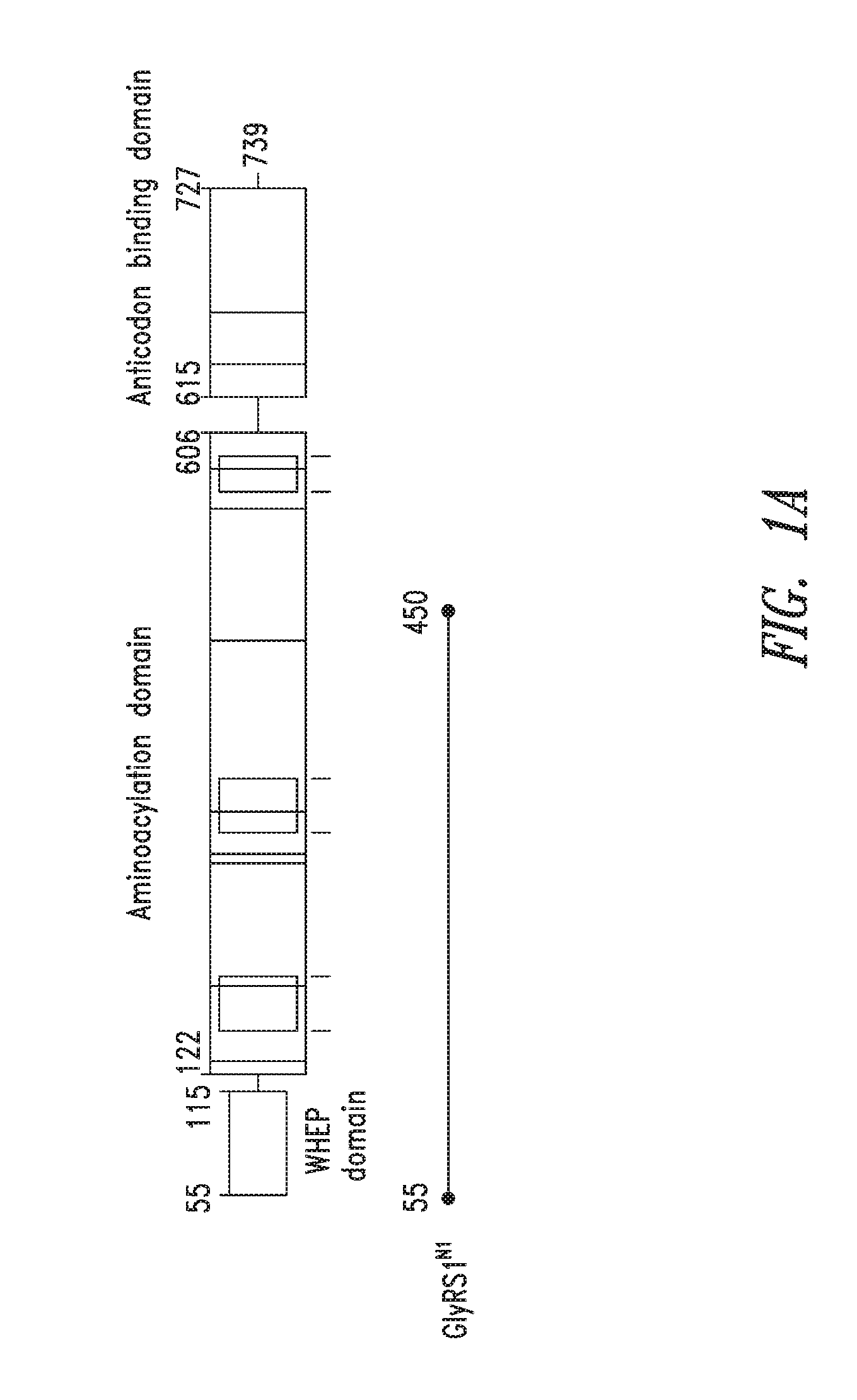
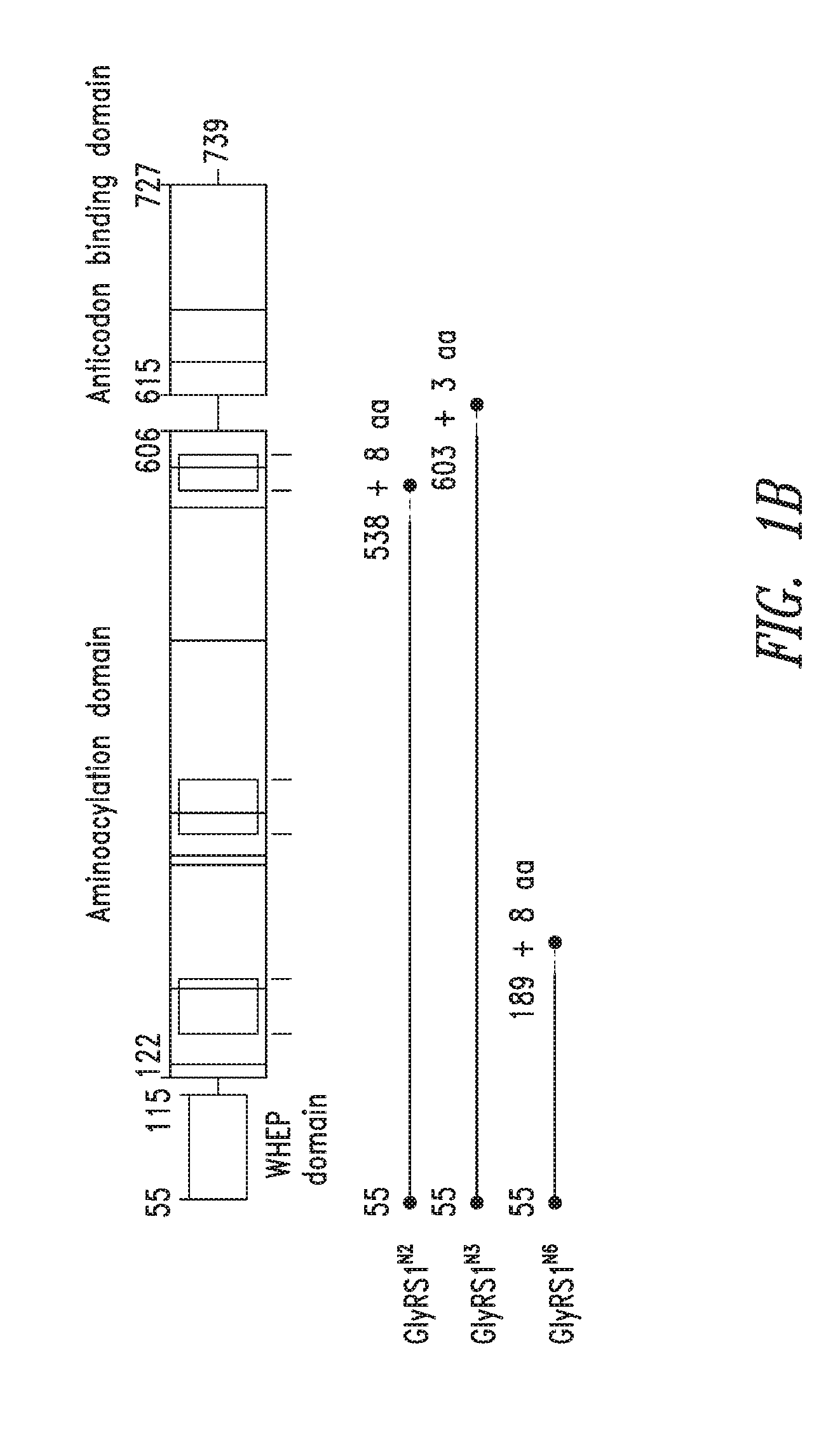
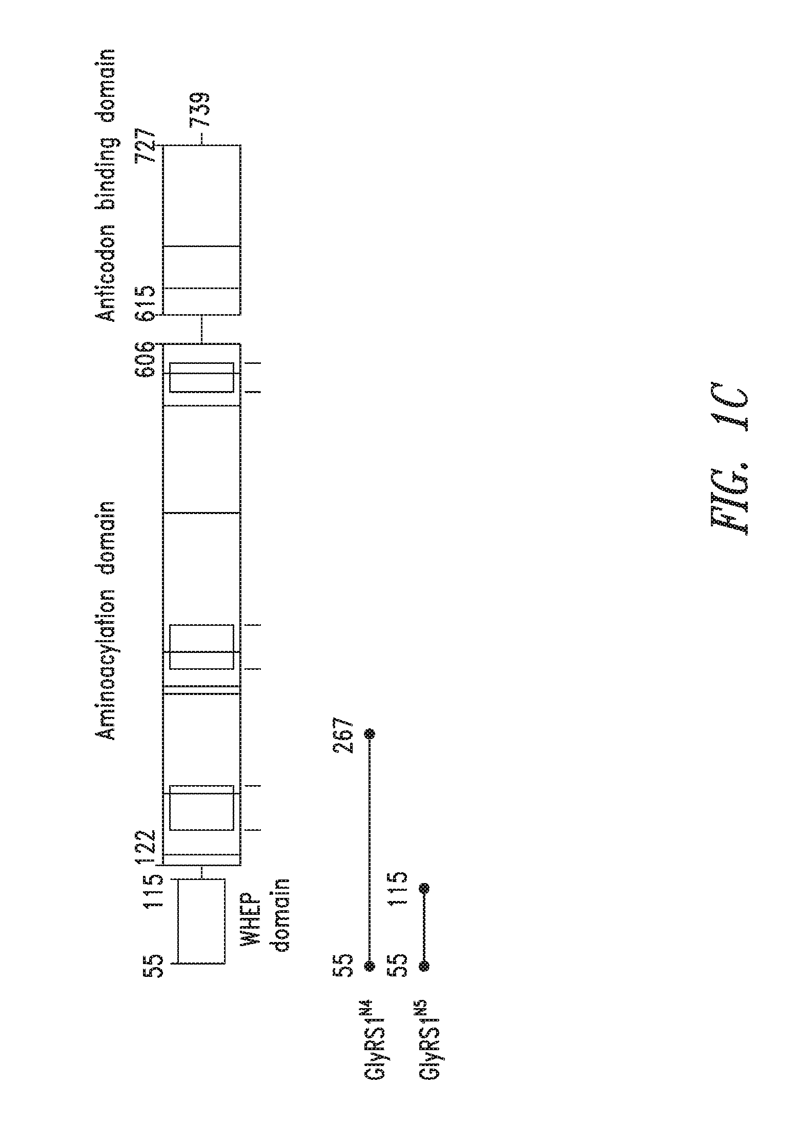
FIGS. 1A-1C show the domain structure of the Glycyl aminoacyl tRNA synthetase overlaid with the relative positions and sizes of the N-terminal AARS polypeptides identified shown schematically. FIG. 1A representing fragments identified from mass spectrometry analysis, FIG. 1B representing the fragments identified from deep sequencing of transcriptomes, and FIG. 1C representing fragments identified from bioinformatics analysis.
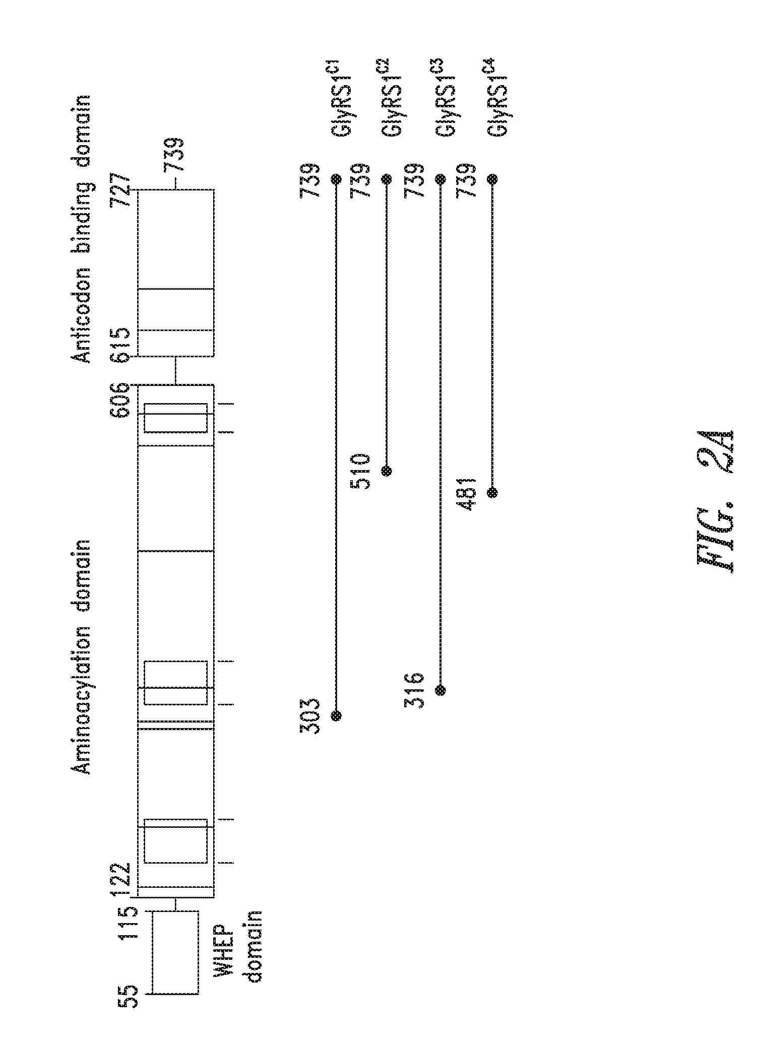
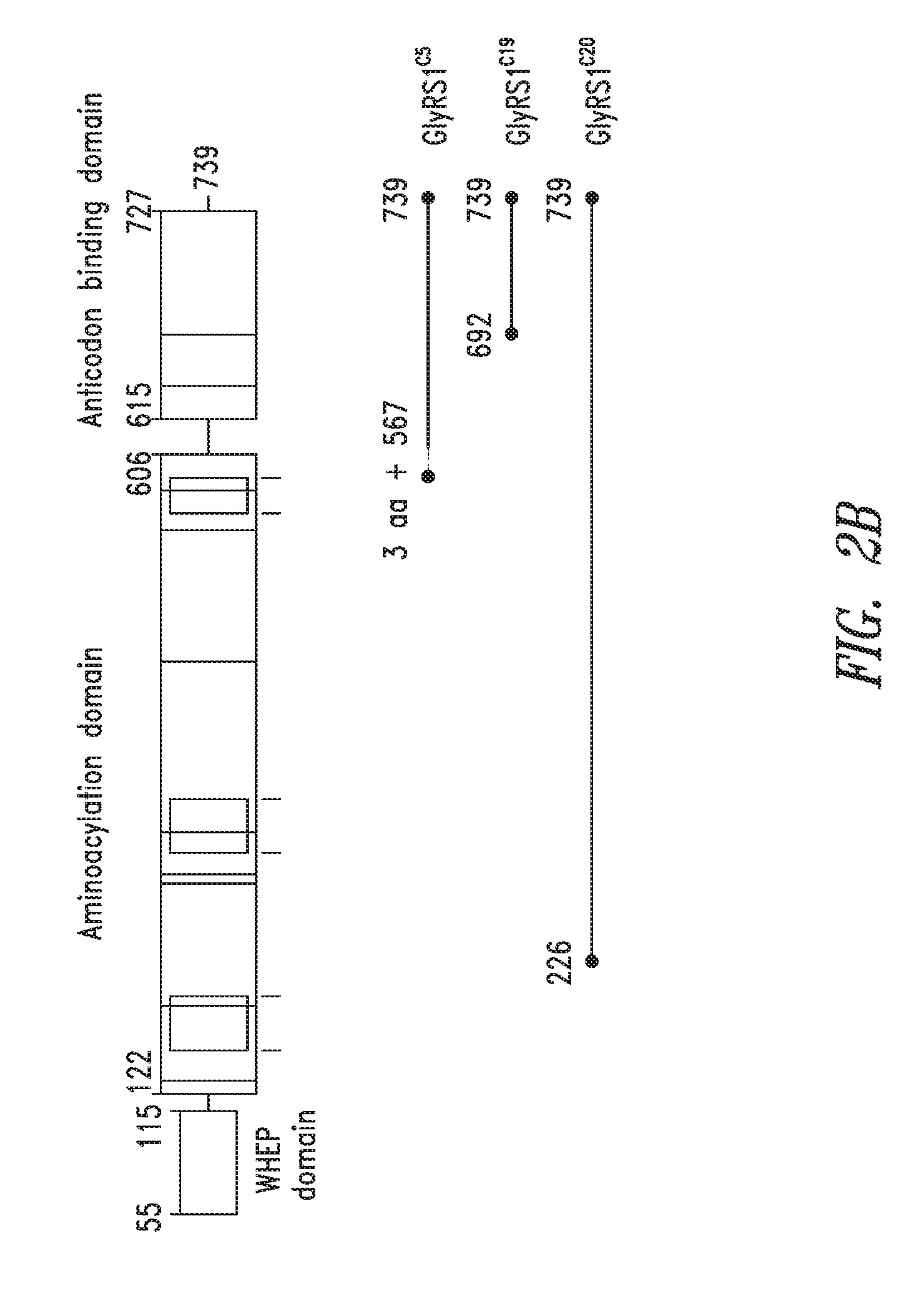
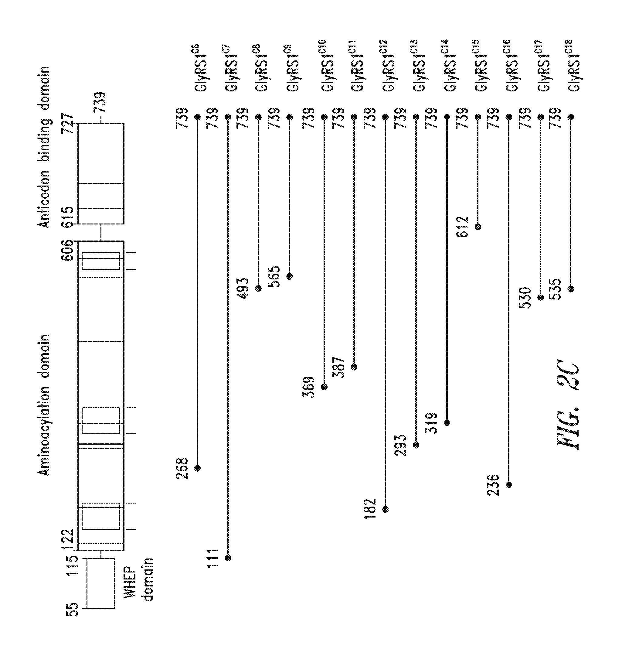
FIGS. 2A-2C show the domain structure of the Glycyl aminoacyl tRNA synthetase overlaid with the relative positions and sizes of the C-terminal AARS polypeptides identified shown schematically. FIG. 2A representing fragments identified from mass spectrometry analysis, FIG. 2B representing the fragments identified from deep sequencing of transcriptomes, and FIG. 2C representing fragments identified from bioinformatics analysis.
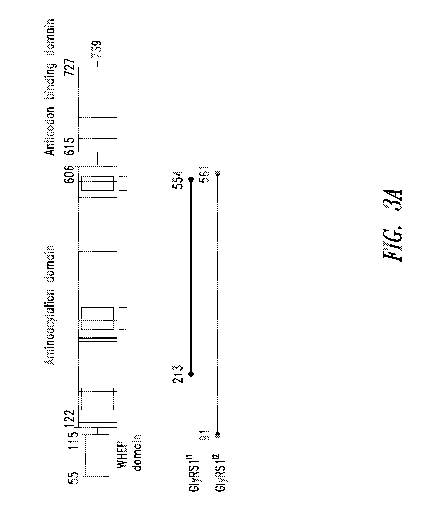
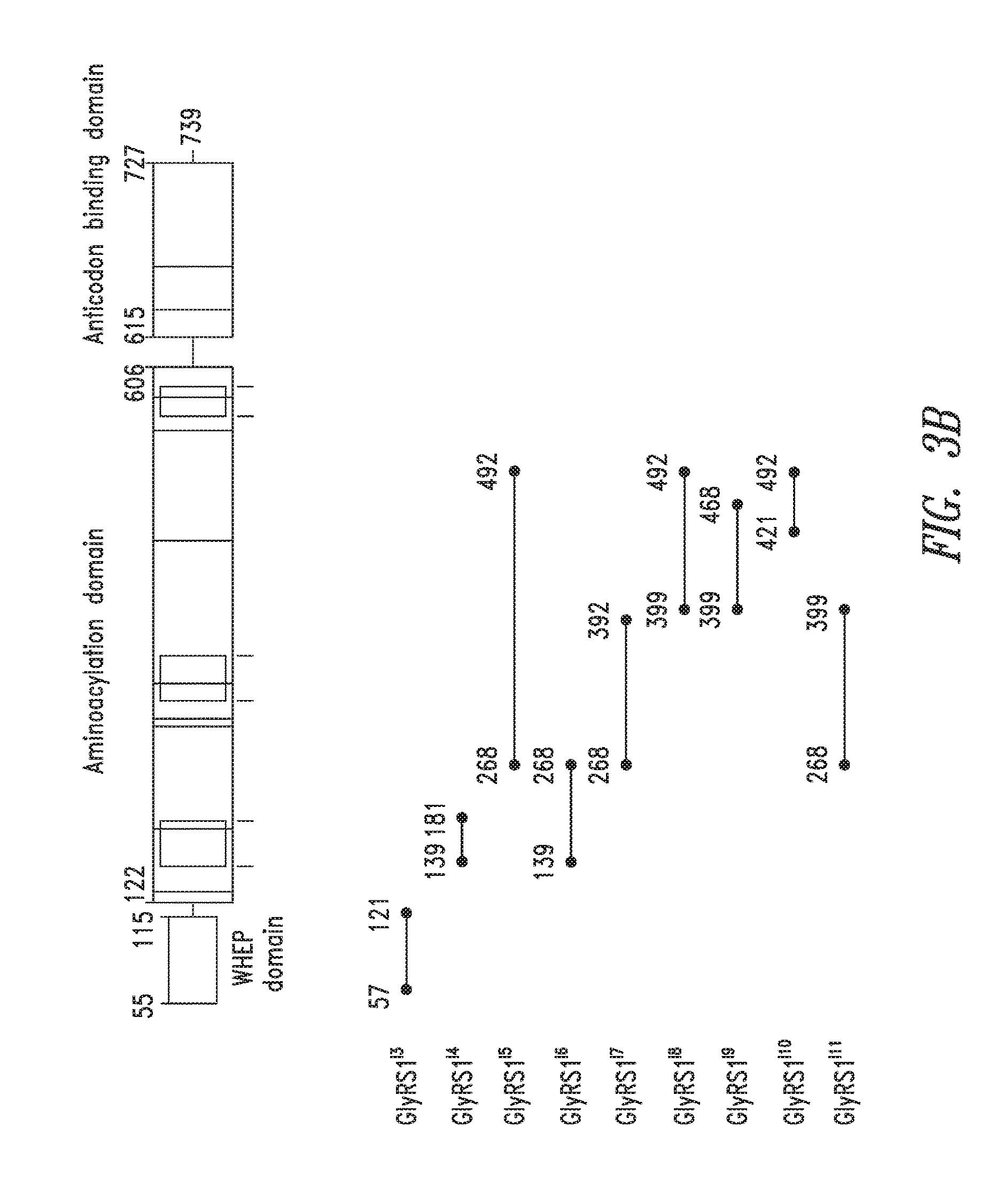
FIGS. 3A-3B show the domain structure of the Glycyl aminoacyl tRNA synthetase overlaid with the relative positions and sizes of the Internal AARS polypeptides identified shown schematically. FIG. 3A representing fragments identified from mass spectrometry analysis, FIG. 3B fragments identified from bioinformatics analysis.
BRIEF SUMMARY OF THE INVENTION
Embodiments of the present invention relate generally to the discovery of protein fragments of aminoacyl-tRNA synthetases (AARSs), which possess non-canonical biological activities, such as extracellular signaling activities, and/or other characteristics of therapeutic and diagnostic relevance. The AARSs are universal and essential elements of the protein synthesis machinery found in all organisms, but human AARSs and their associated proteins have naturally-occurring resected variants, with potent cell signaling activities that contribute to normal functioning of humans. The activities of these protein fragments are distinct from the protein synthesis activities commonly known for AARSs, and the present invention includes the discovery and development of these resected proteins as new biotherapeutic agents, new discovery research reagents, and as new antigens/targets for directed biologics and diagnostic agents that can be used to potentially treat or diagnose a wide variety of human diseases, such as inflammatory, hematological, neurodegenerative, autoimmune, hematopoietic, cardiovascular, and metabolic diseases or disorders.
The AARS protein fragment(s) of the present invention may therefore be referred to as "resectins," or alternatively as "appendacrines." As noted above, the term "resectin" derives from the process of excising or resecting a given AARS protein fragment from the context of its full-length parent AARS sequence, which typically masks its non-canonical activities. In certain instances, the AARS protein fragments and polynucleotides of the present invention were identified through the occurrence of this resection process, whether naturally-occurring (e.g., proteolytic, splice variant), artificially-induced, or predicted. The term "appendacrine" derives from a combination of "append" (from Latin--appender) and to "separate" or "discern" (from Greek--crines)," and also reflects the separation of one or more appended domains of the AARS protein fragments from their corresponding full-length or parent AARS sequences.
Although a few AARS fragments have been previously shown to have non-synthetase activities, the expression, isolation, purification, and characterization of such fragments for biotherapeutic, discovery, or diagnostic utility is limited, and persons skilled in the art would not have readily appreciated such activities to associate with each member of the entire family of AARSs, or with alternative fragments. Here, a methodical approach was utilized to discover and verify AARS protein fragments for the 20 mitochondrial and 20 cytosolic AARSs (and associated proteins) for biotherapeutic discovery and diagnostic utility. For instance, certain of the present AARS protein fragment(s) and polynucleotides that encode them are identified from biological samples using mass spectrometry (MS), mainly to identify proteolytic fragments, and others were identified by deep sequencing techniques, mainly to identify splice variants. Other AARS protein fragment(s) are identified using in silico predictions of amino acid sequences, such as by computationally comparing synthetases from humans and lower organisms along with key demarcations (e.g., protease sites); this approach utilized sequence analysis of the full-length AARS based on specific criteria to discern proteolytic fragments and functional domains possessing non-canonical biological activities.
Novel resectins of the AARSs are unexpected, and their differential expression is also unexpected. Specific resections are typically seen under different treatments (e.g., from cells grown in media with or without serum), at different stages of growth (e.g., adult brain vs. fetal brain) and for different tissue types (e.g., pancreas vs. liver). The pattern of expression is not the same for all aminoacyl tRNA synthetases despite the fact that the canonical functions for all aminoacyl tRNA synthetases are needed in the same cell locations and in relatively proportional amounts. One would not expect the levels of an aminoacyl tRNA synthetase activity to increase without an increase in the amounts of other aminoacyl tRNA synthetase activities at the same time. The mass spectrometry and deep sequencing data indicates that aminoacyl tRNA synthetase resectins do have varying levels and do occur in different sites and at different stages.
In addition, AARS protein fragments can be expressed and purified to sufficiently high purity to discern their biological properties. Previously, fragments were often not of sufficient purity, folding, and stability to enable proper biological characterization of non-synthetase activities. Cell based assays, for instance, are used in conjunction with sufficiently pure, stable, soluble and folded resectins to reveal their important biotherapeutic, discovery or diagnostic activities.
In particular, embodiments of the present invention relate to protein fragments of Glycyl tRNA synthetases, related agents and compositions of biotherapeutic, discovery, or diagnostic utility, and methods of use thereof. The compositions of the present invention are useful in a variety of diagnostic, drug discovery, and therapeutic applications, as described herein. Preferably, the AARS proteins and fragments are purified and stored in suitable condition to the extent required for such biotherapeutic, discovery, or diagnostic uses.
Certain embodiments include compositions, comprising an isolated aminoacyl-tRNA synthetase (AARS) protein fragment of at least about 100, 90, 80, 70, 60, 50 or 40 amino acids that comprises an amino acid sequence as set forth in Table(s) 1-3, or Table(s) 4-6, or Table(s) 7 or 9, and has a solubility of at least about 5 mg/ml, and wherein the composition has a purity of at least about 95% on a protein basis, and less than about 10 EU/mg protein endotoxin. In one aspect, the composition is a therapeutic composition. In specific embodiments, the composition is substantially serum free. In some embodiments the AARS protein fragment comprises a non-canonical activity. In some embodiments, the non-canonical biological activity is selected from modulation of extracellular signaling, modulation of cell proliferation, modulation of cell differentiation, modulation of gene transcription, modulation of cytokine production or activity, modulation of cytokine receptor activity, and modulation of inflammation. In some embodiments, the AARS protein fragment has an EC.sub.50 of less than about 1 nM, about 5 nM, about 10 nM, about 50 nM, about 100 nM or about 200 nM for a cell-based non-canonical biological activity.
In certain embodiments the AARS protein fragment is fused to a heterologous polypeptide. In some embodiments, the AARS fusion protein substantially retains a non-canonical activity of the AARS protein fragment. In some embodiments, the AARS fusion protein suppresses a non-canonical activity of the AARS protein fragment. In some embodiments, the heterologous polypeptide is attached to the N-terminus of the AARS protein fragment. In some embodiments, the heterologous polypeptide is attached to the C-terminus of the AARS protein fragment. In one aspect of any of these embodiments the heterologous polypeptide is selected from the group consisting of purification tags, epitope tags, targeting sequences, signal peptides, membrane translocating sequences, and PK modifiers.
In certain embodiments, the composition comprises an AARS protein fragment at a concentration of least about 10 mg/mL. In certain embodiments the composition comprises an AARS protein fragment which is at least 90% monodisperse. In certain embodiments the composition comprises less than about 3% high molecular weight aggregated proteins. In certain embodiments the composition exhibits less than 3% aggregation when stored at a concentration of at least 10 mg/mL in PBS for one week at 4.degree. C. In certain embodiments the composition exhibits less than 3% aggregation when stored at a concentration of at least 10 mg/mL in PBS for one week at room temperature.
Various assays for measuring such features of resectins are described herein and may be used to define aspects of the invention. In certain aspects, these features will be preferable for biotherapeutic utility of the AARS protein fragments described herein.
Certain embodiments include compositions, comprising an isolated aminoacyl-tRNA synthetase (AARS) protein fragment of at least 40 amino acids that differs from an amino acid sequence set forth in Table(s) 1-3, or Table(s) 4-6, or Table(s) 7 or 9 by substitution, deletion, and/or addition of about 1, 2, 3, 4, 5, 6, 7, 8, 9, 10, 11, 12, 13, 14, 15, 16, 17, 18, 19, or 20 amino acids, wherein the altered protein fragment substantially retains a non-canonical activity of the unaltered protein, or has a dominant negative phenotype in relation to the non-canonical activity, wherein the protein fragment has a solubility of at least about 5 mg/ml, and wherein the composition has a purity of at least about 95% on a protein basis and less than about 10 EU/mg protein endotoxin. In specific embodiments, the composition is substantially serum free.
Other embodiments include compositions, comprising an isolated antibody that specifically binds to an isolated aminoacyl-tRNA synthetase (AARS) protein fragment as set forth in Table(s) 1-3, or Table(s) 4-6, or Table(s) 7 or 9, wherein affinity of the antibody for the AARS protein fragment is about 10.times. stronger than its affinity for a corresponding full-length AARS polypeptide. One of the surprising aspects of the present invention includes certain resectins possessing "new" surfaces accessible to antibody or other directed biologics, whereas the full length AARS "hides" or covers these surfaces with other sequences or adjacent domains. The process of resecting can also create greater aqueous accessibility for revealing previously unidentified biological activities. Some embodiments include compositions, comprising an isolated antibody that specifically binds to an isolated aminoacyl-tRNA synthetase (AARS) protein fragment as set forth in Table(s) 1-3, or Table(s) 4-6, or Table(s) 7 or 9, wherein the antibody has an affinity of at least about 10 nM for the AARS protein fragment, and an affinity of at least about 100 nM for a corresponding full-length AARS polypeptide. In some embodiments, the antibody binds to an epitope located within an AARS polypeptide unique splice junction as set forth in any of Table(s) 1-3, or Table(s) 4-6, or Table(s) 7 or 9, or to an amino acid sequence C-terminal of this splice site. In certain embodiments, the antibody antagonizes the non-canonical activity of the AARS protein fragment. Such antagonists may optionally bind the corresponding parental or full-length AARS.
Other aspects relate to bioassay systems, comprising a substantially pure aminoacyl-tRNA synthetase (AARS) protein fragment of at least 40 amino acids that comprises an amino acid sequence as set forth in Table(s) 1-3, or Table(s) 4-6, or Table(s) 7 or 9, and a binding partner that binds to the AARS protein fragment. In one aspect, the binding partner is selected from the group consisting of a cellular surface receptor protein, nucleic acid, lipid membrane, cell regulatory protein, enzyme, and transcription factor. Optionally, such a receptor may be part of a cell, preferably a cell relevant to the revealed biology of the resectin.
Certain embodiments include cellular compositions, comprising an isolated aminoacyl-tRNA synthetase (AARS) protein fragment of at least 40 amino acids that comprises an amino acid sequence as set forth in Table(s) 1-3, or Table(s) 4-6, or Table(s) 7 or 9, and an engineered population of cells in which at least one cell comprises a polynucleotide encoding said AARS protein fragment. In one aspect, the cells are capable of growing in a serum free medium.
Also included are detection systems, comprising a substantially pure aminoacyl-tRNA synthetase (AARS) protein fragment of at least 50 or 100 amino acids that comprises an amino acid sequence as set forth in Table(s) 1-3, or Table(s) 4-6, or Table(s) 7 or 9, a cell that comprises a cell-surface receptor or an extracellular portion thereof that binds to the protein fragment, and a molecule of less than about 2000 daltons, or a second polypeptide, which modulates binding or interaction between the AARS protein fragment and the extracellular receptor.
Particular embodiments include diagnostic systems, comprising a substantially pure aminoacyl-tRNA synthetase (AARS) protein fragment of at least 40 amino acids that comprises an amino acid sequence as set forth in Table(s) 1-3, or Table(s) 4-6, or Table(s) 7 or 9, and a cell that comprises a cell-surface receptor or an extracellular portion thereof that binds to the AARS protein fragment, wherein the system or cell comprises an indicator molecule that allows detection of a change in the levels or activity of the cell-surface receptor or extracellular portion thereof.
Certain embodiments include cellular growth devices, comprising an isolated aminoacyl-tRNA synthetase (AARS) protein fragment of at least 40 amino acids that comprises an amino acid sequence as set forth in Table(s) 1-3, or Table(s) 4-6, or Table(s) 7 or 9, an engineered population of cells in which at least one cell comprises a polynucleotide encoding said AARS protein fragment, at least about 10 liters of serum-free cell media, and a sterile container. In specific embodiments, the cells utilized for any of the methods or compositions described herein are capable of growing in serum-free media, optionally with an antibiotic and an inducer.
Some embodiments relate to antisense or RNA interference (RNAi) agents, comprising a sequence that is targeted against a unique splice junction of an AARS splice variant as set forth in Table(s) 1-3, or Table(s) 4-6, or Table(s) 7 or 9.
Also included are therapeutic compositions, comprising an isolated aminoacyl-tRNA synthetase (AARS) protein fragment of at least 40 amino acids that comprises an amino acid sequence as set forth in Table(s) 1-3, or Table(s) 4-6, or Table(s) 7 or 9, wherein the protein fragment specifically binds to a binding partner and has a solubility of at least about 5 mg/ml, and wherein the composition has a purity of at least about 95% on a protein basis. In some aspects, the composition may have less than 10 EU endotoxin/mg protein.
Also included are compositions, comprising an isolated aminoacyl-tRNA synthetase (AARS) protein fragment of at least 40 amino acids that is at least 80%, 85%, 90%, 95%, 98%, or 100% identical to an amino acid sequence set forth in Table(s) 1-3, or Table(s) 4-6, or Table(s) 7 or 9, wherein the protein fragment has a solubility of at least about 5 mg/ml, and wherein the composition has a purity of at least about 95% on a protein basis and less than 10 EU endotoxin/mg protein. In any of these embodiments, the compositions may comprise an AARS protein fragment that is at least about 50%, about 60%, about 70%, about 80%, about 90% or about 95% monodisperse with respect to its apparent molecular mass. In another aspect of any of these embodiments, the compositions comprise less than about 10% (on a protein basis) high molecular weight aggregated proteins, or less than about 5% high molecular weight aggregated proteins, or less than about 4% high molecular weight aggregated proteins, or less than about 3% high molecular weight aggregated proteins, or less than 2% high molecular weight aggregated proteins, or less than about 1% high molecular weight aggregated proteins.
In another aspect of any of these embodiments, the compositions exhibits less than about 10% aggregation when stored at a concentration of at least 10 mg/mL in PBS for one week at 4.degree. C., or less than about 5% aggregation when stored at a concentration of at least 10 mg/mL in PBS for one week at 4.degree. C., or less than about 3% aggregation when stored at a concentration of at least 10 mg/mL in PBS for one week at 4.degree. C., or less than about 2% aggregation when stored at a concentration of at least 10 mg/mL in PBS for one week at 4.degree. C., or less than about 1% aggregation when stored at a concentration of at least 10 mg/mL in PBS for one week at 4.degree. C.
Certain embodiments include compositions, comprising a substantially pure aminoacyl-tRNA synthetase (AARS) protein fragment of at least 40 amino acids that comprises an amino acid sequence as set forth in Table(s) 1-3, or Table(s) 4-6, or Table(s) 7 or 9, and at least one covalently or non-covalently moiety attached thereto. In some embodiments, the moiety is a detectable label. In some embodiments, the moiety is a water soluble polymer. In some embodiments, the moiety is PEG. In one aspect of any of these embodiments, the moiety is attached to the N-terminus of the protein fragment. In one aspect of any of these embodiments, the moiety is attached to the C-terminus of the protein fragment.
Particular embodiments include compositions, comprising a solid substrate attached to an isolated aminoacyl-tRNA synthetase (AARS) protein fragment of at least 40 amino acids that comprises an amino acid sequence as set forth in Table(s) 1-3, or Table(s) 4-6, or Table(s) 7 or 9, or a biologically active fragment or variant thereof, wherein the protein fragment has a solubility of at least about 5 mg/ml, and the composition has a purity of at least about 95% on a protein basis.
Also included are compositions, comprising a binding agent that specifically binds to an isolated aminoacyl-tRNA synthetase (AARS) protein fragment as set forth in Table(s) 1-3, or Table(s) 4-6, or Table(s) 7 or 9, wherein the binding agent has an affinity of at least about 1 nM for the protein fragment. In one aspect, the binding agent binds to an epitope located within an AARS polypeptide unique splice junction as set forth in any of Table(s) 1-3, or Table(s) 4-6, or Table(s) 7 or 9, or to an amino acid sequence C-terminal of this splice site. In some embodiments, the binding agent antagonizes a non-canonical activity of the AARS polypeptide.
Certain embodiments include isolated aminoacyl-tRNA synthetase (AARS) polypeptides, comprising an amino acid sequence of an AARS protein fragment as described herein, an amino acid sequence encoded by an AARS polynucleotide as described herein, or a variant or fragment thereof. Certain AARS polypeptides comprise an amino acid sequence that is at least 80%, 85%, 90%, 95%, 98%, or 100% identical to an AARS reference sequence as disclosed in Table(s) 1-3, or Table(s) 4-6, or Table(s) 7 or 9, or Table E2. Certain AARS polypeptides consist essentially of an amino acid sequence that is at least 80%, 85%, 90%, 95%, 98%, or 100% identical to an AARS reference sequence as disclosed in Table(s) 1-3, or Table(s) 4-6, or Table(s) 7 or 9, or Table E2. In certain embodiments, the polypeptide comprises a non-canonical biological activity. In specific embodiments, the non-canonical biological activity is selected from modulation of cell signaling (e.g., extracellular signaling), modulation of cell proliferation, modulation of cell migration, modulation of cell differentiation, modulation of apoptosis or cell death, modulation of angiogenesis, modulation of cell binding, modulation of cellular metabolism, modulation of cellular uptake, modulation of gene transcription, or secretion, modulation of cytokine production or activity, modulation of cytokine receptor activity, and modulation of inflammation.
Other aspects include antibodies and other binding agents that exhibit binding specificity for an isolated AARS polypeptide as described herein, a binding partner of the AARS polypeptide, or the complex of both. In some embodiments, the affinity of the antibody or binding agent for the AARS polypeptide is about 10.times. stronger than its affinity for a corresponding full-length AARS polypeptide. In specific embodiments, the binding agent is selected from a peptide, peptide mimetic, an adnectin, an aptamer, and a small molecule. In certain embodiments, the antibody or binding agent antagonizes a non-canonical activity of the AARS polypeptide. In other embodiments, the antibody or binding agent agonizes a non-canonical activity of the AARS polypeptide.
Certain embodiments include isolated aminoacyl-tRNA synthetase (AARS) polynucleotides, comprising a nucleotide sequence of an AARS polynucleotide as described herein, a nucleotide sequence that encodes an AARS protein fragment as described herein, or a variant, a fragment, or a complement thereof. Certain AARS polynucleotides comprise a nucleotide sequence that is at least 80%, 85%, 90%, 95%, 98%, or 100% identical to an AARS reference polynucleotide, or a complement thereof, as disclosed in Table(s) 1-3, or Table(s) 4-6, or Table(s) 7 or 9, or Table E2. In some embodiments, the nucleotide sequence is codon optimized for bacterial expression. In one aspect, the nucleotide sequence is at least 80% identical a polynucleotide sequence disclosed in Table E2.
Specific AARS polynucleotides consist essentially of a nucleotide sequence that is at least 80%, 85%, 90%, 95%, 98%, or 100% identical to an AARS reference polynucleotide, or a complement thereof, as disclosed in Table(s) 1-3, or Table(s) 4-6, or Table(s) 7 or 9, or Table E2. Other AARS polynucleotides comprise or consist essentially of a nucleotide sequence that specifically hybridizes to an AARS reference polynucleotide, as disclosed in Table(s) 1-3, or Table(s) 4-6, or Table(s) 7 or 9, or Table E2. In certain embodiments, the polynucleotide is selected from a primer, a probe, and an antisense oligonucleotide. In specific embodiments, the primer, probe, or antisense oligonucleotide is targeted to a specific or unique splice junction, and/or sequence 3' of this splice site within an AARS polynucleotide.
Certain embodiments include methods of determining presence or levels of an AARS protein fragment in a sample, comprising contacting the sample with one or more binding agents that specifically bind to an AARS protein fragment as described herein, detecting the presence or absence of the binding agent, and thereby determining the presence or levels of the AARS protein fragment. Other embodiments include methods of determining presence or levels of an AARS protein fragment in a sample, comprising analyzing the sample with a detector that is capable of specifically identifying a protein fragment as described herein, and thereby determining the presence or levels of the AARS protein fragment. In specific embodiments, the detector is a mass spectrometer (MS), a flow cytometer, a protein imaging device, an enzyme-linked immunosorbent assays (ELISA), or a protein microarray. Certain embodiments comprise comparing the presence or levels of the AARS protein fragment to a control sample or a predetermined value. Certain embodiments comprise characterizing the state of the sample to distinguish it from the control. In specific embodiments, the sample and control comprise a cell or tissue, and the method comprises distinguishing between cells or tissues of different species, cells of different tissues or organs, cells at different cellular developmental states, cells at different cellular differentiation states, cells at different physiological states, or healthy and diseased cells. For instance, selected resectins may be more abundant under conditions such as stress or insult.
Certain embodiments include discovery methods of, and related compositions for, identifying a compound that specifically binds to an aminoacyl-tRNA synthetase (AARS) polypeptide as described herein, or one or more of its cellular binding partners, comprising a) combining the AARS polypeptide or its cellular binding partner or both with at least one test compound under suitable conditions, and b) detecting binding of the AARS polypeptide or its cellular binding partner or both to the test compound, thereby identifying a compound that specifically binds to the AARS polypeptide or its cellular binding partner or both. In certain embodiments, the test compound is a polypeptide or peptide, an antibody or antigen-binding fragment thereof, a peptide mimetic, or a small molecule. In certain embodiments, the test compound agonizes a non-canonical biological activity of the AARS polypeptide or its cellular binding partner. In other embodiments, the test compound antagonizes a non-canonical biological activity of the AARS polypeptide or its cellular binding partner. Certain embodiments include a compound identified by the above-method, such as an agonist (e.g., small molecule, peptide).
Certain embodiments include methods of determining presence or levels of a polynucleotide sequence of an AARS splice variant in a sample, comprising contacting the sample with one or more oligonucleotides that specifically hybridize to an AARS polynucleotide as described herein, detecting the presence or absence of the oligonucleotides in the sample, and thereby determining the presence or levels of the polynucleotide sequence of the AARS splice variant. Other embodiments include methods of determining presence or levels of a polynucleotide sequence of an AARS splice variant in a sample, comprising contacting the sample with at least two oligonucleotides that specifically amplify an AARS polynucleotide as described herein, performing an amplification reaction, detecting the presence or absence of an amplified product, and thereby determining presence or levels of the polynucleotide sequence of the AARS splice variant. In specific embodiments, the oligonucleotide(s) specifically hybridize to or specifically amplify a splice junction that is unique to the AARS splice variant. Certain embodiments include comparing the presence or levels of the AARS protein fragment or splice variant to a control sample or a predetermined value. Certain embodiments include characterizing the state of the sample to distinguish it from the control. In specific embodiments, the sample and control comprise a cell or tissue, and the method comprises distinguishing between cells or tissues of different species, cells of different tissues or organs, cells at different cellular developmental states, cells at different cellular differentiation states, or healthy and diseased cells.
Some embodiments include pharmaceutical compositions, comprising an AARS polynucleotide described herein, an AARS polypeptide described herein, a binding agent as described herein, or a compound identified by the above-method or described herein, and a pharmaceutically acceptable excipient or carrier.
Certain embodiments include methods of modulating a cellular activity of a cell, comprising contacting the cell with an AARS polynucleotide described herein, an AARS polypeptide described herein, a binding agent described herein, a compound of the above-method or described herein, or a pharmaceutical composition described herein. In specific embodiments, the cellular activity is selected from cell proliferation, cell migration, cell differentiation, apoptosis or cell death, cell signaling, angiogenesis, cell binding, cellular uptake, cell secretion, metabolism, cytokine production or activity, cytokine receptor activity, gene transcription, and inflammation. In one aspect, the cell is selected from the group consisting of pre-adipocytes, bone marrow, neutrophils, blood cells, hepatocytes, astrocytes, mesenchymal stem cells, and skeletal muscle cells.
In certain embodiments, the cell is in a subject. Certain embodiments comprise treating the subject, wherein the subject has a condition associated with a neoplastic disease, an immune system disease or condition, an infectious disease, a metabolic disease, an inflammatory disorder, neuronal/neurological disease, a muscular/cardiovascular disease, a disease associated with aberrant hematopoiesis, a disease associated with aberrant angiogenesis, or a disease associated with aberrant cell survival.
Also included are processes for manufacturing a pharmaceutical compound, comprising: a) performing an in vitro screen of one or more candidate compounds in the presence an AARS protein fragment of at least 40 amino acids that comprises an amino acid sequence as set forth in Table(s) 1-3, or Table(s) 4-6, or Table(s) 7 or 9, to identify a compound that specifically binds to the AARS protein fragment; b) performing a cell-based or biochemical or receptor assay with the compound identified in step a), to identify a compound that modulates one or more non-canonical activities of the AARS protein fragment; c) optionally assessing the structure-activity relationship (SAR) of the compound identified in step b), to correlate its structure with modulation of the non-canonical activity, and optionally derivatizing the compound to alter its ability to modulate the non-canonical activity; and d) producing sufficient amounts of the compound identified in step b), or the derivatized compound in step c), for use in humans, thereby manufacturing the pharmaceutical compound.
Other embodiments include processes for manufacturing a pharmaceutical compound, comprising: a) performing an in vitro screen of one or more candidate compounds in the presence a cell-surface receptor or an extracellular portion thereof that specifically binds to an AARS protein fragment of Table(s) 1-3, or Table(s) 4-6, or Table(s) 7 or 9, to identify a compound that specifically binds to the cell-surface receptor or extracellular portion thereof; b) performing a cell-based or biochemical or receptor assay with the compound identified in step a), to identify a compound that modulates one or more non-canonical activities of the AARS protein fragment; c) optionally assessing the structure-activity relationship (SAR) of the compound identified in step b), to correlate its structure with modulation of the non-canonical activity, and optionally derivatizing the compound to alter its ability to modulate the non-canonical activity; and d) producing sufficient amounts of the compound identified in step b), or the derivatized compound in step c), for use in humans, thereby manufacturing the pharmaceutical compound.
Some embodiments include a cellular composition, comprising an engineered population of cells in which at least one cell comprises a polynucleotide encoding a heterologous full length aminoacyl-tRNA synthetase (AARS) protein, wherein the cells are capable of growing in a serum-free medium. In one aspect, the full length aminoacyl-tRNA synthetase (AARS) protein comprises a heterologous purification or epitope tag to facilitate purification of an AARS protein fragment. In another aspect, the full length aminoacyl-tRNA synthetase (AARS) protein comprises a heterologous proteolysis site to enable production of the AARS protein fragment upon cleavage.
Some embodiments include a method for producing an AARS polypeptide as set forth in Table(s) 1-3, or Table(s) 4-6, or Table(s) 7 or 9, or Table E2 in situ within a cell, comprising; i) expressing a heterologous full length aminoacyl-tRNA synthetase (AARS) protein within the cell, wherein the cell comprises a protease capable of cleaving the heterologous full length aminoacyl-tRNA synthetase (AARS) protein to produce the AARS polypeptide.
Some embodiments include a method for producing an AARS polypeptide as set forth in Table(s) 1-3, or Table(s) 4-6, or Table(s) 7 or 9, or Table E2 comprising contacting an isolated full length aminoacyl-tRNA synthetase (AARS) protein with a protease that is capable of cleaving the full length aminoacyl-tRNA synthetase (AARS) protein and producing an AARS polypeptide.
Some embodiments include an engineered full length aminoacyl-tRNA synthetase (AARS) protein comprising a heterologous proteolysis site to enable the proteolytic generation of an AARS protein fragment as set forth in any of Table(s) 1-3, or Table(s) 4-6, or Table(s) 7 or 9 or Table E2.
Some embodiments include a composition, comprising an isolated full length aminoacyl-tRNA synthetase protein, wherein the composition has a purity of at least about 95% on a protein basis, less than about 10 EU endotoxin/mg protein, and is substantially serum free. In one aspect, the full length aminoacyl-tRNA synthetase protein is present at a concentration of at least 10 mg/mL, and is at least 90% monodisperse.
Certain embodiments include methods of reducing or ameliorating at least one symptom of a disease mediated by a mutation, inappropriate subcellular association, over-expression, and/or subcellular distribution of a glycyl-tRNA synthetase (GlyRS), comprising administering to a subject an isolated aminoacyl-tRNA synthetase (AARS) protein fragment that consists essentially of an amino acid sequence as set forth in any of Table(s) 1-3, or Table(s) 4-6, or Table(s) 7 or 9, or Table E2. In some embodiments, the AARS protein fragment has at least 40 amino acids. In certain embodiments, the AARS protein fragment has a non canonical activity. In certain embodiments, the AARS protein fragment is fused to a heterologous fusion partner.
In different embodiments, the disease is Charcot-Marie-Tooth Disease Type 2D (CMT2D) or Distal Spinal Muscular Atrophy Type V (dSMA-V). In specific embodiments, the disease is CMT2D, and the at least one symptom includes reduced nerve conduction velocity. In certain embodiments, the subject has been determined to have at least one mutation in Glycyl tRNA synthetase selected from the group consisting of E71G, L129P, P234KY, G240R, 1280F, H418R, D500N, G526R, S581L, and G598A.
In some embodiments, the disease is dSMA-V. In certain embodiments, the at least one symptom is muscle weakness. In these and related embodiments, the subject has been determined to have at least one mutation in Glycyl tRNA synthetase selected from the group consisting of E71G, L129P, G240R, and G526R.
As noted above, also included are fusion proteins that comprise an AARS protein fragment of any one of Table(s) 1-3, or Table(s) 4-6, or Table(s) 7 or 9, or Table E2 and a heterologous fusion partner. In certain embodiments, the AARS fusion protein substantially retains a non-canonical activity of the AARS protein fragment. In some embodiments, the AARS fusion protein suppresses a non-canonical activity of the AARS protein fragment. In particular embodiments, the heterologous polypeptide is attached to the N-terminus of the AARS protein fragment. In some embodiments, the heterologous polypeptide is attached to the C-terminus of the AARS protein fragment. In specific embodiments, the heterologous polypeptide is selected from the group consisting of purification tags, epitope tags, targeting sequences, signal peptides, membrane translocating sequences, and PK modifiers.
Also included are methods of reducing or ameliorating a symptom of a disease mediated by a mutation, inappropriate subcellular association, over-expression, and/or subcellular distribution of a glycyl-tRNA synthetase (GlyRS), comprising administering to a subject an isolated antibody or binding agent that specifically binds to an isolated aminoacyl-tRNA synthetase (AARS) protein fragment as set forth in any of Table(s) 1-3, or Table(s) 4-6, or Table(s) 7 or 9, or Table E2, wherein affinity of the antibody for the AARS protein fragment is at least about 10.times. stronger than its affinity for a corresponding full-length AARS polypeptide. In particular embodiments, the antibody or binding agent antagonizes a non-canonical activity of the AARS polypeptide.
DETAILED DESCRIPTION OF THE INVENTION
TABLE-US-00001 TABLE OF CONTENTS I. OVERVIEW 16 II. DEFINITIONS 17 III. PURIFIED AARS PROTEIN FRAGMENTS AND 30 VARIANTS IV. AARS POLYNUCLEOTIDES 100 V. ANTIBODIES 113 VI. ANTIBODY ALTERNATIVES AND OTHER BINDING 118 AGENTS VII. BIOASSAYS AND ANALYTICAL ASSAYS 122 VIII. EXPRESSION AND PURIFICATION SYSTEMS 125 IX. DIAGNOSTIC METHODS AND COMPOSITIONS 138 X. ANTISENSE AND RNAI AGENTS 154 A. ANTISENSE AGENTS 155 B. RNA INTERFERENCE AGENTS 163 XI. DRUG DISCOVERY 171 XII. METHODS OF USE 180 XIII. PHARMACEUTICAL FORMULATIONS, 187 ADMINISTRATION AND KITS XIV. EXAMPLES 196
I. Overview
The current invention is directed, at least in part, to the discovery of novel AARS polypeptides, and methods for their preparation and use, which represent the transformation of native wild type proteins into new forms that exhibit markedly different characteristics compared to the naturally occurring full length Glycyl tRNA synthetase genes. Such AARS polypeptides were identified based on extensive sequence, and mass spectrum analysis of expressed Glycyl tRNA synthetases in different tissues, followed by the systematic production and testing of each potential AARS polypeptide to identify protein sequences that represent stable and soluble protein domains which exhibit novel biological activities, and favorable therapeutic drug characteristics.
Based on this analysis at least six new novel families of AARS polypeptides derived from Glycyl tRNA synthetase have been identified.
In one aspect, such Glycyl RNA synthetase derived AARS polypeptides comprise polypeptide sequences approximately comprising amino acids 55-450 of the Glycyl tRNA synthetase.
In a second aspect, such Glycyl tRNA synthetase derived AARS polypeptides comprise polypeptide sequences approximately comprising amino acids 55-189 of Glycyl tRNA synthetase.
In a third aspect, such Glycyl tRNA synthetase derived AARS polypeptides comprise polypeptide sequences approximately comprising amino acids 91-561 of the Glycyl tRNA synthetase.
In a fourth aspect, such Glycyl tRNA synthetase derived AARS polypeptides comprise polypeptide sequences approximately comprising amino acids 612 to 739 of the Glycyl tRNA synthetase.
In a fifth aspect, such Glycyl tRNA synthetase derived AARS polypeptides comprise polypeptide sequences approximately comprising amino acids 57 to 121 of the Glycyl tRNA synthetase.
In a six aspect, such Glycyl tRNA synthetase derived AARS polypeptides comprise polypeptide sequences approximately comprising amino acids 316 to 739 of the Glycyl tRNA synthetase.
These new AARS polypeptide families represent novel, previously unknown protein products which exhibit inter alia i) novel biological activity, ii) favorable protein stability and aggregation characteristics, and iii) the ability to be expressed and produced at high level in prokaryotic expression systems, which are materially different characteristics not found in the intact wild type protein.
II. Definitions
Unless defined otherwise, all technical and scientific terms used herein have the same meaning as commonly understood by those of ordinary skill in the art to which the invention belongs. Although any methods and materials similar or equivalent to those described herein can be used in the practice or testing of the present invention, preferred methods and materials are described. For the purposes of the present invention, the following terms are defined below.
The articles "a" and "an" are used herein to refer to one or to more than one (i.e., to at least one) of the grammatical object of the article. By way of example, "an element" means one element or more than one element.
By "about" is meant a quantity, level, value, number, frequency, percentage, dimension, size, amount, weight or length that varies by as much as 30, 25, 20, 25, 10, 9, 8, 7, 6, 5, 4, 3, 2 or 1% to a reference quantity, level, value, number, frequency, percentage, dimension, size, amount, weight or length.
An "agonist" refers to a molecule that intensifies or mimics an activity. For example, a non-canonical biological activity of an AARS, or another protein. Agonists may include proteins, nucleic acids, carbohydrates, small molecules, or any other compound or composition that modulates the activity of an AARS either by directly interacting with the AARS or its binding partner, or by acting on components of the biological pathway in which the AARS participates. Included are partial and full agonists.
As used herein, the term "amino acid" is intended to mean both naturally occurring and non-naturally occurring amino acids as well as amino acid analogs and mimetics. Naturally occurring amino acids include the 20 (L)-amino acids utilized during protein biosynthesis as well as others such as 4-hydroxyproline, hydroxylysine, desmosine, isodesmosine, homocysteine, citrulline and ornithine, for example. Non-naturally occurring amino acids include, for example, (D)-amino acids, norleucine, norvaline, p-fluorophenylalanine, ethionine and the like, which are known to a person skilled in the art. Amino acid analogs include modified forms of naturally and non-naturally occurring amino acids. Such modifications can include, for example, substitution or replacement of chemical groups and moieties on the amino acid or by derivitization of the amino acid. Amino acid mimetics include, for example, organic structures which exhibit functionally similar properties such as charge and charge spacing characteristic of the reference amino acid. For example, an organic structure which mimics Arginine (Arg or R) would have a positive charge moiety located in similar molecular space and having the same degree of mobility as the e-amino group of the side chain of the naturally occurring Arg amino acid. Mimetics also include constrained structures so as to maintain optimal spacing and charge interactions of the amino acid or of the amino acid functional groups. Those skilled in the art know or can determine what structures constitute functionally equivalent amino acid analogs and amino acid mimetics.
In certain aspects, the use of non-natural amino acids can be utilized to modify (e.g., increase) a selected non-canonical activity of an AARS protein fragment, or to alter the in vivo or in vitro half-life of the protein. Non-natural amino acids can also be used to facilitate (selective) chemical modifications (e.g., pegylation) of an AARS protein. For instance, certain non-natural amino acids allow selective attachment of polymers such as PEG to a given protein, and thereby improve their pharmacokinetic properties.
Specific examples of amino acid analogs and mimetics can be found described in, for example, Roberts and Vellaccio, The Peptides: Analysis, Synthesis, Biology, Eds. Gross and Meinhofer, Vol. 5, p. 341, Academic Press, Inc., New York, N.Y. (1983), the entire volume of which is incorporated herein by reference. Other examples include peralkylated amino acids, particularly permethylated amino acids. See, for example, Combinatorial Chemistry, Eds. Wilson and Czarnik, Ch. 11, p. 235, John Wiley & Sons Inc., New York, N.Y. (1997), the entire book of which is incorporated herein by reference. Yet other examples include amino acids whose amide portion (and, therefore, the amide backbone of the resulting peptide) has been replaced, for example, by a sugar ring, steroid, benzodiazepine or carbo cycle. See, for instance, Burger's Medicinal Chemistry and Drug Discovery, Ed. Manfred E. Wolff, Ch. 15, pp. 619-620, John Wiley & Sons Inc., New York, N.Y. (1995), the entire book of which is incorporated herein by reference. Methods for synthesizing peptides, polypeptides, peptidomimetics and proteins are well known in the art (see, for example, U.S. Pat. No. 5,420,109; M. Bodanzsky, Principles of Peptide Synthesis (1st ed. & 2d rev. ed.), Springer-Verlag, New York, N.Y. (1984 & 1993), see Chapter 7; Stewart and Young, Solid Phase Peptide Synthesis, (2d ed.), Pierce Chemical Co., Rockford, Ill. (1984), each of which is incorporated herein by reference). Accordingly, the AARS polypeptides of the present invention may be composed of naturally occurring and non-naturally occurring amino acids as well as amino acid analogs and mimetics.
The term "antagonist" refers to a molecule that reduces or attenuates an activity. For example, a non-canonical biological activity of an AARS, or another protein. Antagonists may include proteins such as antibodies, nucleic acids, carbohydrates, small molecules, or any other compound or composition that modulates the activity of an AARS or its binding partner, either by directly interacting with the AARS or its binding partner or by acting on components of the biological pathway in which the AARS participates. Included are partial and full antagonists.
The term "aminoacyl-tRNA synthetase" (AARS) refers generally to enzymes that in their natural or wild-type form are capable of catalyzing the esterification of a specific amino acid or its precursor to one of all its compatible cognate tRNAs to form an aminoacyl-tRNA. In this "canonical" activity, aminoacyl-tRNA synthetases catalyze a two-step reaction: first, they activate their respective amino acid by forming an aminoacyl-adenylate, in which the carboxyl of the amino acid is linked in to the alpha-phosphate of ATP by displacing pyrophosphate, and then, when the correct tRNA is bound, the aminoacyl group of the aminoacyl-adenylate is transferred to the 2' or 3' terminal OH of the tRNA.
Class I aminoacyl-tRNA synthetases typically have two highly conserved sequence motifs. These enzymes aminoacylate at the 2'-OH of an adenosine nucleotide, and are usually monomeric or dimeric. Class II aminoacyl-tRNA synthetases typically have three highly conserved sequence motifs. These enzymes aminoacylate at the 3'-OH of the same adenosine, and are usually dimeric or tetrameric. The active sites of class II enzymes are mainly made up of a seven-stranded anti-parallel .beta.-sheet flanked by .alpha.-helices. Although phenylalanine-tRNA synthetase is class II, it aminoacylates at the 2'-OH.
AARS polypeptides include sources of mitochondrial and cytoplasmic forms of tyrosyl-tRNA synthetase (TyrRS), a tryptophanyl-tRNA synthetase (TrpRS), a glutaminyl-tRNA synthetase (GlnRS), a glycyl-tRNA synthetase (GlyRS), a histidyl-tRNA synthetase (HisRS), a seryl-tRNA synthetase (SerRS), a phenylalanyl-tRNA synthetase (PheRS), an alanyl-tRNA synthetase (AlaRS), an asparaginyl-tRNA synthetase (AsnRS), an aspartyl-tRNA synthetase (AspRS), a cysteinyl-tRNA synthetase (CysRS), a glutamyl-tRNA synthetase (GluRS), a prolyl-tRNA synthetase (ProRS), an arginyl-tRNA synthetase (ArgRS), an isoleucyl-tRNA synthetase (IleRS), a leucyl-tRNA synthetase (LeuRS), a lysyl-tRNA synthetase (LysRS), a threonyl-tRNA synthetase (ThrRS), a methionyl-tRNA synthetases (MetRS), or a valyl-tRNA synthetase (ValRS). The wild-type or parental sequences of these AARS polypeptides are known in the art.
By "coding sequence" is meant any nucleic acid sequence that contributes to the code for the polypeptide product of a gene. By contrast, the term "non-coding sequence" refers to any nucleic acid sequence that does not contribute to the code for the polypeptide product of a gene.
Throughout this specification, unless the context requires otherwise, the words "comprise," "comprises," and "comprising" will be understood to imply the inclusion of a stated step or element or group of steps or elements but not the exclusion of any other step or element or group of steps or elements.
By "consisting of" is meant including, and limited to, whatever follows the phrase "consisting of" Thus, the phrase "consisting of" indicates that the listed elements are required or mandatory, and that no other elements may be present. By "consisting essentially of" is meant including any elements listed after the phrase, and limited to other elements that do not interfere with or contribute to the activity or action specified in the disclosure for the listed elements. Thus, the phrase "consisting essentially of" indicates that the listed elements are required or mandatory, but that other elements are optional and may or may not be present depending upon whether or not they materially affect the activity or action of the listed elements.
The recitation "endotoxin free" or "substantially endotoxin free" relates generally to compositions, solvents, and/or vessels that contain at most trace amounts (e.g., amounts having no clinically adverse physiological effects to a subject) of endotoxin, and preferably undetectable amounts of endotoxin. Endotoxins are toxins associated with certain bacteria, typically gram-negative bacteria, although endotoxins may be found in gram-positive bacteria, such as Listeria monocytogenes. The most prevalent endotoxins are lipopolysaccharides (LPS) or lipo-oligo-saccharides (LOS) found in the outer membrane of various Gram-negative bacteria, and which represent a central pathogenic feature in the ability of these bacteria to cause disease. Small amounts of endotoxin in humans may produce fever, a lowering of the blood pressure, and activation of inflammation and coagulation, among other adverse physiological effects.
Therefore, in pharmaceutical production of AARS polypeptides, it is often desirable to remove most or all traces of endotoxin from drug products and/or drug containers, because even small amounts may cause adverse effects in humans. A depyrogenation oven may be used for this purpose, as temperatures in excess of 300.degree. C. are typically required to break down most endotoxins. For instance, based on primary packaging material such as syringes or vials, the combination of a glass temperature of 250.degree. C. and a holding time of 30 minutes is often sufficient to achieve a 3 log reduction in endotoxin levels. Other methods of removing endotoxins are contemplated, including, for example, chromatography and filtration methods, as described herein and known in the art. Also included are methods of producing AARS polypeptides in and isolating them from eukaryotic cells such as mammalian cells to reduce, if not eliminate, the risk of endotoxins being present in a composition of the invention. Preferred are methods of producing AARS polypeptides in and isolating them from serum free cells. Such compositions comprising AARS polypeptides represent new formulations which exhibit novel and new biological and therapeutic characteristics not found in AARS polypeptide compositions contaminated with serum or endotoxin which have the potential to bind to and alter the novel biological properties of the AARS polypeptides.
Endotoxins can be detected using routine techniques known in the art. For example, the Limulus Ameobocyte Lysate assay, which utilizes blood from the horseshoe crab, is a very sensitive assay for detecting presence of endotoxin, and reagents, kits and instrumentation for the detection of endotoxin based on this assay are commercially available, for example from the Lonza Group. In this test, very low levels of LPS can cause detectable coagulation of the limulus lysate due a powerful enzymatic cascade that amplifies this reaction. Endotoxins can also be quantitated by enzyme-linked immunosorbent assay (ELISA). To be substantially endotoxin free, endotoxin levels may be less than about 0.001, 0.005, 0.01, 0.02, 0.03, 0.04, 0.05, 0.06, 0.08, 0.09, 0.1, 0.5, 1.0, 1.5, 2, 2.5, 3, 4, 5, 6, 7, 8, 9, or 10 EU/mg of protein. Typically, 1 ng lipopolysaccharide (LPS) corresponds to about 1-10 EU.
In certain embodiments, the "purity" of any given agent (e.g., AARS protein fragment) in a composition may be specifically defined. For instance, certain compositions may comprise an agent that is at least 80, 85, 90, 91, 92, 93, 94, 95, 96, 97, 98, 99, or 100% pure, including all decimals in between, as measured, for example and by no means limiting, by high pressure liquid chromatography (HPLC), a well-known form of column chromatography used frequently in biochemistry and analytical chemistry to separate, identify, and quantify compounds.
As used herein, the terms "function" and "functional" and the like refer to a biological, enzymatic, or therapeutic function.
By "gene" is meant a unit of inheritance that may occupy a specific locus on a chromosome and consists of transcriptional and/or translational regulatory sequences and/or a coding region and/or non-translated sequences (i.e., introns, 5' and 3' untranslated sequences).
"Homology" refers to the percentage number of amino acids that are identical or constitute conservative substitutions. Homology may be determined using sequence comparison programs such as GAP (Deveraux et al., 1984, Nucleic Acids Research 12, 387-395), which is incorporated herein by reference. In this way sequences of a similar or substantially different length to those cited herein could be compared by insertion of gaps into the alignment, such gaps being determined, for example, by the comparison algorithm used by GAP.
The term "host cell" includes an individual cell or cell culture that can be or has been a recipient of any recombinant vector(s), isolated polynucleotide, or polypeptide of the invention. Host cells include progeny of a single host cell, and the progeny may not necessarily be completely identical (in morphology or in total DNA complement) to the original parent cell due to natural, accidental, or deliberate mutation and/or change. A host cell includes cells transfected or infected in vivo or in vitro with a recombinant vector or a polynucleotide of the invention. A host cell which comprises a recombinant vector of the invention is a recombinant host cell.
By "isolated" is meant material that is substantially or essentially free from components that normally accompany it in its native state. For example, an "isolated polynucleotide," as used herein, includes a polynucleotide that has been purified from the sequences that flank it in its naturally-occurring state, e.g., a DNA fragment which has been removed from the sequences that are normally adjacent to the fragment. Alternatively, an "isolated peptide" or an "isolated polypeptide" and the like, as used herein, includes the in vitro isolation and/or purification of a peptide or polypeptide molecule from its natural cellular environment, and from association with other components of the cell; i.e., it is not significantly associated with in vivo substances.
The term "mRNA" or sometimes refer by "mRNA transcripts" as used herein, include, but not limited to pre-mRNA transcript(s), transcript processing intermediates, mature mRNA(s) ready for translation and transcripts of the gene or genes, or nucleic acids derived from the mRNA transcript(s). Transcript processing may include splicing, editing and degradation. As used herein, a nucleic acid derived from an mRNA transcript refers to a nucleic acid for whose synthesis the mRNA transcript or a subsequence thereof has ultimately served as a template. A cDNA reverse transcribed from an mRNA, an RNA transcribed from that cDNA, a DNA amplified from the cDNA, an RNA transcribed from the amplified DNA, etc., are all derived from the mRNA transcript and detection of such derived products is indicative of the presence and/or abundance of the original transcript in a sample. Thus, mRNA derived samples include, but are not limited to, mRNA transcripts of the gene or genes, cDNA reverse transcribed from the mRNA, cRNA transcribed from the cDNA, DNA amplified from the genes, RNA transcribed from amplified DNA, and the like.
"Non-canonical" activity as used herein, refers generally to either i) a new activity possessed by an AARS polypeptide of the invention that is not possessed to any significant degree by the intact native full length parental protein, or ii) an activity that was possessed by the by the intact native full length parental protein, where the AARS polypeptide either exhibits a significantly higher (i.e. at least 20% greater) specific activity compared to the intact native full length parental protein, or exhibits the activity in a new context; for example by isolating the activity from other activities possessed by the intact native full length parental protein. In the case of AARS polypeptides, non-limiting examples of non-canonical activities include extracellular signaling, RNA-binding, amino acid-binding, modulation of cell proliferation, modulation of cell migration, modulation of cell differentiation (e.g., hematopoiesis, neurogenesis, myogenesis, osteogenesis, and adipogenesis), modulation of gene transcription, modulation of apoptosis or other forms of cell death, modulation of cell signaling, modulation of cellular uptake, or secretion, modulation of angiogenesis, modulation of cell binding, modulation of cellular metabolism, modulation of cytokine production or activity, modulation of cytokine receptor activity, modulation of inflammation, and the like.
The term "half maximal effective concentration" or "EC.sub.50" refers to the concentration of an AARS protein fragment, antibody or other agent described herein at which it induces a response halfway between the baseline and maximum after some specified exposure time; the EC.sub.50 of a graded dose response curve therefore represents the concentration of a compound at which 50% of its maximal effect is observed. In certain embodiments, the EC.sub.50 of an agent provided herein is indicated in relation to a "non-canonical" activity, as noted above. EC.sub.50 also represents the plasma concentration required for obtaining 50% of a maximum effect in vivo. Similarly, the "EC.sub.90" refers to the concentration of an agent or composition at which 90% of its maximal effect is observed. The "EC.sub.90" can be calculated from the "EC.sub.50" and the Hill slope, or it can be determined from the data directly, using routine knowledge in the art. In some embodiments, the EC.sub.50 of an AARS protein fragment, antibody, or other agent is less than about 0.01, 0.05, 0.1, 0.2, 0.3, 0.4, 0.5, 0.6, 0.7, 0.8, 0.9, 1, 2, 3, 4, 5, 6, 7, 8, 9, 10, 11, 12, 13, 14, 15, 16, 17, 18, 19, 20, 25, 30, 40, 50, 60, 70, 80, 90, or 100 nM. Preferably, biotherapeutic composition will have an EC.sub.50 value of about 1 nM or less.
The term "modulating" includes "increasing" or "stimulating," as well as "decreasing" or "reducing," typically in a statistically significant or a physiologically significant amount as compared to a control. Accordingly a "modulator" may be an agonist, an antagonist, or any mixture thereof depending upon the conditions used. An "increased" or "enhanced" amount is typically a "statistically significant" amount, and may include an increase that is 1.1, 1.2, 2, 3, 4, 5, 6, 7, 8, 9, 10, 15, 20, 30 or more times (e.g., 500, 1000 times) (including all integers and decimal points in between and above 1, e.g., 1.5, 1.6, 1.7, 1.8, etc.) the amount produced by no composition (the absence of an agent or compound) or a control composition. A "decreased" or reduced amount is typically a "statistically significant" amount, and may include a 1%, 2%, 3%, 4%, 5%, 6%, 7%, 8%, 9%, 10%, 11%, 12%, 13%, 14%, 15%, 16%, 17%, 18%, 19%, 20%, 25%, 30%, 35%, 40%, 45%, 50%, 55%, 60%, 65%, 70%, 75%, 80%, 85%, 90%, 95%, or 100% decrease in the amount produced by no composition (the absence of an agent or compound) or a control composition, including all integers in between. As one non-limiting example, a control in comparing canonical and non-canonical activities could include the AARS protein fragment of interest compared to its corresponding full-length AARS, or a fragment AARS having comparable canonical activity to its corresponding full-length AARS. Other examples of "statistically significant" amounts are described herein.
By "obtained from" is meant that a sample such as, for example, a polynucleotide extract or polypeptide extract is isolated from, or derived from, a particular source of the subject. For example, the extract can be obtained from a tissue or a biological fluid isolated directly from the subject. "Derived" or "obtained from" can also refer to the source of a polypeptide or polynucleotide sequence. For instance, an AARS sequence of the present invention may be "derived" from the sequence information of an AARS proteolytic fragment or AARS splice variant, or a portion thereof, whether naturally-occurring or artificially generated, and may thus comprise, consist essentially of, or consist of that sequence
The terms "polypeptide" and "protein" are used interchangeably herein to refer to a polymer of amino acid residues and to variants and synthetic and naturally occurring analogues of the same. Thus, these terms apply to amino acid polymers in which one or more amino acid residues are synthetic non-naturally occurring amino acids, such as a chemical analogue of a corresponding naturally occurring amino acid, as well as to naturally-occurring amino acid polymers and naturally occurring chemical derivatives thereof. Such derivatives include, for example, post-translational modifications and degradation products including pyroglutamyl, iso-aspartyl, proteolytic, phosphorylated, glycosylated, oxidatized, isomerized, and deaminated variants of the AARS reference fragment.
The recitations "sequence identity" or, for example, comprising a "sequence 50% identical to," as used herein, refer to the extent that sequences are identical on a nucleotide-by-nucleotide basis or an amino acid-by-amino acid basis over a window of comparison. Thus, a "percentage of sequence identity" may be calculated by comparing two optimally aligned sequences over the window of comparison, determining the number of positions at which the identical nucleic acid base (e.g., A, T, C, G, I) or the identical amino acid residue (e.g., Ala, Pro, Ser, Thr, Gly, Val, Leu, Ile, Phe, Tyr, Trp, Lys, Arg, His, Asp, Glu, Asn, Gln, Cys and Met) occurs in both sequences to yield the number of matched positions, dividing the number of matched positions by the total number of positions in the window of comparison (i.e., the window size), and multiplying the result by 100 to yield the percentage of sequence identity.
Terms used to describe sequence relationships between two or more polynucleotides or polypeptides include "reference sequence," "comparison window," "sequence identity," "percentage of sequence identity" and "substantial identity." A "reference sequence" is at least 12 but frequently 15 to 18 and often at least 25 monomer units, inclusive of nucleotides and amino acid residues, in length. Because two polynucleotides may each comprise (1) a sequence (i.e., only a portion of the complete polynucleotide sequence) that is similar between the two polynucleotides, and (2) a sequence that is divergent between the two polynucleotides, sequence comparisons between two (or more) polynucleotides are typically performed by comparing sequences of the two polynucleotides over a "comparison window" to identify and compare local regions of sequence similarity. A "comparison window" refers to a conceptual segment of at least 6 contiguous positions, usually about 50 to about 100, more usually about 100 to about 150 in which a sequence is compared to a reference sequence of the same number of contiguous positions after the two sequences are optimally aligned. The comparison window may comprise additions or deletions (i.e., gaps) of about 20% or less as compared to the reference sequence (which does not comprise additions or deletions) for optimal alignment of the two sequences. Optimal alignment of sequences for aligning a comparison window may be conducted by computerized implementations of algorithms (GAP, BESTFIT, FASTA, and TFASTA in the Wisconsin Genetics Software Package Release 7.0, Genetics Computer Group, 575 Science Drive Madison, Wis., USA) or by inspection and the best alignment (i.e., resulting in the highest percentage homology over the comparison window) generated by any of the various methods selected. Reference also may be made to the BLAST family of programs as for example disclosed by Altschul et al., 1997, Nucl. Acids Res. 25:3389. A detailed discussion of sequence analysis can be found in Unit 19.3 of Ausubel et al., "Current Protocols in Molecular Biology," John Wiley & Sons Inc, 1994-1998, Chapter 15.
Calculations of sequence similarity or sequence identity between sequences (the terms are used interchangeably herein) are performed as follows. To determine the percent identity of two amino acid sequences, or of two nucleic acid sequences, the sequences are aligned for optimal comparison purposes (e.g., gaps can be introduced in one or both of a first and a second amino acid or nucleic acid sequence for optimal alignment and non-homologous sequences can be disregarded for comparison purposes). In certain embodiments, the length of a reference sequence aligned for comparison purposes is at least 30%, preferably at least 40%, more preferably at least 50%, 60%, and even more preferably at least 70%, 80%, 90%, 100% of the length of the reference sequence. The amino acid residues or nucleotides at corresponding amino acid positions or nucleotide positions are then compared. When a position in the first sequence is occupied by the same amino acid residue or nucleotide as the corresponding position in the second sequence, then the molecules are identical at that position.
The percent identity between the two sequences is a function of the number of identical positions shared by the sequences, taking into account the number of gaps, and the length of each gap, which need to be introduced for optimal alignment of the two sequences.
The comparison of sequences and determination of percent identity between two sequences can be accomplished using a mathematical algorithm. In a preferred embodiment, the percent identity between two amino acid sequences is determined using the Needleman and Wunsch, (1970, J. Mol. Biol. 48: 444-453) algorithm which has been incorporated into the GAP program in the GCG software package, using either a Blossum 62 matrix or a PAM250 matrix, and a gap weight of 16, 14, 12, 10, 8, 6, or 4 and a length weight of 1, 2, 3, 4, 5, or 6. In yet another preferred embodiment, the percent identity between two nucleotide sequences is determined using the GAP program in the GCG software package, using a NWSgapdna.CMP matrix and a gap weight of 40, 50, 60, 70, or 80 and a length weight of 1, 2, 3, 4, 5, or 6. A particularly preferred set of parameters (and the one that should be used unless otherwise specified) are a Blossum 62 scoring matrix with a gap penalty of 12, a gap extend penalty of 4, and a frame shift gap penalty of 5.
The percent identity between two amino acid or nucleotide sequences can be determined using the algorithm of E. Meyers and W. Miller (1989, Cabios, 4: 11-17) which has been incorporated into the ALIGN program (version 2.0), using a PAM120 weight residue table, a gap length penalty of 12 and a gap penalty of 4.
The nucleic acid and protein sequences described herein can be used as a "query sequence" to perform a search against public databases to, for example, identify other family members or related sequences. Such searches can be performed using the NBLAST and XBLAST programs (version 2.0) of Altschul, et al., (1990, J. Mol. Biol, 215: 403-10). BLAST nucleotide searches can be performed with the NBLAST program, score=100, wordlength=12 to obtain nucleotide sequences homologous to nucleic acid molecules of the invention. BLAST protein searches can be performed with the XBLAST program, score=50, wordlength=3 to obtain amino acid sequences homologous to protein molecules of the invention. To obtain gapped alignments for comparison purposes, Gapped BLAST can be utilized as described in Altschul et al., (1997, Nucleic Acids Res, 25: 3389-3402). When utilizing BLAST and Gapped BLAST programs, the default parameters of the respective programs (e.g., XBLAST and NBLAST) can be used.
The term "solubility" refers to the property of an agent provided herein to dissolve in a liquid solvent and form a homogeneous solution. Solubility is typically expressed as a concentration, either by mass of solute per unit volume of solvent (g of solute per kg of solvent, g per dL (100 mL), mg/ml, etc.), molarity, molality, mole fraction or other similar descriptions of concentration. The maximum equilibrium amount of solute that can dissolve per amount of solvent is the solubility of that solute in that solvent under the specified conditions, including temperature, pressure, pH, and the nature of the solvent. In certain embodiments, solubility is measured at physiological pH. In certain embodiments, solubility is measured in water or a physiological buffer such as PBS. In certain embodiments, solubility is measured in a biological fluid (solvent) such as blood or serum. In certain embodiments, the temperature can be about room temperature (e.g., about 20, 21, 22, 23, 24, 25.degree. C.) or about body temperature (37.degree. C.). In certain embodiments, an agent such as an AARS protein fragment has a solubility of at least about 0.1, 0.2, 0.3, 0.4, 0.5, 0.6, 0.7, 0.8, 0.9, 1, 2, 3, 4, 5, 6, 7, 8, 9, 10, 11, 12, 13, 14, 15, 16, 17, 18, 19, 20, 25, or 30 mg/ml at room temperature or at 37.degree. C.
A "splice junction" as used herein includes the region in a mature mRNA transcript or the encoded polypeptide where the 3' end of a first exon joins with the 5' end of a second exon. The size of the region may vary, and may include 2, 3, 4, 5, 6, 7, 8, 9, 10, 11, 12, 13, 14, 15, 16, 17, 18, 19, 20, 25, 30, 35, 40, 45, 50, 55, 60, 65, 70, 75, 80, 85, 90, 95, 100 or more (including all integers in between) nucleotide or amino acid residues on either side of the exact residues where the 3' end of one exon joins with the 5' end of another exon. An "exon" refers to a nucleic acid sequence that is represented in the mature form of an RNA molecule after either portions of a precursor RNA (introns) have been removed by cis-splicing or two or more precursor RNA molecules have been ligated by trans-splicing. The mature RNA molecule can be a messenger RNA or a functional form of a non-coding RNA such as rRNA or tRNA. Depending on the context, an exon can refer to the sequence in the DNA or its RNA transcript. An "intron" refers to a non-coding nucleic acid region within a gene, which is not translated into a protein. Non-coding intronic sections are transcribed to precursor mRNA (pre-mRNA) and some other RNAs (such as long noncoding RNAs), and subsequently removed by splicing during the processing to mature RNA.
A "splice variant" refers to a mature mRNA and its encoded protein that are produced by alternative splicing, a process by which the exons of the RNA (a primary gene transcript or pre-mRNA) are reconnected in multiple ways during RNA splicing. The resulting different mRNAs may be translated into different protein isoforms, allowing a single gene to code for multiple proteins.
A "subject," as used herein, includes any animal that exhibits a symptom, or is at risk for exhibiting a symptom, which can be treated or diagnosed with an AARS polynucleotide or polypeptide of the invention. Also included are subjects for which it is desirable to profile levels of AARS polypeptides and/or polynucleotides of the invention, for diagnostic or other purposes. Suitable subjects (patients) include laboratory animals (such as mouse, rat, rabbit, or guinea pig), farm animals, and domestic animals or pets (such as a cat or dog). Non-human primates and, preferably, human patients, are included.
"Treatment" or "treating," as used herein, includes any desirable effect on the symptoms or pathology of a disease or condition that can be effected by the non-canonical activities of an AARS polynucleotide or polypeptide, as described herein, and may include even minimal changes or improvements in one or more measurable markers of the disease or condition being treated. Also included are treatments that relate to non-AARS therapies, in which an AARS sequence described herein provides a clinical marker of treatment. "Treatment" or "treating" does not necessarily indicate complete eradication or cure of the disease or condition, or associated symptoms thereof. The subject receiving this treatment is any subject in need thereof. Exemplary markers of clinical improvement will be apparent to persons skilled in the art.
The practice of the present invention will employ, unless indicated specifically to the contrary, conventional methods of molecular biology and recombinant DNA techniques within the skill of the art, many of which are described below for the purpose of illustration. Such techniques are explained fully in the literature. See, e.g., Sambrook, et al., Molecular Cloning: A Laboratory Manual (3.sup.rd Edition, 2000); DNA Cloning: A Practical Approach, vol. I & II (D. Glover, ed.); Oligonucleotide Synthesis (N. Gait, ed., 1984); Oligonucleotide Synthesis: Methods and Applications (P. Herdewijn, ed., 2004); Nucleic Acid Hybridization (B. Hames & S. Higgins, eds., 1985); Nucleic Acid Hybridization: Modern Applications (Buzdin and Lukyanov, eds., 2009); Transcription and Translation (B. Hames & S. Higgins, eds., 1984); Animal Cell Culture (R. Freshney, ed., 1986); Freshney, R. I. (2005) Culture of Animal Cells, a Manual of Basic Technique, 5.sup.th Ed. Hoboken N.J., John Wiley & Sons; B. Perbal, A Practical Guide to Molecular Cloning (3.sup.rd Edition 2010); Farrell, R., RNA Methodologies: A Laboratory Guide for Isolation and Characterization (3.sup.rd Edition 2005), Methods of Enzymology: DNA Structure Part A: Synthesis and Physical Analysis of DNA Methods in Enzymology, Academic Press; Using Antibodies: A Laboratory Manual: Portable Protocol NO. I by Edward Harlow, David Lane, Ed Harlow (1999, Cold Spring Harbor Laboratory Press, ISBN 0-87969-544-7); Antibodies: A Laboratory Manual by Ed Harlow (Editor), David Lane (Editor) (1988, Cold Spring Harbor Laboratory Press, ISBN 0-87969-3, 4-2), 1855. Handbook of Drug Screening, edited by Ramakrishna Seethala, Prabhavathi B. Fernandes (2001, New York, N.Y., Marcel Dekker, ISBN 0-8247-0562-9); and Lab Ref A Handbook of Recipes, Reagents, and Other Reference Tools for Use at the Bench, Edited Jane Roskams and Linda Rodgers, (2002, Cold Spring Harbor Laboratory, ISBN 0-87969-630-3).
All publications, patents and patent applications cited herein are hereby incorporated by reference in their entirety.
III. Purified AARS Protein Fragments and Variants for Therapeutics and Other Applications
Surprisingly, and unlike their full-length parental sequences that are known only for their aminoacylation-activities, it has been found that AARS fragments possess biological activities important for biotherapeutic, discovery and diagnostic applications. Embodiments of the present invention therefore include full length proteins, mature protein isoforms and protein fragments of aminoacyl-tRNA synthetases (AARS), in addition to biologically active variants and fragments thereof. In certain embodiments, the proteins and fragments may arise through endogenous proteolysis, in vitro proteolysis, splice variation, or in silico prediction, among other mechanisms. The AARS protein fragments described herein, and variants thereof, may possess at least one "non-canonical" biological activity. The AARS protein fragment(s) of the present invention are also referred to herein as "AARS polypeptides" or "AARS reference polypeptides." In certain embodiments, the AARS polypeptides provided herein comprise or consist essentially of all or a portion of the AARS polypeptide "reference sequence(s)" as set forth in Table(s) 1-3, or Table(s) 4-6, or Table(s) 7 or 9 below, which represent the amino acid sequence(s) of various fragments of Glycyl tRNA synthetases. Mouse and human AARS protein sequences are highly related, typically differing by no more than a few amino acids within an entire sequence, a particular domain, or a particular protein fragment.
N-Terminal AARS Polypeptides: (Tables 1, 2 & 3)
TABLE-US-00002 TABLE 1 A AARS polypeptides identified by MS Type/ species/ SEQ. ID. Name Residues Amino acid and Nucleic Acid Sequences NO. GlyRS1.sup.N1 Protein/ MDGAGAEEVLAPLRLAVRQQGDLVRKLKE SEQ. ID. Human/ DKAPQVDVDKAVAELKARKRVLEAKELAL NO. 12 55-450 QPKDDIVDRAKMEDTLKRRFFYDQAFAIYG GVSGLYDFGPVGCALKNNIIQTWRQHFIQEE QILEIDCTMLTPEPVLKTSGHVDKFADFMVK DVKNGECFRADHLLKAHLQKLMSDKKCSV EKKSEMESVLAQLDNYGQQELADLFVNYN VKSPITGNDLSPPVSFNLMFKTFIGPGGNMP GYLRPETAQGIFLNFKRLLEFNQGKLPFAAA QIGNSFRNEISPRSGLIRVREFTMAEIEHFVDP SEKDHPKFQNVADLHLYLYSAKAQVSGQSA RKMRLGDAVEQGVINNTVLGYFIGRIYLYL TKVGISPDKLRFRQHMENEMAHYACDCWD AESK GlyRS1.sup.N1 DNA/ ATGGACGGCGCGGGGGCTGAGGAGGTGCT SEQ. ID. Human/ GGCACCTCTGAGGCTAGCAGTGCGCCAGC NO. 13 AGGGAGATCTTGTGCGAAAACTCAAAGAA GATAAAGCACCCCAAGTAGACGTAGACAA AGCAGTGGCTGAGCTCAAAGCCCGCAAGA GGGTTCTGGAAGCAAAGGAGCTGGCGTTA CAGCCCAAAGATGATATTGTAGACCGAGC AAAAATGGAAGATACCCTGAAGAGGAGG TTTTTCTATGATCAAGCTTTTGCTATTTAT GGAGGTGTTAGTGGTCTGTATGACTTTGG GCCAGTTGGCTGTGCTTTGAAGAACAATA TTATTCAGACCTGGAGGCAGCACTTTATCC AAGAGGAACAGATCCTGGAGATCGATTGC ACCATGCTCACCCCTGAGCCAGTTTTAAA GACCTCTGGCCATGTAGACAAATTTGCTG ACTTCATGGTGAAAGACGTAAAAAATGGA GAATGTTTTCGTGCTGACCATCTATTAAAA GCTCATTTACAGAAATTGATGTCTGATAA GAAGTGTTCTGTCGAAAAGAAATCAGAAA TGGAAAGTGTTTTGGCCCAGCTTGATAAC TATGGACAGCAAGAACTTGCGGATCTTTT TGTGAACTATAATGTAAAATCTCCCATTAC TGGAAATGATCTATCCCCTCCAGTGTCTTT TAACTTAATGTTCAAGACTTTCATTGGGCC TGGAGGAAACATGCCTGGGTACTTGAGAC CAGAAACTGCACAGGGGATTTTCTTGAAT TTCAAACGACTTTTGGAGTTCAACCAAGG AAAGTTGCCTTTTGCTGCTGCCCAGATTGG AAATTCTTTTAGAAATGAGATCTCCCCTCG ATCTGGACTGATCAGAGTCAGAGAATTCA CAATGGCAGAAATTGAGCACTTTGTAGAT CCCAGTGAGAAAGACCACCCCAAGTTCCA GAATGTGGCAGACCTTCACCTTTATTTGTA TTCAGCAAAAGCCCAGGTCAGCGGACAGT CCGCTCGGAAAATGCGCCTGGGAGATGCT GTTGAACAGGGTGTGATTAATAACACAGT ATTAGGCTATTTCATTGGCCGCATCTACCT CTACCTCACGAAGGTTGGAATATCTCCAG ATAAACTCCGCTTCCGGCAGCACATGGAG AATGAGATGGCCCATTATGCCTGTGACTG TTGGGATGCAGAATCCAAA B GlyRS1.sup.N1 Mass spec peptides detected and inferred linking peptides Type/ SEQ. ID. species Sequence NO. Protein/ AIYGGVSGLYDFGPVGCALK SEQ. ID. mouse NO. 14 Protein/ NNIIQAWRQHFIQEEQILEIDCTMLTPEPVLKTSGHVDKF SEQ. ID. mouse ADFMVKDVKNGECFRADHLLKAHLQKLMSDKKCSAE NO. 15 KKSEMESVLAQLDNYGQQELADLFVNYNVKSPTTGND LSPPVPFNLMFQTFIGPGGNMPGYLRPETAQGIFLNFKR LLEFNQGK Protein/ PFAAAQIGNSFR SEQ. ID. mouse NO. 16 Protein/ NEISPRSGLIRVREFTMAEIEHFVDPTEKDHPKFQSVADL SEQ. ID. mouse CLYLYSAKAQVTGQSARKMR NO. 17 Protein/ LGDAVEQGVINNSVLGYFIGR SEQ. ID. mouse NO. 18 C GlyRS1.sup.N1 Concatenated sequences based on mass spec peptides detected Type/ SEQ. ID. species Sequence NO. Protein/ AIYGGVSGLYDFGPVGCALKNNIIQAWRQHFIQEEQIL SEQ. ID. mouse EIDCTMLTPEPVLKTSGHVDKFADFMVKDVKNGECFRA NO. 19 DHLLKAHLQKLMSDKKCSAEKKSEMESVLAQLDNYGQ QELADLFVNYNVKSPTTGNDLSPPVPFNLMFQTFIGPGG NMPGYLRPETAQGIFLNFKRLLEFNQGLPFAAAQIGN SFRNEISPRSGLIRVREFTMAEIEHFVDPTEKDHPKFQSV ADLCLYLYSAKAQVTGQSARKMRLGDAVEQGVINNS VLGYFIGR
TABLE-US-00003 TABLE 2 AARS polypeptides and alternative transcripts identified by Deep Sequencing Type/ species/ SEQ. ID. Name Residues Amino acid and Nucleic Acid Sequences NO. GlyRS1.sup.N2 Protein/ MPSPRPVLLRGARAALLLLLPPRLLARPSLL SEQ. ID. Human/ LRRSLSAASCPPISLPAAASRSSMDGAGAEE NO. 20 55-538 + VLAPLRLAVRQQGDLVRKLKEDKAPQVDV 8 aa DKAVAELKARKRVLEAKELALQPKDDIVDR AKMEDTLKRRFFYDQAFAIYGGVSGLYDFG PVGCALKNNIIQTWRQHFIQEEQILEIDCTML TPEPVLKTSGHVDKFADFMVKDVKNGECFR ADHLLKAHLQKLMSDKKCSVEKKSEMESV LAQLDNYGQQELADLFVNYNVKSPITGNDL SPPVSFNLMFKTFIGPGGNMPGYLRPETAQG IFLNFKRLLEFNQGKLPFAAAQIGNSFRNEIS PRSGLIRVREFTMAEIEHFVDPSEKDHPKFQ NVADLHLYLYSAKAQVSGQSARKMRLGDA VEQGVINNTVLGYFIGRIYLYLTKVGISPDKL RFRQHMENEMAHYACDCWDAESKTSYGWI EIVGCADRSCYDLSCHARATKVPLVAEKPL KEPKTVNVVQFEPSKGAIGKAYKKDAKLV MEYLAICDECYITEMEMLLNEKGGRSCSECN GlyRS1.sup.N2 DNA/ ATGCCCTCTCCGCGTCCAGTGCTGCTTAGA SEQ. ID. Human GGTGCTCGCGCCGCTCTGCTGCTGCTGCTG NO. 21 CCGCCCCGGCTCTTAGCCCGACCCTCGCTC CTGCTCCGCCGGTCCCTCAGCGCGGCCTCC TGCCCCCCGATCTCCTTGCCCGCCGCCGCC TCCCGGAGCAGCATGGACGGCGCGGGGGC TGAGGAGGTGCTGGCACCTCTGAGGCTAG CAGTGCGCCAGCAGGGAGATCTTGTGCGA AAACTCAAAGAAGATAAAGCACCCCAAGT AGACGTAGACAAAGCAGTGGCTGAGCTCA AAGCCCGCAAGAGGGTTCTGGAAGCAAA GGAGCTGGCGTTACAGCCCAAAGATGATA TTGTAGACCGAGCAAAAATGGAAGATACC CTGAAGAGGAGGTTTTTCTATGATCAAGC TTTTGCTATTTATGGAGGTGTTAGTGGTCT GTATGACTTTGGGCCAGTTGGCTGTGCTTT GAAGAACAATATTATTCAGACCTGGAGGC AGCACTTTATCCAAGAGGAACAGATCCTG GAGATCGATTGCACCATGCTCACCCCTGA GCCAGTTTTAAAGACCTCTGGCCATGTAG ACAAATTTGCTGACTTCATGGTGAAAGAC GTAAAAAATGGAGAATGTTTTCGTGCTGA CCATCTATTAAAAGCTCATTTACAGAAATT GATGTCTGATAAGAAGTGTTCTGTCGAAA AGAAATCAGAAATGGAAAGTGTTTTGGCC CAGCTTGATAACTATGGACAGCAAGAACT TGCGGATCTTTTTGTGAACTATAATGTAAA ATCTCCCATTACTGGAAATGATCTATCCCC TCCAGTGTCTTTTAACTTAATGTTCAAGAC TTTCATTGGGCCTGGAGGAAACATGCCTG GGTACTTGAGACCAGAAACTGCACAGGGG ATTTTCTTGAATTTCAAACGACTTTTGGAG TTCAACCAAGGAAAGTTGCCTTTTGCTGCT GCCCAGATTGGAAATTCTTTTAGAAATGA GATCTCCCCTCGATCTGGACTGATCAGAG TCAGAGAATTCACAATGGCAGAAATTGAG CACTTTGTAGATCCCAGTGAGAAAGACCA CCCCAAGTTCCAGAATGTGGCAGACCTTC ACCTTTATTTGTATTCAGCAAAAGCCCAG GTCAGCGGACAGTCCGCTCGGAAAATGCG CCTGGGAGATGCTGTTGAACAGGGTGTGA TTAATAACACAGTATTAGGCTATTTCATTG GCCGCATCTACCTCTACCTCACGAAGGTT GGAATATCTCCAGATAAACTCCGCTTCCG GCAGCACATGGAGAATGAGATGGCCCATT ATGCCTGTGACTGTTGGGATGCAGAATCC AAAACATCCTACGGTTGGATTGAGATTGT TGGATGTGCTGATCGTTCCTGTTATGACCT CTCCTGTCATGCACGAGCCACCAAAGTCC CACTTGTAGCTGAGAAACCTCTGAAAGAA CCCAAAACAGTCAATGTTGTTCAGTTTGA ACCCAGTAAGGGAGCAATTGGTAAGGCAT ATAAGAAGGATGCAAAACTGGTGATGGA GTATCTTGCCATTTGTGATGAGTGCTACAT TACAGAAATGGAGATGCTGCTGAATGAGA AAGGTGGAAGAAGTTGTTCCGAATGTaattga GlyRS1.sup.N3 Protein/ MPSPRPVLLRGARAALLLLLPPRLLARPSLL SEQ. ID. Human/ LRRSLSAASCPPISLPAAASRSSMDGAGAEE NO. 22 55-603 + VLAPLRLAVRQQGDLVRKLKEDKAPQVDV 3 aa DKAVAELKARKRVLEAKELALQPKDDIVDR AKMEDTLKRRFFYDQAFAIYGGVSGLYDFG PVGCALKNNIIQTWRQHFIQEEQILEIDCTML TPEPVLKTSGHVDKFADFMVKDVKNGECFR ADHLLKAHLQKLMSDKKCSVEKKSEMESV LAQLDNYGQQELADLFVNYNVKSPITGNDL SPPVSFNLMFKTFIGPGGNMPGYLRPETAQG IFLNFKRLLEFNQGKLPFAAAQIGNSFRNEIS PRSGLIRVREFTMAEIEHFVDPSEKDHPKFQ NVADLHLYLYSAKAQVSGQSARKMRLGDA VEQGVINNTVLGYFIGRIYLYLTKVGISPDKL RFRQHMENEMAHYACDCWDAESKTSYGWI EIVGCADRSCYDLSCHARATKVPLVAEKPL KEPKTVNVVQFEPSKGAIGKAYKKDAKLV MEYLAICDECYITEMEMLLNEKGEFTIETEG KTFQLTKDMINVKRFQKTLYVEEVVPNVIEP SFGLGRIMYTVFEHTFHVREGDEQRTRKP GlyRS1.sup.N3 DNA/ ATGCCCTCTCCGCGTCCAGTGCTGCTTAGA SEQ. ID. Human GGTGCTCGCGCCGCTCTGCTGCTGCTGCTG NO. 23 CCGCCCCGGCTCTTAGCCCGACCCTCGCTC CTGCTCCGCCGGTCCCTCAGCGCGGCCTCC TGCCCCCCGATCTCCTTGCCCGCCGCCGCC TCCCGGAGCAGCATGGACGGCGCGGGGGC TGAGGAGGTGCTGGCACCTCTGAGGCTAG CAGTGCGCCAGCAGGGAGATCTTGTGCGA AAACTCAAAGAAGATAAAGCACCCCAAGT AGACGTAGACAAAGCAGTGGCTGAGCTCA AAGCCCGCAAGAGGGTTCTGGAAGCAAA GGAGCTGGCGTTACAGCCCAAAGATGATA TTGTAGACCGAGCAAAAATGGAAGATACC CTGAAGAGGAGGTTTTTCTATGATCAAGC TTTTGCTATTTATGGAGGTGTTAGTGGTCT GTATGACTTTGGGCCAGTTGGCTGTGCTTT GAAGAACAATATTATTCAGACCTGGAGGC AGCACTTTATCCAAGAGGAACAGATCCTG GAGATCGATTGCACCATGCTCACCCCTGA GCCAGTTTTAAAGACCTCTGGCCATGTAG ACAAATTTGCTGACTTCATGGTGAAAGAC GTAAAAAATGGAGAATGTTTTCGTGCTGA CCATCTATTAAAAGCTCATTTACAGAAATT GATGTCTGATAAGAAGTGTTCTGTCGAAA AGAAATCAGAAATGGAAAGTGTTTTGGCC CAGCTTGATAACTATGGACAGCAAGAACT TGCGGATCTTTTTGTGAACTATAATGTAAA ATCTCCCATTACTGGAAATGATCTATCCCC TCCAGTGTCTTTTAACTTAATGTTCAAGAC TTTCATTGGGCCTGGAGGAAACATGCCTG GGTACTTGAGACCAGAAACTGCACAGGGG ATTTTCTTGAATTTCAAACGACTTTTGGAG TTCAACCAAGGAAAGTTGCCTTTTGCTGCT GCCCAGATTGGAAATTCTTTTAGAAATGA GATCTCCCCTCGATCTGGACTGATCAGAG TCAGAGAATTCACAATGGCAGAAATTGAG CACTTTGTAGATCCCAGTGAGAAAGACCA CCCCAAGTTCCAGAATGTGGCAGACCTTC ACCTTTATTTGTATTCAGCAAAAGCCCAG GTCAGCGGACAGTCCGCTCGGAAAATGCG CCTGGGAGATGCTGTTGAACAGGGTGTGA TTAATAACACAGTATTAGGCTATTTCATTG GCCGCATCTACCTCTACCTCACGAAGGTT GGAATATCTCCAGATAAACTCCGCTTCCG GCAGCACATGGAGAATGAGATGGCCCATT ATGCCTGTGACTGTTGGGATGCAGAATCC AAAACATCCTACGGTTGGATTGAGATTGT TGGATGTGCTGATCGTTCCTGTTATGACCT CTCCTGTCATGCACGAGCCACCAAAGTCC CACTTGTAGCTGAGAAACCTCTGAAAGAA CCCAAAACAGTCAATGTTGTTCAGTTTGA ACCCAGTAAGGGAGCAATTGGTAAGGCAT ATAAGAAGGATGCAAAACTGGTGATGGA GTATCTTGCCATTTGTGATGAGTGCTACAT TACAGAAATGGAGATGCTGCTGAATGAGA AAGGGGAATTCACAATTGAAACTGAAGGG AAAACATTTCAGTTAACAAAAGACATGAT CAATGTGAAGAGATTCCAGAAAACACTAT ATGTGGAAGAAGTTGTTCCGAATGTAATT GAACCTTCCTTCGGCCTGGGTAGGATCAT GTATACGGTATTTGAACATACATTCCATGT ACGAGAAGGAGATGAACAGAGAACACGG AAGCCCTGA GlyRS1.sup.N6 Protein/ MPSPRPVLLRGARAALLLLLPPRLLARPSLL SEQ. ID. Human/ LRRSLSAASCPPISLPAAASRSSMDGAGAEE NO. 24 55-189 + VLAPLRLAVRQQGDLVRKLKEDKAPQVDV 8aa DKAVAELKARKRVLEAKELALQPKDDIVDR AKMEDTLKRRFFYDQAFAIYGGVSGLYDFG PVGCALKNNIIQTWRQHFIQEEQILEIDCTML TPEPVLNSFTEIDV GlyRS1.sup.N6 DNA/ ATGCCCTCTCCGCGTCCAGTGCTGCTTAGA SEQ. ID. Human GGTGCTCGCGCCGCTCTGCTGCTGCTGCTG NO. 25 CCGCCCCGGCTCTTAGCCCGACCCTCGCTC CTGCTCCGCCGGTCCCTCAGCGCGGCCTCC TGCCCCCCGATCTCCTTGCCCGCCGCCGCC TCCCGGAGCAGCATGGACGGCGCGGGGGC TGAGGAGGTGCTGGCACCTCTGAGGCTAG CAGTGCGCCAGCAGGGAGATCTTGTGCGA AAACTCAAAGAAGATAAAGCACCCCAAGT AGACGTAGACAAAGCAGTGGCTGAGCTCA AAGCCCGCAAGAGGGTTCTGGAAGCAAA GGAGCTGGCGTTACAGCCCAAAGATGATA TTGTAGACCGAGCAAAAATGGAAGATACC CTGAAGAGGAGGTTTTTCTATGATCAAGC TTTTGCTATTTATGGAGGTGTTAGTGGTCT GTATGACTTTGGGCCAGTTGGCTGTGCTTT GAAGAACAATATTATTCAGACCTGGAGGC AGCACTTTATCCAAGAGGAACAGATCCTG GAGATCGATTGCACCATGCTCACCCCTGA GCCAGTTTTAAACTCATTTACAGAAATTG ATGTCTGA B AARS polypeptides unique splice junctions Type/ Amino acid and Nucleic Acid Sequences in the SEQ. ID. Name species vicinity of the unique splice junction NO. G1- DNA/ TGGAGATGCTGCTGAATGAGAAAGG|TGGAAG SEQ. ID. AS01 Human/ AAGTTGTTCCGAATGTAAT NO. 26 Protein/ EMLLNEKGGRSCSECN SEQ. ID. Human/ NO. 27 G1- DNA/ ACGAGAAGGAGATGAACAGAGAACA|CGGAA SEQ. ID. AS02 Human/ GCCCTGACCAGGCATGGAGT NO. 28 Protein/ REGDEQRTRKP SEQ. ID. Human/ NO. 29 G1- DNA/ TGCTCACCCCTGAGCCAGTTTTAAA|CTCATTT SEQ. ID. AS03 Human/ ACAGAAATTGATGTCTGA NO. 30 Protein/ LTPEPVLNSFTEIDV SEQ. ID. Human/ NO. 31
TABLE-US-00004 TABLE 3 AARS polypeptides and nucleic acids identified by Bioinformatics Type/ species/ SEQ. ID. Name Residues Amino acid and Nucleic Acid Sequences NO. GlyRS1.sup.N4 Protein/ MDGAGAEEVLAPLRLAVRQQGDLVRKLKE SEQ. ID. Human/ DKAPQVDVDKAVAELKARKRVLEAKELAL NO. 32 55-267 QPKDDIVDRAKMEDTLKRRFFYDQAFAIYG GVSGLYDFGPVGCALKNNIIQTWRQHFIQEE QILEIDCTMLTPEPVLKTSGHVDKFADFMVK DVKNGECFRADHLLKAHLQKLMSDKKCSV EKKSEMESVLAQLDNYGQQELADLFVNYN VKSPI GlyRS1.sup.N4 DNA/ ATGGACGGCGCGGGGGCTGAGGAGGTGCT SEQ. ID. Human GGCACCTCTGAGGCTAGCAGTGCGCCAGC NO. 33 AGGGAGATCTTGTGCGAAAACTCAAAGAA GATAAAGCACCCCAAGTAGACGTAGACAA AGCAGTGGCTGAGCTCAAAGCCCGCAAGA GGGTTCTGGAAGCAAAGGAGCTGGCGTTA CAGCCCAAAGATGATATTGTAGACCGAGC AAAAATGGAAGATACCCTGAAGAGGAGG TTTTTCTATGATCAAGCTTTTGCTATTTAT GGAGGTGTTAGTGGTCTGTATGACTTTGG GCCAGTTGGCTGTGCTTTGAAGAACAATA TTATTCAGACCTGGAGGCAGCACTTTATCC AAGAGGAACAGATCCTGGAGATCGATTGC ACCATGCTCACCCCTGAGCCAGTTTTAAA GACCTCTGGCCATGTAGACAAATTTGCTG ACTTCATGGTGAAAGACGTAAAAAATGGA GAATGTTTTCGTGCTGACCATCTATTAAAA GCTCATTTACAGAAATTGATGTCTGATAA GAAGTGTTCTGTCGAAAAGAAATCAGAAA TGGAAAGTGTTTTGGCCCAGCTTGATAAC TATGGACAGCAAGAACTTGCGGATCTTTT TGTGAACTATAATGTAAAATCTCCCATT GlyRS1.sup.N5 Protein/ MDGAGAEEVLAPLRLAVRQQGDLVRKLKE SEQ. ID. Human/ DKAPQVDVDKAVAELKARKRVLEAKELAL NO. 34 55-115 QPK GlyRS1.sup.N5 DNA/ ATGGACGGCGCGGGGGCTGAGGAGGTGCT SEQ. ID. Human GGCACCTCTGAGGCTAGCAGTGCGCCAGC NO. 35 AGGGAGATCTTGTGCGAAAACTCAAAGAA GATAAAGCACCCCAAGTAGACGTAGACAA AGCAGTGGCTGAGCTCAAAGCCCGCAAGA GGGTTCTGGAAGCAAAGGAGCTGGCGTTA CAGCCCAAA
C-Terminal AARS Polypeptides: (Tables 4, 5 & 6)
TABLE-US-00005 TABLE 4 A AARS polypeptides identified by MS Type/ species/ SEQ. ID. Name Residues Amino acid and Nucleic Acid Sequences NO. GlyRS1.sup.C1 Protein/ GIFLNFKRLLEFNQGKLPFAAAQIGNSFRNEI SEQ. ID. Human/ SPRSGLIRVREFTMAEIEHFVDPSEKDHPKFQ NO. 39 303-739 NVADLHLYLYSAKAQVSGQSARKMRLGDA VEQGVINNTVLGYFIGRIYLYLTKVGISPDKL RFRQHMENEMAHYACDCWDAESKTSYGWI EIVGCADRSCYDLSCHARATKVPLVAEKPL KEPKTVNVVQFEPSKGAIGKAYKKDAKLV MEYLAICDECYITEMEMLLNEKGEFTIETEG KTFQLTKDMINVKRFQKTLYVEEVVPNVIEP SFGLGRIMYTVFEHTFHVREGDEQRTFFSFP AVVAPFKCSVLPLSQNQEFMPFVKELSEALT RHGVSHKVDDSSGSIGRRYARTDEIGVAFG VTIDFDTVNKTPHTATLRDRDSMRQIRAEIS ELPSIVQDLANGNITWADVEARYPLFEGQET GKKETIEE GlyRS1.sup.C1 DNA/ GGGATTTTCTTGAATTTCAAACGACTTTTG SEQ. ID. Human GAGTTCAACCAAGGAAAGTTGCCTTTTGC NO. 40 TGCTGCCCAGATTGGAAATTCTTTTAGAA ATGAGATCTCCCCTCGATCTGGACTGATC AGAGTCAGAGAATTCACAATGGCAGAAAT TGAGCACTTTGTAGATCCCAGTGAGAAAG ACCACCCCAAGTTCCAGAATGTGGCAGAC CTTCACCTTTATTTGTATTCAGCAAAAGCC CAGGTCAGCGGACAGTCCGCTCGGAAAAT GCGCCTGGGAGATGCTGTTGAACAGGGTG TGATTAATAACACAGTATTAGGCTATTTCA TTGGCCGCATCTACCTCTACCTCACGAAG GTTGGAATATCTCCAGATAAACTCCGCTTC CGGCAGCACATGGAGAATGAGATGGCCCA TTATGCCTGTGACTGTTGGGATGCAGAAT CCAAAACATCCTACGGTTGGATTGAGATT GTTGGATGTGCTGATCGTTCCTGTTATGAC CTCTCCTGTCATGCACGAGCCACCAAAGT CCCACTTGTAGCTGAGAAACCTCTGAAAG AACCCAAAACAGTCAATGTTGTTCAGTTT GAACCCAGTAAGGGAGCAATTGGTAAGGC ATATAAGAAGGATGCAAAACTGGTGATGG AGTATCTTGCCATTTGTGATGAGTGCTACA TTACAGAAATGGAGATGCTGCTGAATGAG AAAGGGGAATTCACAATTGAAACTGAAGG GAAAACATTTCAGTTAACAAAAGACATGA TCAATGTGAAGAGATTCCAGAAAACACTA TATGTGGAAGAAGTTGTTCCGAATGTAAT TGAACCTTCCTTCGGCCTGGGTAGGATCAT GTATACGGTATTTGAACATACATTCCATGT ACGAGAAGGAGATGAACAGAGAACATTC TTCAGTTTCCCTGCTGTAGTTGCTCCATTC AAATGTTCCGTCCTCCCACTGAGCCAAAA CCAGGAGTTCATGCCATTTGTCAAGGAAT TATCGGAAGCCCTGACCAGGCATGGAGTA TCTCACAAAGTAGACGATTCCTCTGGGTC AATCGGAAGGCGCTATGCCAGGACTGATG AGATTGGCGTGGCTTTTGGTGTCACCATTG ACTTTGACACAGTGAACAAGACCCCCCAC ACTGCAACTCTGAGGGACCGTGACTCAAT GCGGCAGATAAGAGCAGAGATCTCTGAGC TGCCCAGCATAGTCCAAGACCTAGCCAAT GGCAACATCACATGGGCTGATGTGGAGGC CAGGTATCCTCTGTTTGAAGGGCAAGAGA CTGGTAAAAAAGAGACAATCGAGGAATGA GlyRS1.sup.C2 Protein/ KDAKLVMEYLAICDECYITEMEMLLNEKGE SEQ. ID. Human/ FTIETEGKTFQLTKDMINVKRFQKTLYVEEV NO. 41 510-739 VPNVIEPSFGLGRIMYTVFEHTFHVREGDEQ RTFFSFPAVVAPFKCSVLPLSQNQEFMPFVK ELSEALTRHGVSHKVDDSSGSIGRRYARTDE IGVAFGVTIDFDTVNKTPHTATLRDRDSMR QIRAEISELPSIVQDLANGNITWADVEARYPL FEGQETGKKETIEE GlyRS1.sup.C2 DNA/ AAGGATGCAAAACTGGTGATGGAGTATCT SEQ. ID. Human/ TGCCATTTGTGATGAGTGCTACATTACAG NO. 42 AAATGGAGATGCTGCTGAATGAGAAAGG GGAATTCACAATTGAAACTGAAGGGAAAA CATTTCAGTTAACAAAAGACATGATCAAT GTGAAGAGATTCCAGAAAACACTATATGT GGAAGAAGTTGTTCCGAATGTAATTGAAC CTTCCTTCGGCCTGGGTAGGATCATGTATA CGGTATTTGAACATACATTCCATGTACGA GAAGGAGATGAACAGAGAACATTCTTCAG TTTCCCTGCTGTAGTTGCTCCATTCAAATG TTCCGTCCTCCCACTGAGCCAAAACCAGG AGTTCATGCCATTTGTCAAGGAATTATCG GAAGCCCTGACCAGGCATGGAGTATCTCA CAAAGTAGACGATTCCTCTGGGTCAATCG GAAGGCGCTATGCCAGGACTGATGAGATT GGCGTGGCTTTTGGTGTCACCATTGACTTT GACACAGTGAACAAGACCCCCCACACTGC AACTCTGAGGGACCGTGACTCAATGCGGC AGATAAGAGCAGAGATCTCTGAGCTGCCC AGCATAGTCCAAGACCTAGCCAATGGCAA CATCACATGGGCTGATGTGGAGGCCAGGT ATCCTCTGTTTGAAGGGCAAGAGACTGGT AAAAAAGAGACAATCGAGGAATGA GlyRS1.sup.C3 Protein/ QGKLPFAAAQIGNSFRNEISPRSGLIRVREFT SEQ. ID. Human/ MAEIEHFVDPSEKDHPKFQNVADLHLYLYS NO. 43 316-739 AKAQVSGQSARKMRLGDAVEQGVINNTVL GYFIGRIYLYLTKVGISPDKLRFRQHMENEM AHYACDCWDAESKTSYGWIEIVGCADRSCY DLSCHARATKVPLVAEKPLKEPKTVNVVQF EPSKGAIGKAYKKDAKLVMEYLAICDECYI TEMEMLLNEKGEFTIETEGKTFQLTKDMINV KRFQKTLYVEEVVPNVIEPSFGLGRIMYTVF EHTFHVREGDEQRTFFSFPAVVAPFKCSVLP LSQNQEFMPFVKELSEALTRHGVSHKVDDS SGSIGRRYARTDEIGVAFGVTIDFDTVNKTP HTATLRDRDSMRQIRAEISELPSIVQDLANG NITWADVEARYPLFEGQETGKKETIEE GlyRS1.sup.C3 DNA/ CAAGGAAAGTTGCCTTTTGCTGCTGCCCA SEQ. ID. Human/ GATTGGAAATTCTTTTAGAAATGAGATCT NO. 44 CCCCTCGATCTGGACTGATCAGAGTCAGA GAATTCACAATGGCAGAAATTGAGCACTT TGTAGATCCCAGTGAGAAAGACCACCCCA AGTTCCAGAATGTGGCAGACCTTCACCTTT ATTTGTATTCAGCAAAAGCCCAGGTCAGC GGACAGTCCGCTCGGAAAATGCGCCTGGG AGATGCTGTTGAACAGGGTGTGATTAATA ACACAGTATTAGGCTATTTCATTGGCCGC ATCTACCTCTACCTCACGAAGGTTGGAAT ATCTCCAGATAAACTCCGCTTCCGGCAGC ACATGGAGAATGAGATGGCCCATTATGCC TGTGACTGTTGGGATGCAGAATCCAAAAC ATCCTACGGTTGGATTGAGATTGTTGGAT GTGCTGATCGTTCCTGTTATGACCTCTCCT GTCATGCACGAGCCACCAAAGTCCCACTT GTAGCTGAGAAACCTCTGAAAGAACCCAA AACAGTCAATGTTGTTCAGTTTGAACCCA GTAAGGGAGCAATTGGTAAGGCATATAAG AAGGATGCAAAACTGGTGATGGAGTATCT TGCCATTTGTGATGAGTGCTACATTACAG AAATGGAGATGCTGCTGAATGAGAAAGG GGAATTCACAATTGAAACTGAAGGGAAAA CATTTCAGTTAACAAAAGACATGATCAAT GTGAAGAGATTCCAGAAAACACTATATGT GGAAGAAGTTGTTCCGAATGTAATTGAAC CTTCCTTCGGCCTGGGTAGGATCATGTATA CGGTATTTGAACATACATTCCATGTACGA GAAGGAGATGAACAGAGAACATTCTTCAG TTTCCCTGCTGTAGTTGCTCCATTCAAATG TTCCGTCCTCCCACTGAGCCAAAACCAGG AGTTCATGCCATTTGTCAAGGAATTATCG GAAGCCCTGACCAGGCATGGAGTATCTCA CAAAGTAGACGATTCCTCTGGGTCAATCG GAAGGCGCTATGCCAGGACTGATGAGATT GGCGTGGCTTTTGGTGTCACCATTGACTTT GACACAGTGAACAAGACCCCCCACACTGC AACTCTGAGGGACCGTGACTCAATGCGGC AGATAAGAGCAGAGATCTCTGAGCTGCCC AGCATAGTCCAAGACCTAGCCAATGGCAA CATCACATGGGCTGATGTGGAGGCCAGGT ATCCTCTGTTTGAAGGGCAAGAGACTGGT AAAAAAGAGACAATCGAGGAATGA GlyRS1.sup.C4 Protein/ VAEKPLKEPKTVNVVQFEPSKGAIGKAYKK SEQ. ID. Human/ DAKLVMEYLAICDECYITEMEMLLNEKGEF NO. 45 481-739 TIETEGKTFQLTKDMINVKRFQKTLYVEEVV PNVIEPSFGLGRIMYTVFEHTFHVREGDEQR TFFSFPAVVAPFKCSVLPLSQNQEFMPFVKE LSEALTRHGVSHKVDDSSGSIGRRYARTDEI GVAFGVTIDFDTVNKTPHTATLRDRDSMRQ IRAEISELPSIVQDLANGNITWADVEARYPLF EGQETGKKETIEE GlyRS1.sup.C4 DNA/ GTAGCTGAGAAACCTCTGAAAGAACCCAA SEQ. ID. Human/ AACAGTCAATGTTGTTCAGTTTGAACCCA NO. 46 GTAAGGGAGCAATTGGTAAGGCATATAAG AAGGATGCAAAACTGGTGATGGAGTATCT TGCCATTTGTGATGAGTGCTACATTACAG AAATGGAGATGCTGCTGAATGAGAAAGG GGAATTCACAATTGAAACTGAAGGGAAAA CATTTCAGTTAACAAAAGACATGATCAAT GTGAAGAGATTCCAGAAAACACTATATGT GGAAGAAGTTGTTCCGAATGTAATTGAAC CTTCCTTCGGCCTGGGTAGGATCATGTATA CGGTATTTGAACATACATTCCATGTACGA GAAGGAGATGAACAGAGAACATTCTTCAG TTTCCCTGCTGTAGTTGCTCCATTCAAATG TTCCGTCCTCCCACTGAGCCAAAACCAGG AGTTCATGCCATTTGTCAAGGAATTATCG GAAGCCCTGACCAGGCATGGAGTATCTCA CAAAGTAGACGATTCCTCTGGGTCAATCG GAAGGCGCTATGCCAGGACTGATGAGATT GGCGTGGCTTTTGGTGTCACCATTGACTTT GACACAGTGAACAAGACCCCCCACACTGC AACTCTGAGGGACCGTGACTCAATGCGGC AGATAAGAGCAGAGATCTCTGAGCTGCCC AGCATAGTCCAAGACCTAGCCAATGGCAA CATCACATGGGCTGATGTGGAGGCCAGGT ATCCTCTGTTTGAAGGGCAAGAGACTGGT AAAAAAGAGACAATCGAGGAATGA B GlyRS1.sup.C1 Mass spec peptides detected and inferred linking peptides Type/ SEQ. ID. species Sequence NO. Protein/ TLHVEEVVPSVIEPSFGLGR SEQ. ID. mouse NO. 47 Protein/ IMYTILEHTFHVREGDEQR SEQ. ID. mouse NO. 48 Protein/ TFFSFPAVVAPFK SEQ. ID. mouse NO. 49 Protein/ CSVLPLSQNQEFMPFVKELSEALTRNGVSHKVDDSSGSI SEQ. ID. mouse GRRYARTDEIGVAFGITIDFDTVNKTPHTATLRDRDSM NO. 50 RQIR Protein/ AEVSELPNVVRDLANGNITWADVEAR SEQ. ID. mouse NO. 51 C GlyRS1.sup.C1 Concatenated sequences based on mass spec peptides detected Type/ SEQ. ID. species Sequence NO. Protein/ TLHVEEVVPSVIEPSFGLGRIMYTILEHTFHVREGDEQ SEQ. ID. mouse RTFFSFPAVVAPFKCSVLPLSQNQEFMPFVKELSEALTR NO. 52 NGVSHKVDDSSGSIGRRYARTDEIGVAFGITIDFDTVNK TPHTATLRDRDSMRQIRAEVSELPNVVRDLANGNITW ADVEAR D GlyRS1.sup.C2 Mass spec peptides detected and inferred linking peptides Type/ SEQ. ID. species Sequence NO. Protein/ MELLLSEKGEFTIETEGK SEQ. ID. mouse NO. 53 Protein/ TFQLTKDMVSVKRFQKTLHVEEVVPSVIEPSFGLGRIMY SEQ. ID. mouse TILEHTFHVREGDEQRTFFSFPAVVAPFKCSVLPLSQNQ NO. 54 EFMPFVKELSEALTRNGVSHKVDDSSGSIGRRYARTDEI GVAFGITIDFDTVNKTPHTATLRDRDSMRQIR
Protein/ AEVSELPNVVRDLANGNITWADVEAR SEQ. ID. mouse NO. 55 Protein/ YPLFEGQETGKK SEQ. ID. mouse NO. 56 E GlyRS1.sup.C2 Concatenated sequences based on mass spec peptides detected Type/ SEQ. ID. species Sequence NO. Protein/ MELLLSEKGEFTIETEGKTFQLTKDMVSVKRFQKTLH SEQ. ID. mouse VEEVVPSVIEPSFGLGRIMYTILEHTFHVREGDEQRTFFS NO. 57 FPAVVAPFKCSVLPLSQNQEFMPFVKELSEALTRNGVSH KVDDSSGSIGRRYARTDEIGVAFGITIDFDTVNKTPHTA TLRDRDSMRQIRAEVSELPNVVRDLANGNITWADVEA RYPLFEGQETGKK F GlyRS1.sup.C3 Mass spec peptides detected and inferred linking peptides Type/ SEQ. ID. species Sequence NO. Protein/ LPFAAAQIGNSFR SEQ. ID. mouse NO. 58 Protein/ NEISPRSGLIRVREFTMAEIEHFVDPTEKDHPKFQSVADL SEQ. ID. mouse CLYLYSAKAQVTGQSARKMR NO. 59 Protein/ LGDAVEQGVINNSVLGYFIGR SEQ. ID. mouse NO. 60 Protein/ IYLYLTKVGISPDKLRFRQHMENEMAHYACDCWDAES SEQ. ID. mouse KTSYGWIEIVGCADRSCYDLSCHARATKVPLVAEKPLK NO. 61 EPK Protein/ TVNVVQFEPNKGAVGK SEQ. ID. mouse NO. 62 Protein/ AYKKDAKLVLEYLSACDECYISEMELLLSEKGEFTIETE SEQ. ID. mouse GKTFQLTKDMVSVKRFQKTLHVEEVVPSVIEPSFGLGRI NO. 63 MYTILEHTFHVREGDEQRTFFSFPAVVAPFKCSVLPLSQ NQEFMPFVKELSEALTRNGVSHKVDDSSGSIGRRYART DEIGVAFGITIDFDTVNKTPHTATLRDRDSMRQIRAEVS ELPNVVRDLANGNITWADVEAR Protein/ YPLFEGQETGKK SEQ. ID. mouse NO. 64 G GlyRS1.sup.C3 Concatenated sequences based on mass spec peptides detected Type/ SEQ. ID. species Sequence NO. Protein/ LPFAAAQIGNSFRNEISPRSGLIRVREFTMAEIEHFVDPT SEQ. ID. mouse EKDHPKFQSVADLCLYLYSAKAQVTGQSARKMRLGDA NO. 65 VEQGVINNSVLGYFIGRIYLYLTKVGISPDKLRFRQHM ENEMAHYACDCWDAESKTSYGWIEIVGCADRSCYDLS CHARATKVPLVAEKPLKEPKTVNVVQFEPNKGAVGK AYKKDAKLVLEYLSACDECYISEMELLLSEKGEFTIETE GKTFQLTKDMVSVKRFQKTLHVEEVVPSVIEPSFGLGRI MYTILEHTFHVREGDEQRTFFSFPAVVAPFKCSVLPLSQ NQEFMPFVKELSEALTRNGVSHKVDDSSGSIGRRYART DEIGVAFGITIDFDTVNKTPHTATLRDRDSMRQIRAEVS ELPNVVRDLANGNITWADVEARYPLFEGQETGKK H GlyRS1.sup.C4 Mass spec peptides detected and inferred linking peptides Type/ SEQ. ID. species Sequence NO. Protein/ TVNVVQFEPNKGAVGK SEQ. ID. mouse NO. 66 Protein/ AYKKDAKLVLEYLSACDECYISEMELLLSEK SEQ. ID. mouse NO. 67 Protein/ GEFTIETEGK SEQ. ID. mouse NO. 68 Protein/ TFQLTKDMVSVKRFQK SEQ. ID. mouse NO. 69 Protein/ TLHVEEVVPSVIEPSFGLGR SEQ. ID. mouse NO. 70 Protein/ IMYTILEHTFHVREGDEQR SEQ. ID. mouse NO. 71 Protein/ TFFSFPAVVAPFK SEQ. ID. mouse NO. 72 Protein/ CSVLPLSQNQEFMPFVK SEQ. ID. mouse NO. 73 Protein/ ELSEALTRNGVSHKVDDSSGSIGRRYARTDEIGVAFGITI SEQ. ID. mouse DFDTVNKTPHTATLRDRDSMRQIRAEVSELPNVVR NO. 74 Protein/ DLANGNITWADVEAR SEQ. ID. mouse NO. 75 I GlyRS1.sup.C4 Concatenated sequences based on mass spec peptides detected Type/ SEQ. ID. species Sequence NO. Protein/ TVNVVQFEPNKGAVGKAYKKDAKLVLEYLSACDECY SEQ. ID. mouse ISEMELLLSEKGEFTIETEGKTFQLTKDMVSVKRFQKT NO. 76 LHVEEVVPSVIEPSFGLGRIMYTILEHTFHVREGDEQR TFFSFPAVVAPFKCSVLPLSQNQEFMPFVKELSEALTR NGVSHKVDDSSGSIGRRYARTDEIGVAFGITIDFDTVNK TPHTATLRDRDSMRQIRAEVSELPNVVRDLANGNITW ADVEAR
TABLE-US-00006 TABLE 5 AARS polypeptides and alternative transcripts identified by Deep Sequencing Type/ species/ SEQ. ID. Name Residues Amino acid and Nucleic Acid Sequences NO. GlyRS1.sup.C5 Protein/ MRKVEEVVPNVIEPSFGLGRIMYTVFEHTFH SEQ. ID. Human/ VREGDEQRTFFSFPAVVAPFKCSVLPLSQNQ NO. 77 3aa + 567-739 EFMPFVKELSEALTRHGVSHKVDDSSGSIGR RYARTDEIGVAFGVTIDFDTVNKTPHTATLR DRDSMRQIRAEISELPSIVQDLANGNITWAD VEARYPLFEGQETGKKETIEE GlyRS1.sup.C5 DNA/ ATGAGAAAGGTGGAAGAAGTTGTTCCGAA SEQ. ID. Human TGTAATTGAACCTTCCTTCGGCCTGGGTAG NO. 78 GATCATGTATACGGTATTTGAACATACATT CCATGTACGAGAAGGAGATGAACAGAGA ACATTCTTCAGTTTCCCTGCTGTAGTTGCT CCATTCAAATGTTCCGTCCTCCCACTGAGC CAAAACCAGGAGTTCATGCCATTTGTCAA GGAATTATCGGAAGCCCTGACCAGGCATG GAGTATCTCACAAAGTAGACGATTCCTCT GGGTCAATCGGAAGGCGCTATGCCAGGAC TGATGAGATTGGCGTGGCTTTTGGTGTCAC CATTGACTTTGACACAGTGAACAAGACCC CCCACACTGCAACTCTGAGGGACCGTGAC TCAATGCGGCAGATAAGAGCAGAGATCTC TGAGCTGCCCAGCATAGTCCAAGACCTAG CCAATGGCAACATCACATGGGCTGATGTG GAGGCCAGGTATCCTCTGTTTGAAGGGCA AGAGACTGGTAAAAAAGAGACAATCGAG GAATGA GlyRS1.sup.C19 Protein/ MRQIRAEISELPSIVQDLANGNITWADVEAR SEQ. ID. Human/ YPLFEGQETGKKETIEE NO. 79 692-739 GlyRS1.sup.C19 DNA/ ATGCGGCAGATAAGAGCAGAGATCTCTGA SEQ. ID. Human GCTGCCCAGCATAGTCCAAGACCTAGCCA NO. 80 ATGGCAACATCACATGGGCTGATGTGGAG GCCAGGTATCCTCTGTTTGAAGGGCAAGA GACTGGTAAAAAAGAGACAATCGAGGAA TGA GlyRS1.sup.C20 Protein/ MSDKKCSVEKKSEMESVLAQLDNYGQQEL SEQ. ID. Human/ ADLFVNYNVKSPITGNDLSPPVSFNLMFKTF NO. 81 226-739 IGPGGNMPGYLRPETAQGIFLNFKRLLEFNQ GKLPFAAAQIGNSFRNEISPRSGLIRVREFTM AEIEHFVDPSEKDHPKFQNVADLHLYLYSA KAQVSGQSARKMRLGDAVEQGVINNTVLG YFIGRIYLYLTKVGISPDKLRFRQHMENEMA HYACDCWDAESKTSYGWIEIVGCADRSCYD LSCHARATKVPLVAEKPLKEPKTVNVVQFE PSKGAIGKAYKKDAKLVMEYLAICDECYIT EMEMLLNEKGEFTIETEGKTFQLTKDMINV KRFQKTLYVEEVVPNVIEPSFGLGRIMYTVF EHTFHVREGDEQRTFFSFPAVVAPFKCSVLP LSQNQEFMPFVKELSEALTRHGVSHKVDDS SGSIGRRYARTDEIGVAFGVTIDFDTVNKTP HTATLRDRDSMRQIRAEISELPSIVQDLANG NITWADVEARYPLFEGQETGKKETIEE GlyRS1.sup.C20 DNA/ ATGTCTGATAAGAAGTGTTCTGTCGAAAA SEQ. ID. Human GAAATCAGAAATGGAAAGTGTTTTGGCCC NO. 82 AGCTTGATAACTATGGACAGCAAGAACTT GCGGATCTTTTTGTGAACTATAATGTAAA ATCTCCCATTACTGGAAATGATCTATCCCC TCCAGTGTCTTTTAACTTAATGTTCAAGAC TTTCATTGGGCCTGGAGGAAACATGCCTG GGTACTTGAGACCAGAAACTGCACAGGGG ATTTTCTTGAATTTCAAACGACTTTTGGAG TTCAACCAAGGAAAGTTGCCTTTTGCTGCT GCCCAGATTGGAAATTCTTTTAGAAATGA GATCTCCCCTCGATCTGGACTGATCAGAG TCAGAGAATTCACAATGGCAGAAATTGAG CACTTTGTAGATCCCAGTGAGAAAGACCA CCCCAAGTTCCAGAATGTGGCAGACCTTC ACCTTTATTTGTATTCAGCAAAAGCCCAG GTCAGCGGACAGTCCGCTCGGAAAATGCG CCTGGGAGATGCTGTTGAACAGGGTGTGA TTAATAACACAGTATTAGGCTATTTCATTG GCCGCATCTACCTCTACCTCACGAAGGTT GGAATATCTCCAGATAAACTCCGCTTCCG GCAGCACATGGAGAATGAGATGGCCCATT ATGCCTGTGACTGTTGGGATGCAGAATCC AAAACATCCTACGGTTGGATTGAGATTGT TGGATGTGCTGATCGTTCCTGTTATGACCT CTCCTGTCATGCACGAGCCACCAAAGTCC CACTTGTAGCTGAGAAACCTCTGAAAGAA CCCAAAACAGTCAATGTTGTTCAGTTTGA ACCCAGTAAGGGAGCAATTGGTAAGGCAT ATAAGAAGGATGCAAAACTGGTGATGGA GTATCTTGCCATTTGTGATGAGTGCTACAT TACAGAAATGGAGATGCTGCTGAATGAGA AAGGGGAATTCACAATTGAAACTGAAGGG AAAACATTTCAGTTAACAAAAGACATGAT CAATGTGAAGAGATTCCAGAAAACACTAT ATGTGGAAGAAGTTGTTCCGAATGTAATT GAACCTTCCTTCGGCCTGGGTAGGATCAT GTATACGGTATTTGAACATACATTCCATGT ACGAGAAGGAGATGAACAGAGAACATTC TTCAGTTTCCCTGCTGTAGTTGCTCCATTC AAATGTTCCGTCCTCCCACTGAGCCAAAA CCAGGAGTTCATGCCATTTGTCAAGGAAT TATCGGAAGCCCTGACCAGGCATGGAGTA TCTCACAAAGTAGACGATTCCTCTGGGTC AATCGGAAGGCGCTATGCCAGGACTGATG AGATTGGCGTGGCTTTTGGTGTCACCATTG ACTTTGACACAGTGAACAAGACCCCCCAC ACTGCAACTCTGAGGGACCGTGACTCAAT GCGGCAGATAAGAGCAGAGATCTCTGAGC TGCCCAGCATAGTCCAAGACCTAGCCAAT GGCAACATCACATGGGCTGATGTGGAGGC CAGGTATCCTCTGTTTGAAGGGCAAGAGA CTGGTAAAAAAGAGACAATCGAGGAATGA B AARS polypeptides unique splice junctions Type/ Amino acid and Nucleic Acid Sequences in the SEQ. ID. Name species vicinity of the unique splice junction NO. G1- DNA/ TGGAGATGCTGCTGAATGAGAAAGG|TGGAAG SEQ. ID. AS01 Human AAGTTGTTCCGAATGTAAT NO. 83 Protein/ MRKVEEVVPNV SEQ. ID. Human NO. 84 G1- DNA/ ACGAGAAGGAGATGAACAGAGAACA|CGGAA SEQ. ID. AS02 Human GCCCTGACCAGGCATGGAGT NO. 85 Protein/ N/A Human G1- DNA/ TGCTCACCCCTGAGCCAGTTTTAAA|CTCATTT SEQ. ID. AS03 Human ACAGAAATTGATGTCTGA NO. 86 Protein/ MS SEQ. ID. Human NO. 87
TABLE-US-00007 TABLE 6 AARS polypeptides and nucleic acids identified by Bioinformatics Type/ species/ SEQ. ID. Name Residues Amino acid and Nucleic Acid Sequences NO. GlyRS1.sup.C6 Protein/ TGNDLSPPVSFNLMFKTFIGPGGNMPGYLR SEQ. ID. Human/ PETAQGIFLNFKRLLEFNQGKLPFAAAQIG NO. 88 268-739 NSFRNEISPRSGLIRVREFTMAEIEHFVDPSE KDHPKFQNVADLHLYLYSAKAQVSGQSAR KMRLGDAVEQGVINNTVLGYFIGRIYLYLT KVGISPDKLRFRQHMENEMAHYACDCWD AESKTSYGWIEIVGCADRSCYDLSCHARAT KVPLVAEKPLKEPKTVNVVQFEPSKGAIGK AYKKDAKLVMEYLAICDECYITEMEMLLN EKGEFTIETEGKTFQLTKDMINVKRFQKTL YVEEVVPNVIEPSFGLGRIMYTVFEHTFHV REGDEQRTFFSFPAVVAPFKCSVLPLSQNQ EFMPFVKELSEALTRHGVSHKVDDSSGSIG RRYARTDEIGVAFGVTIDFDTVNKTPHTAT LRDRDSMRQIRAEISELPSIVQDLANGNITW ADVEARYPLFEGQETGKKETIEE GlyRS1.sup.C6 DNA/ ACTGGAAATGATCTATCCCCTCCAGTGTC SEQ. ID. Human/ TTTTAACTTAATGTTCAAGACTTTCATTG NO. 89 GGCCTGGAGGAAACATGCCTGGGTACTT GAGACCAGAAACTGCACAGGGGATTTTC TTGAATTTCAAACGACTTTTGGAGTTCAA CCAAGGAAAGTTGCCTTTTGCTGCTGCCC AGATTGGAAATTCTTTTAGAAATGAGATC TCCCCTCGATCTGGACTGATCAGAGTCAG AGAATTCACAATGGCAGAAATTGAGCAC TTTGTAGATCCCAGTGAGAAAGACCACC CCAAGTTCCAGAATGTGGCAGACCTTCA CCTTTATTTGTATTCAGCAAAAGCCCAGG TCAGCGGACAGTCCGCTCGGAAAATGCG CCTGGGAGATGCTGTTGAACAGGGTGTG ATTAATAACACAGTATTAGGCTATTTCAT TGGCCGCATCTACCTCTACCTCACGAAGG TTGGAATATCTCCAGATAAACTCCGCTTC CGGCAGCACATGGAGAATGAGATGGCCC ATTATGCCTGTGACTGTTGGGATGCAGAA TCCAAAACATCCTACGGTTGGATTGAGAT TGTTGGATGTGCTGATCGTTCCTGTTATG ACCTCTCCTGTCATGCACGAGCCACCAAA GTCCCACTTGTAGCTGAGAAACCTCTGAA AGAACCCAAAACAGTCAATGTTGTTCAG TTTGAACCCAGTAAGGGAGCAATTGGTA AGGCATATAAGAAGGATGCAAAACTGGT GATGGAGTATCTTGCCATTTGTGATGAGT GCTACATTACAGAAATGGAGATGCTGCT GAATGAGAAAGGGGAATTCACAATTGAA ACTGAAGGGAAAACATTTCAGTTAACAA AAGACATGATCAATGTGAAGAGATTCCA GAAAACACTATATGTGGAAGAAGTTGTT CCGAATGTAATTGAACCTTCCTTCGGCCT GGGTAGGATCATGTATACGGTATTTGAA CATACATTCCATGTACGAGAAGGAGATG AACAGAGAACATTCTTCAGTTTCCCTGCT GTAGTTGCTCCATTCAAATGTTCCGTCCT CCCACTGAGCCAAAACCAGGAGTTCATG CCATTTGTCAAGGAATTATCGGAAGCCCT GACCAGGCATGGAGTATCTCACAAAGTA GACGATTCCTCTGGGTCAATCGGAAGGC GCTATGCCAGGACTGATGAGATTGGCGT GGCTTTTGGTGTCACCATTGACTTTGACA CAGTGAACAAGACCCCCCACACTGCAAC TCTGAGGGACCGTGACTCAATGCGGCAG ATAAGAGCAGAGATCTCTGAGCTGCCCA GCATAGTCCAAGACCTAGCCAATGGCAA CATCACATGGGCTGATGTGGAGGCCAGG TATCCTCTGTTTGAAGGGCAAGAGACTG GTAAAAAAGAGACAATCGAGGAATGA GlyRS1.sup.C7 Protein/ ALQPKDDIVDRAKMEDTLKRRFFYDQAFA SEQ. ID. Human/ IYGGVSGLYDFGPVGCALKNNIIQTWRQHF NO. 90 111-739 IQEEQILEIDCTMLTPEPVLKTSGHVDKFAD FMVKDVKNGECFRADHLLKAHLQKLMSD KKCSVEKKSEMESVLAQLDNYGQQELADL FVNYNVKSPITGNDLSPPVSFNLMFKTFIGP GGNMPGYLRPETAQGIFLNFKRLLEFNQGK LPFAAAQIGNSFRNEISPRSGLIRVREFTMA EIEHFVDPSEKDHPKFQNVADLHLYLYSAK AQVSGQSARKMRLGDAVEQGVINNTVLG YFIGRIYLYLTKVGISPDKLRFRQHMENEM AHYACDCWDAESKTSYGWIEIVGCADRSC YDLSCHARATKVPLVAEKPLKEPKTVNVV QFEPSKGAIGKAYKKDAKLVMEYLAICDE CYITEMEMLLNEKGEFTIETEGKTFQLTKD MINVKRFQKTLYVEEVVPNVIEPSFGLGRI MYTVFEHTFHVREGDEQRTFFSFPAVVAPF KCSVLPLSQNQEFMPFVKELSEALTRHGVS HKVDDSSGSIGRRYARTDEIGVAFGVTIDF DTVNKTPHTATLRDRDSMRQIRAEISELPSI VQDLANGNITWADVEARYPLFEGQETGKK ETIEE GlyRS1.sup.C7 DNA/ GCGTTACAGCCCAAAGATGATATTGTAG SEQ. ID. Human/ ACCGAGCAAAAATGGAAGATACCCTGAA NO. 91 GAGGAGGTTTTTCTATGATCAAGCTTTTG CTATTTATGGAGGTGTTAGTGGTCTGTAT GACTTTGGGCCAGTTGGCTGTGCTTTGAA GAACAATATTATTCAGACCTGGAGGCAG CACTTTATCCAAGAGGAACAGATCCTGG AGATCGATTGCACCATGCTCACCCCTGAG CCAGTTTTAAAGACCTCTGGCCATGTAGA CAAATTTGCTGACTTCATGGTGAAAGAC GTAAAAAATGGAGAATGTTTTCGTGCTG ACCATCTATTAAAAGCTCATTTACAGAAA TTGATGTCTGATAAGAAGTGTTCTGTCGA AAAGAAATCAGAAATGGAAAGTGTTTTG GCCCAGCTTGATAACTATGGACAGCAAG AACTTGCGGATCTTTTTGTGAACTATAAT GTAAAATCTCCCATTACTGGAAATGATCT ATCCCCTCCAGTGTCTTTTAACTTAATGT TCAAGACTTTCATTGGGCCTGGAGGAAA CATGCCTGGGTACTTGAGACCAGAAACT GCACAGGGGATTTTCTTGAATTTCAAACG ACTTTTGGAGTTCAACCAAGGAAAGTTG CCTTTTGCTGCTGCCCAGATTGGAAATTC TTTTAGAAATGAGATCTCCCCTCGATCTG GACTGATCAGAGTCAGAGAATTCACAAT GGCAGAAATTGAGCACTTTGTAGATCCC AGTGAGAAAGACCACCCCAAGTTCCAGA ATGTGGCAGACCTTCACCTTTATTTGTAT TCAGCAAAAGCCCAGGTCAGCGGACAGT CCGCTCGGAAAATGCGCCTGGGAGATGC TGTTGAACAGGGTGTGATTAATAACACA GTATTAGGCTATTTCATTGGCCGCATCTA CCTCTACCTCACGAAGGTTGGAATATCTC CAGATAAACTCCGCTTCCGGCAGCACAT GGAGAATGAGATGGCCCATTATGCCTGT GACTGTTGGGATGCAGAATCCAAAACAT CCTACGGTTGGATTGAGATTGTTGGATGT GCTGATCGTTCCTGTTATGACCTCTCCTG TCATGCACGAGCCACCAAAGTCCCACTT GTAGCTGAGAAACCTCTGAAAGAACCCA AAACAGTCAATGTTGTTCAGTTTGAACCC AGTAAGGGAGCAATTGGTAAGGCATATA AGAAGGATGCAAAACTGGTGATGGAGTA TCTTGCCATTTGTGATGAGTGCTACATTA CAGAAATGGAGATGCTGCTGAATGAGAA AGGGGAATTCACAATTGAAACTGAAGGG AAAACATTTCAGTTAACAAAAGACATGA TCAATGTGAAGAGATTCCAGAAAACACT ATATGTGGAAGAAGTTGTTCCGAATGTA ATTGAACCTTCCTTCGGCCTGGGTAGGAT CATGTATACGGTATTTGAACATACATTCC ATGTACGAGAAGGAGATGAACAGAGAAC ATTCTTCAGTTTCCCTGCTGTAGTTGCTCC ATTCAAATGTTCCGTCCTCCCACTGAGCC AAAACCAGGAGTTCATGCCATTTGTCAA GGAATTATCGGAAGCCCTGACCAGGCAT GGAGTATCTCACAAAGTAGACGATTCCT CTGGGTCAATCGGAAGGCGCTATGCCAG GACTGATGAGATTGGCGTGGCTTTTGGTG TCACCATTGACTTTGACACAGTGAACAA GACCCCCCACACTGCAACTCTGAGGGAC CGTGACTCAATGCGGCAGATAAGAGCAG AGATCTCTGAGCTGCCCAGCATAGTCCA AGACCTAGCCAATGGCAACATCACATGG GCTGATGTGGAGGCCAGGTATCCTCTGTT TGAAGGGCAAGAGACTGGTAAAAAAGA GACAATCGAGGAATGA GlyRS1.sup.C8 Protein/ NVVQFEPSKGAIGKAYKKDAKLVMEYLAI SEQ. ID. Human/ CDECYITEMEMLLNEKGEFTIETEGKTFQL NO. 92 493-739 TKDMINVKRFQKTLYVEEVVPNVIEPSFGL GRIMYTVFEHTFHVREGDEQRTFFSFPAVV APFKCSVLPLSQNQEFMPFVKELSEALTRH GVSHKVDDSSGSIGRRYARTDEIGVAFGVT IDFDTVNKTPHTATLRDRDSMRQIRAEISEL PSIVQDLANGNITWADVEARYPLFEGQETG KKETIEE GlyRS1.sup.C8 DNA/ AATGTTGTTCAGTTTGAACCCAGTAAGGG SEQ. ID. Human/ AGCAATTGGTAAGGCATATAAGAAGGAT NO. 93 GCAAAACTGGTGATGGAGTATCTTGCCA TTTGTGATGAGTGCTACATTACAGAAATG GAGATGCTGCTGAATGAGAAAGGGGAAT TCACAATTGAAACTGAAGGGAAAACATT TCAGTTAACAAAAGACATGATCAATGTG AAGAGATTCCAGAAAACACTATATGTGG AAGAAGTTGTTCCGAATGTAATTGAACCT TCCTTCGGCCTGGGTAGGATCATGTATAC GGTATTTGAACATACATTCCATGTACGAG AAGGAGATGAACAGAGAACATTCTTCAG TTTCCCTGCTGTAGTTGCTCCATTCAAAT GTTCCGTCCTCCCACTGAGCCAAAACCAG GAGTTCATGCCATTTGTCAAGGAATTATC GGAAGCCCTGACCAGGCATGGAGTATCT CACAAAGTAGACGATTCCTCTGGGTCAA TCGGAAGGCGCTATGCCAGGACTGATGA GATTGGCGTGGCTTTTGGTGTCACCATTG ACTTTGACACAGTGAACAAGACCCCCCA CACTGCAACTCTGAGGGACCGTGACTCA ATGCGGCAGATAAGAGCAGAGATCTCTG AGCTGCCCAGCATAGTCCAAGACCTAGC CAATGGCAACATCACATGGGCTGATGTG GAGGCCAGGTATCCTCTGTTTGAAGGGC AAGAGACTGGTAAAAAAGAGACAATCGA GGAATGA GlyRS1.sup.C9 Protein/ LYVEEVVPNVIEPSFGLGRIMYTVFEHTFH SEQ. ID. Human/ VREGDEQRTFFSFPAVVAPFKCSVLPLSQN NO. 94 565-739 QEFMPFVKELSEALTRHGVSHKVDDSSGSI GRRYARTDEIGVAFGVTIDFDTVNKTPHTA TLRDRDSMRQIRAEISELPSIVQDLANGNIT WADVEARYPLFEGQETGKKETIEE GlyRS1.sup.C9 DNA/ CTATATGTGGAAGAAGTTGTTCCGAATGT SEQ. ID. Human/ AATTGAACCTTCCTTCGGCCTGGGTAGGA NO. 95 TCATGTATACGGTATTTGAACATACATTC CATGTACGAGAAGGAGATGAACAGAGAA CATTCTTCAGTTTCCCTGCTGTAGTTGCTC CATTCAAATGTTCCGTCCTCCCACTGAGC CAAAACCAGGAGTTCATGCCATTTGTCA AGGAATTATCGGAAGCCCTGACCAGGCA TGGAGTATCTCACAAAGTAGACGATTCCT CTGGGTCAATCGGAAGGCGCTATGCCAG GACTGATGAGATTGGCGTGGCTTTTGGTG TCACCATTGACTTTGACACAGTGAACAA GACCCCCCACACTGCAACTCTGAGGGAC CGTGACTCAATGCGGCAGATAAGAGCAG AGATCTCTGAGCTGCCCAGCATAGTCCA AGACCTAGCCAATGGCAACATCACATGG GCTGATGTGGAGGCCAGGTATCCTCTGTT TGAAGGGCAAGAGACTGGTAAAAAAGA GACAATCGAGGAATGA GlyRS1.sup.C10 Protein/ ADLHLYLYSAKAQVSGQSARKMRLGDAV SEQ. ID. Human/ EQGVINNTVLGYFIGRIYLYLTKVGISPDKL NO. 96 369-739 RFRQHMENEMAHYACDCWDAESKTSYG WIEIVGCADRSCYDLSCHARATKVPLVAE KPLKEPKTVNVVQFEPSKGAIGKAYKKDA KLVMEYLAICDECYITEMEMLLNEKGEFTI ETEGKTFQLTKDMINVKRFQKTLYVEEVV PNVIEPSFGLGRIMYTVFEHTFHVREGDEQ RTFFSFPAVVAPFKCSVLPLSQNQEFMPFV KELSEALTRHGVSHKVDDSSGSIGRRYART DEIGVAFGVTIDFDTVNKTPHTATLRDRDS MRQIRAEISELPSIVQDLANGNITWADVEA RYPLFEGQETGKKETIEE GlyRS1.sup.C10 DNA/ GCAGACCTTCACCTTTATTTGTATTCAGC SEQ. ID. Human/ AAAAGCCCAGGTCAGCGGACAGTCCGCT NO. 97 CGGAAAATGCGCCTGGGAGATGCTGTTG AACAGGGTGTGATTAATAACACAGTATT
AGGCTATTTCATTGGCCGCATCTACCTCT ACCTCACGAAGGTTGGAATATCTCCAGA TAAACTCCGCTTCCGGCAGCACATGGAG AATGAGATGGCCCATTATGCCTGTGACTG TTGGGATGCAGAATCCAAAACATCCTAC GGTTGGATTGAGATTGTTGGATGTGCTGA TCGTTCCTGTTATGACCTCTCCTGTCATG CACGAGCCACCAAAGTCCCACTTGTAGC TGAGAAACCTCTGAAAGAACCCAAAACA GTCAATGTTGTTCAGTTTGAACCCAGTAA GGGAGCAATTGGTAAGGCATATAAGAAG GATGCAAAACTGGTGATGGAGTATCTTG CCATTTGTGATGAGTGCTACATTACAGAA ATGGAGATGCTGCTGAATGAGAAAGGGG AATTCACAATTGAAACTGAAGGGAAAAC ATTTCAGTTAACAAAAGACATGATCAAT GTGAAGAGATTCCAGAAAACACTATATG TGGAAGAAGTTGTTCCGAATGTAATTGA ACCTTCCTTCGGCCTGGGTAGGATCATGT ATACGGTATTTGAACATACATTCCATGTA CGAGAAGGAGATGAACAGAGAACATTCT TCAGTTTCCCTGCTGTAGTTGCTCCATTC AAATGTTCCGTCCTCCCACTGAGCCAAAA CCAGGAGTTCATGCCATTTGTCAAGGAAT TATCGGAAGCCCTGACCAGGCATGGAGT ATCTCACAAAGTAGACGATTCCTCTGGGT CAATCGGAAGGCGCTATGCCAGGACTGA TGAGATTGGCGTGGCTTTTGGTGTCACCA TTGACTTTGACACAGTGAACAAGACCCC CCACACTGCAACTCTGAGGGACCGTGAC TCAATGCGGCAGATAAGAGCAGAGATCT CTGAGCTGCCCAGCATAGTCCAAGACCT AGCCAATGGCAACATCACATGGGCTGAT GTGGAGGCCAGGTATCCTCTGTTTGAAG GGCAAGAGACTGGTAAAAAAGAGACAAT CGAGGAATGA GlyRS1.sup.C11 Protein/ ARKMRLGDAVEQGVINNTVLGYFIGRIYL SEQ. ID. Human/ YLTKVGISPDKLRFRQHMENEMAHYACDC NO. 98 387-739 WDAESKTSYGWIEIVGCADRSCYDLSCHA RATKVPLVAEKPLKEPKTVNVVQFEPSKG AIGKAYKKDAKLVMEYLAICDECYITEME MLLNEKGEFTIETEGKTFQLTKDMINVKRF QKTLYVEEVVPNVIEPSFGLGRIMYTVFEH TFHVREGDEQRTFFSFPAVVAPFKCSVLPL SQNQEFMPFVKELSEALTRHGVSHKVDDS SGSIGRRYARTDEIGVAFGVTIDFDTVNKTP HTATLRDRDSMRQIRAEISELPSIVQDLANG NITWADVEARYPLFEGQETGKKETIEE GlyRS1.sup.C11 DNA/ GCTCGGAAAATGCGCCTGGGAGATGCTG SEQ. ID. Human/ TTGAACAGGGTGTGATTAATAACACAGT NO. 99 ATTAGGCTATTTCATTGGCCGCATCTACC TCTACCTCACGAAGGTTGGAATATCTCCA GATAAACTCCGCTTCCGGCAGCACATGG AGAATGAGATGGCCCATTATGCCTGTGA CTGTTGGGATGCAGAATCCAAAACATCC TACGGTTGGATTGAGATTGTTGGATGTGC TGATCGTTCCTGTTATGACCTCTCCTGTC ATGCACGAGCCACCAAAGTCCCACTTGT AGCTGAGAAACCTCTGAAAGAACCCAAA ACAGTCAATGTTGTTCAGTTTGAACCCAG TAAGGGAGCAATTGGTAAGGCATATAAG AAGGATGCAAAACTGGTGATGGAGTATC TTGCCATTTGTGATGAGTGCTACATTACA GAAATGGAGATGCTGCTGAATGAGAAAG GGGAATTCACAATTGAAACTGAAGGGAA AACATTTCAGTTAACAAAAGACATGATC AATGTGAAGAGATTCCAGAAAACACTAT ATGTGGAAGAAGTTGTTCCGAATGTAATT GAACCTTCCTTCGGCCTGGGTAGGATCAT GTATACGGTATTTGAACATACATTCCATG TACGAGAAGGAGATGAACAGAGAACATT CTTCAGTTTCCCTGCTGTAGTTGCTCCATT CAAATGTTCCGTCCTCCCACTGAGCCAAA ACCAGGAGTTCATGCCATTTGTCAAGGA ATTATCGGAAGCCCTGACCAGGCATGGA GTATCTCACAAAGTAGACGATTCCTCTGG GTCAATCGGAAGGCGCTATGCCAGGACT GATGAGATTGGCGTGGCTTTTGGTGTCAC CATTGACTTTGACACAGTGAACAAGACC CCCCACACTGCAACTCTGAGGGACCGTG ACTCAATGCGGCAGATAAGAGCAGAGAT CTCTGAGCTGCCCAGCATAGTCCAAGAC CTAGCCAATGGCAACATCACATGGGCTG ATGTGGAGGCCAGGTATCCTCTGTTTGAA GGGCAAGAGACTGGTAAAAAAGAGACA ATCGAGGAATGA GlyRS1.sup.C12 Protein/ MLTPEPVLKTSGHVDKFADFMVKDVKNG SEQ. ID. Human/ ECFRADHLLKAHLQKLMSDKKCSVEKKSE NO. 100 182-739 MESVLAQLDNYGQQELADLFVNYNVKSPI TGNDLSPPVSFNLMFKTFIGPGGNMPGYLR PETAQGIFLNFKRLLEFNQGKLPFAAAQIG NSFRNEISPRSGLIRVREFTMAEIEHFVDPSE KDHPKFQNVADLHLYLYSAKAQVSGQSAR KMRLGDAVEQGVINNTVLGYFIGRIYLYLT KVGISPDKLRFRQHMENEMAHYACDCWD AESKTSYGWIEIVGCADRSCYDLSCHARAT KVPLVAEKPLKEPKTVNVVQFEPSKGAIGK AYKKDAKLVMEYLAICDECYITEMEMLLN EKGEFTIETEGKTFQLTKDMINVKRFQKTL YVEEVVPNVIEPSFGLGRIMYTVFEHTFHV REGDEQRTFFSFPAVVAPFKCSVLPLSQNQ EFMPFVKELSEALTRHGVSHKVDDSSGSIG RRYARTDEIGVAFGVTIDFDTVNKTPHTAT LRDRDSMRQIRAEISELPSIVQDLANGNITW ADVEARYPLFEGQETGKKETIEE GlyRS1.sup.C12 DNA/ ATGCTCACCCCTGAGCCAGTTTTAAAGAC SEQ. ID. Human/ CTCTGGCCATGTAGACAAATTTGCTGACT NO. 101 TCATGGTGAAAGACGTAAAAAATGGAGA ATGTTTTCGTGCTGACCATCTATTAAAAG CTCATTTACAGAAATTGATGTCTGATAAG AAGTGTTCTGTCGAAAAGAAATCAGAAA TGGAAAGTGTTTTGGCCCAGCTTGATAAC TATGGACAGCAAGAACTTGCGGATCTTTT TGTGAACTATAATGTAAAATCTCCCATTA CTGGAAATGATCTATCCCCTCCAGTGTCT TTTAACTTAATGTTCAAGACTTTCATTGG GCCTGGAGGAAACATGCCTGGGTACTTG AGACCAGAAACTGCACAGGGGATTTTCT TGAATTTCAAACGACTTTTGGAGTTCAAC CAAGGAAAGTTGCCTTTTGCTGCTGCCCA GATTGGAAATTCTTTTAGAAATGAGATCT CCCCTCGATCTGGACTGATCAGAGTCAG AGAATTCACAATGGCAGAAATTGAGCAC TTTGTAGATCCCAGTGAGAAAGACCACC CCAAGTTCCAGAATGTGGCAGACCTTCA CCTTTATTTGTATTCAGCAAAAGCCCAGG TCAGCGGACAGTCCGCTCGGAAAATGCG CCTGGGAGATGCTGTTGAACAGGGTGTG ATTAATAACACAGTATTAGGCTATTTCAT TGGCCGCATCTACCTCTACCTCACGAAGG TTGGAATATCTCCAGATAAACTCCGCTTC CGGCAGCACATGGAGAATGAGATGGCCC ATTATGCCTGTGACTGTTGGGATGCAGAA TCCAAAACATCCTACGGTTGGATTGAGAT TGTTGGATGTGCTGATCGTTCCTGTTATG ACCTCTCCTGTCATGCACGAGCCACCAAA GTCCCACTTGTAGCTGAGAAACCTCTGAA AGAACCCAAAACAGTCAATGTTGTTCAG TTTGAACCCAGTAAGGGAGCAATTGGTA AGGCATATAAGAAGGATGCAAAACTGGT GATGGAGTATCTTGCCATTTGTGATGAGT GCTACATTACAGAAATGGAGATGCTGCT GAATGAGAAAGGGGAATTCACAATTGAA ACTGAAGGGAAAACATTTCAGTTAACAA AAGACATGATCAATGTGAAGAGATTCCA GAAAACACTATATGTGGAAGAAGTTGTT CCGAATGTAATTGAACCTTCCTTCGGCCT GGGTAGGATCATGTATACGGTATTTGAA CATACATTCCATGTACGAGAAGGAGATG AACAGAGAACATTCTTCAGTTTCCCTGCT GTAGTTGCTCCATTCAAATGTTCCGTCCT CCCACTGAGCCAAAACCAGGAGTTCATG CCATTTGTCAAGGAATTATCGGAAGCCCT GACCAGGCATGGAGTATCTCACAAAGTA GACGATTCCTCTGGGTCAATCGGAAGGC GCTATGCCAGGACTGATGAGATTGGCGT GGCTTTTGGTGTCACCATTGACTTTGACA CAGTGAACAAGACCCCCCACACTGCAAC TCTGAGGGACCGTGACTCAATGCGGCAG ATAAGAGCAGAGATCTCTGAGCTGCCCA GCATAGTCCAAGACCTAGCCAATGGCAA CATCACATGGGCTGATGTGGAGGCCAGG TATCCTCTGTTTGAAGGGCAAGAGACTG GTAAAAAAGAGACAATCGAGGAATGA GlyRS1.sup.C13 Protein/ PGYLRPETAQGIFLNFKRLLEFNQGKLPFA SEQ. ID. Human/ AAQIGNSFRNEISPRSGLIRVREFTMAEIEHF NO. 102 293-739 VDPSEKDHPKFQNVADLHLYLYSAKAQVS GQSARKMRLGDAVEQGVINNTVLGYFIGRI YLYLTKVGISPDKLRFRQHMENEMAHYAC DCWDAESKTSYGWIEIVGCADRSCYDLSC HARATKVPLVAEKPLKEPKTVNVVQFEPS KGAIGKAYKKDAKLVMEYLAICDECYITE MEMLLNEKGEFTIETEGKTFQLTKDMINVK RFQKTLYVEEVVPNVIEPSFGLGRIMYTVF EHTFHVREGDEQRTFFSFPAVVAPFKCSVL PLSQNQEFMPFVKELSEALTRHGVSHKVD DSSGSIGRRYARTDEIGVAFGVTIDFDTVN KTPHTATLRDRDSMRQIRAEISELPSIVQDL ANGNITWADVEARYPLFEGQETGKKETIEE GlyRS1.sup.C13 DNA/ CCTGGGTACTTGAGACCAGAAACTGCAC SEQ. ID. Human/ AGGGGATTTTCTTGAATTTCAAACGACTT NO. 103 TTGGAGTTCAACCAAGGAAAGTTGCCTTT TGCTGCTGCCCAGATTGGAAATTCTTTTA GAAATGAGATCTCCCCTCGATCTGGACTG ATCAGAGTCAGAGAATTCACAATGGCAG AAATTGAGCACTTTGTAGATCCCAGTGA GAAAGACCACCCCAAGTTCCAGAATGTG GCAGACCTTCACCTTTATTTGTATTCAGC AAAAGCCCAGGTCAGCGGACAGTCCGCT CGGAAAATGCGCCTGGGAGATGCTGTTG AACAGGGTGTGATTAATAACACAGTATT AGGCTATTTCATTGGCCGCATCTACCTCT ACCTCACGAAGGTTGGAATATCTCCAGA TAAACTCCGCTTCCGGCAGCACATGGAG AATGAGATGGCCCATTATGCCTGTGACTG TTGGGATGCAGAATCCAAAACATCCTAC GGTTGGATTGAGATTGTTGGATGTGCTGA TCGTTCCTGTTATGACCTCTCCTGTCATG CACGAGCCACCAAAGTCCCACTTGTAGC TGAGAAACCTCTGAAAGAACCCAAAACA GTCAATGTTGTTCAGTTTGAACCCAGTAA GGGAGCAATTGGTAAGGCATATAAGAAG GATGCAAAACTGGTGATGGAGTATCTTG CCATTTGTGATGAGTGCTACATTACAGAA ATGGAGATGCTGCTGAATGAGAAAGGGG AATTCACAATTGAAACTGAAGGGAAAAC ATTTCAGTTAACAAAAGACATGATCAAT GTGAAGAGATTCCAGAAAACACTATATG TGGAAGAAGTTGTTCCGAATGTAATTGA ACCTTCCTTCGGCCTGGGTAGGATCATGT ATACGGTATTTGAACATACATTCCATGTA CGAGAAGGAGATGAACAGAGAACATTCT TCAGTTTCCCTGCTGTAGTTGCTCCATTC AAATGTTCCGTCCTCCCACTGAGCCAAAA CCAGGAGTTCATGCCATTTGTCAAGGAAT TATCGGAAGCCCTGACCAGGCATGGAGT ATCTCACAAAGTAGACGATTCCTCTGGGT CAATCGGAAGGCGCTATGCCAGGACTGA TGAGATTGGCGTGGCTTTTGGTGTCACCA TTGACTTTGACACAGTGAACAAGACCCC CCACACTGCAACTCTGAGGGACCGTGAC TCAATGCGGCAGATAAGAGCAGAGATCT CTGAGCTGCCCAGCATAGTCCAAGACCT AGCCAATGGCAACATCACATGGGCTGAT GTGGAGGCCAGGTATCCTCTGTTTGAAG GGCAAGAGACTGGTAAAAAAGAGACAAT CGAGGAATGA GlyRS1.sup.C14 Protein/ LPFAAAQIGNSFRNEISPRSGLIRVREFTMA SEQ. ID. Human/ EIEHFVDPSEKDHPKFQNVADLHLYLYSAK NO. 104 319-739 AQVSGQSARKMRLGDAVEQGVINNTVLG YFIGRIYLYLTKVGISPDKLRFRQHMENEM AHYACDCWDAESKTSYGWIEIVGCADRSC YDLSCHARATKVPLVAEKPLKEPKTVNVV QFEPSKGAIGKAYKKDAKLVMEYLAICDE CYITEMEMLLNEKGEFTIETEGKTFQLTKD MINVKRFQKTLYVEEVVPNVIEPSFGLGRI MYTVFEHTFHVREGDEQRTFFSFPAVVAPF KCSVLPLSQNQEFMPFVKELSEALTRHGVS HKVDDSSGSIGRRYARTDEIGVAFGVTIDF DTVNKTPHTATLRDRDSMRQIRAEISELPSI VQDLANGNITWADVEARYPLFEGQETGKK ETIEE GlyRS1.sup.C14 DNA/ TTGCCTTTTGCTGCTGCCCAGATTGGAAA SEQ. ID.
Human/ TTCTTTTAGAAATGAGATCTCCCCTCGAT NO. 105 CTGGACTGATCAGAGTCAGAGAATTCAC AATGGCAGAAATTGAGCACTTTGTAGAT CCCAGTGAGAAAGACCACCCCAAGTTCC AGAATGTGGCAGACCTTCACCTTTATTTG TATTCAGCAAAAGCCCAGGTCAGCGGAC AGTCCGCTCGGAAAATGCGCCTGGGAGA TGCTGTTGAACAGGGTGTGATTAATAAC ACAGTATTAGGCTATTTCATTGGCCGCAT CTACCTCTACCTCACGAAGGTTGGAATAT CTCCAGATAAACTCCGCTTCCGGCAGCAC ATGGAGAATGAGATGGCCCATTATGCCT GTGACTGTTGGGATGCAGAATCCAAAAC ATCCTACGGTTGGATTGAGATTGTTGGAT GTGCTGATCGTTCCTGTTATGACCTCTCC TGTCATGCACGAGCCACCAAAGTCCCAC TTGTAGCTGAGAAACCTCTGAAAGAACC CAAAACAGTCAATGTTGTTCAGTTTGAAC CCAGTAAGGGAGCAATTGGTAAGGCATA TAAGAAGGATGCAAAACTGGTGATGGAG TATCTTGCCATTTGTGATGAGTGCTACAT TACAGAAATGGAGATGCTGCTGAATGAG AAAGGGGAATTCACAATTGAAACTGAAG GGAAAACATTTCAGTTAACAAAAGACAT GATCAATGTGAAGAGATTCCAGAAAACA CTATATGTGGAAGAAGTTGTTCCGAATGT AATTGAACCTTCCTTCGGCCTGGGTAGGA TCATGTATACGGTATTTGAACATACATTC CATGTACGAGAAGGAGATGAACAGAGAA CATTCTTCAGTTTCCCTGCTGTAGTTGCTC CATTCAAATGTTCCGTCCTCCCACTGAGC CAAAACCAGGAGTTCATGCCATTTGTCA AGGAATTATCGGAAGCCCTGACCAGGCA TGGAGTATCTCACAAAGTAGACGATTCCT CTGGGTCAATCGGAAGGCGCTATGCCAG GACTGATGAGATTGGCGTGGCTTTTGGTG TCACCATTGACTTTGACACAGTGAACAA GACCCCCCACACTGCAACTCTGAGGGAC CGTGACTCAATGCGGCAGATAAGAGCAG AGATCTCTGAGCTGCCCAGCATAGTCCA AGACCTAGCCAATGGCAACATCACATGG GCTGATGTGGAGGCCAGGTATCCTCTGTT TGAAGGGCAAGAGACTGGTAAAAAAGA GACAATCGAGGAATGA GlyRS1.sup.C15 Protein/ APFKCSVLPLSQNQEFMPFVKELSEALTRH SEQ. ID. Human/ GVSHKVDDSSGSIGRRYARTDEIGVAFGVT NO. 106 612-739 IDFDTVNKTPHTATLRDRDSMRQIRAEISEL PSIVQDLANGNITWADVEARYPLFEGQETG KKETIEE GlyRS1.sup.C15 DNA/ GCTCCATTCAAATGTTCCGTCCTCCCACT SEQ. ID. Human/ GAGCCAAAACCAGGAGTTCATGCCATTT NO. 107 GTCAAGGAATTATCGGAAGCCCTGACCA GGCATGGAGTATCTCACAAAGTAGACGA TTCCTCTGGGTCAATCGGAAGGCGCTATG CCAGGACTGATGAGATTGGCGTGGCTTTT GGTGTCACCATTGACTTTGACACAGTGAA CAAGACCCCCCACACTGCAACTCTGAGG GACCGTGACTCAATGCGGCAGATAAGAG CAGAGATCTCTGAGCTGCCCAGCATAGT CCAAGACCTAGCCAATGGCAACATCACA TGGGCTGATGTGGAGGCCAGGTATCCTCT GTTTGAAGGGCAAGAGACTGGTAAAAAA GAGACAATCGAGGAATGA GlyRS1.sup.C16 Protein/ KSEMESVLAQLDNYGQQELADLFVNYNV SEQ. ID. Human/ KSPITGNDLSPPVSFNLMFKTFIGPGGNMPG NO. 108 236-739 YLRPETAQGIFLNFKRLLEFNQGKLPFAAA QIGNSFRNEISPRSGLIRVREFTMAEIEHFVD PSEKDHPKFQNVADLHLYLYSAKAQVSGQ SARKMRLGDAVEQGVINNTVLGYFIGRIYL YLTKVGISPDKLRFRQHMENEMAHYACDC WDAESKTSYGWIEIVGCADRSCYDLSCHA RATKVPLVAEKPLKEPKTVNVVQFEPSKG AIGKAYKKDAKLVMEYLAICDECYITEME MLLNEKGEFTIETEGKTFQLTKDMINVKRF QKTLYVEEVVPNVIEPSFGLGRIMYTVFEH TFHVREGDEQRTFFSFPAVVAPFKCSVLPL SQNQEFMPFVKELSEALTRHGVSHKVDDS SGSIGRRYARTDEIGVAFGVTIDFDTVNKTP HTATLRDRDSMRQIRAEISELPSIVQDLANG NITWADVEARYPLFEGQETGKKETIEE GlyRS1.sup.C16 DNA/ AAATCAGAAATGGAAAGTGTTTTGGCCC SEQ. ID. Human/ AGCTTGATAACTATGGACAGCAAGAACT NO. 109 TGCGGATCTTTTTGTGAACTATAATGTAA AATCTCCCATTACTGGAAATGATCTATCC CCTCCAGTGTCTTTTAACTTAATGTTCAA GACTTTCATTGGGCCTGGAGGAAACATG CCTGGGTACTTGAGACCAGAAACTGCAC AGGGGATTTTCTTGAATTTCAAACGACTT TTGGAGTTCAACCAAGGAAAGTTGCCTTT TGCTGCTGCCCAGATTGGAAATTCTTTTA GAAATGAGATCTCCCCTCGATCTGGACTG ATCAGAGTCAGAGAATTCACAATGGCAG AAATTGAGCACTTTGTAGATCCCAGTGA GAAAGACCACCCCAAGTTCCAGAATGTG GCAGACCTTCACCTTTATTTGTATTCAGC AAAAGCCCAGGTCAGCGGACAGTCCGCT CGGAAAATGCGCCTGGGAGATGCTGTTG AACAGGGTGTGATTAATAACACAGTATT AGGCTATTTCATTGGCCGCATCTACCTCT ACCTCACGAAGGTTGGAATATCTCCAGA TAAACTCCGCTTCCGGCAGCACATGGAG AATGAGATGGCCCATTATGCCTGTGACTG TTGGGATGCAGAATCCAAAACATCCTAC GGTTGGATTGAGATTGTTGGATGTGCTGA TCGTTCCTGTTATGACCTCTCCTGTCATG CACGAGCCACCAAAGTCCCACTTGTAGC TGAGAAACCTCTGAAAGAACCCAAAACA GTCAATGTTGTTCAGTTTGAACCCAGTAA GGGAGCAATTGGTAAGGCATATAAGAAG GATGCAAAACTGGTGATGGAGTATCTTG CCATTTGTGATGAGTGCTACATTACAGAA ATGGAGATGCTGCTGAATGAGAAAGGGG AATTCACAATTGAAACTGAAGGGAAAAC ATTTCAGTTAACAAAAGACATGATCAAT GTGAAGAGATTCCAGAAAACACTATATG TGGAAGAAGTTGTTCCGAATGTAATTGA ACCTTCCTTCGGCCTGGGTAGGATCATGT ATACGGTATTTGAACATACATTCCATGTA CGAGAAGGAGATGAACAGAGAACATTCT TCAGTTTCCCTGCTGTAGTTGCTCCATTC AAATGTTCCGTCCTCCCACTGAGCCAAAA CCAGGAGTTCATGCCATTTGTCAAGGAAT TATCGGAAGCCCTGACCAGGCATGGAGT ATCTCACAAAGTAGACGATTCCTCTGGGT CAATCGGAAGGCGCTATGCCAGGACTGA TGAGATTGGCGTGGCTTTTGGTGTCACCA TTGACTTTGACACAGTGAACAAGACCCC CCACACTGCAACTCTGAGGGACCGTGAC TCAATGCGGCAGATAAGAGCAGAGATCT CTGAGCTGCCCAGCATAGTCCAAGACCT AGCCAATGGCAACATCACATGGGCTGAT GTGGAGGCCAGGTATCCTCTGTTTGAAG GGCAAGAGACTGGTAAAAAAGAGACAAT CGAGGAATGA GlyRS1.sup.C17 Protein/ MEMLLNEKGEFTIETEGKTFQLTKDMINVK SEQ. ID. Human/ RFQKTLYVEEVVPNVIEPSFGLGRIMYTVF NO. 110 530-739 EHTFHVREGDEQRTFFSFPAVVAPFKCSVL PLSQNQEFMPFVKELSEALTRHGVSHKVD DSSGSIGRRYARTDEIGVAFGVTIDFDTVN KTPHTATLRDRDSMRQIRAEISELPSIVQDL ANGNITWADVEARYPLFEGQETGKKETIEE GlyRS1.sup.C17 DNA/ ATGGAGATGCTGCTGAATGAGAAAGGGG SEQ. ID. Human/ AATTCACAATTGAAACTGAAGGGAAAAC NO. 111 ATTTCAGTTAACAAAAGACATGATCAAT GTGAAGAGATTCCAGAAAACACTATATG TGGAAGAAGTTGTTCCGAATGTAATTGA ACCTTCCTTCGGCCTGGGTAGGATCATGT ATACGGTATTTGAACATACATTCCATGTA CGAGAAGGAGATGAACAGAGAACATTCT TCAGTTTCCCTGCTGTAGTTGCTCCATTC AAATGTTCCGTCCTCCCACTGAGCCAAAA CCAGGAGTTCATGCCATTTGTCAAGGAAT TATCGGAAGCCCTGACCAGGCATGGAGT ATCTCACAAAGTAGACGATTCCTCTGGGT CAATCGGAAGGCGCTATGCCAGGACTGA TGAGATTGGCGTGGCTTTTGGTGTCACCA TTGACTTTGACACAGTGAACAAGACCCC CCACACTGCAACTCTGAGGGACCGTGAC TCAATGCGGCAGATAAGAGCAGAGATCT CTGAGCTGCCCAGCATAGTCCAAGACCT AGCCAATGGCAACATCACATGGGCTGAT GTGGAGGCCAGGTATCCTCTGTTTGAAG GGCAAGAGACTGGTAAAAAAGAGACAAT CGAGGAATGA GlyRS1.sup.C18 Protein/ NEKGEFTIETEGKTFQLTKDMINVKRFQKT SEQ. ID. Human/ LYVEEVVPNVIEPSFGLGRIMYTVFEHTFH NO. 112 535-739 VREGDEQRTFFSFPAVVAPFKCSVLPLSQN QEFMPFVKELSEALTRHGVSHKVDDSSGSI GRRYARTDEIGVAFGVTIDFDTVNKTPHTA TLRDRDSMRQIRAEISELPSIVQDLANGNIT WADVEARYPLFEGQETGKKETIEE GlyRS1.sup.C18 DNA/ AATGAGAAAGGGGAATTCACAATTGAAA SEQ. ID. Human/ CTGAAGGGAAAACATTTCAGTTAACAAA NO. 113 AGACATGATCAATGTGAAGAGATTCCAG AAAACACTATATGTGGAAGAAGTTGTTC CGAATGTAATTGAACCTTCCTTCGGCCTG GGTAGGATCATGTATACGGTATTTGAAC ATACATTCCATGTACGAGAAGGAGATGA ACAGAGAACATTCTTCAGTTTCCCTGCTG TAGTTGCTCCATTCAAATGTTCCGTCCTC CCACTGAGCCAAAACCAGGAGTTCATGC CATTTGTCAAGGAATTATCGGAAGCCCTG ACCAGGCATGGAGTATCTCACAAAGTAG ACGATTCCTCTGGGTCAATCGGAAGGCG CTATGCCAGGACTGATGAGATTGGCGTG GCTTTTGGTGTCACCATTGACTTTGACAC AGTGAACAAGACCCCCCACACTGCAACT CTGAGGGACCGTGACTCAATGCGGCAGA TAAGAGCAGAGATCTCTGAGCTGCCCAG CATAGTCCAAGACCTAGCCAATGGCAAC ATCACATGGGCTGATGTGGAGGCCAGGT ATCCTCTGTTTGAAGGGCAAGAGACTGG TAAAAAAGAGACAATCGAGGAATGA
Internal AARS Polypeptides: (Tables 7 & 9)
TABLE-US-00008 TABLE 7 A AARS polypeptides identified by MS Type/ species/ SEQ. ID. Name Residues Amino acid and Nucleic Acid Sequences NO. GlyRS1.sup.I1 Protein/ RADHLLKAHLQKLMSDKKCSVEKKSEMES SEQ. ID. Human/ VLAQLDNYGQQELADLFVNYNVKSPITGN NO. 135 213-554 DLSPPVSFNLMFKTFIGPGGNMPGYLRPET AQGIFLNFKRLLEFNQGKLPFAAAQIGNSF RNEISPRSGLIRVREFTMAEIEHFVDPSEKD HPKFQNVADLHLYLYSAKAQVSGQSARK MRLGDAVEQGVINNTVLGYFIGRIYLYLTK VGISPDKLRFRQHMENEMAHYACDCWDA ESKTSYGWIEIVGCADRSCYDLSCHARATK VPLVAEKPLKEPKTVNVVQFEPSKGAIGKA YKKDAKLVMEYLAICDECYITEMEMLLNE KGEFTIETEGKTFQLTKD GlyRS1.sup.I1 DNA/ CGTGCTGACCATCTATTAAAAGCTCATTT SEQ. ID. Human ACAGAAATTGATGTCTGATAAGAAGTGT NO. 136 TCTGTCGAAAAGAAATCAGAAATGGAAA GTGTTTTGGCCCAGCTTGATAACTATGGA CAGCAAGAACTTGCGGATCTTTTTGTGAA CTATAATGTAAAATCTCCCATTACTGGAA ATGATCTATCCCCTCCAGTGTCTTTTAAC TTAATGTTCAAGACTTTCATTGGGCCTGG AGGAAACATGCCTGGGTACTTGAGACCA GAAACTGCACAGGGGATTTTCTTGAATTT CAAACGACTTTTGGAGTTCAACCAAGGA AAGTTGCCTTTTGCTGCTGCCCAGATTGG AAATTCTTTTAGAAATGAGATCTCCCCTC GATCTGGACTGATCAGAGTCAGAGAATT CACAATGGCAGAAATTGAGCACTTTGTA GATCCCAGTGAGAAAGACCACCCCAAGT TCCAGAATGTGGCAGACCTTCACCTTTAT TTGTATTCAGCAAAAGCCCAGGTCAGCG GACAGTCCGCTCGGAAAATGCGCCTGGG AGATGCTGTTGAACAGGGTGTGATTAAT AACACAGTATTAGGCTATTTCATTGGCCG CATCTACCTCTACCTCACGAAGGTTGGAA TATCTCCAGATAAACTCCGCTTCCGGCAG CACATGGAGAATGAGATGGCCCATTATG CCTGTGACTGTTGGGATGCAGAATCCAA AACATCCTACGGTTGGATTGAGATTGTTG GATGTGCTGATCGTTCCTGTTATGACCTC TCCTGTCATGCACGAGCCACCAAAGTCCC ACTTGTAGCTGAGAAACCTCTGAAAGAA CCCAAAACAGTCAATGTTGTTCAGTTTGA ACCCAGTAAGGGAGCAATTGGTAAGGCA TATAAGAAGGATGCAAAACTGGTGATGG AGTATCTTGCCATTTGTGATGAGTGCTAC ATTACAGAAATGGAGATGCTGCTGAATG AGAAAGGGGAATTCACAATTGAAACTGA AGGGAAAACATTTCAGTTAACAAAAGAC GlyRS1.sup.I2 Protein/ VDKAVAELKARKRVLEAKELALQPKDDIV SEQ. ID. Human/ DRAKMEDTLKRRFFYDQAFAIYGGVSGLY NO. 137 91-561 DFGPVGCALKNNIIQTWRQHFIQEEQILEID CTMLTPEPVLKTSGHVDKFADFMVKDVKN GECFRADHLLKAHLQKLMSDKKCSVEKKS EMESVLAQLDNYGQQELADLFVNYNVKSP ITGNDLSPPVSFNLMFKTFIGPGGNMPGYL RPETAQGIFLNFKRLLEFNQGKLPFAAAQIG NSFRNEISPRSGLIRVREFTMAEIEHFVDPSE KDHPKFQNVADLHLYLYSAKAQVSGQSAR KMRLGDAVEQGVINNTVLGYFIGRIYLYLT KVGISPDKLRFRQHMENEMAHYACDCWD AESKTSYGWIEIVGCADRSCYDLSCHARAT KVPLVAEKPLKEPKTVNVVQFEPSKGAIGK AYKKDAKLVMEYLAICDECYITEMEMLLN EKGEFTIETEGKTFQLTKDMINVKRF GlyRS1.sup.I2 DNA/ GTAGACAAAGCAGTGGCTGAGCTCAAAG SEQ. ID. Human CCCGCAAGAGGGTTCTGGAAGCAAAGGA NO. 138 GCTGGCGTTACAGCCCAAAGATGATATT GTAGACCGAGCAAAAATGGAAGATACCC TGAAGAGGAGGTTTTTCTATGATCAAGCT TTTGCTATTTATGGAGGTGTTAGTGGTCT GTATGACTTTGGGCCAGTTGGCTGTGCTT TGAAGAACAATATTATTCAGACCTGGAG GCAGCACTTTATCCAAGAGGAACAGATC CTGGAGATCGATTGCACCATGCTCACCCC TGAGCCAGTTTTAAAGACCTCTGGCCATG TAGACAAATTTGCTGACTTCATGGTGAAA GACGTAAAAAATGGAGAATGTTTTCGTG CTGACCATCTATTAAAAGCTCATTTACAG AAATTGATGTCTGATAAGAAGTGTTCTGT CGAAAAGAAATCAGAAATGGAAAGTGTT TTGGCCCAGCTTGATAACTATGGACAGC AAGAACTTGCGGATCTTTTTGTGAACTAT AATGTAAAATCTCCCATTACTGGAAATG ATCTATCCCCTCCAGTGTCTTTTAACTTA ATGTTCAAGACTTTCATTGGGCCTGGAGG AAACATGCCTGGGTACTTGAGACCAGAA ACTGCACAGGGGATTTTCTTGAATTTCAA ACGACTTTTGGAGTTCAACCAAGGAAAG TTGCCTTTTGCTGCTGCCCAGATTGGAAA TTCTTTTAGAAATGAGATCTCCCCTCGAT CTGGACTGATCAGAGTCAGAGAATTCAC AATGGCAGAAATTGAGCACTTTGTAGAT CCCAGTGAGAAAGACCACCCCAAGTTCC AGAATGTGGCAGACCTTCACCTTTATTTG TATTCAGCAAAAGCCCAGGTCAGCGGAC AGTCCGCTCGGAAAATGCGCCTGGGAGA TGCTGTTGAACAGGGTGTGATTAATAAC ACAGTATTAGGCTATTTCATTGGCCGCAT CTACCTCTACCTCACGAAGGTTGGAATAT CTCCAGATAAACTCCGCTTCCGGCAGCAC ATGGAGAATGAGATGGCCCATTATGCCT GTGACTGTTGGGATGCAGAATCCAAAAC ATCCTACGGTTGGATTGAGATTGTTGGAT GTGCTGATCGTTCCTGTTATGACCTCTCC TGTCATGCACGAGCCACCAAAGTCCCAC TTGTAGCTGAGAAACCTCTGAAAGAACC CAAAACAGTCAATGTTGTTCAGTTTGAAC CCAGTAAGGGAGCAATTGGTAAGGCATA TAAGAAGGATGCAAAACTGGTGATGGAG TATCTTGCCATTTGTGATGAGTGCTACAT TACAGAAATGGAGATGCTGCTGAATGAG AAAGGGGAATTCACAATTGAAACTGAAG GGAAAACATTTCAGTTAACAAAAGACAT GATCAATGTGAAGAGATTC B GlyRS1.sup.I1 Mass spec peptides detected and inferred linking peptides Type/ SEQ. ID. species Sequence NO. Protein/ KSEMESVLAQLDNYGQQELADLFVNYNVK SEQ. ID. mouse NO. 139 Protein/ SPTTGNDLSPPVPFNLMFQTFIGPGGNMPGYLRPETAQG SEQ. ID. mouse IFLNFKRLLEFNQGKLPFAAAQIGNSFRNEISPRSGLIRVR NO. 140 EFTMAEIEHFVDPTEKDHPKFQSVADLCLYLYSAKAQV TGQSARKMR Protein/ LGDAVEQGVINNSVLGYFIGR SEQ. ID. mouse NO. 141 Protein/ IYLYLTKVGISPDKLRFRQHMENEMAHYACDCWDAES SEQ. ID. mouse KTSYGWIEIVGCADRSCYDLSCHARATKVPLVAEKPLK NO. 142 EPK Protein/ TVNVVQFEPNK SEQ. ID. mouse NO. 143 C GlyRS1.sup.I1 Concatenated sequences based on mass spec peptides detected Type/ SEQ. ID. species Sequence NO. Protein/ KSEMESVLAQLDNYGQQELADLFVNYNVKSPTTGNDL SEQ. ID. mouse SPPVPFNLMFQTFIGPGGNMPGYLRPETAQGIFLNFKRL NO. 144 LEFNQGKLPFAAAQIGNSFRNEISPRSGLIRVREFTMAEI EHFVDPTEKDHPKFQSVADLCLYLYSAKAQVTGQSAR KMRLGDAVEQGVINNSVLGYFIGRIYLYLTKVGISPDK LRFRQHMENEMAHYACDCWDAESKTSYGWIEIVGCAD RSCYDLSCHARATKVPLVAEKPLKEPKTVNVVQFEPNK D GlyRS1.sup.I2 Mass spec peptides detected and inferred linking peptides Type/ SEQ. ID. species Sequence NO. Protein/ LPFAAAQIGNSFR SEQ. ID. mouse NO. 145 Protein/ NEISPRSGLIRVREFTMAEIEHFVDPTEKDHPKFQSVADL SEQ. ID. mouse CLYLYSAKAQVTGQSARKMR NO. 146 Protein/ LGDAVEQGVINNSVLGYFIGR SEQ. ID. mouse NO. 147 Protein/ IYLYLTKVGISPDKLRFRQHMENEMAHYACDCWDAES SEQ. ID. mouse KTSYGWIEIVGCADRSCYDLSCHARATKVPLVAEKPLK NO. 148 EPK Protein/ TVNVVQFEPNKGAVGK SEQ. ID. mouse NO. 149 E GlyRS1.sup.I2 Concatenated sequences based on mass spec peptides detected Type/ SEQ. ID. species Sequence NO. Protein/ LPFAAAQIGNSFRNEISPRSGLIRVREFTMAEIEHFVDPT SEQ. ID. mouse EKDHPKFQSVADLCLYLYSAKAQVTGQSARKMRLGDA NO. 150 VEQGVINNSVLGYFIGRIYLYLTKVGISPDKLRFRQHM ENEMAHYACDCWDAESKTSYGWIEIVGCADRSCYDLS CHARATKVPLVAEKPLKEPKTVNVVQFEPNKGAVGK
TABLE-US-00009 TABLE 9 AARS polypeptides and nucleic acids identified by Bioinformatics Type/ species/ SEQ. ID. Name Residues Amino acid and Nucleic Acid Sequences NO. GlyRS1.sup.I3 Protein/ GAGAEEVLAPLRLAVRQQGDLVRKLKEDK SEQ. ID. Human/ APQVDVDKAVAELKARKRVLEAKELALQP NO. 151 57-121 KDDIVDR GlyRS1.sup.I3 DNA/ GGCGCGGGGGCTGAGGAGGTGCTGGCACC SEQ. ID. Human TCTGAGGCTAGCAGTGCGCCAGCAGGGAG NO. 152 ATCTTGTGCGAAAACTCAAAGAAGATAAA GCACCCCAAGTAGACGTAGACAAAGCAGT GGCTGAGCTCAAAGCCCGCAAGAGGGTTC TGGAAGCAAAGGAGCTGGCGTTACAGCCC AAAGATGATATTGTAGACCGA GlyRS1.sup.I4 Protein/ AIYGGVSGLYDFGPVGCALKNNIIQTWRQH SEQ. ID. Human/ FIQEEQILEIDCT NO. 153 139-181 GlyRS1.sup.I4 DNA/ GCTATTTATGGAGGTGTTAGTGGTCTGTAT SEQ. ID. Human GACTTTGGGCCAGTTGGCTGTGCTTTGAA NO. 154 GAACAATATTATTCAGACCTGGAGGCAGC ACTTTATCCAAGAGGAACAGATCCTGGAG ATCGATTGCACC GlyRS1.sup.I5 Protein/ TGNDLSPPVSFNLMFKTFIGPGGNMPGYLRP SEQ. ID. Human/ ETAQGIFLNFKRLLEFNQGKLPFAAAQIGNS NO. 155 268-492 FRNEISPRSGLIRVREFTMAEIEHFVDPSEKD HPKFQNVADLHLYLYSAKAQVSGQSARKM RLGDAVEQGVINNTVLGYFIGRIYLYLTKVG ISPDKLRFRQHMENEMAHYACDCWDAESK TSYGWIEIVGCADRSCYDLSCHARATKVPL VAEKPLKEPKTV GlyRS1.sup.I5 DNA/ ACTGGAAATGATCTATCCCCTCCAGTGTCT SEQ. ID. Human TTTAACTTAATGTTCAAGACTTTCATTGGG NO. 156 CCTGGAGGAAACATGCCTGGGTACTTGAG ACCAGAAACTGCACAGGGGATTTTCTTGA ATTTCAAACGACTTTTGGAGTTCAACCAA GGAAAGTTGCCTTTTGCTGCTGCCCAGATT GGAAATTCTTTTAGAAATGAGATCTCCCCT CGATCTGGACTGATCAGAGTCAGAGAATT CACAATGGCAGAAATTGAGCACTTTGTAG ATCCCAGTGAGAAAGACCACCCCAAGTTC CAGAATGTGGCAGACCTTCACCTTTATTTG TATTCAGCAAAAGCCCAGGTCAGCGGACA GTCCGCTCGGAAAATGCGCCTGGGAGATG CTGTTGAACAGGGTGTGATTAATAACACA GTATTAGGCTATTTCATTGGCCGCATCTAC CTCTACCTCACGAAGGTTGGAATATCTCC AGATAAACTCCGCTTCCGGCAGCACATGG AGAATGAGATGGCCCATTATGCCTGTGAC TGTTGGGATGCAGAATCCAAAACATCCTA CGGTTGGATTGAGATTGTTGGATGTGCTG ATCGTTCCTGTTATGACCTCTCCTGTCATG CACGAGCCACCAAAGTCCCACTTGTAGCT GAGAAACCTCTGAAAGAACCCAAAACAGTC GlyRS1.sup.I6 Protein/ AIYGGVSGLYDFGPVGCALKNNIIQTWRQH SEQ. ID. Human/ FIQEEQILEIDCTMLTPEPVLKTSGHVDKFAD NO. 157 139-268 FMVKDVKNGECFRADHLLKAHLQKLMSDK KCSVEKKSEMESVLAQLDNYGQQELADLFV NYNVKSPIT GlyRS1.sup.I6 DNA/ GCTATTTATGGAGGTGTTAGTGGTCTGTAT SEQ. ID. Human GACTTTGGGCCAGTTGGCTGTGCTTTGAA NO. 158 GAACAATATTATTCAGACCTGGAGGCAGC ACTTTATCCAAGAGGAACAGATCCTGGAG ATCGATTGCACCATGCTCACCCCTGAGCC AGTTTTAAAGACCTCTGGCCATGTAGACA AATTTGCTGACTTCATGGTGAAAGACGTA AAAAATGGAGAATGTTTTCGTGCTGACCA TCTATTAAAAGCTCATTTACAGAAATTGAT GTCTGATAAGAAGTGTTCTGTCGAAAAGA AATCAGAAATGGAAAGTGTTTTGGCCCAG CTTGATAACTATGGACAGCAAGAACTTGC GGATCTTTTTGTGAACTATAATGTAAAATC TCCCATTACT GlyRS1.sup.I7 Protein/ TGNDLSPPVSFNLMFKTFIGPGGNMPGYLRP SEQ. ID. Human/ ETAQGIFLNFKRLLEFNQGKLPFAAAQIGNS NO. 159 268-392 FRNEISPRSGLIRVREFTMAEIEHFVDPSEKD HPKFQNVADLHLYLYSAKAQVSGQSARKM RL GlyRS1.sup.I7 DNA/ ACTGGAAATGATCTATCCCCTCCAGTGTCT SEQ. ID. Human TTTAACTTAATGTTCAAGACTTTCATTGGG NO. 160 CCTGGAGGAAACATGCCTGGGTACTTGAG ACCAGAAACTGCACAGGGGATTTTCTTGA ATTTCAAACGACTTTTGGAGTTCAACCAA GGAAAGTTGCCTTTTGCTGCTGCCCAGATT GGAAATTCTTTTAGAAATGAGATCTCCCCT CGATCTGGACTGATCAGAGTCAGAGAATT CACAATGGCAGAAATTGAGCACTTTGTAG ATCCCAGTGAGAAAGACCACCCCAAGTTC CAGAATGTGGCAGACCTTCACCTTTATTTG TATTCAGCAAAAGCCCAGGTCAGCGGACA GTCCGCTCGGAAAATGCGCCTG GlyRS1.sup.I8 Protein/ GVINNTVLGYFIGRIYLYLTKVGISPDKLRFR SEQ. ID. Human/ QHMENEMAHYACDCWDAESKTSYGWIEIV NO. 161 399-492 GCADRSCYDLSCHARATKVPLVAEKPLKEP KTV GlyRS1.sup.I8 DNA/ GGTGTGATTAATAACACAGTATTAGGCTA SEQ. ID. Human TTTCATTGGCCGCATCTACCTCTACCTCAC NO. 162 GAAGGTTGGAATATCTCCAGATAAACTCC GCTTCCGGCAGCACATGGAGAATGAGATG GCCCATTATGCCTGTGACTGTTGGGATGC AGAATCCAAAACATCCTACGGTTGGATTG AGATTGTTGGATGTGCTGATCGTTCCTGTT ATGACCTCTCCTGTCATGCACGAGCCACC AAAGTCCCACTTGTAGCTGAGAAACCTCT GAAAGAACCCAAAACAGTC GlyRS1.sup.I9 Protein/ GVINNTVLGYFIGRIYLYLTKVGISPDKLRFR SEQ. ID. Human/ QHMENEMAHYACDCWDAESKTSYGWIEIV NO. 163 399-468 GCADRSCYD GlyRS1.sup.I9 DNA/ GGTGTGATTAATAACACAGTATTAGGCTA SEQ. ID. Human TTTCATTGGCCGCATCTACCTCTACCTCAC NO. 164 GAAGGTTGGAATATCTCCAGATAAACTCC GCTTCCGGCAGCACATGGAGAATGAGATG GCCCATTATGCCTGTGACTGTTGGGATGC AGAATCCAAAACATCCTACGGTTGGATTG AGATTGTTGGATGTGCTGATCGTTCCTGTT ATGAC GlyRS1.sup.I10 Protein/ GISPDKLRFRQHMENEMAHYACDCWDAES SEQ. ID. Human/ KTSYGWIEIVGCADRSCYDLSCHARATKVP NO. 165 421-492 LVAEKPLKEPKTV GlyRS1.sup.I10 DNA/ GGAATATCTCCAGATAAACTCCGCTTCCG SEQ. ID. Human GCAGCACATGGAGAATGAGATGGCCCATT NO. 166 ATGCCTGTGACTGTTGGGATGCAGAATCC AAAACATCCTACGGTTGGATTGAGATTGT TGGATGTGCTGATCGTTCCTGTTATGACCT CTCCTGTCATGCACGAGCCACCAAAGTCC CACTTGTAGCTGAGAAACCTCTGAAAGAA CCCAAAACAGTC GlyRS1.sup.I11 Protein/ TGNDLSPPVSFNLMFKTFIGPGGNMPGYLRP SEQ. ID. Human/ ETAQGIFLNFKRLLEFNQGKLPFAAAQIGNS NO. 167 268-399 FRNEISPRSGLIRVREFTMAEIEHFVDPSEKD HPKFQNVADLHLYLYSAKAQVSGQSARKM RLGDAVEQG GlyRS1.sup.I11 DNA/ ACTGGAAATGATCTATCCCCTCCAGTGTCT SEQ. ID. Human TTTAACTTAATGTTCAAGACTTTCATTGGG NO. 168 CCTGGAGGAAACATGCCTGGGTACTTGAG ACCAGAAACTGCACAGGGGATTTTCTTGA ATTTCAAACGACTTTTGGAGTTCAACCAA GGAAAGTTGCCTTTTGCTGCTGCCCAGATT GGAAATTCTTTTAGAAATGAGATCTCCCCT CGATCTGGACTGATCAGAGTCAGAGAATT CACAATGGCAGAAATTGAGCACTTTGTAG ATCCCAGTGAGAAAGACCACCCCAAGTTC CAGAATGTGGCAGACCTTCACCTTTATTTG TATTCAGCAAAAGCCCAGGTCAGCGGACA GTCCGCTCGGAAAATGCGCCTGGGAGATG CTGTTGAACAGGGT
"Protein fragments," or the amino acid sequence of protein fragments, such as proteolytic fragments or splice variant fragments, can be characterized, identified, or derived according to a variety of techniques. For instance, splice variants can be identified by techniques such as deep sequencing (see, e.g., Xing et al., RNA. 14:1470-1479, 2008; and Zhang et al., Genome Research. 17:503-509, 2007). As a further example, protein fragments such as proteolytic fragments can be identified in vitro, such as by incubating full-length or other AARS polypeptides with selected proteases, or they can be identified endogenously (e.g., in vivo). In certain embodiments, protein fragments such as endogenous proteolytic fragments can be generated or identified, for instance, by recombinantly expressing full-length or other AARS polypeptides in a selected microorganism or eukaryotic cell that has been either modified to contain one or more selected proteases, or that naturally contains one or more proteases that are capable of acting on a selected AARS polypeptide, and isolating and characterizing the endogenously produced protein fragments therefrom.
In certain embodiments, protein fragments such as endogenous (e.g., naturally-occurring) proteolytic fragments can be generated or identified, for instance, from various cellular fractions (e.g., cytosolic, membrane, nuclear) and/or growth medium of various cell-types, including, for example, immune cells such as monocytes, dendritic cells, macrophages (e.g., RAW 264.7 macrophages), neutrophils, eosinophils, basophils, and lymphocytes, such as B-cells and T-cells (e.g., CD4+ helper and CD8+ killer cells), including primary T-cells and T-cell lines such as Jurkat T-cells, as well as natural killer (NK) cells.
In certain embodiments, protein fragments such as endogenous proteolytic fragments, however generated, can be identified by techniques such as mass-spectrometry, or equivalent techniques. Once an in vitro or endogenously identified protein fragment has been generated or identified, it can be mapped or sequenced, and, for example, cloned into an expression vector for recombinant production, or produced synthetically.
A wide variety of proteases can be used to produce, identify, derive, or characterize the sequence of AARS protein fragments such as proteolytic fragments. Generally, proteases are usually classified according to three major criteria: (i) the reaction catalyzed, (ii) the chemical nature of the catalytic site, and (iii) the evolutionary relationship, as revealed by the structure. General examples of proteases or proteinases, as classified by mechanism of catalysis, include aspartic proteases, serine proteases, cysteine proteases, and metalloproteases.
Most aspartic proteases belong to the pepsin family. This family includes digestive enzymes, such as pepsin and chymosin, as well as lysosomal cathepsins D and processing enzymes such as renin, and certain fungal proteases (e.g., penicillopepsin, rhizopuspepsin, endothiapepsin). A second family of aspartic proteases includes viral proteinases such as the protease from the AIDS virus (HIV), also called retropepsin.
Serine proteases include two distinct families. First, the chymotrypsin family, which includes the mammalian enzymes such as chymotrypsin, trypsin, elastase, and kallikrein, and second, the substilisin family, which includes the bacterial enzymes such as subtilisin. The general 3D structure between these two families is different, but they have the same active site geometry, and catalysis proceeds via the same mechanism. The serine proteases exhibit different substrate specificities, differences which relate mainly to amino acid substitutions in the various enzyme subsites (substrate residue interacting sites). Some serine proteases have an extended interaction site with the substrate whereas others have a specificity that is restricted to the P1 substrate residue.
The cysteine protease family includes the plant proteases such as papain, actinidin, and bromelain, several mammalian lysosomal cathepsins, the cytosolic calpains (calcium-activated), as well as several parasitic proteases (e.g., Trypanosoma, Schistosoma). Papain is the archetype and the best studied member of the family. Recent elucidation of the X-ray structure of the Interleukin-1-beta Converting Enzyme has revealed a novel type of fold for cysteine proteinases.
The metalloproteases are one of the older classes of proteases, found in bacteria, fungi, and higher organisms. They differ widely in their sequences and their 3D structures, but the great majority of enzymes contain a zinc atom that is catalytically active. In some cases, zinc may be replaced by another metal such as cobalt or nickel without loss of proteolytic activity. Bacterial thermolysin has been well characterized and its crystallographic structure indicates that zinc is bound by two histidines and one glutamic acid. Many metalloproteases contain the sequence motif HEXXH, which provides two histidine ligands for the zinc. The third ligand is either a glutamic acid (thermolysin, neprilysin, alanyl aminopeptidase) or a histidine (astacin, serralysin).
Illustrative proteases include, for example, achromopeptidase, aminopeptidase, ancrod, angiotensin converting enzyme, bromelain, calpain, calpain I, calpain II, carboxypeptidase A, carboxypeptidase B, carboxypeptidase G, carboxypeptidase P, carboxypeptidase W, carboxypeptidase Y, caspase 1, caspase 2, caspase 3, caspase 4, caspase 5, caspase 6, caspase 7, caspase 8, caspase 9, caspase 10, caspase 11, caspase 12, caspase 13, cathepsin B, cathepsin C, cathepsin D, cathepsin E, cathepsin G, cathepsin H, cathepsin L, chymopapain, chymase, chymotrypsin, clostripain, collagenase, complement C1 r, complement C1 s, complement Factor D, complement factor I, cucumisin, dipeptidyl peptidase IV, elastase (leukocyte), elastase (pancreatic), endoproteinase Arg-C, endoproteinase Asp-N, endoproteinase Glu-C, endoproteinase Lys-C, enterokinase, factor Xa, ficin, furin, granzyme A, granzyme B, HIV Protease, IGase, kallikrein tissue, leucine aminopeptidase (general), leucine aminopeptidase (cytosol), leucine aminopeptidase (microsomal), matrix metalloprotease, methionine aminopeptidase, neutrase, papain, pepsin, plasmin, prolidase, pronase E, prostate specific antigen, protease alkalophilic from Streptomyces griseus, protease from Aspergillus, protease from Aspergillus saitoi, protease from Aspergillus sojae, protease (B. licheniformis) (alkaline or alcalase), protease from Bacillus polymyxa, protease from Bacillus sp, protease from Rhizopus sp., protease S, proteasomes, proteinase from Aspergillus oryzae, proteinase 3, proteinase A, proteinase K, protein C, pyroglutamate aminopeptidase, rennin, rennin, streptokinase, subtilisin, thermolysin, thrombin, tissue plasminogen activator, trypsin, tryptase and urokinase.
Certain embodiments relate to isolated AARS polypeptides, comprising, consisting essentially of, or consisting of amino acid sequences that have been derived from endogenous, naturally-occurring AARS polypeptide fragments, and pharmaceutical compositions comprising said fragments, and methods of use thereof. These and related embodiments can be generated or identified in vivo, ex vivo, and/or in vitro. In certain preferred in vitro embodiments, AARS proteolytic fragments are generated or identified by incubating an AARS polypeptide, such as a full-length AARS polypeptide, with one or more isolated human proteases, mainly those proteases that are endogenous or natural to humans, such as elastase and others described herein and known in the art. Other embodiments relate to isolated AARS polypeptides, comprising, consisting essentially of, or consisting of amino acid sequences that have been derived from endogenous, naturally-occurring AARS splice variants, and pharmaceutical compositions comprising said fragments, and methods of use thereof. Essentially, AARS protein fragment can be isolated from samples that have been exposed to proteases, whether in vivo or in vitro.
In certain embodiments, AARS protein fragments can be identified by techniques such as mass-spectrometry, or equivalent techniques. Merely by way of illustration and not limitation, in certain embodiments the proteomes from various cell types, tissues, or body fluids from a variety of physiological states (e.g., hypoxia, diet, age, disease) or fractions thereof may be separated by 1D SDS-PAGE and the gel lanes cut into bands at fixed intervals; after which the bands may be optionally digested with an appropriate protease, such as trypsin, to release the peptides, which may then be analyzed by 1D reverse phase LC-MS/MS. The resulting proteomic data may be integrated into so-called peptographs, which plot, in the left panel, sequence coverage for a given protein in the horizontal dimension (N to C terminus, left to right) versus SDS-PAGE migration in the vertical dimension (high to low molecular weight, top to bottom). The specific peptide fragments can then be sequenced or mapped. In certain embodiments, the AARS reference fragment may be characterized by its unique molecular weight, as compared, for example, to the molecular weight of the corresponding full-length AARS.
As noted above, embodiments of the present invention include the AARS polypeptides set forth in Table(s) 1-3, or Table(s) 4-6, or Table(s) 7 or 9. Also included are "variants" of the AARS reference polypeptides. The recitation polypeptide "variant" refers to polypeptides that are distinguished from a reference AARS polypeptide by the addition, deletion, and/or substitution of at least one amino acid residue, and which typically retain (e.g., mimic) or modulate (e.g., antagonize) one or more non-canonical activities of a reference AARS polypeptide.
Moreover human Glycyl tRNA synthetases include several hundred highly related polymorphic forms, and these are known in the art to be at least partially functionally interchangeable. It would thus be a routine matter to select a naturally occurring variant of Glycyl tRNA synthetase, including, for example the single nucleotide polymorphic forms listed in Table A to create an AARS polypeptide containing one or more amino acid changes based on the sequence of any of the homologues, orthologs, and naturally-occurring isoforms of human as well as other species of Glycyl tRNA synthetase.
TABLE-US-00010 TABLE A Human Glycyl tRNA synthetase SNPs Gene Bank Nucleotide Gene Bank Nucleotide Accession Number Change Accession Number Change rs137852648 C/T rs10487699 A/G rs137852647 A/G rs10280491 C/T rs137852646 C/G rs10265589 C/T rs137852645 A/G rs10264469 C/G rs137852644 C/T rs10249885 A/G rs137852643 C/G rs10239660 A/G rs118125761 C/T rs10239537 A/G rs118095447 A/G rs10233132 C/T rs118002895 C/T rs10229836 C/T rs117787223 A/C rs7808770 C/T rs117761038 A/G rs6970807 A/G rs117699323 A/G rs6950015 C/T rs117689584 A/G rs6947982 C/T rs117604747 A/C rs6945586 C/T rs117576279 A/G rs6945546 A/G rs117561225 C/T rs6945475 A/G rs117505203 A/G rs6945223 A/G rs117427255 G/T rs6945035 A/G rs117407393 C/T rs6462214 A/T rs117386613 G/T rs4000210 A/C rs117298550 A/G rs3886641 A/G rs117293764 G/T rs3840589 --/G rs117253158 C/T rs3840588 --/A rs117179862 C/T rs3807633 A/T rs117131285 C/T rs3807632 A/G rs117100393 A/G rs2970504 C/T rs117061222 A/G rs2952805 A/G rs116974693 A/G rs2952804 A/G rs116902581 G/T rs2893390 G/T rs116870930 G/T rs2893389 A/T rs116780306 A/G rs2893388 A/T rs116391961 G/T rs2709809 A/C rs116372640 A/T rs2709806 G/T rs116141424 G/T rs2709778 C/T rs116125961 A/G rs2709772 A/G rs116058082 A/G rs2709771 A/G rs116043325 G/T rs2529442 A/G rs115985185 G/T rs2529441 C/T rs115983898 C/T rs2529439 C/T rs115872400 C/T rs34725384 --/A rs115869510 A/G rs34668775 --/T rs115729265 A/T rs34495080 --/C rs115725201 A/T rs34457367 --/G rs115643077 C/T rs34230821 --/C rs115584386 C/T rs34149226 --/A rs115478916 C/T rs28937322 C/G rs115392618 A/G rs17159296 C/T rs115248676 A/C rs17159291 C/T rs114737667 A/G rs17159289 A/C rs114669323 A/G rs17159287 A/G rs114606029 C/T rs17159280 A/T rs114592415 A/G rs17159279 A/G rs114559294 A/G rs17159275 C/T rs114399600 A/G rs17159273 A/T rs114371943 C/G rs17159272 C/T rs114018913 C/T rs17159267 C/T rs114014941 A/G rs17159262 C/T rs113958280 A/C rs16875290 A/G rs113916862 A/C rs13228758 A/G rs113850958 C/T rs13228097 C/G rs113821180 C/G rs13228094 C/T rs113610403 A/G rs13228006 A/C rs113598818 A/G rs13226718 A/T rs113551895 A/G rs12532057 A/T rs113524753 C/T rs11982890 C/T rs113382563 A/G rs11981106 C/T rs113343841 C/T rs11980069 C/T rs113278637 C/G rs11979352 A/G rs113192993 G/T rs11976576 C/T rs112769590 --/A rs11761080 A/G rs112606980 C/T rs11553503 C/T rs112480586 C/G rs11553502 C/T rs112464750 C/T rs11553498 C/T rs112431911 A/G rs11553497 A/G rs112298581 A/G rs11465024 --/A rs112256525 C/G rs11454228 --/T rs112192791 C/T rs10951271 A/C rs111981863 A/C rs10487700 C/T rs111934382 C/T rs71732887 --/G rs111733764 --/A rs71703569 --/A rs111704060 C/T rs71530518 G/T rs111657016 C/T rs71530517 A/G rs80130604 C/T rs71530516 A/T rs80120450 C/T rs70983380 --/A rs79977675 A/T rs67692064 --/A rs79975216 C/T rs62636572 C/T rs79870814 C/G rs62446864 A/G rs79725583 G/T rs62446863 A/C rs79652091 A/G rs62446862 A/T rs79295938 A/G rs61613428 --/T rs79259887 A/G rs61128823 A/G rs79258058 C/G rs61098313 --/A rs79036537 C/G rs60956809 --/A rs79003776 A/C rs60584748 C/T rs78980639 C/T rs60323633 A/G rs78960380 --/AA rs60103805 --/T rs78814210 C/T rs59525016 --/T rs78796037 C/T rs58891283 A/G rs78790949 A/G rs57305857 A/G rs78734125 A/G rs57232114 C/T rs78698645 C/G rs56022562 C/T rs78697811 C/G rs55811804 --/T rs78638545 A/G rs55731773 G/T rs78589132 G/T rs41275991 C/T rs78534755 G/T rs41275988 C/T rs78529602 G/T rs36125163 --/C rs78517123 A/C rs35821025 --/A rs78472083 A/C rs35711502 C/T rs78418231 A/C rs35482863 --/C rs78284531 A/G rs35425165 --/T rs78253336 A/C rs35302357 --/T rs78238955 A/G rs35241091 --/A rs78090992 G/T rs35151755 --/G rs77680010 C/T rs35045679 --/G rs77663409 A/C rs34957085 --/A rs77539841 C/T rs34885661 --/A rs77536553 A/G rs34785207 A/G rs77518956 A/G rs2529438 C/G rs77451570 G/T rs2527880 C/T rs77213416 A/G rs2527879 C/T rs77121610 A/G rs2527878 A/T rs76868954 C/T rs2270025 C/T rs76632003 A/C rs2240501 A/G rs76621232 A/G rs2240401 C/T rs76563658 A/G rs2240400 A/G rs76549349 C/G rs2230310 C/T rs76487674 C/T rs2214837 A/G rs76417979 A/T rs2190241 C/T rs76246821 G/T rs2074779 C/G rs76062262 C/T rs2072236 C/T rs76038928 C/T rs1986757 A/T rs75999212 A/G rs1986756 C/T rs75859574 C/T rs1558064 A/C rs75855065 C/T rs1468402 A/G rs75847373 C/T rs1049402 C/G rs75826378 A/G rs1049399 C/G rs75721044 A/C rs758000 A/G rs75717609 C/T rs728539 C/T rs75331182 G/T rs722423 A/C rs75094667 A/G rs14270 C/T rs75091504 A/G rs4593 A/C rs74996042 A/C rs70983320 --/TTTTTTT rs74967934 A/G rs60532382 --/A/AA rs74909389 C/T rs74885007 A/T rs74866034 C/T rs74842788 A/C rs74797735 C/G rs74788734 A/G rs74462425 C/T rs74274581 --/TT rs73294450 A/G rs73294431 A/G rs73085803 A/G rs71890110 --/A rs71735485 --/G
In certain embodiments, a polypeptide variant is distinguished from a reference polypeptide by one or more substitutions, which may be conservative or non-conservative, as described herein and known in the art. In certain embodiments, the polypeptide variant comprises conservative substitutions and, in this regard, it is well understood in the art that some amino acids may be changed to others with broadly similar properties without changing the nature of the activity of the polypeptide.
In certain embodiments, a variant polypeptide includes an amino acid sequence having at least about 50%, 55%, 60%, 65%, 70%, 75%, 80%, 85%, 90%, 91%, 92%, 93%, 94%, 95%, 96%, 97%, 98% or more sequence identity or similarity to a corresponding sequence of an AARS reference polypeptide, as described herein, and substantially retains the non-canonical activity of that reference polypeptide. Also included are sequences differing from the reference AARS sequences by the addition, deletion, or substitution of 1, 2, 3, 4, 5, 6, 7, 8, 9, 10, 11, 12, 13, 14, 15, 16, 17, 18, 19, 20, 30, 40, 50, 60, 70, 80, 90, 100, 110, 120, 130, 140, 150 or more amino acids but which retain the properties of the reference AARS polypeptide. In certain embodiments, the amino acid additions or deletions occur at the C-terminal end and/or the N-terminal end of the AARS reference polypeptide. In certain embodiments, the amino acid additions include 1, 2, 3, 4, 5, 6, 7, 8, 9, 10, 11, 12, 13, 14, 15, 16, 17, 18, 19, 20, 30, 40, 50 or more wild-type residues (i.e., from the corresponding full-length AARS polypeptide) that are proximal to the C-terminal end and/or the N-terminal end of the AARS reference polypeptide.
In certain embodiments, variant polypeptides differ from the corresponding AARS reference sequences by at least 1% but less than 20%, 15%, 10% or 5% of the residues. (If this comparison requires alignment, the sequences should be aligned for maximum similarity. "Looped" out sequences from deletions or insertions, or mismatches, are considered differences.) The differences are, suitably, differences or changes at a non-essential residue or a conservative substitution. In certain embodiments, the molecular weight of a variant AARS polypeptide differs from that of the AARS reference polypeptide by about 1%, 2%, 3%, 4%, 5%, 6%, 7%, 8%, 9%, 10%, 11%, 12%, 13%, 14%, 15%, 16%, 17%, 18%, 19%, 20%, or more.
Also included are biologically active "fragments" of the AARS reference polypeptides, i.e., biologically active fragments of the AARS protein fragments. Representative biologically active fragments generally participate in an interaction, e.g., an intramolecular or an inter-molecular interaction. An inter-molecular interaction can be a specific binding interaction or an enzymatic interaction. An inter-molecular interaction can be between an AARS polypeptide and a cellular binding partner, such as a cellular receptor or other host molecule that participates in the non-canonical activity of the AARS polypeptide. In some embodiments, AARS proteins, variants, and biologically active fragments thereof, bind to one or more cellular binding partners with an affinity of at least about 0.01, 0.05, 0.1, 0.2, 0.3, 0.4, 0.5, 0.6, 0.7, 0.8, 0.9, 1, 2, 3, 4, 5, 6, 7, 8, 9, 10, 11, 12, 13, 14, 15, 16, 17, 18, 19, 20, 21, 22, 23, 24, 25, 26, 27, 28, 29, 30, 40, or 50 nM. The binding affinity of an AARS protein fragment for a selected cellular binding partner, particularly a binding partner that participates in a non-canonical activity, is typically stronger than that of the AARS protein fragment's corresponding full-length AARS polypeptide, by at least about 1.5.times., 2.times., 2.5.times., 3.times., 3.5.times., 4.times., 4.5.times., 5.times., 6.times., 7.times., 8.times., 9.times., 10.times., 15.times., 20.times., 25.times., 30.times., 40.times., 50.times., 60.times., 70.times., 80.times., 90.times., 100.times., 200.times., 300.times., 400.times., 500.times., 600.times., 700.times., 800.times., 900.times., 1000.times. or more (including all integers in between). The binding affinity of an AARS protein fragment for a binding partner that participates in at least one canonical activity of an AARS is typically weaker than that of the AARS protein fragment's corresponding full-length AARS polypeptide, by at least about 1.5.times., 2.times., 2.5.times., 3.times., 3.5.times., 4.times., 4.5.times., 5.times., 6.times., 7.times., 8.times., 9.times., 10.times., 15.times., 20.times., 25.times., 30.times., 40.times., 50.times., 60.times., 70.times., 80.times., 90.times., 100.times., 200.times., 300.times., 400.times., 500.times., 600.times., 700.times., 800.times., 900.times., 1000.times. or more.
Typically, biologically active fragments comprise a domain or motif with at least one activity of an AARS reference polypeptide and may include one or more (and in some cases all) of the various active domains, and include fragments having a non-canonical activity. In some cases, biologically active fragments of an AARS polypeptide have a biological activity that is unique to the particular, truncated fragment, such that the full-length AARS polypeptide may not have that activity. In certain cases, the biological activity may be revealed by separating the biologically active AARS polypeptide fragment from the other full-length AARS polypeptide sequences, or by altering certain residues of the full-length AARS wild-type polypeptide sequence to unmask the biologically active domains.
A biologically active fragment of an AARS reference polypeptide can be a polypeptide fragment which is, for example, 10, 11, 12, 13, 14, 15, 16, 17, 18, 19, 20, 21, 22, 23, 24, 25, 26, 27, 28, 29, 30, 35, 40, 45, 50, 55, 60, 65, 70, 75, 80, 85, 90, 95, 100, 110, 120, 130, 140, 150, 160, 170, 180, 190, 200, 220, 240, 260, 280, 300, 320, 340, 360, 380, 400, 450, 500, 550, 600, 650, 700, 750 or more contiguous or non-contiguous amino acids, including all integers (e.g., 101, 102, 103) and ranges (e.g., 50-100, 50-150, 50-200) in between, of the amino acid sequences set forth in any one of the AARS reference polypeptides described herein, but typically exclude the full-length AARS. In certain embodiments, a biologically active fragment comprises a non-canonical activity-related sequence, domain, or motif. In certain embodiments, the C-terminal or N-terminal region of any AARS reference polypeptide may be truncated by about 1, 2, 3, 4, 5, 6, 7, 8, 9, 10, 15, 20, 25, 30, 35, 40, 45, 50, 60, 70, 80, 90, 100, 110, 120, 130, 140, 150, 160, 170, 180, 190, 200, 250, 300, 350, 400, 450, 500, 550, 600, 650, or 700 or more amino acids, or by about 10-50, 20-50, 50-100, 100-150, 150-200, 200-250, 250-300, 300-350, 350-400, 400-450, 450-500, 500-550, 550-600, 600-650, 650-700 or more amino acids, including all integers and ranges in between (e.g., 101, 102, 103, 104, 105), so long as the truncated AARS polypeptide retains the non-canonical activity of the reference polypeptide. Typically, the biologically-active fragment has no less than about 1%, about 5%, about 10%, about 25%, or about 50% of an activity of the biologically-active (i.e., non-canonical activity) AARS reference polypeptide from which it is derived. Exemplary methods for measuring such non-canonical activities are described in the Examples.
As noted above, an AARS polypeptide may be altered in various ways including amino acid substitutions, deletions, truncations, and insertions. Methods for such manipulations are generally known in the art. For example, amino acid sequence variants of an AARS reference polypeptide can be prepared by mutations in the DNA. Methods for mutagenesis and nucleotide sequence alterations are well known in the art. See, for example, Kunkel (1985, Proc. Natl. Acad. Sci. USA. 82: 488-492), Kunkel et al., (1987, Methods in Enzymol, 154: 367-382), U.S. Pat. No. 4,873,192, Watson, J. D. et al., ("Molecular Biology of the Gene", Fourth Edition, Benjamin/Cummings, Menlo Park, Calif., 1987) and the references cited therein. Guidance as to appropriate amino acid substitutions that do not affect biological activity of the protein of interest may be found in the model of Dayhoff et al., (1978) Atlas of Protein Sequence and Structure (Natl. Biomed. Res. Found., Washington, D.C.).
Similarly it is within the skill in the art to address and/or mitigate immunogenicity concerns if they arise using an AARS polypeptide, e.g., by the use of automated computer recognition programs to identify potential T cell epitopes, and directed evolution approaches to identify less immunogenic forms.
Methods for screening gene products of combinatorial libraries made by point mutations or truncation, and for screening cDNA libraries for gene products having a selected property are known in the art. Such methods are adaptable for rapid screening of the gene libraries generated by combinatorial mutagenesis of AARS polypeptides. Recursive ensemble mutagenesis (REM), a technique which enhances the frequency of functional mutants in the libraries, can be used in combination with the screening assays to identify AARS polypeptide variants (Arkin and Yourvan (1992) Proc. Natl. Acad. Sci. USA 89: 7811-7815; Delgrave et al., (1993) Protein Engineering, 6: 327-331). Conservative substitutions, such as exchanging one amino acid with another having similar properties, may be desirable as discussed in more detail below.
Biologically active truncated and/or variant AARS polypeptides may contain conservative amino acid substitutions at various locations along their sequence, as compared to a reference AARS amino acid residue. Additionally, naturally occurring variants of AARS proteins have been sequenced, and are known in the art to be at least partially functionally interchangeable. It would thus be a routine matter to select an amino acid position to introduce a conservative, or non conservative mutation into an AARS polypeptide based on naturally occurring sequence variation among the known AARS protein homologues, orthologs, and naturally-occurring isoforms of human as well as other species of an AARS protein.
A "conservative amino acid substitution" is one in which the amino acid residue is replaced with an amino acid residue having a similar side chain. Families of amino acid residues having similar side chains have been defined in the art, which can be generally sub-classified as follows:
Acidic: The residue has a negative charge due to loss of H ion at physiological pH and the residue is attracted by aqueous solution so as to seek the surface positions in the conformation of a peptide in which it is contained when the peptide is in aqueous medium at physiological pH. Amino acids having an acidic side chain include glutamic acid and aspartic acid.
Basic: The residue has a positive charge due to association with H ion at physiological pH or within one or two pH units thereof (e.g., histidine) and the residue is attracted by aqueous solution so as to seek the surface positions in the conformation of a peptide in which it is contained when the peptide is in aqueous medium at physiological pH. Amino acids having a basic side chain include arginine, lysine and histidine.
Charged: The residues are charged at physiological pH and, therefore, include amino acids having acidic or basic side chains (i.e., glutamic acid, aspartic acid, arginine, lysine and histidine).
Hydrophobic: The residues are not charged at physiological pH and the residue is repelled by aqueous solution so as to seek the inner positions in the conformation of a peptide in which it is contained when the peptide is in aqueous medium. Amino acids having a hydrophobic side chain include tyrosine, valine, isoleucine, leucine, methionine, phenylalanine and tryptophan.
Neutral/polar: The residues are not charged at physiological pH, but the residue is not sufficiently repelled by aqueous solutions so that it would seek inner positions in the conformation of a peptide in which it is contained when the peptide is in aqueous medium. Amino acids having a neutral/polar side chain include asparagine, glutamine, cysteine, histidine, serine and threonine.
This description also characterizes certain amino acids as "small" since their side chains are not sufficiently large, even if polar groups are lacking, to confer hydrophobicity. With the exception of proline, "small" amino acids are those with four carbons or less when at least one polar group is on the side chain and three carbons or less when not. Amino acids having a small side chain include glycine, serine, alanine and threonine. The gene-encoded secondary amino acid proline is a special case due to its known effects on the secondary conformation of peptide chains. The structure of proline differs from all the other naturally-occurring amino acids in that its side chain is bonded to the nitrogen of the .alpha.-amino group, as well as the .alpha.-carbon. Several amino acid similarity matrices are known in the art (see e.g., PAM120 matrix and PAM250 matrix as disclosed for example by Dayhoff et al., 1978, A model of evolutionary change in proteins). Matrices for determining distance relationships In M. O. Dayhoff, (ed.), Atlas of protein sequence and structure, Vol. 5, pp. 345-358, National Biomedical Research Foundation, Washington D.C.; and by Gonnet et al., (Science, 256: 14430-1445, 1992), however, include proline in the same group as glycine, serine, alanine and threonine. Accordingly, for the purposes of the present invention, proline is classified as a "small" amino acid.
The degree of attraction or repulsion required for classification as polar or nonpolar is arbitrary and, therefore, amino acids specifically contemplated by the invention have been classified as one or the other. Most amino acids not specifically named can be classified on the basis of known behavior.
Amino acid residues can be further sub-classified as cyclic or non-cyclic, and aromatic or non-aromatic, self-explanatory classifications with respect to the side-chain substituent groups of the residues, and as small or large. The residue is considered small if it contains a total of four carbon atoms or less, inclusive of the carboxyl carbon, provided an additional polar substituent is present; three or less if not. Small residues are, of course, always non-aromatic. Dependent on their structural properties, amino acid residues may fall in two or more classes. For the naturally-occurring protein amino acids, sub-classification according to this scheme is presented in Table B.
TABLE-US-00011 TABLE B Amino acid sub-classification Sub-classes Amino acids Acidic Aspartic acid, Glutamic acid Basic Noncyclic: Arginine, Lysine; Cyclic: Histidine Charged Aspartic acid, Glutamic acid, Arginine, Lysine, Histidine Small Glycine, Serine, Alanine, Threonine, Proline Polar/neutral Asparagine, Histidine, Glutamine, Cysteine, Serine, Threonine Polar/large Asparagine, Glutamine Hydrophobic Tyrosine, Valine, Isoleucine, Leucine, Methionine, Phenylalanine, Tryptophan Aromatic Tryptophan, Tyrosine, Phenylalanine Residues that Glycine and Proline influence chain orientation
Conservative amino acid substitution also includes groupings based on side chains. For example, a group of amino acids having aliphatic side chains is glycine, alanine, valine, leucine, and isoleucine; a group of amino acids having aliphatic-hydroxyl side chains is serine and threonine; a group of amino acids having amide-containing side chains is asparagine and glutamine; a group of amino acids having aromatic side chains is phenylalanine, tyrosine, and tryptophan; a group of amino acids having basic side chains is lysine, arginine, and histidine; and a group of amino acids having sulfur-containing side chains is cysteine and methionine. For example, it is reasonable to expect that replacement of a leucine with an isoleucine or valine, an aspartate with a glutamate, a threonine with a serine, or a similar replacement of an amino acid with a structurally related amino acid will not have a major effect on the properties of the resulting variant polypeptide. Whether an amino acid change results in a functional truncated and/or variant AARS polypeptide can readily be determined by assaying its non-canonical activity, as described herein. Conservative substitutions are shown in Table C under the heading of exemplary substitutions. Amino acid substitutions falling within the scope of the invention, are, in general, accomplished by selecting substitutions that do not differ significantly in their effect on maintaining (a) the structure of the peptide backbone in the area of the substitution, (b) the charge or hydrophobicity of the molecule at the target site, (c) the bulk of the side chain, or (d) the biological function. After the substitutions are introduced, the variants are screened for biological activity.
TABLE-US-00012 TABLE C Exemplary Amino Acid Substitutions Original Preferred Residue Exemplary Substitutions Substitutions Ala Val, Leu, Ile Val Arg Lys, Gln, Asn Lys Asn Gln, His, Lys, Arg Gln Asp Glu Glu Cys Ser Ser Gln Asn, His, Lys, Asn Glu Asp, Lys Asp Gly Pro Pro His Asn, Gln, Lys, Arg Arg Ile Leu, Val, Met, Ala, Phe, Norleu Leu Leu Norleu, Ile, Val, Met, Ala, Phe Ile Lys Arg, Gln, Asn Arg Met Leu, Ile, Phe Leu Phe Leu, Val, Ile, Ala Leu Pro Gly Gly Ser Thr Thr Thr Ser Ser Trp Tyr Tyr Tyr Trp, Phe, Thr, Ser Phe Val Ile, Leu, Met, Phe, Ala, Norleu Leu
Alternatively, similar amino acids for making conservative substitutions can be grouped into three categories based on the identity of the side chains. The first group includes glutamic acid, aspartic acid, arginine, lysine, histidine, which all have charged side chains; the second group includes glycine, serine, threonine, cysteine, tyrosine, glutamine, asparagine; and the third group includes leucine, isoleucine, valine, alanine, proline, phenylalanine, tryptophan, methionine, as described in Zubay, G., Biochemistry, third edition, Wm.C. Brown Publishers (1993).
Thus, a predicted non-essential amino acid residue in a truncated and/or variant AARS polypeptide is typically replaced with another amino acid residue from the same side chain family. Alternatively, mutations can be introduced randomly along all or part of an AARS coding sequence, such as by saturation mutagenesis, and the resultant mutants can be screened for an activity of the parent polypeptide to identify mutants which retain that activity. Following mutagenesis of the coding sequences, the encoded peptide can be expressed recombinantly and the activity of the peptide can be determined. A "non-essential" amino acid residue is a residue that can be altered from the reference sequence of an embodiment polypeptide without abolishing or substantially altering one or more of its activities. Suitably, the alteration does not substantially abolish one of these activities, for example, the activity is at least 20%, 40%, 60%, 70% or 80% 100%, 500%, 1000% or more of the reference AARS sequence. An "essential" amino acid residue is a residue that, when altered from the reference sequence of an AARS polypeptide, results in abolition of an activity of the parent molecule such that less than 20% of the reference activity is present. For example, such essential amino acid residues include those that are conserved in AARS polypeptides across different species, including those sequences that are conserved in the active binding site(s) or motif(s) of AARS polypeptides from various sources.
In general, polypeptides and fusion polypeptides (as well as their encoding polynucleotides) are isolated. An "isolated" polypeptide or polynucleotide is one that is removed from its original environment. For example, a naturally-occurring protein is isolated if it is separated from some or all of the coexisting materials in the natural system. Preferably, such polypeptides are at least about 90% pure, more preferably at least about 95% pure and most preferably at least about 99% pure. A polynucleotide is considered to be isolated if, for example, it is cloned into a vector that is not a part of the natural environment.
Certain embodiments also encompass dimers of AARS polypeptides. Dimers may include, for example, homodimers between two identical AARS polypeptides, heterodimers between two different AARS polypeptides (e.g., a full-length YRS polypeptide and a truncated YRS polypeptide; a truncated YRS polypeptide and a truncated WRS polypeptide), and/or heterodimers between an AARS polypeptide and a heterologous polypeptide. Certain heterodimers, such as those between an AARS polypeptide and a heterologous polypeptide, may be bi-functional, as described herein.
Also included are monomers of AARS polypeptides, including isolated AARS polypeptides monomers that do not substantially dimerize with a second AARS polypeptide, whether due to one or more substitutions, truncations, deletions, additions, chemical modifications, or a combination of these alterations. In certain embodiments, monomeric AARS polypeptides possess biological activities, including non-canonical activities, which are not possessed by dimeric or multimeric AARS polypeptide complexes.
Certain embodiments of the present invention also contemplate the use of modified AARS polypeptides, including modifications that improved the desired characteristics of an AARS polypeptide, as described herein. Modifications of AARS polypeptides of the invention include chemical and/or enzymatic derivatizations at one or more constituent amino acid, including side chain modifications, backbone modifications, and N- and C-terminal modifications including acetylation, hydroxylation, methylation, amidation, and the attachment of carbohydrate or lipid moieties, cofactors, and the like. Exemplary modifications also include pegylation of an AARS polypeptide (see, e.g., Veronese and Harris, Advanced Drug Delivery Reviews 54: 453-456, 2002; and Pasut et al., Expert Opinion. Ther. Patents 14(6) 859-894 2004, both herein incorporated by reference).
PEG is a well-known polymer having the properties of solubility in water and in many organic solvents, lack of toxicity, and lack of immunogenicity. It is also clear, colorless, odorless, and chemically stable. For these reasons and others, PEG has been selected as the preferred polymer for attachment, but it has been employed solely for purposes of illustration and not limitation. Similar products may be obtained with other water-soluble polymers, including without limitation; polyvinyl alcohol, other poly(alkylene oxides) such as poly(propylene glycol) and the like, poly(oxyethylated polyols) such as poly(oxyethylated glycerol) and the like, carboxymethylcellulose, dextran, polyvinyl alcohol, polyvinyl purrolidone, poly-1,3-dioxolane, poly-1,3,6-trioxane, ethylene/maleic anhydride, and polyaminoacids. One skilled in the art will be able to select the desired polymer based on the desired dosage, circulation time, resistance to proteolysis, and other considerations.
In particular a wide variety of PEG derivatives are both available and suitable for use in the preparation of PEG-conjugates. For example, NOF Corp.'s PEG reagents sold under the trademark SUNBRIGHT.RTM. Series provides numerous PEG derivatives, including methoxypolyethylene glycols and activated PEG derivatives such as methoxy-PEG amines, maleimides, N-hydroxysuccinimide esters, and carboxylic acids, for coupling by various methods to the N-terminal, C-terminal or any internal amino acid of the AARS polypeptide. Nektar Therapeutics' Advanced PEGylation technology also offers diverse PEG-coupling technologies to potentially improve the safety and efficacy of an AARS polypeptide based therapeutic.
A search of patents, published patent applications, and related publications will also provide those skilled in the art reading this disclosure with significant possible PEG-coupling technologies and PEG-derivatives. For example, U.S. Pat. Nos. 6,436,386; 5,932,462; 5,900,461; 5,824,784; and 4,904,584; the contents of which are incorporated by reference in their entirety, describe such technologies and derivatives, and methods for their manufacture.
In certain aspects, chemoselective ligation technology may be utilized to modify AARS polypeptides of the invention, such as by attaching polymers in a site-specific and controlled manner. Such technology typically relies on the incorporation of chemoselective anchors into the protein backbone by either chemical, or recombinant means, and subsequent modification with a polymer carrying a complementary linker. As a result, the assembly process and the covalent structure of the resulting protein--polymer conjugate may be controlled, enabling the rational optimization of drug properties, such as efficacy and pharmacokinetic properties (see, e.g., Kochendoerfer, Current Opinion in Chemical Biology 9:555-560, 2005).
In other embodiments, fusion proteins of AARS polypeptide to other proteins are also included, and these fusion proteins may increase the AARS polypeptide's biological activity, secretion, targeting, biological life, ability to penetrate cellular membranes, or the blood brain barrier, or pharmacokinetic properties. Examples of fusion proteins that improve pharmacokinetic properties ("PK modifiers") include without limitation, fusions to human albumin (Osborn et al.: Eur. J. Pharmacol. 456(1-3): 149-158, (2002)), antibody Fc domains, poly Glu or poly Asp sequences, and transferrin. Additionally, fusion with conformationally disordered polypeptide sequences composed of the amino acids Pro, Ala, and Ser (`PASylation`) or hydroxyethyl starch (sold under the trademark HESYLATION.RTM.) provides a simple way to increase the hydrodynamic volume of the AARS polypeptide. This additional extension adopts a bulky random structure, which significantly increases the size of the resulting fusion protein. By this means the typically rapid clearance of smaller AARS polypeptides via kidney filtration is retarded by several orders of magnitude. Additionally use of Ig G fusion proteins has also been shown to enable some fusion protein proteins to penetrate the blood brain barrier (Fu et al., (2010) Brain Res. 1352:208-13).
Examples of fusion proteins that improve penetration across cellular membranes include fusions to membrane translocating sequences. In this context, the term "membrane translocating sequences" refers to naturally occurring and synthetic amino acid sequences that are capable of membrane translocation across a cellular membrane. Representative membrane translocating sequences include those based on the naturally occurring membrane translocating sequences derived from the Tat protein, and homeotic transcription protein Antennapedia, as well as synthetic membrane translocating sequences based in whole or part on poly Arginine and Lysine resides. Representative membrane translocating sequences include for example those disclosed in the following patents, U.S. Pat. No. 5,652,122; U.S. Pat. No. 5,670,617; U.S. Pat. No. 5,674,980; U.S. Pat. No. 5,747,641; U.S. Pat. No. 5,804,604; U.S. Pat. No. 6,316,003; U.S. Pat. No. 7,585,834; U.S. Pat. No. 7,312,244; U.S. Pat. No. 7,279,502; U.S. Pat. No. 7,229,961; U.S. Pat. No. 7,169,814; U.S. Pat. No. 7,453,011; U.S. Pat. No. 7,235,695; U.S. Pat. No. 6,982,351; U.S. Pat. No. 6,605,115; U.S. Pat. No. 7,306,784; U.S. Pat. No. 7,306,783; U.S. Pat. No. 6,589,503; U.S. Pat. No. 6,348,185; U.S. Pat. No. 6,881,825; U.S. Pat. No. 7,431,915; WO0074701A2; WO2007111993A2; WO2007106554A2; WO02069930A1; WO03049772A2; WO03106491A2; and WO2008063113A1.
It will be appreciated that a flexible molecular linker (or spacer) optionally may be interposed between, and covalently join, the AARS polypeptide and any of the fusion proteins disclosed herein.
Additionally in some embodiments, the AARS polypeptide can include synthetic, or naturally occurring secretion signal sequences, derived from other well characterized secreted proteins. In some embodiments such proteins, may be processed by proteolytic cleavage to form the AARS polypeptide in situ. Such fusions proteins include for example fusions of AARS polypeptide to ubiquitin to provide a new N-terminal amino acid, or the use of a secretion signal to mediate high level secretion of the AARS polypeptide into the extracellular medium, or N, or C-terminal epitope tags to improve purification or detection.
The AARS polypeptides described herein may be prepared by any suitable procedure known to those of skill in the art, such as by recombinant techniques. In addition to recombinant production methods, polypeptides of the invention may be produced by direct peptide synthesis using solid-phase techniques (Merrifield, J. Am. Chem. Soc. 85:2149-2154 (1963)). Protein synthesis may be performed using manual techniques or by automation. Automated synthesis may be achieved, for example, using Applied Biosystems 431A Peptide Synthesizer (Perkin Elmer). Alternatively, various fragments may be chemically synthesized separately and combined using chemical methods to produce the desired molecule.
IV. AARS Polynucleotides
Embodiments of the present invention include polynucleotides that encode one or more newly identified protein fragments of an aminoacyl-tRNA synthetase (AARS), in addition to complements, variants, and fragments thereof. In certain embodiments, an AARS polynucleotide encodes all or a portion of the AARS polypeptide reference sequence(s) as set forth in Table(s) 1-3, or Table(s) 4-6, or Table(s) 7 or 9, which represent splice variants, proteolytic fragments, or other type of fragments of Glycyl tRNA synthetase. Certain embodiments include polynucleotides, encoding polypeptides or proteins that comprise the sequence of one or more splice junctions of those splice variants, in addition to complements, variants, and fragments thereof. In certain embodiments, typically due to the singular nature of a selected AARS splice variant, which combines exons in a new or exceptional way, the AARS polynucleotide references sequences comprise a unique or exceptional splice junction. Certain embodiments exclude a corresponding full-length AARS polynucleotide.
Also included within the AARS polynucleotides of the present invention are primers, probes, antisense oligonucleotides, and RNA interference agents that comprise all or a portion of these reference polynucleotides, which are complementary to all or a portion of these reference polynucleotides, or which specifically hybridize to these reference polynucleotides, as described herein.
The term "polynucleotide" or "nucleic acid" as used herein designates mRNA, RNA, cRNA, cDNA or DNA. The term typically refers to polymeric form of nucleotides of at least 10 bases in length, either ribonucleotides or deoxynucleotides or a modified form of either type of nucleotide. The term includes single and double stranded forms of DNA. The terms "DNA" and "polynucleotide" and "nucleic acid" refer to a DNA molecule that has been isolated free of total genomic DNA of a particular species. Therefore, an isolated DNA segment encoding a polypeptide refers to a DNA segment that contains one or more coding sequences yet is substantially isolated away from, or purified free from, total genomic DNA of the species from which the DNA segment is obtained. Also included are non-coding polynucleotides (e.g., primers, probes, oligonucleotides), which do not encode an AARS polypeptide. Included within the terms "DNA segment" and "polynucleotide" are DNA segments and smaller fragments of such segments, and also recombinant vectors, including, for example, plasmids, cosmids, phagemids, phage, viruses, and the like.
Additional coding or non-coding sequences may, but need not, be present within a polynucleotide of the present invention, and a polynucleotide may, but need not, be linked to other molecules and/or support materials. Hence, the polynucleotides of the present invention, regardless of the length of the coding sequence itself, may be combined with other DNA sequences, such as promoters, polyadenylation signals, additional restriction enzyme sites, multiple cloning sites, other coding segments, and the like, such that their overall length may vary considerably.
It is therefore contemplated that a polynucleotide fragment of almost any length may be employed; with the total length preferably being limited by the ease of preparation and use in the intended recombinant DNA protocol. Included are polynucleotides of about 10, 11, 12, 13, 14, 15, 16, 17, 18, 19, 20, 21, 22, 23, 24, 25, 26, 27, 28, 29, 30, 31, 32, 33, 34, 35, 36, 37, 38, 39, 40, 41, 41, 43, 44, 45, 46, 47, 48, 49, 50, 60, 70, 80, 90, 100, 110, 120, 130, 140, 150, 160, 170, 180, 190, 200, 220, 240, 260, 270, 280, 300, 350, 400, 450, 500, 550, 600, 650, 700, 750, 800, 850, 900, 950, 1000, 1100, 1200, 1300, 1400, 1500, 1600, 1700, 1800, 1900, 2000, 2100, 2200, 2300, 2400, 2500, 2600, 2700, 2800, 2900, 3000 or more (including all integers in between) bases in length, including any portion or fragment (e.g., greater than about 6, 7, 8, 9, or 10 nucleotides in length) of an AARS reference polynucleotide (e.g., base number X-Y, in which X is about 1-3000 or more and Y is about 10-3000 or more), or its complement.
Embodiments of the present invention also include "variants" of the AARS reference polynucleotide sequences. Polynucleotide "variants" may contain one or more substitutions, additions, deletions and/or insertions in relation to a reference polynucleotide. Generally, variants of an AARS reference polynucleotide sequence may have at least about 30%, 40% 50%, 55%, 60%, 65%, 70%, generally at least about 75%, 80%, 85%, desirably about 90% to 95% or more, and more suitably about 98% or more sequence identity to that particular nucleotide sequence as determined by sequence alignment programs described elsewhere herein using default parameters. In certain embodiments, variants may differ from a reference sequence by about 1, 2, 3, 4, 5, 6, 7, 8, 9, 10, 11, 12, 13, 14, 15, 16, 17, 18, 19, 20, 21, 22, 23, 24, 25, 26, 27, 28, 29, 30, 31, 32, 33, 34, 35, 36, 37, 38, 39, 40, 41, 41, 43, 44, 45, 46, 47, 48, 49, 50, 60, 70, 80, 90, 100 (including all integers in between) or more bases. In certain embodiments, such as when the polynucleotide variant encodes an AARS polypeptide having a non-canonical activity, the desired activity of the encoded AARS polypeptide is not substantially diminished relative to the unmodified polypeptide. The effect on the activity of the encoded polypeptide may generally be assessed as described herein.
Certain embodiments include polynucleotides that hybridize to a reference AARS polynucleotide sequence, or to their complements, under stringency conditions described below. As used herein, the term "hybridizes under low stringency, medium stringency, high stringency, or very high stringency conditions" describes conditions for hybridization and washing. Guidance for performing hybridization reactions can be found in Ausubel et al., (1998, supra), Sections 6.3.1-6.3.6. Aqueous and non-aqueous methods are described in that reference and either can be used.
Reference herein to low stringency conditions include and encompass from at least about 1% v/v to at least about 15% v/v formamide and from at least about 1 M to at least about 2 M salt for hybridization at 42.degree. C., and at least about 1 M to at least about 2 M salt for washing at 42.degree. C. Low stringency conditions also may include 1% Bovine Serum Albumin (BSA), 1 mM EDTA, 0.5 M NaHPO.sub.4 (pH 7.2), 7% SDS for hybridization at 65.degree. C., and (i) 2.times.SSC, 0.1% SDS; or (ii) 0.5% BSA, 1 mM EDTA, 40 mM NaHPO.sub.4 (pH 7.2), 5% SDS for washing at room temperature. One embodiment of low stringency conditions includes hybridization in 6.times. sodium chloride/sodium citrate (SSC) at about 45.degree. C., followed by two washes in 0.2.times.SSC, 0.1% SDS at least at 50.degree. C. (the temperature of the washes can be increased to 55.degree. C. for low stringency conditions).
Medium stringency conditions include and encompass from at least about 16% v/v to at least about 30% v/v formamide and from at least about 0.5 M to at least about 0.9 M salt for hybridization at 42.degree. C., and at least about 0.1 M to at least about 0.2 M salt for washing at 55.degree. C. Medium stringency conditions also may include 1% Bovine Serum Albumin (BSA), 1 mM EDTA, 0.5 M NaHPO.sub.4 (pH 7.2), 7% SDS for hybridization at 65.degree. C., and (i) 2.times.SSC, 0.1% SDS; or (ii) 0.5% BSA, 1 mM EDTA, 40 mM NaHPO.sub.4 (pH 7.2), 5% SDS for washing at 60-65.degree. C. One embodiment of medium stringency conditions includes hybridizing in 6.times.SSC at about 45.degree. C., followed by one or more washes in 0.2.times.SSC, 0.1% SDS at 60.degree. C. High stringency conditions include and encompass from at least about 31% v/v to at least about 50% v/v formamide and from about 0.01 M to about 0.15 M salt for hybridization at 42.degree. C., and about 0.01 M to about 0.02 M salt for washing at 55.degree. C.
High stringency conditions also may include 1% BSA, 1 mM EDTA, 0.5 M NaHPO.sub.4 (pH 7.2), 7% SDS for hybridization at 65.degree. C., and (i) 0.2.times.SSC, 0.1% SDS; or (ii) 0.5% BSA, 1 mM EDTA, 40 mM NaHPO.sub.4 (pH 7.2), 1% SDS for washing at a temperature in excess of 65.degree. C. One embodiment of high stringency conditions includes hybridizing in 6.times.SSC at about 45.degree. C., followed by one or more washes in 0.2.times.SSC, 0.1% SDS at 65.degree. C. One embodiment of very high stringency conditions includes hybridizing in 0.5 M sodium phosphate, 7% SDS at 65.degree. C., followed by one or more washes in 0.2.times.SSC, 1% SDS at 65.degree. C.
Other stringency conditions are well known in the art and a skilled artisan will recognize that various factors can be manipulated to optimize the specificity of the hybridization. Optimization of the stringency of the final washes can serve to ensure a high degree of hybridization. For detailed examples, see Ausubel et al., supra at pages 2.10.1 to 2.10.16 and Sambrook et al. (1989, supra) at sections 1.101 to 1.104.
While stringent washes are typically carried out at temperatures from about 42.degree. C. to 68.degree. C., one skilled in the art will appreciate that other temperatures may be suitable for stringent conditions. Maximum hybridization rate typically occurs at about 20.degree. C. to 25.degree. C. below the T.sub.m for formation of a DNA-DNA hybrid. It is well known in the art that the T.sub.m is the melting temperature, or temperature at which two complementary polynucleotide sequences dissociate. Methods for estimating T.sub.m are well known in the art (see Ausubel et al., supra at page 2.10.8).
In general, the T.sub.m of a perfectly matched duplex of DNA may be predicted as an approximation by the formula: T.sub.m=81.5+16.6 (log.sub.10 M)+0.41 (% G+C)-0.63 (% formamide)-(600/length) wherein: M is the concentration of Na.sup.+, preferably in the range of 0.01 molar to 0.4 molar; % G+C is the sum of guanosine and cytosine bases as a percentage of the total number of bases, within the range between 30% and 75% G+C; % formamide is the percent formamide concentration by volume; length is the number of base pairs in the DNA duplex. The T.sub.m of a duplex DNA decreases by approximately 1.degree. C. with every increase of 1% in the number of randomly mismatched base pairs. Washing is generally carried out at T.sub.m-15.degree. C. for high stringency, or T.sub.m-30.degree. C. for moderate stringency.
In one example of a hybridization procedure, a membrane (e.g., a nitrocellulose membrane or a nylon membrane) containing immobilized DNA is hybridized overnight at 42.degree. C. in a hybridization buffer (50% deionized formamide, 5.times.SSC, 5.times.Denhardt's solution (0.1% ficoll, 0.1% polyvinylpyrollidone and 0.1% bovine serum albumin), 0.1% SDS and 200 mg/mL denatured salmon sperm DNA) containing a labeled probe. The membrane is then subjected to two sequential medium stringency washes (i.e., 2.times.SSC, 0.1% SDS for 15 min at 45.degree. C., followed by 2.times.SSC, 0.1% SDS for 15 min at 50.degree. C.), followed by two sequential higher stringency washes (i.e., 0.2.times.SSC, 0.1% SDS for 12 min at 55.degree. C. followed by 0.2.times.SSC and 0.1% SDS solution for 12 min at 65-68.degree. C.
As noted above, certain embodiments relate to AARS polynucleotides that encode an AARS polypeptide. Among other uses, these embodiments may be utilized to recombinantly produce a desired AARS polypeptide or variant thereof, or to express the AARS polypeptide in a selected cell or subject. It will be appreciated by those of ordinary skill in the art that, as a result of the degeneracy of the genetic code, there are many nucleotide sequences that encode a polypeptide as described herein. Some of these polynucleotides may bear minimal homology to the nucleotide sequence of any native gene. Nonetheless, polynucleotides that vary due to differences in codon usage are specifically contemplated by the present invention, for example polynucleotides that are optimized for human and/or primate codon selection.
Therefore, multiple polynucleotides can encode the AARS polypeptides of the invention. Moreover, the polynucleotide sequence can be manipulated for various reasons. Examples include but are not limited to the incorporation of preferred codons to enhance the expression of the polynucleotide in various organisms (see generally Nakamura et al., Nuc. Acid. Res. (2000) 28 (1): 292). In addition, silent mutations can be incorporated in order to introduce, or eliminate restriction sites, decrease the density of CpG dinucleotide motifs (see for example, Kameda et al., Biochem. Biophys. Res. Commun. (2006) 349(4): 1269-1277) or reduce the ability of single stranded sequences to form stem-loop structures: (see, e.g., Zuker M., Nucl. Acid Res. (2003); 31(13): 3406-3415). In addition, mammalian expression can be further optimized by including a Kozak consensus sequence [i.e., (a/g)cc(a/g)ccATGg] at the start codon. Kozak consensus sequences useful for this purpose are known in the art (Mantyh et al. PNAS 92: 2662-2666 (1995); Mantyh et al. Prot. Exp. & Purif 6,124 (1995)).
The polynucleotides of the present invention, regardless of the length of the coding sequence itself, may be combined with other DNA sequences, such as promoters, polyadenylation signals, additional restriction enzyme sites, multiple cloning sites, other coding segments, and the like, such that their overall length may vary considerably. It is therefore contemplated that a polynucleotide fragment of almost any length may be employed; with the total length preferably being limited by the ease of preparation and use in the intended recombinant DNA protocol.
Polynucleotides and fusions thereof may be prepared, manipulated and/or expressed using any of a variety of well established techniques known and available in the art. For example, polynucleotide sequences which encode polypeptides of the invention, or fusion proteins or functional equivalents thereof, may be used in recombinant DNA molecules to direct expression of an AARS polypeptide in appropriate host cells. Due to the inherent degeneracy of the genetic code, other DNA sequences that encode substantially the same or a functionally equivalent amino acid sequence may be produced and these sequences may be used to clone and express a given polypeptide.
As will be understood by those of skill in the art, it may be advantageous in some instances to produce polypeptide-encoding nucleotide sequences possessing non-naturally occurring codons. For example, codons preferred by a particular prokaryotic or eukaryotic host can be selected to increase the rate of protein expression or to produce a recombinant RNA transcript having desirable properties, such as a half-life which is longer than that of a transcript generated from the naturally occurring sequence. Such polynucleotides are commonly referred to as "codon-optimized." Any of the polynucleotides described herein may be utilized in a codon-optimized form. In certain embodiments, a polynucleotide can be codon optimized for use in specific bacteria such as E. coli or yeast such as S. cerevisiae (see, e.g., Burgess-Brown et al., Protein Expr Purif 59:94-102, 2008; Ermolaeva MD (2001) Curr. Iss. Mol. Biol. 3 (4) 91-7; Welch et al., PLoS ONE 4(9): e7007 doi:10.1371/journal.pone.0007002).
Moreover, the polynucleotide sequences of the present invention can be engineered using methods generally known in the art in order to alter polypeptide encoding sequences for a variety of reasons, including but not limited to, alterations which modify the cloning, processing, expression and/or activity of the gene product.
According to another aspect of the invention, polynucleotides encoding polypeptides of the invention may be delivered to a subject in vivo, e.g., using gene therapy techniques. Gene therapy refers generally to the transfer of heterologous nucleic acids to the certain cells, target cells, of a mammal, particularly a human, with a disorder or conditions for which such therapy is sought. The nucleic acid is introduced into the selected target cells in a manner such that the heterologous DNA is expressed and a therapeutic product encoded thereby is produced.
Various viral vectors that can be utilized for gene therapy as taught herein include adenovirus, herpes virus, vaccinia, adeno-associated virus (AAV), or, preferably, an RNA virus such as a retrovirus. Preferably, the retroviral vector is a derivative of a murine or avian retrovirus, or is a lentiviral vector. The preferred retroviral vector is a lentiviral vector. Examples of retroviral vectors in which a single foreign gene can be inserted include, but are not limited to: Moloney murine leukemia virus (MoMuLV), Harvey murine sarcoma virus (HaMuSV), murine mammary tumor virus (MuMTV), SIV, BIV, HIV and Rous Sarcoma Virus (RSV). A number of additional retroviral vectors can incorporate multiple genes. All of these vectors can transfer or incorporate a gene for a selectable marker so that transduced cells can be identified and generated. By inserting a zinc finger derived-DNA binding polypeptide sequence of interest into the viral vector, along with another gene that encodes the ligand for a receptor on a specific target cell, for example, the vector may be made target specific. Retroviral vectors can be made target specific by inserting, for example, a polynucleotide encoding a protein (dimer). Illustrative targeting may be accomplished by using an antibody to target the retroviral vector. Those of skill in the art will know of, or can readily ascertain without undue experimentation, specific polynucleotide sequences which can be inserted into the retroviral genome to allow target specific delivery of the retroviral vector containing the zinc finger-nucleotide binding protein polynucleotide.
Since recombinant retroviruses are defective, they require assistance in order to produce infectious vector particles. This assistance can be provided, for example, by using helper cell lines that contain plasmids encoding all of the structural genes of the retrovirus under the control of regulatory sequences within the LTR. These plasmids are missing a nucleotide sequence which enables the packaging mechanism to recognize an RNA transcript for encapsulation. Helper cell lines which have deletions of the packaging signal include but are not limited to PSI.2, PA317 and PA12, for example. These cell lines produce empty virions, since no genome is packaged. If a retroviral vector is introduced into such cells in which the packaging signal is intact, but the structural genes are replaced by other genes of interest, the vector can be packaged and vector virion produced. The vector virions produced by this method can then be used to infect a tissue cell line, such as NIH 3T3 cells, to produce large quantities of chimeric retroviral virions.
"Non-viral" delivery techniques for gene therapy can also be used including, for example, DNA-ligand complexes, adenovirus-ligand-DNA complexes, direct injection of DNA, CaPO.sub.4 precipitation, gene gun techniques, electroporation, liposomes, lipofection, and the like. Any of these methods are widely available to one skilled in the art and would be suitable for use in the present invention. Other suitable methods are available to one skilled in the art, and it is to be understood that the present invention can be accomplished using any of the available methods of transfection. Lipofection can be accomplished by encapsulating an isolated DNA molecule within a liposomal particle and contacting the liposomal particle with the cell membrane of the target cell. Liposomes are self-assembling, colloidal particles in which a lipid bilayer, composed of amphiphilic molecules such as phosphatidyl serine or phosphatidyl choline, encapsulates a portion of the surrounding media such that the lipid bilayer surrounds a hydrophilic interior. Unilammellar or multilammellar liposomes can be constructed such that the interior contains a desired chemical, drug, or, as in the instant invention, an isolated DNA molecule.
In another aspect, polynucleotides encoding polypeptides of the invention may be used to express and delivery an AARS polypeptide via cell therapy. Accordingly in another aspect, the current invention includes a cell therapy for treating a disease or disorder, comprising administering a host cell expressing, or capable of expressing, an AARS polypeptide.
Cell therapy involves the administration of cells which have been selected, multiplied and pharmacologically treated or altered (i.e. genetically modified) outside of the body (Bordignon, C. et al, Cell Therapy: Achievements and Perspectives (1999), Haematologica, 84, pp. 1110-1149). Such host cells include for example, primary cells, including macrophages, and stem cells which have been genetically modified to express an AARS polypeptide. The aim of cell therapy is to replace, repair or enhance the biological function of damaged tissues or organs.
The use of transplanted cells has been investigated for the treatment of numerous endocrine disorders such as anemia and dwarfism, hematological disorders, kidney and liver failure, pituitary and CNS deficiencies and diabetes mellitus (Uludag et al., Technology of Mammalian Cell Encapsulation (2000), Advanced Drug Delivery Reviews, 42, pp. 29-64). Transplanted cells may function by releasing bioactive compounds such as an AARS polypeptide of the invention, to replace endogenous AARS polypeptides which are absent or produced in insufficient quantities in an effected system.
Embodiments of the present invention also include oligonucleotides, whether for detection, amplification, antisense therapies, or other purpose. For these and related purposes, the term "oligonucleotide" or "oligo" or "oligomer" is intended to encompass a singular "oligonucleotide" as well as plural "oligonucleotides," and refers to any polymer of two or more of nucleotides, nucleosides, nucleobases or related compounds used as a reagent in the amplification methods of the present invention, as well as subsequent detection methods. The oligonucleotide may be DNA and/or RNA and/or analogs thereof.
The term oligonucleotide does not necessarily denote any particular function to the reagent, rather, it is used generically to cover all such reagents described herein. An oligonucleotide may serve various different functions, e.g., it may function as a primer if it is capable of hybridizing to a complementary strand and can further be extended in the presence of a nucleic acid polymerase, it may provide a promoter if it contains a sequence recognized by an RNA polymerase and allows for transcription, and it may function to prevent hybridization or impede primer extension if appropriately situated and/or modified. An oligonucleotide may also function as a probe, or an antisense agent. An oligonucleotide can be virtually any length, limited only by its specific function, e.g., in an amplification reaction, in detecting an amplification product of the amplification reaction, or in an antisense or RNA interference application. Any of the oligonucleotides described herein can be used as a primer, a probe, an antisense oligomer, or an RNA interference agent.
The term "primer" as used herein refers to a single-stranded oligonucleotide capable of acting as a point of initiation for template-directed DNA synthesis under suitable conditions defined, for example, by buffer and temperature, in the presence of four different nucleoside triphosphates and an agent for polymerization, such as a DNA or RNA polymerase or reverse transcriptase. The length of the primer, in any given case, depends on, for example, the intended use of the primer, and generally ranges from about 15 to 30 nucleotides, although shorter and longer primers may be used. Short primer molecules generally require cooler temperatures to form sufficiently stable hybrid complexes with the template. A primer need not reflect the exact sequence of the template but must be sufficiently complementary to hybridize with such template. The primer site is the area of the template to which a primer hybridizes. The primer pair is a set of primers including a 5' upstream primer that hybridizes with the 5' end of the sequence to be amplified and a 3' downstream primer that hybridizes with the complement of the 3' end of the sequence to be amplified.
The term "probe" as used herein includes a surface-immobilized or soluble but capable of being immobilized molecule that can be recognized by a particular target. See, e.g., U.S. Pat. No. 6,582,908 for an example of arrays having all possible combinations of probes with 10, 12, and more bases. Probes and primers as used herein typically comprise at least 10-15 contiguous nucleotides of a known sequence. In order to enhance specificity, longer probes and primers may also be employed, such as probes and primers that comprise at least 20, 25, 30, 40, 50, 60, 70, 80, 90, 100, or at least 150 nucleotides of an AARS reference sequence or its complement. Probes and primers may be considerably longer than these examples, and it is understood that any length supported by the knowledge in the art and the specification, including the tables, figures, and Sequence Listing, may be used.
Methods for preparing and using probes and primers are described in the references, for example Sambrook, J. et al. (1989) Molecular Cloning: A Laboratory Manual, 2.sup.nd ed., vol. 1-3, Cold Spring Harbor Press, Plainview N.Y.; Ausubel, F. M. et al. (1987) Current Protocols in Molecular Biology, Greene Publ. Assoc. & Wiley-Intersciences, New York N.Y.; Innis, M. et al. (1990) PCR Protocols. A Guide to Methods and Applications, Academic Press, San Diego Calif. PCR primer pairs can be derived from a known sequence, for example, by using computer programs intended for that purpose such as Primer (Version 0.5, 1991, Whitehead Institute for Biomedical Research, Cambridge Mass.).
Oligonucleotides for use as primers or probes may be selected using software known in the art. For example, OLIGO 4.06 software is useful for the selection of PCR primer pairs of up to 100 nucleotides each, and for the analysis of oligonucleotides and larger polynucleotides of up to 5,000 nucleotides from an input polynucleotide sequence of up to 32 kilobases. Similar primer selection programs have incorporated additional features for expanded capabilities. For example, the PrimOU primer selection program (available to the public from the Genome Center at University of Texas South West Medical Center, Dallas Tex.) is capable of choosing specific primers from megabase sequences and is thus useful for designing primers on a genome-wide scope.
The Primer3 primer selection program (available to the public from the Whitehead Institute/MIT Center for Genome Research, Cambridge Mass.) allows the user to input a "mispriming library," in which sequences to avoid as primer binding sites are user-specified. Primer3 is useful, in particular, for the selection of oligonucleotides for microarrays. (The source code for the latter two primer selection programs may also be obtained from their respective sources and modified to meet the user's specific needs.) The PrimeGen program (available to the public from the UK Human Genome Mapping Project Resource Centre, Cambridge UK) designs primers based on multiple sequence alignments, thereby allowing selection of primers that hybridize to either the most conserved or least conserved regions of aligned nucleic acid sequences. Hence, this program is useful for identification of both unique and conserved oligonucleotides and polynucleotide fragments. The oligonucleotides and polynucleotide fragments identified by any of the above selection methods are useful in hybridization technologies, for example, as PCR or sequencing primers, microarray elements, or specific probes to identify fully or partially complementary polynucleotides in a sample of nucleic acids. Methods of oligonucleotide selection are not limited to those described herein.
In certain embodiments, oligonucleotides can be prepared by stepwise solid-phase synthesis, employing methods detailed in the references cited above, and below with respect to the synthesis of oligonucleotides having a mixture or uncharged and cationic backbone linkages. In some cases, it may be desirable to add additional chemical moieties to the oligonucleotide, e.g., to enhance pharmacokinetics or to facilitate capture or detection of the compound. Such a moiety may be covalently attached, typically to a terminus of the oligomer, according to standard synthetic methods. For example, addition of a polyethyleneglycol moiety or other hydrophilic polymer, e.g., one having 10-100 monomeric subunits, may be useful in enhancing solubility. One or more charged groups, e.g., anionic charged groups such as an organic acid, may enhance cell uptake.
A variety of detectable molecules may be used to render an oligonucleotide, or protein detectable, such as a radioisotopes, fluorochromes, dyes, enzymes, nanoparticles, chemiluminescent markers, biotin, or other monomer known in the art that can be detected directly (e.g., by light emission) or indirectly (e.g., by binding of a fluorescently-labeled antibody).
Radioisotopes provide examples of detectable molecules that can be utilized in certain aspects of the present invention. Several radioisotopes can be used as detectable molecules for labeling nucleotides or proteins, including, for example, .sup.32P, .sup.33P, .sup.35S, .sup.3H, and .sup.125I. These radioisotopes have different half-lives, types of decay, and levels of energy which can be tailored to match the needs of a particular protocol. For example, .sup.3H is a low energy emitter which results in low background levels, however this low energy also results in long time periods for autoradiography. Radioactively labeled ribonucleotides, deoxyribonucleotides and amino acids are commercially available. Nucleotides are available that are radioactively labeled at the first, or .alpha., phosphate group, or the third, or .gamma., phosphate group. For example, both [.alpha.-.sup.32P] dATP and [.gamma.-.sup.32P] dATP are commercially available. In addition, different specific activities for radioactively labeled nucleotides are also available commercially and can be tailored for different protocols.
Other examples of detectable molecules that can be utilized to detect an oligonucleotide include fluorophores. Several fluorophores can be used for labeling nucleotides including, for example, fluorescein, tetramethylrhodamine, Texas Red, and a number of others (e.g., Haugland, Handbook of Fluorescent Probes--9th Ed., 2002, Molec. Probes, Inc., Eugene Oreg.; Haugland, The Handbook: A Guide to Fluorescent Probes and Labeling Technologies-10th Ed., 2005, Invitrogen, Carlsbad, Calif.).
As one example, oligonucleotides may be fluorescently labeled during chemical synthesis, since incorporation of amines or thiols during nucleotide synthesis permit addition of fluorophores. Fluorescently labeled nucleotides are commercially available. For example, uridine and deoxyuridine triphosphates are available that are conjugated to ten different fluorophores that cover the spectrum. Fluorescent dyes that can be bound directly to nucleotides can also be utilized as detectable molecules. For example, FAM, JOE, TAMRA, and ROX are amine reactive fluorescent dyes that have been attached to nucleotides and are used in automated DNA sequencing. These fluorescently labeled nucleotides, for example, ROX-ddATP, ROX-ddCTP, ROX-ddGTP and ROX-ddUTP, are commercially available.
Non-radioactive and non-fluorescent detectable molecules are also available. As noted above, biotin can be attached directly to nucleotides and detected by specific and high affinity binding to avidin or streptavidin which has been chemically coupled to an enzyme catalyzing a colorimetric reaction (such as phosphatase, luciferase, or peroxidase). Digoxigenin labeled nucleotides can also similarly be used for non-isotopic detection of nucleic acids. Biotinylated and digoxigenin-labeled nucleotides are commercially available.
Very small particles, termed nanoparticles, also can be used to label oligonucleotide probes. These particles range from 1-1000 nm in size and include diverse chemical structures such as gold and silver particles and quantum dots. When irradiated with angled incident white light, silver or gold nanoparticles ranging from 40-120 nm will scatter monochromatic light with high intensity. The wavelength of the scattered light is dependent on the size of the particle. Four to five different particles in close proximity will each scatter monochromatic light, which when superimposed will give a specific, unique color. The particles are being manufactured by companies such as Genicon Sciences (Carlsbad, Calif.). Derivatized silver or gold particles can be attached to a broad array of molecules including, proteins, antibodies, small molecules, receptor ligands, and nucleic acids. For example, the surface of the particle can be chemically derivatized to allow attachment to a nucleotide.
Other types of nanoparticles that can be used for detection of a detectable molecule include quantum dots. Quantum dots are fluorescing crystals 1-5 nm in diameter that are excitable by light over a large range of wavelengths. Upon excitation by light having an appropriate wavelength, these crystals emit light, such as monochromatic light, with a wavelength dependent on their chemical composition and size. Quantum dots such as CdSe, ZnSe, InP, or InAs possess unique optical properties; these and similar quantum dots are available from a number of commercial sources (e.g., NN-Labs, Fayetteville, Ark.; Ocean Nanotech, Fayetteville, Ark.; Nanoco Technologies, Manchester, UK; Sigma-Aldrich, St. Louis, Mo.).
Many dozens of classes of particles can be created according to the number of size classes of the quantum dot crystals. The size classes of the crystals are created either 1) by tight control of crystal formation parameters to create each desired size class of particle, or 2) by creation of batches of crystals under loosely controlled crystal formation parameters, followed by sorting according to desired size and/or emission wavelengths. Two examples of references in which quantum dots are embedded within intrinsic silicon epitaxial layers of semiconductor light emitting/detecting devices are U.S. Pat. Nos. 5,293,050 and 5,354,707 to Chapple Sokol, et al.
In certain embodiments, oligonucleotide primers or probes may be labeled with one or more light-emitting or otherwise detectable dyes. The light emitted by the dyes can be visible light or invisible light, such as ultraviolet or infrared light. In exemplary embodiments, the dye may be a fluorescence resonance energy transfer (FRET) dye; a xanthene dye, such as fluorescein and rhodamine; a dye that has an amino group in the alpha or beta position (such as a naphthylamine dye, 1-dimethylaminonaphthyl-5-sulfonate, 1-anilino-8-naphthalende sulfonate and 2-p-touidinyl-6-naphthalene sulfonate); a dye that has 3-phenyl-7-isocyanatocoumarin; an acridine, such as 9-isothiocyanatoacridine and acridine orange; a pyrene, a bensoxadiazole and a stilbene; a dye that has 3-(.epsilon.-carboxypentyl)-3'-ethyl-5,5'-dimethyloxacarbocyanine (CYA); 6-carboxy fluorescein (FAM); 5&6-carboxyrhodamine-110 (R110); 6-carboxyrhodamine-6G (R6G); N,N,N',N'-tetramethyl-6-carboxyrhodamine (TAMRA); 6-carboxy-X-rhodamine (ROX); 6-carboxy-4',5'-dichloro-2',7'-dimethoxyfluorescein (JOE); ALEXA FLUOR.TM.; Cyt; Texas Red and Rhodamine Red; 6-carboxy-2',4,7,7'-tetrachlorofluorescein (TET); 6-carboxy-2',4,4',5',7,7'-hexachlorofluorescein (HEX); 5-carboxy-2',4',5',7'-tetrachlorofluorescein (ZOE); NAN; NED; Cy3; Cy3.5; Cy5; Cy5.5; Cy7; and Cy7.5; IR800CW, ICG, Alexa Fluor 350; Alexa Fluor 488; Alexa Fluor 532; Alexa Fluor 546; Alexa Fluor 568; Alexa Fluor 594; Alexa Fluor 647; Alexa Fluor 680, or Alexa Fluor 750.
The AARS polynucleotides and oligonucleotides of the present invention can be used in any of the therapeutic, diagnostic, research, or drug discovery compositions and methods described herein.
V. Antibodies
According to another aspect, the present invention further provides antibodies that exhibit binding specificity for an AARS polypeptide, or its native cellular binding partner (i.e. cellular receptor, lipid, carbohydrate, protein, or nucleic acid binding partner), or complex thereof, and methods of using the same. The term antibody includes the various variations of the same, such as FABs, human antibodies, modified human antibodies, single chains, nonhuman antibodies, and other derivatives of the immunoglobulin fold that underlie immune system ligands for antigens, as described herein and known in the art. Antibodies can be used in any of the therapeutic, diagnostic, drug discovery, or protein expression/purification methods and compositions provided herein.
Certain antibodies of the present invention differ from certain previously made antibodies because they can distinguish between the AARS protein fragments of Table(s) 1-3, or Table(s) 4-6, or Table(s) 7 or 9 and their corresponding full-length AARS, typically by binding with greater affinity to the AARS protein fragments than to the corresponding full-length AARS. Generally, such antibodies may bind to unique sequences or structures generated or revealed by splice variations, proteolysis, or other cellular processing that generates an AARS protein fragment of the invention (e.g., post translational processing, including but not limited to phosphorylation and other modifications that change protein structure). In some aspects the antibodies may bind to sequences around a unique splice junction (for example to one or more regions of at least 5 contiguous amino acids selected from the splice junction sequences listed in Tables 2B or 5B, or alternatively to any amino acid sequence C-terminal of this splice site, for example as listed in Tables 2B or 5B. For example, such antibodies may have binding specificity to one or more non-solvent exposed faces that are exposed in the AARS protein fragment but not in the full-length AARS, or sequences that are not found or are otherwise inaccessible in the full-length AARS. Antibodies may also bind to unique three-dimensional structures that result from differences in folding between the AARS protein fragment and the full-length AARS. Such differences in folding may be localized (e.g., to a specific domain or region) or globalized. As one example, folding of AARS protein fragments may generate unique continuous or discontinuous epitopes that are not found in the corresponding or parent AARS. Examples also include antibodies that specifically bind to N- or C-termini generated by splice variations, proteolysis, or other cellular processing; such termini may be unique compared to the full-length AARS or may not be exposed for antibody binding in the full-length versions due to their termini being completely or partially buried in the overall structure of the larger AARS parent molecule.
In some embodiments, antibodies provided herein do not form aggregates, have a desired solubility, and/or have an immunogenicity profile that is suitable for use in humans, as described herein and known in the art. Also included are antibodies that are suitable for production work, such as to purify the AARS protein fragments described herein. Preferably, active antibodies can be concentrated to at least about 10 mg/ml and optional formulated for biotherapeutic uses.
In certain embodiments, antibodies are effective for modulating one or more of the non-canonical activities mediated by an AARS polypeptide of the invention. In certain embodiments, for example, the antibody is one that binds to an AARS polypeptide and/or its binding partner, inhibits their ability to interact with each other, and/or antagonizes the non-canonical activity of the AARS polypeptide. In certain embodiments, for example, the antibody binds to the cellular binding partner of an AARS polypeptide, and mimics the AARS polypeptide activity, such as by increasing or agonizing the non-canonical activity mediated by the AARS polypeptide. Accordingly, antibodies may be used to diagnose, treat, or prevent diseases, disorders or other conditions that are mediated by an AARS polypeptide of the invention, such as by antagonizing or agonizing its activity partially or fully.
An antibody, or antigen-binding fragment thereof, is said to "specifically bind," "immunologically bind," and/or is "immunologically reactive" to a polypeptide of the invention if it reacts at a detectable level (within, for example, an ELISA assay) with the polypeptide, and does not react detectably in a statistically significant manner with unrelated polypeptides under similar conditions. In certain instances, a binding agent does not significantly interact with a full-length version of the AARS polypeptide.
Immunological binding, as used in this context, generally refers to the non-covalent interactions of the type which occur between an immunoglobulin molecule and an antigen for which the immunoglobulin is specific. The strength, or affinity of binding such as immunological binding interactions can be expressed in terms of the dissociation constant (K.sub.d) of the interaction, wherein a smaller K.sub.d represents a greater affinity. Immunological binding properties of selected polypeptides can be quantified using methods well known in the art. See, e.g., Davies et al. (1990) Annual Rev. Biochem. 59:439-473. In certain illustrative embodiments, an antibody has an affinity for an AARS protein fragment of at least about 0.01, 0.05, 0.1, 0.2, 0.3, 0.4, 0.5, 0.6, 0.7, 0.8, 0.9, 1, 2, 3, 4, 5, 6, 7, 8, 9, 10, 11, 12, 13, 14, 15, 16, 17, 18, 19, 20, 21, 22, 23, 24, 25, 26, 27, 28, 29, 30, 40, or 50 nM. In certain embodiments, the affinity of the antibody for an AARS protein fragment is stronger than its affinity for a corresponding full-length AARS polypeptide, typically by about 1.5.times., 2.times., 2.5.times., 3.times., 3.5.times., 4.times., 4.5.times., 5.times., 6.times., 7.times., 8.times., 9.times., 10.times., 15.times., 20.times., 25.times., 30.times., 40.times., 50.times., 60.times., 70.times., 80.times., 90.times., 100.times., 200.times., 300.times., 400.times., 500.times., 600.times., 700.times., 800.times., 900.times., 1000.times. or more (including all integers in between). In certain embodiments, an antibody as an affinity for a corresponding full-length AARS protein of at least about 0.05, 0.1, 0.25, 0.5, 0.75, 1, 2, 3, 4, 5, 6, 7, 8, 9, 10, 11, 12, 13, 14, 15, 16, 17, 18, 19, or 20 .mu.M. In certain embodiments, an antibody binds weakly or substantially undetectably to a full-length AARS protein.
An "antigen-binding site," or "binding portion" of an antibody, refers to the part of the immunoglobulin molecule that participates in antigen binding. The antigen binding site is formed by amino acid residues of the N-terminal variable ("V") regions of the heavy ("H") and light ("L") chains. Three highly divergent stretches within the V regions of the heavy and light chains are referred to as "hypervariable regions" which are interposed between more conserved flanking stretches known as "framework regions," or "FRs". Thus the term "FR" refers to amino acid sequences which are naturally found between and adjacent to hypervariable regions in immunoglobulins. In an antibody molecule, the three hypervariable regions of a light chain and the three hypervariable regions of a heavy chain are disposed relative to each other in three dimensional space to form an antigen-binding surface. The antigen-binding surface is complementary to the three-dimensional surface of a bound antigen, and the three hypervariable regions of each of the heavy and light chains are referred to as "complementarity-determining regions," or "CDRs."
Antibodies may be prepared by any of a variety of techniques known to those of ordinary skill in the art. See, e.g., Harlow and Lane, Antibodies: A Laboratory Manual, Cold Spring Harbor Laboratory, 1988. Monoclonal antibodies specific for a polypeptide of interest may be prepared, for example, using the technique of Kohler and Milstein, Eur. J. Immunol. 6:511-519, 1976, and improvements thereto. Also included are methods that utilize transgenic animals such as mice to express human antibodies. See, e.g., Neuberger et al., Nature Biotechnology 14:826, 1996; Lonberg et al., Handbook of Experimental Pharmacology 113:49-101, 1994; and Lonberg et al., Internal Review of Immunology 13:65-93, 1995. Particular examples include the VELOCIMMUNE.RTM. platform by REGENERON.RTM. (see, e.g., U.S. Pat. No. 6,596,541). Antibodies can also be generated or identified by the use of phage display or yeast display libraries (see, e.g., U.S. Pat. No. 7,244,592; Chao et al., Nature Protocols. 1:755-768, 2006). Non-limiting examples of available libraries include cloned or synthetic libraries, such as the Human Combinatorial Antibody Library (HuCAL), in which the structural diversity of the human antibody repertoire is represented by seven heavy chain and seven light chain variable region genes. The combination of these genes gives rise to 49 frameworks in the master library. By superimposing highly variable genetic cassettes (CDRs=complementarity determining regions) on these frameworks, the vast human antibody repertoire can be reproduced. Also included are human libraries designed with human-donor-sourced fragments encoding a light-chain variable region, a heavy-chain CDR-3, synthetic DNA encoding diversity in heavy-chain CDR-1, and synthetic DNA encoding diversity in heavy-chain CDR-2. Other libraries suitable for use will be apparent to persons skilled in the art. The polypeptides of this invention may be used in the purification process in, for example, an affinity chromatography step.
An "Fv" fragment can be produced by preferential proteolytic cleavage of an IgM, and on rare occasions IgG or IgA immunoglobulin molecule. Fv fragments are, however, more commonly derived using recombinant techniques known in the art. The Fv fragment includes a non-covalent V.sub.H::V.sub.L heterodimer including an antigen-binding site which retains much of the antigen recognition and binding capabilities of the native antibody molecule. See, e.g., Inbar et al. (1972) Proc. Nat. Acad. Sci. USA 69:2659-2662; Hochman et al. (1976) Biochem 15:2706-2710; and Ehrlich et al. (1980) Biochem 19:4091-4096.
A single chain Fv ("sFv") polypeptide is a covalently linked V.sub.H::V.sub.L heterodimer which is expressed from a gene fusion including V.sub.H- and V.sub.L-encoding genes linked by a peptide-encoding linker. Huston et al. (1988) PNAS USA. 85(16):5879-5883. A number of methods have been described to discern chemical structures for converting the naturally aggregated--but chemically separated--light and heavy polypeptide chains from an antibody V region into an sFv molecule which will fold into a three dimensional structure substantially similar to the structure of an antigen-binding site. See, e.g., U.S. Pat. Nos. 5,091,513 and 5,132,405, to Huston et al.; and U.S. Pat. No. 4,946,778, to Ladner et al.
Each of the above-described molecules includes a heavy chain and a light chain CDR set, respectively interposed between a heavy chain and a light chain FR set which provide support to the CDRS and define the spatial relationship of the CDRs relative to each other. As used herein, the term "CDR set" refers to the three hypervariable regions of a heavy or light chain V region. Proceeding from the N-terminus of a heavy or light chain, these regions are denoted as "CDR1," "CDR2," and "CDR3" respectively. An antigen-binding site, therefore, includes six CDRs, comprising the CDR set from each of a heavy and a light chain V region. A polypeptide comprising a single CDR, (e.g., a CDR1, CDR2 or CDR3) is referred to herein as a "molecular recognition unit." Crystallographic analysis of a number of antigen-antibody complexes has demonstrated that the amino acid residues of CDRs form extensive contact with bound antigen, wherein the most extensive antigen contact is with the heavy chain CDR3. Thus, the molecular recognition units are primarily responsible for the specificity of an antigen-binding site.
As used herein, the term "FR set" refers to the four flanking amino acid sequences which frame the CDRs of a CDR set of a heavy or light chain V region. Some FR residues may contact bound antigen; however, FRs are primarily responsible for folding the V region into the antigen-binding site, particularly the FR residues directly adjacent to the CDRS. Within FRs, certain amino residues and certain structural features are very highly conserved. In this regard, all V region sequences contain an internal disulfide loop of around 90 amino acid residues. When the V regions fold into a binding-site, the CDRs are displayed as projecting loop motifs which form an antigen-binding surface. It is generally recognized that there are conserved structural regions of FRs which influence the folded shape of the CDR loops into certain "canonical" structures--regardless of the precise CDR amino acid sequence. Further, certain FR residues are known to participate in non-covalent interdomain contacts which stabilize the interaction of the antibody heavy and light chains.
Certain embodiments include single domain antibody (sdAbs or "nanobodies"), which refer to an antibody fragment consisting of a single monomeric variable antibody domain (see, e.g., U.S. Pat. Nos. 5,840,526; 5,874,541; 6,005,079, 6,765,087, 5,800,988; 5,874,541; and 6,015,695). Such sdABs typically have a molecular weight of about 12-15 kDa. In certain aspects, sdABs and other antibody molecules can be derived or isolated from the unique heavy-chain antibodies of immunized camels and llamas, often referred to as camelids. See, e.g., Conrath et al., JBC. 276:7346-7350, 2001.
A number of "humanized" antibody molecules comprising an antigen-binding site derived from a non-human immunoglobulin have been described, including chimeric antibodies having rodent V regions and their associated CDRs fused to human constant domains (Winter et al. (1991) Nature 349:293-299; Lobuglio et al. (1989) Proc. Nat. Acad. Sci. USA 86:4220-4224; Shaw et al. (1987) J Immunol. 138:4534-4538; and Brown et al. (1987) Cancer Res. 47:3577-3583), rodent CDRs grafted into a human supporting FR prior to fusion with an appropriate human antibody constant domain (Riechmann et al. (1988) Nature 332:323-327; Verhoeyen et al. (1988) Science 239:1534-1536; and Jones et al. (1986) Nature 321:522-525), and rodent CDRs supported by recombinantly veneered rodent FRs (European Patent Publication No. 519,596, published Dec. 23, 1992). These "humanized" molecules are designed to minimize unwanted immunological response toward rodent antihuman antibody molecules which limits the duration and effectiveness of therapeutic applications of those moieties in human recipients. See, e.g., U.S. Pat. Nos. 5,530,101; 5,585,089; 5,693,762; 6,180,370; and 7,022,500.
The antibodies of the present invention can be used in any of the therapeutic, diagnostic, drug discovery, protein purification, and analytical methods and compositions described herein.
VI. Antibody Alternatives and Other Binding Agents
According to another aspect, the present invention further provides antibody alternatives or other binding agents, such as soluble receptors, adnectins, peptides, peptide mimetics, small molecules, aptamers, etc., that exhibit binding specificity for an AARS polypeptide or its cellular binding partner as disclosed herein, or to a portion, variant or derivative thereof, and compositions and methods of using same. Binding agents can be used in any of the therapeutic, diagnostic, drug discovery, or protein expression/purification, and analytical methods and compositions described herein. Biologic-based binding agents such as adnectins, soluble receptors, avimers, and trinectins are particularly useful.
In certain embodiments, such binding agents are effective for modulating one or more of the non-canonical activities mediated by an AARS polypeptide of the invention. In some embodiments, for example, the binding agent is one that binds to an AARS polypeptide and/or its binding partner, inhibits their ability to interact with each other, and/or antagonizes the non-canonical activity of the AARS polypeptide. In certain embodiments, for example, the binding agent binds to the cellular binding partner of an AARS polypeptide, and mimics the AARS polypeptide activity, such as by increasing or agonizing the non-canonical activity mediated by the AARS polypeptide. Accordingly, such binding agents may be used to diagnose, treat, or prevent diseases, disorders or other conditions that are mediated by an AARS polypeptide of the invention, such as by antagonizing or agonizing its activity partially or fully.
A binding agent is said to "specifically bind" to an AARS polypeptide of the invention, or its cellular binding partner, if it reacts at a detectable level (within, for example, an ELISA assay) with the polypeptide or its cellular binding partner, and does not react detectably in a statistically significant manner with unrelated polypeptides under similar conditions. In certain instances, a binding agent does not significantly interact with a full-length version of the AARS polypeptide. In certain illustrative embodiments, a binding agent has an affinity for an AARS protein fragment or its cellular binding partner of at least about 0.01, 0.05, 0.1, 0.2, 0.3, 0.4, 0.5, 0.6, 0.7, 0.8, 0.9, 1, 2, 3, 4, 5, 6, 7, 8, 9, 10, 11, 12, 13, 14, 15, 16, 17, 18, 19, 20, 21, 22, 23, 24, 25, 26, 27, 28, 29, 30, 40, or 50 nM. In certain embodiments, the affinity of the binding agent for an AARS protein fragment is stronger than its affinity for a corresponding full-length AARS polypeptide, typically by about 1.5.times., 2.times., 2.5.times., 3.times., 3.5.times., 4.times., 4.5.times., 5.times., 6.times., 7.times., 8.times., 9.times., 10.times., 15.times., 20.times., 25.times., 30.times., 40.times., 50.times., 60.times., 70.times., 80.times., 90.times., 100.times., 200.times., 300.times., 400.times., 500.times., 600.times., 700.times., 800.times., 900.times., 1000.times. or more (including all integers in between). In certain embodiments, a binding agent has an affinity for a corresponding full-length AARS protein of at least about 0.5, 1, 2, 3, 4, 5, 6, 7, 8, 9, 10, 11, 12, 13, 14, 15, 16, 17, 18, 19, or 20 .mu.M.
As noted above, "peptides" are included as binding agents. The term peptide typically refers to a polymer of amino acid residues and to variants and synthetic analogues of the same. In certain embodiments, the term "peptide" refers to relatively short polypeptides, including peptides that consist of about 2, 3, 4, 5, 6, 7, 8, 9, 10, 11, 12, 13, 14, 15, 16, 17, 18, 19, 20, 25, 30, 35, 40, 45, or 50 amino acids, including all integers and ranges (e.g., 5-10, 8-12, 10-15) in between, and interact with an AARS polypeptide, its cellular binding partner, or both. Peptides can be composed of naturally-occurring amino acids and/or non-naturally occurring amino acids, as described herein.
In addition to peptides consisting only of naturally-occurring amino acids, peptidomimetics or peptide analogs are also provided. Peptide analogs are commonly used in the pharmaceutical industry as non-peptide drugs with properties analogous to those of the template peptide. These types of non-peptide compound are termed "peptide mimetics" or "peptidomimetics" (Luthman, et al., A Textbook of Drug Design and Development, 14:386-406, 2nd Ed., Harwood Academic Publishers (1996); Joachim Grante, Angew. Chem. Int. Ed. Engl., 33:1699-1720 (1994); Fauchere, J., Adv. Drug Res., 15:29 (1986); Veber and Freidinger TINS, p. 392 (1985); and Evans, et al., J. Med. Chem. 30:229 (1987)). A peptidomimetic is a molecule that mimics the biological activity of a peptide but is no longer peptidic in chemical nature. Peptidomimetic compounds are known in the art and are described, for example, in U.S. Pat. No. 6,245,886.
The present invention also includes peptoids. Peptoid derivatives of peptides represent another form of modified peptides that retain the important structural determinants for biological activity, yet eliminate the peptide bonds, thereby conferring resistance to proteolysis (Simon, et al., PNAS USA. 89:9367-9371, 1992). Peptoids are oligomers of N-substituted glycines. A number of N-alkyl groups have been described, each corresponding to the side chain of a natural amino acid. The peptidomimetics of the present invention include compounds in which at least one amino acid, a few amino acids or all amino acid residues are replaced by the corresponding N-substituted glycines. Peptoid libraries are described, for example, in U.S. Pat. No. 5,811,387.
A binding agent may also include one or more small molecules. A "small molecule" refers to an organic compound that is of synthetic or biological origin (biomolecule), but is typically not a polymer. Organic compounds refer to a large class of chemical compounds whose molecules contain carbon, typically excluding those that contain only carbonates, simple oxides of carbon, or cyanides. A "biomolecule" refers generally to an organic molecule that is produced by a living organism, including large polymeric molecules (biopolymers) such as peptides, polysaccharides, and nucleic acids as well, and small molecules such as primary secondary metabolites, lipids, phospholipids, glycolipids, sterols, glycerolipids, vitamins, and hormones. A "polymer" refers generally to a large molecule or macromolecule composed of repeating structural units, which are typically connected by covalent chemical bond.
In certain embodiments, a small molecule has a molecular weight of less than 1000-2000 Daltons, typically between about 300 and 700 Daltons, and including about 50, 100, 150, 200, 250, 300, 350, 400, 450, 500, 550, 500, 650, 600, 750, 700, 850, 800, 950, 1000 or 2000 Daltons. Small molecule libraries are described elsewhere herein.
Aptamers are also included as binding agents (see, e.g., Ellington et al., Nature. 346, 818-22, 1990; and Tuerk et al., Science. 249, 505-10, 1990). Examples of aptamers included nucleic acid aptamers (e.g., DNA aptamers, RNA aptamers) and peptide aptamers. Nucleic acid aptamers refer generally to nucleic acid species that have been engineered through repeated rounds of in vitro selection or equivalent method, such as SELEX (systematic evolution of ligands by exponential enrichment), to bind to various molecular targets such as small molecules, proteins, nucleic acids, and even cells, tissues and organisms. See, e.g., U.S. Pat. Nos. 6,376,190; and 6,387,620. Hence, included are nucleic acid aptamers that bind to the AARS polypeptides described herein and/or their cellular binding partners.
Peptide aptamers typically include a variable peptide loop attached at both ends to a protein scaffold, a double structural constraint that typically increases the binding affinity of the peptide aptamer to levels comparable to that of an antibody's (e.g., in the nanomolar range). In certain embodiments, the variable loop length may be composed of about 10-20 amino acids (including all integers in between), and the scaffold may include any protein that has good solubility and compacity properties. Certain exemplary embodiments may utilize the bacterial protein Thioredoxin-A as a scaffold protein, the variable loop being inserted within the reducing active site (-Cys-Gly-Pro-Cys-loop in the wild protein), with the two cysteines lateral chains being able to form a disulfide bridge. Methods for identifying peptide aptamers are described, for example, in U.S. Application No. 2003/0108532. Hence, included are peptide aptamers that bind to the AARS polypeptides described herein and/or their cellular binding partners. Peptide aptamer selection can be performed using different systems known in the art, including the yeast two-hybrid system.
Also included are ADNECTINS.TM., AVIMERS.TM., anaphones and anticalins that specifically bind to an AARS protein fragment of the invention. ADNECTINS.TM. refer to a class of targeted biologics derived from human fibronectin, an abundant extracellular protein that naturally binds to other proteins. See, e.g., U.S. Application Nos. 2007/0082365; 2008/0139791; and 2008/0220049. ADNECTINS.TM. typically consists of a natural fibronectin backbone, as well as the multiple targeting domains of a specific portion of human fibronectin. The targeting domains can be engineered to enable an ADNECTIN.TM. to specifically recognize a therapeutic target of interest, such as an AARS protein fragment of the invention.
AVIMERS.TM. refer to multimeric binding proteins or peptides engineered using in vitro exon shuffling and phage display. Multiple binding domains are linked, resulting in greater affinity and specificity compared to single epitope immunoglobulin domains. See, e.g., Silverman et al., Nature Biotechnology. 23:1556-1561, 2005; U.S. Pat. No. 7,166,697; and U.S. Application Nos. 2004/0175756, 2005/0048512, 2005/0053973, 2005/0089932 and 2005/0221384.
Also included are designed ankyrin repeat proteins (DARPins), which include a class of non-immunoglobulin proteins that can offer advantages over antibodies for target binding in drug discovery and drug development. Among other uses, DARPins are ideally suited for in vivo imaging or delivery of toxins or other therapeutic payloads because of their favorable molecular properties, including small size and high stability. The low-cost production in bacteria and the rapid generation of many target-specific DARPins make the DARPin approach useful for drug discovery. Additionally, DARPins can be easily generated in multispecific formats, offering the potential to target an effector DARPin to a specific organ or to target multiple receptors with one molecule composed of several DARPins. See, e.g., Stumpp et al., Curr Opin Drug Discov Devel. 10:153-159, 2007; U.S. Application No. 2009/0082274; and PCT/EP2001/10454.
Certain embodiments include "monobodies," which typically utilize the 10th fibronectin type III domain of human fibronectin (FNfn10) as a scaffold to display multiple surface loops for target binding. FNfn10 is a small (94 residues) protein with a f3-sandwich structure similar to the immunoglobulin fold. It is highly stable without disulfide bonds or metal ions, and it can be expressed in the correctly folded form at a high level in bacteria. The FNfn10 scaffold is compatible with virtually any display technologies. See, e.g., Batori et al., Protein Eng. 15:1015-20, 2002; and Wojcik et al., Nat Struct Mol Biol., 2010; and U.S. Pat. No. 6,673,901.
Anticalins refer to a class of antibody mimetics, which are typically synthesized from human lipocalins, a family of binding proteins with a hypervariable loop region supported by a structurally rigid framework. See, e.g., U.S. Application No. 2006/0058510. Anticalins typically have a size of about 20 kDa. Anticalins can be characterized by a barrel structure formed by eight antiparallel .beta.-strands (a stable .beta.-barrel scaffold) that are pairwise connected by four peptide loops and an attached .alpha.-helix. In certain aspects, conformational deviations to achieve specific binding are made in the hypervariable loop region(s). See, e.g., Skerra, FEBS J. 275:2677-83, 2008, herein incorporated by reference.
VII. Bioassays and Analytical Assays for Drug Release Assays and Product Specifications, Diagnostics, and Reagents
Also included are bioassays that relate to the AARS protein fragments and related agents as therapeutic and diagnostic reagents. Examples include bioassays and analytical assays that measure purity, biological activity, affinity, solubility, pH, endotoxin levels, among others, many of which are described herein. Also included are assays that establish dose response curves and/or provide one or more bases for comparison between different batches of agents. Batch comparisons can be based on any one or more of chemical characterization, biological characterization, and clinical characterization. For protein agents, also included are methods of evaluating the potency, stability, pharmacokinetics, and immunogenicity of a selected agent. Among other uses, these and other methods can be used for lot releasing testing of biologic or chemical agents, including the AARS protein fragments, antibodies, binding agents, polynucleotides such as antisense agents and vectors, and others described herein.
Certain embodiments include the use of bioaffinity assays. Such assays can be used to assess the binding affinity, for example, between an AARS protein fragment and a cellular binding partner, or between an AARS protein fragment and an antibody. Binding affinity can also be measured between an AARS protein fragment and an alternate binding agent such as a candidate or lead test compound (e.g., small molecule modulator of an AARS), or between an AARS cellular binding partner and a candidate or lead test compound. Certain exemplary binding affinity assays may utilize ELISA assays, as described herein and known in the art. Certain assays utilize high-performance receptor binding chromatography (see, e.g., Roswall et al., Biologicals. 24:25-39, 1996). Other exemplary binding affinity assays may utilize surface plasmon resonance (SPR)-based technologies. Examples include BIACore technologies, certain of which integrate SPR technology with a microfluidics system to monitor molecular interactions in real time at concentrations ranging from pM to mM. Also included are KINEXA.TM. assays, which provide accurate measurements of binding specificity, binding affinity, and binding kinetics/rate constants.
Certain embodiments relate to immunoassays for evaluating or optimizing the immunogenicity of protein agents. Examples include ex vivo human cellular assays and in vitro immuno-enzymatic assays to provide useful information on the immunogenic potential of a therapeutic protein. Ex vivo cell-response assays can be used, for example, to reproduce the cellular co-operation between antigen-presenting cells (APCs) and T-cells, and thereby measure T-cells activation after contact with a protein of interest. Certain in vitro enzymatic assays may utilize a collection of recombinant HLA-DR molecules that cover a significant portion of a relevant human population, and may include automated immuno-enzymatic assays for testing the binding of peptides (stemming from the fragmentation of the therapeutic protein) with the HLA-DR molecules. Also included are methods of reducing the immunogenicity of a selected protein, such as by using these and related methods to identify and then remove or alter one or more T-cell epitopes from a protein agent.
Also included are biological release assays (e.g., cell-based assays) for measuring parameters such as specific biological activities, including non-canonical biological activities, and cytotoxicity. Certain specific biological assays include, for example, cell-based assays that utilize a cellular binding partner (e.g., cell-surface receptor) of a selected AARS protein fragment, which is functionally coupled to a readout, such as a fluorescent or luminescent indicator of a non-canonical biological activity, as described herein. For instance, specific embodiments include a cell that comprises a cell-surface receptor or an extracellular portion thereof that binds to an AARS protein fragment, wherein the cell comprises a detector or readout. Also included are in vivo biological assays to characterize the pharmacokinetics of an agent, such as an AARS polypeptide or antibody, typically utilizing engineered mice or other mammal (see, e.g., Lee et al., The Journal of Pharmacology. 281:1431-1439, 1997). Examples of cytotoxicity-based biological assays include release assays (e.g., chromium or europium release assays to measure apoptosis; see, e.g., von Zons et al., Clin Diagn Lab Immunol.4:202-207, 1997), among others, which can assess the cytotoxicity of AARS protein fragments, whether for establishing dose response curves, batch testing, or other properties related to approval by various regulatory agencies, such as the Food and Drug Administration (FDA).
Such assays can be used, for example, to develop a dose response curve for a selected AARS protein fragment or other agent, and/or to compare the dose response curve of different batches of proteins or other agents. A dose-response curve is an X-Y graph that relates the magnitude of a stressor to the response of a receptor; the response may be a physiological or biochemical response, such as a non-canonical biological activity in a cell in vitro or in a cell or tissue in vivo, a therapeutically effective amount as measured in vivo (e.g., as measured by EC.sub.50), or death, whether measured in vitro or in vivo (e.g., cell death, organismal death). Death is usually indicated as an LD.sub.50, a statistically-derived dose that is lethal to 50% of a modeled population, though it can be indicated by LC.sub.01 (lethal dose for 1% of the animal test population), LC.sub.100 (lethal dose for 100% of the animal test population), or L.sub.LO (lowest dose causing lethality). Almost any desired effect or endpoint can be characterized in this manner.
The measured dose of a response curve is typically plotted on the X axis and the response is plotted on the Y axis. More typically, the logarithm of the dose is plotted on the X axis, most often generating a sigmoidal curve with the steepest portion in the middle. The No Observable Effect Level (NOEL) refers to the lowest experimental dose for which no measurable effect is observed, and the threshold dose refers to the first point along the graph that indicates a response above zero. As a general rule, stronger drugs generate steeper dose response curves. For many drugs, the desired effects are found at doses slightly greater than the threshold dose, often because lower doses are relatively ineffective and higher doses lead to undesired side effects. For in vivo generated dose response curves, a curve can be characterized by values such as .mu.g/kg, mg/kg, or g/kg of body-weight, if desired.
For batch comparisons, it can be useful to calculate the coefficient of variation (CV) between different dose response curves of different batches (e.g., between different batches of AARS protein fragments, antibodies, or other agents), in part because the CV allows comparison between data sets with different units or different means. For instance, in certain exemplary embodiments, two or three or more different batches of AARS protein fragments or other agents have a CV between them of less than about 15%, 14%, 13%, 12%, 11%, 10%, 9%, 8%, 7%, 6%, 5%, 4%, 3%, 2%, or 1% for a 4, 5, 6, 7, or 8 point dose curve. In certain embodiments, the dose response curve is measured in a cell-based assay, and its readout relates to an increase or a decrease in a selected non-canonical activity of the AARS protein fragment. In certain embodiments, the dose response curve is measured in a cell release assay or animal model (e.g., mouse model), and its readout relates to cell death or animal death. Other variations will be apparent to persons skilled in the art.
VIII. Expression and Purification Systems
Embodiments of the present invention include methods and related compositions for expressing and purifying the AARS protein fragments or other polypeptide-based agents of the invention. Such recombinant AARS polypeptides can be conveniently prepared using standard protocols as described for example in Sambrook, et al., (1989, supra), in particular Sections 16 and 17; Ausubel et al., (1994, supra), in particular Chapters 10 and 16; and Coligan et al., Current Protocols in Protein Science (John Wiley & Sons, Inc. 1995-1997), in particular Chapters 1, 5 and 6. As one general example, AARS polypeptides may be prepared by a procedure including one or more of the steps of: (a) preparing a construct comprising a polynucleotide sequence that encodes a AARS polypeptide and that is operably linked to a regulatory element; (b) introducing the construct into a host cell; (c) culturing the host cell to express the AARS polypeptide; and (d) isolating the AARS polypeptide from the host cell.
AARS polynucleotides are described elsewhere herein. In order to express a desired polypeptide, a nucleotide sequence encoding the polypeptide, or a functional equivalent, may be inserted into appropriate expression vector, i.e., a vector which contains the necessary elements for the transcription and translation of the inserted coding sequence. Methods which are well known to those skilled in the art may be used to construct expression vectors containing sequences encoding a polypeptide of interest and appropriate transcriptional and translational control elements. These methods include in vitro recombinant DNA techniques, synthetic techniques, and in vivo genetic recombination. Such techniques are described in Sambrook et al., Molecular Cloning, A Laboratory Manual (1989), and Ausubel et al., Current Protocols in Molecular Biology (1989).
A variety of expression vector/host systems are known and may be utilized to contain and express polynucleotide sequences. These include, but are not limited to, microorganisms such as bacteria transformed with recombinant bacteriophage, plasmid, or cosmid DNA expression vectors; yeast transformed with yeast expression vectors; insect cell systems infected with virus expression vectors (e.g., baculovirus); plant cell systems transformed with virus expression vectors (e.g., cauliflower mosaic virus, CaMV; tobacco mosaic virus, TMV) or with bacterial expression vectors (e.g., Ti or pBR322 plasmids); or animal cell systems, including mammalian cell and more specifically human cell systems.
The "control elements" or "regulatory sequences" present in an expression vector are those non-translated regions of the vector--enhancers, promoters, 5' and 3' untranslated regions--which interact with host cellular proteins to carry out transcription and translation. Such elements may vary in their strength and specificity. Depending on the vector system and host utilized, any number of suitable transcription and translation elements, including constitutive and inducible promoters, may be used. For example, when cloning in bacterial systems, inducible promoters such as the hybrid lacZ promoter of the PBLUESCRIPT phagemid (Stratagene, La Jolla, Calif.) or PSPORT1 plasmid (Gibco BRL, Gaithersburg, Md.) and the like may be used. In mammalian cell systems, promoters from mammalian genes or from mammalian viruses are generally preferred. If it is necessary to generate a cell line that contains multiple copies of the sequence encoding a polypeptide, vectors based on SV40 or EBV may be advantageously used with an appropriate selectable marker.
In bacterial systems, a number of expression vectors may be selected depending upon the use intended for the expressed polypeptide. For example, when large quantities are needed, vectors which direct high level expression of fusion proteins that are readily purified may be used. Such vectors include, but are not limited to, the multifunctional E. coli cloning and expression vectors such as BLUESCRIPT (Stratagene), in which the sequence encoding the polypeptide of interest may be ligated into the vector in frame with sequences for the amino-terminal Met and the subsequent 7 residues of .beta.-galactosidase so that a hybrid protein is produced; pIN vectors (Van Heeke & Schuster, J. Biol. Chem. 264:5503 5509 (1989)); and the like. pGEX Vectors (Promega, Madison, Wis.) may also be used to express foreign polypeptides as fusion proteins with glutathione S-transferase (GST). In general, such fusion proteins are soluble and can easily be purified from lysed cells by adsorption to glutathione-agarose beads followed by elution in the presence of free glutathione. Proteins made in such systems may be designed to include heparin, thrombin, or factor XA protease cleavage sites so that the cloned polypeptide of interest can be released from the GST moiety at will.
Certain embodiments may employ E. coli-based expression systems (see, e.g., Structural Genomics Consortium et al., Nature Methods. 5:135-146, 2008). These and related embodiments may rely partially or totally on ligation-independent cloning (LIC) to produce a suitable expression vector. In specific embodiments, protein expression may be controlled by a T7 RNA polymerase (e.g., pET vector series). These and related embodiments may utilize the expression host strain BL21(DE3), a .lamda.DE3 lysogen of BL21 that supports T7-mediated expression and is deficient in lon and ompT proteases for improved target protein stability. Also included are expression host strains carrying plasmids encoding tRNAs rarely used in E. coli, such as ROSETTA' (DE3) and Rosetta 2 (DE3) strains. Cell lysis and sample handling may also be improved using reagents sold under the trademarks BENZONASE.RTM. nuclease and BUGBUSTER.RTM. Protein Extraction Reagent. For cell culture, auto-inducing media can improve the efficiency of many expression systems, including high-throughput expression systems. Media of this type (e.g., OVERNIGHT EXPRESS.TM. Autoinduction System) gradually elicit protein expression through metabolic shift without the addition of artificial inducing agents such as IPTG. Particular embodiments employ hexahistidine tags (such as those sold under the trademark HIS.cndot.TAG.RTM. fusions), followed by immobilized metal affinity chromatography (IMAC) purification, or related techniques. In certain aspects, however, clinical grade proteins can be isolated from E. coli inclusion bodies, without or without the use of affinity tags (see, e.g., Shimp et al., Protein Expr Purif. 50:58-67, 2006). As a further example, certain embodiments may employ a cold-shock induced E. coli high-yield production system, because over-expression of proteins in Escherichia coli at low temperature improves their solubility and stability (see, e.g., Qing et al., Nature Biotechnology. 22:877-882, 2004).
Also included are high-density bacterial fermentation systems. For example, high cell density cultivation of Ralstonia eutropha allows protein production at cell densities of over 150 g/L, and the expression of recombinant proteins at titers exceeding 10 g/L.
In the yeast Saccharomyces cerevisiae, a number of vectors containing constitutive or inducible promoters such as alpha factor, alcohol oxidase, and PGH may be used. For reviews, see Ausubel et al. (supra) and Grant et al., Methods Enzymol. 153:516-544 (1987). Also included are Pichia pandoris expression systems (see, e.g., Li et al., Nature Biotechnology. 24, 210-215, 2006; and Hamilton et al., Science, 301:1244, 2003). Certain embodiments include yeast systems that are engineered to selectively glycosylate proteins, including yeast that have humanized N-glycosylation pathways, among others (see, e.g., Hamilton et al., Science. 313:1441-1443, 2006; Wildt et al., Nature Reviews Microbiol. 3:119-28, 2005; and Gerngross et al., Nature-Biotechnology. 22:1409-1414, 2004; U.S. Pat. Nos. 7,629,163; 7,326,681; and 7,029,872). Merely by way of example, recombinant yeast cultures can be grown in Fernbach Flasks or 15 L, 50 L, 100 L, and 200 L fermentors, among others.
In cases where plant expression vectors are used, the expression of sequences encoding polypeptides may be driven by any of a number of promoters. For example, viral promoters such as the 35S and 19S promoters of CaMV may be used alone or in combination with the omega leader sequence from TMV (Takamatsu, EMBO J 6:307-311 (1987)). Alternatively, plant promoters such as the small subunit of RUBISCO or heat shock promoters may be used (Coruzzi et al., EMBO J. 3:1671-1680 (1984); Broglie et al., Science 224:838-843 (1984); and Winter et al., Results Probl. Cell Differ. 17:85-105 (1991)). These constructs can be introduced into plant cells by direct DNA transformation or pathogen-mediated transfection. Such techniques are described in a number of generally available reviews (see, e.g., Hobbs in McGraw Hill, Yearbook of Science and Technology, pp. 191-196 (1992)).
An insect system may also be used to express a polypeptide of interest. For example, in one such system, Autographa californica nuclear polyhedrosis virus (AcNPV) is used as a vector to express foreign genes in Spodoptera frugiperda cells or in Trichoplusia cells. The sequences encoding the polypeptide may be cloned into a non-essential region of the virus, such as the polyhedrin gene, and placed under control of the polyhedrin promoter. Successful insertion of the polypeptide-encoding sequence will render the polyhedrin gene inactive and produce recombinant virus lacking coat protein. The recombinant viruses may then be used to infect, for example, S. frugiperda cells or Trichoplusia cells in which the polypeptide of interest may be expressed (Engelhard et al., Proc. Natl. Acad. Sci. U.S.A. 91:3224-3227 (1994)). Also included are baculovirus expression systems, including those that utilize SF9, SF21, and T. ni cells (see, e.g., Murphy and Piwnica-Worms, Curr Protoc Protein Sci. Chapter 5:Unit5.4, 2001). Insect systems can provide post-translation modifications that are similar to mammalian systems.
In mammalian host cells, a number of viral-based expression systems are generally available. For example, in cases where an adenovirus is used as an expression vector, sequences encoding a polypeptide of interest may be ligated into an adenovirus transcription/translation complex consisting of the late promoter and tripartite leader sequence. Insertion in a non-essential E1 or E3 region of the viral genome may be used to obtain a viable virus which is capable of expressing the polypeptide in infected host cells (Logan & Shenk, Proc. Natl. Acad. Sci. U.S.A. 81:3655-3659 (1984)). In addition, transcription enhancers, such as the Rous sarcoma virus (RSV) enhancer, may be used to increase expression in mammalian host cells.
Examples of useful mammalian host cell lines include monkey kidney CV1 line transformed by SV40 (COS-7, ATCC CRL 1651); human embryonic kidney line (293 or 293 cells sub-cloned for growth in suspension culture, Graham et al., J. Gen Virol. 36:59 (1977)); baby hamster kidney cells (BHK, ATCC CCL 10); mouse sertoli cells (TM4, Mather, Biol. Reprod. 23:243-251 (1980)); monkey kidney cells (CV1 ATCC CCL 70); African green monkey kidney cells (VERO-76, ATCC CRL-1587); human cervical carcinoma cells (HELA, ATCC CCL 2); canine kidney cells (MDCK, ATCC CCL 34); buffalo rat liver cells (BRL 3A, ATCC CRL 1442); human lung cells (W138, ATCC CCL 75); human liver cells (Hep G2, HB 8065); mouse mammary tumor (MMT 060562, ATCC CCL51); TR1 cells (Mather et al., Annals N.Y. Acad. Sci. 383:44-68 (1982)); MRC 5 cells; FS4 cells; and a human hepatoma line (Hep G2). Other useful mammalian host cell lines include Chinese hamster ovary (CHO) cells, including DHFR-CHO cells (Urlaub et al., PNAS USA 77:4216 (1980)); and myeloma cell lines such as NSO and Sp2/0. For a review of certain mammalian host cell lines suitable for antibody production, see, e.g., Yazaki and Wu, Methods in Molecular Biology, Vol. 248 (B. K.C. Lo, ed., Humana Press, Totowa, N.J., 2003), pp. 255-268. Certain preferred mammalian cell expression systems include CHO and HEK293-cell based expression systems. Mammalian expression systems can utilize attached cell lines, for example, in T-flasks, roller bottles, or cell factories, or suspension cultures, for example, in 1 L and 5 L spinners, 5 L, 14 L, 40 L, 100 L and 200 L stir tank bioreactors, or 20/50 L and 100/200 L WAVE bioreactors, among others known in the art.
Also included is cell-free expression of proteins. These and related embodiments typically utilize purified RNA polymerase, ribosomes, tRNA and ribonucleotides; these reagents may be produced by extraction from cells or from a cell-based expression system.
Specific initiation signals may also be used to achieve more efficient translation of sequences encoding a polypeptide of interest. Such signals include the ATG initiation codon and adjacent sequences. In cases where sequences encoding the polypeptide, its initiation codon, and upstream sequences are inserted into the appropriate expression vector, no additional transcriptional or translational control signals may be needed. However, in cases where only coding sequence, or a portion thereof, is inserted, exogenous translational control signals including the ATG initiation codon should be provided. Furthermore, the initiation codon should be in the correct reading frame to ensure translation of the entire insert. Exogenous translational elements and initiation codons may be of various origins, both natural and synthetic. The efficiency of expression may be enhanced by the inclusion of enhancers which are appropriate for the particular cell system which is used, such as those described in the literature (Scharf. et al., Results Probl. Cell Differ. 20:125-162 (1994)).
In addition, a host cell strain may be chosen for its ability to modulate the expression of the inserted sequences or to process the expressed protein in the desired fashion. Such modifications of the polypeptide include, but are not limited to, post-translational modifications such as acetylation, carboxylation, glycosylation, phosphorylation, lipidation, and acylation. Post-translational processing which cleaves a "prepro" form of the protein may also be used to facilitate correct insertion, folding and/or function. Different host cells such as yeast, CHO, HeLa, MDCK, HEK293, and W138, in addition to bacterial cells, which have or even lack specific cellular machinery and characteristic mechanisms for such post-translational activities, may be chosen to ensure the correct modification and processing of the foreign protein.
For long-term, high-yield production of recombinant proteins, stable expression is generally preferred. For example, cell lines which stably express a polynucleotide of interest may be transformed using expression vectors which may contain viral origins of replication and/or endogenous expression elements and a selectable marker gene on the same or on a separate vector. Following the introduction of the vector, cells may be allowed to grow for about 1-2 days in an enriched media before they are switched to selective media. The purpose of the selectable marker is to confer resistance to selection, and its presence allows growth and recovery of cells which successfully express the introduced sequences. Resistant clones of stably transformed cells may be proliferated using tissue culture techniques appropriate to the cell type. Transient production, such as by transient transfection or infection, can also be employed. Exemplary mammalian expression systems that are suitable for transient production include HEK293 and CHO-based systems.
Any number of selection systems may be used to recover transformed or transduced cell lines. These include, but are not limited to, the herpes simplex virus thymidine kinase (Wigler et al., Cell 11:223-232 (1977)) and adenine phosphoribosyltransferase (Lowy et al., Cell 22:817-823 (1990)) genes which can be employed in tk- or aprt-cells, respectively. Also, antimetabolite, antibiotic or herbicide resistance can be used as the basis for selection; for example, dhfr which confers resistance to methotrexate (Wigler et al., Proc. Natl. Acad. Sci. U.S.A. 77:3567-70 (1980)); npt, which confers resistance to the aminoglycosides, neomycin and G-418 (Colbere-Garapin et al., J. Mol. Biol. 150:1-14 (1981)); and als or pat, which confer resistance to chlorsulfuron and phosphinotricin acetyltransferase, respectively (Murry, supra). Additional selectable genes have been described, for example, trpB, which allows cells to utilize indole in place of tryptophan, or hisD, which allows cells to utilize histinol in place of histidine (Hartman & Mulligan, Proc. Natl. Acad. Sci. U.S.A. 85:8047-51 (1988)). The use of visible markers has gained popularity with such markers as green fluorescent protein (GFP) and other fluorescent proteins (e.g., RFP, YFP), anthocyanins, .beta.-glucuronidase and its substrate GUS, and luciferase and its substrate luciferin, being widely used not only to identify transformants, but also to quantify the amount of transient or stable protein expression attributable to a specific vector system (see, e.g., Rhodes et al., Methods Mol. Biol. 55:121-131 (1995)).
Embodiments of the present invention also include high-throughput protein production systems, or micro-production systems. Certain aspects may utilize, for example, hexa-histidine fusion tags for protein expression and purification on metal chelate-modified slide surfaces or MagneHis Ni-Particles (see, e.g., Kwon et al., BMC Biotechnol. 9:72, 2009; and Lin et al., Methods Mol Biol. 498:129-41, 2009)). Also included are high-throughput cell-free protein expression systems (see, e.g., Sitaraman et al., Methods Mol Biol. 498:229-44, 2009). These and related embodiments can be used, for example, to generate microarrays of AARS protein fragment(s), which can then be used for screening libraries to identify agents that interact with the AARS protein fragment(s).
A variety of protocols for detecting and measuring the expression of polynucleotide-encoded products, using binding agents or antibodies such as polyclonal or monoclonal antibodies specific for the product, are known in the art. Examples include enzyme-linked immunosorbent assay (ELISA), western immunoblots, radioimmunoassays (RIA), and fluorescence activated cell sorting (FACS). These and other assays are described, among other places, in Hampton et al., Serological Methods, a Laboratory Manual (1990) and Maddox et al., J. Exp. Med. 158:1211-1216 (1983).
A wide variety of labels and conjugation techniques are known by those skilled in the art and may be used in various nucleic acid and amino acid assays. Means for producing labeled hybridization or PCR probes for detecting sequences related to polynucleotides include oligolabeling, nick translation, end-labeling or PCR amplification using a labeled nucleotide. Alternatively, the sequences, or any portions thereof may be cloned into a vector for the production of an mRNA probe. Such vectors are known in the art, are commercially available, and may be used to synthesize RNA probes in vitro by addition of an appropriate RNA polymerase such as T7, T3, or SP6 and labeled nucleotides. These procedures may be conducted using a variety of commercially available kits. Suitable reporter molecules or labels, which may be used include radionuclides, enzymes, fluorescent, chemiluminescent, or chromogenic agents as well as substrates, cofactors, inhibitors, magnetic particles, and the like.
Host cells transformed with a polynucleotide sequence of interest may be cultured under conditions suitable for the expression and recovery of the protein from cell culture. Certain specific embodiments utilize serum free cell expression systems. Examples include HEK293 cells and CHO cells that can grown on serum free medium (see, e.g., Rosser et al., Protein Expr. Purif. 40:237-43, 2005; and U.S. Pat. No. 6,210,922).
The protein produced by a recombinant cell may be secreted or contained intracellularly depending on the sequence and/or the vector used. As will be understood by those of skill in the art, expression vectors containing polynucleotides of the invention may be designed to contain signal sequences which direct secretion of the encoded polypeptide through a prokaryotic or eukaryotic cell membrane. Other recombinant constructions may be used to join sequences encoding a polypeptide of interest to nucleotide sequence encoding a polypeptide domain which will facilitate purification and/or detection of soluble proteins. Examples of such domains include cleavable and non-cleavable affinity purification and epitope tags such as avidin, FLAG tags, poly-histidine tags (e.g., 6.times.His), cMyc tags, V5-tags, glutathione S-transferase (GST) tags, and others.
The protein produced by a recombinant cell can be purified and characterized according to a variety of techniques known in the art. Exemplary systems for performing protein purification and analyzing protein purity include fast protein liquid chromatography (FPLC) (e.g., AKTA and Bio-Rad FPLC systems), high-pressure liquid chromatography (HPLC) (e.g., Beckman and Waters HPLC). Exemplary chemistries for purification include ion exchange chromatography (e.g., Q, S), size exclusion chromatography, salt gradients, affinity purification (e.g., Ni, Co, FLAG, maltose, glutathione, protein A/G), gel filtration, reverse-phase, ceramic HYPERD.RTM. ion exchange chromatography, and hydrophobic interaction columns (HIC), among others known in the art. Also included are analytical methods such as SDS-PAGE (e.g., coomassie, silver stain), immunoblot, Bradford, and ELISA, which may be utilized during any step of the production or purification process, typically to measure the purity of the protein composition.
Also included are methods of concentrating AARS protein fragments, and composition comprising concentrated soluble proteins. In different aspects such concentrated solutions of AARS polypeptides may comprise proteins at a concentration of about 5 mg/mL; or about 8 mg/mL; or about 10 mg/mL; about 15 mg/mL; or about 20 mg/mL.
In one aspect such compositions may be substantially monodisperse, meaning that the AARS polypeptide compositions exist primarily (i.e. at least about 90%, or greater) in one apparent molecular weight form when assessed for example, by size exclusion chromatography, dynamic light scattering, or analytical ultracentrifugation.
In another aspect, such compositions have a purity (on a protein basis) of at least about 90%, or in some aspects at least about 95% purity, or in some embodiments, at least 98% purity. Purity may be determined via any routine analytical method as known in the art.
In another aspect, such compositions have a high molecular weight aggregate content of less than about 10%, compared to the total amount of protein present, or in some embodiments such compositions have a high molecular weight aggregate content of less than about 5%, or in some aspects such compositions have a high molecular weight aggregate content of less than about 3%, or in some embodiments a high molecular weight aggregate content of less than about 1%. High molecular weight aggregate content may be determined via a variety of analytical techniques including for example, by size exclusion chromatography, dynamic light scattering, or analytical ultracentrifugation.
In certain embodiments, as noted herein, the AARS polypeptide compositions have an endotoxin content of less than about 10 EU/mg of AARS polypeptide, or less than about 5 EU/mg of AARS polypeptide, less than about 3 EU/mg of AARS polypeptide, or less than about 1 EU/mg of AARS polypeptide.
Examples of concentration approaches contemplated herein include lyophilization, which is typically employed when the solution contains few soluble components other than the protein of interest. Lyophilization is often performed after HPLC run, and can remove most or all volatile components from the mixture. Also included are ultrafiltration techniques, which typically employ one or more selective permeable membranes to concentrate a protein solution. The membrane allows water and small molecules to pass through and retains the protein; the solution can be forced against the membrane by mechanical pump, gas pressure, or centrifugation, among other techniques.
In certain embodiments, the reagents, AARS protein fragments, or related agents (e.g., antibodies) have a purity of at least about 90%, as measured according to routine techniques in the art. In certain embodiments, such as diagnostic compositions or certain therapeutic compositions, the AARS compositions of the present invention have a purity of at least about 95%. In specific embodiments, such as therapeutic or pharmaceutical compositions, the AARS compositions of the present invention have a purity of at least about 97% or 98% or 99%. In other embodiments, such as when being used as reference or research reagents, AARS protein fragments can be of lesser purity, and may have a purity of at least about 50%, 60%, 70%, or 80%. Purity can be measured overall or in relation to selected components, such as other proteins, e.g., purity on a protein basis.
Purified AARS protein fragments can also be characterized according to their biological characteristics. Examples include binding affinity or binding kinetics to a selected ligand (e.g., a cellular binding partner of the AARS protein fragment such as a cell-surface receptor or an extracellular domain thereof), and the presence or levels of one or more canonical or non-canonical biological activity, as described herein. Binding affinity and binding kinetics can be measured according to a variety of techniques known in the art, such as Biacore.RTM. and related technologies that utilize surface plasmon resonance (SPR), an optical phenomenon that enables detection of unlabeled interactants in real time. SPR-based biosensors can be used in determination of active concentration, screening and characterization in terms of both affinity and kinetics. The presence or levels of one or more canonical or non-canonical biological activities can be measured according to cell-based assays, including those that utilize a cellular binding partner (e.g., cell-surface receptor) of a selected AARS protein fragment, which is functionally coupled to a readout or indicator, such as a fluorescent or luminescent indicator of a non-canonical biological activity, as described herein.
In certain embodiments, as noted above, the AARS polypeptide compositions are about substantially endotoxin free, including, for example, about 95% endotoxin free, preferably about 99% endotoxin free, and more preferably about 99.99% endotoxin free. The presence of endotoxins can be detected according to routine techniques in the art, as described herein. In specific embodiments, the AARS compositions are made from a eukaryotic cell such as a mammalian or human cell in substantially serum free media.
In certain embodiments, the AARS polypeptide compositions comprise less than about 10% wt/wt high molecular weight aggregates, or less than about 5% wt/wt high molecular weight aggregates, or less than about 2% wt/wt high molecular weight aggregates, or less than about or less than about 1% wt/wt high molecular weight aggregates.
Also included are protein-based analytical assays and methods, which can be used to assess, for example, protein purity, size, solubility, and degree of aggregation, among other characteristics. Protein purity can be assessed a number of ways. For instance, purity can be assessed based on primary structure, higher order structure, size, charge, hydrophobicity, and glycosylation. Examples of methods for assessing primary structure include N- and C-terminal sequencing and peptide-mapping (see, e.g., Allen et al., Biologicals. 24:255-275, 1996)). Examples of methods for assessing higher order structure include circular dichroism (see, e.g., Kelly et al., Biochim Biophys Acta. 1751:119-139, 2005), fluorescent spectroscopy (see, e.g., Meagher et al., J. Biol. Chem. 273:23283-89, 1998), FT-IR, amide hydrogen-deuterium exchange kinetics, differential scanning calorimetry, NMR spectroscopy, immunoreactivity with conformationally sensitive antibodies. Higher order structure can also be assessed as a function of a variety of parameters such as pH, temperature, or added salts. Examples of methods for assessing protein characteristics such as size include analytical ultracentrifugation and size exclusion HPLC (SEC-HPLC), and exemplary methods for measuring charge include ion-exchange chromatography and isolectric focusing. Hydrophobicity can be assessed, for example, by reverse-phase HPLC and hydrophobic interaction chromatography HPLC. Glycosylation can affect pharmacokinetics (e.g., clearance), conformation or stability, receptor binding, and protein function, and can be assessed, for example, by mass spectrometry and nuclear magnetic resonance (NMR) spectroscopy.
As noted above, certain embodiments include the use of SEC-HPLC to assess protein characteristics such as purity, size (e.g., size homogeneity) or degree of aggregation, and/or to purify proteins, among other uses. SEC, also including gel-filtration chromatography (GFC) and gel-permeation chromatography (GPC), refers to a chromatographic method in which molecules in solution are separated in a porous material based on their size, or more specifically their hydrodynamic volume, diffusion coefficient, and/or surface properties. The process is generally used to separate biological molecules, and to determine molecular weights and molecular weight distributions of polymers. Typically, a biological or protein sample (such as a protein extract produced according to the protein expression methods provided herein and known in the art) is loaded into a selected size-exclusion column with a defined stationary phase (the porous material), preferably a phase that does not interact with the proteins in the sample. In certain aspects, the stationary phase is composed of inert particles packed into a dense three-dimensional matrix within a glass or steel column. The mobile phase can be pure water, an aqueous buffer, an organic solvent, or a mixture thereof. The stationary-phase particles typically have small pores and/or channels which only allow molecules below a certain size to enter. Large particles are therefore excluded from these pores and channels, and their limited interaction with the stationary phase leads them to elute as a "totally-excluded" peak at the beginning of the experiment. Smaller molecules, which can fit into the pores, are removed from the flowing mobile phase, and the time they spend immobilized in the stationary-phase pores depends, in part, on how far into the pores they penetrate. Their removal from the mobile phase flow causes them to take longer to elute from the column and results in a separation between the particles based on differences in their size. A given size exclusion column has a range of molecular weights that can be separated. Overall, molecules larger than the upper limit will not be trapped by the stationary phase, molecules smaller than the lower limit will completely enter the solid phase and elute as a single band, and molecules within the range will elute at different rates, defined by their properties such as hydrodynamic volume. For examples of these methods in practice with pharmaceutical proteins, see Bruner et al., Journal of Pharmaceutical and Biomedical Analysis. 15: 1929-1935, 1997.
Protein purity for clinical applications is also discussed, for example, by Anicetti et al. (Trends in Biotechnology. 7:342-349, 1989). More recent techniques for analyzing protein purity include, without limitation, the LabChip GXII, an automated platform for rapid analysis of proteins and nucleic acids, which provides high throughput analysis of titer, sizing, and purity analysis of proteins. In certain non-limiting embodiments, clinical grade proteins such as protein fragments and antibodies can be obtained by utilizing a combination of chromatographic materials in at least two orthogonal steps, among other methods (see, e.g., Therapeutic Proteins: Methods and Protocols. Vol. 308, Eds., Smales and James, Humana Press Inc., 2005). Typically, protein agents (e.g., AARS protein fragments, antibodies, binding agents) and other agents (e.g., antisense, RNAi, small molecules) are substantially endotoxin-free, as measured according to techniques known in the art and described herein.
Protein solubility assays are also included. Such assays can be utilized, for example, to determine optimal growth and purification conditions for recombinant production, to optimize the choice of buffer(s), and to optimize the choice of AARS protein fragments or variants thereof. Solubility or aggregation can be evaluated according to a variety of parameters, including temperature, pH, salts, and the presence or absence of other additives. Examples of solubility screening assays include, without limitation, microplate-based methods of measuring protein solubility using turbidity or other measure as an end point, high-throughput assays for analysis of the solubility of purified recombinant proteins (see, e.g., Stenvall et al., Biochim Biophys Acta. 1752:6-10, 2005), assays that use structural complementation of a genetic marker protein to monitor and measure protein folding and solubility in vivo (see, e.g., Wigley et al., Nature Biotechnology. 19:131-136, 2001), and electrochemical screening of recombinant protein solubility in Escherichia coli using scanning electrochemical microscopy (SECM) (see, e.g., Nagamine et al., Biotechnology and Bioengineering. 96:1008-1013, 2006), among others. AARS protein fragments with increased solubility (or reduced aggregation) can be identified or selected for according to routine techniques in the art, including simple in vivo assays for protein solubility (see, e.g., Maxwell et al., Protein Sci. 8:1908-11, 1999).
Protein solubility and aggregation can also be measured by dynamic light scattering techniques. Aggregation is a general term that encompasses several types of interactions or characteristics, including soluble/insoluble, covalent/noncovalent, reversible/irreversible, and native/denatured interactions and characteristics. For protein therapeutics, the presence of aggregates is typically considered undesirable because of the concern that aggregates may cause an immunogenic reaction (e.g., small aggregates), or may cause adverse events on administration (e.g., particulates). Dynamic light scattering refers to a technique that can be used to determine the size distribution profile of small particles in suspension or polymers such as proteins in solution. This technique, also referred to as photon correlation spectroscopy (PCS) or quasi-elastic light scattering (QELS), uses scattered light to measure the rate of diffusion of the protein particles. Fluctuations of the scattering intensity can be observed due to the Brownian motion of the molecules and particles in solution. This motion data can be conventionally processed to derive a size distribution for the sample, wherein the size is given by the Stokes radius or hydrodynamic radius of the protein particle. The hydrodynamic size depends on both mass and shape (conformation). Dynamic scattering can detect the presence of very small amounts of aggregated protein (<0.01% by weight), even in samples that contain a large range of masses. It can also be used to compare the stability of different formulations, including, for example, applications that rely on real-time monitoring of changes at elevated temperatures. Accordingly, certain embodiments include the use of dynamic light scattering to analyze the solubility and/or presence of aggregates in a sample that contains an AARS protein fragment, antibody, or other agent of the invention.
IX. Diagnostic Methods and Compositions
AARS agents such as AARS protein fragments, AARS polynucleotides, and antibodies and other binding agents described herein can be used in diagnostic assays and diagnostic compositions. Included are biochemical, histological, and cell-based methods and compositions, among others.
These and related embodiments include the detection of the AARS polynucleotide sequence(s) or corresponding AARS polypeptide sequence(s) or portions thereof of one or more newly identified AARS protein fragments, also referred to as AARS polypeptides. For instance, certain aspects include detection of the AARS polynucleotide sequence(s) or corresponding polypeptide sequence(s) or portions thereof of one or more newly identified AARS splice variants, and/or one or more splice junctions of those splice variants. In certain embodiments, the polynucleotide or corresponding polypeptide sequence(s) of at least one of the splice junctions is unique to that particular AARS splice variant.
Also included is the direct detection of AARS protein fragments, including splice variants, proteolytic fragments, and others. In certain embodiments, the presence or levels of one or more newly identified AARS protein fragments associate or correlate with one or more cellular types or cellular states. Hence, the presence or levels of an AARS polypeptide or polynucleotide can be used to distinguish between different cellular types or different cellular states. The presence or levels of AARS protein fragments or their related polynucleotides can be detected according to polynucleotide and/or polypeptide-based diagnostic techniques, as described herein and known in the art.
Certain aspects can employ the AARS protein fragments, antibody, or AARS polynucleotides as part of a companion diagnostic method, typically to assess whether a subject or population subjects will respond favorably to a specific medical treatment. For instance, a given AARS therapeutic agent (e.g., protein fragment, antisense, RNAi, antibody, binding agent) could be identified as suitable for a subject or certain populations of subjects based on whether the subject(s) have one or more selected biomarkers for a given disease or condition. Examples of biomarkers include serum/tissue markers as well as markers that can be identified by medical imaging techniques. In certain embodiments, a naturally-occurring AARS protein fragment (or its corresponding polynucleotide) may itself provide a serum and/or tissue biomarker that can be utilized to measure drug outcome or assess the desirability of drug use in a specific subject or a specific population of subjects. In certain aspects, the identification of an AARS polypeptide or polynucleotide reference sequence may include characterizing the differential expression of that sequence, whether in a selected subject, selected tissue, or otherwise, as described herein and known in the art.
Certain of the methods provided herein rely on the differential expression of an AARS polypeptide or polynucleotide to characterize the condition or state of a cell, tissue, or subject, and to distinguish it from another cell, tissue, or subject. Non-limiting examples include methods of detecting the presence or levels of an AARS polypeptide or polynucleotide in a biological sample to distinguish between cells or tissues of different species, cells of different tissues or organs, cellular developmental states such as neonatal and adult, cellular differentiation states, conditions such as healthy, diseased and treated, intracellular and extracellular fractions, in addition to primary cell cultures and other cell cultures, such as immortalized cell cultures.
Differential expression includes a statistically significant difference in one or more gene expression levels of an AARS polynucleotide or polypeptide reference sequence compared to the expression levels of the same sequence in an appropriate control. The statistically significant difference may relate to either an increase or a decrease in expression levels, as measured by RNA levels, protein levels, protein function, or any other relevant measure of gene expression such as those described herein. Also included is a comparison between an AARS polynucleotide or polypeptide of the invention and a full-length or wild-type cytosolic or mitochondrial AARS sequence, typically of the same or corresponding type. Differential expression can be detected by a variety of techniques in the art and described herein, including polynucleotide and polypeptide based techniques, such as real-time PCR, subtractive hybridization, polynucleotide and polypeptide arrays, and others.
A result is typically referred to as statistically significant if it is unlikely to have occurred by chance. The significance level of a test or result relates traditionally to a frequentist statistical hypothesis testing concept. In simple cases, statistical significance may be defined as the probability of making a decision to reject the null hypothesis when the null hypothesis is actually true (a decision known as a Type I error, or "false positive determination"). This decision is often made using the p-value: if the p-value is less than the significance level, then the null hypothesis is rejected. The smaller the p-value, the more significant the result. Bayes factors may also be utilized to determine statistical significance (see, e.g., Goodman S., Ann Intern Med 130:1005-13, 1999).
In more complicated, but practically important cases, the significance level of a test or result may reflect an analysis in which the probability of making a decision to reject the null hypothesis when the null hypothesis is actually true is no more than the stated probability. This type of analysis allows for those applications in which the probability of deciding to reject may be much smaller than the significance level for some sets of assumptions encompassed within the null hypothesis.
In certain exemplary embodiments, statistically significant differential expression may include situations wherein the expression level of a given AARS sequence provides at least about a 1.2.times., 1.3.times., 1.4.times., 1.5.times., 1.6.times., 1.7.times., 1.8.times., 1.9.times., 2.0.times., 2.2.times., 2.4.times., 2.6.times., 2.8.times., 3.0.times., 4.0.times., 5.0.times., 6.0.times., 7.0.times., 8.0.times., 9.0.times., 10.0.times., 15.0.times., 20.0.times., 50.0.times., 100.0.times., or greater difference in expression (i.e., differential expression that may be higher or lower expression) in a suspected biological sample as compared to an appropriate control, including all integers and decimal points in between (e.g., 1.24.times., 1.25.times., 2.1.times., 2.5.times., 60.0.times., 75.0.times., etc.). In certain embodiments, statistically significant differential expression may include situations wherein the expression level of a given AARS sequence provides at least about 4, 5, 6, 7, 8, 9, 10, 11, 12, 13, 14, 15, 16, 17, 18, 19, 20, 25, 30, 35, 40, 45, 50, 60, 70, 80, 90, 100, 200, 300, 400, 500, 600, 700, 800, 900, 1000 percent (%) or greater difference in expression (i.e., differential expression that may be higher or lower) in a suspected biological sample as compared to an appropriate control, including all integers and decimal points in between.
As an additional example, differential expression may also be determined by performing Z-testing, i.e., calculating an absolute Z score, as described herein and known in the art (see Example 1). Z-testing is typically utilized to identify significant differences between a sample mean and a population mean. For example, as compared to a standard normal table (e.g., a control tissue), at a 95% confidence interval (i.e., at the 5% significance level), a Z-score with an absolute value greater than 1.96 indicates non-randomness. For a 99% confidence interval, if the absolute Z is greater than 2.58, it means that p<0.01, and the difference is even more significant--the null hypothesis can be rejected with greater confidence. In these and related embodiments, an absolute Z-score of 1.96, 2, 2.58, 3, 4, 5, 6, 7, 8, 9, 10, 11, 12, 13, 14, 15, 16, 17, 18, 19, 20 or more, including all decimal points in between (e.g., 10.1, 10.6, 11.2, etc.), may provide a strong measure of statistical significance. In certain embodiments, an absolute Z-score of greater than 6 may provide exceptionally high statistical significance.
Substantial similarly relates generally to the lack of a statistically significant difference in the expression levels between the biological sample and the reference control. Examples of substantially similar expression levels may include situations wherein the expression level of a given SSCIGS provides less than about a 0.05.times., 0.1.times., 0.2.times., 0.3.times., 0.4.times., 0.5.times., 0.6.times., 0.7.times., 0.8.times., 0.9.times., 1.0.times., 1.1.times., 1.2.times., 1.3.times., or 1.4.times. difference in expression (i.e., differential expression that may be higher or lower expression) in a suspected biological sample as compared to a reference sample, including all decimal points in between (e.g., 0.15.times., 0.25.times., 0.35.times., etc.). In certain embodiments, differential expression may include situations wherein the expression level of a given AARS sequence provides less than about 0.25, 0.5, 1, 2, 3, 4, 5, 6, 7, 8, 9, 10, 11, 12, 13, 14, 15, 16, 17, 18, 19, 20, 30, 40, 50 percent (%) difference in expression (i.e., differential expression that may be higher or lower) in a suspected biological sample as compared to a reference sample, including all decimal points in between.
In certain embodiments, such as when using an Affymetrix Microarray to measure the expression levels of an AARS polynucleotide or polypeptide reference sequence, differential expression may also be determined by the mean expression value summarized by Affymetrix Microarray Suite 5 software (Affymetrix, Santa Clara, Calif.), or other similar software, typically with a scaled mean expression value of 1000.
Embodiments of the present invention include methods of detecting the presence or levels of an AARS polynucleotide or polypeptide reference sequence or a portion thereof to distinguish between cells or tissues or other biological sample of a different organism or species, wherein the presence or levels of that sequence associates with a selected organism or species. General examples include methods of distinguishing between humans and any combination of bacteria, fungi, plants, and other non-human animals. Included within animals are methods of distinguishing between humans and any combination of vertebrates and invertebrates, including vertebrates such as fish, amphibians, reptiles, birds, and non-human mammals, and invertebrates such as insects, mollusks, crustaceans, and corals. Included within non-human mammals are methods of distinguishing between humans and any combination of non-human mammals from the Order Afrosoricida, Macroscelidea, Tubulidentata, Hyracoidea, Proboscidea, Sirenia, Cingulata, Pilosa, Scandentia, Dermoptera, Primates, Rodentia, Lagomorpha, Erinaceomorpha, Soricomorpha, Chiroptera, Pholidota, Cetacea, Carnivora, Perissodactyla, or Artiodactyla. Included within the Primate Order are monkeys, apes, gorillas, and chimpanzees, among others known in the art. Accordingly, the presence or levels of an AARS polynucleotide or polypeptide reference sequence or variant, as described herein, may be used to identify the source of a given biological sample, such as a cell, tissue, or organ, by distinguishing between any combination of these organisms, or by distinguishing between humans and any one or more of these organisms, such as a panel of organisms. In certain embodiments, the source of a given biological sample may also be determined by comparing the presence or levels of an AARS sequence or a portion thereof to a pre-determined value.
Embodiments of the present invention include methods of detecting the presence or levels of an AARS polynucleotide or polypeptide reference sequence or a portion thereof to distinguish between cells or other biological samples that originate from different tissues or organs. Non-limiting examples include methods of distinguishing between a cell or other biological sample that originates from any combination of skin (e.g., dermis, epidermis, subcutaneous layer), hair follicles, nervous system (e.g., brain, spinal cord, peripheral nerves), auditory system or balance organs (e.g., inner ear, middle ear, outer ear), respiratory system (e.g., nose, trachea, lungs), gastroesophogeal tissues, the gastrointestinal system (e.g., mouth, esophagus, stomach, small intestines, large intestines, rectum), vascular system (e.g., heart, blood vessels and arteries), liver, gallbladder, lymphatic/immune system (e.g., lymph nodes, lymphoid follicles, spleen, thymus, bone marrow), uro-genital system (e.g., kidneys, ureter, bladder, urethra, cervix, Fallopian tubes, ovaries, uterus, vulva, prostate, bulbourethral glands, epididymis, prostate, seminal vesicles, testicles), musculoskeletal system (e.g., skeletal muscles, smooth muscles, bone, cartilage, tendons, ligaments), adipose tissue, mammary tissue, and the endocrine system (e.g., hypothalamus, pituitary, thyroid, pancreas, adrenal glands). Hence, based on the association of an AARS polynucleotide or polypeptide sequence as described herein, these methods may be used to identify or characterize the tissue or organ from which a cell or other biological sample is derived.
Embodiments of the present invention include methods of detecting the presence or levels of an AARS polynucleotide or polypeptide reference sequence or a portion thereof to distinguish between or characterize the developmental or differentiation state of the cell. Also included are methods of differentiating between germ cells, stem cells, and somatic cells. Examples of developmental states include neonatal and adult. Examples of cellular differentiation states include all of the discreet and identifiable stages between a totipotent cell, a pluripotent cell, a multipotent progenitor stem cell and a mature, fully differentiated cell.
A totipotent cell has total potential, typically arises during sexual and asexual reproduction, and includes and spores and zygotes, though in certain instances cells can dedifferentiate and regain totipotency. A pluripotent cell includes a stem cell that has the potential to differentiate into any of the three germ layers, including the endoderm (interior stomach lining, gastrointestinal tract, the lungs), the mesoderm (muscle, bone, blood, urogenital), and the ectoderm (epidermal tissues and nervous system). Multipotent progenitor cells are typically capable of differentiating into a limited number of tissue types. Examples of multipotent cells include, without limitation, hematopoietic stem cells (adult stem cells) from the bone marrow that give rise to immune cells such as red blood cells, white blood cells, and platelets, mesenchymal stem cells (adult stem cells) from the bone marrow that give rise to stromal cells, fat cells, and various types of bone cells, epithelial stem cells (progenitor cells) that give rise to the various types of skin cells, and muscle satellite cells (progenitor cells) that contribute to differentiated muscle tissue. Accordingly, the presence or levels of particular AARS polynucleotide or polypeptide sequence (e.g., splice junction of an AARS splice variant, AARS proteolytic fragment), can be used to distinguish between or characterize the above-noted cellular differentiation states, as compared to a control or a predetermined level.
Embodiments of the present invention include methods of detecting the presence or levels of an AARS polynucleotide or polypeptide reference sequence to characterize or diagnose the condition or a cell, tissue, organ, or subject, in which that condition may be characterized as healthy, diseased, at risk for being diseased, or treated. For such diagnostic purposes, the term "diagnostic" or "diagnosed" includes identifying the presence or nature of a pathologic condition, characterizing the risk of developing such a condition, and/or measuring the change (or no change) of a pathologic condition in response to therapy. Diagnostic methods may differ in their sensitivity and specificity. In certain embodiments, the "sensitivity" of a diagnostic assay refers to the percentage of diseased cells, tissues or subjects which test positive (percent of "true positives"). Diseased cells, tissues or subjects not detected by the assay are typically referred to as "false negatives." Cells, tissues or subjects that are not diseased and which test negative in the assay may be termed "true negatives." In certain embodiments, the "specificity" of a diagnostic assay may be defined as one (1) minus the false positive rate, where the "false positive" rate is defined as the proportion of those samples or subjects without the disease and which test positive. While a particular diagnostic method may not provide a definitive diagnosis of a condition, it suffices if the method provides a positive indication that aids in diagnosis.
In certain instances, the presence or risk of developing a pathologic condition can be diagnosed by comparing the presence or levels of one or more selected AARS polynucleotide or polypeptide reference sequences or portions thereof that correlate with the condition, whether by increased or decreased levels, as compared to a suitable control. A "suitable control" or "appropriate control" includes a value, level, feature, characteristic, or property determined in a cell or other biological sample of a tissue or organism, e.g., a control or normal cell, tissue or organism, exhibiting, for example, normal traits, such as the absence of the condition. In certain embodiments, a "suitable control" or "appropriate control" is a predefined value, level, feature, characteristic, or property. Other suitable controls will be apparent to persons skilled in the art. Examples of diseases and conditions are described elsewhere herein.
Embodiments of the present invention include AARS polynucleotide or nucleic acid-based detection techniques, which offer certain advantages due to sensitivity of detection. Hence, certain embodiments relate to the use or detection of AARS polynucleotides as part of a diagnostic method or assay. The presence and/or levels of AARS polynucleotides may be measured by any method known in the art, including hybridization assays such as Northern blot, quantitative or qualitative polymerase chain reaction (PCR), quantitative or qualitative reverse transcriptase PCR (RT-PCR), microarray, dot or slot blots, or in situ hybridization such as fluorescent in situ hybridization (FISH), among others. Certain of these methods are described in greater detail below.
AARS polynucleotides such as DNA and RNA can be collected and/or generated from blood, biological fluids, tissues, organs, cell lines, or other relevant sample using techniques known in the art, such as those described in Kingston. (2002 Current Protocols in Molecular Biology, Greene Publ. Assoc. Inc. & John Wiley & Sons, Inc., NY, NY (see, e.g., as described by Nelson et al. Proc Natl Acad Sci USA, 99: 11890-11895, 2002) and elsewhere. Further, a variety of commercially available kits for constructing RNA are useful for making the RNA to be used in the present invention. RNA may be constructed from organs/tissues/cells procured from normal healthy subjects; however, this invention also contemplates construction of RNA from diseased subjects. Certain embodiments contemplate using any type of organ from any type of subject or animal. For test samples RNA may be procured from an individual (e.g., any animal, including mammals) with or without visible disease and from tissue samples, biological fluids (e.g., whole blood) or the like.
In certain embodiments, amplification or construction of cDNA sequences may be helpful to increase detection capabilities. The instant disclosure, as well as the art, provides the requisite level of detail to perform such tasks. In one exemplary embodiment, whole blood is used as the source of RNA and accordingly, RNA stabilizing reagents are optionally used, such as PAX tubes, as described, for example, in Thach et al., J. Immunol. Methods. December 283(1-2):269-279, 2003 and Chai et al., J. Clin. Lab Anal. 19(5):182-188, 2005 (both of which are incorporated by reference). Complementary DNA (cDNA) libraries can be generated using techniques known in the art, such as those described in Ausubel et al. (2001 Current Protocols in Molecular Biology, Greene Publ. Assoc. Inc. & John Wiley & Sons, Inc., NY, NY); Sambrook et al. (1989 Molecular Cloning, Second Ed., Cold Spring Harbor Laboratory, Plainview, N.Y.); Maniatis et al. (1982 Molecular Cloning, Cold Spring Harbor Laboratory, Plainview, N.Y.) and elsewhere. Further, a variety of commercially available kits for constructing cDNA libraries are useful for making the cDNA libraries of the present invention. Libraries can be constructed from organs/tissues/cells procured from normal, healthy subjects.
Certain embodiments may employ hybridization methods for detecting AARS polynucleotide sequences. Methods for conducting polynucleotide hybridization assays have been well developed in the art. Hybridization assay procedures and conditions will vary depending on the application and are selected in accordance with the general binding methods known including those referred to in: Maniatis et al. Molecular Cloning: A Laboratory Manual (2nd Ed. Cold Spring Harbor, N.Y., 1989); Berger and Kimmel Methods in Enzymology, Vol. 152, Guide to Molecular Cloning Techniques (Academic Press, Inc., San Diego, Calif., 1987); Young and Davis, PNAS. 80: 1194 (1983). Methods and apparatus for carrying out repeated and controlled hybridization reactions have been described in U.S. Pat. Nos. 5,871,928, 5,874,219, 6,045,996 and 6,386,749, 6,391,623 each of which are incorporated herein by reference
Certain embodiments may employ nucleic acid amplification methods for detecting AARS polynucleotide sequences. The term "amplification" or "nucleic acid amplification" refers to the production of multiple copies of a target nucleic acid that contains at least a portion of the intended specific target nucleic acid sequence. The multiple copies may be referred to as amplicons or amplification products. In certain embodiments, the amplified target contains less than the complete target gene sequence (introns and exons) or an expressed target gene sequence (spliced transcript of exons and flanking untranslated sequences). For example, specific amplicons may be produced by amplifying a portion of the target polynucleotide by using amplification primers that hybridize to, and initiate polymerization from, internal positions of the target polynucleotide. Preferably, the amplified portion contains a detectable target sequence that may be detected using any of a variety of well-known methods.
"Selective amplification" or "specific amplification," as used herein, refers to the amplification of a target nucleic acid sequence according to the present invention wherein detectable amplification of the target sequence is substantially limited to amplification of target sequence contributed by a nucleic acid sample of interest that is being tested and is not contributed by target nucleic acid sequence contributed by some other sample source, e.g., contamination present in reagents used during amplification reactions or in the environment in which amplification reactions are performed.
The term "amplification conditions" refers to conditions permitting nucleic acid amplification according to the present invention. Amplification conditions may, in some embodiments, be less stringent than "stringent hybridization conditions" as described herein. Oligonucleotides used in the amplification reactions of the present invention hybridize to their intended targets under amplification conditions, but may or may not hybridize under stringent hybridization conditions. On the other hand, detection probes of the present invention typically hybridize under stringent hybridization conditions. Acceptable conditions to carry out nucleic acid amplifications according to the present invention can be easily ascertained by someone having ordinary skill in the art depending on the particular method of amplification employed.
Many well-known methods of nucleic acid amplification require thermocycling to alternately denature double-stranded nucleic acids and hybridize primers; however, other well-known methods of nucleic acid amplification are isothermal. The polymerase chain reaction (U.S. Pat. Nos. 4,683,195; 4,683,202; 4,800,159; 4,965,188), commonly referred to as PCR, uses multiple cycles of denaturation, annealing of primer pairs to opposite strands, and primer extension to exponentially increase copy numbers of the target sequence. In a variation called RT-PCR, reverse transcriptase (RT) is used to make a complementary DNA (cDNA) from mRNA, and the cDNA is then amplified by PCR to produce multiple copies of DNA.
As noted above, the term "PCR" refers to multiple amplification cycles that selectively amplify a target nucleic acid species. Included are quantitative PCR (qPCR), real-time PCR), reverse transcription PCR (RT-PCR) and quantitative reverse transcription PCR (qRT-PCR) is well described in the art. The term "pPCR" refers to quantitative polymerase chain reaction, and the term "qRT-PCR" refers to quantitative reverse transcription polymerase chain reaction. qPCR and qRT-PCR may be used to amplify and simultaneously quantify a targeted cDNA molecule. It enables both detection and quantification of a specific sequence in a cDNA pool, such as a selected AARS gene or transcript.
The term "real-time PCR" may use DNA-binding dye to bind to all double-stranded (ds) DNA in PCR, causing fluorescence of the dye. An increase in DNA product during PCR therefore leads to an increase in fluorescence intensity and is measured at each cycle, thus allowing DNA concentrations to be quantified. However, dsDNA dyes such as SYBR Green will bind to all dsDNA PCR products. Fluorescence is detected and measured in the real-time PCR thermocycler, and its geometric increase corresponding to exponential increase of the product is used to determine the threshold cycle ("Ct") in each reaction.
The term "Ct Score" refers to the threshold cycle number, which is the cycle at which PCR amplification has surpassed a threshold level. If there is a higher quantity of mRNA for a particular gene in a sample, it will cross the threshold earlier than a lowly expressed gene since there is more starting RNA to amplify. Therefore, a low Ct score indicates high gene expression in a sample and a high Ct score is indicative of low gene expression.
Certain embodiments may employ the ligase chain reaction (Weiss, Science. 254: 1292, 1991), commonly referred to as LCR, which uses two sets of complementary DNA oligonucleotides that hybridize to adjacent regions of the target nucleic acid. The DNA oligonucleotides are covalently linked by a DNA ligase in repeated cycles of thermal denaturation, hybridization and ligation to produce a detectable double-stranded ligated oligonucleotide product.
Another method is strand displacement amplification (Walker, G. et al., 1992, Proc. Natl. Acad. Sci. USA 89:392-396; U.S. Pat. Nos. 5,270,184 and 5,455,166), commonly referred to as SDA, which uses cycles of annealing pairs of primer sequences to opposite strands of a target sequence, primer extension in the presence of a dNTPaS to produce a duplex hemiphosphorothioated primer extension product, endonuclease-mediated nicking of a hemimodified restriction endonuclease recognition site, and polymerase-mediated primer extension from the 3' end of the nick to displace an existing strand and produce a strand for the next round of primer annealing, nicking and strand displacement, resulting in geometric amplification of product. Thermophilic SDA (tSDA) uses thermophilic endonucleases and polymerases at higher temperatures in essentially the same method (European Pat. No. 0 684 315).
Other amplification methods include, for example: nucleic acid sequence based amplification (U.S. Pat. No. 5,130,238), commonly referred to as NASBA; one that uses an RNA replicase to amplify the probe molecule itself (Lizardi, P. et al., 1988, BioTechnol. 6: 1197-1202), commonly referred to as Q.beta. replicase; a transcription based amplification method (Kwoh, D. et al., 1989, Proc. Natl. Acad. Sci. USA 86:1173-1177); self-sustained sequence replication (Guatelli, J. et al., 1990, Proc. Natl. Acad. Sci. USA 87: 1874-1878); and, transcription mediated amplification (U.S. Pat. Nos. 5,480,784 and 5,399,491), commonly referred to as TMA. For further discussion of known amplification methods see Persing, David H., 1993, "In Vitro Nucleic Acid Amplification Techniques" in Diagnostic Medical Microbiology: Principles and Applications (Persing et al., Eds.), pp. 51-87 (American Society for Microbiology, Washington, D.C.).
Illustrative transcription-based amplification systems of the present invention include TMA, which employs an RNA polymerase to produce multiple RNA transcripts of a target region (U.S. Pat. Nos. 5,480,784 and 5,399,491). TMA uses a "promoter-primer" that hybridizes to a target nucleic acid in the presence of a reverse transcriptase and an RNA polymerase to form a double-stranded promoter from which the RNA polymerase produces RNA transcripts. These transcripts can become templates for further rounds of TMA in the presence of a second primer capable of hybridizing to the RNA transcripts. Unlike PCR, LCR or other methods that require heat denaturation, TMA is an isothermal method that uses an RNase H activity to digest the RNA strand of an RNA:DNA hybrid, thereby making the DNA strand available for hybridization with a primer or promoter-primer. Generally, the RNase H activity associated with the reverse transcriptase provided for amplification is used.
In an illustrative TMA method, one amplification primer is an oligonucleotide promoter-primer that comprises a promoter sequence which becomes functional when double-stranded, located 5' of a target-binding sequence, which is capable of hybridizing to a binding site of a target RNA at a location 3' to the sequence to be amplified. A promoter-primer may be referred to as a "T7-primer" when it is specific for T7 RNA polymerase recognition. Under certain circumstances, the 3' end of a promoter-primer, or a subpopulation of such promoter-primers, may be modified to block or reduce primer extension. From an unmodified promoter-primer, reverse transcriptase creates a cDNA copy of the target RNA, while RNase H activity degrades the target RNA. A second amplification primer then binds to the cDNA. This primer may be referred to as a "non-T7 primer" to distinguish it from a "T7-primer." From this second amplification primer, reverse transcriptase creates another DNA strand, resulting in a double-stranded DNA with a functional promoter at one end. When double-stranded, the promoter sequence is capable of binding an RNA polymerase to begin transcription of the target sequence to which the promoter-primer is hybridized. An RNA polymerase uses this promoter sequence to produce multiple RNA transcripts (i.e., amplicons), generally about 100 to 1,000 copies. Each newly-synthesized amplicon can anneal with the second amplification primer. Reverse transcriptase can then create a DNA copy, while the RNase H activity degrades the RNA of this RNA:DNA duplex. The promoter-primer can then bind to the newly synthesized DNA, allowing the reverse transcriptase to create a double-stranded DNA, from which the RNA polymerase produces multiple amplicons. Thus, a billion-fold isothermic amplification can be achieved using two amplification primers.
In certain embodiments, other techniques may be used to evaluate RNA transcripts of the transcripts from a particular cDNA library, including microarray analysis (Han, M., et al., Nat Biotechnol, 19: 631-635, 2001; Bao, P., et al., Anal Chem, 74: 1792-1797, 2002; Schena et al., Proc. Natl. Acad. Sci. USA 93:10614-19, 1996; and Heller et al., Proc. Natl. Acad. Sci. USA 94:2150-55, 1997) and SAGE (serial analysis of gene expression). Like MPSS, SAGE is digital and can generate a large number of signature sequences. (see e.g., Velculescu, V. E., et al., Trends Genet, 16: 423-425., 2000; Tuteja R. and Tuteja N. Bioessays. 2004 August; 26(8):916-22), although orders of magnitude fewer than that are available from techniques such as MPSS.
In certain embodiments, the term "microarray" includes a "nucleic acid microarray" having a substrate-bound plurality of nucleic acids, hybridization to each of the plurality of bound nucleic acids being separately detectable. The substrate can be solid or porous, planar or non-planar, unitary or distributed. Nucleic acid microarrays include all the devices so called in Schena (ed.), DNA Microarrays: A Practical Approach (Practical Approach Series), Oxford University Press (1999); Nature Genet. 21(1) (suppl.): 1-60 (1999); Schena (ed.), Microarray Biochip: Tools and Technology, Eaton Publishing Company/BioTechniques Books Division (2000). Nucleic acid microarrays may include a substrate-bound plurality of nucleic acids in which the plurality of nucleic acids are disposed on a plurality of beads, rather than on a unitary planar substrate, as described, for example, in Brenner et al., Proc. Natl. Acad. Sci. USA 97(4): 1665-1670 (2000). Examples of nucleic acid microarrays may be found in U.S. Pat. Nos. 6,391,623, 6,383,754, 6,383,749, 6,380,377, 6,379,897, 6,376,191, 6,372,431, 6,351,712 6,344,316, 6,316,193, 6,312,906, 6,309,828, 6,309,824, 6,306,643, 6,300,063, 6,287,850, 6,284,497, 6,284,465, 6,280,954, 6,262,216, 6,251,601, 6,245,518, 6,263,287, 6,251,601, 6,238,866, 6,228,575, 6,214,587, 6,203,989, 6,171,797, 6,103,474, 6,083,726, 6,054,274, 6,040,138, 6,083,726, 6,004,755, 6,001,309, 5,958,342, 5,952,180, 5,936,731, 5,843,655, 5,814,454, 5,837,196, 5,436,327, 5,412,087, and 5,405,783, the disclosures of which are incorporated by reference.
Additional examples include nucleic acid arrays that are commercially available from Affymetrix (Santa Clara, Calif.) under the brand name GENECHIP.TM. Further exemplary methods of manufacturing and using arrays are provided in, for example, U.S. Pat. Nos. 7,028,629; 7,011,949; 7,011,945; 6,936,419; 6,927,032; 6,924,103; 6,921,642; and 6,818,394.
The present invention as related to arrays and microarrays also contemplates many uses for polymers attached to solid substrates. These uses include gene expression monitoring, profiling, library screening, genotyping and diagnostics. Gene expression monitoring and profiling methods and methods useful for gene expression monitoring and profiling are shown in U.S. Pat. Nos. 5,800,992, 6,013,449, 6,020,135, 6,033,860, 6,040,138, 6,177,248 and 6,309,822. Genotyping and uses therefore are shown in U.S. Ser. Nos. 10/442,021, 10/013,598 (U.S. Application No. 2003/0036069), and U.S. Pat. Nos. 5,925,525, 6,268,141, 5,856,092, 6,267,152, 6,300,063, 6,525,185, 6,632,611, 5,858,659, 6,284,460, 6,361,947, 6,368,799, 6,673,579 and 6,333,179. Other methods of nucleic acid amplification, labeling and analysis that may be used in combination with the methods disclosed herein are embodied in U.S. Pat. Nos. 5,871,928, 5,902,723, 6,045,996, 5,541,061, and 6,197,506.
As will be apparent to persons skilled in the art, certain embodiments may employ oligonucleotides, such as primers or probes, for amplification or detection, as described herein. Oligonucleotides of a defined sequence and chemical structure may be produced by techniques known to those of ordinary skill in the art, such as by chemical or biochemical synthesis, and by in vitro or in vivo expression from recombinant nucleic acid molecules, e.g., bacterial or viral vectors. In certain embodiments, an oligonucleotide does not consist solely of wild-type chromosomal DNA or the in vivo transcription products thereof.
Oligonucleotides or primers may be modified in any way, as long as a given modification is compatible with the desired function of a given oligonucleotide. One of ordinary skill in the art can easily determine whether a given modification is suitable or desired for any given oligonucleotide of the present invention. Relevant AARS oligonucleotides are described in greater detail elsewhere herein.
While the design and sequence of oligonucleotides depends on their function as described herein, several variables are generally taken into account. Among the most relevant are: length, melting temperature (Tm), specificity, complementarity with other oligonucleotides in the system, G/C content, polypyrimidine (T, C) or polypurine (A, G) stretches, and the 3'-end sequence. Controlling for these and other variables is a standard and well known aspect of oligonucleotide design, and various computer programs are readily available to screen large numbers of potential oligonucleotides for optimal ones.
Certain embodiments therefore include methods for detecting a target AARS polynucleotide in a sample, the polynucleotide comprising the sequence of a reference AARS polynucleotide, as described herein, comprising a) hybridizing the sample with a probe comprising a sequence complementary to the target polynucleotide in the sample, and which probe specifically hybridizes to said target polynucleotide, under conditions whereby a hybridization complex is formed between said probe and said target polynucleotide or fragments thereof, and b) detecting the presence or absence of said hybridization complex, and optionally, if present, the amount thereof. Also included are methods for detecting a target AARS polynucleotide in a sample, the polynucleotide comprising the sequence of a reference AARS polynucleotide, as described herein, comprising a) amplifying the target polynucleotide or fragment thereof, and b) detecting the presence or absence of said amplified target polynucleotide or fragment thereof, and, optionally, if present, the amount thereof. Specific embodiments relate to the detection of AARS splice variants, such as by detecting a unique splice junction of the splice variant, whether by hybridization, amplification, or other detection method.
Embodiments of the present invention include a variety of AARS polypeptide-based detection techniques, including antibody-based detection techniques. Included in these embodiments are the use of AARS polypeptides to generate antibodies or other binders, which may then be used in diagnostic methods and compositions to detect or quantitate selected AARS polypeptides in a cell or other biological sample, typically from a subject.
Certain embodiments may employ standard methodologies and detectors such as western blotting and immunoprecipitation, enzyme-linked immunosorbent assays (ELISA), flow cytometry, and immunofluorescence assays (IFA), which utilize an imaging device. These well-known methods typically utilize one or more monoclonal or polyclonal antibodies as described herein that specifically bind to a selected AARS polypeptide of the invention, or a unique region of that AARS polypeptide, and generally do not bind significantly to other AARS polypeptides, such as a full-length AARS polypeptide. In certain embodiments, the unique region of the AARS polypeptide may represent a unique three-dimensional structure that is possessed by a newly identified protein fragment of an AARS.
Certain embodiments may employ "arrays," such as "microarrays." In certain embodiments, a "microarray" may also refer to a "peptide microarray" or "protein microarray" having a substrate-bound collection or plurality of polypeptides, the binding to each of the plurality of bound polypeptides being separately detectable. Alternatively, the peptide microarray may have a plurality of binders, including but not limited to monoclonal antibodies, polyclonal antibodies, phage display binders, yeast 2 hybrid binders, and aptamers, which can specifically detect the binding of the AARS polypeptides described herein. The array may be based on autoantibody detection of these AARS polypeptides, as described, for example, in Robinson et al., Nature Medicine 8(3):295-301 (2002). Examples of peptide arrays may be found in WO 02/31463, WO 02/25288, WO 01/94946, WO 01/88162, WO 01/68671, WO 01/57259, WO 00/61806, WO 00/54046, WO 00/47774, WO 99/40434, WO 99/39210, and WO 97/42507 and U.S. Pat. Nos. 6,268,210, 5,766,960, and 5,143,854, each of which are incorporated by reference.
Certain embodiments may employ MS or other molecular weight-based methods for diagnostically detecting AARS polypeptide sequences. Mass spectrometry (MS) refers generally to an analytical technique for determining the elemental composition of a sample or molecule. MS may also be used for determining the chemical structures of molecules, such as peptides and other chemical compounds.
Generally, the MS principle consists of ionizing chemical compounds to generate charged molecules or molecule fragments, and then measuring their mass-to-charge ratios. In an illustrative MS procedure: a sample is loaded onto the MS instrument, and undergoes vaporization, the components of the sample are ionized by one of a variety of methods (e.g., by impacting them with an electron beam), which results in the formation of positively charged particles, the positive ions are then accelerated by a magnetic field, computations are performed on the mass-to-charge ratio (m/z) of the particles based on the details of motion of the ions as they transit through electromagnetic fields, and, detection of the ions, which in step prior were sorted according to m/z.
An illustrative MS instruments has three modules: an ion source, which converts gas phase sample molecules into ions (or, in the case of electrospray ionization, move ions that exist in solution into the gas phase); a mass analyzer, which sorts the ions by their masses by applying electromagnetic fields; and a detector, which measures the value of an indicator quantity and thus provides data for calculating the abundances of each ion present.
The MS technique has both qualitative and quantitative uses, including identifying unknown compounds, determining the isotopic composition of elements in a molecule, and determining the structure of a compound by observing its fragmentation. Other uses include quantifying the amount of a compound in a sample or studying the fundamentals of gas phase ion chemistry (the chemistry of ions and neutrals in a vacuum). Included are gas chromatography-mass spectrometry (GC/MS or GC-MS), liquid chromatography mass spectrometry (LC/MS or LC-MS), and ion mobility spectrometry/mass spectrometry (IMS/MS or IMMS). Accordingly, MS techniques may be used according to any of the methods provided herein to measure the presence or levels of an AARS polypeptide of the invention in a biological sample, and to compare those levels to a control sample or a pre-determined value.
Certain embodiments may employ cell-sorting or cell visualization or imaging devices/techniques to detect or quantitate the presence or levels of AARS polynucleotides or polypeptides. Examples include flow cytometry or FACS, immunofluorescence analysis (IFA), and in situ hybridization techniques, such as fluorescent in situ hybridization (FISH).
Certain embodiments may employ conventional biology methods, software and systems for diagnostic purposes. Computer software products of the invention typically include computer readable medium having computer-executable instructions for performing the logic steps of the method of the invention. Suitable computer readable medium include floppy disk, CD-ROM/DVD/DVD-ROM, hard-disk drive, flash memory, ROM/RAM, magnetic tapes and etc. The computer executable instructions may be written in a suitable computer language or combination of several languages. Basic computational biology methods are described in, for example Setubal and Meidanis et al., Introduction to Computational Biology Methods (PWS Publishing Company, Boston, 1997); Salzberg, Searles, Kasif, (Ed.), Computational Methods in Molecular Biology, (Elsevier, Amsterdam, 1998); Rashidi and Buehler, Bioinformatics Basics: Application in Biological Science and Medicine (CRC Press, London, 2000) and Ouelette and Bzevanis Bioinformatics: A Practical Guide for Analysis of Gene and Proteins (Wiley & Sons, Inc., 2nd ed., 2001). See U.S. Pat. No. 6,420,108.
Certain embodiments may employ various computer program products and software for a variety of purposes, such as probe design, management of data, analysis, and instrument operation. See, U.S. Pat. Nos. 5,593,839, 5,795,716, 5,733,729, 5,974,164, 6,066,454, 6,090,555, 6,185,561, 6,188,783, 6,223,127, 6,229,911 and 6,308,170.
The whole genome sampling assay (WGSA) is described, for example in Kennedy et al., Nat. Biotech. 21, 1233-1237 (2003), Matsuzaki et al., Gen. Res. 14: 414-425, (2004), and Matsuzaki, et al., Nature Methods 1:109-111 (2004). Algorithms for use with mapping assays are described, for example, in Liu et al., Bioinformatics. 19: 2397-2403 (2003) and Di et al. Bioinformatics. 21:1958 (2005). Additional methods related to WGSA and arrays useful for WGSA and applications of WGSA are disclosed, for example, in U.S. Patent Application Nos. 60/676,058 filed Apr. 29, 2005, 60/616,273 filed Oct. 5, 2004, Ser. Nos. 10/912,445, 11/044,831, 10/442,021, 10/650,332 and 10/463,991. Genome wide association studies using mapping assays are described in, for example, Hu et al., Cancer Res.; 65(7):2542-6 (2005), Mitra et al., Cancer Res., 64(21):8116-25 (2004), Butcher et al., Hum Mol Genet., 14(10):1315-25 (2005), and Klein et al., Science. 308(5720):385-9 (2005).
Additionally, certain embodiments may include methods for providing genetic information over networks such as the Internet as shown, for example, in U.S. application Ser. Nos. 10/197,621, 10/063,559 (United States Publication Number 2002/0183936), Ser. Nos. 10/065,856, 10/065,868, 10/328,818, 10/328,872, 10/423,403, and 60/482,389.
X. Antisense and RNAi Agents
Embodiments of the present invention also include antisense oligonucleotides and RNAi agents that target the AARS polynucleotide sequences, and methods of use thereof to reduce expression of a selected AARS transcript and/or protein fragment. Certain embodiments relate to targeting one or more splice junctions (often unique) that generate a splice variant, AARS protein fragment of instant invention. Also included are methods of antisense or RNAi inhibition that target certain splice forms, either to encourage or discourage splicing of a selected protein fragment. In certain preferred embodiments, the splice junctions that generate the AARS protein fragments are over-expressed with respect to particular tissues, and are unique to that splice variant. In these and related embodiments, such splice variants are not the only source of cytosolic AARS activity in the targeted cell type. For instance, certain splice variants to be targeted may represent about 10% to 50% of the total copy number of the AARS RNA splice variants in a given cell or tissue, and preferably about 1-10% of the total copy number of the AARS RNA splice variants in a given cell or tissue. Splice variants that are about <1% of the total copy number of the AARS RNA splice variants in a given cell or tissue may also be targeted.
In certain embodiments, the antisense or RNAi agent does not target the full-length protein, because such full-length proteins are responsible for a key step in protein synthesis, and thereby avoids lethality that often results from wild-type AARS knockouts. Certain of the methods described herein can therefore by used to avoid undesired effects such as toxicities in both chronic and acute treatments, and to selectively modulate the non-canonical activities of the AARS protein fragment. However, certain embodiments may generically target AARS sequences, including full-length AARS sequences, such as to kill or substantially derange the cell physiology of a target cell or tissue.
In certain embodiments, the AARS splice variant to be targeted possesses a non-canonical biological activity. In some embodiments, the AARS splice variant has reduced or undetectable canonical AARS activity, and the antisense or RNAi-related method more specifically modulates its non-canonical activity. In certain embodiments, the antisense or RNAi-related agents can be combined with a targeted or local delivery approach to lessen systemic undesired effects to non-targeted cells or tissues. Among others described herein, exemplary cells or tissues that could be targeted this way include cancer cells, and cells to tissues that lend themselves to localized targeting, such as tumors or epithelia via topical application.
A. Antisense Agents
The terms "antisense oligomer" or "antisense compound" or "antisense oligonucleotide" are used interchangeably and refer to a sequence of cyclic subunits, each bearing a base-pairing moiety, linked by intersubunit linkages that allow the base-pairing moieties to hybridize to a target sequence in a nucleic acid (typically an RNA) by Watson-Crick base pairing, to form a nucleic acid:oligomer heteroduplex within the target sequence, and typically thereby prevent translation of that RNA. Also included are methods of use thereof to modulate expression of a selected AARS transcript, such as a splice variant or proteolytic fragment, and/or its corresponding polyeptide.
Antisense oligonucleotides may contain between about 8 and 40 subunits, typically about 8-25 subunits, and preferably about 12 to 25 subunits. In certain embodiments, oligonucleotides may have exact sequence complementarity to the target sequence or near complementarity, as defined below. In certain embodiments, the degree of complementarity between the target and antisense targeting sequence is sufficient to form a stable duplex. The region of complementarity of the antisense oligomers with the target RNA sequence may be as short as 8-11 bases, but is preferably 12-15 bases or more, e.g., 12-20 bases, or 12-25 bases, including all integers in between these ranges. An antisense oligomer of about 14-15 bases is generally long enough to have a unique complementary sequence in targeting the selected AARS gene. In certain embodiments, a minimum length of complementary bases may be required to achieve the requisite binding Tm, as discussed herein.
In certain embodiments, antisense oligomers as long as 40 bases may be suitable, where at least a minimum number of bases, e.g., 10-12 bases, are complementary to the target sequence. In general, however, facilitated or active uptake in cells is optimized at oligomer lengths less than about 30. For certain oligomers, described further below, an optimum balance of binding stability and uptake generally occurs at lengths of 18-25 bases. Included are antisense oligomers (e.g., PNAs, LNAs, 2'-OMe, MOE) that consist of about 10, 11, 12, 13, 14, 15, 16, 17, 18, 19, 20, 21, 22, 23, 24, 25, 26, 27, 28, 29, 30, 31, 32, 33, 34, 35, 36, 37, 38, 39, or 40 bases, in which at least about 6, 8, 9, 10, 11, 12, 13, 14, 15, 16, 17, 18, 19, 20, 21, 22, 23, 24, 25, 26, 27, 28, 29, 30, 31, 32, 33, 34, 35, 36, 37, 38, 39, or 40 contiguous or non-contiguous bases are complementary to their AARS target sequence, or variants thereof.
In certain embodiments, antisense oligomers may be 100% complementary to an AARS nucleic acid target sequence, or it may include mismatches, e.g., to accommodate variants, as long as a heteroduplex formed between the oligomer and AARS nucleic acid target sequence is sufficiently stable to withstand the action of cellular nucleases and other modes of degradation which may occur in vivo. The term "target sequence" refers to a portion of the target RNA against which the oligonucleotide is directed, that is, the sequence to which the oligonucleotide will hybridize by Watson-Crick base pairing of a complementary sequence. In certain embodiments, the target sequence may be a contiguous region of an AARS mRNA (e.g., a unique splice junction of an AARS mRNA), or may be composed of non-contiguous regions of the mRNA.
Oligomer backbones which are less susceptible to cleavage by nucleases are discussed below. Mismatches, if present, are less destabilizing toward the end regions of the hybrid duplex than in the middle. The number of mismatches allowed will depend on the length of the oligomer, the percentage of G:C base pairs in the duplex, and the position of the mismatch(es) in the duplex, according to well understood principles of duplex stability. Although such an antisense oligomer is not necessarily 100% complementary to the AARS nucleic acid target sequence, it is effective to stably and specifically bind to the target sequence, such that a biological activity of the nucleic acid target, e.g., expression of AARS protein(s), is modulated.
The stability of the duplex formed between an oligomer and a target sequence is a function of the binding Tm and the susceptibility of the duplex to cellular enzymatic cleavage. The Tm of an antisense oligonucleotide with respect to complementary-sequence RNA may be measured by conventional methods, such as those described by Hames et al., Nucleic Acid Hybridization, IRL Press, 1985, pp. 107-108 or as described in Miyada C. G. and Wallace R. B., 1987, Oligonucleotide hybridization techniques, Methods Enzymol. Vol. 154 pp. 94-107. In certain embodiments, antisense oligomer may have a binding Tm, with respect to a complementary-sequence RNA, of greater than body temperature and preferably greater than 50.degree. C. Tm's in the range 60-80.degree. C. or greater are preferred. According to well known principles, the Tm of an oligomer compound, with respect to a complementary-based RNA hybrid, can be increased by increasing the ratio of C:G paired bases in the duplex, and/or by increasing the length (in base pairs) of the heteroduplex. At the same time, for purposes of optimizing cellular uptake, it may be advantageous to limit the size of the antisense oligomer. For this reason, compounds that show high Tm (50.degree. C. or greater) at a length of 25 bases or less are generally preferred over those requiring greater than 25 bases for high Tm values.
Antisense oligomers can be designed to block or inhibit translation of mRNA or to inhibit natural pre-mRNA splice processing, or induce degradation of targeted mRNAs, and may be said to be "directed to" or "targeted against" a target sequence with which it hybridizes. In certain embodiments, the target sequence may include any coding or non-coding sequence of an AARS mRNA transcript, and may thus by within an exon or within an intron. In certain embodiments, the target sequence is relatively unique or exceptional among AARSs (e.g., a full-length AARS) and is selective for reducing expression of a selected AARS protein fragment, such as a proteolytic fragment or splice variant. In certain embodiments, the target site includes a 3' or 5' splice site of a pre-processed mRNA, or a branch point. The target sequence for a splice site may include an mRNA sequence having its 5' end 1 to about 25 to about 50 base pairs downstream of a splice acceptor junction or upstream of a splice donor junction in a preprocessed mRNA. In certain embodiments, a target sequence may include a splice junction of an alternatively splice AARS mRNA, such as a splice junction that does not occur in the full-length AARS, or is unique or exceptional to that transcript, in that it either does not occur or only seldom occurs in other AARS splice variants. An oligomer is more generally said to be "targeted against" a biologically relevant target, such as reference AARS polynucleotide, when it is targeted against the nucleic acid of the target in the manner described herein.
An oligonucleotide is typically complementary to a target sequence, such as a target DNA or RNA. The terms "complementary" and "complementarity" refer to polynucleotides (i.e., a sequence of nucleotides) related by the base-pairing rules. For example, the sequence "A-G-T," is complementary to the sequence "T-C-A." Complementarity may be "partial," in which only some of the nucleic acids' bases are matched according to the base pairing rules. Or, there may be "complete" or "total" complementarity (100%) between the nucleic acids. The degree of complementarity between nucleic acid strands has significant effects on the efficiency and strength of hybridization between nucleic acid strands. While perfect complementarity is often desired, some embodiments can include one or more but preferably 20, 19, 18, 17, 16, 15, 14, 13, 12, 11, 10, 9, 8, 7, 6, 5, 4, 3, 2, or 1 mismatches with respect to the target sequence. Variations at any location within the oligomer are included. In certain embodiments, variations in sequence near the termini of an oligomer are generally preferable to variations in the interior, and if present are typically within about 10, 9, 8, 7, 6, 5, 4, 3, 2, or 1 nucleotides of the 5' and/or 3' terminus.
The term "targeting sequence" or in certain embodiments "antisense targeting sequence" refers to the sequence in an oligonucleotide that is complementary (meaning, in addition, substantially complementary) to the target sequence in the DNA or RNA target molecule. The entire sequence, or only a portion, of the antisense compound may be complementary to the target sequence. For example, in an oligonucleotide having 20-30 bases, about 6, 7, 8, 9, 10, 11, 12, 13, 14, 15, 16, 17, 18, 19, 20, 21, 22, 23, 24, 25, 26, 27, 28, or 29 may be targeting sequences that are complementary to the target region. Typically, the targeting sequence is formed of contiguous bases, but may alternatively be formed of non-contiguous sequences that when placed together, e.g., from opposite ends of the oligonucleotide, constitute sequence that spans the target sequence.
Target and targeting sequences are described as "complementary" to one another when hybridization occurs in an antiparallel configuration. A targeting sequence may have "near" or "substantial" complementarity to the target sequence and still function for the purpose of the present invention, that is, it may still be functionally "complementary." In certain embodiments, an oligonucleotide may have at most one mismatch with the target sequence out of 10 nucleotides, and preferably at most one mismatch out of 20. Alternatively, an oligonucleotide may have at least about 80%, 85%, 90% sequence homology, and preferably at least 95% sequence homology, with an AARS reference polynucleotide sequence described herein, or its complement.
An oligonucleotide "specifically hybridizes" to a target polynucleotide if the oligomer hybridizes to a target (e.g., an AARS reference polynucleotide or its complement) under physiological conditions, with a Tm substantially greater than 45.degree. C., preferably at least 50.degree. C., and typically 60.degree. C.-80.degree. C. or higher. Such hybridization preferably corresponds to stringent hybridization conditions. At a given ionic strength and pH, the Tm is the temperature at which 50% of a target sequence hybridizes to a complementary polynucleotide. Again, such hybridization may occur with "near" or "substantial" complementarity of the antisense oligomer to the target sequence, as well as with exact complementarity.
The terms specifically binds or specifically hybridizes refer generally to an oligonucleotide probe or polynucleotide sequence that not only binds to its intended target gene sequence in a sample under selected hybridization conditions, but does not bind significantly to other target sequences in the sample, and thereby discriminates between its intended target and all other targets in the target pool. A probe that specifically hybridizes to its intended target sequence may also detect concentration differences under the selected hybridization conditions, as described herein.
A "nuclease-resistant" oligomeric molecule (oligomer) refers to one whose backbone is substantially resistant to nuclease cleavage, in non-hybridized or hybridized form; by common extracellular and intracellular nucleases in the body; that is, the oligomer shows little or no nuclease cleavage under normal nuclease conditions in the body to which the oligomer is exposed.
A "heteroduplex" refers to a duplex between an oligonucleotide and the complementary portion of a target polynucleotide, such as a target DNA or RNA. A "nuclease-resistant heteroduplex" refers to a heteroduplex formed by the binding of an oligomer to its complementary target, such that the heteroduplex is substantially resistant to in vivo degradation by intracellular and extracellular nucleases, such as RNaseH, which are capable of cutting double-stranded RNA/RNA or RNA/DNA complexes.
A "subunit" of an oligonucleotide refers to one nucleotide (or nucleotide analog) unit. The term may refer to the nucleotide unit with or without the attached intersubunit linkage, although, when referring to a "charged subunit", the charge typically resides within the intersubunit linkage (e.g., a phosphate or phosphorothioate linkage or a cationic linkage).
The cyclic subunits of an oligonucleotide may be based on ribose or another pentose sugar or, in certain embodiments, alternate or modified groups. Examples of modified oligonucleotide backbones include, without limitation, phosphorothioates, chiral phosphorothioates, phosphorodithioates, phosphotriesters, aminoalkylphosphotriesters, methyl and other alkyl phosphonates including 3'-alkylene phosphonates and chiral phosphonates, phosphinates, phosphoramidates including 3'-amino phosphoramidate and aminoalkylphosphoramidates, thionophosphoramidates, thionoalkylphosphonates, thionoalkylphosphotriesters, and boranophosphates having normal 3'-5' linkages, 2'-5' linked analogs of these, and those having inverted polarity wherein the adjacent pairs of nucleoside units are linked 3'-5' to 5'-3' or 2'-5' to 5'-2'. Also contemplated are peptide nucleic acids (PNAs), locked nucleic acids (LNAs), 2'-O-Methyl oligonucleotides (2'-OMe), 2'-methoxyethoxy oligonucleotides (MOE), among other oligonucleotides known in the art.
The purine or pyrimidine base pairing moiety is typically adenine, cytosine, guanine, uracil, thymine or inosine. Also included are bases such as pyridin-4-one, pyridin-2-one, phenyl, pseudouracil, 2,4,6-trime115thoxy benzene, 3-methyl uracil, dihydrouridine, naphthyl, aminophenyl, 5-alkylcytidines (e.g., 5-methylcytidine), 5-alkyluridines (e.g., ribothymidine), 5-halouridine (e.g., 5-bromouridine) or 6-azapyrimidines or 6-alkylpyrimidines (e.g. 6-methyluridine), propyne, quesosine, 2-thiouridine, 4-thiouridine, wybutosine, wybutoxosine, 4-acetyltidine, 5-(carboxyhydroxymethyl)uridine, 5'-carboxymethylaminomethyl-2-thiouridine, 5-carboxymethylaminomethyluridine, .beta.-D-galactosylqueosine, 1-methy ladenosine, 1-methylinosine, 2,2-dimethylguanosine, 3-methylcytidine, 2-methyladenosine, 2-methy lguanosine, N6-methyladenosine, 7-methylguanosine, 5-methoxyaminomethyl-2-thiouridine, 5-methylaminomethyluridine, 5-methylcarbonyhnethyluridine, 5-methyloxyuridine, 5-methyl-2-thiouridine, 2-methylthio-N6-isopentenyladenosine, .beta.-D-mannosylqueosine, uridine-5-oxyacetic acid, 2-thiocytidine, threonine derivatives and others (Burgin et al., 1996, Biochemistry, 35, 14090; Uhlman & Peyman, supra). By "modified bases" in this aspect is meant nucleotide bases other than adenine (A), guanine (G), cytosine (C), thymine (T), and uracil (U), as illustrated above; such bases can be used at any position in the antisense molecule. Persons skilled in the art will appreciate that depending on the uses of the oligomers, Ts and Us are interchangeable. For instance, with other antisense chemistries such as 2'-O-methyl antisense oligonucleotides that are more RNA-like, the T bases may be shown as U.
As noted above, certain oligonucleotides provided herein include peptide nucleic acids (PNAs). Peptide nucleic acids (PNAs) are analogs of DNA in which the backbone is structurally homomorphous with a deoxyribose backbone, consisting of N-(2-aminoethyl) glycine units to which pyrimidine or purine bases are attached. PNAs containing natural pyrimidine and purine bases hybridize to complementary oligonucleotides obeying Watson-Crick base-pairing rules, and mimic DNA in terms of base pair recognition (Egholm, Buchardt et al. 1993). The backbone of PNAs is formed by peptide bonds rather than phosphodiester bonds, making them well-suited for antisense applications (see structure below). The backbone is uncharged, resulting in PNA/DNA or PNA/RNA duplexes that exhibit greater than normal thermal stability. PNAs are not recognized by nucleases or proteases.
PNAs may be produced synthetically using any technique known in the art. PNA is a DNA analog in which a polyamide backbone replaces the traditional phosphate ribose ring of DNA. Despite a radical structural change to the natural structure, PNA is capable of sequence-specific binding in a helix form to DNA or RNA. Characteristics of PNA include a high binding affinity to complementary DNA or RNA, a destabilizing effect caused by single-base mismatch, resistance to nucleases and proteases, hybridization with DNA or RNA independent of salt concentration and triplex formation with homopurine DNA. Panagene.TM. has developed its proprietary Bts PNA monomers (Bts; benzothiazole-2-sulfonyl group) and proprietary oligomerisation process. The PNA oligomerisation using Bts PNA monomers is composed of repetitive cycles of deprotection, coupling and capping. Panagene's patents to this technology include U.S. Pat. No. 6,969,766, U.S. Pat. No. 7,211,668, U.S. Pat. No. 7,022,851, U.S. Pat. No. 7,125,994, U.S. Pat. No. 7,145,006 and U.S. Pat. No. 7,179,896. Representative United States patents that teach the preparation of PNA compounds include, but are not limited to, U.S. Pat. Nos. 5,539,082; 5,714,331; and 5,719,262, each of which is herein incorporated by reference. Further teaching of PNA compounds can be found in Nielsen et al., Science, 1991, 254, 1497.
Also included are "locked nucleic acid" subunits (LNAs). The structures of LNAs are known in the art: for example, Wengel, et al., Chemical Communications (1998) 455; Tetrahedron (1998) 54, 3607, and Accounts of Chem. Research (1999) 32, 301); Obika, et al., Tetrahedron Letters (1997) 38, 8735; (1998) 39, 5401, and Bioorganic Medicinal Chemistry (2008) 16, 9230.
Oligonucleotides may incorporate one or more LNAs; in some cases, the compounds may be entirely composed of LNAs. Methods for the synthesis of individual LNA nucleoside subunits and their incorporation into oligonucleotides are known in the art: U.S. Pat. Nos. 7,572,582; 7,569,575; 7,084,125; 7,060,809; 7,053,207; 7,034,133; 6,794,499; and 6,670,461. Typical intersubunit linkers include phosphodiester and phosphorothioate moieties; alternatively, non-phosphorous containing linkers may be employed. A preferred embodiment is an LNA containing compound where each LNA subunit is separated by a DNA subunit (i.e., a deoxyribose nucleotide). Further preferred compounds are composed of alternating LNA and DNA subunits where the intersubunit linker is phosphorothioate.
Certain oligonucleotides may comprise morpholino-based subunits bearing base-pairing moieties, joined by uncharged or substantially uncharged linkages. The terms "morpholino oligomer" or "PMO" (phosphoramidate- or phosphorodiamidate morpholino oligomer) refer to an oligonucleotide analog composed of morpholino subunit structures, where (i) the structures are linked together by phosphorus-containing linkages, one to three atoms long, preferably two atoms long, and preferably uncharged or cationic, joining the morpholino nitrogen of one subunit to a 5' exocyclic carbon of an adjacent subunit, and (ii) each morpholino ring bears a purine or pyrimidine or an equivalent base-pairing moiety effective to bind, by base specific hydrogen bonding, to a base in a polynucleotide.
Variations can be made to this linkage as long as they do not interfere with binding or activity. For example, the oxygen attached to phosphorus may be substituted with sulfur (thiophosphorodiamidate). The 5' oxygen may be substituted with amino or lower alkyl substituted amino. The pendant nitrogen attached to phosphorus may be unsubstituted, monosubstituted, or disubstituted with (optionally substituted) lower alkyl. The purine or pyrimidine base pairing moiety is typically adenine, cytosine, guanine, uracil, thymine or inosine. The synthesis, structures, and binding characteristics of morpholino oligomers are detailed in U.S. Pat. Nos. 5,698,685, 5,217,866, 5,142,047, 5,034,506, 5,166,315, 5,521,063, and 5,506,337, and PCT Appn. Nos. PCT/US07/11435 (cationic linkages) and U.S. Ser. No. 08/012,804 (improved synthesis), all of which are incorporated herein by reference.
The morpholino subunits may also be linked by non-phosphorus-based intersubunit linkages, as described further below, where at least one linkage is modified with a pendant cationic group as described above. Other oligonucleotide analog linkages which are uncharged in their unmodified state but which could also bear a pendant amine substituent could be used. For example, a 5'nitrogen atom on a morpholino ring could be employed in a sulfamide linkage or a urea linkage (where phosphorus is replaced with carbon or sulfur, respectively) and modified in a manner analogous to the 5'-nitrogen atom in structure (b3) above
Certain embodiments include substantially uncharged morpholino oligomers, such as a substantially uncharged phosphorodiamidate-linked morpholino oligomer. A substantially uncharged, phosphorus containing backbone in an oligonucleotide analog is one in which a majority of the subunit linkages, e.g., between 50-100%, typically at least 60% to 100% or 75% or 80% of its linkages, are uncharged at physiological pH, and contain a single phosphorous atom. Examples of morpholino oligonucleotides having phosphorus-containing backbone linkages include phosphoroamidate and phosphorodiamidate-linked morpholino oligonucleotides. Certain embodiments may contain positively charged groups at preferably about 10%-50% of their backbone linkages.
Properties of the morpholino-based subunits include, for example, the ability to be linked in a oligomeric form by stable, uncharged or positively charged backbone linkages, the ability to support a nucleotide base (e.g., adenine, cytosine, guanine, thymidine, uracil and hypoxanthine) such that the polymer formed can hybridize with a complementary-base target nucleic acid, including target RNA, T.sub.m values above about 45.degree. C. in relatively short oligonucleotides (e.g., 10-15 bases), the ability of the oligonucleotide to be actively or passively transported into mammalian cells, and the ability of the antisense oligonucleotide:RNA heteroduplex to resist RNase and RNaseH degradation, respectively.
In certain embodiments, a substantially uncharged oligonucleotide may be modified to include charged linkages, e.g., up to about 1 per every 2-5 uncharged linkages, such as about 4-5 per every 10 uncharged linkages. In certain embodiments, optimal improvement in antisense activity may be seen when about 25% of the backbone linkages are cationic. In certain embodiments, enhancement may be seen with a small number e.g., 10-20% cationic linkages, or where the number of cationic linkages are in the range 50-80%, such as about 60%. In certain embodiments the cationic backbone charges may be further enhanced by distributing the bulk of the charges close of the "center-region" backbone linkages of the antisense oligonucleotide, e.g., in a 20-mer oligonucleotide with 8 cationic backbone linkages, having at least 70% of these charged linkages localized in the 10 centermost linkages.
Oligonucleotides that target one or more portions of an AARS polynucleotide reference sequence or its complement may be used in any of the therapeutic, diagnostic, or drug screening methods described herein and apparent to persons skilled in the art.
B. RNA Interference Agents
Certain embodiments relate to RNA interference (RNAi) agents that target one or more mRNA transcripts of an aminoacyl-tRNA synthetase (AARS) reference polynucleotide, including fragments and splice variants thereof. Also included are methods of use thereof to modulate the levels of a selected AARS transcript, such as an AARS splice variant or endogenous proteolytic fragment.
The term "double-stranded" means two separate nucleic acid strands comprising a region in which at least a portion of the strands are sufficiently complementary to hydrogen bond and form a duplex structure. The term "duplex" or "duplex structure" refers to the region of a double stranded molecule wherein the two separate strands are substantially complementary, and thus hybridize to each other. "dsRNA" refers to a ribonucleic acid molecule having a duplex structure comprising two complementary and anti-parallel nucleic acid strands (i.e., the sense and antisense strands). Not all nucleotides of a dsRNA must exhibit Watson-Crick base pairs; the two RNA strands may be substantially complementary. The RNA strands may have the same or a different number of nucleotides.
In certain embodiments, a dsRNA is or includes a region which is at least partially complementary to the target RNA. In certain embodiments, the dsRNA is fully complementary to the target RNA. It is not necessary that there be perfect complementarity between the dsRNA and the target, but the correspondence must be sufficient to enable the dsRNA, or a cleavage product thereof, to direct sequence specific silencing, such as by RNAi cleavage of the target RNA. Complementarity, or degree of homology with the target strand, is typically most critical in the antisense strand. While perfect complementarity, particularly in the antisense strand, is often desired some embodiments can include one or more but preferably 6, 5, 4, 3, 2, or fewer mismatches with respect to the target RNA. The mismatches are most tolerated in the terminal regions, and if present are preferably in a terminal region or regions, e.g., within 6, 5, 4, or 3 nucleotides of the 5' and/or 3' terminus. The sense strand need only be substantially complementary with the antisense strand to maintain the overall double-strand character of the molecule.
As used herein, "modified dsRNA" refers to a dsRNA molecule that comprises at least one alteration that renders it more resistant to nucleases (e.g., protein kinase) than an identical dsRNA molecule that recognizes the same target RNA. Modified dsRNAs may include a single-stranded nucleotide overhang and/or at least one substituted nucleotide.
As used herein, a "nucleotide overhang" refers to the unpaired nucleotide or nucleotides that protrude from the duplex structure when a 3'-end of one RNA strand extends beyond the 5'-end of the other complementary strand, or vice versa. "Blunt" or "blunt end" means that there are no unpaired nucleotides at that end of the dsRNA, i.e., no nucleotide overhang. A "blunt ended" dsRNA is a dsRNA that is double stranded over its entire length, i.e., no nucleotide overhang at either end of the molecule.
The term "terminal base pair," as used herein, refers to the last nucleotide base pair on one end of the duplex region of a double-stranded molecule. For example, if a dsRNA or other molecule is blunt ended (i.e., has no nucleotide overhangs), the last nucleotide base pairs at both ends of the molecule are terminal base pairs. Where a dsRNA or other molecule has a nucleotide overhang at one or both ends of the duplex structure, the last nucleotide base pair(s) immediately adjacent the nucleotide overhang(s) is the terminal base pair at that end(s) of the molecule.
In certain embodiments, the methods provided herein may utilize double-stranded ribonucleic acid (dsRNA) molecules as modulating agents, for reducing expression of an AARS transcript such as a selected fragment or splice variant. dsRNAs generally comprise two single strands. One strand of the dsRNA comprises a nucleotide sequence that is substantially identical to a portion of the target gene or target region (the "sense" strand), and the other strand (the "complementary" or "antisense" strand) comprises a sequence that is substantially complementary to a portion of the target region. The strands are sufficiently complementary to hybridize to form a duplex structure. In certain embodiments, the complementary RNA strand may be less than 30 nucleotides, less than 25 nucleotides in length, or even 19 to 24 nucleotides in length. In certain aspects, the complementary nucleotide sequence may be 20-23 nucleotides in length, or 22 nucleotides in length.
In certain embodiments, at least one of the RNA strands comprises a nucleotide overhang of 1 to 4 nucleotides in length. In other embodiments, the dsRNA may further comprise at least one chemically modified nucleotide. In certain aspects, a dsRNA comprising a single-stranded overhang of 1 to 4 nucleotides may comprise a molecule wherein the unpaired nucleotide of the single-stranded overhang that is directly adjacent to the terminal nucleotide pair contains a purine base. In other aspects, the last complementary nucleotide pairs on both ends of a dsRNA are a G-C pair, or, at least two of the last four terminal nucleotide pairs are G-C pairs.
Certain embodiments of the present invention may comprise microRNAs. Micro-RNAs represent a large group of small RNAs produced naturally in organisms, some of which regulate the expression of target genes. Micro-RNAs are formed from an approximately 70 nucleotide single-stranded hairpin precursor transcript by Dicer. (V. Ambros et al. Current Biology 13:807, 2003). Certain micro-RNAs may be transcribed as hairpin RNA precursors, which are then processed to their mature forms by Dicer enzyme.
Certain embodiments may also employ short-interfering RNAs (siRNA). In certain embodiments, the first strand of the double-stranded oligonucleotide contains two more nucleoside residues than the second strand. In other embodiments, the first strand and the second strand have the same number of nucleosides; however, the first and second strands may be offset such that the two terminal nucleosides on the first and second strands are not paired with a residue on the complimentary strand. In certain instances, the two nucleosides that are not paired are thymidine resides.
Also included are short hairpin RNAs (shRNAs) and micro RNAs (miRNAs). A double-stranded structure of an shRNA is formed by a single self-complementary RNA strand, and RNA duplex formation may be initiated either inside or outside the cell. MicroRNAs (miRNAs) are small non-coding RNAs of 20-22 nucleotides, typically excised from .about.70 nucleotide foldback RNA precursor structures known as pre-miRNAs.
In instances when the modulating agent comprises siRNA, the agent should include a region of sufficient homology to the target region, and be of sufficient length in terms of nucleotides, such that the siRNA agent, or a fragment thereof, can mediate down regulation of the target RNA. It will be understood that the term "ribonucleotide" or "nucleotide" can, in the case of a modified RNA or nucleotide surrogate, also refer to a modified nucleotide, or surrogate replacement moiety at one or more positions. Thus, an siRNA agent is or includes a region which is at least partially complementary to the target RNA, as described herein.
In addition, an siRNA modulating agent may be modified or include nucleoside surrogates. Single stranded regions of an siRNA agent may be modified or include nucleoside surrogates, e.g., the unpaired region or regions of a hairpin structure, e.g., a region which links two complementary regions, can have modifications or nucleoside surrogates. Modification to stabilize one or more 3'- or 5'-terminus of an siRNA agent, e.g., against exonucleases, or to favor the antisense siRNA agent to enter into RISC are also useful. Modifications can include C3 (or C6, C7, C12) amino linkers, thiol linkers, carboxyl linkers, non-nucleotidic spacers (C3, C6, C9, C12, abasic, triethylene glycol, hexaethylene glycol), special biotin or fluorescein reagents that come as phosphoramidites and that have another DMT-protected hydroxyl group, allowing multiple couplings during RNA synthesis.
siRNA agents may include, for example, molecules that are long enough to trigger the interferon response (which can be cleaved by Dicer (Bernstein et al. 2001. Nature, 409:363-366) and enter a RISC (RNAi-induced silencing complex)), in addition to molecules which are sufficiently short that they do not trigger the interferon response (which molecules can also be cleaved by Dicer and/or enter a RISC), e.g., molecules which are of a size which allows entry into a RISC, e.g., molecules which resemble Dicer-cleavage products. An siRNA modulating agent, or a cleavage product thereof, can down regulate a target gene, e.g., by inducing RNAi with respect to a target RNA, preferably an AARS target such as a selected splice variant.
Each strand of an siRNA agent can be equal to or less than 35, 30, 25, 24, 23, 22, 21, 20, 19, 18, 17, 16, or 15 nucleotides in length. The strand is preferably at least 19 nucleotides in length. For example, each strand can be between 21 and 25 nucleotides in length. Preferred siRNA agents have a duplex region of 17, 18, 19, 29, 21, 22, 23, 24, or 25 nucleotide pairs, and one or more overhangs, preferably one or two 3' overhangs, of 2-3 nucleotides.
In addition to homology to target RNA and the ability to down regulate a target gene, an siRNA agent may have one or more of the following properties: it may, despite modifications, even to a very large number, or all of the nucleosides, have an antisense strand that can present bases (or modified bases) in the proper three dimensional framework so as to be able to form correct base pairing and form a duplex structure with a homologous target RNA which is sufficient to allow down regulation of the target, e.g., by cleavage of the target RNA; it may, despite modifications, even to a very large number, or all of the nucleosides, still have "RNA-like" properties, i.e., it may possess the overall structural, chemical and physical properties of an RNA molecule, even though not exclusively, or even partly, of ribonucleotide-based content. For example, an siRNA agent can contain, e.g., a sense and/or an antisense strand in which all of the nucleotide sugars contain e.g., 2' fluoro in place of 2' hydroxyl. This deoxyribonucleotide-containing agent can still be expected to exhibit RNA-like properties. While not wishing to be bound by theory, the electronegative fluorine prefers an axial orientation when attached to the C2' position of ribose. This spatial preference of fluorine can, in turn, force the sugars to adopt a C.sub.3'-endo pucker. This is the same puckering mode as observed in RNA molecules and gives rise to the RNA-characteristic A-family-type helix. Further, since fluorine is a good hydrogen bond acceptor, it can participate in the same hydrogen bonding interactions with water molecules that are known to stabilize RNA structures. Generally, it is preferred that a modified moiety at the 2' sugar position will be able to enter into H-bonding which is more characteristic of the OH moiety of a ribonucleotide than the H moiety of a deoxyribonucleotide.
A "single strand RNAi agent" as used herein, is an RNAi agent which is made up of a single molecule. It may include a duplexed region, formed by intra-strand pairing, e.g., it may be, or include, a hairpin or pan-handle structure. Single strand RNAi modulating agents are preferably antisense with regard to the target molecule. A single strand RNAi agent should be sufficiently long that it can enter the RISC and participate in RISC mediated cleavage of a target mRNA. A single strand RNAi agent is at least 14, and more preferably at least 15, 20, 25, 29, 35, 40, or 50 nucleotides in length. It is preferably less than 200, 100, or 60 nucleotides in length.
Hairpin RNAi modulating agents may have a duplex region equal to or at least 17, 18, 19, 29, 21, 22, 23, 24, or 25 nucleotide pairs. The duplex region may preferably be equal to or less than 200, 100, or 50, in length. Certain ranges for the duplex region are 15-30, 17 to 23, 19 to 23, and 19 to 21 nucleotides pairs in length. The hairpin may have a single strand overhang or terminal unpaired region, preferably the 3', and preferably of the antisense side of the hairpin. In certain embodiments, overhangs are 2-3 nucleotides in length.
Certain modulating agents utilized according to the methods provided herein may comprise RNAi oligonucleotides such as chimeric oligonucleotides, or "chimeras," which contain two or more chemically distinct regions, each made up of at least one monomer unit, i.e., a nucleotide in the case of an oligonucleotide compound. These oligonucleotides typically contain at least one region wherein the oligonucleotide is modified so as to confer upon the oligonucleotide increased resistance to nuclease degradation, increased cellular uptake, and/or increased binding affinity for the target nucleic acid. Consequently, comparable results can often be obtained with shorter oligonucleotides when chimeric oligonucleotides are used, compared to phosphorothioate oligodeoxynucleotides. Chimeric oligonucleotides may be formed as composite structures of two or more oligonucleotides, modified oligonucleotides, oligonucleotides and/or oligonucleotide mimetics as described above. Such oligonucleotides have also been referred to in the art as hybrids or gapmers. Representative United States patents that teach the preparation of such hybrid structures include, but are not limited to, U.S. Pat. Nos. 5,013,830; 5,149,797; 5,220,007; 5,256,775; 5,366,878; 5,403,711; 5,491,133; 5,565,350; 5,623,065; 5,652,355; 5,652,356; 5,700,922; and 5,955,589, each of which is herein incorporated by reference. In certain embodiments, the chimeric oligonucleotide is RNA-DNA, DNA-RNA, RNA-DNA-RNA, DNA-RNA-DNA, or RNA-DNA-RNA-DNA, wherein the oligonucleotide is between 5 and 60 nucleotides in length.
In one aspect of the invention RNAi agents relate to an oligonucleotide comprising at least one ligand tethered to an altered or non-natural nucleobase. A large number of compounds can function as the altered base. The structure of the altered base is important to the extent that the altered base should not substantially prevent binding of the oligonucleotide to its target, e.g., mRNA. In certain embodiments, the altered base is difluorotolyl, nitropyrrolyl, nitroimidazolyl, nitroindolyl, napthalenyl, anthrancenyl, pyridinyl, quinolinyl, pyrenyl, or the divalent radical of any one of the non-natural nucleobases described herein. In certain embodiments, the non-natural nucleobase is difluorotolyl, nitropyrrolyl, or nitroimidazolyl. In certain embodiments, the non-natural nucleobase is difluorotolyl. A wide variety of ligands are known in the art and are amenable to the present invention. For example, the ligand can be a steroid, bile acid, lipid, folic acid, pyridoxal, B12, riboflavin, biotin, aromatic compound, polycyclic compound, crown ether, intercalator, cleaver molecule, protein-binding agent, or carbohydrate. In certain embodiments, the ligand is a steroid or aromatic compound. In certain instances, the ligand is cholesteryl.
In other embodiments, the RNAi agent is an oligonucleotide tethered to a ligand for the purposes of improving cellular targeting and uptake. For example, an RNAi agent may be tethered to an antibody, or antigen binding fragment thereof. As an additional example, an RNAi agent may be tethered to a specific ligand binding molecule, such as a polypeptide or polypeptide fragment that specifically binds a particular cell-surface receptor.
In other embodiments, the modulating agent comprises a non-natural nucleobase, as described herein. In certain instances, the ribose sugar moiety that naturally occurs in nucleosides is replaced with a hexose sugar. In certain aspects, the hexose sugar is an allose, altrose, glucose, mannose, gulose, idose, galactose, talose, or a derivative thereof. In a preferred embodiment, the hexose is a D-hexose. In certain instances, the ribose sugar moiety that naturally occurs in nucleosides is replaced with a polycyclic heteroalkyl ring or cyclohexenyl group. In certain instances, the polycyclic heteroalkyl group is a bicyclic ring containing one oxygen atom in the ring. In certain instances, the polycyclic heteroalkyl group is a bicyclo[2.2.1]heptane, a bicyclo[3.2.1]octane, or a bicyclo[3.3.1]nonane. Examples of modified RNAi agents also include oligonucleotides containing modified backbones or non-natural internucleoside linkages, as described herein.
The present invention further encompasses oligonucleotides employing ribozymes. Synthetic RNA molecules and derivatives thereof that catalyze highly specific endoribonuclease activities are known as ribozymes. (see, e.g., U.S. Pat. No. 5,543,508 to Haseloff et al., and U.S. Pat. No. 5,545,729 to Goodchild et al.) The cleavage reactions are catalyzed by the RNA molecules themselves. In naturally occurring RNA molecules, the sites of self-catalyzed cleavage are located within highly conserved regions of RNA secondary structure (Buzayan et al., Proc. Natl. Acad. Sci. U.S.A., 1986, 83, 8859; Forster et al., Cell, 1987, 50, 9). Naturally occurring autocatalytic RNA molecules have been modified to generate ribozymes which can be targeted to a particular cellular or pathogenic RNA molecule with a high degree of specificity. Thus, ribozymes serve the same general purpose as antisense oligonucleotides (i.e., modulation of expression of a specific gene) and, like oligonucleotides, are nucleic acids possessing significant portions of single-strandedness.
In certain instances, the RNAi agents or antisense oligonucleotides for use with the methods provided herein may be modified by non-ligand group. A number of non-ligand molecules have been conjugated to oligonucleotides in order to enhance the activity, cellular distribution, cellular targeting, or cellular uptake of the oligonucleotide, and procedures for performing such conjugations are available in the scientific literature. Such non-ligand moieties have included lipid moieties, such as cholesterol (Letsinger et al., Proc. Natl. Acad. Sci. USA, 1989, 86:6553), arginine-rich peptides, cholic acid (Manoharan et al., Bioorg. Med. Chem. Lett., 1994, 4:1053), a thioether, e.g., hexyl-5-tritylthiol (Manoharan et al., Ann. N.Y. Acad. Sci., 1992, 660:306; Manoharan et al., Bioorg. Med. Chem. Let., 1993, 3:2765), a thiocholesterol (Oberhauser et al., Nucl. Acids Res., 1992, 20:533), an aliphatic chain, e.g., dodecandiol or undecyl residues (Saison-Behmoaras et al., EMBO J., 1991, 10:111; Kabanov et al., FEBS Lett., 1990, 259:327; Svinarchuk et al., Biochimie, 1993, 75:49), a phospholipid, e.g., di-hexadecyl-rac-glycerol or triethylammonium 1,2-di-O-hexadecyl-rac-glycero-3-H-phosphonate (Manoharan et al., Tetrahedron Lett., 1995, 36:3651; Shea et al., Nucl. Acids Res., 1990, 18:3777), a polyamine or a polyethylene glycol chain (Manoharan et al., Nucleosides & Nucleotides, 1995, 14:969), or adamantane acetic acid (Manoharan et al., Tetrahedron Lett., 1995, 36:3651), a palmityl moiety (Mishra et al., Biochim. Biophys. Acta, 1995, 1264:229), or an octadecylamine or hexylamino-carbonyl-oxycholesterol moiety (Crooke et al., J. Pharmacol. Exp. Ther., 1996, 277:923). Representative United States patents that teach the preparation of such oligonucleotide conjugates have been listed above. Typical conjugation protocols involve the synthesis of oligonucleotides bearing an aminolinker at one or more positions of the sequence. The amino group is then reacted with the molecule being conjugated using appropriate coupling or activating reagents. The conjugation reaction may be performed either with the oligonucleotide still bound to the solid support or following cleavage of the oligonucleotide in solution phase. Purification of the oligonucleotide conjugate by HPLC typically affords the pure conjugate.
Additional examples of RNAi agents may be found in U.S. Application Publication Nos. 2007/0275465, 2007/0054279, 2006/0287260, 2006/0035254, 2006/0008822, which are incorporated by reference. Also included are vector delivery systems that are capable of expressing the AARS-targeting sequences described herein. Included are vectors that express siRNA or other duplex-forming RNA interference molecules.
A vector or nucleic acid construct system can comprise a single vector or plasmid, two or more vectors or plasmids, which together contain the total DNA to be introduced into the genome of the host cell, or a transposon. The choice of the vector will typically depend on the compatibility of the vector with the host cell into which the vector is to be introduced. In the present case, the vector or nucleic acid construct is preferably one which is operably functional in a mammalian cell, such as a muscle cell. The vector can also include a selection marker such as an antibiotic or drug resistance gene, or a reporter gene (i.e., green fluorescent protein, luciferase), that can be used for selection or identification of suitable transformants or transfectants. Exemplary delivery systems may include viral vector systems (i.e., viral-mediated transduction) including, but not limited to, retroviral (e.g., lentiviral) vectors, adenoviral vectors, adeno-associated viral vectors, and herpes viral vectors, among others known in the art.
XI. Drug Discovery
Certain embodiments relate to the use of AARS polypeptides, antibodies, or polynucleotides in drug discovery, typically to identify agents that modulate one or more of the non-canonical activities of the reference AARS polypeptide, e.g., the AARS protein fragment. For example, certain embodiments include methods of identifying one or more "cellular binding partners" of an AARS reference polypeptide, such as a cellular protein, lipid, nucleic acid or other host molecule that directly or physically interacts with the AARS polypeptide. Particular examples include for example cell-surface receptors, such as GPCRs, protein-protein interaction domains, and extracellular or intracellular domains thereof.
Also included are methods of identifying host molecules that participate in one or more non-canonical activities of the AARS polypeptide, including molecules that directly or indirectly interact with the cellular binding partner, and either regulate its role in a non-canonical activity, or are regulated by the binding partner. Such host molecules include both upstream and downstream components of the non-canonical pathway, typically related by about 1, 2, 3, 4, 5 or more identifiable steps in the pathway, relative to the cellular binding partner/AARS protein interaction.
Certain aspects include methods of identifying a compound (e.g., polypeptide) or other agent that agonizes or antagonizes the non-canonical activity of an AARS reference polypeptide or active variant thereof, such as by interacting with the AARS polypeptide and/or one or more of its cellular binding partners. Also included are methods of identifying agents that modulate the expression (e.g., splicing) of AARS splice variants, or modulate the activity of proteases that otherwise regulate the production of endogenous AARS protein fragments (resectins) at the protein level.
Certain embodiments therefore include methods of identifying a binding partner of an AARS reference polypeptide, comprising a) combining the AARS polypeptide with a biological sample under suitable conditions, and b) detecting specific binding of the AARS polypeptide to a binding partner, thereby identifying a binding partner that specifically binds to the AARS reference polypeptide. Also included are methods of screening for a compound that specifically binds to an AARS reference polypeptide or a binding partner of the AARS polypeptide, comprising a) combining the polypeptide or the binding partner with at least one test compound under suitable conditions, and b) detecting binding of the polypeptide or the binding partner to the test compound, thereby identifying a compound that specifically binds to the polypeptide or its binding partner. In certain embodiments, the compound is a polypeptide or peptide. In certain embodiments, the compound is a small molecule or other (e.g., non-biological) chemical compound. In certain embodiments, the compound is a peptide mimetic.
Any method suitable for detecting protein-protein interactions may be employed for identifying cellular proteins that interact with an AARS reference polypeptide, interact with one or more of its cellular binding partners, or both. Examples of traditional methods that may be employed include co-immunoprecipitation, cross-linking, and co-purification through gradients or chromatographic columns of cell lysates or proteins obtained from cell lysates, mainly to identify proteins in the lysate that interact with the AARS polypeptide.
In these and related embodiments, at least a portion of the amino acid sequence of a protein that interacts with an AARS polypeptide or its binding partner can be ascertained using techniques well known to those of skill in the art, such as via the Edman degradation technique. See, e.g., Creighton Proteins: Structures and Molecular Principles, W. H. Freeman & Co., N.Y., pp. 34 49, 1983. The amino acid sequence obtained may be used as a guide for the generation of oligonucleotide mixtures that can be used to screen for gene sequences encoding such proteins. Screening may be accomplished, for example, by standard hybridization or PCR techniques, as described herein and known in the art. Techniques for the generation of oligonucleotide mixtures and the screening are well known. See, e.g., Ausubel et al. Current Protocols in Molecular Biology Green Publishing Associates and Wiley Interscience, N.Y., 1989; and Innis et al., eds. PCR Protocols: A Guide to Methods and Applications Academic Press, Inc., New York, 1990.
Additionally, methods may be employed in the simultaneous identification of genes that encode the binding partner or other polypeptide. These methods include, for example, probing expression libraries, in a manner similar to the well known technique of antibody probing of lambda-gtl 1 libraries, using labeled AARS protein, or another polypeptide, peptide or fusion protein, e.g., a variant AARS polypeptide or AARS domain fused to a marker (e.g., an enzyme, fluor, luminescent protein, or dye), or an Ig-Fc domain.
One method that detects protein interactions in vivo, the two-hybrid system, is described in detail for illustration only and not by way of limitation. One example of this system has been described (Chien et al., PNAS USA 88:9578 9582, 1991) and is commercially available from Clontech (Palo Alto, Calif.).
Briefly, utilizing such a system, plasmids may be constructed that encode two hybrid proteins: one plasmid consists of nucleotides encoding the DNA-binding domain of a transcription activator protein fused to an AARS reference nucleotide sequence (or, in certain embodiments, its binding partner), or a variant thereof, and the other plasmid consists of nucleotides encoding the transcription activator protein's activation domain fused to a cDNA (or collection of cDNAs) encoding an unknown protein(s) that has been recombined into the plasmid as part of a cDNA library. The DNA-binding domain fusion plasmid and the activator cDNA library may be transformed into a strain of the yeast Saccharomyces cerevisiae that contains a reporter gene (e.g., HBS or lacZ) whose regulatory region contains the transcription activator's binding site. Either hybrid protein alone cannot activate transcription of the reporter gene: the DNA-binding domain hybrid cannot because it does not provide activation function and the activation domain hybrid cannot because it cannot localize to the activator's binding sites. Interaction of the two hybrid proteins reconstitutes the functional activator protein and results in expression of the reporter gene, which is detected by an assay for the reporter gene product.
The two-hybrid system or other such methodology may be used to screen activation domain libraries for proteins that interact with the "bait" gene product. By way of example, and not by way of limitation, an AARS reference polypeptide or variant may be used as the bait gene product. An AARS binding partner may also be used as a "bait" gene product. Total genomic or cDNA sequences are fused to the DNA encoding an activation domain. This library and a plasmid encoding a hybrid of a bait AARS gene product fused to the DNA-binding domain are co-transformed into a yeast reporter strain, and the resulting transformants are screened for those that express the reporter gene.
A cDNA library of the cell line from which proteins that interact with bait AARS gene products are to be detected can be made using methods routinely practiced in the art. For example, the cDNA fragments can be inserted into a vector such that they are translationally fused to the transcriptional activation domain of GAL4. This library can be co-transformed along with the bait gene-GAL4 fusion plasmid into a yeast strain, which contains a lacZ gene driven by a promoter that contains GAL4 activation sequence. A cDNA encoded protein, fused to GAL4 transcriptional activation domain, that interacts with bait gene product will reconstitute an active GAL4 protein and thereby drive expression of the HIS3 gene. Colonies, which express HIS3, can be detected by their growth on Petri dishes containing semi-solid agar based media lacking histidine. The cDNA can then be purified from these strains, and used to produce and isolate the bait AARS gene-interacting protein using techniques routinely practiced in the art.
Also included are three-hybrid systems, which allow the detection of RNA-protein interactions in yeast. See, e.g., Hook et al., RNA. 11:227-233, 2005. Accordingly, these and related methods can be used to identify a cellular binding partner of an AARS polypeptide, and to identify other proteins or nucleic acids that interact with the AARS polypeptide, the cellular binding partner, or both.
Certain embodiments relate to the use of interactome screening approaches. Particular examples include protein domain-based screening (see, e.g., Boxem et al., Cell. 134:534-545, 2008; and Yu et al., Science. 322:10-110, 2008).
As noted above, once isolated, binding partners can be identified and can, in turn, be used in conjunction with standard techniques to identify proteins or other compounds with which it interacts. Certain embodiments thus relate to methods of screening for a compound that specifically binds to the binding partner of an AARS reference polypeptide, comprising a) combining the binding partner with at least one test compound under suitable conditions, and b) detecting binding of the binding partner to the test compound, thereby identifying a compound that specifically binds to the binding partner. In certain embodiments, the test compound is a polypeptide. In certain embodiments, the test compound is a chemical compound, such as a small molecule compound or peptide mimetic.
Certain embodiments include methods of screening for a compound that modulates the activity of an AARS reference polypeptide, comprising a) combining the polypeptide with at least one test compound under conditions permissive for the activity of the polypeptide, b) assessing the activity of the polypeptide in the presence of the test compound, and c) comparing the activity of the polypeptide in the presence of the test compound with the activity of the polypeptide in the absence of the test compound, wherein a change in the activity of the polypeptide in the presence of the test compound is indicative of a compound that modulates the activity of the polypeptide. Certain embodiments include methods of screening for a compound that modulates the activity of a binding partner of an AARS reference polypeptide, comprising a) combining the polypeptide with at least one test compound under conditions permissive for the activity of the binding partner, b) assessing the activity of the binding partner in the presence of the test compound, and c) comparing the activity of the binding partner in the presence of the test compound with the activity of the binding partner in the absence of the test compound, wherein a change in the activity of the binding partner in the presence of the test compound is indicative of a compound that modulates the activity of the binding partner. Typically, these and related embodiments include assessing a selected non-canonical activity that is associated with the AARS polypeptide or its binding partner. Included are in vitro and in vivo conditions, such as cell culture conditions.
Certain embodiments include methods of screening a compound for effectiveness as a full or partial agonist of an AARS reference polypeptide or an active fragment or variant thereof, comprising a) exposing a sample comprising the polypeptide to a compound, and b) detecting agonist activity in the sample, typically by measuring an increase in the non-canonical activity of the AARS polypeptide. Certain methods include a) exposing a sample comprising a binding partner of the AARS polypeptide to a compound, and b) detecting agonist activity in the sample, typically by measuring an increase in the selected non-canonical activity of the AARS polypeptide. Certain embodiments include compositions that comprise an agonist compound identified by the method and a pharmaceutically acceptable carrier or excipient.
Also included are methods of screening a compound for effectiveness as a full or partial antagonist of an AARS reference polypeptide, comprising a) exposing a sample comprising the polypeptide to a compound, and b) detecting antagonist activity in the sample, typically by measuring a decrease in the non-canonical activity of the AARS polypeptide. Certain methods include a) exposing a sample comprising a binding partner of the AARS polypeptide to a compound, and b) detecting antagonist activity in the sample, typically by measuring a decrease in the selected non-canonical activity of the AARS polypeptide. Certain embodiments include compositions that comprise an antagonist compound identified by the method and a pharmaceutically acceptable carrier or excipient.
In certain embodiments, in vitro systems may be designed to identify compounds capable of interacting with or modulating an AARS reference sequence or its binding partner. Certain of the compounds identified by such systems may be useful, for example, in modulating the activity of the pathway, and in elaborating components of the pathway itself. They may also be used in screens for identifying compounds that disrupt interactions between components of the pathway; or may disrupt such interactions directly. One exemplary approach involves preparing a reaction mixture of the AARS polypeptide and a test compound under conditions and for a time sufficient to allow the two to interact and bind, thus forming a complex that can be removed from and/or detected in the reaction mixture
In vitro screening assays can be conducted in a variety of ways. For example, an AARS polypeptide, a cellular binding partner, or test compound(s) can be anchored onto a solid phase. In these and related embodiments, the resulting complexes may be captured and detected on the solid phase at the end of the reaction. In one example of such a method, the AARS polypeptide and/or its binding partner are anchored onto a solid surface, and the test compound(s), which are not anchored, may be labeled, either directly or indirectly, so that their capture by the component on the solid surface can be detected. In other examples, the test compound(s) are anchored to the solid surface, and the AARS polypeptide and/or its binding partner, which are not anchored, are labeled or in some way detectable. In certain embodiments, microtiter plates may conveniently be utilized as the solid phase. The anchored component (or test compound) may be immobilized by non-covalent or covalent attachments. Non-covalent attachment may be accomplished by simply coating the solid surface with a solution of the protein and drying. Alternatively, an immobilized antibody, preferably a monoclonal antibody, specific for the protein to be immobilized may be used to anchor the protein to the solid surface. The surfaces may be prepared in advance and stored.
To conduct an exemplary assay, the non-immobilized component is typically added to the coated surface containing the anchored component. After the reaction is complete, un-reacted components are removed (e.g., by washing) under conditions such that any specific complexes formed will remain immobilized on the solid surface. The detection of complexes anchored on the solid surface can be accomplished in a number of ways. For instance, where the previously non-immobilized component is pre-labeled, the detection of label immobilized on the surface indicates that complexes were formed. Where the previously non-immobilized component is not pre-labeled, an indirect label can be used to detect complexes anchored on the surface; e.g., using a labeled antibody specific for the previously non-immobilized component (the antibody, in turn, may be directly labeled or indirectly labeled with a labeled anti-Ig antibody).
Alternatively, the presence or absence of binding of a test compound can be determined, for example, using surface plasmon resonance (SPR) and the change in the resonance angle as an index, wherein an AARS polypeptide or a cellular binding partner is immobilized onto the surface of a commercially available sensorchip (e.g., manufactured by BIACORE.TM.) according to a conventional method, the test compound is contacted therewith, and the sensorchip is illuminated with a light of a particular wavelength from a particular angle. The binding of a test compound can also be measured by detecting the appearance of a peak corresponding to the test compound by a method wherein an AARS polypeptide or a cellular binding partner is immobilized onto the surface of a protein chip adaptable to a mass spectrometer, a test compound is contacted therewith, and an ionization method such as MALDI-MS, ESI-MS, FAB-MS and the like is combined with a mass spectrometer (e.g., double-focusing mass spectrometer, quadrupole mass spectrometer, time-of-flight mass spectrometer, Fourier transformation mass spectrometer, ion cyclotron mass spectrometer and the like).
In certain embodiments, cell-based assays, membrane vesicle-based assays, or membrane fraction-based assays can be used to identify compounds that modulate interactions in the non-canonical pathway of the selected AARS polypeptide. To this end, cell lines that express an AARS polypeptide and/or a binding partner, or a fusion protein containing a domain or fragment of such proteins (or a combination thereof), or cell lines (e.g., COS cells, CHO cells, HEK293 cells, Hela cells etc.) that have been genetically engineered to express such protein(s) or fusion protein(s) can be used. Test compound(s) that influence the non-canonical activity can be identified by monitoring a change (e.g., a statistically significant change) in that activity as compared to a control or a predetermined amount.
For embodiments that relate to antisense and RNAi agents, for example, also included are methods of screening a compound for effectiveness in altering expression of an AARS reference polynucleotide, comprising a) exposing a sample comprising the AARS reference polynucleotide to a compound such as a potential antisense oligonucleotide, and b) detecting altered expression of the AARS polynucleotide. In certain non-limiting examples, these and related embodiments can be employed in cell-based assays or in cell-free translation assays, according to routine techniques in the art. Also included are the antisense and RNAi agents identified by such methods.
Antibodies to AARS protein fragments can also be used in screening assays, such as to identify an agent that specifically binds to an AARS, confirm the specificity or affinity of an agent that binds to an AARS protein fragment, or identify the site of interaction between the agent and the AARS protein fragment. Included are assays in which the antibody is used as a competitive inhibitor of the agent. For instance, an antibody that specifically binds to an AARS protein fragment with a known affinity can act as a competitive inhibitor of a selected agent, and be used to calculate the affinity of the agent for the AARS protein fragment. Also, one or more antibodies that specifically bind to known epitopes or sites of an AARS protein fragment can be used as a competitive inhibitor to confirm whether or not the agent binds at that same site. Other variations will be apparent to persons skilled in the art.
Also included are any of the above methods, or other screening methods known in the art, which are adapted for high-throughput screening (HTS). HTS typically uses automation to run a screen of an assay against a library of candidate compounds, for instance, an assay that measures an increase or a decrease in a non-canonical activity, as described herein.
Any of the screening methods provided herein may utilize small molecule libraries or libraries generated by combinatorial chemistry. Libraries of chemical and/or biological mixtures, such as fungal, bacterial, or algal extracts, are known in the art and can be screened with any of the assays of the invention. Examples of methods for the synthesis of molecular libraries can be found in: (Carell et al., 1994a; Carell et al., 1994b; Cho et al., 1993; DeWitt et al., 1993; Gallop et al., 1994; Zuckermann et al., 1994).
Libraries of compounds may be presented in solution (Houghten et al., 1992) or on beads (Lam et al., 1991), on chips (Fodor et al., 1993), bacteria, spores (Ladner et al., U.S. Pat. No. 5,223,409, 1993), plasmids (Cull et al., 1992) or on phage (Cwirla et al., 1990; Devlin et al., 1990; Felici et al., 1991; Ladner et al., U.S. Pat. No. 5,223,409, 1993; Scott and Smith, 1990). Embodiments of the present invention encompass the use of different libraries for the identification of small molecule modulators of one or more AARS protein fragments, their cellular binding partners, and/or their related non-canonical activities. Libraries useful for the purposes of the invention include, but are not limited to, (1) chemical libraries, (2) natural product libraries, and (3) combinatorial libraries comprised of random peptides, oligonucleotides and/or organic molecules.
Chemical libraries consist of structural analogs of known compounds or compounds that are identified as "hits" or "leads" via natural product screening. Natural product libraries are derived from collections of microorganisms, animals, plants, or marine organisms which are used to create mixtures for screening by: (1) fermentation and extraction of broths from soil, plant or marine microorganisms or (2) extraction of plants or marine organisms. Natural product libraries include polyketides, non-ribosomal peptides, and variants (non-naturally occurring) thereof. See, e.g., Cane et al., Science 282:63-68, 1998. Combinatorial libraries may be composed of large numbers of peptides, oligonucleotides or organic compounds as a mixture. They are relatively easy to prepare by traditional automated synthesis methods, PCR, cloning or proprietary synthetic methods.
More specifically, a combinatorial chemical library is a collection of diverse chemical compounds generated by either chemical synthesis or biological synthesis, by combining a number of chemical "building blocks" such as reagents. For example, a linear combinatorial chemical library such as a polypeptide library is formed by combining a set of chemical building blocks (amino acids) in every possible way for a given compound length (i.e., the number of amino acids in a polypeptide compound). Millions of chemical compounds can be synthesized through such combinatorial mixing of chemical building blocks.
For a review of combinatorial chemistry and libraries created therefrom, see, e.g., Huc, I. and Nguyen, R. (2001) Comb. Chem. High Throughput Screen 4:53-74; Lepre, C A. (2001) Drug Discov. Today 6:133-140; Peng, S. X. (2000) Biomed. Chromatogr. 14:430-441; Bohm, H. J. and Stahl, M. (2000) Curr. Opin. Chem. Biol. 4:283-286; Barnes, C and Balasubramanian, S. (2000) Curr. Opin. Chem. Biol. 4:346-350; Lepre, Enjalbal, C, et al., (2000) Mass Septrom Rev. 19:139-161; Hall, D. G., (2000) Nat. Biotechnol. 18:262-262; Lazo, J. S., and Wipf, P. (2000) J. Pharmacol. Exp. Ther. 293:705-709; Houghten, R. A., (2000) Ann. Rev. Pharmacol. Toxicol. 40:273-282; Kobayashi, S. (2000) Curr. Opin. Chem. Biol. (2000) 4:338-345; Kopylov, A. M. and Spiridonova, V. A. (2000) Mol. Biol. (Mock) 34:1097-1113; Weber, L. (2000) Curr. Opin. Chem. Biol. 4:295-302; Dolle, R. E. (2000) J. Comb. Chem. 2:383-433; Floyd, C D., et al., (1999) Prog. Med. Chem. 36:91-168; Kundu, B., et al., (1999) Prog. Drug Res. 53:89-156; Cabilly, S. (1999) Mol. Biotechnol. 12:143-148; Lowe, G. (1999) Nat. Prod. Rep. 16:641-651; Dolle, R. E. and Nelson, K. H. (1999) J. Comb. Chem. 1:235-282; Czamick, A. W. and Keene, J. D. (1998) Curr. Biol. 8:R705-R707; Dolle, R. E. (1998) Mol. Divers. 4:233-256; Myers, P. L., (1997) Curr. Opin. Biotechnol. 8:701-707; and Pluckthun, A. and Cortese, R. (1997) Biol. Chem. 378:443.
Devices for the preparation of combinatorial libraries are commercially available (see, e.g., 357 MPS, 390 MPS, Advanced Chem Tech, Louisville Ky., Symphony, Rainin, Woburn, Mass., 433A Applied Biosystems, Foster City, Calif., 9050 Plus, Millipore, Bedford, Mass.). In addition, numerous combinatorial libraries are themselves commercially available (see, e.g., ComGenex, Princeton, N.J., Asinex, Moscow, Ru, Tripos, Inc., St. Louis, Mo., ChemStar, Ltd., Moscow, RU, 3D Pharmaceuticals, Exton, Pa., Martek Biosciences, Columbia, Md., etc.).
XII. Methods of Use
Embodiments of the present invention include therapeutic methods of treatment. Accordingly, the AARS agents described herein, including AARS polypeptides, AARS polynucleotides, AARS polynucleotide-based vectors, AARS expressing host cells, antisense oligonucleotides, RNAi agents, as well as binding agents such as peptides, antibodies and antigen-binding fragments, peptide mimetics and other small molecules, can be used to treat a variety of non-limiting diseases or conditions associated with the non-canonical activities of a reference AARS. Examples of such non-canonical activities include modulation of extracellular signaling, modulation of cell proliferation, modulation of cell migration, modulation of cell differentiation (e.g., hematopoiesis, neurogenesis, myogenesis, osteogenesis, and adipogenesis), modulation of apoptosis or other forms of cell death, modulation of angiogenesis, modulation of cell binding, modulation of cellular metabolism, modulation of cytokine production or activity, modulation of cytokine receptor activity, modulation of cellular uptake, or secretion, immunomodulation, modulation of inflammation, modulation of metabolic processes such as glucose control, and the like.
Included are polynucleotide-based therapies, such as antisense therapies and RNAi interference therapies, which typically relate to reducing the expression of a target molecule, such as an endogenous fragment of an AARS, or a cellular binding partner of an AARS polypeptide, which otherwise contributes to its non-canonical activity. Antisense or RNAi therapies typically antagonize the non-canonical activity, such as by reducing expression of the AARS reference polypeptide. Also included are polypeptides or peptides, antibodies or antigen-binding fragment, peptide mimetics, or other small molecule-based therapies, which either agonize or antagonize the non-canonical activity of an AARS reference polypeptide, such as by interacting directly with the AARS polypeptide, its cellular binding partner(s), or both.
These and related embodiments include methods of using the AARS agents or compositions of the present invention for treating a cell, tissue or subject. The cells or tissues that may be treated or modulated by the present invention are preferably mammalian cells or tissues, or more preferably human cells or tissues. Such cells or tissues can be of a healthy state or of a diseased state.
In certain embodiments, for example, methods are provided for modulating therapeutically relevant cellular activities including, but not limited to, cellular metabolism, cell differentiation, cell proliferation, cellular uptake, cell secretion, cell death, cell mobilization, cell migration, gene transcription, mRNA translation, cell impedance, immune responses, inflammatory responses, and the like, comprising contacting a cell with an AARS agent or composition as described herein. In certain embodiments, the cell is in a subject. Accordingly, the AARS compositions may be employed in treating essentially any cell or tissue or subject that would benefit from modulation of one or more such activities.
The AARS agents and compositions may also be used in any of a number of therapeutic contexts including, for example, those relating to the treatment or prevention of neoplastic diseases, immune system diseases or conditions (e.g., autoimmune diseases and inflammation), infectious diseases, metabolic diseases, neuronal/neurological diseases, muscular/cardiovascular diseases, diseases associated with aberrant hematopoiesis, diseases associated with aberrant myogenesis, diseases associated with aberrant neurogenesis, diseases associated with aberrant adipogenesis, diseases associated with aberrant osteogenesis, diseases associated with aberrant angiogenesis, diseases associated with aberrant cell survival, diseases associated with aberrant lipid uptake, diseases associated with aging (e.g. hearing loss, peripheral or autonomic neuropathies, senile dementia, retinopathy) and others.
For example, in certain illustrative embodiments, the AARS compositions of the invention may be used to modulate angiogenesis, e.g., via modulation of endothelial cell proliferation and/or signaling. Endothelial cell proliferation and/or signaling may be monitored using an appropriate cell line (e.g., human microvascular endothelial lung cells (HMVEC-L) and human umbilical vein endothelial cells (HUVEC)), and using an appropriate assay (e.g., endothelial cell migration assays, endothelial cell proliferation assays, tube-forming assays, matrigel plug assays, etc.), many of which are known and available in the art.
Therefore, in related embodiments, the compositions of the invention may be employed in the treatment of essentially any cell or tissue or subject that would benefit from modulation of angiogenesis. For example, in some embodiments, a cell or tissue or subject experiencing or susceptible to angiogenesis (e.g., an angiogenic condition) may be contacted with a suitable composition of the invention to inhibit an angiogenic condition. In other embodiments, a cell or tissue experiencing or susceptible to insufficient angiogenesis (e.g., an angiostatic condition) may be contacted with an appropriate composition of the invention in order to interfere with angiostatic activity and/or promote angiogenesis.
Also included are methods of modulating hematopoiesis and related conditions. Examples of hematopoietic processes that may be modulated by the AARS polypeptides of the invention include, without limitation, the formation of myeloid cells (e.g., erythroid cells, mast cells monocytes/macrophages, myeloid dendritic cells, granulocytes such as basophils, neutrophils, and eosinophils, megakaryocytes, platelets) and lymphoid cells (e.g., natural killer cells, lymphoid dendritic cells, B-cells, and T-cells). Certain specific hematopoietic processes include erythropoiesis, granulopoiesis, lymphopoiesis, megakaryopoiesis, thrombopoiesis, and others. Also included are methods of modulating the trafficking or mobilization of hematopoietic cells, including hematopoietic stem cells, progenitor cells, erythrocytes, granulocytes, lymphocytes, megakaryocytes, and thrombocytes.
The methods of modulating hematopoiesis may be practiced in vivo, in vitro, ex vivo, or in any combination thereof. These methods can be practiced on any biological sample, cell culture, or tissue that contains hematopoietic stem cells, hematopoietic progenitor cells, or other stem or progenitor cells that are capable of differentiating along the hematopoietic lineage (e.g., adipose tissue derived stem cells). For in vitro and ex vivo methods, stem cells and progenitor cells, whether of hematopoietic origin or otherwise, can be isolated and/or identified according to the techniques and characteristics described herein and known in the art.
The compositions of the invention may also be useful as immunomodulators for treating anti- or pro-inflammatory indications by modulating the cells that mediate, either directly or indirectly, autoimmune and/or inflammatory diseases, conditions and disorders. The utility of the compositions of the invention as immunomodulators or modulators of inflammation can be monitored using any of a number of known and available techniques in the art including, for example, migration assays (e.g., using leukocytes or lymphocytes) or cell viability assays (e.g., using B-cells, T-cells, monocytes or NK cells).
"Inflammation" refers generally to the biological response of tissues to harmful stimuli, such as pathogens, damaged cells (e.g., wounds), and irritants. The term "inflammatory response" refers to the specific mechanisms by which inflammation is achieved and regulated, including, merely by way of illustration, immune cell activation or migration, cytokine production, vasodilation, including kinin release, fibrinolysis, and coagulation, among others described herein and known in the art.
Clinical signs of chronic inflammation are dependent upon duration of the illness, inflammatory lesions, cause and anatomical area affected. (see, e.g., Kumar et al., Robbins Basic Pathology-8.sup.th Ed., 2009 Elsevier, London; Miller, L M, Pathology Lecture Notes, Atlantic Veterinary College, Charlottetown, PEI, Canada). Chronic inflammation is associated with a variety of pathological conditions or diseases, including, for example, allergies, Alzheimer's disease, anemia, aortic valve stenosis, arthritis such as rheumatoid arthritis and osteoarthritis, cancer, congestive heart failure, fibromyalgia, fibrosis, heart attack, kidney failure, lupus, pancreatitis, stroke, surgical complications, inflammatory lung disease, inflammatory bowel disease, atherosclerosis, neurological disorders, diabetes, metabolic disorders, obesity, and psoriasis, among others described herein and known in the art. Hence, AARS compositions may be used to treat or manage chronic inflammation, modulate any of one or more of the individual chronic inflammatory responses, or treat any one or more diseases or conditions associated with chronic inflammation.
Criteria for assessing the signs and symptoms of inflammatory and other conditions, including for purposes of making differential diagnosis and also for monitoring treatments such as determining whether a therapeutically effective dose has been administered in the course of treatment, e.g., by determining improvement according to accepted clinical criteria, will be apparent to those skilled in the art and are exemplified by the teachings of e.g., Berkow et al., eds., The Merck Manual, 16.sup.th edition, Merck and Co., Rahway, N.J., 1992; Goodman et al., eds., Goodman and Gilman's The Pharmacological Basis of Therapeutics, 10th edition, Pergamon Press, Inc., Elmsford, N.Y., (2001); Avery's Drug Treatment: Principles and Practice of Clinical Pharmacology and Therapeutics, 3rd edition, ADIS Press, Ltd., Williams and Wilkins, Baltimore, Md. (1987); Ebadi, Pharmacology, Little, Brown and Co., Boston, (1985); Osolci al., eds., Remington's Pharmaceutical Sciences, 18.sup.th edition, Mack Publishing Co., Easton, Pa. (1990); Katzung, Basic and Clinical Pharmacology, Appleton and Lange, Norwalk, Conn. (1992).
In other embodiments, the AARS compositions of the invention may be used to modulate cellular proliferation and/or survival and, accordingly, for treating or preventing diseases, disorders or conditions characterized by abnormalities in cellular proliferation and/or survival. For example, in certain embodiments, the AARS compositions may be used to modulate apoptosis and/or to treat diseases or conditions associated with abnormal apoptosis. Apoptosis can be monitored by any of a number of available techniques known and available in the art including, for example, assays that measure fragmentation of DNA, alterations in membrane asymmetry, activation of apoptotic caspases and/or release of cytochrome C and AIF.
Embodiments of the present invention also include methods of treating and preventing diseases associated with the mutation, inappropriate subcellular association, over-expression, and/or subcellular distribution of a parental aminoacyl-tRNA synthetase (AARS) (See, e.g., Park et al., PNAS USA. 105:11043-11049, 2008).
Examples of diseases associated with the mutation of AARSs include, for example, Charcot-Marie-Tooth Disease and Distal Spinal Muscular Atrophy Type V, caused by mutations in Glycyl tRNA synthetase and Tyrosyl tRNA synthetase; diabetic nephropathy, associated with mutations in Cysteinyl tRNA synthetase (see Pezzolesi et al., Diabetes. 58:1403-1410, 2009); and leukoenchalopathy, associated with mutations in mitochrondrial Aspartyl tRNA synthetase. Examples of diseases related to inappropriate subcellular association of aminoacyl-tRNA synthetases include, for example, amyotrophic lateral sclerosis; associated with the interaction between Lysyl tRNA synthetase and superoxide dismutase 1 (Banks et al., PLOSone 4(7)e6218:1-12, 2009); and Parkinson's disease, associated with the interaction between AIMP-2 (p38) and Parkin (Choi et al., 7(3)e1001351:1-13, 2011). Examples of diseases caused by, or associated with, the over-expression of tRNA synthetases, include for example, the association of various aminoacyl-tRNA synthetases, such as methionyl, cysteinyl, isoleucyl, glutamyl-prolyl, phenylalanyl, glycyl, lysyl, tyrosyl and tryptophanyl tRNA synthetases in cancer development and progression (see, e.g., Kushner et al., Proc. Soc. Exp. Biol. Med. 153 273-276, 1976; Wasenius et al., Clin. Cancer. Res. 9:68-75, 2003; Scandurro et al., Int. J. Oncol. 19:129-135, 2001; and Park et al., PNAS USA 105: 11043-11049, 2008).
Thus in certain embodiments, the present invention provides soluble AARS protein fragments that exhibit favorable protein stability and aggregation characteristics, and the ability to be expressed and produced at high level in prokaryotic expression systems, which can be used to complement, or suppress the activity of a parental tRNA synthetase associated with a disease.
Accordingly the present invention also includes therapeutic methods for the use of such AARS protein fragments for the treatment and prevention of diseases associated with aminoacyl-tRNA synthetases. Without being bound by any particular theory of operation, it is believed that such AARS protein fragments may act to complement a lost function of a mutant aminoacyl-tRNA synthetase, suppress a change in the conformation, rigidity, alter the dimerization state of a mutant AARS, and/or act as a decoy to a second molecule which would otherwise inappropriately interact with the wild type or mutant AARS.
In certain embodiments, such therapeutic methods include the administration of one or more of the Glycyl AARS protein fragments of Tables 1-3, or Tables 4-6, or Tables 7 or 9 to a subject which has, or is at risk of developing, a disease which is characterized by the mutation, inappropriate subcellular association, over-expression, and/or subcellular distribution of a Glycyl tRNA synthetase.
In certain embodiments, such AARS protein fragments may be used to develop antibodies and binding agents which enable the development of antibodies and binding agents to novel cryptic epitopes which may also act to suppress a disease phenotype associated with the parental aminoacyl-tRNA synthetase. Accordingly, certain embodiments include the administration of one or more antibodies or binding agents to the Glycyl AARS polypeptides of Tables 1-3, or Tables 4-6, or Tables 7 or 9, to a subject which has, or is at risk of developing, a disease which is characterized by the mutation, inappropriate subcellular association, over-expression, and/or subcellular distribution of a Glycyl tRNA synthetase.
Specific examples of diseases associated with the mutation of Glycyl tRNA synthetase that can be treated with the AARS polypeptides and other compounds of the present invention include, for example, distal spinal muscular atrophies (dSMA) and distal hereditary motor neuropathies (dHMN), such as Charcot-Marie-Tooth Disease Type 2D (CMT2D) and Distal Spinal Muscular Atrophy Type V (dSMA-V), among others described herein and known in the art. See, e.g., Sivakamur et al., Brain. 128:2304-2314, 2005; and Antonellis et al., Am. J. Hum. Genet. 72:1293-1299, 2003.
Distal spinal muscular atrophies (dSMA) and distal hereditary motor neuropathies (dHMNs) such as CMT represent a group of inherited disorders that affect the peripheral nerves (i.e., the nerves outside the brain and spine). These diseases can be characterized by slowly progressive muscle weakness and atrophy in the distal parts of the limbs caused, for example, by progressive anterior horn cell degeneration, among other processes. CMT Type 1 (CMT1) is a peripheral motor and sensory demyelinating neuropathy caused, for example, by degeneration of Schwann cells, and CMT Type 2 (CMT2) is typically characterized by primary axonal degeneration. CMT2 is often characterized by distal muscular atrophy, reduced compound motor action potentials, and/or reduced sensory nerve action potentials, but normal or mildly slowed motor nerve conduction velocity.
CMT disease is also known as known also as Morbus Charcot-Marie-Tooth, Charcot-Marie-Tooth neuropathy, hereditary motor and sensory neuropathy (HMSN), hereditary motor neuropathy type V (dHMN-V), hereditary sensor and motor neuropathy (HSMN), and peroneal muscular atrophy. Currently incurable, this family of diseases is one of the most common inherited neurological disorders, with an estimated 1 in 2500 individuals affected (see, e.g., Krajewski et al., Brain. 123:1516-27, 2000; and Skre, Clin. Genet. 6:98-118, 1974).
In certain embodiments, the invention includes a method of treating a subject which has, or is at risk of developing, a CMT disease such as CMT1 or CMT2 (including CMT2D), or dSMA-V, which is characterized by one or more mutations in GlyRS, comprising the step of administering one or more of the Glycyl AARS protein fragments of Tables 1-3, or Tables 4-6, or Tables 7 or 9, or an antibody, or binding agent directed to any of the Glycyl AARS protein fragments of Tables 1-3, or Tables 4-6, or Tables 7 or 9, which treats, reduces, or reduces the risk of developing, one or more symptoms of that disease. In certain embodiments, the subject to be treated has a particular GlyRS mutant. Examples of specific disease-associated GlyRS mutants include, for example, A57V, E71G, L129P, C157R, P234KY, G240R, P244L, 1280F, H418R, D500N, G526R, S581L, and G598A of human GlyRS. See, e.g., Nangle et al., PNAS USA. 104:11239-11244, 2007; Park et al., PNAS USA. 105:11043-11049, 2008).
Further to the characteristics described above, exemplary clinical symptoms of neuronal diseases such as CMT include, without limitation, foot deformity (very high arch to feet), foot drop (inability to hold foot horizontal), loss of lower leg muscle, leading to skinny calves, numbness in the foot or leg, "slapping" gait (feet hit the floor hard when walking), and weakness of the hips, legs, or feet. These symptoms often present between mid-childhood and early adulthood. Later, similar symptoms may appear in the arms and hands, including, for example, claw-like hand deformities. Also included are complications such as a progressive inability to walk, a progressive weakness, and injury to areas of the body that have decreased sensation. One specific symptom is reduced nerve conduction velocity. Accordingly, the GlyRS polypeptides, antibodies, binding agents, and other compounds described herein may reduce or ameliorate any one or more of these characteristics, symptoms or complications of neuronal diseases such as dSMA and dHMN, including CMT disease such as CMT1 and CMT2 (including CMT2D).
Neuronal diseases such as CMT can be diagnosed according to routine techniques in the art. For example, physical examination may be used to observe signs such as difficulty lifting up the foot and making toe-out movements, lack of stretch reflexes in the legs, loss of muscle control and atrophy (shrinking of the muscles) in the foot or leg, and thickened nerve bundles under the skin of the legs. Muscle biopsies and/or nerve biopsies may be used to confirm a diagnosis, and nerve conduction tests can be used to tell the difference between different forms of the disease.
In certain methods, the subject has, or is at risk of developing a cancer, which is characterized by the over-expression of GlyRS, and a therapeutic method comprises the step of administering one or more of the Glycyl AARS protein fragments of Tables 1-3, or Tables 4-6, or Tables 7 or 9, or an antibody, or binding agent directed to any of the Glycyl AARS protein fragments of Tables 1-3, or Tables 4-6, or Tables 7 or 9, which treats or reduces the cancer, reduces the risk of developing the cancer, and/or reduces the recurrence of cancer. In specific embodiments, the cancer is selected from thyroid carcinoma and liver cancer.
The progress of these and other therapies (e.g., ex vivo therapies) can be readily monitored by conventional methods and assays and based on criteria known to the physician or other persons of skill in the art.
XIII. Pharmaceutical Formulations, Administration and Kits
Embodiments of the present invention include AARS polynucleotides, AARS polypeptides, host cells expressing AARS polypeptides, binding agents, modulatory agents, or other compounds described herein, formulated in pharmaceutically-acceptable or physiologically-acceptable solutions for administration to a cell or an animal, either alone, or in combination with one or more other modalities of therapy. It will also be understood that, if desired, the compositions of the invention may be administered in combination with other agents as well, such as, e.g., other proteins or polypeptides or various pharmaceutically-active agents. There is virtually no limit to other components that may also be included in the compositions, provided that the additional agents do not adversely affect the modulatory or other effects desired to be achieved.
In the pharmaceutical compositions of the invention, formulation of pharmaceutically-acceptable excipients and carrier solutions is well-known to those of skill in the art, as is the development of suitable dosing and treatment regimens for using the particular compositions described herein in a variety of treatment regimens, including e.g., oral, parenteral, intravenous, intranasal, subcutaneous, and intramuscular administration and formulation.
In certain applications, the pharmaceutical or therapeutic compositions of the invention do not stimulate an immune reaction. In other embodiments, the pharmaceutical or therapeutic compositions of the invention, typically comprising one or more AARS polypeptides or polynucleotides, stimulate an immune reaction, such as by serving as an adjuvant in a vaccine or related composition, or being present in a composition together with a separate adjuvant or agent stimulates an immune response.
In certain embodiments, the AARS agents such as AARS polypeptides, AARS polynucleotides, and antibodies have a solubility that is desirable for the particular mode of administration, such intravenous administration. Examples of desirable solubilities include at least about 1 mg/ml, at least about 10 mg/ml, at least about 25 mg/ml, and at least about 50 mg/ml.
In certain applications, the pharmaceutical compositions disclosed herein may be delivered via oral administration to a subject. As such, these compositions may be formulated with an inert diluent or with an assimilable edible carrier, or they may be enclosed in hard- or soft-shell gelatin capsule, or they may be compressed into tablets, or they may be incorporated directly with the food of the diet.
In certain circumstances it will be desirable to deliver the pharmaceutical compositions disclosed herein parenterally, subcutaneously, intravenously, intramuscularly, intra-arterially, intrathecally, intraparenchymally, intracisternally, intraventricularlly, intraurethrally, intrasternally, intracranially, intrasynovially, or even intraperitoneally as described, for example, in U.S. Pat. No. 5,543,158; U.S. Pat. No. 5,641,515 and U.S. Pat. No. 5,399,363 (each specifically incorporated herein by reference in its entirety). Suitable devices for parenteral administration include needle (including microneedle) injectors, needle-free injectors, and infusion techniques.
Solutions of the active compounds as free base or pharmacologically acceptable salts may be prepared in water suitably mixed with a surfactant, such as hydroxypropylcellulose. Dispersions may also be prepared in glycerol, liquid polyethylene glycols, and mixtures thereof and in oils. Under ordinary conditions of storage and use, these preparations contain a preservative to prevent the growth of microorganisms.
The pharmaceutical forms suitable for injectable use include sterile aqueous solutions or dispersions and sterile powders for the extemporaneous preparation of sterile injectable solutions or dispersions (U.S. Pat. No. 5,466,468, specifically incorporated herein by reference in its entirety). In all cases the form should be sterile and should be fluid to the extent that easy syringability exists. It should be stable under the conditions of manufacture and storage and should be preserved against the contaminating action of microorganisms, such as bacteria and fungi. The carrier can be a solvent or dispersion medium containing, for example, water, ethanol, polyol (e.g., glycerol, propylene glycol, and liquid polyethylene glycol, and the like), suitable mixtures thereof, and/or vegetable oils. Proper fluidity may be maintained, for example, by the use of a coating, such as lecithin, by the maintenance of the required particle size in the case of dispersion and by the use of surfactants. The prevention of the action of microorganisms can be facilitated by various antibacterial and antifungal agents, for example, parabens, chlorobutanol, phenol, sorbic acid, thimerosal, and the like. In many cases, it will be preferable to include isotonic agents, for example, sugars or sodium chloride. Prolonged absorption of the injectable compositions can be brought about by the use in the compositions of agents delaying absorption, for example, aluminum monostearate and gelatin.
For parenteral administration in an aqueous solution, for example, the solution should be suitably buffered if necessary and the liquid diluent first rendered isotonic with sufficient saline or glucose. These particular aqueous solutions are especially suitable for intravenous, intramuscular, subcutaneous and intraperitoneal administration. In this connection, a sterile aqueous medium that can be employed will be known to those of skill in the art in light of the present disclosure. For example, one dosage may be dissolved in 1 ml of isotonic NaCl solution and either added to 1000 ml of hypodermoclysis fluid or injected at the proposed site of infusion (see, e.g., Remington's Pharmaceutical Sciences, 15th Edition, pp. 1035-1038 and 1570-1580). Some variation in dosage will necessarily occur depending on the condition of the subject being treated. The person responsible for administration will, in any event, determine the appropriate dose for the individual subject. Moreover, for human administration, preparations should meet sterility, pyrogenicity, and the general safety and purity standards as required by FDA Office of Biologics standards.
Sterile injectable solutions can be prepared by incorporating the active compounds in the required amount in the appropriate solvent with the various other ingredients enumerated above, as required, followed by filtered sterilization. Generally, dispersions are prepared by incorporating the various sterilized active ingredients into a sterile vehicle which contains the basic dispersion medium and the required other ingredients from those enumerated above. In the case of sterile powders for the preparation of sterile injectable solutions, the preferred methods of preparation are vacuum-drying and freeze-drying techniques which yield a powder of the active ingredient plus any additional desired ingredient from a previously sterile-filtered solution thereof.
The compositions disclosed herein may be formulated in a neutral or salt form. Pharmaceutically-acceptable salts, include the acid addition salts (formed with the free amino groups of the protein) and which are formed with inorganic acids such as, for example, hydrochloric or phosphoric acids, or such organic acids as acetic, oxalic, tartaric, mandelic, and the like. Salts formed with the free carboxyl groups can also be derived from inorganic bases such as, for example, sodium, potassium, ammonium, calcium, or ferric hydroxides, and such organic bases as isopropylamine, trimethylamine, histidine, procaine and the like. Upon formulation, solutions will be administered in a manner compatible with the dosage formulation and in such amount as is therapeutically effective. The formulations are easily administered in a variety of dosage forms such as injectable solutions, drug-release capsules, and the like.
As used herein, "carrier" includes any and all solvents, dispersion media, vehicles, coatings, diluents, antibacterial and antifungal agents, isotonic and absorption delaying agents, buffers, carrier solutions, suspensions, colloids, and the like. The use of such media and agents for pharmaceutical active substances is well known in the art. Except insofar as any conventional media or agent is incompatible with the active ingredient, its use in the therapeutic compositions is contemplated. Supplementary active ingredients can also be incorporated into the compositions.
The phrase "pharmaceutically-acceptable" refers to molecular entities and compositions that do not produce an allergic or similar untoward reaction when administered to a human. The preparation of an aqueous composition that contains a protein as an active ingredient is well understood in the art. Typically, such compositions are prepared as injectables, either as liquid solutions or suspensions; solid forms suitable for solution in, or suspension in, liquid prior to injection can also be prepared. The preparation can also be emulsified.
In certain embodiments, the pharmaceutical compositions may be delivered by intranasal sprays, inhalation, and/or other aerosol delivery vehicles. Methods for delivering genes, polynucleotides, and peptide compositions directly to the lungs via nasal aerosol sprays have been described e.g., in U.S. Pat. No. 5,756,353 and U.S. Pat. No. 5,804,212 (each specifically incorporated herein by reference in its entirety). Likewise, the delivery of drugs using intranasal microparticle resins (Takenaga et al., 1998) and lysophosphatidyl-glycerol compounds (U.S. Pat. No. 5,725,871, specifically incorporated herein by reference in its entirety) are also well-known in the pharmaceutical arts. Likewise, transmucosal drug delivery in the form of a polytetrafluoroetheylene support matrix is described in U.S. Pat. No. 5,780,045 (specifically incorporated herein by reference in its entirety).
The pharmaceutical compositions may be formulated to be immediate and/or sustained release. Sustained release compositions include delayed, modified, pulsed, controlled, targeted and programmed release. Thus the compositions may be formulated as a suspension or as a solid, semi-solid, or thixotropic liquid for administration as an implanted depot providing sustained release of the AARS polynucleotides, AARS polypeptides, binding agents, modulatory agents and other active agents. Examples of such formulations include without limitation, drug-coated stents and semi-solids and suspensions comprising drug-loaded poly(DL-lactic-co-glycolic)acid (PGLA), poly(DL-lactide-co-glycolide) (PLG) or poly(lactide) (PLA) lamellar vesicles or microparticles, hydrogels (Hoffman AS: Ann. N.Y. Acad. Sci. 944: 62-73 (2001)), poly-amino acid nanoparticles systems, sold under the trademark MEDUSA.RTM. developed by Flamel Technologies Inc., non aqueous gel systems sold under the trademark ATRIGEL.RTM. developed by Atrix, Inc., and Sucrose Acetate Isobutyrate Extended Release formulations sold under the trademark SABER.RTM. developed by Durect Corporation, and lipid-based systems developed by SkyePharma and sold under the trademark DEPOFOAM.RTM..
Sustained release devices capable of delivering desired doses of the pharmaceutical compositions over extended periods of time are known in the art. For example, U.S. Pat. Nos. 5,034,229; 5,557,318; 5,110,596; 5,728,396; 5,985,305; 6,113,938; 6,156,331; 6,375,978; and 6,395,292; teach osmotically-driven devices capable of delivering an active agent formulation, such as a solution or a suspension, at a desired rate over an extended period of time (i.e., a period ranging from more than one week up to one year or more). Other exemplary sustained release devices include regulator-type pumps that provide constant flow, adjustable flow, or programmable flow of beneficial agent formulations, which are available from Medtronic including the Intrathecal pumps sold under the trademark SYNCHROMED INFUSION SYSTEM.RTM., the Johnson and Johnson systems sold under the trademark CODMAN.RTM. division pumps, and INSET.RTM. technologies pumps. Further examples of devices are described in U.S. Pat. Nos. 6,283,949; 5,976,109; 5,836,935; and 5,511,355.
In certain embodiments, the delivery may occur by use of liposomes, nanocapsules, microparticles, microspheres, lipid particles, vesicles, and the like, for the introduction of the compositions of the present invention into suitable host cells. In particular, the compositions of the present invention may be formulated for delivery either encapsulated in a lipid particle, a liposome, a vesicle, a nanosphere, a nanoparticle or the like. The formulation and use of such delivery vehicles can be carried out using known and conventional techniques.
In certain embodiments, the agents provided herein may be attached to a pharmaceutically acceptable solid substrate, including biocompatible and biodegradable substrates such as polymers and matrices. Examples of such solid substrates include, without limitation, polyesters, hydrogels (for example, poly(2-hydroxyethyl-methacrylate), or poly(vinylalcohol)), polylactides (U.S. Pat. No. 3,773,919), copolymers of L-glutamic acid and .gamma.-ethyl-L-glutamate, non-degradable ethylene-vinyl acetate, degradable lactic acid-glycolic acid copolymers such as poly(lactic-co-glycolic acid) (PLGA) and the LUPRON DEPOT.TM. (injectable microspheres composed of lactic acid-glycolic acid copolymer and leuprolide acetate), poly-D-(-)-3-hydroxybutyric acid, collagen, metal, hydroxyapatite, bioglass, aluminate, bioceramic materials, and purified proteins.
In one particular embodiment, the solid substrate comprises biodegradable polymers sold under the trademark ATRIGEL.TM. (QLT, Inc., Vancouver, B.C.). The ATRIGEL.RTM. drug delivery system consists of biodegradable polymers dissolved in biocompatible carriers. Pharmaceuticals may be blended into this liquid delivery system at the time of manufacturing or, depending upon the product, may be added later by the physician at the time of use. When the liquid product is injected into the subcutaneous space through a small gauge needle or placed into accessible tissue sites through a cannula, water in the tissue fluids causes the polymer to precipitate and trap the drug in a solid implant. The drug encapsulated within the implant is then released in a controlled manner as the polymer matrix biodegrades with time.
Pharmaceutical compositions for use in the present invention may also be administered topically, (intra)dermally, or transdermally to the skin or mucosa. Typical formulations for this purpose include gels, hydrogels, lotions, solutions, creams, ointments, dusting powders, dressings, foams, films, skin patches, wafers, implants, sponges, fibers, bandages, and microemulsions. Liposomes may also be used. Typical carriers include alcohol, water, mineral oil, liquid petrolatum, white petrolatum, glycerin, polyethylene glycol, and propylene glycol. Penetration enhancers may be incorporated--see, e.g., Finnin and Morgan: J. Pharm. Sci. 88(10): 955-958, (1999). Other means of topical administration include delivery by electroporation, iontophoresis, phonophoresis, sonophoresis, and microneedle or needle-free injection for example using the systems sold under the trademarks POWDERJECT.TM., and BIOJECT.TM..
Methods of formulation are well known in the art and are disclosed, for example, in Remington: The Science and Practice of Pharmacy, Mack Publishing Company, Easton, Pa., 20th edition, ISBN: 0683306472 (2000). The compositions and agents provided herein may be administered according to the methods of the present invention in any therapeutically effective dosing regimen. The dosage amount and frequency are selected to create an effective level of the agent without harmful effects. The effective amount of a compound of the present invention will depend on the route of administration, the type of warm-blooded animal being treated, and the physical characteristics of the specific warm-blooded animal under consideration. These factors and their relationship to determining this amount are well known to skilled practitioners in the medical arts. This amount and the method of administration can be tailored to achieve optimal efficacy but will depend on such factors as weight, diet, concurrent medication and other factors which those skilled in the medical arts will recognize.
In particular embodiments, the amount of a composition or agent administered will generally range from a dosage of from about 0.1 to about 100 mg/kg/day, and typically from about 0.1 to 10 mg/kg where administered orally or intravenously. In particular embodiments, a dosage is 5 mg/kg or 7.5 mg/kg. In various embodiments, the dosage is about 50-2500 mg per day, 100-2500 mg/day, 300-1800 mg/day, or 500-1800 mg/day. In one embodiment, the dosage is between about 100 to 600 mg/day. In another embodiment, the dosage is between about 300 and 1200 mg/day. In particular embodiments, the composition or agent is administered at a dosage of 100 mg/day, 240 mg/day 300 mg/day, 600 mg/day, 1000 mg/day, 1200 mg/day, or 1800 mg/day, in one or more doses per day (i.e., where the combined doses achieve the desired daily dosage). In related embodiments, a dosage is 100 mg bid, 150 mg bid, 240 mg bid, 300 mg bid, 500 mg bid, or 600 mg bid. In various embodiments, the composition or agent is administered in single or repeat dosing. The initial dosage and subsequent dosages may be the same or different.
In certain embodiments, a composition or agent is administered in a single dosage of 0.1 to 10 mg/kg or 0.5 to 5 mg/kg. In other embodiments, a composition or agent is administered in a dosage of 0.1 to 50 mg/kg/day, 0.5 to 20 mg/kg/day, or 5 to 20 mg/kg/day.
In certain embodiments, a composition or agent is administered orally or intravenously, e.g., by infusion over a period of time of about, e.g., 10 minutes to 90 minutes. In other related embodiments, a composition or agent is administered by continuous infusion, e.g., at a dosage of between about 0.1 to about 10 mg/kg/hr over a time period. While the time period can vary, in certain embodiments the time period may be between about 10 minutes to about 24 hours or between about 10 minutes to about three days.
In particular embodiments, an effective amount or therapeutically effective amount is an amount sufficient to achieve a total concentration of the composition or agent in the blood plasma of a subject with a C.sub.max of between about 0.1 .mu.g/ml and about 20 .mu.g/ml or between about 0.3 .mu.g/ml and about 20 .mu.g/ml. In certain embodiments, an oral dosage is an amount sufficient to achieve a blood plasma concentration (C.sub.max) of between about 0.1 .mu.g/ml to about 5 .mu.g/ml or between about 0.3 .mu.g/ml to about 3 .mu.g/ml. In certain embodiments, an intravenous dosage is an amount sufficient to achieve a blood plasma concentration (C.sub.max) of between about 1 .mu.g/ml to about 10 .mu.g/ml or between about 2 .mu.g/ml and about 6 .mu.g/ml. In a related embodiment, the total concentration of an agent in the blood plasma of the subject has a mean trough concentration of less than about 20 .mu.g/ml and/or a steady state concentration of less than about 20 .mu.g/ml. In a further embodiment, the total concentration of an agent in the blood plasma of the subject has a mean trough concentration of less than about 10 .mu.g/ml and/or a steady state concentration of less than about 10 .mu.g/ml.
In yet another embodiment, the total concentration of an agent in the blood plasma of the subject has a mean trough concentration of between about 1 ng/ml and about 10 .mu.g/ml and/or a steady state concentration of between about 1 ng/ml and about 10 .mu.g/ml. In one embodiment, the total concentration of an agent in the blood plasma of the subject has a mean trough concentration of between about 0.3 .mu.g/ml and about 3 .mu.g/ml and/or a steady state concentration of between about 0.3 .mu.g/ml and about 3 .mu.g/ml.
In particular embodiments, a composition or agent is administered in an amount sufficient to achieve in the mammal a blood plasma concentration having a mean trough concentration of between about 1 ng/ml and about 10 .mu.g/ml and/or a steady state concentration of between about 1 ng/ml and about 10 .mu.g/ml. In related embodiments, the total concentration of the agent in the blood plasma of the mammal has a mean trough concentration of between about 0.3 .mu.g/ml and about 3 .mu.g/ml and/or a steady state concentration of between about 0.3 .mu.g/ml and about 3 .mu.g/ml.
In particular embodiments of the present invention, the effective amount of a composition or agent, or the blood plasma concentration of composition or agent is achieved or maintained, e.g., for at least 15 minutes, at least 30 minutes, at least 45 minutes, at least 60 minutes, at least 90 minutes, at least 2 hours, at least 3 hours, at least 4 hours, at least 8 hours, at least 12 hours, at least 24 hours, at least 48 hours, at least 3 days, at least 4 days, at least 5 days, at least 6 days, at least one week, at least 2 weeks, at least one month, at least 2 months, at least 4 months, at least 6 months, at least one year, at least 2 years, or greater than 2 years.
In certain polypeptide-based embodiments, the amount of polypeptide administered will typically be in the range of about 0.1 .mu.g/kg to about 0.1 mg/kg to about 50 mg/kg of patient body weight. Depending on the type and severity of the disease, about 0.1 .mu.g/kg to about 0.1 mg/kg to about 50 mg/kg body weight (e.g., about 0.1-15 mg/kg/dose) of polypeptide can be an initial candidate dosage for administration to the patient, whether, for example, by one or more separate administrations, or by continuous infusion. For example, a dosing regimen may comprise administering an initial loading dose of about 4 mg/kg, followed by a weekly maintenance dose of about 2 mg/kg of the polypeptide, or about half of the loading dose. However, other dosage regimens may be useful. A typical daily dosage might range from about 0.1 .mu.g/kg to about 1 .mu.g/kg to 100 mg/kg or more, depending on the factors mentioned above. For repeated administrations over several days or longer, depending on the condition, the treatment is sustained until a desired suppression of disease symptoms occurs.
In particular embodiments, the effective dosage achieves the blood plasma levels or mean trough concentration of a composition or agent described herein. These may be readily determined using routine procedures.
Embodiments of the present invention, in other aspects, provide kits comprising one or more containers filled with one or more of the polypeptides, polynucleotides, antibodies, multiunit complexes, compositions thereof, etc., of the invention, as described herein. The kits can include written instructions on how to use such compositions (e.g., to modulate cellular signaling, angiogenesis, cancer, inflammatory conditions, diagnosis etc.).
The kits herein may also include a one or more additional therapeutic agents or other components suitable or desired for the indication being treated, or for the desired diagnostic application. An additional therapeutic agent may be contained in a second container, if desired. Examples of additional therapeutic agents include, but are not limited to anti-neoplastic agents, anti-inflammatory agents, antibacterial agents, antiviral agents, angiogenic agents, etc.
The kits herein can also include one or more syringes or other components necessary or desired to facilitate an intended mode of delivery (e.g., stents, implantable depots, etc.).
All publications, patent applications, and issued patents cited in this specification are herein incorporated by reference as if each individual publication, patent application, or issued patent were specifically and individually indicated to be incorporated by reference.
Although the foregoing invention has been described in some detail by way of illustration and example for purposes of clarity of understanding, it will be readily apparent to one of ordinary skill in the art in light of the teachings of this invention that certain changes and modifications may be made thereto without departing from the spirit or scope of the appended claims. The following examples are provided by way of illustration only and not by way of limitation. Those of skill in the art will readily recognize a variety of noncritical parameters that could be changed or modified to yield essentially similar results.
XIV. Examples
General Methods
Unless indicated otherwise in the examples below, the following general methods for gene optimization, small and large scale protein expression, protein purification, transcriptional profiling and screening were used to make and characterize the AARS polypeptides described in the Examples below.
Gene Synthesis and Cloning into Expression Vectors
Polynucleotide sequences encoding epitope tagged versions of the AARS polypeptides were codon optimized and cloned into bacterial expression vectors using the methods listed below.
In method (1), E. coli codon-optimized DNA (Welch et al., PLoS ONE 4(9): e7007 doi:10.1371/journal.pone.0007002) encoding each AARS polypeptide is synthesized by DNA 2.0 (Menlo Park, Calif.), and two versions of each AARS polypeptide are synthesized, containing either an N-terminal, or C-terminal combined epitope tag comprising both a six histidine tag and V5 epitope tag.
DNA encoding the N-terminally tagged AARS polypeptides is synthesized with a 5' extension encoding in 5' to 3' orientation, a ribosome binding site (rbs (underlined below)), Ndel restriction site, six histidine tag, and a V5 epitope tag, (AGGAGGTAAAACATATGCATCATCATCATCATCACGGTAAGCCTATCCCTA ACCCTTTGCTCGGTCTCGATTCTACG) (SEQ. ID. No. 1), which is fused in frame to the predicted AARS polypeptide open reading frame. In cases where the AARS polypeptide comprises a predicted native initiation methionine (ATG) residue, or the first amino acid residue of the predicted AARS polypeptide is Met, this was deleted. At the end of the predicted AARS polypeptide open reading frame, two stop codons and a Xhol site (TAATGACTCGAG) (SEQ. ID. No. 2) are added.
DNA encoding the C-terminally tagged AARS polypeptides is synthesized with a 5' extension encoding a rbs (underlined below) and Ndel restriction site that either recapitulates the predicted native start codon for the AARS polypeptide, or inserts an ATG in frame with the predicted AARS polypeptide open reading frame, (AGGAGATAAAACATATG) (SEQ. ID. No. 3). In different embodiments, the ribosome binding site can comprise the sequences "AGGAGGTAAAACAT" (SEQ. ID. No. 4), "AGGAGATAAAACAT" (SEQ. ID. No. 5), or GAAGGAGATATACAT (SEQ. ID. No. 6). At the 3' end of the predicted AARS polypeptide open reading frame, a 3' extension is synthesized which encodes in 5' to 3' order, a V5 epitope tag, six histidine tag, two stop codons and a Xhol site, (GGTAAGCCTATCCCTAACCCTCTCCTCGGTCTCGATTCTACGCACCACCATC ATCACCATTAATGACTCGAG) (SEQ. ID. No. 7), which is fused in frame to the predicted AARS polypeptide open reading frame. If the AARS polypeptide included a predicted native stop codon, this was deleted.
Synthesized DNA sequences encoding the AARS polypeptides are subcloned into pJExpress411 vector (DNA 2.0). After sequencing to confirm synthesis of the correct product, expression vectors are transformed into bacteria for protein expression as described more fully below.
In method (2), E. coli codon-optimized DNA (Ermolaeva MD (2001) Curr. Iss. Mol. Biol. 3 (4) 91-7) encoding each AARS polypeptide is synthesized by GENEWIZ (South Plainfield, N.J.). Each polynucleotide sequence encoding the AARS polypeptide was synthesized with short 5' and 3' extensions comprising unique restriction sites for subsequent cloning.
Specifically a BamHI restriction site was inserted at the 5' end of the predicted open reading frame. In cases where the AARS polypeptide comprises a predicted native initiation methionine residue (ATG), or the first amino acid residue of the predicted AARS polypeptide is Met, this was deleted. Additionally a Xhol restriction site was inserted at the 3' end of the predicted open reading frame. In cases where the AARS polypeptide comprises a predicted native stop codon, this was deleted.
After restriction digestion, the resulting DNA sequences are subcloned into modified pET-24b vectors (EMD, Gibbstown, N.J.) containing either an N-terminal(pET24b_N-6.times.His/V5), or C-terminal (pET24b_C-V5/6.times.His) combined epitope tag comprising both a six histidine and V5 epitope tag (vector modification by GENEWIZ, (South Plainfield, N.J.).
After restriction digestion, and cloning, the DNA encoding the N-tagged AARS polypeptide is cloned into the N-tagged vector (pET24b_N-6.times.His/V5), which comprises a 5' DNA sequence encoding six histidines and a V5 epitope tag, (CATATGCATCATCATCATCATCACGGTAAGCCTATCCCTAACCCTCTCCTCG GTCTCGATTCTACGGGATCC) (SEQ. ID. No. 8), in frame with an initiation codon (ATG) embedded within the Ndel restriction site. This 5' extension is fused to the predicted AARS polypeptide open reading frame through a short 2 amino acid linker (GS).
At the 3' end of the predicted open reading frame, the DNA encoding the N-tagged AARS polypeptide comprises a DNA sequence encoding a 2 amino acid extension (LE) followed by two termination codons (CTCGAGTAATGA) (SEQ. ID. No. 9).
After restriction digestion, and cloning, the DNA encoding the C-tagged AARS polypeptide cloned into the C-tagged vector (pET24b_C-V5/6.times.His), comprises a 5' sequence encoding an initiation codon (ATG) embedded within the Ndel restriction site which is fused to the predicted AARS polypeptide open reading frame through a short 2 amino acid linker (GS), (CATATGGGATCC) (SEQ. ID. No. 10).
At the 3' end of the predicted open reading frame, the DNA encoding the C-tagged AARS polypeptide comprises a 3' DNA sequence encoding a short linker 2 amino acid linker (LE) followed by a V5 epitope tag followed by six histidines, and two stop codons, CTCGAGGGTAAGCCTATCCCTAACCCTCTCCTCGGTCTCGATTCTACGCACC ACCACCACCACCACTAATGA (SEQ. ID. No. 11).
AARS Polypeptide Expression, Purification and Biophysical Characterization
6.times.His-tagged AARS polypeptides are expressed in bacteria in a medium-throughput format and/or in larger scale flask cultures depending upon the amount of protein required. AARS polypeptides are purified using affinity and ion exchange chromatography as described below, and as specified for specific experiments.
Bacterial Cultures:
100 ng of expression vector comprising codon optimized DNA encoding each AARS polypeptide (as described above) is transformed into BL21(DE3) (EMD chemicals, cat. no. 69450) competent E. coli bacteria at 42.degree. C. for 30 seconds in PCR plates. C41(DE3) (Lucigen, cat. no. 60442), HMS174(DE3) (EMD chemicals, cat. no. 69453) and Origami2(DE3) (EMD chemicals, cat. no. 71345) strains are also evaluated. The plates are placed on ice for 2 minutes and 100 .mu.L of SOC medium is added, followed by a 1-hour incubation at 37.degree. C. 5 mL of auto-induction medium (EMD chemicals, cat. no. 71491) supplemented with kanamycin (100 .mu.g/mL) is added into each well of a 24-well block (Qiagen, cat. no. 19583). The transformation reactions are added to the individual wells, the block is sealed with adhesive film (VWR, cat. no 60941-078) and incubated overnight at 250 rpm in a 37.degree. C. shaker. When low temperature (25.degree. C.) conditions are used, incubation is carried out for 48 hours instead.
For larger scale expression, 200 mL of auto-induction medium supplemented with kanamycin (100 .mu.g/mL) is added into 500-mL Erlenmeyer flasks with vent caps (Corning, cat. no. 431401). The transformation reactions are added to the individual flasks and incubated for 30 hours at 250 rpm in a 37.degree. C. shaker. For 1 L scale, TB medium (BD Biosciences, cat. no. 243820) is used instead of auto-induction medium and a 4-hour induction (0.5 mM IPTG) is initiated when the culture reaches 0.6-1 OD.sub.600.
Protein Isolation:
After the culture reached stationary phase (typical OD.sub.600 of 3-6), the blocks are centrifuged at 3600.times.g for 10 minutes. The medium is carefully aspirated and the blocks are frozen at -80.degree. C. or -20.degree. C. for 10 minutes. The blocks are then allowed to thaw at room temperature and 1 mL lysis buffer (100 mL Bugbuster supplemented with 200 .mu.L lysonase (EMD chemicals, cat. no 71370) and protease inhibitors "complete mini EDTA-free" (Roche, cat. no. 11 836 170 001)) is added into each well. The pellets are resuspended by repeat pipetting until no clump is visible and transferred to eppendorf tubes, followed by a 10-20 minute incubation on a shaker at room temperature. After centrifugation at 16,000 g for 10 minutes at 4.degree. C., the lysates are loaded onto a TurboFilter 96 Plate included in the Ni-NTA Superflow 96 BioRobot Kit (Qiagen, cat. no. 969261) and centrifuged at 500 g for 5-10 minutes.
For larger scale expression, the stationary phase culture is transferred into 500-mL bottles and centrifuged at 6,000 g for 10 minutes. The medium is decanted and the pellet is stored at -80.degree. C. or -20.degree. C. before further processing. The pellet is then allowed to thaw at room temperature and 20 mL lysis buffer is added into each bottle. The pellets are resuspended by repeat pipetting until no clump is visible, followed by 20 minute incubation on a shaker at room temperature. After centrifugation at 10,000 g for 30 minutes at 4.degree. C., the lysates are transferred to clean tubes or bottles. If trace amounts of debris are carried over during the transfer, the sample is centrifuged again or passed through a 0.45 .mu.m cellulose acetate membrane (Corning, cat. no. 430314) for further clarification. For 1 L scale, microfluidization at 14,000 psi (Microfluidics, cat. no. 110L) is used instead of lysis buffer.
Affinity Purification:
A QIAFilter 96 Plate is loaded with 200 .mu.L Ni-NTA Superflow slurry included in the Ni-NTA Superflow 96 BioRobot Kit and the resin is equilibrated by adding 600 .mu.L binding buffer (20 mM sodium phosphate, 500 mM sodium chloride and 10 mM imidazole, pH 7.5). A vacuum of -15 in. Hg is applied until all the buffer has passed through the resin. The clarified cell lysates from the previous step are then loaded onto the QIAFilter.RTM. 96 Plate and allowed to bind for 5 minutes. A vacuum of -3 in. Hg is applied for approximately 5 minutes until all the samples have passed through the resin. The resin is then washed with 1 mL binding buffer, followed by two washes with 1 mL binding buffer containing 0.1% Triton X-100. The resin is then washed 10 times with 1 mL binding buffer without Triton X-100. The bound 6.times.His-tagged AARS polypeptides are eluted with 450 .mu.L elution buffer (20 mM sodium phosphate, 500 mM sodium chloride and 500 mM imidazole, pH 7.5) and stored at 4.degree. C.
For larger scale expression, an empty disposable column "Poly-Prep" (Bio-Rad, cat. no. 731-1550) is loaded with 1 mL Ni-NTA Superflow slurry (Qiagen, cat. no. 30450) and the 0.5 mL resin is equilibrated by adding 5 mL binding buffer. The clarified cell lysate from the previous step is then loaded onto the column and allowed to pass through by gravity. The resin is first washed with 50 mL binding buffer plus 0.1% Triton X-100, then washed with 50 mL binding buffer without Triton X-100. The bound 6.times.His-tagged AARS polypeptides are eluted with 2 mL elution buffer and stored at 4.degree. C.
Desalting and Polishing Steps:
For AARS polypeptides with a molecular mass of >10 kDa, the Omega 10K membrane of an AcroPrep 96 filter plate (Pall, cat. no. 5034) is rinsed with 20 .mu.L 1.times.PBS and the plate is placed onto a vacuum manifold (>10 in Hg) until all the liquid passes through. The eluates from the previous step (Ni-NTA) are dispensed into each well and the vacuum applied until all the liquid passes through. These steps are repeated until the total eluate volume (450 .mu.L) has been processed. AARS polypeptides are recovered by adding 180 .mu.L of 1.times.PBS pH 7.4 to each well, pipetting up and down 10 times carefully and then transferred to a clean block. This step is repeated to yield a total volume of 360 .mu.L per well and the block is stored at 4.degree. C. For AARS polypeptides with a molecular mass of <10 kDa, the eluates from Ni-NTA are loaded onto an Amicon Ultra-15 Centrifugal Filter Unit with Ultracel-3 membrane (Millipore, cat. no. UFC900308), followed by the addition of 10 mL 1.times.PBS and a centrifugation at 3,600 g for 10-30 minutes until the volume is less than 360 .mu.L. The samples are recovered and IX PBS is added to a final volume of 360 .mu.L.
In order to remove endotoxins, an AcroPrep Advance filter plate with Mustang Q membrane (Pall, cat. no. 8171) is rinsed with 300 .mu.L of 1.times.PBS and centrifuged at 1,000 g for 5 minutes to remove the buffer. The desalted AARS polypeptides (360 .mu.L/well) are added to the filter plate and incubated on a shaker for 5-10 minutes. The plate is then centrifuged at 1,000 g for 5-10 minutes and the flow through fractions containing the AARS polypeptides are collected and stored at 4.degree. C.
For larger scale expression, the eluates from Ni-NTA are loaded onto an Amicon Ultra-15 Centrifugal Filter Unit with Ultracel-3 or Ultracel-10 membrane (Millipore, cat. no. UFC900308 or UFC901008) depending on the molecular weight of the AARS polypeptide and then centrifuged at 3,600 g for 10-30 minutes until the volume is reduced to 250 .mu.L. The samples are mixed in 10 mL 1.times.PBS, pH7.4 and centrifuged again at 3,600 g for 10-30 minutes until the volume is about 250 .mu.L. This step is repeated one more time, the supernatants are recovered and 1.times.PBS is added to a final volume of 1.5 mL. For 1 L scale, the Ni-NTA eluate is dialyzed against 1.times.PBS overnight instead of using filtration.
In order to remove endotoxins, a Sartobind Q 5 strong anion exchanger membrane (Sartorius, cat. no. Q5F) is flushed with 1 mL IX PBS and the AARS polypeptides are slowly passed through the membrane using a plastic syringe. The flow through fraction containing the AARS polypeptides is collected in a 96-deep well block that is sealed and stored at 4.degree. C.
6.times.His-tagged AARS polypeptides expressed in bacteria and found in inclusion bodies are purified using affinity chromatography and a series of refolding steps, as described below.
Bacterial Cultures:
100 ng of plasmid encoding each AARS polypeptide is transformed into BL21(DE3) (EMD chemicals, cat. no. 69450) or C41(DE3) (Lucigen, cat. no. 60442) competent E. coli bacteria at 42.degree. C. for 30 seconds in PCR plates. The plates are placed on ice for 2 minutes and 100 .mu.L of SOC medium is added, followed by a 1-hour incubation at 37.degree. C. 5 mL of auto-induction medium (EMD chemicals, cat. no. 71491) supplemented with kanamycin (100 .mu.g/mL) is added into each well of a 24-well block (Qiagen, cat. no. 19583). The transformation reactions are added to the individual wells, the block is sealed with adhesive film (VWR, cat. no 60941-078) and incubated overnight at 250 rpm in a 37.degree. C. shaker.
For larger scale expression, 200 mL of auto-induction medium supplemented with kanamycin (100 .mu.g/mL) is added into 500-mL Erlenmeyer flasks with vent caps (Corning, cat. no. 431401). The transformation reactions are added to the individual flasks and incubated for 30 hours at 250 rpm in a 37.degree. C. shaker. For 1 L scale, TB medium (BD Biosciences, cat. no. 243820) is used instead of auto-induction medium and a 4-hour induction (0.5 mM IPTG) is initiated when the culture reaches 0.6-1 OD.sub.600.
Isolation:
After the cultures reach stationary phase (typical OD.sub.600 of 3-6), the blocks are centrifuged at 3,600.times.g for 10 minutes. The medium is carefully aspirated and the blocks are frozen at -80.degree. C. or -20.degree. C. for 10 minutes. The blocks are then allowed to thaw at room temperature and 1 mL lysis buffer (100 mL Bugbuster supplemented with 200 .mu.l lysonase (EMD chemicals, cat. no 71370) and protease inhibitor "complete mini EDTA-free" (Roche, cat. no. 11 836 170 001)) is added into each well. The pellets are resuspended by repeat pipetting until no clump is visible and transferred to eppendorf tubes, followed by a 10-20 minute incubation on a shaker at room temperature. After centrifugation at 16,000.times.g for 10 minutes at 4.degree. C., the soluble lysates are discarded and the inclusion bodies are thoroughly resuspended in denaturing binding buffer (20 mM sodium phosphate, 500 mM sodium chloride, 6 M guanidine hydrochloride, 10 mM imidazole, pH 7.5). The samples are centrifuged at 16,000 g for 10 minutes and the supernatants loaded onto a TurboFilter 96 Plate included in the Ni-NTA Superflow 96 BioRobot Kit (Qiagen, cat. no. 969261) followed by centrifugation at 500 g for 5-10 minutes. The filtrates are collected in a clean 96-well block (Greiner, cat. no. 780286).
For larger scale expression, the stationary phase culture is transferred into 500-mL bottles and centrifuged at 6,000 g for 10 minutes. The medium is decanted and the pellet is stored at -80.degree. C. or -20.degree. C. before further processing. The pellet is then allowed to thaw at room temperature and 20 mL lysis buffer is added into each bottle. The pellets are resuspended by repeat pipetting until no clump is visible, followed by 20 minute incubation on a shaker at room temperature. After centrifugation at 10,000 g for 30 minutes at 4.degree. C., the soluble lysates are discarded and the insoluble inclusion bodies thoroughly resuspended in denaturing binding buffer. For 1 L scale, microfluidization at 14,000 psi (Microfluidics, cat. no. 110L) is used instead of lysis buffer. After resuspension of the inclusion bodies, a centrifugation step at 10,000 g for 30 minutes is added, followed by filtration through a 0.45 .mu.m PES membrane (VWR, ca. no. 87006).
Affinity Purification:
A QIAFilter 96 Plate is loaded with 200 .mu.L Ni-NTA Superflow slurry included in the Ni-NTA Superflow 96 BioRobot Kit and the resin is equilibrated by adding 600 .mu.L denaturing binding buffer (see above). A vacuum of -15 in. Hg is applied until all of the buffer passes through the resin. The clarified denatured samples from the previous step are then loaded onto the QIAFilter.RTM. 96 Plate and allowed to bind for 5 minutes. A vacuum of approximately 3 inches of mercury is applied for approximately 5 minutes until all the samples pass through the resin. The resin is then washed with 1 mL denaturing binding buffer, followed by five washes with 1 mL denaturing binding buffer containing 0.1% Triton X-100. The resin is then washed 15 times with 1 mL denaturing binding buffer without Triton X-100. The bound 6.times.His-tagged AARS polypeptides are then eluted with 450 .mu.L denaturing elution buffer (20 mM sodium phosphate, 500 mM sodium chloride, 6 M guanidine hydrochloride and 500 mM imidazole, pH 7.5) and stored at 4.degree. C.
For larger scale expression, an empty disposable column "Poly-Prep" (Bio-Rad, cat. no. 731-1550) is loaded with 1 mL Ni-NTA Superflow slurry (Qiagen, cat. no. 30450) and the 0.5 mL resin is equilibrated by adding 5 mL denaturing binding buffer (see above). The denatured inclusion bodies from the previous step are then loaded onto the column and allowed to pass through by gravity. The resin is first washed with 50 mL denaturing binding buffer plus 0.1% Triton X-100, then washed with 50 mL denaturing binding buffer without Triton X-100. The bound 6.times.His-tagged AARS polypeptides are eluted with 2 mL denaturing elution buffer and stored at 4.degree. C.
Refolding:
For AARS polypeptides >10 kDa, the Omega 10K membrane of an AcroPrep 96 filter plate (Pall, cat. no. 5034) is rinsed with 20 .mu.L 1.times.PBS and the plate is placed onto a vacuum manifold (>10 in. Hg) until all the liquid passes through. The eluates from the previous step (Ni-NTA) are dispensed into each well and the vacuum applied until all the liquid passes through. These steps are repeated until the total eluate volume (450 .mu.L) has been processed. AARS polypeptides are recovered by adding 200 .mu.L of refolding buffer containing 50 mM Tris, 250 mM sodium chloride, 10 mM potassium chloride, 2 mM magnesium chloride, 2 mM calcium chloride, 400 mM sucrose, 500 mM arginine, 1 mM DTT and 0.01% polysorbate 80, pH 7.4) to each well, pipetting up and down 10 times carefully, and then transferred to a clean block. This step is repeated to yield a total volume of 400 .mu.L per well and the block is placed on the shaker overnight at 4.degree. C. For AARS polypeptides <10 kDa, the eluates from Ni-NTA are loaded onto an Amicon Ultra-15 Centrifugal Filter Unit with Ultracel-3 membrane (Millipore, cat. no. UFC900308), followed by the addition of 10 mL refolding buffer and a centrifugation at 3,600 g for 10-30 minutes until the volume is less than 400 .mu.L. The samples are recovered and extra refolding buffer is added to a final volume of 400 .mu.L. The samples are transferred to a 96-well block, sealed with film and placed on a shaker overnight at 4.degree. C.
For larger scale cultures, the eluates from Ni-NTA are loaded onto an Amicon Ultra-15 centrifugal filter unit with Ultracel-3 or Ultracel-10 membrane (Millipore, cat. no. UFC900308 or UFC901008 depending on the molecular weight of the AARS polypeptide) and then centrifuged at 3,600 g for 10-30 minutes until the volume is reduced to about 500 .mu.L. For AARS polypeptides with pI >7, the samples are diluted 20-fold in the following buffer: 50 mM sodium acetate, 10 mM sodium chloride, 0.4 mM potassium chloride, 1 mM EDTA, 400 mM sucrose, 500 mM arginine, 1 mM DTT and 0.01% polysorbate 80, pH 6.0. For AARS polypeptides with pI<7, the samples are diluted 20-fold in the following buffer: 50 mM Tris, 250 mM sodium chloride, 10 mM potassium chloride, 2 mM magnesium chloride, 2 mM calcium chloride, 400 mM sucrose, 500 mM arginine, 1 mM DTT and 0.01% polysorbate 80, pH 8.0. The samples are incubated on a shaker at 4.degree. C. overnight.
Desalting and Polishing Steps:
After overnight incubation, the 96-well block is centrifuged at 3,600 g to remove any potential aggregates. The supernatants are then subjected to buffer exchange with IX PBS (Invitrogen, cat. no. 10010). For AARS polypeptides >10 kDa, the Omega 10K membrane of an AcroPrep 96 filter plate is rinsed with 20 .mu.L 1.times.PBS and the plate is placed onto a vacuum manifold (>10 in. Hg) until all the liquid passes through. The samples in the refolding buffer are dispensed into each well and the vacuum applied until all the liquid passes through. These steps are repeated until the total sample volume (400 .mu.L) has been processed. AARS polypeptides are recovered by adding 180 .mu.L of 1.times.PBS pH 7.4 to each well, pipetting up and down 10 times carefully, and then transferred to a clean block. This step is repeated to yield a total volume of 360 .mu.L per well and the block is stored at 4.degree. C. For AARS polypeptides <10 kDa, the refolded samples are loaded onto an Amicon Ultra-15 Centrifugal Filter Unit with Ultracel-3 membrane (Millipore, cat. no. UFC900308) followed by the addition of 10 mL 1.times.PBS and centrifugation at 3,600 g for 10-30 minutes until the volume is less than 360 .mu.L. The samples are recovered and 1.times.PBS is added to a final volume of 360 .mu.L.
In order to remove endotoxins, an AcroPrep Advance filter plate with Mustang Q membrane (Pall, cat. no. 8171) is rinsed with 300 .mu.L of 1.times.PBS and centrifuged at 1,000 g for 5 minutes to remove the buffer. The AARS polypeptides (360 .mu.L/well) are added to the filter plate and incubated on a shaker for 5-10 minutes. The plate is then centrifuged at 1,000 g for 5-10 minutes and the flow through fractions containing the AARS polypeptides are collected and stored at 4.degree. C.
For larger scale cultures, after overnight incubation, the refolded samples are centrifuged at 10,000 g for 10 minutes to remove any insoluble aggregates. The supernatant is loaded onto an Amicon Ultra-15 Centrifugal Filter Unit and centrifuged at 3,600 g until the volume is reduced to 250 .mu.L. The samples are mixed in 10 mL 1.times.PBS and centrifuged again at 3,600 g for 10-30 minutes until the volume is about 250 .mu.L. Note that the pH of 1.times.PBS is adjusted to match the pH of the refolding buffer, either pH 6.0 or pH 8.0. This step is repeated one more time, the supernatants are recovered and 1.times.PBS is added to a final volume of 1.5 mL. For 1 L scale, the refolded samples are dialyzed against 1.times.PBS overnight instead of using filtration.
In order to remove endotoxins, a Sartobind Q 5 strong anion exchanger membrane (Sartorius, cat. no. Q5F) is flushed with 1 mL 1.times.PBS and the AARS polypeptides are slowly passed through the membrane using a plastic syringe. The flow through fraction containing the AARS polypeptides is collected in a 96-deep well block that is sealed and stored at 4.degree. C.
Biophysical Characterization:
All purified AARS polypeptides are analyzed by SDS-PAGE, their concentration determined based on Also and calculated extinction coefficient (ProtParam on ExPASy server). Endotoxin levels are measured by the QCL-1000 Endpoint Chromogenic LAL assay (Lonza, cat. no. 50-648U) according to the manufacturer's instructions.
Dynamic Light Scattering:
A Wyatt Technology DynaPro 99 instrument and the temperature controller (20.degree. C.) are warmed up for 15 minutes before the experiment followed by connection of the Dynamics software to the instrument. The acquisition time is set to 10 seconds for multiple acquisitions and the laser power is set to 100%. The quartz cuvette is washed thoroughly with deionized water and methanol before the addition of the protein sample (15 .mu.L at a concentration of approximately 1 mg/mL in PBS). Air bubbles are removed by tapping the cuvette before it is inserted into the holder with the frosted side to the left. If the intensity is too high (warning message shown on the screen), the sample is further diluted with PBS until the intensity is decreased to a normal range. The data collected include hydrodynamic radius, polydispersity, predicted average molecular weight, percentage of intensity and percentage of mass.
Size Exclusion Chromatography:
The protein sample is diluted to a concentration of about 5-10 mg/mL in PBS before being loaded into a 100 .mu.L sample loop on the General Electric AKTA FPLC. The Superdex 200 10/300 GL size exclusion column (General Electric, cat. no. 17-5175-01) is used for separation. The column is first equilibrated with 1.5 column volume (CV) of 1.times.PBS buffer, followed by sample injection. The column is run in 1 CV of 1.times.PBS buffer (isocratic flow) with absorbance at 280 nm monitoring. The peak area is integrated and the percentage calculated with the Unicorn software. The elution volume is used to estimate the molecular weight based on comparison with gel filtration calibration kits (General Electric, cat. no. 28-4038-41 and 28-4038-42).
Protein Recovery Upon Storage at High Concentration:
10 .mu.L of the AARS polypeptides concentrated to .gtoreq.10 mg/mL using an Amicon Ultra-15 filter unit (Millipore, cat. no. UFC901024 or UFC900324 depending on molecular weight) are transferred to a clean microcentrifuge tube. The sample is stored at room temperature for one week followed by centrifugation at 16,000 g for 10 minutes to pellet any precipitates. The concentration of the supernatant is determined by a Bradford protein assay and compared to the concentration measured prior to the week-long exposure to room temperature. The recovery is expressed as percentage of the starting concentration.
Characterization of AARS Polypeptides by LC-MS:
Purified AARS polypeptides (1 mg/mL) are diluted 1:10 into 0.1% formic acid and 0.6 .mu.g protein is loaded with a Dionex autosampler onto a C4 capillary column. The capillary column is prepared by cutting 150 mm of fused silica tubing (0.36 mm OD by 0.1 mm ID, Polymicro Technologies, cat. no. 2000023). The capillary is pulled at one end with a Suter Instrument Laser Fiber Puller and cut with a fused silica cutter to generate a 5 .mu.m tip. The capillary is packed to the length of 75 mm with C4 resin (5 .mu.m, 300 .ANG., Michrom, cat. no. PM5/64300/00) using pressure bomb. The LC-MS analysis is performed on an ThermoFisher LTQ ion trap mass spectrometer coupled to a Dionex Ultimate3000 HPLC system. The analyte is eluted from the column using a 35-minute gradient of 5-70% acetonitrile in 0.1% formic acid at a flow rate of 0.9 .mu.L/min. The LTQ is operated on a full MS scan mode (300-2,000 m/z) with a spray voltage of 2.5 kV.
Data collection and analysis: raw mass spectrometry data are stored in RAW files generated by XCalibur running on the LTQ XL mass spectrometer. The MS spectra of the major peaks on the chromatograph are further analyzed with ThermoFisher deconvoluting algorithm ProMass to obtain the AARS polypeptide molecular weights.
Functional Analysis of AARS Polypeptides
Transcriptional Profiling
Background and Therapeutic Relevance:
In addition to traditional target identification techniques, genomic tools have recently emerged as important approaches to aid in elucidating the mechanism of action of AARS polypeptides and can provide direct insight into therapeutic relevance early in the drug discovery process. To facilitate an understanding of potential therapeutic utility, primary human cell types are cultured with AARS polypeptides and transcriptional profiling is assessed at two separate time points following incubation with AARS polypeptides.
The cell types chosen for transcriptional profiling are based on the pluripotent capabilities of the cells in question and potential to identify AARS polypeptides of direct therapeutic value. For example, Mesenchymal stem cells (MSCs) can differentiate into osteogenic, adipogenic, chondrogenic, myocardial, or neural lineages when exposed to specific stimuli, making them attractive for understanding the potential relevance of the AARS polypeptides to a broad range of cell types, and diseases.
In addition to supporting hematopoietic cells, marrow stromal cells can also be induced to differentiate into cells of different connective tissue lineage, such as bone, cartilage, and fat. The potential of Human Mesenchymal stem cells (hMSCs) to maintain multipotency and proliferate extensively in vitro provides new avenues for cell-based therapy in the restoration of damaged or diseased tissue. Recent reports also indicate that HMSCs are capable of cell fate crossing germ layer boundaries. In addition to differentiating into multi-lineages of the mesoderm, these cells can also differentiate into neurons of ectodermal origin and hepatocyte-like cells of endodermal origin. During the process of differentiation, these cells may modify expression patterns of certain lineage specific transcripts.
Accordingly the ability of specific AARS polypeptides to modulate specific patterns of genes in HMSCs in a time dependent manner demonstrates that these proteins play potentially significant roles in a broad array of differentiation pathways, as well as diseases and disorders resulting from the dysfunction, or deterioration of these processes, or the corresponding cell types. Moreover AARS polypeptides with the ability to modulate gene transcription in MSCs have significant therapeutic utility to enable the in vitro or in vivo modulation of hematopoiesis, neurogenesis, myogenesis, osteogenesis, and adipogenesis, as well as in a broad range of disorders and diseases, including for example inflammatory responses, autoimmunity, cancer, neuronal degeneration, muscular dystrophy, osteoporosis, and lipodystrophy.
Human Skeletal Muscle Cells (HSkMC) can undergo differentiation to exhibit actin and myosin myofilaments, and have been used in the study of genetic muscular diseases such as Malignant Hyperthermial. HSkMC also have the potential to act as a cardiac graft, mending damage to the heart. Recently, cultured Human Skeletal Muscle cells have been used in micro gravity experiments to study the effects of low gravity environments on Human Skeletal Muscle.
Accordingly the ability of specific AARS polypeptides to modulate specific patterns of genes in HSkMC in a time dependent manner demonstrates that these proteins play potentially significant roles in the processes of myogenesis, as well as diseases and disorders resulting from the dysfunction, or deterioration of these processes as well as muscle cell development or metabolism. Accordingly AARS polypeptides with the ability to modulate gene transcription in muscle cells have therapeutic utility in a broad range of diseases including for example, the treatment of metabolic disease, cachexia, various muscle wasting conditions, as well as musculoskeletal diseases.
Methods:
The ability of AARS polypeptides to modulate gene expression is assessed using a high-throughput microfluidic real-time quantitative PCR (RT-qPCR) approach (Fluidigm Corporation). (See Petriv et al., (2010) PNAS (doi/10.1073/pnas.1009320107) in Human Marrow Stromal Cells (HMSC) and Human Skeletal Muscle Cells (HSkMC). In the experiments reported here, Human HSkMC (Cat #150-05f) and HMSC (Cat #492-05f) were purchased from Cell Applications. HMSC cells are cryopreserved at second passage and can be cultured and propagated to 10 population doublings. Here HMSC in the 6th Passage are used. Human Skeletal Muscle Cells (HSkMC) are cryopreserved at second passage and can be cultured and propagated for at least 15 population doublings. In the experiments reported here HSkMC at passage 6 post harvest from normal human donor are used.
In both cases, cells are plated at 50000 cells/mL in 100 .mu.L volume of growth media and exposed to AARS polypeptides at a concentration of 250 nM, or as otherwise indicated below, for 24 hours and 72 hours. Controls include Differentiation media with a standard cocktail to promote (1) Adipogenesis, (2) Osteogenesis, (3) Chondrogenesis and (4) Skeletal muscle myotube formation. Additional controls include untreated wells containing only growth media. Two wells were run for each Differentiation control. Controls: all media was made utilizing DMEM as the basal media. Standard literature was followed and Differentiation media was purchased from Cell Applications. Per the vendor, differentiation media contained the following additives: Skeletal muscle differentiation cocktail: FBS, insulin, glutamine, FGF, EGF; Adipogenesis cocktail: insulin, dexamethasone and IBMX; Osteogenesis cocktail: FBS, dexamethasone, ascorbate 2 phosphate, beta-glycerophosphate; Chondrogenesis cocktail: insulin, ascorbate-2-phosphate, and TGF-.beta.1.
Standard protocols for using an ABI (Applied Biosystems, Item # AM1728) TAQMAN.RTM. Gene Expression Cells-to-CT.TM. Kit are utilized to lyse cells and harvest genomic material. An ABI Pre-Amp Mix (Applied Biosystems, Item#4391128) is used to initiate pre-amplification. Gene specific primers are created using a Primer 3 program and purchased from IDT technologies. Fluidigm profiling arrays (Item # BMK-M-96.96) were used for actual quantitative PCR with standard Fluidigm loading reagents and pipetting devices. Table E1 below lists the genes profiled.
TABLE-US-00013 TABLE E1 List of genes assessed in transcriptional profiling Compiled Unique List refseq_nt Full_name_ Synonyms ABCA1 NM_005502 ATP-binding cassette, sub- ABC- family A (ABC1), member 1 1|ABC1|CERP|FLJ14958|HDLDT1| MGC164864|MGC165011|TGD ACTB NM_001101 actin, beta PS1TP5BP1 ACTG1 NM_001614 actin, gamma 1 ACT|ACTG|DFNA20|DFNA26 ACVR2B NM_001106 activin A receptor, type IIB ACTRIIB|ActR-IIB|MGC116908 APOA1 NM_000039 apolipoprotein A-I MGC117399 ARNT NM_178427 aryl hydrocarbon receptor HIF-1beta|HIF1B|HIF1BETA|TANGO| nuclear translocator bHLHe2 BAD NM_032989 BCL2-associated agonist of BBC2|BCL2L8 cell death BCL2 NM_000657 B-cell CLL/lymphoma 2 Bcl-2 BMP2 NM_001200 bone morphogenetic protein 2 BMP2A BMP4 NM_130851 bone morphogenetic protein 4 BMP2B|BMP2B1|MCOPS6|OFC11| ZYME C3AR1 NM_004054 complement component 3a AZ3B|C3AR|HNFAG09 receptor 1 CASP3 NM_032991 caspase 3, apoptosis-related CPP32|CPP32B|SCA-1 cysteine peptidase CAV1 NM_001753 caveolin 1, caveolae protein, BSCL3|CGL3|MSTP085|VIP21 22 kDa CDH5 NM_001795 cadherin 5, type 2 (vascular 7B4|CD144|FLJ17376 endothelium) CFLAR NM_003879 CASP8 and FADD-like CASH|CASP8AP1|CLARP|Casper| apoptosis regulator FLAME|FLAME-1|FLAME1|FLIP| I-FLICE| MRIT|c-FLIP|c-FLIPL|c-FLIPR| c-FLIPS COMP NM_000095 cartilage oligomeric matrix EDM1|EPD1|MED|MGC131819| protein MGC149768| PSACH|THBS5 CSF1 NM_172212 colony stimulating factor 1 MCSF|MGC31930 (macrophage) CTGF NM_001901 connective tissue growth CCN2|HCS24|IGFBP8|MGC102839|NOV2 factor CTNNB1 NM_001904 catenin (cadherin-associated CTNNB|DKFZp686D02253|FLJ25606| protein), beta 1, 88 kDa FLJ37923 DAAM1 NM_014992 dishevelled associated FLJ41657|KIAA0666 activator of morphogenesis 1 ELN NM_001081755 elastin FLJ38671|FLJ43523|SVAS|WBS|WS ENO1 NM_001428 enolase 1, (alpha) ENO1L1|MPB1|NNE|PPH FABP3 NM_004102 fatty acid binding protein 3, FABP11|H-FABP|MDGI|O-FABP muscle and heart (mammary- derived growth inhibitor) FAK NM_001199649 focal adhesion kinase fak1 FGF4 NM_002007 fibroblast growth factor 4 HBGF-4|HST|HST-1|HSTF1|K-FGF| KFGF FIGF NM_004469 c-fos induced growth factor VEGF-D|VEGFD (vascular endothelial growth factor D) FLT1 NM_002019 fms-related tyrosine kinase 1 FLT|VEGFR1 (vascular endothelial growth factor/vascular permeability factor receptor) FOXA1 NM_004496 forkhead box A1 HNF3A|MGC33105|TCF3A GAPDH NM_002046 glyceraldehyde-3-phosphate G3PD|GAPD|MGC88685 dehydrogenase GFAP NM_002055 glial fibrillary acidic protein FLJ45472 SLC2A4 NM_001042 solute carrier family 2 GLUT4 (facilitated glucose transporter), member 4 HAND1 NM_004821 heart and neural crest Hxt|Thing1|bHLHa27|eHand derivatives expressed 1 HIF1A NM_181054 hypoxia inducible factor 1, HIF-1alpha|HIF1|HIF1- alpha subunit (basic helix- ALPHA|MOP1|PASD8|bHLHe78 loop-helix transcription factor) HK2 NM_000189 hexokinase 2 DKFZp686M1669|HKII|HXK2 HMGB1 NM_002128 high-mobility group box 1 DKFZp686A04236|HMG1|HMG3| SBP-1 HNF4A NM_178850 hepatocyte nuclear factor 4, FLJ39654|HNF4|HNF4a7|HNF4a8| alpha HNF4a9| HNF4alpha|MODY|MODY1|NR2A1| NR2A21| TCF|TCF14 HPRT1 NM_000194 hypoxanthine HGPRT|HPRT phosphoribosyltransferase 1 HSPB1 NM_001540 heat shock 27 kDa protein 1 CMT2F|DKFZp586P1322|HMN2B| HS.76067| HSP27|HSP28|Hsp25|SRP27 ICAM1 NM_000201 intercellular adhesion BB2|CD54|P3.58 molecule 1 IFNG NM_000619 interferon, gamma IFG|IFI IGF1 NM_001111285 insulin-like growth factor 1 IGF-I|IGF1A|IGFI (somatomedin C) IGF2 NM_001127598 insulin-like growth factor 2 C11orf43|FLJ22066|FLJ44734|INSIGF| (somatomedin A) pp9974 IGFBP3 NM_001013398 insulin-like growth factor BP-53|IBP3 binding protein 3 IGFBP5 NM_000599 insulin-like growth factor IBP5 binding protein 5 IKBKB NM_001556 inhibitor of kappa light FLJ33771|FLJ36218|FLJ38368| polypeptide gene enhancer in FLJ40509| B-cells, kinase beta IKK-beta|IKK2|IKKB|MGC131801| NFKBIKB IL10 NM_000572 interleukin 10 CSIF|IL-10|IL10A|MGC126450| MGC126451| TGIF IL1B NM_000576 interleukin 1, beta IL-1|IL1-BETA|IL1F2 IL3 NM_000588 interleukin 3 (colony- IL-3|MCGF|MGC79398|MGC79399| stimulating factor, multiple) MULTI-CSF IL4 NM_172348 interleukin 4 BCGF-1|BCGF1|BSF1|IL-4|MGC79402 IL5 NM_000879 interleukin 5 (colony- EDF|IL-5|TRF stimulating factor, eosinophil) IL6R NM_181359 interleukin 6 receptor CD126|IL-6R-1|IL-6R-alpha|IL6RA| MGC104991 IL8 NM_000584 interleukin 8 CXCL8|GCP-1|GCP1|LECT| LUCT|LYNAP| MDNCF| MONAP|NAF|NAP-1|NAP1 ITGA5 NM_002205 integrin, alpha 5 (fibronectin CD49e|FNRA|VLA5A receptor, alpha polypeptide) KDR NM_002253 kinase insert domain receptor CD309|FLK1|VEGFR|VEGFR2 (a type III receptor tyrosine kinase) LEP NM_000230 leptin FLJ94114|OB|OBS LPL NM_000237 lipoprotein lipase HDLCQ11|LIPD MAPK11 NM_002751 mitogen-activated protein P38B|P38BETA2|PRKM11|SAPK2| kinase 11 SAPK2B|p38-2|p38Beta MMP1 NM_002421 matrix metallopeptidase 1 CLG|CLGN (interstitial collagenase) MMP3 NM_002422 matrix metallopeptidase 3 CHDS6|MGC126102|MGC126103| (stromelysin 1, progelatinase) MGC126104| MMP-3|SL-1|STMY|STMY1|STR1 MYH1 NM_005963 myosin, heavy chain 1, MGC133384|MYHSA1|MYHa| skeletal muscle, adult MyHC-2X/D| MyHC-2x MYH11 NM_022844 myosin, heavy chain 11, AAT4|DKFZp686D10126| smooth muscle DKFZp686D19237| FAA4|FLJ35232|MGC126726|MGC32963| SMHC|SMMHC MYH7 NM_000257 myosin, heavy chain 7, CMD1S|CMH1|DKFZp451F047| cardiac muscle, beta MGC138376| MGC138378|MPD1|MYHCB|SPMD| SPMM MYOD1 NM_002478 myogenic differentiation 1 MYF3|MYOD|PUM|bHLHc1 NFATC1 NM_172390 nuclear factor of activated T- MGC138448|NF-ATC|NFAT2|NFATc cells, cytoplasmic, calcineurin-dependent 1 NFATC2 NM_173091 nuclear factor of activated T- NFAT1|NFATP cells, cytoplasmic, calcineurin-dependent 2 NFKB1 NM_003998 nuclear factor of kappa light DKFZp686C01211|EBP-1|KBF1| polypeptide gene enhancer in MGC54151| B-cells 1 NF-kappa-B|NF-kappaB|NFKB-p105| NFKB-p50|p105|p50 NOS2 NM_000625 nitric oxide synthase 2, HEP-NOS|INOS|NOS|NOS2A inducible NOTCH1 NM_017617 notch 1 TAN1|hN1 NR3C1 NM_001024094 nuclear receptor subfamily 3, GCCR|GCR|GR|GRL group C, member 1 (glucocorticoid receptor) NRP2 NM_201279 neuropilin 2 MGC126574|NP2|NPN2|PRO2714| VEGF165R2 PAX7 NM_013945 paired box 7 FLJ37460|HUP1|PAX7B|RMS2 PDGFB NM_033016 platelet-derived growth factor FLJ12858|PDGF2|SIS|SSV|c-sis beta polypeptide (simian sarcoma viral (v-sis) oncogene homolog) PDK4 NM_002612 pyruvate dehydrogenase FLJ40832 kinase, isozyme 4 PLA2G1B NM_000928 phospholipase A2, group IB MGC119834|MGC119835|PLA2|PLA2A| (pancreas) PPLA2 PLIN1 NM_002666 lipid droplet associated perilipin protein PPARG NM_138712 peroxisome proliferator- CIMT1|GLM1|NR1C3|PPARG1|PPARG2| activated receptor gamma PPARgamma QARS NM_005051 glutaminyl-tRNA synthetase GLNRS|PRO2195 RHOA NM_001664 ras homolog gene family, ARH12|ARHA|RHO12|RHOH12 member A RUNX1 NM_001754 runt-related transcription AML1|AML1-EVI-1|AMLCR1|CBFA2| factor 1 EVI-1|PEBP2aB RXRA NM_002957 retinoid X receptor, alpha FLJ00280|FLJ00318|FLJ16020|FLJ16733| MGC102720|NR2B1 SERPINE1 NM_001165413 serpin peptidase inhibitor, PAI|PAI-1|PAI1|PLANH1 clade E (nexin, plasminogen activator inhibitor type 1), member 1 SMAD2 NM_005901 SMAD family member 2 JV18|JV18-1|MADH2|MADR2| MGC22139| MGC34440|hMAD-2|hSMAD2 SMAD4 NM_005359 SMAD family member 4 DPC4|JIP|MADH4 TERT NM_198255 telomerase reverse EST2|TCS1|TP2|TRT|hEST2 transcriptase TGFB1 NM_000660 transforming growth factor, CED|DPD1|LAP|TGFB|TGFbeta beta 1 TGFB3 NM_003239 transforming growth factor, ARVD|FLJ16571|TGF-beta3 beta 3 THBS4 NM_003248 thrombospondin 4 TSP4 TNF NM_000594 tumor necrosis factor DIF|TNF-alpha|TNFA|TNFSF2 TUBB NM_178014 tubulin, beta M40|MGC117247|MGC16435|OK/SW- cl.56|TUBB1| TUBB5 TUBB1 NM_030773 tubulin, beta 1 tubulin isoform beta (1) TUBG1 NM_001070 tubulin, gamma 1 GCP-1|TUBG|TUBGCP1 VCAM1 NM_080682 vascular cell adhesion CD106|DKFZp779G2333|INCAM- molecule 1 100|MGC99561 VEGFA NM_003376 vascular endothelial growth MGC70609|MVCD1|VEGF|VPF factor A VIM NM_003380 vimentin FLJ36605 WISP1 NM_080838 WNT1 inducible signaling CCN4|WISP1c|WISP1i|WISP1tc pathway protein 1 WNT1 NM_005430 wingless-type MMTV INT1 integration site family, member 1
Bioinformatics Analysis:
Data retrieved in .csv format from the Biomark machine by Fluidigm is converted to a tabular format including sample, mRNA, and replicate information along with the raw fluorescence value. PCR reactions that failed are marked as missing. Multiple experiments were combined after normalizing to total expression of mRNA species. All measured mRNA expression is filtered based on the requirement of detection in at least 2 of all of the biological replicates tested. We assessed technical, biological and set deviation mean in entire dataset.
For data analysis Ct values for all genes of interest are first normalized to the averaged Ct values for housekeeping genes from the corresponding sample to obtain .DELTA.Ct values (.DELTA.Ct=Ct gene-Ct average housekeeping genes). Genes from each sample are then normalized to the same gene in untreated control to obtain .DELTA..DELTA.Ct values (.DELTA..DELTA.Ct=.DELTA.Ct control sample-.DELTA.Ct treated sample).
To obtain fold change values up-regulated genes (i.e. .DELTA..DELTA.Cts greater than 0) are subject to the following calculation: Fold Change=2^.DELTA..DELTA.Ct. For down-regulated genes (i.e. .DELTA..DELTA.Cts less than 0): Fold Change=-(2^|.DELTA..DELTA.Ct|).
Cellular Proliferation Assays (Assays A1-A11 in the Data Tables Below)
Background and Therapeutic Relevance:
The ability to modulate the rate of cellular proliferation and apoptosis of different cell types represents a fundamental property of many therapeutic compounds, and is of direct relevance to the treatment and prevention of a broad range of diseases and disorders.
Accordingly AARS polypeptides with the ability to modulate the rate of cellular proliferation and or apoptosis have significant therapeutic utility in a broad range of diseases including, as growth factors, and differentiation factors for stem cells, and in treatment regimens to enhance or suppress the proliferation of specific cell types of interest in vivo or in vitro, including for example, haemopoietic cells, immunomodulatory cells, cancer, and for the treatment and prevention of diseases associated with aging, including for example neurodegeneration, peripheral neuropathy, and loss of muscular and soft tissue tone.
Methods:
Effects of the AARS polypeptides on cellular proliferation is assessed using one or more of the methods listed below, and as more specifically elaborated in the methods below.
Hoechst 33432.
Standard cell counts to assess proliferation are performed using Hoechst 33432, which is a cell-permeant nuclear counterstain that emits blue fluorescence when bound to dsDNA. It is available as a solution (Invitrogen Cat # H-3570) that is used at a final concentration of 1 .mu.g/mL in either media or PBS. Cells are grown in 96 well plates in the presence of AARS polypeptides for a standard growth time of 48 hours, or longer depending on cell type and as described in the examples below.
Atp-Lite.
Cellular ATP levels correlate with cellular health and can be readily determined using a variety of commercially available kits. ATP-lite (Perkin-Elmer, Cat #6016947 Boston, Mass. 02481) which is a homogenous mixture of lysis solution and ATP-detection reagent. is pre-mixed before use and is used 1:1 v:v ratio with cultured cells. Plates are incubated for 5 minutes to promote lysis and plates are measured using a luminescent plate reader. Cells are grown in 96 well plates in the presence of AARS polypeptides for a standard growth time of 48 hours, or longer depending on cell type and as described in the examples below.
ALAMARBLUE.RTM.
(Resazurin) is a cell viability indicator which is based on the redox state of the cells. Resazurin, the active ingredient, is a nontoxic, cell permeable compound that is blue in color and virtually nonfluorescent when present in its oxidized form. However upon entering normal viable cells, resazurin is rapidly reduced to resorufin, which produces a red fluorescence signal. Viable cells continuously convert resazurin to resorufin, thereby generating a quantitative measure of viability--and cytotoxicity. The lack of toxicity allows long-term exposure of cells to resazurin without negative impact; cells grown in the presence of resazurin were found to produce similar numbers of viable cells as control cells, as determined by flow cytometric analysis.
Measurements are made by adding a solution of Resazurin/ALAMARBLUE.RTM. to cells, incubating them for 1-4 hours, and reading the fluorescence or absorbance. The amount of fluorescence or absorbance is proportional to the number of living cells and corresponds to the cells metabolic activity. Damaged and nonviable cells have lower innate metabolic activity and thus generate a proportionally lower signal than healthy cells. After incubation with ALAMARBLUE.RTM., samples can readily be measured on fluorescence and absorbance instrumentation. For fluorescence readings: 530 nm excitation and 590 nm emission filter settings are used.
Cells are grown in 96 well plates in the presence of AARS polypeptides for a standard growth time of 48 hours, or longer depending on cell type and as described in the examples below.
Acetylated LDL Uptake in HepG2C3a Human Hepatocyte Cells. (Assay B1 in the Data Tables Below)
Background and Therapeutic Relevance:
LDL is the major carrier of cholesterol in the blood, accounting for more than 60% of the cholesterol in plasma. In humans, the hepatic LDL receptor is responsible for clearing around 70% of plasma LDL from circulation. Internalized LDL is degraded to free cholesterol and amino acids in the lysosome. The liver is the most important organ for LDL catabolism and LDL receptor activity in humans. LDL that is not internalized and remains in circulation can be transported by endothelial cells into the vessel wall, resulting in the formation of atherosclerotic plaques. Circulating LDL can also be taken up by macrophages and this can also contribute to the formation of plaques. Increasing LDL uptake into hepatic tissue is thought to be beneficial to human health and finding safe and efficacious therapeutics that may the positively regulate this process may provide new therapies for cardiovascular and metabolic diseases. To investigate whether the unique properties of AARS polypeptides can regulate uptake of acetylated LDL, a standard assay for measuring acetylated LDL uptake is employed in HepG2C3a cells.
Accordingly AARS polypeptides with the ability to modulate LDL uptake have significant therapeutic utility in a broad range of diseases including for example, the treatment of hypercholesteremia, hyperlipidemia, type 1 and 2 diabetes, metabolic syndrome, and vascular diseases including atherosclerosis
Methods:
HEPG2C3a cells (ATCC# CRL-10741) are maintained in Eagles Minimal Essential (EMEM) medium supplemented with 10% FBS (HyClone Cat#SH30910.03), 50 u/mL penicillin/50 .mu.g/mL streptomycin, (Invitrogen) in 15 mL medium in 75 mL flasks. Cells are grown at 37.degree. C., 5% CO.sub.2, in a humidified environment and utilized in BSL2 certified tissue culture hoods using sterile technique and appropriate personal protective equipment including goggles, gloves and lab coats. HEPG2C3a express the LDL-receptor and are competent for acetylated LDL uptake when grown on clear bottom collagen coated plates. A 100 .mu.L volume of cells is plated on collagen coated plates (Invitrogen Cat#A11428) overnight in complete medium (above) at a cell density of 50,000 cells/mL. Cells are washed once with PBS (Invitrogen Cat#10010) and 80 .mu.L of serum free EMEM is added to each well. AARS polypeptides at a final concentration of 250 nM per well are added in a consistent volume in sterile PBS to each well. A unique AARS polypeptide is placed in each well. Cells are serum starved and exposed to the AARS polypeptides for 16 hours. Following the 16 hour incubation, the, supernatant is collected and soluble ICAM is measured using a standard ELISA kit from RND Systems (Cat # DY643), and serum free media supplemented with 5 .mu.g/mL ac-LDL (Alexa Fluor 488 labeled Cat # L23380, Invitrogen) is added to each well. Following a 2 hour incubation at 37.degree. C. 5% CO.sub.2, cells are washed twice with sterile PBS before 100 .mu.L PBS is added to each well for quantification. Plates were analyzed for total fluorescent intensity using a bottom read on a Victor X5 fluorescent plate reader (Perkin Elmer) at an excitation wavelength centered around 485 nm, and an emission wavelength centered around 535 nm. Cells are stained with Hoechst dye and fluorescent intensity 405 nm Excitation/450 nM Emission is read to confirm total cell number is consistent across the plate.
Regulation of Human Neutrophil Oxidative Burst and Elastase Production (Assays C1-C3 in the Data Tables Below)
Neutrophil Oxidative Burst
Background and Therapeutic Relevance:
Phagocytosis by polymorphonuclear neutrophils and monocytes constitutes an essential arm of host defense against infections by microorganisms including bacteria and fungi. The phagocytic process can be separated into several major stages: chemotaxis (migration of phagocytes to inflammatory sites), attachment of particles to the cell surface of phagocytes, ingestion (phagocytosis) and intracellular killing by oxygen-dependent (oxidative burst) and oxygen-independent mechanisms. Reduced or missing burst activity is observed in inborne defects like the chronic granulomatous disease (CGD). CGD is a heterogeneous group of inherited disorders that usually manifests itself during the first two years of life. The disease is characterized by repeated and life-threatening infections caused by bacterial and fungal organisms. These infections typically consist of pneumonia, lymphadenitis, or abscesses that involve lymph nodes, lungs, and liver. The NADPH oxidase is the enzyme system responsible for producing superoxide anion, which is quickly converted to hydrogen peroxide and hydroxyl radicals. Abnormalities in the constituent peptides of the NADPH oxidase enzyme system lead to the dysfunctions characteristic of CGD. Neutrophils from CGD patients fail to produce a significant oxidative burst following stimulation. Different forms of CGD are described (classical X-linked CGD and autosomal recessive patterns). The oxidative burst of granulocytes is impaired in transplantation, later stages of HIV infection, and in the elderly, making these populations more susceptible to secondary infection and exacerbations of inflammatory disease. Various immunomodulators (e.g., cytokines (GM-CSF, G-CSF, TNF) or drugs) also seem to have effects on the oxidative burst. There is the potential for proteins with the ability to up-regulate or down-regulate oxidative burst in a therapeutic fashion to be useful for a variety of different disease states.
Methods:
The protein kinase C ligand phorbol 12-myristate 13-acetate (PMA) can be utilized in this assay as an agonist of the oxidative burst process. Heparinized whole blood is mixed with sterile dextran (0.6% final concentration) for 1 hour and allowed to separate into layers. The lower layer contains neutrophil, monocytes and red blood cells. An ammonium chloride lysis step is utilized to remove all RBCs and a 97% pure population of neutrophils with approximately 3% monocyte contamination remains following lysis step. Upon stimulation, granulocytes and monocytes produce reactive oxygen metabolites (superoxide anion, hydrogen peroxide, hypochlorous acid) which destroy bacteria inside the phagosome. Formation of the reactive oxidants during the oxidative burst can be monitored by the addition and oxidation of Amplex Red. The percentage of cells having produced reactive oxygen radicals are then analyzed as well as their mean fluorescence intensity using a fluorescent plate reader. The typical time course for this reaction is 10 minutes, with obvious burst being seen by 2 minutes and a drop off of signal being seen by 20 minutes. This assay can be run in agonist mode in the absence of PMA or in antagonist mode, with concomitant administration of AARS polypeptides and PMA at a concentration that is below the EC.sub.50 for this compound.
Regulation of Human Neutrophil Elastase Production
Background and Therapeutic Relevance:
Neutrophil elastase is a serine protease that has been implicated as having a specific role in the development of a wide range of human diseases, including inflammatory disorders of the lung and cardiovascular system. Although its key physiologic role is in innate host defense, it can also participate in tissue remodeling and possesses secretagogue actions that are now recognized as important to local inflammatory signals. Neutrophil elastase activity has been implicated in the development of emphysema for several decades, however only relatively recently has a pathogenetic function been ascribed to this serine proteinase in situations where excessive extracellular matrix deposition occurs. The use of genetically manipulated animal models is starting to uncover the potential ways in which its actions might influence fibrotic lung repair. Emerging evidence suggests that the engagement of cellular pathways with more direct effects on fibrogenic mediator generation and collagen synthesis appears to underpin the actions of neutrophil elastase in promoting lung matrix accumulation. Human neutrophil elastase is also present within atherosclerotic plaques where it contributes to matrix degradation and weakening of the vessel wall associated with the complications of aneurysm formation and plaque rupture. It is joined by other extracellular proteases in these actions but the broad range of substrates and potency of this enzyme coupled with activity associated with neutrophil degranulation single this disruptive protease out as therapeutic target in atherosclerotic disease.
Methods:
This assay uses the ENZCHEK.RTM. Elastase Assay Kit (Invitrogen Catalog # E-12056). Neutrophils are prepared from fresh human blood using a 6% dextran solution and red blood cells are lysed before plating cells in RPMI media (media should be un-supplemented with no serum, no antibiotics). A 1.0 mg/mL stock solution of the DQ elastin substrate is prepared by adding 1.0 mL of deionized water (dH2O) directly to one of the three vials containing the lyophilized substrate and mixing to dissolve. 1.times. Reaction Buffer is prepared by diluting 6 mL of the 10.times. Reaction Buffer in 54 mL dH2O. A 100 .mu.g/mL working solution of the DQ elastin substrate is prepared by diluting the DQ elastin stock solution tenfold in 1.times. Reaction Buffer. Porcine pancreatic elastase stock solution is prepared by making a 100 U/mL stock solution in dH2O. To assay for elastase activity, 50 .mu.L of 1.times. Reaction Buffer is pipette into each assay well containing 500,000 neutrophils/mL in a 30 .mu.L volume. 8 .mu.L of each AARS polypeptide is added per well, and the sample incubated for 20 minutes at 37.degree. C. 50 .mu.L of 100 .mu.g/mL DQ elastin working solution is added to each well and mixed. Samples are incubated at room temperature, protected from light, for 30 minutes. Fluorescence intensity in a fluorescence microplate reader equipped with standard fluorescein filters (ex 485/Em 535) fluorescence may be measured over multiple time points.
Binding to Toll-Like Receptors and Activation of Nfkb (Assays D1-D4 in the Data Tables Below)
Background and Therapeutic Relevance:
Macrophages are major players in the innate immune system and express a large repertoire of different classes of pattern recognition receptors (PRRs), including the family of Toll-like receptors (TLRs) which are powerful regulators and controllers of the immune response.
Stimulation of TLRs by microbial pathogens and endogenous ligands initiates signaling cascades that induce the secretion of pro-inflammatory cytokines and effector cytokines that direct downstream adaptive immune responses. Endogenous ligands, as well as microbial components, are recognized by and can activate TLRs, raising the possibility that these receptors may be critical targets for the development of new therapies for multiple diseases.
Accordingly AARS polypeptides that modulate TLR receptor activity, have therapeutic utility in a broad range of diseases and disorders including for example, inflammatory diseases and disorders, autoimmune diseases, tissue transplantation/organ rejection, cancer prevention or treatment, the modulation of haematopoiesis and infection.
Measurement of TLR Activation in RAW-BLUE Cells
Mouse macrophages sold under the trademark RAW-BLUE.TM. cells (Invivogen, Catalog code: raw-sp) express all TLRs except TLR5 and include a secreted embryonic alkaline phosphatase (SEAP) gene which is inducible by NF-kB and AP-1 transcription factors. Upon TLR stimulation, RAW-BLUE.TM. cells activate NF-kB and/or AP-1 leading to the secretion of SEAP which is measurable when using SEAP detection medium.
Methods:
RAW-BLUE.TM. cells are washed twice with PBS, trypsinized and resuspended in fresh Growth Medium (Growth Medium: DMEM, 4.5 g/l glucose, 10% heat-inactivated fetal bovine serum (30 minutes at 56.degree. C.), 100 mg/mL ZEOCIN.TM., 2 mM L-glutamine). Cells are plated at a concentration of 50,000 cells/well in a 96 well plate in a total volume of 100 .mu.L, and AARS polypeptides, controls, or AARS polypeptides (+LPS) are added to each well at the concentrations shown in the experiments outlined below. Cells are incubated at 37.degree. C. in a 5% CO.sub.2 incubator for 18 hours. On experimental day 2, SEAP detection medium (QUANTI-BLUE.TM.) (Invivogen Catalog code: rep-qb1) is prepared following the instructions and 120 .mu.L is added per well to a clear flat-bottom 96-well plate, and cell supernatant is added (20 .mu.L). Samples are incubated at 37.degree. C. for about 30 minutes to up to 2 hours. SEAP levels are determined using a spectrophotometer and reading absorbance at 650 nM.
To detect AARS polypeptides that specifically block TLR activation this assay can be modified to identify potential TLR antagonists. In this case AARS polypeptides are added to the cells at a final concentration of about 250 nM per well, (or as otherwise specified in the Examples below) 1 hour prior to adding 50 ng/mL LPS. Cells are incubated and SEAP detected as described above. PBS control wells with no LPS or AARS polypeptide alone added are used to find the basal level of TLR stimulation at the time of the measurement. Control wells are pretreated with PBS and known TLR agonists and antagonists. The ratio of the background subtracted [PBS plus LPS signal] to [AARS polypeptide plus LPS signal] is used to determine percent antagonism.
Human TLR Screening in Hek293 Cells
Human HEK293 cells are genetically modified and sold under the trademark HEK-Blue.TM. TLR cells (Invivogen). The TLR2 and TLR4 versions of this cell type selectively express all TLR2 or TLR4 and include a secreted embryonic alkaline phosphatase (SEAP)reporter gene under the control of an IFN-beta minimal promoter which is fused to five NF-kB and AP-1 transcription factors binding sites. With the use of specific TLR 2 or 4 agonists (respectively), HEK-BLUE.TM. TLR2 and HEK-BLUE.TM. TLR4 cells activate NF-kB and/or AP-1 leading to the secretion of SEAP which is measurable when using SEAP detection reagent. The HEK-BLUE.TM. TLR2 cells are co-transfected with the LPS co-receptor protein CD14 to enhance TLR2 responsiveness and improve signal quality. The parent cell expresses endogenous levels of TLR1, 3, 5, 6 and also NOD1.
Methods:
HEK-BLUE.TM.-TLR2 or HEK-BLUE.TM.-TLR4 cells are washed twice with PBS, trypsinized and resuspended in fresh Growth Medium (Growth Medium: DMEM, 4.5 g/L glucose, 10% heat-inactivated fetal bovine serum (30 minutes at 56.degree. C.), 100 mg/mL ZEOCIN.TM., 2 mM L-glutamine). Cells are plated at a concentration of 50,000 cells/well in a 96 well plate in a total volume of 100 .mu.L, and AARS polypeptides, controls, or AARS polypeptides (+LPS) are added to each well at the concentrations shown in the experiments outlined below. Cells are incubated at 37.degree. C. in a 5% CO.sub.2 incubator for 18 hours. On experimental day 2, SEAP detection medium (QUANTI-BLUE.TM.) (Invivogen Catalog code: rep-qb1) is prepared following the instructions and 120 .mu.L is added per well to a clear flat-bottom 96-well plate, and cell supernatant is added (20 .mu.L). Samples are incubated at 37.degree. C. for about 30 minutes to up to 2 hours. SEAP levels are determined using a spectrophotometer and reading absorbance at 650 nM. Control wells are pretreated with PBS and known TLR agonists such as UltraPure LPS (TLR-4) or PAM3CSK4 (TLR-2). The ratio of the background subtracted [PBS plus LPS signal] to [AARS polypeptide plus LPS signal] is used to determine percent agonism.
Cytokine Release (Assays E1-E17 in the Data Tables Below)
Background and Therapeutic Relevance:
Cytokines are a diverse set of small cell signaling protein molecules that are used extensively for intercellular communication, and play significant roles in normal body homeostasis, including immunomodulation and regulation. Accordingly AARS polypeptides that modulate the release, or biological activities of cytokines, have therapeutic utility in a broad range of diseases and disorders including for example, inflammatory diseases and disorders, autoimmune diseases, tissue transplantation/organ rejection, cancer prevention or treatment, the modulation of haematopoiesis and infection.
Cytokine Release from Cells in Culture
Methods:
Test cells are seeded into a 24-well plate at density of about 1 million cells/well in 1 mL of growth media. Cells are treated with either AARS polypeptide (at the concentrations shown in the examples below) or an equal volume of PBS and incubated overnight at 37.degree. with 5% CO.sub.2. Following cell treatment, samples are centrifuged at 4.degree. C. in a swinging bucket centrifuge at 2,000.times.g for 5 minutes. Media is carefully removed so as to not disturb the cell pellet and transferred to a new tube. Samples are assayed immediately or snap frozen in liquid nitrogen for subsequent analysis. Cytokine release (including the cytokines MIF, IL-8, IL-10, Serpin E1, GM-CSF, GRO, IL-1 alpha, IL-1 beta, IL-1 ra, IL-6, MCP-1, MIP-1, RANTES and TNF-alpha) is determined using commercially available kits (R&D Systems, Inc, MN, USA) or via a contract research organization (MD Biosciences (St. Paul, Minn.).
Cytokine Release from Human Whole Blood
Methods:
Human whole blood is obtained from normal human donors and collected with heparin in standard collection tubes. Blood is used on the same day as it is collected to ensure adequate cell health. Blood is mixed gently and plated in an 100 .mu.L volume into 96 well polycarbonate V bottom plates. AARS polypeptides are added and slowly mixed into blood 2.times. using a multichannel pipet set on 50 .mu.L. Filter tips are used for all experimentation and full PPE is worn. All experimentation occurs in a dedicated biosafety hood that is suitable for experimentation with human blood. Blood is incubated overnight at 37.degree. C. with 5% CO.sub.2. Following cell treatment, samples are centrifuged in a swinging bucket centrifuge at 2,000.times.g for 5 minutes. Supernatant is collected for cytokine ELISAs ELISA are performed as described previously.
Cytokine Release from PBMCs
Methods:
To isolate peripheral blood mononuclear cells freshly isolated human whole blood is gently layered over Sigma HISTOPAQUE.RTM.-1077 at a ratio of 1:1 in 50 mL conical tubes at room temperature. Layered samples are centrifuged at 400.times.g in a swinging bucket clinical centrifuge for 30 minutes at room temperature with no brake. The white cellular layer at the interface between the plasma and density gradient is then removed by pipet. These peripheral blood mononuclear cells are washed twice with RPMI-1640 (Invitrogen #22400-105) by dilution and centrifugation for 10 minutes at 250.times.g. The washed PBMC are resuspended in RPMI-1640+10% FBS and plated at 1.times.10.sup.6 cells/mL.
Cytokine Release from THP-1 and HL60 Cells
Methods.
THP1 and HL60 cells are grown in RPMI-1640+10% FBS and plated at 1.times.10.sup.6 cells/mL in 96 well plates. Cells are treated for 48 hours with AARS polypeptides at a concentration of 250 nM unless otherwise noted. These two cells types are typically considered to me monocyte-like or macrophage-like and have many markers that would suggest them to be of myeloid lineage. Consequently, these cells produce a wide variety of cytokines in response to various biological stimuli and are often used to look for inflammatory and anti-inflammatory responses in vitro. Cytokine release (including the cytokines MIF, IL-8, IL-10, Serpin E1, GM-CSF, GRO, IL-1 alpha, IL-1beta, IL-1ra, IL-6, MCP-1, MIP-1, RANTES and TNF-alpha) is determined using commercially available kits (R&D Systems, Inc, MN, USA) or via a contract research organization (MD Biosciences (St. Paul, Minn.).
Cytokine Release from Human Synoviocytes
Background and Therapeutic Relevance:
A large number of studies have demonstrated that IL-6 and IL-8 are overproduced in several diseases, and thus may play a fundamental role in the pathogenesis of inflammatory disease. IL-6 activates endothelial cell production, leading to the release of IL-8 and monocyte chemoattractant protein, expression of adhesion molecules, and recruitment of leukocytes to inflammatory sites. These cytokines are expressed in cell types associated with inflammatory disease, including cells involved in the pathogenesis of systemic juvenile arthritis, systemic lupus erythematosus, Crohn's disease, and rheumatoid arthritis. One of the most important systemic actions of cytokine production is the induction of the acute phase response. Acute phase proteins are produced primarily by the liver and include proteins that promote the immune response through activation of complement, induction of proinflammatory cytokines, and stimulation of neutrophil chemotaxis. Alternatively, the acute phase response can be helpful, and acute-phase proteins, such as proteinase antagonists, opsonins, and procoagulants, help limit tissue destruction by resolving inflammation. In particular, IL-6 can stimulate synoviocyte proliferation and osteoclast activation, leading to synovial pannus formation and repair. IL-6 acts with IL-1 to increase production of matrix metalloproteinases, which may contribute to joint and cartilage destruction. However, IL-6 may also have protective effects in the joint, as suggested by the finding that this cytokine induces the expression of the tissue inhibitor of metalloproteinase and stimulates proteoglycan synthesis when injected into the joints of mice with antigen-induced arthritis. Human Fibroblast-Like Synoviocytes-Rheumatoid Arthritis (HFLS-RA) are isolated from synovial tissues obtained from patients with Rheumatoid Arthritis (RA). They are cryopreserved at second passage and can be cultured and propagated at least 5 population doublings. HFLS are long known for their role in joint destruction by producing cytokines and metalloproteinases that contribute to cartilage degradation.
Accordingly AARS polypeptides with the ability to modulate the growth, differentiation, or cytokine release profile of fibroblast-like synoviocytes-rheumatoid arthritis (HFLS-RA) have therapeutic utility in a broad range of diseases including for example, the treatment of inflammatory diseases and disorders including systemic juvenile arthritis, systemic lupus erythematosus, Crohn's disease, and rheumatoid arthritis.
Methods:
HFLS-RA, adult cells (Cell Applications Cat #408RA-05a) are maintained in Synoviocyte Growth Medium (Cell Applications Cat #415-50) in 15 mL medium in 125 mL flasks for 1 passage before use. Cells are maintained at 37.degree. C., 5% CO.sub.2, in a humidified environment and utilized in BSL2 certified tissue culture hoods using sterile technique and appropriate personal protective equipment including goggles, gloves and lab coats. An 80 .mu.L volume of cells is plated overnight in growth medium at a cell density of about 50,000 cells/mL. AARS polypeptides at a final concentration of 250 nM per well (or as otherwise indicated in the examples below) are added in sterile PBS to each well following overnight adherence. Control wells contain untreated cells and are incubated with an equivalent volume of PBS. Cells are exposed to proteins or PBS in basal media (Cell Applications Cat #310-470) for 24 hours. Supernatant is removed and IL-8, IL-6 and TNFa ELISA assays are run according to manufacturer's instructions (RND Systems, Cat # DY206 and DY-208, DY-210 Duo-set kits). Proliferation is assessed with Resazurin as described previously by adding fresh media containing Resazurin to plates following supernatant removal and incubating for three hours at 37.degree. C. Plates are read on a fluorescent plate reader and viability/proliferation is expressed as a function of resorufin associated fluorescence of AARS polypeptide treated wells divided by resorufin associated fluorescence of PBS only treated wells.
Human Astrocyte Proliferation and Inflammatory Cytokine Production
Background and Therapeutic Relevance:
Human astrocytes (HA) are derived from human cerebral cortex. They are cryopreserved at second passage and can be cultured and propagated 10 population doublings. HA are the most abundant cells in the central nervous system and they perform many functions such as provision of mechanical support and nutrients to neurons, and removal of wastes from neurons. In addition to playing a critical support role for optimal neuronal functioning, they also provide biochemical support of endothelial cells which form the blood-brain barrier. Recent studies have shown that astrocytes are capable of regulating neurogenesis by instructing the stem cells to adopt a neuronal fate and controlling the function of single synapses, participate actively in the transfer and storage of information in the brain. Recognition of the importance of astrocytes in nervous system functioning is increasing, HA can serve as useful in vitro model for exploring the diversity of astrocytes functions. Astrocytes have been shown to proliferate in response to IL6 and TNFalpha. In addition, these cells are capable of making their own IL6 and TNFalpha. Thus AARS polypeptides which modulate the proliferation and cytokine production in HA have therapeutic utility in a variety of neurological diseases including neuro-inflammation, neurodegeneration, tumorigenesis of the brain, and brain ischemia and repair.
Methods:
Human Astrocytes (HA) from Cell Applications (Cat #882K-051) are maintained in Cell Applications HA Cell Growth Medium (Cat #821-500) according to manufacturer's instructions. Cells are maintained at 37.degree. C., 5% CO.sub.2, in a humidified environment and utilized in BSL2 certified tissue culture hoods using sterile technique and appropriate personal protective equipment including goggles, gloves and lab coats. An 80 .mu.L volume of cells is plated on collagen coated plates overnight in complete medium (above) at a cell density of 50,000 cells/mL. Cells are washed once with PBS and 80 .mu.L of serum free growth media is added to each well. AARS polypeptides at a final concentration of 250 nM per well (or as otherwise described in the examples below) are added in a consistent volume in sterile PBS to each well. Cells are exposed to AARS polypeptides for 48 hours and spent media is removed for cytokine assessment (as described previously). Cells are exposed to proteins or PBS in basal media (Cell Applications Cat #310-470) for 48 hours. Supernatant is removed and IL-8 and IL-6 ELISA assays are run according to manufacturer's instructions (RND Systems, Cat # DY206 and DY-208, DY-210 Duo-set kits). Proliferation is assessed with Resazurin as described previously by adding fresh media containing Resazurin to plates following supernatant removal and incubating for three hours at 37.degree. C. Plates are read on a fluorescent plate reader and viability/proliferation is expressed as a function of resorufin associated fluorescence of AARS polypeptide treated wells divided by resorufin associated fluorescence of PBS only treated wells.
Human Lung Microvascular Endothelial Cell (HLMVEC) Proliferation and Inflammatory Cytokine Production.
Background and Therapeutic Relevance:
The pulmonary vasculature is of great physiological/pathological significance. It is now recognized to be a tissue composed of metabolically active, functionally responsive cells, that interact with circulating substrates and formed elements in ways that regulate the composition of systemic arterial blood, affect target organ functions, and contribute to thrombosis, hemostasis and immune reactions, as well as tumor metastasis. Human lung microvascular endothelial cells (HLMVEC) exhibit elevated expression of chemoattractant cytokines and cell adhesion molecules that provide critical cues for directed migration of leucocytes into the lung during acute lung injury. This primary cell type can be useful tool for studying various aspects of pathology and biology of the pulmonary microvasculature in vitro. Alteration in the structure and function of the microvasculature in response to inflammatory stimuli is believed to be a key factor in organ damage and under appropriate conditions, may provide a stimulus for repair. A significant cause of these vascular alterations is the induction of an inflammatory reaction involving leukocyte infiltration. A variety of studies focused on granulocyte adhesion to the endothelium have revealed that leukocyte recruitment and emigration involves a well-orchestrated adhesion cascade. The adhesion cascade begins when the granulocyte attaches to the endothelium and begins to roll in the direction of fluid flow at a low velocity. As the granulocyte rolls, it becomes activated, subsequently firmly adheres to the endothelium, and migrates across the endothelium into the extravascular space. These adhesion events are mediated, in part, by molecular interactions that occur between CAMs on the surface of the granulocytes and cognate glycoproteins present on the endothelium. A variety of studies have revealed that the endothelial cell adhesion molecule E-selectin can interact with SLex-type glycan presenting granulocyte ligands to mediate the attachment and rolling steps of the adhesion cascade. The downstream steps of the cascade involve the interaction of endothelial-expressed intercellular adhesion molecule with granulocyte-expressed CD18 integrins.
Thus AARS polypeptides which modulate proliferation and/or cytokine production of human lung microvascular endothelial cells have therapeutic utility in a variety of vascular and pulmonary diseases including inflammatory and obstructive lung diseases including for example, pulmonary hypertension, chronic obstructive pulmonary disease, idiopathic pulmonary fibrosis, and asthma.
Methods:
HLMVEC (Cell Applications, Catalog #540-05) are maintained in Cell Applications Microvascular Endothelial Cell Growth Medium (Cat #111-500), For appropriate growth, an Attachment Factor Solution containing collagen (Cell Applications, Catalog #123-100), is used to coat plates and flasks before plating cells. Cells are maintained at 37.degree. C., 5% CO.sub.2, in a humidified environment and utilized in BSL2 certified tissue culture hoods using sterile technique and appropriate personal protective equipment including goggles, gloves and lab coats. A 80 .mu.L volume of cells is plated on collagen coated plates overnight in complete medium (above) at a cell density of 50,000 cells/mL. Cells are washed once with PBS and 80 .mu.L of serum free growth media is added to each well. AARS polypeptides at a final concentration of 250 nM per well (or as otherwise described in the examples below) are added in a consistent volume in sterile PBS to each well. Cells are exposed to AARS polypeptides for 48 hours and spent media is removed for ELISA for cell adhesion molecules and cytokine assessment (as described previously). Cell adhesion molecules including soluble VCAM and/or ICAM are measured using a standard ELISA kit from RND Systems (Cat # DY643 and DY720 respectively). Proliferation is assessed with Resazurin as described previously by adding fresh media containing Resazurin to plates following supernatant removal and incubating for three hours at 37.degree. C. Plates are read on a fluorescent plate reader and viability/proliferation is expressed as a function of resorufin associated fluorescence of AARS polypeptide treated wells divided by resorufin associated fluorescence of PBS only treated wells.
Cell Adhesion ((Assays F1-F7 in the Data Tables Below)
Background and Therapeutic Relevance:
Cell Adhesion Molecules (CAMs) are proteins located on the cell surface which are involved with the binding with other cells or with the extracellular matrix (ECM) in the process called cell adhesion. These proteins are typically transmembrane receptors and are composed of three domains: an intracellular domain that interacts with the cytoskeleton, a transmembrane domain, and an extracellular domain that interacts either with other CAMs of the same kind (homophilic binding) or with other CAMs or the extracellular matrix (heterophilic binding). Most of the CAMs belong to four protein families: Ig (immunoglobulin) superfamily (IgSF CAMs), the integrins, the cadherins, and the selectins. The immunoglobulin superfamily (IgSF) cell adhesion molecules are calcium-independent transmembrane glycoproteins, including: neural cell adhesion molecules (NCAMs), intercellular cell adhesion molecules (ICAMs), vascular cell adhesion molecule (VCAM), platelet-endothelial cell adhesion molecule (PECAM-1), endothelial cell-selective adhesion molecule (ESAM), junctional adhesion molecule (JAMs), nectins, and other cell adhesion molecules.
Cell adhesion molecules are cell surface glycoproteins that are critical for leukocyte adhesion to the sinusoidal endothelium and transmigration and cytotoxicity in a variety of inflammatory liver diseases. ICAM-1 plays an important role in inflammation, and the increased expression of ICAM-1 on endothelial cells is reflected in the activation of endothelial cells. ICAM-1 is of particular importance since it mediates firm endothelial adhesion and facilitates leukocyte transmigration. Studies have shown that there is an upregulation of ICAM-1 on both sinusoidal cells and hepatocytes in inflammatory liver conditions such as hepatitis B viral infection, autoimmune liver disorders, alcoholic hepatitis, and liver allograft rejection.
Thus AARS polypeptides which modulate cell adhesion molecule production and cell adhesion to endothelial cells have therapeutic utility in a variety of inflammatory diseases including for example, cardiovascular diseases, atherosclerosis, autoimmunity and pulmonary hypertension.
Methods:
Human umbilical vein cells (ATCC, Cat # CRL-2873) (HUVEC) are seeded at a concentration of about 1.2.times.10.sup.5 cells/well in 12 well plates coated with human fibronectin attachment solution in the suggested ATCC media and supplements and grown according to manufacturer's instructions. Cells are stimulated with AARS polypeptides at the indicated concentrations, or PBS alone, and incubated overnight in growth media. Human acute monocytic leukemia (THP-1 (TIB-202)), cells are resuspended into 0.1% BSA/RPMI serum free medium with calcein AM (6 .mu.L/mL; Invitrogen Cat # C1430) and incubated for 30 minutes. Labeled cells are collected and resuspended in RPMI medium containing 10% FBS, and the density adjusted to 2.times.10.sup.6 cells/mL.
100 .mu.L (2.times.10.sup.5) labeled THP-1 cells are placed into each well of the HUVEC monolayer in 1 mL of growth media and incubated for 15 minutes. The wells are washed twice with PBS to remove unbound cells, and then the cells are read by fluorescent plate reader with an Excitation wavelength of 488 nm and an Emission wavelength of 530 nm.
Cell Adhesion Molecule Release from THP-1 Cells
Methods:
THP1 cells were grown in RPMI-1640+10% FBS and plated at 1.times.10.sup.6 cells/mL. Cells were treated for 48 hours with AARS polypeptides at a concentration of 250 nM unless otherwise noted, and ELISA for TNF-alpha, IL-6 and IL-8 as well as various cell adhesion molecules was assessed as described previously. This cell types are typically considered to me monocyte-like or macrophage-like and have many markers that would suggest them to be of myeloid lineage. Consequently, these cells produce a wide variety of cytokines in response to various biological stimuli and are often used to look for inflammatory and anti-inflammatory responses in vitro, including cell adhesion molecule regulation and production.
Cellular Differentiation (Assays G1-G4 in the Data Tables Below)
Adipocyte Differentiation and Proliferation in Primary Human Pre-Adipocyte Cells.
Background and Therapeutic Relevance:
Both obesity and lipodystrophy are commonly associated with pathologies including diabetes and cardiovascular diseases. It is now recognized that adipose tissue is an endocrine organ that secretes a wide variety of factors, and dysregulated secretion affects adipogenesis as well as whole-body glucose/insulin homeostasis. Excess adipose tissue leading to obesity has become a severe public health threat. Adipose tissue development can be affected by genetic background, hormonal balance, diet, and physical activity. Adipose tissue mass can increase when fat cells are increased in size due to higher triacylglycerol accumulation. In addition, an increase in fat cell number, arising from differentiation of precursor cells into adipocytes, can also occur even in adults as observed in severe human obesity and in rodents fed a high-carbohydrate or high-fat diet. Adipocytes specifically are thought to arise from mesenchymal cells that undergo the commitment and differentiation process, adipogenesis. Pre-adipocyte cell lines can undergo adipocyte differentiation upon treatment with adipogenic agents comprised of synthetic glucocorticoid, dexamethasone (DEX), isobutylmethylxanthine (IBMX), and insulin, have been valuable in these studies. Peroxisome proliferator-activated receptor .gamma. (PPAR.gamma.) and CCAAT enhancer-binding protein (C/EBP) family of transcription factors have been firmly established to play critical roles in adipocyte differentiation. Early during adipocyte differentiation, C/EBP.beta. and C/EBP.delta. are induced by DEX and IBMX, respectively, which together then induce PPAR.gamma. and C/EBP.alpha. to activate various adipocyte markers that are required for adipocyte function. Other transcription factors have also been reported to either positive or negatively regulate adipogenesis and various growth factors and hormones can affect adipocyte differentiation by regulating expression of adipogenic transcription factors. In fact, in addition to being the main site for energy storage in mammals by storing triacyglycerol and releasing fatty acids in times of need, adipose tissue secretes a wide array of molecules that are involved in diverse physiological processes including immune response, vascular function, and energy homeostasis. Cytokines such as TNF-.alpha. and IL-6 are secreted from adipocytes. Some of these factors may also affect growth and development of adipose tissue by autocrine/paracrine action.
Thus AARS polypeptides which have the ability to modulate the differentiation and/or proliferation of normal human pre-adipocytes have therapeutic utility in a broad range of diseases including for example, the treatment and prevention of metabolic disease, cardiovascular diseases, obesity and lipodystrophies, as well as the long term complications of diabetes.
Methods:
HPAd (human pre-adipocytes) (Cell Application Cat #803sD) are maintained according to vendor instructions. For culturing, cells are thawed quickly, and transferred immediately into 15 mL of Adipocyte Growth Medium (Cell Application Cat #811M-250) and plated into a standard sterile tissue culture treated flask. Media is replaced with fresh Adipocyte Growth Medium every other day until cell is >60% confluent. Cells are grown at 37.degree. C., 5% CO.sub.2, in a humidified environment and utilized in BSL2 certified tissue culture hoods using sterile technique and appropriate personal protective equipment including goggles, gloves and lab coats. Cells are plated in clear bottom black walled 96 well tissue culture treated assay plates for differentiation at a concentration of about 50,000 cells/mL. AARS polypeptides at a final concentration of 250 nM per well (or as otherwise indicated in the Examples below) are added to each assay well. All cells are maintained in growth media for 2 days with the exception of the positive controls which are stimulated with adipogenic differentiation media (Cell Applications Cat #811D-250). Cells are exposed to AARS polypeptides for 48 hours. Cell adhesion molecules including soluble VCAM and/or ICAM are measured using a standard ELISA kit from RND Systems (Cat # DY643 and DY720 respectively). Proliferation is assessed with Resazurin as described previously by adding fresh media containing Resazurin to plates following supernatant removal and incubating for three hours at 37.degree. C. Plates are read on a fluorescent plate reader and viability/proliferation is expressed as a function of resorufin associated fluorescence of AARS polypeptide treated wells divided by resorufin associated fluorescence of PBS only treated wells. Fresh media is added and differentiation is maintained for 16 days post initial media exchange, with fresh media exchanged every other day to maintain cell health. On day 15, cells are placed in serum free media. On day 16, differentiation to mature adipocytes is assessed with Nile Red (Invitrogen, concentration of 3 .mu.M final) staining and quantified with a fluorescent plate reader with the appropriate wavelengths. To perform this assay cells are fixed with 10% paraformaldehyde, washed in PBS and permeabilized in PBS containing 0.5% BSA and 0.1% Triton X-100. Cell proliferation is assessed with an intensity measurement on a fluorescent reader with Hoechst dye 33432 at a concentration of 1 ug/mL final, as described previously. Adipogenesis is expressed as intensity of Nile Red signal. Hoechst dye signal is used to assess cellular number.
Human Skeletal Muscle Cell Differentiation and Proliferation.
Background and Therapeutic Relevance:
The development of skeletal muscle is a multistep process that involves the determination of pluripotential mesodermal cells to give rise to myoblasts, withdrawal of the myoblasts from the cell cycle and differentiation into muscle cells, and finally growth and maturation of skeletal muscle fibers. Skeletal muscle differentiation involves myoblast alignment, elongation, and fusion into multinucleate myotubes, together with the induction of regulatory and structural muscle-specific genes. At the molecular level, myogenic commitment and muscle-specific gene expression involve the skeletal muscle-specific helix-loop-helix (bHLH) MyoD family of proteins, which includes MyoD, myogenin, myf-5, and MRF4, and the myocyte enhancer-binding factor 2 (MEF2). The DNA binding activity of MyoD family proteins is attenuated by Id, which forms complexes with E2a gene products in proliferating cells and is down-regulated when they are induced to differentiate. The decision to differentiate into myotubes is influenced negatively by several factors. Treatment of myoblasts with fetal bovine serum, basic fibroblast growth factor 2, or transforming growth factor .beta.1 is known to inhibit differentiation of myoblasts. Myogenesis is also regulated negatively by oncogenes such as c-myc, c-jun, c-fos, H-ras, and E1a. There is very little information regarding the signaling that is triggered in the myoblast upon serum withdrawal which leads to the induction of the MyoD family gene expression and to muscle differentiation. Myogenic differentiation appears to depend on the activation of integrins present on the plasma membrane of myoblasts suggesting the operation of an "outside-in" biochemical pathway in which integrin is the upstream molecular species. Interactions of insulin-like growth factor (IGF)-I and -II with their receptors are also positive regulators of skeletal muscle differentiation.
Accordingly AARS polypeptides with the ability to modulate muscle development have therapeutic utility in a broad range of diseases including for example, the treatment of metabolic disease, cachexia, various muscle wasting conditions, as well as musculoskeletal disease where muscle atrophy plays a key role in the pathogenesis and symptomology. Human Skeletal Muscle Cells (HSkMC) can undergo differentiation to exhibit actin and myosin myofilaments. HSkMC have been used in the study of genetic muscular diseases such as Malignant Hyperthermia. HSkMC also have the potential to act as a cardiac graft, mending damage to the heart, and thus AARS polypeptides with the ability to modulate muscle development also have utility as in vitro and in vivo regulators of myogenesis.
Methods:
To assess the potential role of AARS polypeptides in this process, a standard assay of skeletal muscle cell differentiation was employed. For this assay, Human Adult Skeletal Muscle Cells (HSkMC, Cell Application Cat #150-050 are isolated from healthy human donors from limbal skeletal muscle. Cells are maintained in HSkMC Growth Medium (Cell Applications, Cat #151-500). These cells can be cultured and propagated for at least 15 population doublings. For differentiation, cells are maintained in growth media for one passage and then plated at 50,000 cells per mL media in to 96 well clear bottom black walled TC treated plates treated with collagen at 100 .mu.L per well. Cells are allowed to adhere overnight. AARS polypeptides in PBS, or PBS alone, is added to each well at a final concentration of 250 nM protein (or as otherwise indicated in the examples below). Control wells received the same volume of Differentiation Media (Cell Applications Cat #151D-250) at this time. Cells are incubated with protein or differentiation media for 48 hours. At 48 hours, cell culture supernatant is collected from all wells and differentiation media is added at a volume of 150 .mu.L to the entire plate with the exception of control wells which are maintained in growth media only. Supernatant is utilized to assess cytokine production including IL6 and IL8 as described previously. Proliferation is assessed with Resazurin as described previously by adding fresh media containing Resazurin to plates following supernatant removal and incubating for three hours at 37.degree. C. Cells are monitored under the microscope and media is exchanged for fresh Differentiation media every 2 days. On Day 10, media is removed and cells are fixed with 10% paraformaldehyde for 30 minutes. Cells are permeabilized with 0.1% Triton X-100 in PBS for 15 minutes and cells are stained with TR-Labeled phalloidin and Hoechst 33432 (as described previously) to define actin and nuclei respectively. Nuclear intensity is used to determine cell proliferation in each well and phalloidin intensity is used to determine total actin content. Cells are also stained with alpha actin skeletal muscle antibody (GenTex Cat # GTX101362). Digital photos using a fluorescent microscope as well as visual inspections and scoring are made of all wells.
Human Bone Marrow Mesenchymal Stem Differentiation and Proliferation.
Background and Therapeutic Relevance:
Mesenchymal stem cells (MSCs) are multipotent stem cells that can differentiate into a variety of cell types, including osteoblasts, chondrocytes, myocytes, adipocytes, beta-pancreatic islets cells, and potentially, neuronal cells. Many different events contribute to the commitment of the MSC to other lineages including the coordination of a complex network of transcription factors, cofactors and signaling intermediates from numerous pathways. MSCs are of intense therapeutic interest because they represent a population of cells with the potential treat a wide range of acute and degenerative diseases.
Moreover AARS polypeptides with the ability to modulate the differentiation of MSCs into different developmental pathways have significant therapeutic utility to enable the in vitro or in vivo modulation of hematopoiesis, neurogenesis, myogenesis, osteogenesis, and adipogenesis, as well as in a broad range of disorders and diseases, including for example inflammatory responses, autoimmunity, cancer, neuronal degeneration, muscular dystrophy, osteoporosis, and lipodystrophy. Human MSCs are immuno-privileged, and represent an advantageous cell type for allogenic transplantation, reducing the risks of rejection and complications of transplantation. Recently, there have also been significant advances in the use of autologous mesenchymal stem cells to regenerate human tissues, including cartilage and meniscus, tendons, and bone fractures. Many studies have also investigated the use of MSCs for gene therapy, including transplantation of MSCs transfected with vascular endothelial growth factor for the improvement of heart function after MI in rats, MSCs as vehicles for interferon-.beta. delivery into tumors in mice and gene therapy with MSCs expressing BMPs to promote bone formation. Accordingly due to the intense interest as MSCs as direct and modified therapeutics, as well as the potential of AARS polypeptides to act as therapeutic agents to regulate the differentiation of MSCs in vivo, AARS polypeptides were tested as potential inducers of MSC proliferation and differentiation.
Methods:
hMSC (human marrow stromal cells) (Cell Application Cat #492-05f) are maintained according to vendor instructions. For culturing, cells are thawed quickly, and transferred immediately into 15 mL of Marrow Stromal cell Growth Medium (Cell Application Cat #419-500) and plated into a standard sterile tissue culture treated flask. Media is replaced with fresh Marrow Stromal cell Growth Medium every other day until cells are >60% confluent. Cells are grown at 37.degree. C., 5% CO.sub.2, in a humidified environment and utilized in BSL2 certified tissue culture hoods using sterile technique and appropriate personal protective equipment including goggles, gloves and lab coats. Cells are plated in clear bottom black walled 96 well tissue culture treated assay plates for differentiation at a concentration of 50,000 cells/mL. tRNA synthetase derived proteins at a final concentration of 250 nM per well (or as otherwise specified in the Examples below) are added to each assay well. All cells are maintained in growth media for 2 days with the exception of the positive controls, which was stimulated with osteogenic or chonodrogenic differentiation media (StemPro, Invitrogen, Cat # A10072-01 and A10071-01 respectively). Cells are exposed to AARS polypeptides for 48 hours. Soluble VCAM is measured using a standard ELISA kit from RND Systems (Cat # DY643). Proliferation is assessed with Resazurin as described previously by adding fresh media containing Resazurin to plates following supernatant removal and incubating for three hours at 37.degree. C. Plates are read on a fluorescent plate reader and viability/proliferation is expressed as a function of resorufin associated fluorescence of AARS polypeptide treated wells divided by resorufin associated fluorescence of PBS only treated wells. Following an assessment of cell viability, resazurin is removed with two media exchanges and 0.5.times. differentiation media is added to all wells. Differentiation is monitored by visual inspections of all wells for 10 days post media exchange, with fresh media exchanged every other day to maintain cell health. Differentiation was assessed with alkaline phosphatase staining using ELF-97 stain (Invitrogen Cat# E6601) at day 10 post first differentiation exchange. (Yang et al, Nature Protocols (6) 187-213 (2011) doi:10.1038/nprot.2010.189).
Human Pulmonary Artery Smooth Muscle Cell (hPASMC) Proliferation and Differentiation.
Background and Therapeutic Relevance:
Pulmonary artery smooth muscle cells (PASMCs) in normal human adult lung blood vessels are mostly quiescent, non-migratory and are largely committed to executing their contractile function in the lung. However, PASMCs are not terminally differentiated and possess the ability to modulate their phenotype and exit their quiescent state in response to changing local environmental cues. This differentiation state may occur in development, tissue injury, and vessel remodeling in response to changes in tissue demand. Pulmonary hypertension (PH) is associated with a variety of underlying conditions including an increase in peripheral pulmonary vascular resistance as a result of increased vascular tone and PASMC contractility and vascular remodeling. Vascular remodeling involves PASMC growth, synthesis of matrix material, and alterations in cell-cell and cell-matrix interactions in the walls of small pulmonary arteries (PAs), which lead to increased thickness of the smooth muscle component of the vessel wall and abnormal muscularization of the normally nonmuscularized, distal PAs. This process contributes to reduced lumen diameter and increased peripheral resistance. Although the precise role of the PASMCs in the initial cause of the disease is controversial, the changes that occur play a key role in the clinical consequences of the disease. A crucial step in studying cellular differentiation is identifying a set of cell-specific or cell-selective genes that contribute to the differentiated function(s) of the cell. A variety of smooth muscle cell (SMC) genes have been identified that serve as useful markers of the relative state of differentiation or maturation of the vascular SMCs, such as SM alpha-actin, SM MHC, hl-calponin, SM22-alpha, desmin, metavinculin, smoothelin and others. The most widely used marker is SM alpha-actin, partially because of the commercial availability of a number of very high-affinity and highly selective antibodies for this protein. Whether changes in PASMCs result from their inherent characteristics or from dysregulation of molecular events that govern PASMC growth remains an open question. However determining the regulatory cues and managing potential dis-regulation provides significant therapeutic insight to managing a variety of vascular and pulmonary diseases including pulmonary hypertension, vascular diseases.
Thus AARS polypeptides which have the ability to modulate the differentiation and/or proliferation of normal human PASMCs derived from adult humans have therapeutic utility in a variety of vascular and pulmonary diseases including inflammatory and obstructive lung diseases including for example, pulmonary hypertension, chronic obstructive pulmonary disease, idiopathic pulmonary fibrosis, and asthma.
Methods:
HPASMC (Cell Applications Cat #352-05a) are maintained in HPASMC growth media (Cell Applications Cat #352-05a) in 15 mL medium in 125 mL flasks for 1 passage before use. Cells are maintained at 37.degree. C., 5% CO.sub.2, in a humidified environment and utilized in BSL2 certified tissue culture hoods using sterile technique and appropriate personal protective equipment including goggles, gloves and lab coats. An 80 .mu.L volume of cells is plated on collagen coated overnight in growth medium at a cell density of 50,000 cells/mL. AARS polypeptides were added in sterile PBS to each well at a final concentration of 250 nM (or as otherwise specified in the Examples below). Control wells held only an equivalent volume of PBS. Positive control samples were incubated with vendor supplied HPASMC differentiation media (Cell Applications Cat #311D-250). Cells are exposed to AARS polypeptides or PBS in basal media (Cell Applications Cat #310-470) for 48 hours followed by a media exchange to differentiation media for the entire plate. Supernatant is collected and utilized to assess cytokine production including IL6 and IL8 as described previously. Proliferation is assessed with Resazurin as described previously by adding fresh media containing Resazurin to plates following supernatant removal and incubating for three hours at 37.degree. C. Cells are monitored for 10 days with a media exchange every other day. Differentiation is assessed after fixation as described above, and permeabilzation with 0.1% Triton X-100, by quantifying staining to smooth muscle actin-alpha staining using an anti-SMA-alpha antibody (GeneTex Cat #GTX101362) and an Alexa 405 conjugated secondary antibody. Proliferation is assessed with Hoechst staining after cell fixation in 10% formaldehyde for 30 minutes. Hoechst dye is read using a bottom reading fluorescent plate reader with an excitation wavelength (Ex) of 405 nm, and an emission wavelength (Em) of 450 nm. Total actin staining is assessed via the use of an Alexa-488 labeled phalloidin stain (Invitrogen Cat# A12379).
Analysis of the Binding of Aars Polypeptides to Cells (Assays H1-H10 in the Data Tables Below)
Background and Therapeutic Relevance:
The binding of AARS polypeptides to specific cell types demonstrates that the cell type in question expresses specific receptors for the AARS polypeptide in question. Depending upon the cell type in question, cell binding implies a potential role for the AARS polypeptide in regulating the activity or behavior of the cell, or similar types of cell, in vivo. Specific examples of such regulatory roles include for example, the binding and modulation of B-cells and T-cells (immunomodulation/chemotaxis/autoimmunity/inflammation); HepG2 cells (control of metabolism, cholesterol uptake or metabolism); THP-1, jurkat, Raji cells (immunomodulation/chemotaxis/autoimmunity/inflammation), platelets (thrombopoiesis), 3T3L1 adipocytes (lipogenesis/metabolism), and C2C12 mouse myoblasts (myogenesis, osteogenesis).
Binding to Blood Cells
Methods:
Blood is collected in EDTA tubes from healthy donors. 2 mL whole blood is placed into 5 mL Falcon FACS tube. 2 mL of staining buffer (PBS+2% FBS) is added, vortexed 3-5 seconds, centrifuged for 5 minutes at 300.times.g. The supernatant aspirated, the wash repeated, and the pellet resuspended in 2 mL of staining buffer.
100 .mu.l of washed blood is transferred to clean 5 mL FACS sample tubes. His6- or V5-His6-tagged AARS polypeptides are added to tubes at the concentrations indicated in the specific experiments outlined below and incubated on ice for 45 minutes. After incubation, antibodies for the different cell type surface markers (BD Pharmigen Cat Nos. 560910, 555398, 555415, 340953, 560361), and FITC labeled anti-V5 tag antibody (V5-FITC, Invitrogen Cat # R96325) or FITC labeled anti-His6 antibody (AbCam Cat #ab1206) are added to tubes, incubated in the dark on ice 30 minutes. After incubation 2 mL of BD FACS Lysing Solution (cat #349202) was added to tubes. Samples are vortexed, and placed on ice for 15 minutes. Samples are washed with 1.times.2 mL PBS and resuspended in 2 mL of 2% formaldehyde in PBS prior to FACS analysis. AARS polypeptides that bind greater than 25% of a cellular population, where antibody alone has no significant signal, is deemed a hit.
Platelet Binding Assays:
50 .mu.L of washed blood is transferred to clean 5 mL FACS sample tubes, His6- or V5-His6-tagged AARS polypeptides are added to tubes at the concentrations indicated in the specific experiments outlined below and tubes are placed on ice for 45 minutes. 20 .mu.L CD61 pan platelet antibody (BD Pharmigen, Cat #555754) and 0.5 .mu.L anti-V5-FITC labeled antibody (Invitrogen, R96325) or FITC labeled anti-His6 antibody (AbCam Cat #ab1206) are added to each tube. Tubes are placed on ice and protected from light for 30 minutes. Samples are brought up to a total volume in 2 mL of 1% formaldehyde in PBS and analyzed by flow cytometry within 24 hours. AARS polypeptides that bind greater than 25% of a cellular population, where antibody alone has no significant signal, is deemed a hit.
Binding to Cells in Culture:
Approximately 1.times.10.sup.6 cells in 100 .mu.L complete RPMI medium are placed into 5 mL FACS tubes. His6- or V5-His6-tagged AARS polypeptides are added to tubes at the concentrations indicated in the specific experiments outlined below and tubes are placed on ice for 45 minutes. Cell samples are washed twice with 1 mL staining buffer (PBS+2% FBS), and then 0.5 .mu.L of anti-V5-FITC antibody (Invitrogen R96325) or FITC labeled anti-His6 antibody (AbCam Cat #ab1206) in staining buffer with 200 .mu.g/mL human IgG, is added and the samples incubated on ice, protected from light, for 30 minutes. Samples are washed twice with 1 mL staining buffer, and then brought up to a total volume in 2 mL of 1% formaldehyde in PBS and analyzed by flow cytometry within 24 hours. AARS polypeptides that bind greater than 25% of a cellular population, where antibody alone has no significant signal, is deemed a hit.
Animal Studies: Modulation of Haematopoiesis and Circulating Cytokines
Background and Therapeutic Relevance:
Hematopoiesis (alternatively haemopoiesis or hemopoiesis) is the formation of blood cellular components. All cellular blood components are derived from haematopoietic stem cells (HSCs) which reside in the medulla of the bone (bone marrow) and have the unique ability to give rise to all of the different mature blood cell types. HSCs are self renewing: when they proliferate, at least some of their daughter cells remain as HSCs, so the pool of stem cells does not become depleted. The other daughters of HSCs (myeloid and lymphoid progenitor cells), however can each commit to any of the alternative differentiation pathways that lead to the production of one or more specific types of blood cells, but cannot themselves self-renew. A change in the blood components in response to exposure to an AARS polypeptide therefore suggests that the AARS polypeptide is capable of modulating hematopoiesis, and regulating the development of haematopoietic stem cells.
All blood cells can be divided into three lineages; Erythroid cells, lymphocytes and myelocytes.
Erythroid cells are the oxygen carrying red blood cells. Both reticulocytes and erythrocytes are functional and are released into the blood. Accordingly a reticulocyte count estimates the rate of erythropoiesis, and a change in red blood cell count suggests that an AARS polypeptide modulates erythropoiesis.
Lymphocytes are the cornerstone of the adaptive immune system. They are derived from common lymphoid progenitors. The lymphoid lineage is primarily composed of T-cells and B-cells (types of white blood cells). Accordingly a change in white blood cell count or composition in response to exposure to an AARS polypeptide suggests that that the AARS polypeptide modulates lymphopoiesis.
Myelocytes, which include granulocytes, megakaryocytes and macrophages, and are derived from common myeloid progenitors, are involved in a variety of roles, including innate immunity, adaptive immunity, and blood clotting. Accordingly a change in myeloid cell count or composition in response to exposure to an AARS polypeptide suggests that that the AARS polypeptide modulates myelopoiesis. The same rationale can be used to establish whether the AARS polypeptides modulate granulopoiesis, by measuring changes in granulocyte number in response to exposure to the AARS polypeptides. A role for the AARS polypeptide in modulating megakaryocytopoiesis may be inferred by a change in megakaryocyte or platelet composition or number in the blood.
Cytokine release in either wild type mice, or in various animal model systems of inflammation, provides an initial assessment of the potential ability of the AARS polypeptides to modulate inflammatory responses. The role of AARS polypeptides in modulating acute chronic inflammatory processes for example, can be readily assessed using a mouse model of diet induced obesity (DIO). The DIO model centers upon placing rodents on a high fat diet for several months leading to increased obesity, insulin resistance and immune system dysfunction. A particular consequence of this immune system dysregulation results in increased production of proinflammatory cytokines in DIO animals leading to a condition of chronic systemic inflammation. There is a growing body of evidence suggesting that low grade inflammation contributes to the development and maintenance of obesity and a diabetic phenotype that is similarly observed in the human condition termed metabolic syndrome. As such, the ability of AARS polypeptides to modulate the immune system and restore homeostatic balance towards a resolution of this chronic inflammatory state would be particularly beneficial in numerous diseases and disorders including but not limited to the treatment and prevention of the symptoms and side effects of metabolic disease, diabetes, cardiovascular diseases, atherosclerosis, obesity, as well as various autoimmune diseases and disorders, including for example, multiple sclerosis, vascular and allergic disorders.
Methods:
Male wild type control (C57BL/6) or diet induced obesity mice (C57BL/6NHsd) are purchased from Harlan (Indianapolis, Ind.) and housed individually. DIO mice are fed a high fat diet (Cat. #TD.06414-60% kcal from fat) and control mice are fed a normal diet (Cat. #2018S-18% kcal from fat). DIO mice are placed on the high fat diet starting at 6 weeks of age for a total of 10 weeks. Both DIO and control mice are allowed to feed and drink ad libitum. At 16 weeks of age, mice are sorted and randomized into groups of 5 animals based on weight. On day 2, mice are weighed and tail vein bled (1004) for pre-treatment complete blood count (CBC) analysis. On day 1, mice are weighed and intravenously injected via the tail vein with vehicle (PBS) or individual AARS polypeptides at 10 mg/kg. Four hours post-injection, mice are facial vein bled (150-200 .mu.L) for subsequent cytokine analysis. On days 2, 3, & 4, mice are intravenously dosed as on day 1. On day 5, mice are weighed, terminated and blood are collected by heart puncture for Complete Blood Count (CBC analysis) (plasma-EDTA) and cytokine examination (serum).
CBC and Cytokine Analysis:
Complete blood counts are analyzed from blood draws preceding injections (day -2) and 24 hours after the final injection (day 5). CBC values are assessed for total white blood cell counts and overall red blood cell morphology. White blood cells are further characterized by total and fractional percentage of neutrophils, lymphocytes, monocytes, eosinophils, & basophils. Red blood cell breakdown included measurements of hemoglobin (dL), hematocrit (%), mean corpuscular volume (fL), mean corpuscular hemoglobin, mean corpuscular hemoglobin concentration (%), and total platelet count (10.sup.3/.mu.L). CBC analysis is performed by Antech Diagnostics (Fishers, Ind.).
Circulating cytokine levels are examined at 4 hours post-injection (day 1) and 24 hours after the final injection (day 5). Serum is isolated, snap frozen and sent to Rules Based Medicine (Austin, Tex.) for multi-analyte profiling. Serum samples are analyzed using the RodentMap panel encompassing 59 unique biomarkers including Apo A-1, CD40, CD40-L, CRP, ET-1, eotaxin, EGF, Factor VII, fibrinogen, FGF-9, FGF-basic, GST-.alpha., GCP-2, GM-CSF, KC/GROa, haptoglobin, IgA, IFN.gamma., IP-10, IL-1.alpha., IL-1.beta., IL-10, IL-11, IL-12p70, Il-17A, IL-18, IL-2, IL-3, IL-4, IL-5, IL-6, IL-7, LIF, lymphotactin, M-CSF-1, MIP-1.alpha., MIP-1.beta., MIP-1.gamma., MIP-2, MIP-3.beta., MDC, MMP-9, MCP-1, MCP-3, MCP-5, MPO, myoglobin, SAP, SGOT, SCF, RANTES, TPO, tissue factor, TIMP-1, TNF-.alpha., VCAM-1, VEGF-A, and vWF. A change in cytokine levels was counted as a hit if the cytokine increased by at least 2-fold or decreased by at least 50% compared to vehicle controls.
Example 1
Identification of Proteolytic Fragments and Products of Alternative Splicing from AARSs Using Protein Topography and Migration Analysis Platform
To identify AARS fragments from cell lines, conditioned media and tissues, samples are prepared in the following ways:
Mouse macrophage (RAW 264.7), cytosol and conditioned media: Cells are treated with serum free DMEM media at a density of 15.times.10.sup.6 cells/flasks. After 48 hours conditioned media and cell pellets are collected and processed. 200 .mu.g of protein from secreted and cytosolic proteomic fractions are separated by SDS-PAGE and gel slices are prepared for analysis by mass spectrometry.
Mouse pancreas tissue: The pancreas from three mice are chopped, dounce homogenized, and sonicated in PBS with protease inhibitors. Cytosolic proteome is isolated by centrifugation and 200 .mu.g of protein is separated by SDS-PAGE and gel slices are prepared for analysis by mass spectrometry.
Mouse liver tissue: Three mouse livers are chopped, dounced homogenized, and sonicated in PBS with protease inhibitors. Cytosolic proteome is isolated by centrifugation and 200 .mu.g of protein is separated by SDS-PAGE and gel slices are prepared for analysis by mass spectrometry.
In-gel digests are analyzed by LTQ XL ion trap mass spectrometer (ThermoFisher) equipped with ultimate 3000 .mu.LC system (Dionex). The samples are first loaded on PepTrap (michrom) for 10 min with 5% Acetonitrile in 0.1% formic acid using Dionex autosampler. Then the samples are analyzed with a 100 .mu.m (inner diameter) fused silica capillary column containing 10 cm of C18 resin (michrom). Peptides are eluted from the column into mass spectrometer with a flow rate of 0.45 .mu.l/min using a linear gradient of 5-33.5% acetonitrile in 0.1% formic acid within 110 min.
LTQ is operated in data-dependent scanning mode such that one full MS scan is followed by seven MS/MS scans of the seven most abundant ions. Dynamic exclusion is enabled with repeat count equals to 1, repeat duration equals to 20 seconds, exclusion list size is 300 and exclusion duration is 60 seconds.
After LC-MS/MS analysis, the raw data is searched with BioWorks3.3.1(SEQUEST) using a concatenated target/decoy variant of the mouse IPI database. The SEQUEST data are filtered and sorted with DTASelect. Tables 1, 4 and 7 show sequences identified in this way.
Example 2
Identification of Splice Variants Using Deep Sequencing
Splice variants of the aminoacyl tRNA synthetase are identified using high throughput sequencing of cDNA libraries enriched for aminoacyl tRNA synthetase transcripts. The cDNA templates are prepared from total RNA extracts of tissues such as human adult and fetal brains and enriched for aminoacyl tRNA synthetase transcripts by using primer sequences specific for all annotated exons of all annotated human aminoacyl tRNA synthetases and their associated proteins.
Human Total RNAs are obtained from Clontech. For cell line and mouse tissue samples, total RNAs are extracted using RNA Extract II Kit (MN). Genomic DNA is digested in the total RNA samples by DNAase I. To obtain mature messenger RNAs (mRNAs), the RNA samples are enriched twice by binding polyA+ RNA and digestion of RNA without 5'-cap by 5'-phosphate dependent exonuclease. Complementary DNA (cDNA) is synthesized from mature RNAs using primers that anneal to exon sequences of aminoacyl tRNA synthetase genes. A transcriptome enriched for aminoacyl tRNA synthetase genes is amplified by multiplex PCR using the aminoacyl tRNA synthetase-exon specific cDNA and different combinations of aminoacyl tRNA synthetase-exon primers. The double-stranded aminoacyl tRNA synthetase--enriched transcriptome PCR products are enzymatically repaired at both ends before adding A-overhangs to the 3' ends of the repaired fragments. Sequencing adaptors and index sequences are then added to the aminoacyl tRNA synthetase-enriched transcriptome PCRs products to generate cDNA libraries for deep sequencing with Illumina's Multiplex Sequencing Kit. In brief, the aminoacyl tRNA synthetase-enriched transcriptome PCR products with 3'-A overhangs are ligated to the InPE adaptor oligonucleotides provided in the kits. Index sequences are added to the PCR products with InPE adaptors. To obtain enough DNA fragments for deep sequencing, the PCR products with index sequences are further amplified by PCR. Aminoacyl tRNA synthetase-enriched cDNA libraries with different indexes are pooled and sequenced using an Illumina DNA sequencing machine to get 50 base pair end reads. Sequencing reads are mapped to human or mouse genome for identification of alternative splicing events. "Splicemap" software is used to identify splice junctions.
Deep sequencing of these cDNAs are performed to generate about 1 million sequencing reads of about 50 nucleotides in length. The sequences specific for exons of the aminoacyl tRNA synthetases are queried against annotated exon junctions and new exon junctions are identified as alternative splicing events.
The columns in Tables 2 and 5 labeled "5' exon" and "3'exon" indicate, when present, which exons are fused together in the cDNA sequence. Tables 2 and 5 show sequences that were identified for alternative splice events, transcripts containing such splice events, and the polypeptides expressed by those transcripts. Alternative splice variants identified by deep sequencing are identified in Tables 2 and 5 as those ones in which there are numbers greater than zero in the columns labeled as "Sequencing reads" in the human adult or fetal brain.
Example 3
Identification of Aars Polypeptides Using Bioinformatics
AARS protein fragments (resectin or appendacrine peptides) are identified using bioinformatics. Amino acid sequences of the full length human aminoacyl tRNA synthetase are aligned with the full length amino acid sequence of its ortholog from the bacterium Escherichia coli using a program such as FASTA or the BLASTP program from the NCBI. Resectin sequences from the human proteins are identified as sequences covering regions where there are gaps in the bacterial sequence in the alignment, or regions with low homology between the two species. The peptide, and corresponding DNA sequences in Tables 3, 6, and 9 include examples identified in this way.
Example 4
Differential Expression of AARS Polypeptides Identified by Mass Spectrometry
The PROTOMAP technique is used as described in Example 1 to compare the differential expression of Glycyl tRNA synthetases in different tissues/cell types (refer to Tables 1, 4, and 7 for sequences and comparisons): Aminoacyl-tRNA synthetase resectin expression is compared between mouse liver tissue and mouse pancreas tissue. Aminoacyl-tRNA synthetase resectin expression is compared between cytosol of RAW264.7 and conditioned media from RAW264.7 cells harvested after 48 hours of serum starvation.
Example 5
Differential Expression of AARS Polypeptides Identified by Deep Sequencing
To test for differential expression of spice events, the deep sequencing is done for cDNAs prepared from different tissues.
Expression of specific alternative splice events for aminoacyl tRNA synthetases is unexpected and indicates biological importance. The variation in relative number of reads seen in the deep sequencing of different transcriptome samples indicates that alternative splice events of aminoacyl tRNA synthetases are differentially regulated and not just artifacts due to sample handling.
Example 6
Antibody Screening
To facilitate the discovery of antibodies displaying preferential binding to specific aminoacyl tRNA synthetase fragments (e.g., .gtoreq.10-fold higher affinity when compared to the parental full length enzyme), a human antibody phage display library is screened by AbD Serotec (a division of MORPHOSYS.TM., Martinsried/Planegg, Germany) using affinity enrichment techniques (panning). Antibodies enriched after multiple rounds of screening with the aminoacyl tRNA synthetase fragments are subsequently characterized by ELISA for reactivity to the fragments, and to the parental, full length enzyme. Clones demonstrating preferential binding (e.g., .gtoreq.10-fold higher affinity) to the aminoacyl tRNA synthetase fragments are further characterized.
If the necessary specificity is not achieved at the end of this process, subtraction strategies, such as pre-adsorption steps with the full length enzyme and/or counter-screening, are used to eliminate cross reacting antibodies and drive the selection process towards the unique epitope(s) on the aminoacyl tRNA synthetase fragments.
Example 7
Identification of Splice Variants Using Systematic PCR
cDNA templates for PCR reactions are reverse transcribed from total RNA extracts of tissues or cells (e.g., human brain, IMR-32 and HEK293T). PCR reactions are performed using aminoacyl tRNA synthetase specific primers, pairing a forward primer (FP1) designed to anneal to the 5' untranslated region or exons in the 5' half of the gene with a reverse primer (RP1) designed to anneal to exons in the 3' half of the gene or the 3'UTR. Amplified DNA products are analyzed by agarose gel electrophoresis to identify PCR products that are a different size then the fragment amplified from the canonical transcripts. These different PCR products are excised and purified from the gel and ligated into a standard cloning vector for DNA sequence analysis. Alternative splicing variants are identified as different sequences from the canonical transcripts. Splice variants identified by this systematic PCR approach are shown in Tables 2 and 5.
Example 8
Codon Optimization of Selected AARS Polynucleotides
Representative AARS polypeptides (summarized in Table E2) are selected for further biochemical, biophysical and functional characterization based on one or more of the following criteria, i) the identification of AARS polypeptide proteolytic fragments, ii) the identification of AARS polypeptide splice variants, iii) the identification of AARS polypeptides by bioinformatic analysis, iv) evidence of differential expression of specific AARS polypeptides, v) the domain structure of the AARS protein, vi) the size of the AARS polypeptide, and vii) the minimization of similar duplicative sequences.
TABLE-US-00014 TABLE E2 Summary of AARS Polypeptides Selected for Codon Optimization and Bacterial Expression SEQ. ID Nos. AARS for Epitope Cloning/ Poly- Tagged SEQ. ID. Nos. Location synthesis peptide AARS for AARS Residues of of epitope method Name polypeptides Polynucleotides AARS protein tag used GlyRS1.sup.N1 SEQ. ID. NO. SEQ. ID. NO. 38 55-450 N-terminal 2 36 GlyRS1.sup.N1 SEQ. ID. NO. SEQ. ID. NO. 38 55-450 C-terminal 2 37 GlyRS1.sup.N2 SEQ. ID. NO. SEQ. ID. NO. 55-538 + 8aa N-terminal 2 178 182 GlyRS1.sup.N2 SEQ. ID. NO. SEQ. ID. NO. 55-538 + 8aa C-terminal 2 179 182 GlyRS1.sup.N6 SEQ. ID. NO. SEQ. ID. NO. 55-189 + 8aa N-terminal 2 180 183 GlyRS1.sup.N6 SEQ. ID. NO. SEQ. ID. NO. 55-189 + 8aa C-terminal 2 181 183 GlyRS1.sup.C1 SEQ. ID. NO. SEQ. ID. NO. 303-739 N-terminal 2 114 128 GlyRS1.sup.C1 SEQ. ID. NO. SEQ. ID. NO. 303-739 C-terminal 2 115 128 GlyRS1.sup.C2 SEQ. ID. NO. SEQ. ID. NO. 510-739 N-terminal 2 116 129 GlyRS1.sup.C2 SEQ. ID. NO. SEQ. ID. NO. 510-739 C-terminal 2 117 129 GlyRS1.sup.C3 SEQ. ID. NO. SEQ. ID. NO. 316-739 N-terminal 2 118 130 GlyRS1.sup.C3 SEQ. ID. NO. SEQ. ID. NO. 316-739 C-terminal 2 119 130 GlyRS1.sup.C4 SEQ. ID. NO. SEQ. ID. NO. 481-739 N-terminal 2 120 131 GlyRS1.sup.C4 SEQ. ID. NO. SEQ. ID. NO. 481-739 C-terminal 2 121 131 GlyRS1.sup.C5 SEQ. ID. NO. SEQ. ID. NO. 3aa + 567-739 N-terminal 2 124 133 GlyRS1.sup.C5 SEQ. ID. NO. SEQ. ID. NO. 3aa + 567-739 C-terminal 2 125 133 GlyRS1.sup.C15 SEQ. ID. NO. SEQ. ID. NO. 612-739 N-terminal 2 122 132 GlyRS1.sup.C15 SEQ. ID. NO. SEQ. ID. NO. 612-739 C-terminal 2 123 132 GlyRS1.sup.C19 SEQ. ID. NO. SEQ. ID. NO. 692-739 N-terminal 2 126 134 GlyRS1.sup.C19 SEQ. ID. NO. SEQ. ID. NO. 692-739 C-terminal 2 127 134 GlyRS1.sup.I1 SEQ. ID. NO. SEQ. ID. NO. 213-554 N-terminal 2 169 175 GlyRS1.sup.I1 SEQ. ID. NO. SEQ. ID. NO. 213-554 C-terminal 2 170 175 GlyRS1.sup.I2 SEQ. ID. NO. SEQ. ID. NO. 91-561 N-terminal 2 171 176 GlyRS1.sup.I2 SEQ. ID. NO. SEQ. ID. NO. 91-561 C-terminal 2 172 176 GlyRS1.sup.I3 SEQ. ID. NO. SEQ. ID. NO. 57-121 N-terminal 2 173 177 GlyRS1.sup.I3 SEQ. ID. NO. SEQ. ID. NO. 57-121 C-terminal 2 174 177
Polynucleotides encoding the selected AARS polypeptides listed in Table E2, along with the appropriate N or C-terminal epitope tag, are synthesized and cloned as described in the General Materials and Methods section using the gene synthesis methodology listed in Table E2.
Example 9
Small Scale Bacterial Expression and Purification
The AARS polypeptides listed in Table E2 are expressed in E. coli. as described in the General Materials and Methods section. The relative expression of soluble and inclusion body localized AARS polypeptides is summarized in Table E3 below.
TABLE-US-00015 TABLE E3 Summary of AARS Polypeptide Bacterial Expression Characteristics Amount of Protein Amount of Protein AARS Location of Recovered from Recovered from Polypeptide Epitope Tag Soluble Fraction Inclusion Bodies GlyRS1.sup.N1 N-terminal + +++++ GlyRS1.sup.N1 C-terminal + + GlyRS1.sup.N2 N-terminal + + GlyRS1.sup.N2 C-terminal + + GlyRS1.sup.N6 N-terminal + +++++ GlyRS1.sup.N6 C-terminal + + GlyRS1.sup.C1 N-terminal + + GlyRS1.sup.C1 C-terminal + +++ GlyRS1.sup.C2 N-terminal + +++ GlyRS1.sup.C2 C-terminal + +++ GlyRS1.sup.C3 N-terminal + ++ GlyRS1.sup.C3 C-terminal + ++ GlyRS1.sup.C4 N-terminal + +++ GlyRS1.sup.C4 C-terminal + +++ GlyRS1.sup.C5 N-terminal + ++ GlyRS1.sup.C5 C-terminal + ++ GlyRS1.sup.C15 N-terminal ++ ++ GlyRS1.sup.C15 C-terminal +++ + GlyRS1.sup.C19 N-terminal + + GlyRS1.sup.C19 C-terminal + + GlyRS1.sup.I1 N-terminal + +++ GlyRS1.sup.I1 C-terminal + +++ GlyRS1.sup.I2 N-terminal + +++ GlyRS1.sup.I2 C-terminal + +++ GlyRS1.sup.I3 N-terminal ++ ++++ GlyRS1.sup.I3 C-terminal ++ ++++ "+" represents 0-1 mg/L AARS polypeptide expression "++" represents 1-5 mg/L AARS polypeptide expression; "+++" represents 5-10 mg/L AARS polypeptide expression; "++++" represents 10-15 mg/L AARS polypeptide expression; "+++++" represents .gtoreq.15 mg/L AARS polypeptide expression; ND: not determined
Surprisingly, the protein expression data demonstrates the existence of at least two protein domain families that exhibit high level expression of soluble protein when expressed in E. coli. Specifically the data demonstrates that the AARS polypeptide GlyRS1.sup.C15, (amino acids 612-739), define the boundary of a first novel protein domain that is highly expressed in E. coli; and the AARS polypeptide GlyRS1.sup.13, (amino acids 57-121), define the boundary of a second novel protein domain that is highly expressed in E. coli. Additionally the majority of the remaining proteins could be readily expressed within inclusion bodies for subsequent refolding.
Example 10
Large Scale Production of AARS Polypeptides
Representative AARS polypeptides are prepared in larger amounts to enable further functional and biophysical characterization. The AARS polypeptides listed in Table E4 are expressed in E. coli. in large scale culture as described in the General Materials and Methods section. The yields, and specified biophysical characteristics, for each expressed soluble protein are summarized below in Table E4.
TABLE-US-00016 TABLE E4 Summary of representative AARS Polypeptides yield and biophysical characterization Working stock Stability AARS Location of Yield Purity Endotoxin Molecular concentration [percent Aggregation Polypeptide Epitope Tag [mg/L].sup.(1) [%] [EU/mg] Weight [mg/ml] recovery].sup.(2) [DLS] GlyRS1.sup.N6 N-terminal 2.3 80 42.2 C: 18789 11.3 92 +++++ M: 18788 GlyRS1.sup.I3 N-terminal 1.2 95 8.7 ND 1.7 ND ND GlyRS1.sup.I3 C-terminal 1.0 95 11.0 ND 1.7 ND ND GlyRS1.sup.C15 N-terminal 23.3 95 0.4 C: 17055 22 85 + M: 17056 GlyRS1.sup.C15 C-terminal 19.6 95 0.4 C: 17055 16.8 98 + M: 16926.sup.(4) Notes .sup.(1)Yield determined by measuring protein recovery after last purification step .sup.(2)Determined as percent recovery of non aggregated material after 1 week at 25.degree. C. (3): Measured after final Amicon concentration step .sup.(4)Likely to represent MW without N-terminal methionine C: Calculated D: Determined molecular weight(s) Key: "+" represents less than 1% high molecular protein aggregates "++" represents less than 2% high molecular protein aggregates "+++" represents less than 5% high molecular protein aggregates "++++" represents less than 10% high molecular protein aggregates "+++++" represents more than 10% high molecular protein aggregates ND: Not Determined
The results from these studies establish that representative AARS proteins from the GlyRS1.sup.N6, GlyRS1.sup.13 and GlyRS1.sup.C15 families of AARS proteins, exhibit reasonable initial protein expression yields and solubility characteristics.
Example 11
Transcriptional Profiling of Representative AARS Polypeptides
To test for the ability of the AARS polypeptides to modulate gene expression, selected AARS polypeptides were incubated with Mesenchymal stems cells or human skeletal muscle cells for the times and at the concentrations shown in Table E5.
TABLE-US-00017 TABLE E5 Transcriptional profiling of representative AARS Polypeptides in Mesenchymal Stem Cells (MSC) or Human Skeletal Muscle Cells (HSkMC) Test Sample Description Cell type and Exposure Time AARS Concen- MSC MSC HSkMC HSkMC Poly- Location of tration 24 72 24 72 peptides Epitope Tag nM hours hours hours hours GlyRS1.sup.N1 N-terminal 250 >20 ND 3 6 GlyRS1.sup.N6 N-terminal 250 14 ND 5 5 GlyRS1.sup.C1 C-terminal 250 >20 4 18 4 GlyRS1.sup.C2 N-terminal 250 9 2 4 2 GlyRS1.sup.C2 C-terminal 250 9 1 5 3 GlyRS1.sup.C3 N-terminal 250 15 1 9 4 GlyRS1.sup.C3 C-terminal 250 15 ND 5 4 GlyRS1.sup.C4 N-terminal 250 21 3 6 4 GlyRS1.sup.C4 C-terminal 250 21 ND 3 3 GlyRS1.sup.C5 N-terminal 250 15 2 15 3 GlyRS1.sup.C5 C-terminal 250 24 4 6 4 GlyRS1.sup.C15 N-terminal 250 13 1 16 4 GlyRS1.sup.C15 C-terminal 250 16 2 2 7 GlyRS1.sup.I1 N-terminal 250 22 16 2 4 GlyRS1.sup.I1 C-terminal 250 22 1 2 5 GlyRS1.sup.I2 N-terminal 250 19 3 6 2 GlyRS1.sup.I2 C-terminal 250 22 1 4 5 GlyRS1.sup.I3 N-terminal 250 0 15 4 ND GlyRS1.sup.I3 C-terminal 250 0 4 10 5 Controls Average across all AARS 6 2 5 4 polypeptides screened Osteogenesis cocktail 16 18 NA NA Chondrogenesis cocktail 11 16 NA NA Adipogenesis cocktail 11 11 NA NA SKMC Pos Ctrl NA NA 32 20 Untreated 0 0 8 3
In Table E5, the numbers in each column represent the number of genes which were modulated, either positively or negatively by at least 4 fold compared to the control samples as described in the general methods section. The data shows that specific forms of the AARS polypeptides tested have the surprising ability to regulate the transcription, and hence potentially modulate the developmental fate or differentiation status when added to either Mesenchymal Stem Cells (MSC) and/or Human Skeletal Muscle Cells (HSkMC). Shaded cells with bolded numbers in the table represent examples where the AARS polypeptide exhibits a significant impact on the regulation of gene transcription in the cell lines and times indicated in the table.
It is concluded that GlyRS1.sup.N1, GlyRS1.sup.N6, GlyRS1.sub.C1, GlyRS1.sup.C2, GlyRS1.sup.C3, GlyRS1.sup.C4, GlyRS1.sup.C5, GlyRS1.sup.C15, GlyRS1.sup.11, GlyRS1.sup.12, and GlyRS1.sup.13 appear to be major regulators of Mesenchymal Stem Cell and/or human skeletal muscle cell gene expression.
Example 12
Functional Profiling of AARS Polypeptides
To test for the ability of the AARS polypeptides to modulate a range of phenotypic processes, selected AARS polypeptides were incubated with the cell types, and the conditions provided in the general methods section, and Tables E5 and E6.
TABLE-US-00018 TABLE E6 Key to Assays and criteria for indicating a hit Proliferation assays Assay Source and cell type Number Human megakaryocytic leukemia cells/Mo7e A1 Human acute promyelocytic leukemia cells/HL60 A2 Human lymphoblast (cancer cell line)/RPMI8226 A3 Human mesenchymal stem cells/hMSC A4 Human astrocytes A5 Human acute monocytic leukemia cells/THP1 A6 Human bone marrow aspirate cells/Bone Marrow Cells (Long Term Culture) A7 Human Synoviocyte/HFLS-SynRA A8 Human pre-adipocyte cells/hPAD A9 Human pulmonary artery smooth muscle cell/hPASMC A10 Human skeletal muscle cell/hSKMC A11 Data analysis for proliferation assays was performed by dividing the numerical value in the assay well by the average PBS value for the assay plate. AARS polypeptides were considered to be proliferative if the measured value was greater than greater than 3 SD away from the PBS value in the positive direction. A tRNA synthetase derived AARS polypeptide was considered to be cytotoxic if greater than 3 SD away from the PBS value in the negative direction. A cytotoxic compound was utilized as a negative control and the average value for this was always greater than 3 SD away from PBS average value. Assay Assay Description Number Cellular differentiation and phenotype assays Human hepatocyte (HepG2C3a cells) acetylated LDL uptake B1 Data analysis for ac-LDL uptake assay was performed by dividing the numerical value in the assay well by the average PBS value for the assay plate. AARS polypeptides were considered to be a modulator of ac-LDL uptake if the measured value was greater than greater than 2 SD away from the PBS value in the positive or negative direction. A visual check to confirm plate reader results was made using a fluorescent microscope. Human Neutrophil assays Neutrophil oxidative burst (agonist) C1 Neutrophil oxidative burst (antagonist) C2 Neutrophil Elastase C3 Data analysis for neutrophil assays was performed by dividing the numerical value in the assay well by the average PBS value for the assay plate. AARS polypeptides were considered to be a modulator of neutrophil elastase production or oxidative burst biology if the measured value was greater than greater than 2 SD away from the PBS value in the positive or negative direction. Modulation of Toll-like receptors (TLR) TLR activation in RAW BLUE cells D1 TLR antagonism in RAW BLUE cells D2 Activation of hTLR2 D3 Activation of hTLR4 D4 Data analysis for TLR modulation assays was performed by dividing the numerical value in the assay well by the average PBS value for the assay plate. AARS polypeptides were considered to be a modulator of TLR specific biology if the measured value was greater than greater than 3 SD away from the PBS value in the positive or negative direction. Positive controls, including LPS and detection reagent were always significantly distinct and >3 SD from PBS average value. Cytokine Release Human Synoviocyte cytokine production (IL6 release) E1 Human pulmonary artery smooth muscle cell (hPASMC) cytokine production (IL6 E2 release) Human skeletal muscle cell (hSKMC) cytokine production (IL6 release) E3 Human Astrocyte cytokine production (IL6 release) E4 Whole blood IL6 release E5 Human pulmonary artery smooth muscle cell (hPASMC) cytokine production E6 (IL8release) 72 h Incubation IL8 production Human Synoviocyte cytokine production (IL8 release) E7 Human pulmonary artery smooth muscle cell (hPASMC) cytokine production E8 (IL8release) Human skeletal muscle cell (hSKMC) cytokine production (IL8 release) E9 Human Astrocyte cytokine production (IL8 release) E10 Human acute monocytic leukemia cells/THP1 (IL8 release) E11 Human acute promyelocytic leukemia cells/HL60 (IL8 release) E12 Human lymphoblast (cancer cell line)/RPMI8226 (IL8 Release) E13 TNF alpha production Human hepatocyte/HepG2C3a cells (TNF alpha release) E14 Human acute monocytic leukemia cells/THP1 (TNF alpha release) E15 IL10 Release Human acute promyelocytic leukemia cells/HL60 IL10 release E16 Human Primary Blood Mononuclear cells (IL10 Release) E17 Data analysis for cytokine release assays was performed by dividing the numerical value in the assay well by the average PBS value for the assay plate. AARS polypeptides were considered to be a modulator of cytokine production or cytokine related biology if the measured value was greater than 2 SD away from the PBS value in the positive or negative direction. A protein standard (specific to each assay kit) was run on every plate to insure good assay quality. Only assays with protein standard curves that had an R.sup.2 value of > than 0.9 were chosen for data analysis. Cell Adhesion and Chemotaxis Monocyte THP 1/Human umbilical vein endothelial cell (HUVEC) cell adhesion F1 Human hepatocyte (HepG2C3a cells) (ICAM release) F2 Human lung microvascular endothelial cell (HLMVEC) cell adhesion regulation F3 (ICAM release) Human umbilical vein endothelial cell (HUVEC) cell adhesion regulation (VCAM F4 release) Human mesenchymal stem cell (hMSC) cell adhesion regulation (ICAM release) F5 Human umbilical vein endothelial cell (HUVEC) cell adhesion regulation (ICAM F6 release) Human pulmonary artery smooth muscle cell (hPASMC) cell adhesion regulation F7 (VCAM release) Data analysis for cell adhesion regulation assays was performed by dividing the numerical value in the assay well by the average PBS value for the assay plate. AARS polypeptides were considered to be a modulator of cell adhesion or a regulator of biology related to cell adhesion if a value of greater than 2 SD away from the PBS value in the positive or negative direction was obtained. In the case of the ELISA assays, a protein standard (specific to each assay kit) was run on every plate to insure good assay quality. Only assays with protein standard curves that had an R.sup.2 value of > than 0.9 were chosen for data analysis. Cellular Differentiation Human pre-adipocyte (hPAD) cell differentiation G1 Human skeletal muscle (hSKMC) cell differentiation G2 Human mesenchymal stem (hMSC) cell differentiation G3 Human pulmonary artery smooth muscle cell (hPASMC) differentiation G4 Data analysis for cellular differentiation assays was performed by dividing the numerical value in the assay well by the average PBS value for the assay plate. Differentiation assays were scored based upon fluorescent intensity of particular antibodies as described in the methods section. AARS polypeptides were considered to be a modulator of cellular differentiation if an intensity value for a specific marker of differentiation was greater than 2 SD away from the PBS value in the positive or negative direction in a given treated well. Cell Binding PBMC H1 Primary T cell H2 Primary B cell H3 Primary Monocyte H4 HepG2 H5 3T3L1 H6 C2C12 H7 THP1 H8 Jurkat H9 Raji H10 AARS polypeptides were considered to be binding to a particular cell type if the mean cell bound fluorescence intensity was greater than 2 SD away from the reagent control values for that cell type.
TABLE-US-00019 TABLE E7 Results of Functional Profiling studies of AARS Polypeptides Location Concen- AARS of Epitope tration Polypeptides Tag [nM] Assay Hits GlyRS1.sup.N1 N-terminal 250 C2 (Neutrophil Oxidative Burst Biology) D2 (TLR-like Receptor Biology) E12, E13, E14 (Cytokine Release) F2, F6 (Cell Adhesion and Chemotaxis) GlyRS1.sup.N6 N-terminal 250 C2 (Neutrophil Oxidative Burst Biology) D2 (TLR-like Receptor Biology) E11, E7, E12 (Cytokine Release) F2 (Cell Adhesion and Chemotaxis) GlyRS1.sup.C1 C-terminal 250 A3 (Proliferation) D1 (TLR-like Receptor Biology) E11, E12 (Cytokine Release) F2, F6 (Cell Adhesion and Chemotaxis) GlyRS1.sup.C2 N-terminal 250 C2 (Neutrophil Oxidative Burst Biology) D2 (TLR-like Receptor Biology) E7, E10, E11, E12 (Cytokine Release) F2, F6 (Cell Adhesion and Chemotaxis) GlyRS1.sup.C2 C-terminal 250 D2 (TLR-like Receptor Biology) E1, E7, E11 (Cytokine Release) F2 (Cell Adhesion and Chemotaxis) GlyRS1.sup.C3 N-terminal 250 A3, A4 (Proliferation) C2 (Neutrophil Oxidative Burst Biology) E1, E7, E11, E12 (Cytokine Release) F2, F3 (Cell Adhesion and Chemotaxis) GlyRS1.sup.C3 C-terminal 250 A3 (Proliferation) E7 (Cytokine Release) F2, F3 (Cell Adhesion and Chemotaxis) G1 (Cell Differentiation) GlyRS1.sup.C4 N-terminal 250 D2 (TLR-like Receptor Biology) E12, E13 (Cytokine Release) F2, F3 (Cell Adhesion and Chemotaxis) G1 (Cell Differentiation) GlyRS1.sup.C4 C-terminal 250 F2, F6 (Cell Adhesion and Chemotaxis) GlyRS1.sup.C5 N-terminal 250 A6 (Proliferation) B1 (Ac-LDL Uptake) D1 (TLR-like Receptor Biology) E1 (Cytokine Release) G3 (Cell Differentiation) GlyRS1.sup.C5 C-terminal 250 D1 (TLR-like Receptor Biology) GlyRS1.sup.C15 N-terminal 250 A4, A7, A10 (Proliferation) B1 (Ac-LDL Uptake) G4 (Cell Differentiation) GlyRS1.sup.C15 C-terminal 250 A7 (Proliferation) B1 (Ac-LDL Uptake) E1, E7, E13 (Cytokine Release) F2 (Cell Adhesion and Chemotaxis) G3 (Cell Differentiation) GlyRS1.sup.I1 N-terminal 250 D2 (TLR-like Receptor Biology) E11, E12 (Cytokine Release) F2, F6 (Cell Adhesion and Chemotaxis) GlyRS1.sup.I1 C-terminal 250 B1 (Ac-LDL Uptake) D2 (TLR-like Receptor Biology) F2 (Cell Adhesion and Chemotaxis) E12 (Cytokine Release) G3 (Cell Differentiation) GlyRS1.sup.I2 N-terminal 250 A3, A4 (Proliferation) B1 (Ac-LDL Uptake) C2 (Neutrophil Oxidative Burst Biology) D2 (TLR-like Receptor Biology) E11, E12 (Cytokine Release) F2, F5, F6 (Cell Adhesion and Chemotaxis) GlyRS1.sup.I2 C-terminal 250 A4 (Proliferation) B1 (Ac-LDL Uptake) C2 (Neutrophil Oxidative Burst Biology) E12 (Cytokine Release) F2, F6 (Cell Adhesion and Chemotaxis) GlyRS1.sup.I3 N-terminal 250 D2 (TLR-like Receptor Biology) F2 (Cell Adhesion and Chemotaxis) G1 (Cell Differentiation) GlyRS1.sup.I3 C-terminal 250 F2 (Cell Adhesion and Chemotaxis) G2 (Cell Differentiation)
It is concluded that GlyRS1.sup.N1, GlyRS1.sup.N6, GlyRS1.sup.C1, GlyRS1.sup.C2, GlyRS1.sup.C3, GlyRS1.sup.C4, GlyRS1.sup.C5, GlyRS1.sup.C15, GlyRS1.sup.11, GlyRS1.sup.12, and GlyRS1.sup.13 appear to be major regulators of proliferation, differentiation, cytokine release, neutrophil activation, acetylated LDL Uptake, cell adhesion and chemotaxis. Of note is that in many cases, the N and C-terminal fusion proteins have differential patterns of activity in both the transcriptional profiling experiments, as well as in the phenotypic screening experiments. This data is consistent with the hypothesis that for these AARS polypeptides, the novel biological activity is suppressed when the AARS polypeptide is part of the intact tRNA synthetase, or translationally fused at either terminus to another protein, but that this biological activity is revealed when the AARS polypeptides has a free amino or carboxy terminus.
When viewed in the context of the transcriptional profiling studies, the phenotypic screening data demonstrates that the AARS polypeptide GlyRS1.sup.N1 (amino acids 55-450), defines the boundary of a first novel protein domain that is highly active in a broad array of phenotypic screening assays.
Accordingly it is concluded that AARS polypeptides comprising amino acids 55-450 of Glycyl tRNA synthetase define the approximate boundaries (i.e. within about +1-5 amino acids) of a first novel, highly active AARS polypeptide domain, that is i) highly functionally active, ii) can be readily made and produced in E. coli, and iii) exhibits favorable protein stability and aggregation characteristics.
The phenotypic screening data also demonstrates that the AARS polypeptides GlyRS1.sup.N6 (amino acids 55-189+8 aa), defines the boundary of a second novel protein domain that is highly active in a broad array of phenotypic screening assays. Accordingly it is concluded that AARS polypeptides comprising amino acids 55 to 189 of Glycyl tRNA synthetase define the approximate boundaries (i.e. within about +1-5 amino acids) of a second novel, highly active AARS polypeptide domain, that is i) highly functionally active, ii) can be readily made and produced in E. coli, and iii) exhibits favorable protein stability and aggregation characteristics.
The phenotypic screening data also demonstrates that the AARS polypeptides GlyRS1.sup.11 (amino acids 213-554), and GlyRS1.sup.12 (amino acids 91-561), defines the boundaries of a third novel protein domain that is highly active in a broad array of phenotypic screening assays. Accordingly it is concluded that AARS polypeptides comprising amino acids 213-554 of Glycyl tRNA synthetase define the approximate boundaries (i.e. within about +/-5 amino acids) of a third novel, highly active AARS polypeptide domain, that is i) highly functionally active, ii) can be readily made and produced in E. coli, and iii) exhibits favorable protein stability and aggregation characteristics.
It will be appreciated by those of skill in the art that any AARS polypeptides comprising as few as amino acids 213-554 of the Glycyl tRNA synthetase, to as large as polypeptides comprising amino acids 91-561 of Glycyl tRNA synthetase represent functional equivalents of the specific AARS polypeptides described.
The phenotypic screening data also demonstrates that the AARS polypeptides GlyRS1.sup.C2 (amino acids 510-739), GlyRS1.sup.C4, (amino acids 481-739), GlyRS1.sup.C5 (amino acids 3aa+567-739), and GlyRS1.sup.C15 (amino acids 612-739) define the boundaries of a fourth novel protein domain that is highly active in a broad array of phenotypic screening assays. Accordingly it is concluded that AARS polypeptides comprising amino acids 612 to 739 of Glycyl tRNA synthetase define the approximate boundaries (i.e. within about +1-5 amino acids) of a further novel, highly active AARS polypeptide domain, that is i) highly functionally active, ii) can be readily made and produced in E. coli, and iii) exhibits favorable protein stability and aggregation characteristics.
It will be appreciated by those of skill in the art that any AARS polypeptides comprising as few as amino acids 612-739 of the Glycyl tRNA synthetase, to as large as polypeptides comprising amino acids 510-739 of Glycyl tRNA synthetase represent functional equivalents of the specific AARS polypeptides described.
The phenotypic screening data also demonstrates that the AARS polypeptides GlyRS1.sup.13 (amino acids 57-121), defines the boundary of an additional fifth novel protein domain that is highly active in a broad array of phenotypic screening assays. Accordingly it is concluded that AARS polypeptides comprising amino acids 57 to 121 of Glycyl tRNA synthetase define the approximate boundaries (i.e. within about +1-5 amino acids) of an additional novel, highly active AARS polypeptide domain, that is i) highly functionally active, ii) can be readily made and produced in E. coli, and iii) exhibits favorable protein stability and aggregation characteristics.
The phenotypic screening data also demonstrates that the AARS polypeptides GlyRS1.sup.C1 (amino acids 303-739), and GlyRS1.sup.C3, (amino acids 316-739), define the boundaries of a sixth novel protein domain that is highly active in a broad array of phenotypic screening assays. Accordingly it is concluded that AARS polypeptides comprising amino acids 316 to 739 of Glycyl tRNA synthetase define the approximate boundaries (i.e. within about +1-5 amino acids) of a further novel, highly active AARS polypeptide domain, that is i) highly functionally active, ii) can be readily made and produced in E. coli, and iii) exhibits favorable protein stability and aggregation characteristics.
It will be appreciated by those of skill in the art that any AARS polypeptides comprising as few as amino acids 316-739 of the Glycyl tRNA synthetase, to as large as polypeptides comprising amino acids 303-739 of Glycyl tRNA synthetase represent functional equivalents of the specific AARS polypeptides described.
SEQUENCE LISTINGS
1
183177DNAArtificial SequenceOligonucleotide 1aggaggtaaa acatatgcat catcatcatc atcacggtaa gcctatccct aaccctttgc 60tcggtctcga ttctacg 77212DNAArtificial SequenceOligonucleotide 2taatgactcg ag 12317DNAArtificial SequenceOligonucleotide 3aggagataaa acatatg 17414DNAArtificial SequenceOligonucleotide 4aggaggtaaa acat 14514DNAArtificial SequenceOligonucleotide 5aggagataaa acat 14615DNAArtificial SequenceOligonucleotide 6gaaggagata tacat 15772DNAArtificial SequenceOligonucleotide 7ggtaagccta tccctaaccc tctcctcggt ctcgattcta cgcaccacca tcatcaccat 60taatgactcg ag 72872DNAArtificial SequenceOligonucleotide 8catatgcatc atcatcatca tcacggtaag cctatcccta accctctcct cggtctcgat 60tctacgggat cc 72912DNAArtificial SequenceOligonucleotide 9ctcgagtaat ga 121012DNAArtificial SequenceOligonucleotide 10catatgggat cc 121172DNAArtificial SequenceOligonucleotide 11ctcgagggta agcctatccc taaccctctc ctcggtctcg attctacgca ccaccaccac 60caccactaat ga 7212396PRTHomo sapiens 12Met Asp Gly Ala Gly Ala Glu Glu Val Leu Ala Pro Leu Arg Leu Ala 1 5 10 15 Val Arg Gln Gln Gly Asp Leu Val Arg Lys Leu Lys Glu Asp Lys Ala 20 25 30 Pro Gln Val Asp Val Asp Lys Ala Val Ala Glu Leu Lys Ala Arg Lys 35 40 45 Arg Val Leu Glu Ala Lys Glu Leu Ala Leu Gln Pro Lys Asp Asp Ile 50 55 60 Val Asp Arg Ala Lys Met Glu Asp Thr Leu Lys Arg Arg Phe Phe Tyr 65 70 75 80 Asp Gln Ala Phe Ala Ile Tyr Gly Gly Val Ser Gly Leu Tyr Asp Phe 85 90 95 Gly Pro Val Gly Cys Ala Leu Lys Asn Asn Ile Ile Gln Thr Trp Arg 100 105 110 Gln His Phe Ile Gln Glu Glu Gln Ile Leu Glu Ile Asp Cys Thr Met 115 120 125 Leu Thr Pro Glu Pro Val Leu Lys Thr Ser Gly His Val Asp Lys Phe 130 135 140 Ala Asp Phe Met Val Lys Asp Val Lys Asn Gly Glu Cys Phe Arg Ala 145 150 155 160 Asp His Leu Leu Lys Ala His Leu Gln Lys Leu Met Ser Asp Lys Lys 165 170 175 Cys Ser Val Glu Lys Lys Ser Glu Met Glu Ser Val Leu Ala Gln Leu 180 185 190 Asp Asn Tyr Gly Gln Gln Glu Leu Ala Asp Leu Phe Val Asn Tyr Asn 195 200 205 Val Lys Ser Pro Ile Thr Gly Asn Asp Leu Ser Pro Pro Val Ser Phe 210 215 220 Asn Leu Met Phe Lys Thr Phe Ile Gly Pro Gly Gly Asn Met Pro Gly 225 230 235 240 Tyr Leu Arg Pro Glu Thr Ala Gln Gly Ile Phe Leu Asn Phe Lys Arg 245 250 255 Leu Leu Glu Phe Asn Gln Gly Lys Leu Pro Phe Ala Ala Ala Gln Ile 260 265 270 Gly Asn Ser Phe Arg Asn Glu Ile Ser Pro Arg Ser Gly Leu Ile Arg 275 280 285 Val Arg Glu Phe Thr Met Ala Glu Ile Glu His Phe Val Asp Pro Ser 290 295 300 Glu Lys Asp His Pro Lys Phe Gln Asn Val Ala Asp Leu His Leu Tyr 305 310 315 320 Leu Tyr Ser Ala Lys Ala Gln Val Ser Gly Gln Ser Ala Arg Lys Met 325 330 335 Arg Leu Gly Asp Ala Val Glu Gln Gly Val Ile Asn Asn Thr Val Leu 340 345 350 Gly Tyr Phe Ile Gly Arg Ile Tyr Leu Tyr Leu Thr Lys Val Gly Ile 355 360 365 Ser Pro Asp Lys Leu Arg Phe Arg Gln His Met Glu Asn Glu Met Ala 370 375 380 His Tyr Ala Cys Asp Cys Trp Asp Ala Glu Ser Lys 385 390 395 131188DNAHomo sapiens 13atggacggcg cgggggctga ggaggtgctg gcacctctga ggctagcagt gcgccagcag 60ggagatcttg tgcgaaaact caaagaagat aaagcacccc aagtagacgt agacaaagca 120gtggctgagc tcaaagcccg caagagggtt ctggaagcaa aggagctggc gttacagccc 180aaagatgata ttgtagaccg agcaaaaatg gaagataccc tgaagaggag gtttttctat 240gatcaagctt ttgctattta tggaggtgtt agtggtctgt atgactttgg gccagttggc 300tgtgctttga agaacaatat tattcagacc tggaggcagc actttatcca agaggaacag 360atcctggaga tcgattgcac catgctcacc cctgagccag ttttaaagac ctctggccat 420gtagacaaat ttgctgactt catggtgaaa gacgtaaaaa atggagaatg ttttcgtgct 480gaccatctat taaaagctca tttacagaaa ttgatgtctg ataagaagtg ttctgtcgaa 540aagaaatcag aaatggaaag tgttttggcc cagcttgata actatggaca gcaagaactt 600gcggatcttt ttgtgaacta taatgtaaaa tctcccatta ctggaaatga tctatcccct 660ccagtgtctt ttaacttaat gttcaagact ttcattgggc ctggaggaaa catgcctggg 720tacttgagac cagaaactgc acaggggatt ttcttgaatt tcaaacgact tttggagttc 780aaccaaggaa agttgccttt tgctgctgcc cagattggaa attcttttag aaatgagatc 840tcccctcgat ctggactgat cagagtcaga gaattcacaa tggcagaaat tgagcacttt 900gtagatccca gtgagaaaga ccaccccaag ttccagaatg tggcagacct tcacctttat 960ttgtattcag caaaagccca ggtcagcgga cagtccgctc ggaaaatgcg cctgggagat 1020gctgttgaac agggtgtgat taataacaca gtattaggct atttcattgg ccgcatctac 1080ctctacctca cgaaggttgg aatatctcca gataaactcc gcttccggca gcacatggag 1140aatgagatgg cccattatgc ctgtgactgt tgggatgcag aatccaaa 11881420PRTMus musculus 14Ala Ile Tyr Gly Gly Val Ser Gly Leu Tyr Asp Phe Gly Pro Val Gly 1 5 10 15 Cys Ala Leu Lys 20 15160PRTMus musculus 15Asn Asn Ile Ile Gln Ala Trp Arg Gln His Phe Ile Gln Glu Glu Gln 1 5 10 15 Ile Leu Glu Ile Asp Cys Thr Met Leu Thr Pro Glu Pro Val Leu Lys 20 25 30 Thr Ser Gly His Val Asp Lys Phe Ala Asp Phe Met Val Lys Asp Val 35 40 45 Lys Asn Gly Glu Cys Phe Arg Ala Asp His Leu Leu Lys Ala His Leu 50 55 60 Gln Lys Leu Met Ser Asp Lys Lys Cys Ser Ala Glu Lys Lys Ser Glu 65 70 75 80 Met Glu Ser Val Leu Ala Gln Leu Asp Asn Tyr Gly Gln Gln Glu Leu 85 90 95 Ala Asp Leu Phe Val Asn Tyr Asn Val Lys Ser Pro Thr Thr Gly Asn 100 105 110 Asp Leu Ser Pro Pro Val Pro Phe Asn Leu Met Phe Gln Thr Phe Ile 115 120 125 Gly Pro Gly Gly Asn Met Pro Gly Tyr Leu Arg Pro Glu Thr Ala Gln 130 135 140 Gly Ile Phe Leu Asn Phe Lys Arg Leu Leu Glu Phe Asn Gln Gly Lys 145 150 155 160 1612PRTMus musculus 16Pro Phe Ala Ala Ala Gln Ile Gly Asn Ser Phe Arg 1 5 10 1760PRTMus musculus 17Asn Glu Ile Ser Pro Arg Ser Gly Leu Ile Arg Val Arg Glu Phe Thr 1 5 10 15 Met Ala Glu Ile Glu His Phe Val Asp Pro Thr Glu Lys Asp His Pro 20 25 30 Lys Phe Gln Ser Val Ala Asp Leu Cys Leu Tyr Leu Tyr Ser Ala Lys 35 40 45 Ala Gln Val Thr Gly Gln Ser Ala Arg Lys Met Arg 50 55 60 1821PRTMus musculus 18Leu Gly Asp Ala Val Glu Gln Gly Val Ile Asn Asn Ser Val Leu Gly 1 5 10 15 Tyr Phe Ile Gly Arg 20 19274PRTMus musculus 19Ala Ile Tyr Gly Gly Val Ser Gly Leu Tyr Asp Phe Gly Pro Val Gly 1 5 10 15 Cys Ala Leu Lys Asn Asn Ile Ile Gln Ala Trp Arg Gln His Phe Ile 20 25 30 Gln Glu Glu Gln Ile Leu Glu Ile Asp Cys Thr Met Leu Thr Pro Glu 35 40 45 Pro Val Leu Lys Thr Ser Gly His Val Asp Lys Phe Ala Asp Phe Met 50 55 60 Val Lys Asp Val Lys Asn Gly Glu Cys Phe Arg Ala Asp His Leu Leu 65 70 75 80 Lys Ala His Leu Gln Lys Leu Met Ser Asp Lys Lys Cys Ser Ala Glu 85 90 95 Lys Lys Ser Glu Met Glu Ser Val Leu Ala Gln Leu Asp Asn Tyr Gly 100 105 110 Gln Gln Glu Leu Ala Asp Leu Phe Val Asn Tyr Asn Val Lys Ser Pro 115 120 125 Thr Thr Gly Asn Asp Leu Ser Pro Pro Val Pro Phe Asn Leu Met Phe 130 135 140 Gln Thr Phe Ile Gly Pro Gly Gly Asn Met Pro Gly Tyr Leu Arg Pro 145 150 155 160 Glu Thr Ala Gln Gly Ile Phe Leu Asn Phe Lys Arg Leu Leu Glu Phe 165 170 175 Asn Gln Gly Lys Leu Pro Phe Ala Ala Ala Gln Ile Gly Asn Ser Phe 180 185 190 Arg Asn Glu Ile Ser Pro Arg Ser Gly Leu Ile Arg Val Arg Glu Phe 195 200 205 Thr Met Ala Glu Ile Glu His Phe Val Asp Pro Thr Glu Lys Asp His 210 215 220 Pro Lys Phe Gln Ser Val Ala Asp Leu Cys Leu Tyr Leu Tyr Ser Ala 225 230 235 240 Lys Ala Gln Val Thr Gly Gln Ser Ala Arg Lys Met Arg Leu Gly Asp 245 250 255 Ala Val Glu Gln Gly Val Ile Asn Asn Ser Val Leu Gly Tyr Phe Ile 260 265 270 Gly Arg 20546PRTHomo sapiens 20Met Pro Ser Pro Arg Pro Val Leu Leu Arg Gly Ala Arg Ala Ala Leu 1 5 10 15 Leu Leu Leu Leu Pro Pro Arg Leu Leu Ala Arg Pro Ser Leu Leu Leu 20 25 30 Arg Arg Ser Leu Ser Ala Ala Ser Cys Pro Pro Ile Ser Leu Pro Ala 35 40 45 Ala Ala Ser Arg Ser Ser Met Asp Gly Ala Gly Ala Glu Glu Val Leu 50 55 60 Ala Pro Leu Arg Leu Ala Val Arg Gln Gln Gly Asp Leu Val Arg Lys 65 70 75 80 Leu Lys Glu Asp Lys Ala Pro Gln Val Asp Val Asp Lys Ala Val Ala 85 90 95 Glu Leu Lys Ala Arg Lys Arg Val Leu Glu Ala Lys Glu Leu Ala Leu 100 105 110 Gln Pro Lys Asp Asp Ile Val Asp Arg Ala Lys Met Glu Asp Thr Leu 115 120 125 Lys Arg Arg Phe Phe Tyr Asp Gln Ala Phe Ala Ile Tyr Gly Gly Val 130 135 140 Ser Gly Leu Tyr Asp Phe Gly Pro Val Gly Cys Ala Leu Lys Asn Asn 145 150 155 160 Ile Ile Gln Thr Trp Arg Gln His Phe Ile Gln Glu Glu Gln Ile Leu 165 170 175 Glu Ile Asp Cys Thr Met Leu Thr Pro Glu Pro Val Leu Lys Thr Ser 180 185 190 Gly His Val Asp Lys Phe Ala Asp Phe Met Val Lys Asp Val Lys Asn 195 200 205 Gly Glu Cys Phe Arg Ala Asp His Leu Leu Lys Ala His Leu Gln Lys 210 215 220 Leu Met Ser Asp Lys Lys Cys Ser Val Glu Lys Lys Ser Glu Met Glu 225 230 235 240 Ser Val Leu Ala Gln Leu Asp Asn Tyr Gly Gln Gln Glu Leu Ala Asp 245 250 255 Leu Phe Val Asn Tyr Asn Val Lys Ser Pro Ile Thr Gly Asn Asp Leu 260 265 270 Ser Pro Pro Val Ser Phe Asn Leu Met Phe Lys Thr Phe Ile Gly Pro 275 280 285 Gly Gly Asn Met Pro Gly Tyr Leu Arg Pro Glu Thr Ala Gln Gly Ile 290 295 300 Phe Leu Asn Phe Lys Arg Leu Leu Glu Phe Asn Gln Gly Lys Leu Pro 305 310 315 320 Phe Ala Ala Ala Gln Ile Gly Asn Ser Phe Arg Asn Glu Ile Ser Pro 325 330 335 Arg Ser Gly Leu Ile Arg Val Arg Glu Phe Thr Met Ala Glu Ile Glu 340 345 350 His Phe Val Asp Pro Ser Glu Lys Asp His Pro Lys Phe Gln Asn Val 355 360 365 Ala Asp Leu His Leu Tyr Leu Tyr Ser Ala Lys Ala Gln Val Ser Gly 370 375 380 Gln Ser Ala Arg Lys Met Arg Leu Gly Asp Ala Val Glu Gln Gly Val 385 390 395 400 Ile Asn Asn Thr Val Leu Gly Tyr Phe Ile Gly Arg Ile Tyr Leu Tyr 405 410 415 Leu Thr Lys Val Gly Ile Ser Pro Asp Lys Leu Arg Phe Arg Gln His 420 425 430 Met Glu Asn Glu Met Ala His Tyr Ala Cys Asp Cys Trp Asp Ala Glu 435 440 445 Ser Lys Thr Ser Tyr Gly Trp Ile Glu Ile Val Gly Cys Ala Asp Arg 450 455 460 Ser Cys Tyr Asp Leu Ser Cys His Ala Arg Ala Thr Lys Val Pro Leu 465 470 475 480 Val Ala Glu Lys Pro Leu Lys Glu Pro Lys Thr Val Asn Val Val Gln 485 490 495 Phe Glu Pro Ser Lys Gly Ala Ile Gly Lys Ala Tyr Lys Lys Asp Ala 500 505 510 Lys Leu Val Met Glu Tyr Leu Ala Ile Cys Asp Glu Cys Tyr Ile Thr 515 520 525 Glu Met Glu Met Leu Leu Asn Glu Lys Gly Gly Arg Ser Cys Ser Glu 530 535 540 Cys Asn 545 211641DNAHomo sapiens 21atgccctctc cgcgtccagt gctgcttaga ggtgctcgcg ccgctctgct gctgctgctg 60ccgccccggc tcttagcccg accctcgctc ctgctccgcc ggtccctcag cgcggcctcc 120tgccccccga tctccttgcc cgccgccgcc tcccggagca gcatggacgg cgcgggggct 180gaggaggtgc tggcacctct gaggctagca gtgcgccagc agggagatct tgtgcgaaaa 240ctcaaagaag ataaagcacc ccaagtagac gtagacaaag cagtggctga gctcaaagcc 300cgcaagaggg ttctggaagc aaaggagctg gcgttacagc ccaaagatga tattgtagac 360cgagcaaaaa tggaagatac cctgaagagg aggtttttct atgatcaagc ttttgctatt 420tatggaggtg ttagtggtct gtatgacttt gggccagttg gctgtgcttt gaagaacaat 480attattcaga cctggaggca gcactttatc caagaggaac agatcctgga gatcgattgc 540accatgctca cccctgagcc agttttaaag acctctggcc atgtagacaa atttgctgac 600ttcatggtga aagacgtaaa aaatggagaa tgttttcgtg ctgaccatct attaaaagct 660catttacaga aattgatgtc tgataagaag tgttctgtcg aaaagaaatc agaaatggaa 720agtgttttgg cccagcttga taactatgga cagcaagaac ttgcggatct ttttgtgaac 780tataatgtaa aatctcccat tactggaaat gatctatccc ctccagtgtc ttttaactta 840atgttcaaga ctttcattgg gcctggagga aacatgcctg ggtacttgag accagaaact 900gcacagggga ttttcttgaa tttcaaacga cttttggagt tcaaccaagg aaagttgcct 960tttgctgctg cccagattgg aaattctttt agaaatgaga tctcccctcg atctggactg 1020atcagagtca gagaattcac aatggcagaa attgagcact ttgtagatcc cagtgagaaa 1080gaccacccca agttccagaa tgtggcagac cttcaccttt atttgtattc agcaaaagcc 1140caggtcagcg gacagtccgc tcggaaaatg cgcctgggag atgctgttga acagggtgtg 1200attaataaca cagtattagg ctatttcatt ggccgcatct acctctacct cacgaaggtt 1260ggaatatctc cagataaact ccgcttccgg cagcacatgg agaatgagat ggcccattat 1320gcctgtgact gttgggatgc agaatccaaa acatcctacg gttggattga gattgttgga 1380tgtgctgatc gttcctgtta tgacctctcc tgtcatgcac gagccaccaa agtcccactt 1440gtagctgaga aacctctgaa agaacccaaa acagtcaatg ttgttcagtt tgaacccagt 1500aagggagcaa ttggtaaggc atataagaag gatgcaaaac tggtgatgga gtatcttgcc 1560atttgtgatg agtgctacat tacagaaatg gagatgctgc tgaatgagaa aggtggaaga 1620agttgttccg aatgtaattg a 164122606PRTHomo sapiens 22Met Pro Ser Pro Arg Pro Val Leu Leu Arg Gly Ala Arg Ala Ala Leu 1 5 10 15 Leu Leu Leu Leu Pro Pro Arg Leu Leu Ala Arg Pro Ser Leu Leu Leu 20 25 30 Arg Arg Ser Leu Ser Ala Ala Ser Cys Pro Pro Ile Ser Leu Pro Ala 35 40 45 Ala Ala Ser Arg Ser Ser Met Asp Gly Ala Gly Ala Glu Glu Val Leu 50 55 60 Ala Pro Leu Arg Leu Ala Val Arg Gln Gln Gly Asp Leu Val Arg Lys 65 70 75 80 Leu Lys Glu Asp Lys Ala Pro Gln Val Asp Val Asp Lys Ala Val Ala 85 90 95 Glu Leu Lys Ala Arg Lys Arg Val Leu Glu Ala Lys Glu Leu Ala Leu 100 105 110 Gln Pro Lys Asp Asp Ile Val Asp Arg Ala Lys Met Glu Asp Thr Leu 115 120 125 Lys Arg Arg Phe Phe Tyr Asp Gln Ala Phe Ala Ile Tyr Gly Gly Val 130
135 140 Ser Gly Leu Tyr Asp Phe Gly Pro Val Gly Cys Ala Leu Lys Asn Asn 145 150 155 160 Ile Ile Gln Thr Trp Arg Gln His Phe Ile Gln Glu Glu Gln Ile Leu 165 170 175 Glu Ile Asp Cys Thr Met Leu Thr Pro Glu Pro Val Leu Lys Thr Ser 180 185 190 Gly His Val Asp Lys Phe Ala Asp Phe Met Val Lys Asp Val Lys Asn 195 200 205 Gly Glu Cys Phe Arg Ala Asp His Leu Leu Lys Ala His Leu Gln Lys 210 215 220 Leu Met Ser Asp Lys Lys Cys Ser Val Glu Lys Lys Ser Glu Met Glu 225 230 235 240 Ser Val Leu Ala Gln Leu Asp Asn Tyr Gly Gln Gln Glu Leu Ala Asp 245 250 255 Leu Phe Val Asn Tyr Asn Val Lys Ser Pro Ile Thr Gly Asn Asp Leu 260 265 270 Ser Pro Pro Val Ser Phe Asn Leu Met Phe Lys Thr Phe Ile Gly Pro 275 280 285 Gly Gly Asn Met Pro Gly Tyr Leu Arg Pro Glu Thr Ala Gln Gly Ile 290 295 300 Phe Leu Asn Phe Lys Arg Leu Leu Glu Phe Asn Gln Gly Lys Leu Pro 305 310 315 320 Phe Ala Ala Ala Gln Ile Gly Asn Ser Phe Arg Asn Glu Ile Ser Pro 325 330 335 Arg Ser Gly Leu Ile Arg Val Arg Glu Phe Thr Met Ala Glu Ile Glu 340 345 350 His Phe Val Asp Pro Ser Glu Lys Asp His Pro Lys Phe Gln Asn Val 355 360 365 Ala Asp Leu His Leu Tyr Leu Tyr Ser Ala Lys Ala Gln Val Ser Gly 370 375 380 Gln Ser Ala Arg Lys Met Arg Leu Gly Asp Ala Val Glu Gln Gly Val 385 390 395 400 Ile Asn Asn Thr Val Leu Gly Tyr Phe Ile Gly Arg Ile Tyr Leu Tyr 405 410 415 Leu Thr Lys Val Gly Ile Ser Pro Asp Lys Leu Arg Phe Arg Gln His 420 425 430 Met Glu Asn Glu Met Ala His Tyr Ala Cys Asp Cys Trp Asp Ala Glu 435 440 445 Ser Lys Thr Ser Tyr Gly Trp Ile Glu Ile Val Gly Cys Ala Asp Arg 450 455 460 Ser Cys Tyr Asp Leu Ser Cys His Ala Arg Ala Thr Lys Val Pro Leu 465 470 475 480 Val Ala Glu Lys Pro Leu Lys Glu Pro Lys Thr Val Asn Val Val Gln 485 490 495 Phe Glu Pro Ser Lys Gly Ala Ile Gly Lys Ala Tyr Lys Lys Asp Ala 500 505 510 Lys Leu Val Met Glu Tyr Leu Ala Ile Cys Asp Glu Cys Tyr Ile Thr 515 520 525 Glu Met Glu Met Leu Leu Asn Glu Lys Gly Glu Phe Thr Ile Glu Thr 530 535 540 Glu Gly Lys Thr Phe Gln Leu Thr Lys Asp Met Ile Asn Val Lys Arg 545 550 555 560 Phe Gln Lys Thr Leu Tyr Val Glu Glu Val Val Pro Asn Val Ile Glu 565 570 575 Pro Ser Phe Gly Leu Gly Arg Ile Met Tyr Thr Val Phe Glu His Thr 580 585 590 Phe His Val Arg Glu Gly Asp Glu Gln Arg Thr Arg Lys Pro 595 600 605 231821DNAHomo sapiens 23atgccctctc cgcgtccagt gctgcttaga ggtgctcgcg ccgctctgct gctgctgctg 60ccgccccggc tcttagcccg accctcgctc ctgctccgcc ggtccctcag cgcggcctcc 120tgccccccga tctccttgcc cgccgccgcc tcccggagca gcatggacgg cgcgggggct 180gaggaggtgc tggcacctct gaggctagca gtgcgccagc agggagatct tgtgcgaaaa 240ctcaaagaag ataaagcacc ccaagtagac gtagacaaag cagtggctga gctcaaagcc 300cgcaagaggg ttctggaagc aaaggagctg gcgttacagc ccaaagatga tattgtagac 360cgagcaaaaa tggaagatac cctgaagagg aggtttttct atgatcaagc ttttgctatt 420tatggaggtg ttagtggtct gtatgacttt gggccagttg gctgtgcttt gaagaacaat 480attattcaga cctggaggca gcactttatc caagaggaac agatcctgga gatcgattgc 540accatgctca cccctgagcc agttttaaag acctctggcc atgtagacaa atttgctgac 600ttcatggtga aagacgtaaa aaatggagaa tgttttcgtg ctgaccatct attaaaagct 660catttacaga aattgatgtc tgataagaag tgttctgtcg aaaagaaatc agaaatggaa 720agtgttttgg cccagcttga taactatgga cagcaagaac ttgcggatct ttttgtgaac 780tataatgtaa aatctcccat tactggaaat gatctatccc ctccagtgtc ttttaactta 840atgttcaaga ctttcattgg gcctggagga aacatgcctg ggtacttgag accagaaact 900gcacagggga ttttcttgaa tttcaaacga cttttggagt tcaaccaagg aaagttgcct 960tttgctgctg cccagattgg aaattctttt agaaatgaga tctcccctcg atctggactg 1020atcagagtca gagaattcac aatggcagaa attgagcact ttgtagatcc cagtgagaaa 1080gaccacccca agttccagaa tgtggcagac cttcaccttt atttgtattc agcaaaagcc 1140caggtcagcg gacagtccgc tcggaaaatg cgcctgggag atgctgttga acagggtgtg 1200attaataaca cagtattagg ctatttcatt ggccgcatct acctctacct cacgaaggtt 1260ggaatatctc cagataaact ccgcttccgg cagcacatgg agaatgagat ggcccattat 1320gcctgtgact gttgggatgc agaatccaaa acatcctacg gttggattga gattgttgga 1380tgtgctgatc gttcctgtta tgacctctcc tgtcatgcac gagccaccaa agtcccactt 1440gtagctgaga aacctctgaa agaacccaaa acagtcaatg ttgttcagtt tgaacccagt 1500aagggagcaa ttggtaaggc atataagaag gatgcaaaac tggtgatgga gtatcttgcc 1560atttgtgatg agtgctacat tacagaaatg gagatgctgc tgaatgagaa aggggaattc 1620acaattgaaa ctgaagggaa aacatttcag ttaacaaaag acatgatcaa tgtgaagaga 1680ttccagaaaa cactatatgt ggaagaagtt gttccgaatg taattgaacc ttccttcggc 1740ctgggtagga tcatgtatac ggtatttgaa catacattcc atgtacgaga aggagatgaa 1800cagagaacac ggaagccctg a 182124197PRTHomo sapiens 24Met Pro Ser Pro Arg Pro Val Leu Leu Arg Gly Ala Arg Ala Ala Leu 1 5 10 15 Leu Leu Leu Leu Pro Pro Arg Leu Leu Ala Arg Pro Ser Leu Leu Leu 20 25 30 Arg Arg Ser Leu Ser Ala Ala Ser Cys Pro Pro Ile Ser Leu Pro Ala 35 40 45 Ala Ala Ser Arg Ser Ser Met Asp Gly Ala Gly Ala Glu Glu Val Leu 50 55 60 Ala Pro Leu Arg Leu Ala Val Arg Gln Gln Gly Asp Leu Val Arg Lys 65 70 75 80 Leu Lys Glu Asp Lys Ala Pro Gln Val Asp Val Asp Lys Ala Val Ala 85 90 95 Glu Leu Lys Ala Arg Lys Arg Val Leu Glu Ala Lys Glu Leu Ala Leu 100 105 110 Gln Pro Lys Asp Asp Ile Val Asp Arg Ala Lys Met Glu Asp Thr Leu 115 120 125 Lys Arg Arg Phe Phe Tyr Asp Gln Ala Phe Ala Ile Tyr Gly Gly Val 130 135 140 Ser Gly Leu Tyr Asp Phe Gly Pro Val Gly Cys Ala Leu Lys Asn Asn 145 150 155 160 Ile Ile Gln Thr Trp Arg Gln His Phe Ile Gln Glu Glu Gln Ile Leu 165 170 175 Glu Ile Asp Cys Thr Met Leu Thr Pro Glu Pro Val Leu Asn Ser Phe 180 185 190 Thr Glu Ile Asp Val 195 25594DNAHomo sapiens 25atgccctctc cgcgtccagt gctgcttaga ggtgctcgcg ccgctctgct gctgctgctg 60ccgccccggc tcttagcccg accctcgctc ctgctccgcc ggtccctcag cgcggcctcc 120tgccccccga tctccttgcc cgccgccgcc tcccggagca gcatggacgg cgcgggggct 180gaggaggtgc tggcacctct gaggctagca gtgcgccagc agggagatct tgtgcgaaaa 240ctcaaagaag ataaagcacc ccaagtagac gtagacaaag cagtggctga gctcaaagcc 300cgcaagaggg ttctggaagc aaaggagctg gcgttacagc ccaaagatga tattgtagac 360cgagcaaaaa tggaagatac cctgaagagg aggtttttct atgatcaagc ttttgctatt 420tatggaggtg ttagtggtct gtatgacttt gggccagttg gctgtgcttt gaagaacaat 480attattcaga cctggaggca gcactttatc caagaggaac agatcctgga gatcgattgc 540accatgctca cccctgagcc agttttaaac tcatttacag aaattgatgt ctga 5942650DNAHomo sapiens 26tggagatgct gctgaatgag aaaggtggaa gaagttgttc cgaatgtaat 502716PRTHomo sapiens 27Glu Met Leu Leu Asn Glu Lys Gly Gly Arg Ser Cys Ser Glu Cys Asn 1 5 10 15 2850DNAHomo sapiens 28acgagaagga gatgaacaga gaacacggaa gccctgacca ggcatggagt 502911PRTHomo sapiens 29Arg Glu Gly Asp Glu Gln Arg Thr Arg Lys Pro 1 5 10 3050DNAHomo sapiens 30tgctcacccc tgagccagtt ttaaactcat ttacagaaat tgatgtctga 503115PRTHomo sapiens 31Leu Thr Pro Glu Pro Val Leu Asn Ser Phe Thr Glu Ile Asp Val 1 5 10 15 32213PRTHomo sapiens 32Met Asp Gly Ala Gly Ala Glu Glu Val Leu Ala Pro Leu Arg Leu Ala 1 5 10 15 Val Arg Gln Gln Gly Asp Leu Val Arg Lys Leu Lys Glu Asp Lys Ala 20 25 30 Pro Gln Val Asp Val Asp Lys Ala Val Ala Glu Leu Lys Ala Arg Lys 35 40 45 Arg Val Leu Glu Ala Lys Glu Leu Ala Leu Gln Pro Lys Asp Asp Ile 50 55 60 Val Asp Arg Ala Lys Met Glu Asp Thr Leu Lys Arg Arg Phe Phe Tyr 65 70 75 80 Asp Gln Ala Phe Ala Ile Tyr Gly Gly Val Ser Gly Leu Tyr Asp Phe 85 90 95 Gly Pro Val Gly Cys Ala Leu Lys Asn Asn Ile Ile Gln Thr Trp Arg 100 105 110 Gln His Phe Ile Gln Glu Glu Gln Ile Leu Glu Ile Asp Cys Thr Met 115 120 125 Leu Thr Pro Glu Pro Val Leu Lys Thr Ser Gly His Val Asp Lys Phe 130 135 140 Ala Asp Phe Met Val Lys Asp Val Lys Asn Gly Glu Cys Phe Arg Ala 145 150 155 160 Asp His Leu Leu Lys Ala His Leu Gln Lys Leu Met Ser Asp Lys Lys 165 170 175 Cys Ser Val Glu Lys Lys Ser Glu Met Glu Ser Val Leu Ala Gln Leu 180 185 190 Asp Asn Tyr Gly Gln Gln Glu Leu Ala Asp Leu Phe Val Asn Tyr Asn 195 200 205 Val Lys Ser Pro Ile 210 33639DNAHomo sapiens 33atggacggcg cgggggctga ggaggtgctg gcacctctga ggctagcagt gcgccagcag 60ggagatcttg tgcgaaaact caaagaagat aaagcacccc aagtagacgt agacaaagca 120gtggctgagc tcaaagcccg caagagggtt ctggaagcaa aggagctggc gttacagccc 180aaagatgata ttgtagaccg agcaaaaatg gaagataccc tgaagaggag gtttttctat 240gatcaagctt ttgctattta tggaggtgtt agtggtctgt atgactttgg gccagttggc 300tgtgctttga agaacaatat tattcagacc tggaggcagc actttatcca agaggaacag 360atcctggaga tcgattgcac catgctcacc cctgagccag ttttaaagac ctctggccat 420gtagacaaat ttgctgactt catggtgaaa gacgtaaaaa atggagaatg ttttcgtgct 480gaccatctat taaaagctca tttacagaaa ttgatgtctg ataagaagtg ttctgtcgaa 540aagaaatcag aaatggaaag tgttttggcc cagcttgata actatggaca gcaagaactt 600gcggatcttt ttgtgaacta taatgtaaaa tctcccatt 6393461PRTHomo sapiens 34Met Asp Gly Ala Gly Ala Glu Glu Val Leu Ala Pro Leu Arg Leu Ala 1 5 10 15 Val Arg Gln Gln Gly Asp Leu Val Arg Lys Leu Lys Glu Asp Lys Ala 20 25 30 Pro Gln Val Asp Val Asp Lys Ala Val Ala Glu Leu Lys Ala Arg Lys 35 40 45 Arg Val Leu Glu Ala Lys Glu Leu Ala Leu Gln Pro Lys 50 55 60 35183DNAHomo sapiens 35atggacggcg cgggggctga ggaggtgctg gcacctctga ggctagcagt gcgccagcag 60ggagatcttg tgcgaaaact caaagaagat aaagcacccc aagtagacgt agacaaagca 120gtggctgagc tcaaagcccg caagagggtt ctggaagcaa aggagctggc gttacagccc 180aaa 18336420PRTArtificial SequenceGlycyl tRNA Synthetase polypeptide with N- terminal 6xHis affinity tag 36Met His His His His His His Gly Lys Pro Ile Pro Asn Pro Leu Leu 1 5 10 15 Gly Leu Asp Ser Thr Gly Ser Asp Gly Ala Gly Ala Glu Glu Val Leu 20 25 30 Ala Pro Leu Arg Leu Ala Val Arg Gln Gln Gly Asp Leu Val Arg Lys 35 40 45 Leu Lys Glu Asp Lys Ala Pro Gln Val Asp Val Asp Lys Ala Val Ala 50 55 60 Glu Leu Lys Ala Arg Lys Arg Val Leu Glu Ala Lys Glu Leu Ala Leu 65 70 75 80 Gln Pro Lys Asp Asp Ile Val Asp Arg Ala Lys Met Glu Asp Thr Leu 85 90 95 Lys Arg Arg Phe Phe Tyr Asp Gln Ala Phe Ala Ile Tyr Gly Gly Val 100 105 110 Ser Gly Leu Tyr Asp Phe Gly Pro Val Gly Cys Ala Leu Lys Asn Asn 115 120 125 Ile Ile Gln Thr Trp Arg Gln His Phe Ile Gln Glu Glu Gln Ile Leu 130 135 140 Glu Ile Asp Cys Thr Met Leu Thr Pro Glu Pro Val Leu Lys Thr Ser 145 150 155 160 Gly His Val Asp Lys Phe Ala Asp Phe Met Val Lys Asp Val Lys Asn 165 170 175 Gly Glu Cys Phe Arg Ala Asp His Leu Leu Lys Ala His Leu Gln Lys 180 185 190 Leu Met Ser Asp Lys Lys Cys Ser Val Glu Lys Lys Ser Glu Met Glu 195 200 205 Ser Val Leu Ala Gln Leu Asp Asn Tyr Gly Gln Gln Glu Leu Ala Asp 210 215 220 Leu Phe Val Asn Tyr Asn Val Lys Ser Pro Ile Thr Gly Asn Asp Leu 225 230 235 240 Ser Pro Pro Val Ser Phe Asn Leu Met Phe Lys Thr Phe Ile Gly Pro 245 250 255 Gly Gly Asn Met Pro Gly Tyr Leu Arg Pro Glu Thr Ala Gln Gly Ile 260 265 270 Phe Leu Asn Phe Lys Arg Leu Leu Glu Phe Asn Gln Gly Lys Leu Pro 275 280 285 Phe Ala Ala Ala Gln Ile Gly Asn Ser Phe Arg Asn Glu Ile Ser Pro 290 295 300 Arg Ser Gly Leu Ile Arg Val Arg Glu Phe Thr Met Ala Glu Ile Glu 305 310 315 320 His Phe Val Asp Pro Ser Glu Lys Asp His Pro Lys Phe Gln Asn Val 325 330 335 Ala Asp Leu His Leu Tyr Leu Tyr Ser Ala Lys Ala Gln Val Ser Gly 340 345 350 Gln Ser Ala Arg Lys Met Arg Leu Gly Asp Ala Val Glu Gln Gly Val 355 360 365 Ile Asn Asn Thr Val Leu Gly Tyr Phe Ile Gly Arg Ile Tyr Leu Tyr 370 375 380 Leu Thr Lys Val Gly Ile Ser Pro Asp Lys Leu Arg Phe Arg Gln His 385 390 395 400 Met Glu Asn Glu Met Ala His Tyr Ala Cys Asp Cys Trp Asp Ala Glu 405 410 415 Ser Lys Leu Glu 420 37420PRTArtificial SequenceGlycyl tRNA Synthetase polypeptide with C-terminal 6xHis affinity tag 37Met Gly Ser Asp Gly Ala Gly Ala Glu Glu Val Leu Ala Pro Leu Arg 1 5 10 15 Leu Ala Val Arg Gln Gln Gly Asp Leu Val Arg Lys Leu Lys Glu Asp 20 25 30 Lys Ala Pro Gln Val Asp Val Asp Lys Ala Val Ala Glu Leu Lys Ala 35 40 45 Arg Lys Arg Val Leu Glu Ala Lys Glu Leu Ala Leu Gln Pro Lys Asp 50 55 60 Asp Ile Val Asp Arg Ala Lys Met Glu Asp Thr Leu Lys Arg Arg Phe 65 70 75 80 Phe Tyr Asp Gln Ala Phe Ala Ile Tyr Gly Gly Val Ser Gly Leu Tyr 85 90 95 Asp Phe Gly Pro Val Gly Cys Ala Leu Lys Asn Asn Ile Ile Gln Thr 100 105 110 Trp Arg Gln His Phe Ile Gln Glu Glu Gln Ile Leu Glu Ile Asp Cys 115 120 125 Thr Met Leu Thr Pro Glu Pro Val Leu Lys Thr Ser Gly His Val Asp 130 135 140 Lys Phe Ala Asp Phe Met Val Lys Asp Val Lys Asn Gly Glu Cys Phe 145 150 155 160 Arg Ala Asp His Leu Leu Lys Ala His Leu Gln Lys Leu Met Ser Asp 165 170 175 Lys Lys Cys Ser Val Glu Lys Lys Ser Glu Met Glu Ser Val Leu Ala 180 185 190 Gln Leu Asp Asn Tyr Gly Gln Gln Glu Leu Ala Asp Leu Phe Val Asn 195 200 205 Tyr Asn Val Lys Ser Pro Ile Thr Gly Asn Asp Leu Ser Pro Pro Val 210 215 220 Ser Phe Asn Leu Met Phe Lys Thr Phe Ile Gly Pro Gly Gly Asn Met 225 230 235 240 Pro Gly Tyr Leu Arg Pro Glu Thr Ala Gln Gly Ile Phe Leu Asn Phe 245 250 255 Lys Arg Leu Leu Glu Phe Asn Gln Gly Lys Leu Pro Phe Ala Ala Ala 260 265 270 Gln Ile Gly Asn Ser Phe Arg Asn Glu Ile Ser Pro Arg Ser Gly Leu 275 280 285 Ile Arg Val Arg Glu Phe Thr Met Ala Glu Ile Glu His
Phe Val Asp 290 295 300 Pro Ser Glu Lys Asp His Pro Lys Phe Gln Asn Val Ala Asp Leu His 305 310 315 320 Leu Tyr Leu Tyr Ser Ala Lys Ala Gln Val Ser Gly Gln Ser Ala Arg 325 330 335 Lys Met Arg Leu Gly Asp Ala Val Glu Gln Gly Val Ile Asn Asn Thr 340 345 350 Val Leu Gly Tyr Phe Ile Gly Arg Ile Tyr Leu Tyr Leu Thr Lys Val 355 360 365 Gly Ile Ser Pro Asp Lys Leu Arg Phe Arg Gln His Met Glu Asn Glu 370 375 380 Met Ala His Tyr Ala Cys Asp Cys Trp Asp Ala Glu Ser Lys Leu Glu 385 390 395 400 Gly Lys Pro Ile Pro Asn Pro Leu Leu Gly Leu Asp Ser Thr His His 405 410 415 His His His His 420 381197DNAArtificial SequenceCodon optimized glycyl tRNA Synthetase polynucleotide 38ggatccgatg gtgccggagc tgaagaagtc ctggcccctc tgcgtctggc tgttcgtcaa 60caaggtgatc tggtccgtaa actgaaagag gacaaagcac ctcaggttga tgtcgataaa 120gcggtggcgg aactgaaagc ccgtaaacgt gttctggaag cgaaagagct ggccctgcaa 180ccaaaagatg atatcgtgga ccgtgctaaa atggaggata ccctgaaacg tcgttttttc 240tatgatcagg cgttcgccat ctatggtggt gttagcggtc tgtatgattt cggcccggtg 300ggttgtgctc tgaaaaacaa catcatccaa acgtggcgtc agcatttcat ccaagaggag 360caaatcctgg agattgactg taccatgctg acacctgagc cagtgctgaa aacctctggt 420catgtggaca aattcgccga ctttatggtg aaagacgtga aaaatgggga gtgttttcgt 480gccgaccatc tgctgaaagc acatctgcaa aaactgatga gcgataaaaa atgttccgtg 540gaaaaaaaat ccgagatgga gagcgtactg gcccaactgg ataattatgg gcagcaagag 600ctggcggacc tgtttgtgaa ctataacgtg aaaagcccga ttaccggcaa tgatctgtct 660cctccggtga gcttcaatct gatgttcaaa acgttcatcg gtccgggtgg taatatgccg 720ggttatctgc gcccagaaac agcacagggc atctttctga acttcaaacg tctgctggag 780tttaatcagg gcaaactgcc gttcgctgct gctcaaatcg gcaacagctt tcgtaacgag 840attagccctc gtagtgggct gatccgtgtc cgtgaattca ctatggctga gatcgaacac 900tttgtggacc cgagcgaaaa agaccatcca aaattccaaa atgtggccga cctgcatctg 960tatctgtata gtgccaaagc ccaggttagt ggtcagtctg ctcgtaaaat gcgcctgggt 1020gatgccgtag aacagggcgt tatcaacaat accgtgctgg ggtatttcat tggccgtatc 1080tatctgtatc tgacaaaagt gggcatttcg ccggataaac tgcgtttccg tcaacacatg 1140gagaatgaga tggcccatta tgcctgtgat tgctgggatg ccgaatccaa actcgag 119739437PRTHomo sapiens 39Gly Ile Phe Leu Asn Phe Lys Arg Leu Leu Glu Phe Asn Gln Gly Lys 1 5 10 15 Leu Pro Phe Ala Ala Ala Gln Ile Gly Asn Ser Phe Arg Asn Glu Ile 20 25 30 Ser Pro Arg Ser Gly Leu Ile Arg Val Arg Glu Phe Thr Met Ala Glu 35 40 45 Ile Glu His Phe Val Asp Pro Ser Glu Lys Asp His Pro Lys Phe Gln 50 55 60 Asn Val Ala Asp Leu His Leu Tyr Leu Tyr Ser Ala Lys Ala Gln Val 65 70 75 80 Ser Gly Gln Ser Ala Arg Lys Met Arg Leu Gly Asp Ala Val Glu Gln 85 90 95 Gly Val Ile Asn Asn Thr Val Leu Gly Tyr Phe Ile Gly Arg Ile Tyr 100 105 110 Leu Tyr Leu Thr Lys Val Gly Ile Ser Pro Asp Lys Leu Arg Phe Arg 115 120 125 Gln His Met Glu Asn Glu Met Ala His Tyr Ala Cys Asp Cys Trp Asp 130 135 140 Ala Glu Ser Lys Thr Ser Tyr Gly Trp Ile Glu Ile Val Gly Cys Ala 145 150 155 160 Asp Arg Ser Cys Tyr Asp Leu Ser Cys His Ala Arg Ala Thr Lys Val 165 170 175 Pro Leu Val Ala Glu Lys Pro Leu Lys Glu Pro Lys Thr Val Asn Val 180 185 190 Val Gln Phe Glu Pro Ser Lys Gly Ala Ile Gly Lys Ala Tyr Lys Lys 195 200 205 Asp Ala Lys Leu Val Met Glu Tyr Leu Ala Ile Cys Asp Glu Cys Tyr 210 215 220 Ile Thr Glu Met Glu Met Leu Leu Asn Glu Lys Gly Glu Phe Thr Ile 225 230 235 240 Glu Thr Glu Gly Lys Thr Phe Gln Leu Thr Lys Asp Met Ile Asn Val 245 250 255 Lys Arg Phe Gln Lys Thr Leu Tyr Val Glu Glu Val Val Pro Asn Val 260 265 270 Ile Glu Pro Ser Phe Gly Leu Gly Arg Ile Met Tyr Thr Val Phe Glu 275 280 285 His Thr Phe His Val Arg Glu Gly Asp Glu Gln Arg Thr Phe Phe Ser 290 295 300 Phe Pro Ala Val Val Ala Pro Phe Lys Cys Ser Val Leu Pro Leu Ser 305 310 315 320 Gln Asn Gln Glu Phe Met Pro Phe Val Lys Glu Leu Ser Glu Ala Leu 325 330 335 Thr Arg His Gly Val Ser His Lys Val Asp Asp Ser Ser Gly Ser Ile 340 345 350 Gly Arg Arg Tyr Ala Arg Thr Asp Glu Ile Gly Val Ala Phe Gly Val 355 360 365 Thr Ile Asp Phe Asp Thr Val Asn Lys Thr Pro His Thr Ala Thr Leu 370 375 380 Arg Asp Arg Asp Ser Met Arg Gln Ile Arg Ala Glu Ile Ser Glu Leu 385 390 395 400 Pro Ser Ile Val Gln Asp Leu Ala Asn Gly Asn Ile Thr Trp Ala Asp 405 410 415 Val Glu Ala Arg Tyr Pro Leu Phe Glu Gly Gln Glu Thr Gly Lys Lys 420 425 430 Glu Thr Ile Glu Glu 435 401314DNAHomo sapiens 40gggattttct tgaatttcaa acgacttttg gagttcaacc aaggaaagtt gccttttgct 60gctgcccaga ttggaaattc ttttagaaat gagatctccc ctcgatctgg actgatcaga 120gtcagagaat tcacaatggc agaaattgag cactttgtag atcccagtga gaaagaccac 180cccaagttcc agaatgtggc agaccttcac ctttatttgt attcagcaaa agcccaggtc 240agcggacagt ccgctcggaa aatgcgcctg ggagatgctg ttgaacaggg tgtgattaat 300aacacagtat taggctattt cattggccgc atctacctct acctcacgaa ggttggaata 360tctccagata aactccgctt ccggcagcac atggagaatg agatggccca ttatgcctgt 420gactgttggg atgcagaatc caaaacatcc tacggttgga ttgagattgt tggatgtgct 480gatcgttcct gttatgacct ctcctgtcat gcacgagcca ccaaagtccc acttgtagct 540gagaaacctc tgaaagaacc caaaacagtc aatgttgttc agtttgaacc cagtaaggga 600gcaattggta aggcatataa gaaggatgca aaactggtga tggagtatct tgccatttgt 660gatgagtgct acattacaga aatggagatg ctgctgaatg agaaagggga attcacaatt 720gaaactgaag ggaaaacatt tcagttaaca aaagacatga tcaatgtgaa gagattccag 780aaaacactat atgtggaaga agttgttccg aatgtaattg aaccttcctt cggcctgggt 840aggatcatgt atacggtatt tgaacataca ttccatgtac gagaaggaga tgaacagaga 900acattcttca gtttccctgc tgtagttgct ccattcaaat gttccgtcct cccactgagc 960caaaaccagg agttcatgcc atttgtcaag gaattatcgg aagccctgac caggcatgga 1020gtatctcaca aagtagacga ttcctctggg tcaatcggaa ggcgctatgc caggactgat 1080gagattggcg tggcttttgg tgtcaccatt gactttgaca cagtgaacaa gaccccccac 1140actgcaactc tgagggaccg tgactcaatg cggcagataa gagcagagat ctctgagctg 1200cccagcatag tccaagacct agccaatggc aacatcacat gggctgatgt ggaggccagg 1260tatcctctgt ttgaagggca agagactggt aaaaaagaga caatcgagga atga 131441230PRTHomo sapiens 41Lys Asp Ala Lys Leu Val Met Glu Tyr Leu Ala Ile Cys Asp Glu Cys 1 5 10 15 Tyr Ile Thr Glu Met Glu Met Leu Leu Asn Glu Lys Gly Glu Phe Thr 20 25 30 Ile Glu Thr Glu Gly Lys Thr Phe Gln Leu Thr Lys Asp Met Ile Asn 35 40 45 Val Lys Arg Phe Gln Lys Thr Leu Tyr Val Glu Glu Val Val Pro Asn 50 55 60 Val Ile Glu Pro Ser Phe Gly Leu Gly Arg Ile Met Tyr Thr Val Phe 65 70 75 80 Glu His Thr Phe His Val Arg Glu Gly Asp Glu Gln Arg Thr Phe Phe 85 90 95 Ser Phe Pro Ala Val Val Ala Pro Phe Lys Cys Ser Val Leu Pro Leu 100 105 110 Ser Gln Asn Gln Glu Phe Met Pro Phe Val Lys Glu Leu Ser Glu Ala 115 120 125 Leu Thr Arg His Gly Val Ser His Lys Val Asp Asp Ser Ser Gly Ser 130 135 140 Ile Gly Arg Arg Tyr Ala Arg Thr Asp Glu Ile Gly Val Ala Phe Gly 145 150 155 160 Val Thr Ile Asp Phe Asp Thr Val Asn Lys Thr Pro His Thr Ala Thr 165 170 175 Leu Arg Asp Arg Asp Ser Met Arg Gln Ile Arg Ala Glu Ile Ser Glu 180 185 190 Leu Pro Ser Ile Val Gln Asp Leu Ala Asn Gly Asn Ile Thr Trp Ala 195 200 205 Asp Val Glu Ala Arg Tyr Pro Leu Phe Glu Gly Gln Glu Thr Gly Lys 210 215 220 Lys Glu Thr Ile Glu Glu 225 230 42693DNAHomo sapiens 42aaggatgcaa aactggtgat ggagtatctt gccatttgtg atgagtgcta cattacagaa 60atggagatgc tgctgaatga gaaaggggaa ttcacaattg aaactgaagg gaaaacattt 120cagttaacaa aagacatgat caatgtgaag agattccaga aaacactata tgtggaagaa 180gttgttccga atgtaattga accttccttc ggcctgggta ggatcatgta tacggtattt 240gaacatacat tccatgtacg agaaggagat gaacagagaa cattcttcag tttccctgct 300gtagttgctc cattcaaatg ttccgtcctc ccactgagcc aaaaccagga gttcatgcca 360tttgtcaagg aattatcgga agccctgacc aggcatggag tatctcacaa agtagacgat 420tcctctgggt caatcggaag gcgctatgcc aggactgatg agattggcgt ggcttttggt 480gtcaccattg actttgacac agtgaacaag accccccaca ctgcaactct gagggaccgt 540gactcaatgc ggcagataag agcagagatc tctgagctgc ccagcatagt ccaagaccta 600gccaatggca acatcacatg ggctgatgtg gaggccaggt atcctctgtt tgaagggcaa 660gagactggta aaaaagagac aatcgaggaa tga 69343424PRTHomo sapiens 43Gln Gly Lys Leu Pro Phe Ala Ala Ala Gln Ile Gly Asn Ser Phe Arg 1 5 10 15 Asn Glu Ile Ser Pro Arg Ser Gly Leu Ile Arg Val Arg Glu Phe Thr 20 25 30 Met Ala Glu Ile Glu His Phe Val Asp Pro Ser Glu Lys Asp His Pro 35 40 45 Lys Phe Gln Asn Val Ala Asp Leu His Leu Tyr Leu Tyr Ser Ala Lys 50 55 60 Ala Gln Val Ser Gly Gln Ser Ala Arg Lys Met Arg Leu Gly Asp Ala 65 70 75 80 Val Glu Gln Gly Val Ile Asn Asn Thr Val Leu Gly Tyr Phe Ile Gly 85 90 95 Arg Ile Tyr Leu Tyr Leu Thr Lys Val Gly Ile Ser Pro Asp Lys Leu 100 105 110 Arg Phe Arg Gln His Met Glu Asn Glu Met Ala His Tyr Ala Cys Asp 115 120 125 Cys Trp Asp Ala Glu Ser Lys Thr Ser Tyr Gly Trp Ile Glu Ile Val 130 135 140 Gly Cys Ala Asp Arg Ser Cys Tyr Asp Leu Ser Cys His Ala Arg Ala 145 150 155 160 Thr Lys Val Pro Leu Val Ala Glu Lys Pro Leu Lys Glu Pro Lys Thr 165 170 175 Val Asn Val Val Gln Phe Glu Pro Ser Lys Gly Ala Ile Gly Lys Ala 180 185 190 Tyr Lys Lys Asp Ala Lys Leu Val Met Glu Tyr Leu Ala Ile Cys Asp 195 200 205 Glu Cys Tyr Ile Thr Glu Met Glu Met Leu Leu Asn Glu Lys Gly Glu 210 215 220 Phe Thr Ile Glu Thr Glu Gly Lys Thr Phe Gln Leu Thr Lys Asp Met 225 230 235 240 Ile Asn Val Lys Arg Phe Gln Lys Thr Leu Tyr Val Glu Glu Val Val 245 250 255 Pro Asn Val Ile Glu Pro Ser Phe Gly Leu Gly Arg Ile Met Tyr Thr 260 265 270 Val Phe Glu His Thr Phe His Val Arg Glu Gly Asp Glu Gln Arg Thr 275 280 285 Phe Phe Ser Phe Pro Ala Val Val Ala Pro Phe Lys Cys Ser Val Leu 290 295 300 Pro Leu Ser Gln Asn Gln Glu Phe Met Pro Phe Val Lys Glu Leu Ser 305 310 315 320 Glu Ala Leu Thr Arg His Gly Val Ser His Lys Val Asp Asp Ser Ser 325 330 335 Gly Ser Ile Gly Arg Arg Tyr Ala Arg Thr Asp Glu Ile Gly Val Ala 340 345 350 Phe Gly Val Thr Ile Asp Phe Asp Thr Val Asn Lys Thr Pro His Thr 355 360 365 Ala Thr Leu Arg Asp Arg Asp Ser Met Arg Gln Ile Arg Ala Glu Ile 370 375 380 Ser Glu Leu Pro Ser Ile Val Gln Asp Leu Ala Asn Gly Asn Ile Thr 385 390 395 400 Trp Ala Asp Val Glu Ala Arg Tyr Pro Leu Phe Glu Gly Gln Glu Thr 405 410 415 Gly Lys Lys Glu Thr Ile Glu Glu 420 441275DNAHomo sapiens 44caaggaaagt tgccttttgc tgctgcccag attggaaatt cttttagaaa tgagatctcc 60cctcgatctg gactgatcag agtcagagaa ttcacaatgg cagaaattga gcactttgta 120gatcccagtg agaaagacca ccccaagttc cagaatgtgg cagaccttca cctttatttg 180tattcagcaa aagcccaggt cagcggacag tccgctcgga aaatgcgcct gggagatgct 240gttgaacagg gtgtgattaa taacacagta ttaggctatt tcattggccg catctacctc 300tacctcacga aggttggaat atctccagat aaactccgct tccggcagca catggagaat 360gagatggccc attatgcctg tgactgttgg gatgcagaat ccaaaacatc ctacggttgg 420attgagattg ttggatgtgc tgatcgttcc tgttatgacc tctcctgtca tgcacgagcc 480accaaagtcc cacttgtagc tgagaaacct ctgaaagaac ccaaaacagt caatgttgtt 540cagtttgaac ccagtaaggg agcaattggt aaggcatata agaaggatgc aaaactggtg 600atggagtatc ttgccatttg tgatgagtgc tacattacag aaatggagat gctgctgaat 660gagaaagggg aattcacaat tgaaactgaa gggaaaacat ttcagttaac aaaagacatg 720atcaatgtga agagattcca gaaaacacta tatgtggaag aagttgttcc gaatgtaatt 780gaaccttcct tcggcctggg taggatcatg tatacggtat ttgaacatac attccatgta 840cgagaaggag atgaacagag aacattcttc agtttccctg ctgtagttgc tccattcaaa 900tgttccgtcc tcccactgag ccaaaaccag gagttcatgc catttgtcaa ggaattatcg 960gaagccctga ccaggcatgg agtatctcac aaagtagacg attcctctgg gtcaatcgga 1020aggcgctatg ccaggactga tgagattggc gtggcttttg gtgtcaccat tgactttgac 1080acagtgaaca agacccccca cactgcaact ctgagggacc gtgactcaat gcggcagata 1140agagcagaga tctctgagct gcccagcata gtccaagacc tagccaatgg caacatcaca 1200tgggctgatg tggaggccag gtatcctctg tttgaagggc aagagactgg taaaaaagag 1260acaatcgagg aatga 127545259PRTHomo sapiens 45Val Ala Glu Lys Pro Leu Lys Glu Pro Lys Thr Val Asn Val Val Gln 1 5 10 15 Phe Glu Pro Ser Lys Gly Ala Ile Gly Lys Ala Tyr Lys Lys Asp Ala 20 25 30 Lys Leu Val Met Glu Tyr Leu Ala Ile Cys Asp Glu Cys Tyr Ile Thr 35 40 45 Glu Met Glu Met Leu Leu Asn Glu Lys Gly Glu Phe Thr Ile Glu Thr 50 55 60 Glu Gly Lys Thr Phe Gln Leu Thr Lys Asp Met Ile Asn Val Lys Arg 65 70 75 80 Phe Gln Lys Thr Leu Tyr Val Glu Glu Val Val Pro Asn Val Ile Glu 85 90 95 Pro Ser Phe Gly Leu Gly Arg Ile Met Tyr Thr Val Phe Glu His Thr 100 105 110 Phe His Val Arg Glu Gly Asp Glu Gln Arg Thr Phe Phe Ser Phe Pro 115 120 125 Ala Val Val Ala Pro Phe Lys Cys Ser Val Leu Pro Leu Ser Gln Asn 130 135 140 Gln Glu Phe Met Pro Phe Val Lys Glu Leu Ser Glu Ala Leu Thr Arg 145 150 155 160 His Gly Val Ser His Lys Val Asp Asp Ser Ser Gly Ser Ile Gly Arg 165 170 175 Arg Tyr Ala Arg Thr Asp Glu Ile Gly Val Ala Phe Gly Val Thr Ile 180 185 190 Asp Phe Asp Thr Val Asn Lys Thr Pro His Thr Ala Thr Leu Arg Asp 195 200 205 Arg Asp Ser Met Arg Gln Ile Arg Ala Glu Ile Ser Glu Leu Pro Ser 210 215 220 Ile Val Gln Asp Leu Ala Asn Gly Asn Ile Thr Trp Ala Asp Val Glu 225 230 235 240 Ala Arg Tyr Pro Leu Phe Glu Gly Gln Glu Thr Gly Lys Lys Glu Thr 245 250 255 Ile Glu Glu 46780DNAHomo sapiens 46gtagctgaga aacctctgaa agaacccaaa acagtcaatg ttgttcagtt tgaacccagt 60aagggagcaa ttggtaaggc atataagaag gatgcaaaac tggtgatgga gtatcttgcc 120atttgtgatg agtgctacat tacagaaatg gagatgctgc tgaatgagaa aggggaattc 180acaattgaaa ctgaagggaa aacatttcag ttaacaaaag acatgatcaa tgtgaagaga 240ttccagaaaa cactatatgt ggaagaagtt gttccgaatg taattgaacc ttccttcggc 300ctgggtagga tcatgtatac ggtatttgaa catacattcc atgtacgaga aggagatgaa 360cagagaacat tcttcagttt ccctgctgta gttgctccat tcaaatgttc cgtcctccca 420ctgagccaaa accaggagtt catgccattt gtcaaggaat tatcggaagc cctgaccagg 480catggagtat ctcacaaagt agacgattcc tctgggtcaa tcggaaggcg ctatgccagg 540actgatgaga ttggcgtggc ttttggtgtc accattgact ttgacacagt gaacaagacc 600ccccacactg caactctgag ggaccgtgac tcaatgcggc
agataagagc agagatctct 660gagctgccca gcatagtcca agacctagcc aatggcaaca tcacatgggc tgatgtggag 720gccaggtatc ctctgtttga agggcaagag actggtaaaa aagagacaat cgaggaatga 7804720PRTMus musculus 47Thr Leu His Val Glu Glu Val Val Pro Ser Val Ile Glu Pro Ser Phe 1 5 10 15 Gly Leu Gly Arg 20 4819PRTMus musculus 48Ile Met Tyr Thr Ile Leu Glu His Thr Phe His Val Arg Glu Gly Asp 1 5 10 15 Glu Gln Arg 4913PRTMus musculus 49Thr Phe Phe Ser Phe Pro Ala Val Val Ala Pro Phe Lys 1 5 10 5081PRTMus musculus 50Cys Ser Val Leu Pro Leu Ser Gln Asn Gln Glu Phe Met Pro Phe Val 1 5 10 15 Lys Glu Leu Ser Glu Ala Leu Thr Arg Asn Gly Val Ser His Lys Val 20 25 30 Asp Asp Ser Ser Gly Ser Ile Gly Arg Arg Tyr Ala Arg Thr Asp Glu 35 40 45 Ile Gly Val Ala Phe Gly Ile Thr Ile Asp Phe Asp Thr Val Asn Lys 50 55 60 Thr Pro His Thr Ala Thr Leu Arg Asp Arg Asp Ser Met Arg Gln Ile 65 70 75 80 Arg 5126PRTMus musculus 51Ala Glu Val Ser Glu Leu Pro Asn Val Val Arg Asp Leu Ala Asn Gly 1 5 10 15 Asn Ile Thr Trp Ala Asp Val Glu Ala Arg 20 25 52159PRTMus musculus 52Thr Leu His Val Glu Glu Val Val Pro Ser Val Ile Glu Pro Ser Phe 1 5 10 15 Gly Leu Gly Arg Ile Met Tyr Thr Ile Leu Glu His Thr Phe His Val 20 25 30 Arg Glu Gly Asp Glu Gln Arg Thr Phe Phe Ser Phe Pro Ala Val Val 35 40 45 Ala Pro Phe Lys Cys Ser Val Leu Pro Leu Ser Gln Asn Gln Glu Phe 50 55 60 Met Pro Phe Val Lys Glu Leu Ser Glu Ala Leu Thr Arg Asn Gly Val 65 70 75 80 Ser His Lys Val Asp Asp Ser Ser Gly Ser Ile Gly Arg Arg Tyr Ala 85 90 95 Arg Thr Asp Glu Ile Gly Val Ala Phe Gly Ile Thr Ile Asp Phe Asp 100 105 110 Thr Val Asn Lys Thr Pro His Thr Ala Thr Leu Arg Asp Arg Asp Ser 115 120 125 Met Arg Gln Ile Arg Ala Glu Val Ser Glu Leu Pro Asn Val Val Arg 130 135 140 Asp Leu Ala Asn Gly Asn Ile Thr Trp Ala Asp Val Glu Ala Arg 145 150 155 5318PRTMus musculus 53Met Glu Leu Leu Leu Ser Glu Lys Gly Glu Phe Thr Ile Glu Thr Glu 1 5 10 15 Gly Lys 54149PRTMus musculus 54Thr Phe Gln Leu Thr Lys Asp Met Val Ser Val Lys Arg Phe Gln Lys 1 5 10 15 Thr Leu His Val Glu Glu Val Val Pro Ser Val Ile Glu Pro Ser Phe 20 25 30 Gly Leu Gly Arg Ile Met Tyr Thr Ile Leu Glu His Thr Phe His Val 35 40 45 Arg Glu Gly Asp Glu Gln Arg Thr Phe Phe Ser Phe Pro Ala Val Val 50 55 60 Ala Pro Phe Lys Cys Ser Val Leu Pro Leu Ser Gln Asn Gln Glu Phe 65 70 75 80 Met Pro Phe Val Lys Glu Leu Ser Glu Ala Leu Thr Arg Asn Gly Val 85 90 95 Ser His Lys Val Asp Asp Ser Ser Gly Ser Ile Gly Arg Arg Tyr Ala 100 105 110 Arg Thr Asp Glu Ile Gly Val Ala Phe Gly Ile Thr Ile Asp Phe Asp 115 120 125 Thr Val Asn Lys Thr Pro His Thr Ala Thr Leu Arg Asp Arg Asp Ser 130 135 140 Met Arg Gln Ile Arg 145 5526PRTMus musculus 55Ala Glu Val Ser Glu Leu Pro Asn Val Val Arg Asp Leu Ala Asn Gly 1 5 10 15 Asn Ile Thr Trp Ala Asp Val Glu Ala Arg 20 25 5612PRTMus musculus 56Tyr Pro Leu Phe Glu Gly Gln Glu Thr Gly Lys Lys 1 5 10 57205PRTMus musculus 57Met Glu Leu Leu Leu Ser Glu Lys Gly Glu Phe Thr Ile Glu Thr Glu 1 5 10 15 Gly Lys Thr Phe Gln Leu Thr Lys Asp Met Val Ser Val Lys Arg Phe 20 25 30 Gln Lys Thr Leu His Val Glu Glu Val Val Pro Ser Val Ile Glu Pro 35 40 45 Ser Phe Gly Leu Gly Arg Ile Met Tyr Thr Ile Leu Glu His Thr Phe 50 55 60 His Val Arg Glu Gly Asp Glu Gln Arg Thr Phe Phe Ser Phe Pro Ala 65 70 75 80 Val Val Ala Pro Phe Lys Cys Ser Val Leu Pro Leu Ser Gln Asn Gln 85 90 95 Glu Phe Met Pro Phe Val Lys Glu Leu Ser Glu Ala Leu Thr Arg Asn 100 105 110 Gly Val Ser His Lys Val Asp Asp Ser Ser Gly Ser Ile Gly Arg Arg 115 120 125 Tyr Ala Arg Thr Asp Glu Ile Gly Val Ala Phe Gly Ile Thr Ile Asp 130 135 140 Phe Asp Thr Val Asn Lys Thr Pro His Thr Ala Thr Leu Arg Asp Arg 145 150 155 160 Asp Ser Met Arg Gln Ile Arg Ala Glu Val Ser Glu Leu Pro Asn Val 165 170 175 Val Arg Asp Leu Ala Asn Gly Asn Ile Thr Trp Ala Asp Val Glu Ala 180 185 190 Arg Tyr Pro Leu Phe Glu Gly Gln Glu Thr Gly Lys Lys 195 200 205 5813PRTMus musculus 58Leu Pro Phe Ala Ala Ala Gln Ile Gly Asn Ser Phe Arg 1 5 10 5960PRTMus musculus 59Asn Glu Ile Ser Pro Arg Ser Gly Leu Ile Arg Val Arg Glu Phe Thr 1 5 10 15 Met Ala Glu Ile Glu His Phe Val Asp Pro Thr Glu Lys Asp His Pro 20 25 30 Lys Phe Gln Ser Val Ala Asp Leu Cys Leu Tyr Leu Tyr Ser Ala Lys 35 40 45 Ala Gln Val Thr Gly Gln Ser Ala Arg Lys Met Arg 50 55 60 6021PRTMus musculus 60Leu Gly Asp Ala Val Glu Gln Gly Val Ile Asn Asn Ser Val Leu Gly 1 5 10 15 Tyr Phe Ile Gly Arg 20 6178PRTMus musculus 61Ile Tyr Leu Tyr Leu Thr Lys Val Gly Ile Ser Pro Asp Lys Leu Arg 1 5 10 15 Phe Arg Gln His Met Glu Asn Glu Met Ala His Tyr Ala Cys Asp Cys 20 25 30 Trp Asp Ala Glu Ser Lys Thr Ser Tyr Gly Trp Ile Glu Ile Val Gly 35 40 45 Cys Ala Asp Arg Ser Cys Tyr Asp Leu Ser Cys His Ala Arg Ala Thr 50 55 60 Lys Val Pro Leu Val Ala Glu Lys Pro Leu Lys Glu Pro Lys 65 70 75 6216PRTMus musculus 62Thr Val Asn Val Val Gln Phe Glu Pro Asn Lys Gly Ala Val Gly Lys 1 5 10 15 63216PRTMus musculus 63Ala Tyr Lys Lys Asp Ala Lys Leu Val Leu Glu Tyr Leu Ser Ala Cys 1 5 10 15 Asp Glu Cys Tyr Ile Ser Glu Met Glu Leu Leu Leu Ser Glu Lys Gly 20 25 30 Glu Phe Thr Ile Glu Thr Glu Gly Lys Thr Phe Gln Leu Thr Lys Asp 35 40 45 Met Val Ser Val Lys Arg Phe Gln Lys Thr Leu His Val Glu Glu Val 50 55 60 Val Pro Ser Val Ile Glu Pro Ser Phe Gly Leu Gly Arg Ile Met Tyr 65 70 75 80 Thr Ile Leu Glu His Thr Phe His Val Arg Glu Gly Asp Glu Gln Arg 85 90 95 Thr Phe Phe Ser Phe Pro Ala Val Val Ala Pro Phe Lys Cys Ser Val 100 105 110 Leu Pro Leu Ser Gln Asn Gln Glu Phe Met Pro Phe Val Lys Glu Leu 115 120 125 Ser Glu Ala Leu Thr Arg Asn Gly Val Ser His Lys Val Asp Asp Ser 130 135 140 Ser Gly Ser Ile Gly Arg Arg Tyr Ala Arg Thr Asp Glu Ile Gly Val 145 150 155 160 Ala Phe Gly Ile Thr Ile Asp Phe Asp Thr Val Asn Lys Thr Pro His 165 170 175 Thr Ala Thr Leu Arg Asp Arg Asp Ser Met Arg Gln Ile Arg Ala Glu 180 185 190 Val Ser Glu Leu Pro Asn Val Val Arg Asp Leu Ala Asn Gly Asn Ile 195 200 205 Thr Trp Ala Asp Val Glu Ala Arg 210 215 6412PRTMus musculus 64Tyr Pro Leu Phe Glu Gly Gln Glu Thr Gly Lys Lys 1 5 10 65416PRTMus musculus 65Leu Pro Phe Ala Ala Ala Gln Ile Gly Asn Ser Phe Arg Asn Glu Ile 1 5 10 15 Ser Pro Arg Ser Gly Leu Ile Arg Val Arg Glu Phe Thr Met Ala Glu 20 25 30 Ile Glu His Phe Val Asp Pro Thr Glu Lys Asp His Pro Lys Phe Gln 35 40 45 Ser Val Ala Asp Leu Cys Leu Tyr Leu Tyr Ser Ala Lys Ala Gln Val 50 55 60 Thr Gly Gln Ser Ala Arg Lys Met Arg Leu Gly Asp Ala Val Glu Gln 65 70 75 80 Gly Val Ile Asn Asn Ser Val Leu Gly Tyr Phe Ile Gly Arg Ile Tyr 85 90 95 Leu Tyr Leu Thr Lys Val Gly Ile Ser Pro Asp Lys Leu Arg Phe Arg 100 105 110 Gln His Met Glu Asn Glu Met Ala His Tyr Ala Cys Asp Cys Trp Asp 115 120 125 Ala Glu Ser Lys Thr Ser Tyr Gly Trp Ile Glu Ile Val Gly Cys Ala 130 135 140 Asp Arg Ser Cys Tyr Asp Leu Ser Cys His Ala Arg Ala Thr Lys Val 145 150 155 160 Pro Leu Val Ala Glu Lys Pro Leu Lys Glu Pro Lys Thr Val Asn Val 165 170 175 Val Gln Phe Glu Pro Asn Lys Gly Ala Val Gly Lys Ala Tyr Lys Lys 180 185 190 Asp Ala Lys Leu Val Leu Glu Tyr Leu Ser Ala Cys Asp Glu Cys Tyr 195 200 205 Ile Ser Glu Met Glu Leu Leu Leu Ser Glu Lys Gly Glu Phe Thr Ile 210 215 220 Glu Thr Glu Gly Lys Thr Phe Gln Leu Thr Lys Asp Met Val Ser Val 225 230 235 240 Lys Arg Phe Gln Lys Thr Leu His Val Glu Glu Val Val Pro Ser Val 245 250 255 Ile Glu Pro Ser Phe Gly Leu Gly Arg Ile Met Tyr Thr Ile Leu Glu 260 265 270 His Thr Phe His Val Arg Glu Gly Asp Glu Gln Arg Thr Phe Phe Ser 275 280 285 Phe Pro Ala Val Val Ala Pro Phe Lys Cys Ser Val Leu Pro Leu Ser 290 295 300 Gln Asn Gln Glu Phe Met Pro Phe Val Lys Glu Leu Ser Glu Ala Leu 305 310 315 320 Thr Arg Asn Gly Val Ser His Lys Val Asp Asp Ser Ser Gly Ser Ile 325 330 335 Gly Arg Arg Tyr Ala Arg Thr Asp Glu Ile Gly Val Ala Phe Gly Ile 340 345 350 Thr Ile Asp Phe Asp Thr Val Asn Lys Thr Pro His Thr Ala Thr Leu 355 360 365 Arg Asp Arg Asp Ser Met Arg Gln Ile Arg Ala Glu Val Ser Glu Leu 370 375 380 Pro Asn Val Val Arg Asp Leu Ala Asn Gly Asn Ile Thr Trp Ala Asp 385 390 395 400 Val Glu Ala Arg Tyr Pro Leu Phe Glu Gly Gln Glu Thr Gly Lys Lys 405 410 415 6616PRTMus musculus 66Thr Val Asn Val Val Gln Phe Glu Pro Asn Lys Gly Ala Val Gly Lys 1 5 10 15 6731PRTMus musculus 67Ala Tyr Lys Lys Asp Ala Lys Leu Val Leu Glu Tyr Leu Ser Ala Cys 1 5 10 15 Asp Glu Cys Tyr Ile Ser Glu Met Glu Leu Leu Leu Ser Glu Lys 20 25 30 6810PRTMus musculus 68Gly Glu Phe Thr Ile Glu Thr Glu Gly Lys 1 5 10 6916PRTMus musculus 69Thr Phe Gln Leu Thr Lys Asp Met Val Ser Val Lys Arg Phe Gln Lys 1 5 10 15 7020PRTMus musculus 70Thr Leu His Val Glu Glu Val Val Pro Ser Val Ile Glu Pro Ser Phe 1 5 10 15 Gly Leu Gly Arg 20 7119PRTMus musculus 71Ile Met Tyr Thr Ile Leu Glu His Thr Phe His Val Arg Glu Gly Asp 1 5 10 15 Glu Gln Arg 7213PRTMus musculus 72Thr Phe Phe Ser Phe Pro Ala Val Val Ala Pro Phe Lys 1 5 10 7317PRTMus musculus 73Cys Ser Val Leu Pro Leu Ser Gln Asn Gln Glu Phe Met Pro Phe Val 1 5 10 15 Lys 7475PRTMus musculus 74Glu Leu Ser Glu Ala Leu Thr Arg Asn Gly Val Ser His Lys Val Asp 1 5 10 15 Asp Ser Ser Gly Ser Ile Gly Arg Arg Tyr Ala Arg Thr Asp Glu Ile 20 25 30 Gly Val Ala Phe Gly Ile Thr Ile Asp Phe Asp Thr Val Asn Lys Thr 35 40 45 Pro His Thr Ala Thr Leu Arg Asp Arg Asp Ser Met Arg Gln Ile Arg 50 55 60 Ala Glu Val Ser Glu Leu Pro Asn Val Val Arg 65 70 75 7515PRTMus musculus 75Asp Leu Ala Asn Gly Asn Ile Thr Trp Ala Asp Val Glu Ala Arg 1 5 10 15 76232PRTMus musculus 76Thr Val Asn Val Val Gln Phe Glu Pro Asn Lys Gly Ala Val Gly Lys 1 5 10 15 Ala Tyr Lys Lys Asp Ala Lys Leu Val Leu Glu Tyr Leu Ser Ala Cys 20 25 30 Asp Glu Cys Tyr Ile Ser Glu Met Glu Leu Leu Leu Ser Glu Lys Gly 35 40 45 Glu Phe Thr Ile Glu Thr Glu Gly Lys Thr Phe Gln Leu Thr Lys Asp 50 55 60 Met Val Ser Val Lys Arg Phe Gln Lys Thr Leu His Val Glu Glu Val 65 70 75 80 Val Pro Ser Val Ile Glu Pro Ser Phe Gly Leu Gly Arg Ile Met Tyr 85 90 95 Thr Ile Leu Glu His Thr Phe His Val Arg Glu Gly Asp Glu Gln Arg 100 105 110 Thr Phe Phe Ser Phe Pro Ala Val Val Ala Pro Phe Lys Cys Ser Val 115 120 125 Leu Pro Leu Ser Gln Asn Gln Glu Phe Met Pro Phe Val Lys Glu Leu 130 135 140 Ser Glu Ala Leu Thr Arg Asn Gly Val Ser His Lys Val Asp Asp Ser 145 150 155 160 Ser Gly Ser Ile Gly Arg Arg Tyr Ala Arg Thr Asp Glu Ile Gly Val 165 170 175 Ala Phe Gly Ile Thr Ile Asp Phe Asp Thr Val Asn Lys Thr Pro His 180 185 190 Thr Ala Thr Leu Arg Asp Arg Asp Ser Met Arg Gln Ile Arg Ala Glu 195 200 205 Val Ser Glu Leu Pro Asn Val Val Arg Asp Leu Ala Asn Gly Asn Ile 210 215 220 Thr Trp Ala Asp Val Glu Ala Arg 225 230 77176PRTHomo sapiens 77Met Arg Lys Val Glu Glu Val Val Pro Asn Val Ile Glu Pro Ser Phe 1 5 10 15 Gly Leu Gly Arg Ile Met Tyr Thr Val Phe Glu His Thr Phe His Val 20 25 30 Arg Glu Gly Asp Glu Gln Arg Thr Phe Phe Ser Phe Pro Ala Val Val 35 40 45 Ala Pro Phe Lys Cys Ser Val Leu Pro Leu Ser Gln Asn Gln Glu Phe 50 55 60 Met Pro Phe Val Lys Glu Leu Ser Glu Ala Leu Thr Arg His Gly Val 65 70 75 80 Ser His Lys Val Asp Asp Ser Ser Gly Ser Ile Gly Arg Arg Tyr Ala 85 90 95 Arg Thr Asp Glu Ile Gly Val Ala Phe Gly Val Thr Ile Asp Phe Asp 100 105 110 Thr Val Asn Lys Thr Pro His Thr Ala Thr Leu Arg Asp Arg Asp Ser 115 120 125 Met Arg Gln Ile Arg Ala Glu Ile Ser Glu Leu Pro Ser Ile Val Gln 130 135 140 Asp Leu Ala Asn Gly Asn Ile Thr Trp Ala Asp Val Glu Ala Arg Tyr 145 150 155 160 Pro Leu Phe Glu Gly Gln Glu Thr Gly Lys Lys
Glu Thr Ile Glu Glu 165 170 175 78531DNAHomo sapiens 78atgagaaagg tggaagaagt tgttccgaat gtaattgaac cttccttcgg cctgggtagg 60atcatgtata cggtatttga acatacattc catgtacgag aaggagatga acagagaaca 120ttcttcagtt tccctgctgt agttgctcca ttcaaatgtt ccgtcctccc actgagccaa 180aaccaggagt tcatgccatt tgtcaaggaa ttatcggaag ccctgaccag gcatggagta 240tctcacaaag tagacgattc ctctgggtca atcggaaggc gctatgccag gactgatgag 300attggcgtgg cttttggtgt caccattgac tttgacacag tgaacaagac cccccacact 360gcaactctga gggaccgtga ctcaatgcgg cagataagag cagagatctc tgagctgccc 420agcatagtcc aagacctagc caatggcaac atcacatggg ctgatgtgga ggccaggtat 480cctctgtttg aagggcaaga gactggtaaa aaagagacaa tcgaggaatg a 5317948PRTHomo sapiens 79Met Arg Gln Ile Arg Ala Glu Ile Ser Glu Leu Pro Ser Ile Val Gln 1 5 10 15 Asp Leu Ala Asn Gly Asn Ile Thr Trp Ala Asp Val Glu Ala Arg Tyr 20 25 30 Pro Leu Phe Glu Gly Gln Glu Thr Gly Lys Lys Glu Thr Ile Glu Glu 35 40 45 80147DNAHomo sapiens 80atgcggcaga taagagcaga gatctctgag ctgcccagca tagtccaaga cctagccaat 60ggcaacatca catgggctga tgtggaggcc aggtatcctc tgtttgaagg gcaagagact 120ggtaaaaaag agacaatcga ggaatga 14781514PRTHomo sapiens 81Met Ser Asp Lys Lys Cys Ser Val Glu Lys Lys Ser Glu Met Glu Ser 1 5 10 15 Val Leu Ala Gln Leu Asp Asn Tyr Gly Gln Gln Glu Leu Ala Asp Leu 20 25 30 Phe Val Asn Tyr Asn Val Lys Ser Pro Ile Thr Gly Asn Asp Leu Ser 35 40 45 Pro Pro Val Ser Phe Asn Leu Met Phe Lys Thr Phe Ile Gly Pro Gly 50 55 60 Gly Asn Met Pro Gly Tyr Leu Arg Pro Glu Thr Ala Gln Gly Ile Phe 65 70 75 80 Leu Asn Phe Lys Arg Leu Leu Glu Phe Asn Gln Gly Lys Leu Pro Phe 85 90 95 Ala Ala Ala Gln Ile Gly Asn Ser Phe Arg Asn Glu Ile Ser Pro Arg 100 105 110 Ser Gly Leu Ile Arg Val Arg Glu Phe Thr Met Ala Glu Ile Glu His 115 120 125 Phe Val Asp Pro Ser Glu Lys Asp His Pro Lys Phe Gln Asn Val Ala 130 135 140 Asp Leu His Leu Tyr Leu Tyr Ser Ala Lys Ala Gln Val Ser Gly Gln 145 150 155 160 Ser Ala Arg Lys Met Arg Leu Gly Asp Ala Val Glu Gln Gly Val Ile 165 170 175 Asn Asn Thr Val Leu Gly Tyr Phe Ile Gly Arg Ile Tyr Leu Tyr Leu 180 185 190 Thr Lys Val Gly Ile Ser Pro Asp Lys Leu Arg Phe Arg Gln His Met 195 200 205 Glu Asn Glu Met Ala His Tyr Ala Cys Asp Cys Trp Asp Ala Glu Ser 210 215 220 Lys Thr Ser Tyr Gly Trp Ile Glu Ile Val Gly Cys Ala Asp Arg Ser 225 230 235 240 Cys Tyr Asp Leu Ser Cys His Ala Arg Ala Thr Lys Val Pro Leu Val 245 250 255 Ala Glu Lys Pro Leu Lys Glu Pro Lys Thr Val Asn Val Val Gln Phe 260 265 270 Glu Pro Ser Lys Gly Ala Ile Gly Lys Ala Tyr Lys Lys Asp Ala Lys 275 280 285 Leu Val Met Glu Tyr Leu Ala Ile Cys Asp Glu Cys Tyr Ile Thr Glu 290 295 300 Met Glu Met Leu Leu Asn Glu Lys Gly Glu Phe Thr Ile Glu Thr Glu 305 310 315 320 Gly Lys Thr Phe Gln Leu Thr Lys Asp Met Ile Asn Val Lys Arg Phe 325 330 335 Gln Lys Thr Leu Tyr Val Glu Glu Val Val Pro Asn Val Ile Glu Pro 340 345 350 Ser Phe Gly Leu Gly Arg Ile Met Tyr Thr Val Phe Glu His Thr Phe 355 360 365 His Val Arg Glu Gly Asp Glu Gln Arg Thr Phe Phe Ser Phe Pro Ala 370 375 380 Val Val Ala Pro Phe Lys Cys Ser Val Leu Pro Leu Ser Gln Asn Gln 385 390 395 400 Glu Phe Met Pro Phe Val Lys Glu Leu Ser Glu Ala Leu Thr Arg His 405 410 415 Gly Val Ser His Lys Val Asp Asp Ser Ser Gly Ser Ile Gly Arg Arg 420 425 430 Tyr Ala Arg Thr Asp Glu Ile Gly Val Ala Phe Gly Val Thr Ile Asp 435 440 445 Phe Asp Thr Val Asn Lys Thr Pro His Thr Ala Thr Leu Arg Asp Arg 450 455 460 Asp Ser Met Arg Gln Ile Arg Ala Glu Ile Ser Glu Leu Pro Ser Ile 465 470 475 480 Val Gln Asp Leu Ala Asn Gly Asn Ile Thr Trp Ala Asp Val Glu Ala 485 490 495 Arg Tyr Pro Leu Phe Glu Gly Gln Glu Thr Gly Lys Lys Glu Thr Ile 500 505 510 Glu Glu 821545DNAHomo sapiens 82atgtctgata agaagtgttc tgtcgaaaag aaatcagaaa tggaaagtgt tttggcccag 60cttgataact atggacagca agaacttgcg gatctttttg tgaactataa tgtaaaatct 120cccattactg gaaatgatct atcccctcca gtgtctttta acttaatgtt caagactttc 180attgggcctg gaggaaacat gcctgggtac ttgagaccag aaactgcaca ggggattttc 240ttgaatttca aacgactttt ggagttcaac caaggaaagt tgccttttgc tgctgcccag 300attggaaatt cttttagaaa tgagatctcc cctcgatctg gactgatcag agtcagagaa 360ttcacaatgg cagaaattga gcactttgta gatcccagtg agaaagacca ccccaagttc 420cagaatgtgg cagaccttca cctttatttg tattcagcaa aagcccaggt cagcggacag 480tccgctcgga aaatgcgcct gggagatgct gttgaacagg gtgtgattaa taacacagta 540ttaggctatt tcattggccg catctacctc tacctcacga aggttggaat atctccagat 600aaactccgct tccggcagca catggagaat gagatggccc attatgcctg tgactgttgg 660gatgcagaat ccaaaacatc ctacggttgg attgagattg ttggatgtgc tgatcgttcc 720tgttatgacc tctcctgtca tgcacgagcc accaaagtcc cacttgtagc tgagaaacct 780ctgaaagaac ccaaaacagt caatgttgtt cagtttgaac ccagtaaggg agcaattggt 840aaggcatata agaaggatgc aaaactggtg atggagtatc ttgccatttg tgatgagtgc 900tacattacag aaatggagat gctgctgaat gagaaagggg aattcacaat tgaaactgaa 960gggaaaacat ttcagttaac aaaagacatg atcaatgtga agagattcca gaaaacacta 1020tatgtggaag aagttgttcc gaatgtaatt gaaccttcct tcggcctggg taggatcatg 1080tatacggtat ttgaacatac attccatgta cgagaaggag atgaacagag aacattcttc 1140agtttccctg ctgtagttgc tccattcaaa tgttccgtcc tcccactgag ccaaaaccag 1200gagttcatgc catttgtcaa ggaattatcg gaagccctga ccaggcatgg agtatctcac 1260aaagtagacg attcctctgg gtcaatcgga aggcgctatg ccaggactga tgagattggc 1320gtggcttttg gtgtcaccat tgactttgac acagtgaaca agacccccca cactgcaact 1380ctgagggacc gtgactcaat gcggcagata agagcagaga tctctgagct gcccagcata 1440gtccaagacc tagccaatgg caacatcaca tgggctgatg tggaggccag gtatcctctg 1500tttgaagggc aagagactgg taaaaaagag acaatcgagg aatga 15458350DNAHomo sapiens 83tggagatgct gctgaatgag aaaggtggaa gaagttgttc cgaatgtaat 508411PRTHomo sapiens 84Met Arg Lys Val Glu Glu Val Val Pro Asn Val 1 5 10 8550DNAHomo sapiens 85acgagaagga gatgaacaga gaacacggaa gccctgacca ggcatggagt 508650DNAHomo sapiens 86tgctcacccc tgagccagtt ttaaactcat ttacagaaat tgatgtctga 50872PRTHomo sapiens 87Met Ser 1 88472PRTHomo sapiens 88Thr Gly Asn Asp Leu Ser Pro Pro Val Ser Phe Asn Leu Met Phe Lys 1 5 10 15 Thr Phe Ile Gly Pro Gly Gly Asn Met Pro Gly Tyr Leu Arg Pro Glu 20 25 30 Thr Ala Gln Gly Ile Phe Leu Asn Phe Lys Arg Leu Leu Glu Phe Asn 35 40 45 Gln Gly Lys Leu Pro Phe Ala Ala Ala Gln Ile Gly Asn Ser Phe Arg 50 55 60 Asn Glu Ile Ser Pro Arg Ser Gly Leu Ile Arg Val Arg Glu Phe Thr 65 70 75 80 Met Ala Glu Ile Glu His Phe Val Asp Pro Ser Glu Lys Asp His Pro 85 90 95 Lys Phe Gln Asn Val Ala Asp Leu His Leu Tyr Leu Tyr Ser Ala Lys 100 105 110 Ala Gln Val Ser Gly Gln Ser Ala Arg Lys Met Arg Leu Gly Asp Ala 115 120 125 Val Glu Gln Gly Val Ile Asn Asn Thr Val Leu Gly Tyr Phe Ile Gly 130 135 140 Arg Ile Tyr Leu Tyr Leu Thr Lys Val Gly Ile Ser Pro Asp Lys Leu 145 150 155 160 Arg Phe Arg Gln His Met Glu Asn Glu Met Ala His Tyr Ala Cys Asp 165 170 175 Cys Trp Asp Ala Glu Ser Lys Thr Ser Tyr Gly Trp Ile Glu Ile Val 180 185 190 Gly Cys Ala Asp Arg Ser Cys Tyr Asp Leu Ser Cys His Ala Arg Ala 195 200 205 Thr Lys Val Pro Leu Val Ala Glu Lys Pro Leu Lys Glu Pro Lys Thr 210 215 220 Val Asn Val Val Gln Phe Glu Pro Ser Lys Gly Ala Ile Gly Lys Ala 225 230 235 240 Tyr Lys Lys Asp Ala Lys Leu Val Met Glu Tyr Leu Ala Ile Cys Asp 245 250 255 Glu Cys Tyr Ile Thr Glu Met Glu Met Leu Leu Asn Glu Lys Gly Glu 260 265 270 Phe Thr Ile Glu Thr Glu Gly Lys Thr Phe Gln Leu Thr Lys Asp Met 275 280 285 Ile Asn Val Lys Arg Phe Gln Lys Thr Leu Tyr Val Glu Glu Val Val 290 295 300 Pro Asn Val Ile Glu Pro Ser Phe Gly Leu Gly Arg Ile Met Tyr Thr 305 310 315 320 Val Phe Glu His Thr Phe His Val Arg Glu Gly Asp Glu Gln Arg Thr 325 330 335 Phe Phe Ser Phe Pro Ala Val Val Ala Pro Phe Lys Cys Ser Val Leu 340 345 350 Pro Leu Ser Gln Asn Gln Glu Phe Met Pro Phe Val Lys Glu Leu Ser 355 360 365 Glu Ala Leu Thr Arg His Gly Val Ser His Lys Val Asp Asp Ser Ser 370 375 380 Gly Ser Ile Gly Arg Arg Tyr Ala Arg Thr Asp Glu Ile Gly Val Ala 385 390 395 400 Phe Gly Val Thr Ile Asp Phe Asp Thr Val Asn Lys Thr Pro His Thr 405 410 415 Ala Thr Leu Arg Asp Arg Asp Ser Met Arg Gln Ile Arg Ala Glu Ile 420 425 430 Ser Glu Leu Pro Ser Ile Val Gln Asp Leu Ala Asn Gly Asn Ile Thr 435 440 445 Trp Ala Asp Val Glu Ala Arg Tyr Pro Leu Phe Glu Gly Gln Glu Thr 450 455 460 Gly Lys Lys Glu Thr Ile Glu Glu 465 470 891419DNAHomo sapiens 89actggaaatg atctatcccc tccagtgtct tttaacttaa tgttcaagac tttcattggg 60cctggaggaa acatgcctgg gtacttgaga ccagaaactg cacaggggat tttcttgaat 120ttcaaacgac ttttggagtt caaccaagga aagttgcctt ttgctgctgc ccagattgga 180aattctttta gaaatgagat ctcccctcga tctggactga tcagagtcag agaattcaca 240atggcagaaa ttgagcactt tgtagatccc agtgagaaag accaccccaa gttccagaat 300gtggcagacc ttcaccttta tttgtattca gcaaaagccc aggtcagcgg acagtccgct 360cggaaaatgc gcctgggaga tgctgttgaa cagggtgtga ttaataacac agtattaggc 420tatttcattg gccgcatcta cctctacctc acgaaggttg gaatatctcc agataaactc 480cgcttccggc agcacatgga gaatgagatg gcccattatg cctgtgactg ttgggatgca 540gaatccaaaa catcctacgg ttggattgag attgttggat gtgctgatcg ttcctgttat 600gacctctcct gtcatgcacg agccaccaaa gtcccacttg tagctgagaa acctctgaaa 660gaacccaaaa cagtcaatgt tgttcagttt gaacccagta agggagcaat tggtaaggca 720tataagaagg atgcaaaact ggtgatggag tatcttgcca tttgtgatga gtgctacatt 780acagaaatgg agatgctgct gaatgagaaa ggggaattca caattgaaac tgaagggaaa 840acatttcagt taacaaaaga catgatcaat gtgaagagat tccagaaaac actatatgtg 900gaagaagttg ttccgaatgt aattgaacct tccttcggcc tgggtaggat catgtatacg 960gtatttgaac atacattcca tgtacgagaa ggagatgaac agagaacatt cttcagtttc 1020cctgctgtag ttgctccatt caaatgttcc gtcctcccac tgagccaaaa ccaggagttc 1080atgccatttg tcaaggaatt atcggaagcc ctgaccaggc atggagtatc tcacaaagta 1140gacgattcct ctgggtcaat cggaaggcgc tatgccagga ctgatgagat tggcgtggct 1200tttggtgtca ccattgactt tgacacagtg aacaagaccc cccacactgc aactctgagg 1260gaccgtgact caatgcggca gataagagca gagatctctg agctgcccag catagtccaa 1320gacctagcca atggcaacat cacatgggct gatgtggagg ccaggtatcc tctgtttgaa 1380gggcaagaga ctggtaaaaa agagacaatc gaggaatga 141990629PRTHomo sapiens 90Ala Leu Gln Pro Lys Asp Asp Ile Val Asp Arg Ala Lys Met Glu Asp 1 5 10 15 Thr Leu Lys Arg Arg Phe Phe Tyr Asp Gln Ala Phe Ala Ile Tyr Gly 20 25 30 Gly Val Ser Gly Leu Tyr Asp Phe Gly Pro Val Gly Cys Ala Leu Lys 35 40 45 Asn Asn Ile Ile Gln Thr Trp Arg Gln His Phe Ile Gln Glu Glu Gln 50 55 60 Ile Leu Glu Ile Asp Cys Thr Met Leu Thr Pro Glu Pro Val Leu Lys 65 70 75 80 Thr Ser Gly His Val Asp Lys Phe Ala Asp Phe Met Val Lys Asp Val 85 90 95 Lys Asn Gly Glu Cys Phe Arg Ala Asp His Leu Leu Lys Ala His Leu 100 105 110 Gln Lys Leu Met Ser Asp Lys Lys Cys Ser Val Glu Lys Lys Ser Glu 115 120 125 Met Glu Ser Val Leu Ala Gln Leu Asp Asn Tyr Gly Gln Gln Glu Leu 130 135 140 Ala Asp Leu Phe Val Asn Tyr Asn Val Lys Ser Pro Ile Thr Gly Asn 145 150 155 160 Asp Leu Ser Pro Pro Val Ser Phe Asn Leu Met Phe Lys Thr Phe Ile 165 170 175 Gly Pro Gly Gly Asn Met Pro Gly Tyr Leu Arg Pro Glu Thr Ala Gln 180 185 190 Gly Ile Phe Leu Asn Phe Lys Arg Leu Leu Glu Phe Asn Gln Gly Lys 195 200 205 Leu Pro Phe Ala Ala Ala Gln Ile Gly Asn Ser Phe Arg Asn Glu Ile 210 215 220 Ser Pro Arg Ser Gly Leu Ile Arg Val Arg Glu Phe Thr Met Ala Glu 225 230 235 240 Ile Glu His Phe Val Asp Pro Ser Glu Lys Asp His Pro Lys Phe Gln 245 250 255 Asn Val Ala Asp Leu His Leu Tyr Leu Tyr Ser Ala Lys Ala Gln Val 260 265 270 Ser Gly Gln Ser Ala Arg Lys Met Arg Leu Gly Asp Ala Val Glu Gln 275 280 285 Gly Val Ile Asn Asn Thr Val Leu Gly Tyr Phe Ile Gly Arg Ile Tyr 290 295 300 Leu Tyr Leu Thr Lys Val Gly Ile Ser Pro Asp Lys Leu Arg Phe Arg 305 310 315 320 Gln His Met Glu Asn Glu Met Ala His Tyr Ala Cys Asp Cys Trp Asp 325 330 335 Ala Glu Ser Lys Thr Ser Tyr Gly Trp Ile Glu Ile Val Gly Cys Ala 340 345 350 Asp Arg Ser Cys Tyr Asp Leu Ser Cys His Ala Arg Ala Thr Lys Val 355 360 365 Pro Leu Val Ala Glu Lys Pro Leu Lys Glu Pro Lys Thr Val Asn Val 370 375 380 Val Gln Phe Glu Pro Ser Lys Gly Ala Ile Gly Lys Ala Tyr Lys Lys 385 390 395 400 Asp Ala Lys Leu Val Met Glu Tyr Leu Ala Ile Cys Asp Glu Cys Tyr 405 410 415 Ile Thr Glu Met Glu Met Leu Leu Asn Glu Lys Gly Glu Phe Thr Ile 420 425 430 Glu Thr Glu Gly Lys Thr Phe Gln Leu Thr Lys Asp Met Ile Asn Val 435 440 445 Lys Arg Phe Gln Lys Thr Leu Tyr Val Glu Glu Val Val Pro Asn Val 450 455 460 Ile Glu Pro Ser Phe Gly Leu Gly Arg Ile Met Tyr Thr Val Phe Glu 465 470 475 480 His Thr Phe His Val Arg Glu Gly Asp Glu Gln Arg Thr Phe Phe Ser 485 490 495 Phe Pro Ala Val Val Ala Pro Phe Lys Cys Ser Val Leu Pro Leu Ser 500 505 510 Gln Asn Gln Glu Phe Met Pro Phe Val Lys Glu Leu Ser Glu Ala Leu 515 520 525 Thr Arg His Gly Val Ser His Lys Val Asp Asp Ser Ser Gly Ser Ile 530 535 540 Gly Arg Arg Tyr Ala Arg Thr Asp Glu Ile Gly Val Ala Phe Gly Val 545 550 555 560 Thr Ile Asp Phe Asp Thr Val Asn Lys Thr Pro His Thr Ala Thr Leu 565 570 575 Arg Asp Arg Asp Ser Met Arg Gln Ile Arg Ala Glu Ile Ser Glu Leu 580 585 590 Pro Ser Ile Val Gln Asp Leu Ala Asn Gly Asn Ile Thr Trp Ala Asp 595 600 605 Val Glu Ala Arg Tyr Pro Leu
Phe Glu Gly Gln Glu Thr Gly Lys Lys 610 615 620 Glu Thr Ile Glu Glu 625 911890DNAHomo sapiens 91gcgttacagc ccaaagatga tattgtagac cgagcaaaaa tggaagatac cctgaagagg 60aggtttttct atgatcaagc ttttgctatt tatggaggtg ttagtggtct gtatgacttt 120gggccagttg gctgtgcttt gaagaacaat attattcaga cctggaggca gcactttatc 180caagaggaac agatcctgga gatcgattgc accatgctca cccctgagcc agttttaaag 240acctctggcc atgtagacaa atttgctgac ttcatggtga aagacgtaaa aaatggagaa 300tgttttcgtg ctgaccatct attaaaagct catttacaga aattgatgtc tgataagaag 360tgttctgtcg aaaagaaatc agaaatggaa agtgttttgg cccagcttga taactatgga 420cagcaagaac ttgcggatct ttttgtgaac tataatgtaa aatctcccat tactggaaat 480gatctatccc ctccagtgtc ttttaactta atgttcaaga ctttcattgg gcctggagga 540aacatgcctg ggtacttgag accagaaact gcacagggga ttttcttgaa tttcaaacga 600cttttggagt tcaaccaagg aaagttgcct tttgctgctg cccagattgg aaattctttt 660agaaatgaga tctcccctcg atctggactg atcagagtca gagaattcac aatggcagaa 720attgagcact ttgtagatcc cagtgagaaa gaccacccca agttccagaa tgtggcagac 780cttcaccttt atttgtattc agcaaaagcc caggtcagcg gacagtccgc tcggaaaatg 840cgcctgggag atgctgttga acagggtgtg attaataaca cagtattagg ctatttcatt 900ggccgcatct acctctacct cacgaaggtt ggaatatctc cagataaact ccgcttccgg 960cagcacatgg agaatgagat ggcccattat gcctgtgact gttgggatgc agaatccaaa 1020acatcctacg gttggattga gattgttgga tgtgctgatc gttcctgtta tgacctctcc 1080tgtcatgcac gagccaccaa agtcccactt gtagctgaga aacctctgaa agaacccaaa 1140acagtcaatg ttgttcagtt tgaacccagt aagggagcaa ttggtaaggc atataagaag 1200gatgcaaaac tggtgatgga gtatcttgcc atttgtgatg agtgctacat tacagaaatg 1260gagatgctgc tgaatgagaa aggggaattc acaattgaaa ctgaagggaa aacatttcag 1320ttaacaaaag acatgatcaa tgtgaagaga ttccagaaaa cactatatgt ggaagaagtt 1380gttccgaatg taattgaacc ttccttcggc ctgggtagga tcatgtatac ggtatttgaa 1440catacattcc atgtacgaga aggagatgaa cagagaacat tcttcagttt ccctgctgta 1500gttgctccat tcaaatgttc cgtcctccca ctgagccaaa accaggagtt catgccattt 1560gtcaaggaat tatcggaagc cctgaccagg catggagtat ctcacaaagt agacgattcc 1620tctgggtcaa tcggaaggcg ctatgccagg actgatgaga ttggcgtggc ttttggtgtc 1680accattgact ttgacacagt gaacaagacc ccccacactg caactctgag ggaccgtgac 1740tcaatgcggc agataagagc agagatctct gagctgccca gcatagtcca agacctagcc 1800aatggcaaca tcacatgggc tgatgtggag gccaggtatc ctctgtttga agggcaagag 1860actggtaaaa aagagacaat cgaggaatga 189092247PRTHomo sapiens 92Asn Val Val Gln Phe Glu Pro Ser Lys Gly Ala Ile Gly Lys Ala Tyr 1 5 10 15 Lys Lys Asp Ala Lys Leu Val Met Glu Tyr Leu Ala Ile Cys Asp Glu 20 25 30 Cys Tyr Ile Thr Glu Met Glu Met Leu Leu Asn Glu Lys Gly Glu Phe 35 40 45 Thr Ile Glu Thr Glu Gly Lys Thr Phe Gln Leu Thr Lys Asp Met Ile 50 55 60 Asn Val Lys Arg Phe Gln Lys Thr Leu Tyr Val Glu Glu Val Val Pro 65 70 75 80 Asn Val Ile Glu Pro Ser Phe Gly Leu Gly Arg Ile Met Tyr Thr Val 85 90 95 Phe Glu His Thr Phe His Val Arg Glu Gly Asp Glu Gln Arg Thr Phe 100 105 110 Phe Ser Phe Pro Ala Val Val Ala Pro Phe Lys Cys Ser Val Leu Pro 115 120 125 Leu Ser Gln Asn Gln Glu Phe Met Pro Phe Val Lys Glu Leu Ser Glu 130 135 140 Ala Leu Thr Arg His Gly Val Ser His Lys Val Asp Asp Ser Ser Gly 145 150 155 160 Ser Ile Gly Arg Arg Tyr Ala Arg Thr Asp Glu Ile Gly Val Ala Phe 165 170 175 Gly Val Thr Ile Asp Phe Asp Thr Val Asn Lys Thr Pro His Thr Ala 180 185 190 Thr Leu Arg Asp Arg Asp Ser Met Arg Gln Ile Arg Ala Glu Ile Ser 195 200 205 Glu Leu Pro Ser Ile Val Gln Asp Leu Ala Asn Gly Asn Ile Thr Trp 210 215 220 Ala Asp Val Glu Ala Arg Tyr Pro Leu Phe Glu Gly Gln Glu Thr Gly 225 230 235 240 Lys Lys Glu Thr Ile Glu Glu 245 93744DNAHomo sapiens 93aatgttgttc agtttgaacc cagtaaggga gcaattggta aggcatataa gaaggatgca 60aaactggtga tggagtatct tgccatttgt gatgagtgct acattacaga aatggagatg 120ctgctgaatg agaaagggga attcacaatt gaaactgaag ggaaaacatt tcagttaaca 180aaagacatga tcaatgtgaa gagattccag aaaacactat atgtggaaga agttgttccg 240aatgtaattg aaccttcctt cggcctgggt aggatcatgt atacggtatt tgaacataca 300ttccatgtac gagaaggaga tgaacagaga acattcttca gtttccctgc tgtagttgct 360ccattcaaat gttccgtcct cccactgagc caaaaccagg agttcatgcc atttgtcaag 420gaattatcgg aagccctgac caggcatgga gtatctcaca aagtagacga ttcctctggg 480tcaatcggaa ggcgctatgc caggactgat gagattggcg tggcttttgg tgtcaccatt 540gactttgaca cagtgaacaa gaccccccac actgcaactc tgagggaccg tgactcaatg 600cggcagataa gagcagagat ctctgagctg cccagcatag tccaagacct agccaatggc 660aacatcacat gggctgatgt ggaggccagg tatcctctgt ttgaagggca agagactggt 720aaaaaagaga caatcgagga atga 74494175PRTHomo sapiens 94Leu Tyr Val Glu Glu Val Val Pro Asn Val Ile Glu Pro Ser Phe Gly 1 5 10 15 Leu Gly Arg Ile Met Tyr Thr Val Phe Glu His Thr Phe His Val Arg 20 25 30 Glu Gly Asp Glu Gln Arg Thr Phe Phe Ser Phe Pro Ala Val Val Ala 35 40 45 Pro Phe Lys Cys Ser Val Leu Pro Leu Ser Gln Asn Gln Glu Phe Met 50 55 60 Pro Phe Val Lys Glu Leu Ser Glu Ala Leu Thr Arg His Gly Val Ser 65 70 75 80 His Lys Val Asp Asp Ser Ser Gly Ser Ile Gly Arg Arg Tyr Ala Arg 85 90 95 Thr Asp Glu Ile Gly Val Ala Phe Gly Val Thr Ile Asp Phe Asp Thr 100 105 110 Val Asn Lys Thr Pro His Thr Ala Thr Leu Arg Asp Arg Asp Ser Met 115 120 125 Arg Gln Ile Arg Ala Glu Ile Ser Glu Leu Pro Ser Ile Val Gln Asp 130 135 140 Leu Ala Asn Gly Asn Ile Thr Trp Ala Asp Val Glu Ala Arg Tyr Pro 145 150 155 160 Leu Phe Glu Gly Gln Glu Thr Gly Lys Lys Glu Thr Ile Glu Glu 165 170 175 95528DNAHomo sapiens 95ctatatgtgg aagaagttgt tccgaatgta attgaacctt ccttcggcct gggtaggatc 60atgtatacgg tatttgaaca tacattccat gtacgagaag gagatgaaca gagaacattc 120ttcagtttcc ctgctgtagt tgctccattc aaatgttccg tcctcccact gagccaaaac 180caggagttca tgccatttgt caaggaatta tcggaagccc tgaccaggca tggagtatct 240cacaaagtag acgattcctc tgggtcaatc ggaaggcgct atgccaggac tgatgagatt 300ggcgtggctt ttggtgtcac cattgacttt gacacagtga acaagacccc ccacactgca 360actctgaggg accgtgactc aatgcggcag ataagagcag agatctctga gctgcccagc 420atagtccaag acctagccaa tggcaacatc acatgggctg atgtggaggc caggtatcct 480ctgtttgaag ggcaagagac tggtaaaaaa gagacaatcg aggaatga 52896371PRTHomo sapiens 96Ala Asp Leu His Leu Tyr Leu Tyr Ser Ala Lys Ala Gln Val Ser Gly 1 5 10 15 Gln Ser Ala Arg Lys Met Arg Leu Gly Asp Ala Val Glu Gln Gly Val 20 25 30 Ile Asn Asn Thr Val Leu Gly Tyr Phe Ile Gly Arg Ile Tyr Leu Tyr 35 40 45 Leu Thr Lys Val Gly Ile Ser Pro Asp Lys Leu Arg Phe Arg Gln His 50 55 60 Met Glu Asn Glu Met Ala His Tyr Ala Cys Asp Cys Trp Asp Ala Glu 65 70 75 80 Ser Lys Thr Ser Tyr Gly Trp Ile Glu Ile Val Gly Cys Ala Asp Arg 85 90 95 Ser Cys Tyr Asp Leu Ser Cys His Ala Arg Ala Thr Lys Val Pro Leu 100 105 110 Val Ala Glu Lys Pro Leu Lys Glu Pro Lys Thr Val Asn Val Val Gln 115 120 125 Phe Glu Pro Ser Lys Gly Ala Ile Gly Lys Ala Tyr Lys Lys Asp Ala 130 135 140 Lys Leu Val Met Glu Tyr Leu Ala Ile Cys Asp Glu Cys Tyr Ile Thr 145 150 155 160 Glu Met Glu Met Leu Leu Asn Glu Lys Gly Glu Phe Thr Ile Glu Thr 165 170 175 Glu Gly Lys Thr Phe Gln Leu Thr Lys Asp Met Ile Asn Val Lys Arg 180 185 190 Phe Gln Lys Thr Leu Tyr Val Glu Glu Val Val Pro Asn Val Ile Glu 195 200 205 Pro Ser Phe Gly Leu Gly Arg Ile Met Tyr Thr Val Phe Glu His Thr 210 215 220 Phe His Val Arg Glu Gly Asp Glu Gln Arg Thr Phe Phe Ser Phe Pro 225 230 235 240 Ala Val Val Ala Pro Phe Lys Cys Ser Val Leu Pro Leu Ser Gln Asn 245 250 255 Gln Glu Phe Met Pro Phe Val Lys Glu Leu Ser Glu Ala Leu Thr Arg 260 265 270 His Gly Val Ser His Lys Val Asp Asp Ser Ser Gly Ser Ile Gly Arg 275 280 285 Arg Tyr Ala Arg Thr Asp Glu Ile Gly Val Ala Phe Gly Val Thr Ile 290 295 300 Asp Phe Asp Thr Val Asn Lys Thr Pro His Thr Ala Thr Leu Arg Asp 305 310 315 320 Arg Asp Ser Met Arg Gln Ile Arg Ala Glu Ile Ser Glu Leu Pro Ser 325 330 335 Ile Val Gln Asp Leu Ala Asn Gly Asn Ile Thr Trp Ala Asp Val Glu 340 345 350 Ala Arg Tyr Pro Leu Phe Glu Gly Gln Glu Thr Gly Lys Lys Glu Thr 355 360 365 Ile Glu Glu 370 971116DNAHomo sapiens 97gcagaccttc acctttattt gtattcagca aaagcccagg tcagcggaca gtccgctcgg 60aaaatgcgcc tgggagatgc tgttgaacag ggtgtgatta ataacacagt attaggctat 120ttcattggcc gcatctacct ctacctcacg aaggttggaa tatctccaga taaactccgc 180ttccggcagc acatggagaa tgagatggcc cattatgcct gtgactgttg ggatgcagaa 240tccaaaacat cctacggttg gattgagatt gttggatgtg ctgatcgttc ctgttatgac 300ctctcctgtc atgcacgagc caccaaagtc ccacttgtag ctgagaaacc tctgaaagaa 360cccaaaacag tcaatgttgt tcagtttgaa cccagtaagg gagcaattgg taaggcatat 420aagaaggatg caaaactggt gatggagtat cttgccattt gtgatgagtg ctacattaca 480gaaatggaga tgctgctgaa tgagaaaggg gaattcacaa ttgaaactga agggaaaaca 540tttcagttaa caaaagacat gatcaatgtg aagagattcc agaaaacact atatgtggaa 600gaagttgttc cgaatgtaat tgaaccttcc ttcggcctgg gtaggatcat gtatacggta 660tttgaacata cattccatgt acgagaagga gatgaacaga gaacattctt cagtttccct 720gctgtagttg ctccattcaa atgttccgtc ctcccactga gccaaaacca ggagttcatg 780ccatttgtca aggaattatc ggaagccctg accaggcatg gagtatctca caaagtagac 840gattcctctg ggtcaatcgg aaggcgctat gccaggactg atgagattgg cgtggctttt 900ggtgtcacca ttgactttga cacagtgaac aagacccccc acactgcaac tctgagggac 960cgtgactcaa tgcggcagat aagagcagag atctctgagc tgcccagcat agtccaagac 1020ctagccaatg gcaacatcac atgggctgat gtggaggcca ggtatcctct gtttgaaggg 1080caagagactg gtaaaaaaga gacaatcgag gaatga 111698353PRTHomo sapiens 98Ala Arg Lys Met Arg Leu Gly Asp Ala Val Glu Gln Gly Val Ile Asn 1 5 10 15 Asn Thr Val Leu Gly Tyr Phe Ile Gly Arg Ile Tyr Leu Tyr Leu Thr 20 25 30 Lys Val Gly Ile Ser Pro Asp Lys Leu Arg Phe Arg Gln His Met Glu 35 40 45 Asn Glu Met Ala His Tyr Ala Cys Asp Cys Trp Asp Ala Glu Ser Lys 50 55 60 Thr Ser Tyr Gly Trp Ile Glu Ile Val Gly Cys Ala Asp Arg Ser Cys 65 70 75 80 Tyr Asp Leu Ser Cys His Ala Arg Ala Thr Lys Val Pro Leu Val Ala 85 90 95 Glu Lys Pro Leu Lys Glu Pro Lys Thr Val Asn Val Val Gln Phe Glu 100 105 110 Pro Ser Lys Gly Ala Ile Gly Lys Ala Tyr Lys Lys Asp Ala Lys Leu 115 120 125 Val Met Glu Tyr Leu Ala Ile Cys Asp Glu Cys Tyr Ile Thr Glu Met 130 135 140 Glu Met Leu Leu Asn Glu Lys Gly Glu Phe Thr Ile Glu Thr Glu Gly 145 150 155 160 Lys Thr Phe Gln Leu Thr Lys Asp Met Ile Asn Val Lys Arg Phe Gln 165 170 175 Lys Thr Leu Tyr Val Glu Glu Val Val Pro Asn Val Ile Glu Pro Ser 180 185 190 Phe Gly Leu Gly Arg Ile Met Tyr Thr Val Phe Glu His Thr Phe His 195 200 205 Val Arg Glu Gly Asp Glu Gln Arg Thr Phe Phe Ser Phe Pro Ala Val 210 215 220 Val Ala Pro Phe Lys Cys Ser Val Leu Pro Leu Ser Gln Asn Gln Glu 225 230 235 240 Phe Met Pro Phe Val Lys Glu Leu Ser Glu Ala Leu Thr Arg His Gly 245 250 255 Val Ser His Lys Val Asp Asp Ser Ser Gly Ser Ile Gly Arg Arg Tyr 260 265 270 Ala Arg Thr Asp Glu Ile Gly Val Ala Phe Gly Val Thr Ile Asp Phe 275 280 285 Asp Thr Val Asn Lys Thr Pro His Thr Ala Thr Leu Arg Asp Arg Asp 290 295 300 Ser Met Arg Gln Ile Arg Ala Glu Ile Ser Glu Leu Pro Ser Ile Val 305 310 315 320 Gln Asp Leu Ala Asn Gly Asn Ile Thr Trp Ala Asp Val Glu Ala Arg 325 330 335 Tyr Pro Leu Phe Glu Gly Gln Glu Thr Gly Lys Lys Glu Thr Ile Glu 340 345 350 Glu 991062DNAHomo sapiens 99gctcggaaaa tgcgcctggg agatgctgtt gaacagggtg tgattaataa cacagtatta 60ggctatttca ttggccgcat ctacctctac ctcacgaagg ttggaatatc tccagataaa 120ctccgcttcc ggcagcacat ggagaatgag atggcccatt atgcctgtga ctgttgggat 180gcagaatcca aaacatccta cggttggatt gagattgttg gatgtgctga tcgttcctgt 240tatgacctct cctgtcatgc acgagccacc aaagtcccac ttgtagctga gaaacctctg 300aaagaaccca aaacagtcaa tgttgttcag tttgaaccca gtaagggagc aattggtaag 360gcatataaga aggatgcaaa actggtgatg gagtatcttg ccatttgtga tgagtgctac 420attacagaaa tggagatgct gctgaatgag aaaggggaat tcacaattga aactgaaggg 480aaaacatttc agttaacaaa agacatgatc aatgtgaaga gattccagaa aacactatat 540gtggaagaag ttgttccgaa tgtaattgaa ccttccttcg gcctgggtag gatcatgtat 600acggtatttg aacatacatt ccatgtacga gaaggagatg aacagagaac attcttcagt 660ttccctgctg tagttgctcc attcaaatgt tccgtcctcc cactgagcca aaaccaggag 720ttcatgccat ttgtcaagga attatcggaa gccctgacca ggcatggagt atctcacaaa 780gtagacgatt cctctgggtc aatcggaagg cgctatgcca ggactgatga gattggcgtg 840gcttttggtg tcaccattga ctttgacaca gtgaacaaga ccccccacac tgcaactctg 900agggaccgtg actcaatgcg gcagataaga gcagagatct ctgagctgcc cagcatagtc 960caagacctag ccaatggcaa catcacatgg gctgatgtgg aggccaggta tcctctgttt 1020gaagggcaag agactggtaa aaaagagaca atcgaggaat ga 1062100558PRTHomo sapiens 100Met Leu Thr Pro Glu Pro Val Leu Lys Thr Ser Gly His Val Asp Lys 1 5 10 15 Phe Ala Asp Phe Met Val Lys Asp Val Lys Asn Gly Glu Cys Phe Arg 20 25 30 Ala Asp His Leu Leu Lys Ala His Leu Gln Lys Leu Met Ser Asp Lys 35 40 45 Lys Cys Ser Val Glu Lys Lys Ser Glu Met Glu Ser Val Leu Ala Gln 50 55 60 Leu Asp Asn Tyr Gly Gln Gln Glu Leu Ala Asp Leu Phe Val Asn Tyr 65 70 75 80 Asn Val Lys Ser Pro Ile Thr Gly Asn Asp Leu Ser Pro Pro Val Ser 85 90 95 Phe Asn Leu Met Phe Lys Thr Phe Ile Gly Pro Gly Gly Asn Met Pro 100 105 110 Gly Tyr Leu Arg Pro Glu Thr Ala Gln Gly Ile Phe Leu Asn Phe Lys 115 120 125 Arg Leu Leu Glu Phe Asn Gln Gly Lys Leu Pro Phe Ala Ala Ala Gln 130 135 140 Ile Gly Asn Ser Phe Arg Asn Glu Ile Ser Pro Arg Ser Gly Leu Ile 145 150 155 160 Arg Val Arg Glu Phe Thr Met Ala Glu Ile Glu His Phe Val Asp Pro 165 170 175 Ser Glu Lys Asp His Pro Lys Phe Gln Asn Val Ala Asp Leu His Leu 180 185 190 Tyr Leu Tyr Ser Ala Lys Ala Gln Val Ser Gly Gln Ser Ala Arg Lys 195 200 205 Met Arg Leu Gly Asp Ala Val Glu Gln Gly Val Ile Asn Asn Thr Val 210 215 220 Leu Gly Tyr Phe Ile Gly Arg Ile Tyr Leu Tyr Leu Thr Lys Val Gly 225 230 235 240 Ile Ser Pro Asp Lys Leu Arg Phe Arg Gln His Met Glu Asn Glu Met 245 250 255 Ala His Tyr Ala Cys Asp Cys Trp Asp Ala Glu Ser Lys Thr Ser Tyr 260 265 270 Gly Trp Ile Glu Ile Val Gly Cys
Ala Asp Arg Ser Cys Tyr Asp Leu 275 280 285 Ser Cys His Ala Arg Ala Thr Lys Val Pro Leu Val Ala Glu Lys Pro 290 295 300 Leu Lys Glu Pro Lys Thr Val Asn Val Val Gln Phe Glu Pro Ser Lys 305 310 315 320 Gly Ala Ile Gly Lys Ala Tyr Lys Lys Asp Ala Lys Leu Val Met Glu 325 330 335 Tyr Leu Ala Ile Cys Asp Glu Cys Tyr Ile Thr Glu Met Glu Met Leu 340 345 350 Leu Asn Glu Lys Gly Glu Phe Thr Ile Glu Thr Glu Gly Lys Thr Phe 355 360 365 Gln Leu Thr Lys Asp Met Ile Asn Val Lys Arg Phe Gln Lys Thr Leu 370 375 380 Tyr Val Glu Glu Val Val Pro Asn Val Ile Glu Pro Ser Phe Gly Leu 385 390 395 400 Gly Arg Ile Met Tyr Thr Val Phe Glu His Thr Phe His Val Arg Glu 405 410 415 Gly Asp Glu Gln Arg Thr Phe Phe Ser Phe Pro Ala Val Val Ala Pro 420 425 430 Phe Lys Cys Ser Val Leu Pro Leu Ser Gln Asn Gln Glu Phe Met Pro 435 440 445 Phe Val Lys Glu Leu Ser Glu Ala Leu Thr Arg His Gly Val Ser His 450 455 460 Lys Val Asp Asp Ser Ser Gly Ser Ile Gly Arg Arg Tyr Ala Arg Thr 465 470 475 480 Asp Glu Ile Gly Val Ala Phe Gly Val Thr Ile Asp Phe Asp Thr Val 485 490 495 Asn Lys Thr Pro His Thr Ala Thr Leu Arg Asp Arg Asp Ser Met Arg 500 505 510 Gln Ile Arg Ala Glu Ile Ser Glu Leu Pro Ser Ile Val Gln Asp Leu 515 520 525 Ala Asn Gly Asn Ile Thr Trp Ala Asp Val Glu Ala Arg Tyr Pro Leu 530 535 540 Phe Glu Gly Gln Glu Thr Gly Lys Lys Glu Thr Ile Glu Glu 545 550 555 1011677DNAHomo sapiens 101atgctcaccc ctgagccagt tttaaagacc tctggccatg tagacaaatt tgctgacttc 60atggtgaaag acgtaaaaaa tggagaatgt tttcgtgctg accatctatt aaaagctcat 120ttacagaaat tgatgtctga taagaagtgt tctgtcgaaa agaaatcaga aatggaaagt 180gttttggccc agcttgataa ctatggacag caagaacttg cggatctttt tgtgaactat 240aatgtaaaat ctcccattac tggaaatgat ctatcccctc cagtgtcttt taacttaatg 300ttcaagactt tcattgggcc tggaggaaac atgcctgggt acttgagacc agaaactgca 360caggggattt tcttgaattt caaacgactt ttggagttca accaaggaaa gttgcctttt 420gctgctgccc agattggaaa ttcttttaga aatgagatct cccctcgatc tggactgatc 480agagtcagag aattcacaat ggcagaaatt gagcactttg tagatcccag tgagaaagac 540caccccaagt tccagaatgt ggcagacctt cacctttatt tgtattcagc aaaagcccag 600gtcagcggac agtccgctcg gaaaatgcgc ctgggagatg ctgttgaaca gggtgtgatt 660aataacacag tattaggcta tttcattggc cgcatctacc tctacctcac gaaggttgga 720atatctccag ataaactccg cttccggcag cacatggaga atgagatggc ccattatgcc 780tgtgactgtt gggatgcaga atccaaaaca tcctacggtt ggattgagat tgttggatgt 840gctgatcgtt cctgttatga cctctcctgt catgcacgag ccaccaaagt cccacttgta 900gctgagaaac ctctgaaaga acccaaaaca gtcaatgttg ttcagtttga acccagtaag 960ggagcaattg gtaaggcata taagaaggat gcaaaactgg tgatggagta tcttgccatt 1020tgtgatgagt gctacattac agaaatggag atgctgctga atgagaaagg ggaattcaca 1080attgaaactg aagggaaaac atttcagtta acaaaagaca tgatcaatgt gaagagattc 1140cagaaaacac tatatgtgga agaagttgtt ccgaatgtaa ttgaaccttc cttcggcctg 1200ggtaggatca tgtatacggt atttgaacat acattccatg tacgagaagg agatgaacag 1260agaacattct tcagtttccc tgctgtagtt gctccattca aatgttccgt cctcccactg 1320agccaaaacc aggagttcat gccatttgtc aaggaattat cggaagccct gaccaggcat 1380ggagtatctc acaaagtaga cgattcctct gggtcaatcg gaaggcgcta tgccaggact 1440gatgagattg gcgtggcttt tggtgtcacc attgactttg acacagtgaa caagaccccc 1500cacactgcaa ctctgaggga ccgtgactca atgcggcaga taagagcaga gatctctgag 1560ctgcccagca tagtccaaga cctagccaat ggcaacatca catgggctga tgtggaggcc 1620aggtatcctc tgtttgaagg gcaagagact ggtaaaaaag agacaatcga ggaatga 1677102447PRTHomo sapiens 102Pro Gly Tyr Leu Arg Pro Glu Thr Ala Gln Gly Ile Phe Leu Asn Phe 1 5 10 15 Lys Arg Leu Leu Glu Phe Asn Gln Gly Lys Leu Pro Phe Ala Ala Ala 20 25 30 Gln Ile Gly Asn Ser Phe Arg Asn Glu Ile Ser Pro Arg Ser Gly Leu 35 40 45 Ile Arg Val Arg Glu Phe Thr Met Ala Glu Ile Glu His Phe Val Asp 50 55 60 Pro Ser Glu Lys Asp His Pro Lys Phe Gln Asn Val Ala Asp Leu His 65 70 75 80 Leu Tyr Leu Tyr Ser Ala Lys Ala Gln Val Ser Gly Gln Ser Ala Arg 85 90 95 Lys Met Arg Leu Gly Asp Ala Val Glu Gln Gly Val Ile Asn Asn Thr 100 105 110 Val Leu Gly Tyr Phe Ile Gly Arg Ile Tyr Leu Tyr Leu Thr Lys Val 115 120 125 Gly Ile Ser Pro Asp Lys Leu Arg Phe Arg Gln His Met Glu Asn Glu 130 135 140 Met Ala His Tyr Ala Cys Asp Cys Trp Asp Ala Glu Ser Lys Thr Ser 145 150 155 160 Tyr Gly Trp Ile Glu Ile Val Gly Cys Ala Asp Arg Ser Cys Tyr Asp 165 170 175 Leu Ser Cys His Ala Arg Ala Thr Lys Val Pro Leu Val Ala Glu Lys 180 185 190 Pro Leu Lys Glu Pro Lys Thr Val Asn Val Val Gln Phe Glu Pro Ser 195 200 205 Lys Gly Ala Ile Gly Lys Ala Tyr Lys Lys Asp Ala Lys Leu Val Met 210 215 220 Glu Tyr Leu Ala Ile Cys Asp Glu Cys Tyr Ile Thr Glu Met Glu Met 225 230 235 240 Leu Leu Asn Glu Lys Gly Glu Phe Thr Ile Glu Thr Glu Gly Lys Thr 245 250 255 Phe Gln Leu Thr Lys Asp Met Ile Asn Val Lys Arg Phe Gln Lys Thr 260 265 270 Leu Tyr Val Glu Glu Val Val Pro Asn Val Ile Glu Pro Ser Phe Gly 275 280 285 Leu Gly Arg Ile Met Tyr Thr Val Phe Glu His Thr Phe His Val Arg 290 295 300 Glu Gly Asp Glu Gln Arg Thr Phe Phe Ser Phe Pro Ala Val Val Ala 305 310 315 320 Pro Phe Lys Cys Ser Val Leu Pro Leu Ser Gln Asn Gln Glu Phe Met 325 330 335 Pro Phe Val Lys Glu Leu Ser Glu Ala Leu Thr Arg His Gly Val Ser 340 345 350 His Lys Val Asp Asp Ser Ser Gly Ser Ile Gly Arg Arg Tyr Ala Arg 355 360 365 Thr Asp Glu Ile Gly Val Ala Phe Gly Val Thr Ile Asp Phe Asp Thr 370 375 380 Val Asn Lys Thr Pro His Thr Ala Thr Leu Arg Asp Arg Asp Ser Met 385 390 395 400 Arg Gln Ile Arg Ala Glu Ile Ser Glu Leu Pro Ser Ile Val Gln Asp 405 410 415 Leu Ala Asn Gly Asn Ile Thr Trp Ala Asp Val Glu Ala Arg Tyr Pro 420 425 430 Leu Phe Glu Gly Gln Glu Thr Gly Lys Lys Glu Thr Ile Glu Glu 435 440 445 1031344DNAHomo sapiens 103cctgggtact tgagaccaga aactgcacag gggattttct tgaatttcaa acgacttttg 60gagttcaacc aaggaaagtt gccttttgct gctgcccaga ttggaaattc ttttagaaat 120gagatctccc ctcgatctgg actgatcaga gtcagagaat tcacaatggc agaaattgag 180cactttgtag atcccagtga gaaagaccac cccaagttcc agaatgtggc agaccttcac 240ctttatttgt attcagcaaa agcccaggtc agcggacagt ccgctcggaa aatgcgcctg 300ggagatgctg ttgaacaggg tgtgattaat aacacagtat taggctattt cattggccgc 360atctacctct acctcacgaa ggttggaata tctccagata aactccgctt ccggcagcac 420atggagaatg agatggccca ttatgcctgt gactgttggg atgcagaatc caaaacatcc 480tacggttgga ttgagattgt tggatgtgct gatcgttcct gttatgacct ctcctgtcat 540gcacgagcca ccaaagtccc acttgtagct gagaaacctc tgaaagaacc caaaacagtc 600aatgttgttc agtttgaacc cagtaaggga gcaattggta aggcatataa gaaggatgca 660aaactggtga tggagtatct tgccatttgt gatgagtgct acattacaga aatggagatg 720ctgctgaatg agaaagggga attcacaatt gaaactgaag ggaaaacatt tcagttaaca 780aaagacatga tcaatgtgaa gagattccag aaaacactat atgtggaaga agttgttccg 840aatgtaattg aaccttcctt cggcctgggt aggatcatgt atacggtatt tgaacataca 900ttccatgtac gagaaggaga tgaacagaga acattcttca gtttccctgc tgtagttgct 960ccattcaaat gttccgtcct cccactgagc caaaaccagg agttcatgcc atttgtcaag 1020gaattatcgg aagccctgac caggcatgga gtatctcaca aagtagacga ttcctctggg 1080tcaatcggaa ggcgctatgc caggactgat gagattggcg tggcttttgg tgtcaccatt 1140gactttgaca cagtgaacaa gaccccccac actgcaactc tgagggaccg tgactcaatg 1200cggcagataa gagcagagat ctctgagctg cccagcatag tccaagacct agccaatggc 1260aacatcacat gggctgatgt ggaggccagg tatcctctgt ttgaagggca agagactggt 1320aaaaaagaga caatcgagga atga 1344104421PRTHomo sapiens 104Leu Pro Phe Ala Ala Ala Gln Ile Gly Asn Ser Phe Arg Asn Glu Ile 1 5 10 15 Ser Pro Arg Ser Gly Leu Ile Arg Val Arg Glu Phe Thr Met Ala Glu 20 25 30 Ile Glu His Phe Val Asp Pro Ser Glu Lys Asp His Pro Lys Phe Gln 35 40 45 Asn Val Ala Asp Leu His Leu Tyr Leu Tyr Ser Ala Lys Ala Gln Val 50 55 60 Ser Gly Gln Ser Ala Arg Lys Met Arg Leu Gly Asp Ala Val Glu Gln 65 70 75 80 Gly Val Ile Asn Asn Thr Val Leu Gly Tyr Phe Ile Gly Arg Ile Tyr 85 90 95 Leu Tyr Leu Thr Lys Val Gly Ile Ser Pro Asp Lys Leu Arg Phe Arg 100 105 110 Gln His Met Glu Asn Glu Met Ala His Tyr Ala Cys Asp Cys Trp Asp 115 120 125 Ala Glu Ser Lys Thr Ser Tyr Gly Trp Ile Glu Ile Val Gly Cys Ala 130 135 140 Asp Arg Ser Cys Tyr Asp Leu Ser Cys His Ala Arg Ala Thr Lys Val 145 150 155 160 Pro Leu Val Ala Glu Lys Pro Leu Lys Glu Pro Lys Thr Val Asn Val 165 170 175 Val Gln Phe Glu Pro Ser Lys Gly Ala Ile Gly Lys Ala Tyr Lys Lys 180 185 190 Asp Ala Lys Leu Val Met Glu Tyr Leu Ala Ile Cys Asp Glu Cys Tyr 195 200 205 Ile Thr Glu Met Glu Met Leu Leu Asn Glu Lys Gly Glu Phe Thr Ile 210 215 220 Glu Thr Glu Gly Lys Thr Phe Gln Leu Thr Lys Asp Met Ile Asn Val 225 230 235 240 Lys Arg Phe Gln Lys Thr Leu Tyr Val Glu Glu Val Val Pro Asn Val 245 250 255 Ile Glu Pro Ser Phe Gly Leu Gly Arg Ile Met Tyr Thr Val Phe Glu 260 265 270 His Thr Phe His Val Arg Glu Gly Asp Glu Gln Arg Thr Phe Phe Ser 275 280 285 Phe Pro Ala Val Val Ala Pro Phe Lys Cys Ser Val Leu Pro Leu Ser 290 295 300 Gln Asn Gln Glu Phe Met Pro Phe Val Lys Glu Leu Ser Glu Ala Leu 305 310 315 320 Thr Arg His Gly Val Ser His Lys Val Asp Asp Ser Ser Gly Ser Ile 325 330 335 Gly Arg Arg Tyr Ala Arg Thr Asp Glu Ile Gly Val Ala Phe Gly Val 340 345 350 Thr Ile Asp Phe Asp Thr Val Asn Lys Thr Pro His Thr Ala Thr Leu 355 360 365 Arg Asp Arg Asp Ser Met Arg Gln Ile Arg Ala Glu Ile Ser Glu Leu 370 375 380 Pro Ser Ile Val Gln Asp Leu Ala Asn Gly Asn Ile Thr Trp Ala Asp 385 390 395 400 Val Glu Ala Arg Tyr Pro Leu Phe Glu Gly Gln Glu Thr Gly Lys Lys 405 410 415 Glu Thr Ile Glu Glu 420 1051266DNAHomo sapiens 105ttgccttttg ctgctgccca gattggaaat tcttttagaa atgagatctc ccctcgatct 60ggactgatca gagtcagaga attcacaatg gcagaaattg agcactttgt agatcccagt 120gagaaagacc accccaagtt ccagaatgtg gcagaccttc acctttattt gtattcagca 180aaagcccagg tcagcggaca gtccgctcgg aaaatgcgcc tgggagatgc tgttgaacag 240ggtgtgatta ataacacagt attaggctat ttcattggcc gcatctacct ctacctcacg 300aaggttggaa tatctccaga taaactccgc ttccggcagc acatggagaa tgagatggcc 360cattatgcct gtgactgttg ggatgcagaa tccaaaacat cctacggttg gattgagatt 420gttggatgtg ctgatcgttc ctgttatgac ctctcctgtc atgcacgagc caccaaagtc 480ccacttgtag ctgagaaacc tctgaaagaa cccaaaacag tcaatgttgt tcagtttgaa 540cccagtaagg gagcaattgg taaggcatat aagaaggatg caaaactggt gatggagtat 600cttgccattt gtgatgagtg ctacattaca gaaatggaga tgctgctgaa tgagaaaggg 660gaattcacaa ttgaaactga agggaaaaca tttcagttaa caaaagacat gatcaatgtg 720aagagattcc agaaaacact atatgtggaa gaagttgttc cgaatgtaat tgaaccttcc 780ttcggcctgg gtaggatcat gtatacggta tttgaacata cattccatgt acgagaagga 840gatgaacaga gaacattctt cagtttccct gctgtagttg ctccattcaa atgttccgtc 900ctcccactga gccaaaacca ggagttcatg ccatttgtca aggaattatc ggaagccctg 960accaggcatg gagtatctca caaagtagac gattcctctg ggtcaatcgg aaggcgctat 1020gccaggactg atgagattgg cgtggctttt ggtgtcacca ttgactttga cacagtgaac 1080aagacccccc acactgcaac tctgagggac cgtgactcaa tgcggcagat aagagcagag 1140atctctgagc tgcccagcat agtccaagac ctagccaatg gcaacatcac atgggctgat 1200gtggaggcca ggtatcctct gtttgaaggg caagagactg gtaaaaaaga gacaatcgag 1260gaatga 1266106128PRTHomo sapiens 106Ala Pro Phe Lys Cys Ser Val Leu Pro Leu Ser Gln Asn Gln Glu Phe 1 5 10 15 Met Pro Phe Val Lys Glu Leu Ser Glu Ala Leu Thr Arg His Gly Val 20 25 30 Ser His Lys Val Asp Asp Ser Ser Gly Ser Ile Gly Arg Arg Tyr Ala 35 40 45 Arg Thr Asp Glu Ile Gly Val Ala Phe Gly Val Thr Ile Asp Phe Asp 50 55 60 Thr Val Asn Lys Thr Pro His Thr Ala Thr Leu Arg Asp Arg Asp Ser 65 70 75 80 Met Arg Gln Ile Arg Ala Glu Ile Ser Glu Leu Pro Ser Ile Val Gln 85 90 95 Asp Leu Ala Asn Gly Asn Ile Thr Trp Ala Asp Val Glu Ala Arg Tyr 100 105 110 Pro Leu Phe Glu Gly Gln Glu Thr Gly Lys Lys Glu Thr Ile Glu Glu 115 120 125 107387DNAHomo sapiens 107gctccattca aatgttccgt cctcccactg agccaaaacc aggagttcat gccatttgtc 60aaggaattat cggaagccct gaccaggcat ggagtatctc acaaagtaga cgattcctct 120gggtcaatcg gaaggcgcta tgccaggact gatgagattg gcgtggcttt tggtgtcacc 180attgactttg acacagtgaa caagaccccc cacactgcaa ctctgaggga ccgtgactca 240atgcggcaga taagagcaga gatctctgag ctgcccagca tagtccaaga cctagccaat 300ggcaacatca catgggctga tgtggaggcc aggtatcctc tgtttgaagg gcaagagact 360ggtaaaaaag agacaatcga ggaatga 387108504PRTHomo sapiens 108Lys Ser Glu Met Glu Ser Val Leu Ala Gln Leu Asp Asn Tyr Gly Gln 1 5 10 15 Gln Glu Leu Ala Asp Leu Phe Val Asn Tyr Asn Val Lys Ser Pro Ile 20 25 30 Thr Gly Asn Asp Leu Ser Pro Pro Val Ser Phe Asn Leu Met Phe Lys 35 40 45 Thr Phe Ile Gly Pro Gly Gly Asn Met Pro Gly Tyr Leu Arg Pro Glu 50 55 60 Thr Ala Gln Gly Ile Phe Leu Asn Phe Lys Arg Leu Leu Glu Phe Asn 65 70 75 80 Gln Gly Lys Leu Pro Phe Ala Ala Ala Gln Ile Gly Asn Ser Phe Arg 85 90 95 Asn Glu Ile Ser Pro Arg Ser Gly Leu Ile Arg Val Arg Glu Phe Thr 100 105 110 Met Ala Glu Ile Glu His Phe Val Asp Pro Ser Glu Lys Asp His Pro 115 120 125 Lys Phe Gln Asn Val Ala Asp Leu His Leu Tyr Leu Tyr Ser Ala Lys 130 135 140 Ala Gln Val Ser Gly Gln Ser Ala Arg Lys Met Arg Leu Gly Asp Ala 145 150 155 160 Val Glu Gln Gly Val Ile Asn Asn Thr Val Leu Gly Tyr Phe Ile Gly 165 170 175 Arg Ile Tyr Leu Tyr Leu Thr Lys Val Gly Ile Ser Pro Asp Lys Leu 180 185 190 Arg Phe Arg Gln His Met Glu Asn Glu Met Ala His Tyr Ala Cys Asp 195 200 205 Cys Trp Asp Ala Glu Ser Lys Thr Ser Tyr Gly Trp Ile Glu Ile Val 210 215 220 Gly Cys Ala Asp Arg Ser Cys Tyr Asp Leu Ser Cys His Ala Arg Ala 225 230 235 240 Thr Lys Val Pro Leu Val Ala Glu Lys Pro Leu Lys Glu Pro Lys Thr 245 250 255 Val Asn Val Val Gln Phe Glu Pro Ser Lys Gly Ala Ile Gly Lys Ala 260
265 270 Tyr Lys Lys Asp Ala Lys Leu Val Met Glu Tyr Leu Ala Ile Cys Asp 275 280 285 Glu Cys Tyr Ile Thr Glu Met Glu Met Leu Leu Asn Glu Lys Gly Glu 290 295 300 Phe Thr Ile Glu Thr Glu Gly Lys Thr Phe Gln Leu Thr Lys Asp Met 305 310 315 320 Ile Asn Val Lys Arg Phe Gln Lys Thr Leu Tyr Val Glu Glu Val Val 325 330 335 Pro Asn Val Ile Glu Pro Ser Phe Gly Leu Gly Arg Ile Met Tyr Thr 340 345 350 Val Phe Glu His Thr Phe His Val Arg Glu Gly Asp Glu Gln Arg Thr 355 360 365 Phe Phe Ser Phe Pro Ala Val Val Ala Pro Phe Lys Cys Ser Val Leu 370 375 380 Pro Leu Ser Gln Asn Gln Glu Phe Met Pro Phe Val Lys Glu Leu Ser 385 390 395 400 Glu Ala Leu Thr Arg His Gly Val Ser His Lys Val Asp Asp Ser Ser 405 410 415 Gly Ser Ile Gly Arg Arg Tyr Ala Arg Thr Asp Glu Ile Gly Val Ala 420 425 430 Phe Gly Val Thr Ile Asp Phe Asp Thr Val Asn Lys Thr Pro His Thr 435 440 445 Ala Thr Leu Arg Asp Arg Asp Ser Met Arg Gln Ile Arg Ala Glu Ile 450 455 460 Ser Glu Leu Pro Ser Ile Val Gln Asp Leu Ala Asn Gly Asn Ile Thr 465 470 475 480 Trp Ala Asp Val Glu Ala Arg Tyr Pro Leu Phe Glu Gly Gln Glu Thr 485 490 495 Gly Lys Lys Glu Thr Ile Glu Glu 500 1091515DNAHomo sapiens 109aaatcagaaa tggaaagtgt tttggcccag cttgataact atggacagca agaacttgcg 60gatctttttg tgaactataa tgtaaaatct cccattactg gaaatgatct atcccctcca 120gtgtctttta acttaatgtt caagactttc attgggcctg gaggaaacat gcctgggtac 180ttgagaccag aaactgcaca ggggattttc ttgaatttca aacgactttt ggagttcaac 240caaggaaagt tgccttttgc tgctgcccag attggaaatt cttttagaaa tgagatctcc 300cctcgatctg gactgatcag agtcagagaa ttcacaatgg cagaaattga gcactttgta 360gatcccagtg agaaagacca ccccaagttc cagaatgtgg cagaccttca cctttatttg 420tattcagcaa aagcccaggt cagcggacag tccgctcgga aaatgcgcct gggagatgct 480gttgaacagg gtgtgattaa taacacagta ttaggctatt tcattggccg catctacctc 540tacctcacga aggttggaat atctccagat aaactccgct tccggcagca catggagaat 600gagatggccc attatgcctg tgactgttgg gatgcagaat ccaaaacatc ctacggttgg 660attgagattg ttggatgtgc tgatcgttcc tgttatgacc tctcctgtca tgcacgagcc 720accaaagtcc cacttgtagc tgagaaacct ctgaaagaac ccaaaacagt caatgttgtt 780cagtttgaac ccagtaaggg agcaattggt aaggcatata agaaggatgc aaaactggtg 840atggagtatc ttgccatttg tgatgagtgc tacattacag aaatggagat gctgctgaat 900gagaaagggg aattcacaat tgaaactgaa gggaaaacat ttcagttaac aaaagacatg 960atcaatgtga agagattcca gaaaacacta tatgtggaag aagttgttcc gaatgtaatt 1020gaaccttcct tcggcctggg taggatcatg tatacggtat ttgaacatac attccatgta 1080cgagaaggag atgaacagag aacattcttc agtttccctg ctgtagttgc tccattcaaa 1140tgttccgtcc tcccactgag ccaaaaccag gagttcatgc catttgtcaa ggaattatcg 1200gaagccctga ccaggcatgg agtatctcac aaagtagacg attcctctgg gtcaatcgga 1260aggcgctatg ccaggactga tgagattggc gtggcttttg gtgtcaccat tgactttgac 1320acagtgaaca agacccccca cactgcaact ctgagggacc gtgactcaat gcggcagata 1380agagcagaga tctctgagct gcccagcata gtccaagacc tagccaatgg caacatcaca 1440tgggctgatg tggaggccag gtatcctctg tttgaagggc aagagactgg taaaaaagag 1500acaatcgagg aatga 1515110210PRTHomo sapiens 110Met Glu Met Leu Leu Asn Glu Lys Gly Glu Phe Thr Ile Glu Thr Glu 1 5 10 15 Gly Lys Thr Phe Gln Leu Thr Lys Asp Met Ile Asn Val Lys Arg Phe 20 25 30 Gln Lys Thr Leu Tyr Val Glu Glu Val Val Pro Asn Val Ile Glu Pro 35 40 45 Ser Phe Gly Leu Gly Arg Ile Met Tyr Thr Val Phe Glu His Thr Phe 50 55 60 His Val Arg Glu Gly Asp Glu Gln Arg Thr Phe Phe Ser Phe Pro Ala 65 70 75 80 Val Val Ala Pro Phe Lys Cys Ser Val Leu Pro Leu Ser Gln Asn Gln 85 90 95 Glu Phe Met Pro Phe Val Lys Glu Leu Ser Glu Ala Leu Thr Arg His 100 105 110 Gly Val Ser His Lys Val Asp Asp Ser Ser Gly Ser Ile Gly Arg Arg 115 120 125 Tyr Ala Arg Thr Asp Glu Ile Gly Val Ala Phe Gly Val Thr Ile Asp 130 135 140 Phe Asp Thr Val Asn Lys Thr Pro His Thr Ala Thr Leu Arg Asp Arg 145 150 155 160 Asp Ser Met Arg Gln Ile Arg Ala Glu Ile Ser Glu Leu Pro Ser Ile 165 170 175 Val Gln Asp Leu Ala Asn Gly Asn Ile Thr Trp Ala Asp Val Glu Ala 180 185 190 Arg Tyr Pro Leu Phe Glu Gly Gln Glu Thr Gly Lys Lys Glu Thr Ile 195 200 205 Glu Glu 210 111633DNAHomo sapiens 111atggagatgc tgctgaatga gaaaggggaa ttcacaattg aaactgaagg gaaaacattt 60cagttaacaa aagacatgat caatgtgaag agattccaga aaacactata tgtggaagaa 120gttgttccga atgtaattga accttccttc ggcctgggta ggatcatgta tacggtattt 180gaacatacat tccatgtacg agaaggagat gaacagagaa cattcttcag tttccctgct 240gtagttgctc cattcaaatg ttccgtcctc ccactgagcc aaaaccagga gttcatgcca 300tttgtcaagg aattatcgga agccctgacc aggcatggag tatctcacaa agtagacgat 360tcctctgggt caatcggaag gcgctatgcc aggactgatg agattggcgt ggcttttggt 420gtcaccattg actttgacac agtgaacaag accccccaca ctgcaactct gagggaccgt 480gactcaatgc ggcagataag agcagagatc tctgagctgc ccagcatagt ccaagaccta 540gccaatggca acatcacatg ggctgatgtg gaggccaggt atcctctgtt tgaagggcaa 600gagactggta aaaaagagac aatcgaggaa tga 633112205PRTHomo sapiens 112Asn Glu Lys Gly Glu Phe Thr Ile Glu Thr Glu Gly Lys Thr Phe Gln 1 5 10 15 Leu Thr Lys Asp Met Ile Asn Val Lys Arg Phe Gln Lys Thr Leu Tyr 20 25 30 Val Glu Glu Val Val Pro Asn Val Ile Glu Pro Ser Phe Gly Leu Gly 35 40 45 Arg Ile Met Tyr Thr Val Phe Glu His Thr Phe His Val Arg Glu Gly 50 55 60 Asp Glu Gln Arg Thr Phe Phe Ser Phe Pro Ala Val Val Ala Pro Phe 65 70 75 80 Lys Cys Ser Val Leu Pro Leu Ser Gln Asn Gln Glu Phe Met Pro Phe 85 90 95 Val Lys Glu Leu Ser Glu Ala Leu Thr Arg His Gly Val Ser His Lys 100 105 110 Val Asp Asp Ser Ser Gly Ser Ile Gly Arg Arg Tyr Ala Arg Thr Asp 115 120 125 Glu Ile Gly Val Ala Phe Gly Val Thr Ile Asp Phe Asp Thr Val Asn 130 135 140 Lys Thr Pro His Thr Ala Thr Leu Arg Asp Arg Asp Ser Met Arg Gln 145 150 155 160 Ile Arg Ala Glu Ile Ser Glu Leu Pro Ser Ile Val Gln Asp Leu Ala 165 170 175 Asn Gly Asn Ile Thr Trp Ala Asp Val Glu Ala Arg Tyr Pro Leu Phe 180 185 190 Glu Gly Gln Glu Thr Gly Lys Lys Glu Thr Ile Glu Glu 195 200 205 113618DNAHomo sapiens 113aatgagaaag gggaattcac aattgaaact gaagggaaaa catttcagtt aacaaaagac 60atgatcaatg tgaagagatt ccagaaaaca ctatatgtgg aagaagttgt tccgaatgta 120attgaacctt ccttcggcct gggtaggatc atgtatacgg tatttgaaca tacattccat 180gtacgagaag gagatgaaca gagaacattc ttcagtttcc ctgctgtagt tgctccattc 240aaatgttccg tcctcccact gagccaaaac caggagttca tgccatttgt caaggaatta 300tcggaagccc tgaccaggca tggagtatct cacaaagtag acgattcctc tgggtcaatc 360ggaaggcgct atgccaggac tgatgagatt ggcgtggctt ttggtgtcac cattgacttt 420gacacagtga acaagacccc ccacactgca actctgaggg accgtgactc aatgcggcag 480ataagagcag agatctctga gctgcccagc atagtccaag acctagccaa tggcaacatc 540acatgggctg atgtggaggc caggtatcct ctgtttgaag ggcaagagac tggtaaaaaa 600gagacaatcg aggaatga 618114462PRTArtificial SequenceGlycyl tRNA Synthetase polypeptide with N-terminal 6xHis affinity tag 114Met His His His His His His Gly Lys Pro Ile Pro Asn Pro Leu Leu 1 5 10 15 Gly Leu Asp Ser Thr Gly Ser Gly Ile Phe Leu Asn Phe Lys Arg Leu 20 25 30 Leu Glu Phe Asn Gln Gly Lys Leu Pro Phe Ala Ala Ala Gln Ile Gly 35 40 45 Asn Ser Phe Arg Asn Glu Ile Ser Pro Arg Ser Gly Leu Ile Arg Val 50 55 60 Arg Glu Phe Thr Met Ala Glu Ile Glu His Phe Val Asp Pro Ser Glu 65 70 75 80 Lys Asp His Pro Lys Phe Gln Asn Val Ala Asp Leu His Leu Tyr Leu 85 90 95 Tyr Ser Ala Lys Ala Gln Val Ser Gly Gln Ser Ala Arg Lys Met Arg 100 105 110 Leu Gly Asp Ala Val Glu Gln Gly Val Ile Asn Asn Thr Val Leu Gly 115 120 125 Tyr Phe Ile Gly Arg Ile Tyr Leu Tyr Leu Thr Lys Val Gly Ile Ser 130 135 140 Pro Asp Lys Leu Arg Phe Arg Gln His Met Glu Asn Glu Met Ala His 145 150 155 160 Tyr Ala Cys Asp Cys Trp Asp Ala Glu Ser Lys Thr Ser Tyr Gly Trp 165 170 175 Ile Glu Ile Val Gly Cys Ala Asp Arg Ser Cys Tyr Asp Leu Ser Cys 180 185 190 His Ala Arg Ala Thr Lys Val Pro Leu Val Ala Glu Lys Pro Leu Lys 195 200 205 Glu Pro Lys Thr Val Asn Val Val Gln Phe Glu Pro Ser Lys Gly Ala 210 215 220 Ile Gly Lys Ala Tyr Lys Lys Asp Ala Lys Leu Val Met Glu Tyr Leu 225 230 235 240 Ala Ile Cys Asp Glu Cys Tyr Ile Thr Glu Met Glu Met Leu Leu Asn 245 250 255 Glu Lys Gly Glu Phe Thr Ile Glu Thr Glu Gly Lys Thr Phe Gln Leu 260 265 270 Thr Lys Asp Met Ile Asn Val Lys Arg Phe Gln Lys Thr Leu Tyr Val 275 280 285 Glu Glu Val Val Pro Asn Val Ile Glu Pro Ser Phe Gly Leu Gly Arg 290 295 300 Ile Met Tyr Thr Val Phe Glu His Thr Phe His Val Arg Glu Gly Asp 305 310 315 320 Glu Gln Arg Thr Phe Phe Ser Phe Pro Ala Val Val Ala Pro Phe Lys 325 330 335 Cys Ser Val Leu Pro Leu Ser Gln Asn Gln Glu Phe Met Pro Phe Val 340 345 350 Lys Glu Leu Ser Glu Ala Leu Thr Arg His Gly Val Ser His Lys Val 355 360 365 Asp Asp Ser Ser Gly Ser Ile Gly Arg Arg Tyr Ala Arg Thr Asp Glu 370 375 380 Ile Gly Val Ala Phe Gly Val Thr Ile Asp Phe Asp Thr Val Asn Lys 385 390 395 400 Thr Pro His Thr Ala Thr Leu Arg Asp Arg Asp Ser Met Arg Gln Ile 405 410 415 Arg Ala Glu Ile Ser Glu Leu Pro Ser Ile Val Gln Asp Leu Ala Asn 420 425 430 Gly Asn Ile Thr Trp Ala Asp Val Glu Ala Arg Tyr Pro Leu Phe Glu 435 440 445 Gly Gln Glu Thr Gly Lys Lys Glu Thr Ile Glu Glu Leu Glu 450 455 460 115462PRTArtificial SequenceGlycyl tRNA Synthetase polypeptide with C-terminal 6xHis affinity tag 115Met Gly Ser Gly Ile Phe Leu Asn Phe Lys Arg Leu Leu Glu Phe Asn 1 5 10 15 Gln Gly Lys Leu Pro Phe Ala Ala Ala Gln Ile Gly Asn Ser Phe Arg 20 25 30 Asn Glu Ile Ser Pro Arg Ser Gly Leu Ile Arg Val Arg Glu Phe Thr 35 40 45 Met Ala Glu Ile Glu His Phe Val Asp Pro Ser Glu Lys Asp His Pro 50 55 60 Lys Phe Gln Asn Val Ala Asp Leu His Leu Tyr Leu Tyr Ser Ala Lys 65 70 75 80 Ala Gln Val Ser Gly Gln Ser Ala Arg Lys Met Arg Leu Gly Asp Ala 85 90 95 Val Glu Gln Gly Val Ile Asn Asn Thr Val Leu Gly Tyr Phe Ile Gly 100 105 110 Arg Ile Tyr Leu Tyr Leu Thr Lys Val Gly Ile Ser Pro Asp Lys Leu 115 120 125 Arg Phe Arg Gln His Met Glu Asn Glu Met Ala His Tyr Ala Cys Asp 130 135 140 Cys Trp Asp Ala Glu Ser Lys Thr Ser Tyr Gly Trp Ile Glu Ile Val 145 150 155 160 Gly Cys Ala Asp Arg Ser Cys Tyr Asp Leu Ser Cys His Ala Arg Ala 165 170 175 Thr Lys Val Pro Leu Val Ala Glu Lys Pro Leu Lys Glu Pro Lys Thr 180 185 190 Val Asn Val Val Gln Phe Glu Pro Ser Lys Gly Ala Ile Gly Lys Ala 195 200 205 Tyr Lys Lys Asp Ala Lys Leu Val Met Glu Tyr Leu Ala Ile Cys Asp 210 215 220 Glu Cys Tyr Ile Thr Glu Met Glu Met Leu Leu Asn Glu Lys Gly Glu 225 230 235 240 Phe Thr Ile Glu Thr Glu Gly Lys Thr Phe Gln Leu Thr Lys Asp Met 245 250 255 Ile Asn Val Lys Arg Phe Gln Lys Thr Leu Tyr Val Glu Glu Val Val 260 265 270 Pro Asn Val Ile Glu Pro Ser Phe Gly Leu Gly Arg Ile Met Tyr Thr 275 280 285 Val Phe Glu His Thr Phe His Val Arg Glu Gly Asp Glu Gln Arg Thr 290 295 300 Phe Phe Ser Phe Pro Ala Val Val Ala Pro Phe Lys Cys Ser Val Leu 305 310 315 320 Pro Leu Ser Gln Asn Gln Glu Phe Met Pro Phe Val Lys Glu Leu Ser 325 330 335 Glu Ala Leu Thr Arg His Gly Val Ser His Lys Val Asp Asp Ser Ser 340 345 350 Gly Ser Ile Gly Arg Arg Tyr Ala Arg Thr Asp Glu Ile Gly Val Ala 355 360 365 Phe Gly Val Thr Ile Asp Phe Asp Thr Val Asn Lys Thr Pro His Thr 370 375 380 Ala Thr Leu Arg Asp Arg Asp Ser Met Arg Gln Ile Arg Ala Glu Ile 385 390 395 400 Ser Glu Leu Pro Ser Ile Val Gln Asp Leu Ala Asn Gly Asn Ile Thr 405 410 415 Trp Ala Asp Val Glu Ala Arg Tyr Pro Leu Phe Glu Gly Gln Glu Thr 420 425 430 Gly Lys Lys Glu Thr Ile Glu Glu Leu Glu Gly Lys Pro Ile Pro Asn 435 440 445 Pro Leu Leu Gly Leu Asp Ser Thr His His His His His His 450 455 460 116255PRTArtificial SequenceGlycyl tRNA Synthetase polypeptide with N-terminal 6xHis affinity tag 116Met His His His His His His Gly Lys Pro Ile Pro Asn Pro Leu Leu 1 5 10 15 Gly Leu Asp Ser Thr Gly Ser Lys Asp Ala Lys Leu Val Met Glu Tyr 20 25 30 Leu Ala Ile Cys Asp Glu Cys Tyr Ile Thr Glu Met Glu Met Leu Leu 35 40 45 Asn Glu Lys Gly Glu Phe Thr Ile Glu Thr Glu Gly Lys Thr Phe Gln 50 55 60 Leu Thr Lys Asp Met Ile Asn Val Lys Arg Phe Gln Lys Thr Leu Tyr 65 70 75 80 Val Glu Glu Val Val Pro Asn Val Ile Glu Pro Ser Phe Gly Leu Gly 85 90 95 Arg Ile Met Tyr Thr Val Phe Glu His Thr Phe His Val Arg Glu Gly 100 105 110 Asp Glu Gln Arg Thr Phe Phe Ser Phe Pro Ala Val Val Ala Pro Phe 115 120 125 Lys Cys Ser Val Leu Pro Leu Ser Gln Asn Gln Glu Phe Met Pro Phe 130 135 140 Val Lys Glu Leu Ser Glu Ala Leu Thr Arg His Gly Val Ser His Lys 145 150 155 160 Val Asp Asp Ser Ser Gly Ser Ile Gly Arg Arg Tyr Ala Arg Thr Asp 165 170 175 Glu Ile Gly Val Ala Phe Gly Val Thr Ile Asp Phe Asp Thr Val Asn 180 185 190 Lys Thr Pro His Thr Ala Thr Leu Arg Asp Arg Asp Ser Met Arg Gln 195 200 205 Ile Arg Ala Glu Ile Ser Glu Leu Pro Ser Ile Val Gln Asp Leu Ala 210 215 220 Asn Gly Asn Ile
Thr Trp Ala Asp Val Glu Ala Arg Tyr Pro Leu Phe 225 230 235 240 Glu Gly Gln Glu Thr Gly Lys Lys Glu Thr Ile Glu Glu Leu Glu 245 250 255 117255PRTArtificial SequenceGlycyl tRNA Synthetase polypeptide with C-terminal 6xHis affinity tag 117Met Gly Ser Lys Asp Ala Lys Leu Val Met Glu Tyr Leu Ala Ile Cys 1 5 10 15 Asp Glu Cys Tyr Ile Thr Glu Met Glu Met Leu Leu Asn Glu Lys Gly 20 25 30 Glu Phe Thr Ile Glu Thr Glu Gly Lys Thr Phe Gln Leu Thr Lys Asp 35 40 45 Met Ile Asn Val Lys Arg Phe Gln Lys Thr Leu Tyr Val Glu Glu Val 50 55 60 Val Pro Asn Val Ile Glu Pro Ser Phe Gly Leu Gly Arg Ile Met Tyr 65 70 75 80 Thr Val Phe Glu His Thr Phe His Val Arg Glu Gly Asp Glu Gln Arg 85 90 95 Thr Phe Phe Ser Phe Pro Ala Val Val Ala Pro Phe Lys Cys Ser Val 100 105 110 Leu Pro Leu Ser Gln Asn Gln Glu Phe Met Pro Phe Val Lys Glu Leu 115 120 125 Ser Glu Ala Leu Thr Arg His Gly Val Ser His Lys Val Asp Asp Ser 130 135 140 Ser Gly Ser Ile Gly Arg Arg Tyr Ala Arg Thr Asp Glu Ile Gly Val 145 150 155 160 Ala Phe Gly Val Thr Ile Asp Phe Asp Thr Val Asn Lys Thr Pro His 165 170 175 Thr Ala Thr Leu Arg Asp Arg Asp Ser Met Arg Gln Ile Arg Ala Glu 180 185 190 Ile Ser Glu Leu Pro Ser Ile Val Gln Asp Leu Ala Asn Gly Asn Ile 195 200 205 Thr Trp Ala Asp Val Glu Ala Arg Tyr Pro Leu Phe Glu Gly Gln Glu 210 215 220 Thr Gly Lys Lys Glu Thr Ile Glu Glu Leu Glu Gly Lys Pro Ile Pro 225 230 235 240 Asn Pro Leu Leu Gly Leu Asp Ser Thr His His His His His His 245 250 255 118449PRTArtificial SequenceGlycyl tRNA Synthetase polypeptide with N-terminal 6xHis affinity tag 118Met His His His His His His Gly Lys Pro Ile Pro Asn Pro Leu Leu 1 5 10 15 Gly Leu Asp Ser Thr Gly Ser Gln Gly Lys Leu Pro Phe Ala Ala Ala 20 25 30 Gln Ile Gly Asn Ser Phe Arg Asn Glu Ile Ser Pro Arg Ser Gly Leu 35 40 45 Ile Arg Val Arg Glu Phe Thr Met Ala Glu Ile Glu His Phe Val Asp 50 55 60 Pro Ser Glu Lys Asp His Pro Lys Phe Gln Asn Val Ala Asp Leu His 65 70 75 80 Leu Tyr Leu Tyr Ser Ala Lys Ala Gln Val Ser Gly Gln Ser Ala Arg 85 90 95 Lys Met Arg Leu Gly Asp Ala Val Glu Gln Gly Val Ile Asn Asn Thr 100 105 110 Val Leu Gly Tyr Phe Ile Gly Arg Ile Tyr Leu Tyr Leu Thr Lys Val 115 120 125 Gly Ile Ser Pro Asp Lys Leu Arg Phe Arg Gln His Met Glu Asn Glu 130 135 140 Met Ala His Tyr Ala Cys Asp Cys Trp Asp Ala Glu Ser Lys Thr Ser 145 150 155 160 Tyr Gly Trp Ile Glu Ile Val Gly Cys Ala Asp Arg Ser Cys Tyr Asp 165 170 175 Leu Ser Cys His Ala Arg Ala Thr Lys Val Pro Leu Val Ala Glu Lys 180 185 190 Pro Leu Lys Glu Pro Lys Thr Val Asn Val Val Gln Phe Glu Pro Ser 195 200 205 Lys Gly Ala Ile Gly Lys Ala Tyr Lys Lys Asp Ala Lys Leu Val Met 210 215 220 Glu Tyr Leu Ala Ile Cys Asp Glu Cys Tyr Ile Thr Glu Met Glu Met 225 230 235 240 Leu Leu Asn Glu Lys Gly Glu Phe Thr Ile Glu Thr Glu Gly Lys Thr 245 250 255 Phe Gln Leu Thr Lys Asp Met Ile Asn Val Lys Arg Phe Gln Lys Thr 260 265 270 Leu Tyr Val Glu Glu Val Val Pro Asn Val Ile Glu Pro Ser Phe Gly 275 280 285 Leu Gly Arg Ile Met Tyr Thr Val Phe Glu His Thr Phe His Val Arg 290 295 300 Glu Gly Asp Glu Gln Arg Thr Phe Phe Ser Phe Pro Ala Val Val Ala 305 310 315 320 Pro Phe Lys Cys Ser Val Leu Pro Leu Ser Gln Asn Gln Glu Phe Met 325 330 335 Pro Phe Val Lys Glu Leu Ser Glu Ala Leu Thr Arg His Gly Val Ser 340 345 350 His Lys Val Asp Asp Ser Ser Gly Ser Ile Gly Arg Arg Tyr Ala Arg 355 360 365 Thr Asp Glu Ile Gly Val Ala Phe Gly Val Thr Ile Asp Phe Asp Thr 370 375 380 Val Asn Lys Thr Pro His Thr Ala Thr Leu Arg Asp Arg Asp Ser Met 385 390 395 400 Arg Gln Ile Arg Ala Glu Ile Ser Glu Leu Pro Ser Ile Val Gln Asp 405 410 415 Leu Ala Asn Gly Asn Ile Thr Trp Ala Asp Val Glu Ala Arg Tyr Pro 420 425 430 Leu Phe Glu Gly Gln Glu Thr Gly Lys Lys Glu Thr Ile Glu Glu Leu 435 440 445 Glu 119449PRTArtificial SequenceGlycyl tRNA Synthetase polypeptide with C-terminal 6xHis affinity tag 119Met Gly Ser Gln Gly Lys Leu Pro Phe Ala Ala Ala Gln Ile Gly Asn 1 5 10 15 Ser Phe Arg Asn Glu Ile Ser Pro Arg Ser Gly Leu Ile Arg Val Arg 20 25 30 Glu Phe Thr Met Ala Glu Ile Glu His Phe Val Asp Pro Ser Glu Lys 35 40 45 Asp His Pro Lys Phe Gln Asn Val Ala Asp Leu His Leu Tyr Leu Tyr 50 55 60 Ser Ala Lys Ala Gln Val Ser Gly Gln Ser Ala Arg Lys Met Arg Leu 65 70 75 80 Gly Asp Ala Val Glu Gln Gly Val Ile Asn Asn Thr Val Leu Gly Tyr 85 90 95 Phe Ile Gly Arg Ile Tyr Leu Tyr Leu Thr Lys Val Gly Ile Ser Pro 100 105 110 Asp Lys Leu Arg Phe Arg Gln His Met Glu Asn Glu Met Ala His Tyr 115 120 125 Ala Cys Asp Cys Trp Asp Ala Glu Ser Lys Thr Ser Tyr Gly Trp Ile 130 135 140 Glu Ile Val Gly Cys Ala Asp Arg Ser Cys Tyr Asp Leu Ser Cys His 145 150 155 160 Ala Arg Ala Thr Lys Val Pro Leu Val Ala Glu Lys Pro Leu Lys Glu 165 170 175 Pro Lys Thr Val Asn Val Val Gln Phe Glu Pro Ser Lys Gly Ala Ile 180 185 190 Gly Lys Ala Tyr Lys Lys Asp Ala Lys Leu Val Met Glu Tyr Leu Ala 195 200 205 Ile Cys Asp Glu Cys Tyr Ile Thr Glu Met Glu Met Leu Leu Asn Glu 210 215 220 Lys Gly Glu Phe Thr Ile Glu Thr Glu Gly Lys Thr Phe Gln Leu Thr 225 230 235 240 Lys Asp Met Ile Asn Val Lys Arg Phe Gln Lys Thr Leu Tyr Val Glu 245 250 255 Glu Val Val Pro Asn Val Ile Glu Pro Ser Phe Gly Leu Gly Arg Ile 260 265 270 Met Tyr Thr Val Phe Glu His Thr Phe His Val Arg Glu Gly Asp Glu 275 280 285 Gln Arg Thr Phe Phe Ser Phe Pro Ala Val Val Ala Pro Phe Lys Cys 290 295 300 Ser Val Leu Pro Leu Ser Gln Asn Gln Glu Phe Met Pro Phe Val Lys 305 310 315 320 Glu Leu Ser Glu Ala Leu Thr Arg His Gly Val Ser His Lys Val Asp 325 330 335 Asp Ser Ser Gly Ser Ile Gly Arg Arg Tyr Ala Arg Thr Asp Glu Ile 340 345 350 Gly Val Ala Phe Gly Val Thr Ile Asp Phe Asp Thr Val Asn Lys Thr 355 360 365 Pro His Thr Ala Thr Leu Arg Asp Arg Asp Ser Met Arg Gln Ile Arg 370 375 380 Ala Glu Ile Ser Glu Leu Pro Ser Ile Val Gln Asp Leu Ala Asn Gly 385 390 395 400 Asn Ile Thr Trp Ala Asp Val Glu Ala Arg Tyr Pro Leu Phe Glu Gly 405 410 415 Gln Glu Thr Gly Lys Lys Glu Thr Ile Glu Glu Leu Glu Gly Lys Pro 420 425 430 Ile Pro Asn Pro Leu Leu Gly Leu Asp Ser Thr His His His His His 435 440 445 His 120284PRTArtificial SequenceGlycyl tRNA Synthetase polypeptide with N-terminal 6xHis affinity tag 120Met His His His His His His Gly Lys Pro Ile Pro Asn Pro Leu Leu 1 5 10 15 Gly Leu Asp Ser Thr Gly Ser Val Ala Glu Lys Pro Leu Lys Glu Pro 20 25 30 Lys Thr Val Asn Val Val Gln Phe Glu Pro Ser Lys Gly Ala Ile Gly 35 40 45 Lys Ala Tyr Lys Lys Asp Ala Lys Leu Val Met Glu Tyr Leu Ala Ile 50 55 60 Cys Asp Glu Cys Tyr Ile Thr Glu Met Glu Met Leu Leu Asn Glu Lys 65 70 75 80 Gly Glu Phe Thr Ile Glu Thr Glu Gly Lys Thr Phe Gln Leu Thr Lys 85 90 95 Asp Met Ile Asn Val Lys Arg Phe Gln Lys Thr Leu Tyr Val Glu Glu 100 105 110 Val Val Pro Asn Val Ile Glu Pro Ser Phe Gly Leu Gly Arg Ile Met 115 120 125 Tyr Thr Val Phe Glu His Thr Phe His Val Arg Glu Gly Asp Glu Gln 130 135 140 Arg Thr Phe Phe Ser Phe Pro Ala Val Val Ala Pro Phe Lys Cys Ser 145 150 155 160 Val Leu Pro Leu Ser Gln Asn Gln Glu Phe Met Pro Phe Val Lys Glu 165 170 175 Leu Ser Glu Ala Leu Thr Arg His Gly Val Ser His Lys Val Asp Asp 180 185 190 Ser Ser Gly Ser Ile Gly Arg Arg Tyr Ala Arg Thr Asp Glu Ile Gly 195 200 205 Val Ala Phe Gly Val Thr Ile Asp Phe Asp Thr Val Asn Lys Thr Pro 210 215 220 His Thr Ala Thr Leu Arg Asp Arg Asp Ser Met Arg Gln Ile Arg Ala 225 230 235 240 Glu Ile Ser Glu Leu Pro Ser Ile Val Gln Asp Leu Ala Asn Gly Asn 245 250 255 Ile Thr Trp Ala Asp Val Glu Ala Arg Tyr Pro Leu Phe Glu Gly Gln 260 265 270 Glu Thr Gly Lys Lys Glu Thr Ile Glu Glu Leu Glu 275 280 121284PRTArtificial SequenceGlycyl tRNA Synthetase polypeptide with C-terminal 6xHis affinity tag 121Met Gly Ser Val Ala Glu Lys Pro Leu Lys Glu Pro Lys Thr Val Asn 1 5 10 15 Val Val Gln Phe Glu Pro Ser Lys Gly Ala Ile Gly Lys Ala Tyr Lys 20 25 30 Lys Asp Ala Lys Leu Val Met Glu Tyr Leu Ala Ile Cys Asp Glu Cys 35 40 45 Tyr Ile Thr Glu Met Glu Met Leu Leu Asn Glu Lys Gly Glu Phe Thr 50 55 60 Ile Glu Thr Glu Gly Lys Thr Phe Gln Leu Thr Lys Asp Met Ile Asn 65 70 75 80 Val Lys Arg Phe Gln Lys Thr Leu Tyr Val Glu Glu Val Val Pro Asn 85 90 95 Val Ile Glu Pro Ser Phe Gly Leu Gly Arg Ile Met Tyr Thr Val Phe 100 105 110 Glu His Thr Phe His Val Arg Glu Gly Asp Glu Gln Arg Thr Phe Phe 115 120 125 Ser Phe Pro Ala Val Val Ala Pro Phe Lys Cys Ser Val Leu Pro Leu 130 135 140 Ser Gln Asn Gln Glu Phe Met Pro Phe Val Lys Glu Leu Ser Glu Ala 145 150 155 160 Leu Thr Arg His Gly Val Ser His Lys Val Asp Asp Ser Ser Gly Ser 165 170 175 Ile Gly Arg Arg Tyr Ala Arg Thr Asp Glu Ile Gly Val Ala Phe Gly 180 185 190 Val Thr Ile Asp Phe Asp Thr Val Asn Lys Thr Pro His Thr Ala Thr 195 200 205 Leu Arg Asp Arg Asp Ser Met Arg Gln Ile Arg Ala Glu Ile Ser Glu 210 215 220 Leu Pro Ser Ile Val Gln Asp Leu Ala Asn Gly Asn Ile Thr Trp Ala 225 230 235 240 Asp Val Glu Ala Arg Tyr Pro Leu Phe Glu Gly Gln Glu Thr Gly Lys 245 250 255 Lys Glu Thr Ile Glu Glu Leu Glu Gly Lys Pro Ile Pro Asn Pro Leu 260 265 270 Leu Gly Leu Asp Ser Thr His His His His His His 275 280 122153PRTArtificial SequenceGlycyl tRNA Synthetase polypeptide with N-terminal 6xHis affinity tag 122Met His His His His His His Gly Lys Pro Ile Pro Asn Pro Leu Leu 1 5 10 15 Gly Leu Asp Ser Thr Gly Ser Ala Pro Phe Lys Cys Ser Val Leu Pro 20 25 30 Leu Ser Gln Asn Gln Glu Phe Met Pro Phe Val Lys Glu Leu Ser Glu 35 40 45 Ala Leu Thr Arg His Gly Val Ser His Lys Val Asp Asp Ser Ser Gly 50 55 60 Ser Ile Gly Arg Arg Tyr Ala Arg Thr Asp Glu Ile Gly Val Ala Phe 65 70 75 80 Gly Val Thr Ile Asp Phe Asp Thr Val Asn Lys Thr Pro His Thr Ala 85 90 95 Thr Leu Arg Asp Arg Asp Ser Met Arg Gln Ile Arg Ala Glu Ile Ser 100 105 110 Glu Leu Pro Ser Ile Val Gln Asp Leu Ala Asn Gly Asn Ile Thr Trp 115 120 125 Ala Asp Val Glu Ala Arg Tyr Pro Leu Phe Glu Gly Gln Glu Thr Gly 130 135 140 Lys Lys Glu Thr Ile Glu Glu Leu Glu 145 150 123153PRTArtificial SequenceGlycyl tRNA Synthetase polypeptide with C-terminal 6xHis affinity tag 123Met Gly Ser Ala Pro Phe Lys Cys Ser Val Leu Pro Leu Ser Gln Asn 1 5 10 15 Gln Glu Phe Met Pro Phe Val Lys Glu Leu Ser Glu Ala Leu Thr Arg 20 25 30 His Gly Val Ser His Lys Val Asp Asp Ser Ser Gly Ser Ile Gly Arg 35 40 45 Arg Tyr Ala Arg Thr Asp Glu Ile Gly Val Ala Phe Gly Val Thr Ile 50 55 60 Asp Phe Asp Thr Val Asn Lys Thr Pro His Thr Ala Thr Leu Arg Asp 65 70 75 80 Arg Asp Ser Met Arg Gln Ile Arg Ala Glu Ile Ser Glu Leu Pro Ser 85 90 95 Ile Val Gln Asp Leu Ala Asn Gly Asn Ile Thr Trp Ala Asp Val Glu 100 105 110 Ala Arg Tyr Pro Leu Phe Glu Gly Gln Glu Thr Gly Lys Lys Glu Thr 115 120 125 Ile Glu Glu Leu Glu Gly Lys Pro Ile Pro Asn Pro Leu Leu Gly Leu 130 135 140 Asp Ser Thr His His His His His His 145 150 124200PRTArtificial SequenceGlycyl tRNA Synthetase polypeptide with N-terminal 6xHis affinity tag 124Met His His His His His His Gly Lys Pro Ile Pro Asn Pro Leu Leu 1 5 10 15 Gly Leu Asp Ser Thr Gly Ser Arg Lys Val Glu Glu Val Val Pro Asn 20 25 30 Val Ile Glu Pro Ser Phe Gly Leu Gly Arg Ile Met Tyr Thr Val Phe 35 40 45 Glu His Thr Phe His Val Arg Glu Gly Asp Glu Gln Arg Thr Phe Phe 50 55 60 Ser Phe Pro Ala Val Val Ala Pro Phe Lys Cys Ser Val Leu Pro Leu 65 70 75 80 Ser Gln Asn Gln Glu Phe Met Pro Phe Val Lys Glu Leu Ser Glu Ala 85 90 95 Leu Thr Arg His Gly Val Ser His Lys Val Asp Asp Ser Ser Gly Ser 100 105 110 Ile Gly Arg Arg Tyr Ala Arg Thr Asp Glu Ile Gly Val Ala Phe Gly 115 120 125 Val Thr Ile Asp Phe Asp Thr Val Asn Lys Thr Pro His Thr
Ala Thr 130 135 140 Leu Arg Asp Arg Asp Ser Met Arg Gln Ile Arg Ala Glu Ile Ser Glu 145 150 155 160 Leu Pro Ser Ile Val Gln Asp Leu Ala Asn Gly Asn Ile Thr Trp Ala 165 170 175 Asp Val Glu Ala Arg Tyr Pro Leu Phe Glu Gly Gln Glu Thr Gly Lys 180 185 190 Lys Glu Thr Ile Glu Glu Leu Glu 195 200 125200PRTArtificial SequenceGlycyl tRNA Synthetase polypeptide with C-terminal 6xHis affinity tag 125Met Gly Ser Arg Lys Val Glu Glu Val Val Pro Asn Val Ile Glu Pro 1 5 10 15 Ser Phe Gly Leu Gly Arg Ile Met Tyr Thr Val Phe Glu His Thr Phe 20 25 30 His Val Arg Glu Gly Asp Glu Gln Arg Thr Phe Phe Ser Phe Pro Ala 35 40 45 Val Val Ala Pro Phe Lys Cys Ser Val Leu Pro Leu Ser Gln Asn Gln 50 55 60 Glu Phe Met Pro Phe Val Lys Glu Leu Ser Glu Ala Leu Thr Arg His 65 70 75 80 Gly Val Ser His Lys Val Asp Asp Ser Ser Gly Ser Ile Gly Arg Arg 85 90 95 Tyr Ala Arg Thr Asp Glu Ile Gly Val Ala Phe Gly Val Thr Ile Asp 100 105 110 Phe Asp Thr Val Asn Lys Thr Pro His Thr Ala Thr Leu Arg Asp Arg 115 120 125 Asp Ser Met Arg Gln Ile Arg Ala Glu Ile Ser Glu Leu Pro Ser Ile 130 135 140 Val Gln Asp Leu Ala Asn Gly Asn Ile Thr Trp Ala Asp Val Glu Ala 145 150 155 160 Arg Tyr Pro Leu Phe Glu Gly Gln Glu Thr Gly Lys Lys Glu Thr Ile 165 170 175 Glu Glu Leu Glu Gly Lys Pro Ile Pro Asn Pro Leu Leu Gly Leu Asp 180 185 190 Ser Thr His His His His His His 195 200 12672PRTArtificial SequenceGlycyl tRNA Synthetase polypeptide with N-terminal 6xHis affinity tag 126Met His His His His His His Gly Lys Pro Ile Pro Asn Pro Leu Leu 1 5 10 15 Gly Leu Asp Ser Thr Gly Ser Arg Gln Ile Arg Ala Glu Ile Ser Glu 20 25 30 Leu Pro Ser Ile Val Gln Asp Leu Ala Asn Gly Asn Ile Thr Trp Ala 35 40 45 Asp Val Glu Ala Arg Tyr Pro Leu Phe Glu Gly Gln Glu Thr Gly Lys 50 55 60 Lys Glu Thr Ile Glu Glu Leu Glu 65 70 12772PRTArtificial SequenceGlycyl tRNA Synthetase polypeptide with C-terminal 6xHis affinity tag 127Met Gly Ser Arg Gln Ile Arg Ala Glu Ile Ser Glu Leu Pro Ser Ile 1 5 10 15 Val Gln Asp Leu Ala Asn Gly Asn Ile Thr Trp Ala Asp Val Glu Ala 20 25 30 Arg Tyr Pro Leu Phe Glu Gly Gln Glu Thr Gly Lys Lys Glu Thr Ile 35 40 45 Glu Glu Leu Glu Gly Lys Pro Ile Pro Asn Pro Leu Leu Gly Leu Asp 50 55 60 Ser Thr His His His His His His 65 70 1281323DNAArtificial SequenceCodon optimized Glycyl tRNA Synthetase polynucleotide 128ggatccggca ttttcctgaa ctttaaacgt ctgctggaat tcaaccaggg caaactgcct 60tttgctgccg ctcaaattgg taacagcttc cgtaacgaga ttagcccacg ctctggactg 120attcgtgtcc gtgagtttac aatggccgaa atcgagcact ttgttgatcc gagcgagaaa 180gatcacccaa aatttcaaaa cgtggcggac ctgcatctgt atctgtattc agccaaagcc 240caggtttctg gtcaatcagc tcgtaaaatg cgcctgggag atgctgtaga acagggtgtg 300attaataaca cggtactggg gtatttcatt ggccgtatct atctgtatct gacgaaagtc 360ggcattagtc cggataaact gcgctttcgc caacacatgg agaacgaaat ggcccattat 420gcctgtgact gctgggatgc tgagtctaaa acgtcctatg gatggatcga gatcgtcggt 480tgtgctgacc gttcatgcta tgacctgtcc tgtcatgccc gtgctaccaa agtccctctg 540gtggccgaaa aaccgctgaa agagccgaaa accgttaacg tggtacagtt cgaaccgagc 600aaaggtgcta ttggaaaagc ctataaaaaa gacgccaaac tggtgatgga atatctggcc 660atctgtgatg agtgctatat caccgagatg gaaatgctgc tgaacgagaa aggtgagttc 720accatcgaaa ccgaaggcaa aacgtttcaa ctgaccaaag acatgatcaa tgttaaacgc 780ttccaaaaaa cactgtatgt ggaggaagtt gtcccgaacg ttatcgaacc ttcatttggg 840ctgggtcgta tcatgtatac ggtgttcgag cacaccttcc atgttcgtga aggcgacgaa 900cagcgtacct ttttcagctt tccggcagtt gtagcaccat tcaaatgttc cgttctgcca 960ctgagccaga atcaagagtt catgccgttc gtgaaagaac tgagcgaagc actgactcgt 1020catggtgtta gccataaagt ggacgatagc agcggctcaa tcggtcgtcg ttatgctcgt 1080accgatgaga ttggtgtggc gtttggtgta accattgact tcgacacagt gaacaaaacc 1140cctcataccg ctactctgcg tgatcgtgac tctatgcgtc aaatccgtgc tgagatttct 1200gagctgccgt ctattgtaca agatctggca aatggtaata tcacatgggc ggacgtggag 1260gcccgttatc ctctgtttga aggccaagag acaggcaaaa aagaaaccat cgaggagctc 1320gag 1323129702DNAArtificial SequenceCodon optimized Glycyl tRNA Synthetase polynucleotide 129ggatccaaag acgccaaact ggtgatggaa tatctggcca tctgtgatga gtgctatatc 60accgagatgg aaatgctgct gaacgagaaa ggtgagttca ccatcgaaac cgaaggcaaa 120acgtttcaac tgaccaaaga catgatcaat gttaaacgct tccaaaaaac actgtatgtg 180gaggaagttg tcccgaacgt tatcgaacct tcatttgggc tgggtcgtat catgtatacg 240gtgttcgagc acaccttcca tgttcgtgaa ggcgacgaac agcgtacctt tttcagcttt 300ccggcagttg tagcaccatt caaatgttcc gttctgccac tgagccagaa tcaagagttc 360atgccgttcg tgaaagaact gagcgaagca ctgactcgtc atggtgttag ccataaagtg 420gacgatagca gcggctcaat cggtcgtcgt tatgctcgta ccgatgagat tggtgtggcg 480tttggtgtaa ccattgactt cgacacagtg aacaaaaccc ctcataccgc tactctgcgt 540gatcgtgact ctatgcgtca aatccgtgct gagatttctg agctgccgtc tattgtacaa 600gatctggcaa atggtaatat cacatgggcg gacgtggagg cccgttatcc tctgtttgaa 660ggccaagaga caggcaaaaa agaaaccatc gaggagctcg ag 7021301284DNAArtificial SequenceCodon optimized Glycyl tRNA Synthetase polynucleotide 130ggatcccagg gcaaactgcc ttttgctgcc gctcaaattg gtaacagctt ccgtaacgag 60attagcccac gctctggact gattcgtgtc cgtgagttta caatggccga aatcgagcac 120tttgttgatc cgagcgagaa agatcaccca aaatttcaaa acgtggcgga cctgcatctg 180tatctgtatt cagccaaagc ccaggtttct ggtcaatcag ctcgtaaaat gcgcctggga 240gatgctgtag aacagggtgt gattaataac acggtactgg ggtatttcat tggccgtatc 300tatctgtatc tgacgaaagt cggcattagt ccggataaac tgcgctttcg ccaacacatg 360gagaacgaaa tggcccatta tgcctgtgac tgctgggatg ctgagtctaa aacgtcctat 420ggatggatcg agatcgtcgg ttgtgctgac cgttcatgct atgacctgtc ctgtcatgcc 480cgtgctacca aagtccctct ggtggccgaa aaaccgctga aagagccgaa aaccgttaac 540gtggtacagt tcgaaccgag caaaggtgct attggaaaag cctataaaaa agacgccaaa 600ctggtgatgg aatatctggc catctgtgat gagtgctata tcaccgagat ggaaatgctg 660ctgaacgaga aaggtgagtt caccatcgaa accgaaggca aaacgtttca actgaccaaa 720gacatgatca atgttaaacg cttccaaaaa acactgtatg tggaggaagt tgtcccgaac 780gttatcgaac cttcatttgg gctgggtcgt atcatgtata cggtgttcga gcacaccttc 840catgttcgtg aaggcgacga acagcgtacc tttttcagct ttccggcagt tgtagcacca 900ttcaaatgtt ccgttctgcc actgagccag aatcaagagt tcatgccgtt cgtgaaagaa 960ctgagcgaag cactgactcg tcatggtgtt agccataaag tggacgatag cagcggctca 1020atcggtcgtc gttatgctcg taccgatgag attggtgtgg cgtttggtgt aaccattgac 1080ttcgacacag tgaacaaaac ccctcatacc gctactctgc gtgatcgtga ctctatgcgt 1140caaatccgtg ctgagatttc tgagctgccg tctattgtac aagatctggc aaatggtaat 1200atcacatggg cggacgtgga ggcccgttat cctctgtttg aaggccaaga gacaggcaaa 1260aaagaaacca tcgaggagct cgag 1284131789DNAArtificial SequenceCodon optimized Glycyl tRNA Synthetase polynucleotide 131ggatccgtgg ccgaaaaacc gctgaaagag ccgaaaaccg ttaacgtggt acagttcgaa 60ccgagcaaag gtgctattgg aaaagcctat aaaaaagacg ccaaactggt gatggaatat 120ctggccatct gtgatgagtg ctatatcacc gagatggaaa tgctgctgaa cgagaaaggt 180gagttcacca tcgaaaccga aggcaaaacg tttcaactga ccaaagacat gatcaatgtt 240aaacgcttcc aaaaaacact gtatgtggag gaagttgtcc cgaacgttat cgaaccttca 300tttgggctgg gtcgtatcat gtatacggtg ttcgagcaca ccttccatgt tcgtgaaggc 360gacgaacagc gtaccttttt cagctttccg gcagttgtag caccattcaa atgttccgtt 420ctgccactga gccagaatca agagttcatg ccgttcgtga aagaactgag cgaagcactg 480actcgtcatg gtgttagcca taaagtggac gatagcagcg gctcaatcgg tcgtcgttat 540gctcgtaccg atgagattgg tgtggcgttt ggtgtaacca ttgacttcga cacagtgaac 600aaaacccctc ataccgctac tctgcgtgat cgtgactcta tgcgtcaaat ccgtgctgag 660atttctgagc tgccgtctat tgtacaagat ctggcaaatg gtaatatcac atgggcggac 720gtggaggccc gttatcctct gtttgaaggc caagagacag gcaaaaaaga aaccatcgag 780gagctcgag 789132396DNAArtificial SequenceCodon optimized Glycyl tRNA Synthetase polynucleotide 132ggatccgcac cattcaaatg ttccgttctg ccactgagcc agaatcaaga gttcatgccg 60ttcgtgaaag aactgagcga agcactgact cgtcatggtg ttagccataa agtggacgat 120agcagcggct caatcggtcg tcgttatgct cgtaccgatg agattggtgt ggcgtttggt 180gtaaccattg acttcgacac agtgaacaaa acccctcata ccgctactct gcgtgatcgt 240gactctatgc gtcaaatccg tgctgagatt tctgagctgc cgtctattgt acaagatctg 300gcaaatggta atatcacatg ggcggacgtg gaggcccgtt atcctctgtt tgaaggccaa 360gagacaggca aaaaagaaac catcgaggag ctcgag 396133537DNAArtificial SequenceCodon optimized Glycyl tRNA Synthetase polynucleotide 133ggatcccgta aagtggagga agttgtcccg aacgttatcg aaccttcatt tgggctgggt 60cgtatcatgt atacggtgtt cgagcacacc ttccatgttc gtgaaggcga cgaacagcgt 120acctttttca gctttccggc agttgtagca ccattcaaat gttccgttct gccactgagc 180cagaatcaag agttcatgcc gttcgtgaaa gaactgagcg aagcactgac tcgtcatggt 240gttagccata aagtggacga tagcagcggc tcaatcggtc gtcgttatgc tcgtaccgat 300gagattggtg tggcgtttgg tgtaaccatt gacttcgaca cagtgaacaa aacccctcat 360accgctactc tgcgtgatcg tgactctatg cgtcaaatcc gtgctgagat ttctgagctg 420ccgtctattg tacaagatct ggcaaatggt aatatcacat gggcggacgt ggaggcccgt 480tatcctctgt ttgaaggcca agagacaggc aaaaaagaaa ccatcgagga gctcgag 537134153DNAArtificial SequenceCodon optimized Glycyl tRNA Synthetase polynucleotide 134ggatcccgtc aaatccgtgc tgagatttct gagctgccgt ctattgtaca agatctggca 60aatggtaata tcacatgggc ggacgtggag gcccgttatc ctctgtttga aggccaagag 120acaggcaaaa aagaaaccat cgaggagctc gag 153135342PRTHomo sapiens 135Arg Ala Asp His Leu Leu Lys Ala His Leu Gln Lys Leu Met Ser Asp 1 5 10 15 Lys Lys Cys Ser Val Glu Lys Lys Ser Glu Met Glu Ser Val Leu Ala 20 25 30 Gln Leu Asp Asn Tyr Gly Gln Gln Glu Leu Ala Asp Leu Phe Val Asn 35 40 45 Tyr Asn Val Lys Ser Pro Ile Thr Gly Asn Asp Leu Ser Pro Pro Val 50 55 60 Ser Phe Asn Leu Met Phe Lys Thr Phe Ile Gly Pro Gly Gly Asn Met 65 70 75 80 Pro Gly Tyr Leu Arg Pro Glu Thr Ala Gln Gly Ile Phe Leu Asn Phe 85 90 95 Lys Arg Leu Leu Glu Phe Asn Gln Gly Lys Leu Pro Phe Ala Ala Ala 100 105 110 Gln Ile Gly Asn Ser Phe Arg Asn Glu Ile Ser Pro Arg Ser Gly Leu 115 120 125 Ile Arg Val Arg Glu Phe Thr Met Ala Glu Ile Glu His Phe Val Asp 130 135 140 Pro Ser Glu Lys Asp His Pro Lys Phe Gln Asn Val Ala Asp Leu His 145 150 155 160 Leu Tyr Leu Tyr Ser Ala Lys Ala Gln Val Ser Gly Gln Ser Ala Arg 165 170 175 Lys Met Arg Leu Gly Asp Ala Val Glu Gln Gly Val Ile Asn Asn Thr 180 185 190 Val Leu Gly Tyr Phe Ile Gly Arg Ile Tyr Leu Tyr Leu Thr Lys Val 195 200 205 Gly Ile Ser Pro Asp Lys Leu Arg Phe Arg Gln His Met Glu Asn Glu 210 215 220 Met Ala His Tyr Ala Cys Asp Cys Trp Asp Ala Glu Ser Lys Thr Ser 225 230 235 240 Tyr Gly Trp Ile Glu Ile Val Gly Cys Ala Asp Arg Ser Cys Tyr Asp 245 250 255 Leu Ser Cys His Ala Arg Ala Thr Lys Val Pro Leu Val Ala Glu Lys 260 265 270 Pro Leu Lys Glu Pro Lys Thr Val Asn Val Val Gln Phe Glu Pro Ser 275 280 285 Lys Gly Ala Ile Gly Lys Ala Tyr Lys Lys Asp Ala Lys Leu Val Met 290 295 300 Glu Tyr Leu Ala Ile Cys Asp Glu Cys Tyr Ile Thr Glu Met Glu Met 305 310 315 320 Leu Leu Asn Glu Lys Gly Glu Phe Thr Ile Glu Thr Glu Gly Lys Thr 325 330 335 Phe Gln Leu Thr Lys Asp 340 1361026DNAHomo sapiens 136cgtgctgacc atctattaaa agctcattta cagaaattga tgtctgataa gaagtgttct 60gtcgaaaaga aatcagaaat ggaaagtgtt ttggcccagc ttgataacta tggacagcaa 120gaacttgcgg atctttttgt gaactataat gtaaaatctc ccattactgg aaatgatcta 180tcccctccag tgtcttttaa cttaatgttc aagactttca ttgggcctgg aggaaacatg 240cctgggtact tgagaccaga aactgcacag gggattttct tgaatttcaa acgacttttg 300gagttcaacc aaggaaagtt gccttttgct gctgcccaga ttggaaattc ttttagaaat 360gagatctccc ctcgatctgg actgatcaga gtcagagaat tcacaatggc agaaattgag 420cactttgtag atcccagtga gaaagaccac cccaagttcc agaatgtggc agaccttcac 480ctttatttgt attcagcaaa agcccaggtc agcggacagt ccgctcggaa aatgcgcctg 540ggagatgctg ttgaacaggg tgtgattaat aacacagtat taggctattt cattggccgc 600atctacctct acctcacgaa ggttggaata tctccagata aactccgctt ccggcagcac 660atggagaatg agatggccca ttatgcctgt gactgttggg atgcagaatc caaaacatcc 720tacggttgga ttgagattgt tggatgtgct gatcgttcct gttatgacct ctcctgtcat 780gcacgagcca ccaaagtccc acttgtagct gagaaacctc tgaaagaacc caaaacagtc 840aatgttgttc agtttgaacc cagtaaggga gcaattggta aggcatataa gaaggatgca 900aaactggtga tggagtatct tgccatttgt gatgagtgct acattacaga aatggagatg 960ctgctgaatg agaaagggga attcacaatt gaaactgaag ggaaaacatt tcagttaaca 1020aaagac 1026137471PRTHomo sapiens 137Val Asp Lys Ala Val Ala Glu Leu Lys Ala Arg Lys Arg Val Leu Glu 1 5 10 15 Ala Lys Glu Leu Ala Leu Gln Pro Lys Asp Asp Ile Val Asp Arg Ala 20 25 30 Lys Met Glu Asp Thr Leu Lys Arg Arg Phe Phe Tyr Asp Gln Ala Phe 35 40 45 Ala Ile Tyr Gly Gly Val Ser Gly Leu Tyr Asp Phe Gly Pro Val Gly 50 55 60 Cys Ala Leu Lys Asn Asn Ile Ile Gln Thr Trp Arg Gln His Phe Ile 65 70 75 80 Gln Glu Glu Gln Ile Leu Glu Ile Asp Cys Thr Met Leu Thr Pro Glu 85 90 95 Pro Val Leu Lys Thr Ser Gly His Val Asp Lys Phe Ala Asp Phe Met 100 105 110 Val Lys Asp Val Lys Asn Gly Glu Cys Phe Arg Ala Asp His Leu Leu 115 120 125 Lys Ala His Leu Gln Lys Leu Met Ser Asp Lys Lys Cys Ser Val Glu 130 135 140 Lys Lys Ser Glu Met Glu Ser Val Leu Ala Gln Leu Asp Asn Tyr Gly 145 150 155 160 Gln Gln Glu Leu Ala Asp Leu Phe Val Asn Tyr Asn Val Lys Ser Pro 165 170 175 Ile Thr Gly Asn Asp Leu Ser Pro Pro Val Ser Phe Asn Leu Met Phe 180 185 190 Lys Thr Phe Ile Gly Pro Gly Gly Asn Met Pro Gly Tyr Leu Arg Pro 195 200 205 Glu Thr Ala Gln Gly Ile Phe Leu Asn Phe Lys Arg Leu Leu Glu Phe 210 215 220 Asn Gln Gly Lys Leu Pro Phe Ala Ala Ala Gln Ile Gly Asn Ser Phe 225 230 235 240 Arg Asn Glu Ile Ser Pro Arg Ser Gly Leu Ile Arg Val Arg Glu Phe 245 250 255 Thr Met Ala Glu Ile Glu His Phe Val Asp Pro Ser Glu Lys Asp His 260 265 270 Pro Lys Phe Gln Asn Val Ala Asp Leu His Leu Tyr Leu Tyr Ser Ala 275 280 285 Lys Ala Gln Val Ser Gly Gln Ser Ala Arg Lys Met Arg Leu Gly Asp 290 295 300 Ala Val Glu Gln Gly Val Ile Asn Asn Thr Val Leu Gly Tyr Phe Ile 305 310 315 320 Gly Arg Ile Tyr Leu Tyr Leu Thr Lys Val Gly Ile Ser Pro Asp Lys 325 330 335 Leu Arg Phe Arg Gln His Met Glu Asn Glu Met Ala His Tyr Ala Cys 340 345 350 Asp Cys Trp Asp Ala Glu Ser Lys Thr Ser Tyr Gly Trp Ile Glu Ile 355 360 365 Val Gly Cys Ala Asp Arg Ser Cys Tyr Asp Leu Ser Cys His Ala Arg 370 375 380 Ala Thr Lys Val Pro Leu Val Ala Glu Lys Pro Leu Lys Glu Pro Lys 385 390 395 400 Thr Val Asn Val Val Gln Phe Glu Pro Ser Lys Gly Ala Ile Gly Lys 405 410 415 Ala Tyr Lys Lys Asp Ala Lys Leu Val Met Glu Tyr Leu Ala Ile Cys
420 425 430 Asp Glu Cys Tyr Ile Thr Glu Met Glu Met Leu Leu Asn Glu Lys Gly 435 440 445 Glu Phe Thr Ile Glu Thr Glu Gly Lys Thr Phe Gln Leu Thr Lys Asp 450 455 460 Met Ile Asn Val Lys Arg Phe 465 470 1381413DNAHomo sapiens 138gtagacaaag cagtggctga gctcaaagcc cgcaagaggg ttctggaagc aaaggagctg 60gcgttacagc ccaaagatga tattgtagac cgagcaaaaa tggaagatac cctgaagagg 120aggtttttct atgatcaagc ttttgctatt tatggaggtg ttagtggtct gtatgacttt 180gggccagttg gctgtgcttt gaagaacaat attattcaga cctggaggca gcactttatc 240caagaggaac agatcctgga gatcgattgc accatgctca cccctgagcc agttttaaag 300acctctggcc atgtagacaa atttgctgac ttcatggtga aagacgtaaa aaatggagaa 360tgttttcgtg ctgaccatct attaaaagct catttacaga aattgatgtc tgataagaag 420tgttctgtcg aaaagaaatc agaaatggaa agtgttttgg cccagcttga taactatgga 480cagcaagaac ttgcggatct ttttgtgaac tataatgtaa aatctcccat tactggaaat 540gatctatccc ctccagtgtc ttttaactta atgttcaaga ctttcattgg gcctggagga 600aacatgcctg ggtacttgag accagaaact gcacagggga ttttcttgaa tttcaaacga 660cttttggagt tcaaccaagg aaagttgcct tttgctgctg cccagattgg aaattctttt 720agaaatgaga tctcccctcg atctggactg atcagagtca gagaattcac aatggcagaa 780attgagcact ttgtagatcc cagtgagaaa gaccacccca agttccagaa tgtggcagac 840cttcaccttt atttgtattc agcaaaagcc caggtcagcg gacagtccgc tcggaaaatg 900cgcctgggag atgctgttga acagggtgtg attaataaca cagtattagg ctatttcatt 960ggccgcatct acctctacct cacgaaggtt ggaatatctc cagataaact ccgcttccgg 1020cagcacatgg agaatgagat ggcccattat gcctgtgact gttgggatgc agaatccaaa 1080acatcctacg gttggattga gattgttgga tgtgctgatc gttcctgtta tgacctctcc 1140tgtcatgcac gagccaccaa agtcccactt gtagctgaga aacctctgaa agaacccaaa 1200acagtcaatg ttgttcagtt tgaacccagt aagggagcaa ttggtaaggc atataagaag 1260gatgcaaaac tggtgatgga gtatcttgcc atttgtgatg agtgctacat tacagaaatg 1320gagatgctgc tgaatgagaa aggggaattc acaattgaaa ctgaagggaa aacatttcag 1380ttaacaaaag acatgatcaa tgtgaagaga ttc 141313929PRTMus musculus 139Lys Ser Glu Met Glu Ser Val Leu Ala Gln Leu Asp Asn Tyr Gly Gln 1 5 10 15 Gln Glu Leu Ala Asp Leu Phe Val Asn Tyr Asn Val Lys 20 25 140127PRTMus musculus 140Ser Pro Thr Thr Gly Asn Asp Leu Ser Pro Pro Val Pro Phe Asn Leu 1 5 10 15 Met Phe Gln Thr Phe Ile Gly Pro Gly Gly Asn Met Pro Gly Tyr Leu 20 25 30 Arg Pro Glu Thr Ala Gln Gly Ile Phe Leu Asn Phe Lys Arg Leu Leu 35 40 45 Glu Phe Asn Gln Gly Lys Leu Pro Phe Ala Ala Ala Gln Ile Gly Asn 50 55 60 Ser Phe Arg Asn Glu Ile Ser Pro Arg Ser Gly Leu Ile Arg Val Arg 65 70 75 80 Glu Phe Thr Met Ala Glu Ile Glu His Phe Val Asp Pro Thr Glu Lys 85 90 95 Asp His Pro Lys Phe Gln Ser Val Ala Asp Leu Cys Leu Tyr Leu Tyr 100 105 110 Ser Ala Lys Ala Gln Val Thr Gly Gln Ser Ala Arg Lys Met Arg 115 120 125 14121PRTMus musculus 141Leu Gly Asp Ala Val Glu Gln Gly Val Ile Asn Asn Ser Val Leu Gly 1 5 10 15 Tyr Phe Ile Gly Arg 20 14278PRTMus musculus 142Ile Tyr Leu Tyr Leu Thr Lys Val Gly Ile Ser Pro Asp Lys Leu Arg 1 5 10 15 Phe Arg Gln His Met Glu Asn Glu Met Ala His Tyr Ala Cys Asp Cys 20 25 30 Trp Asp Ala Glu Ser Lys Thr Ser Tyr Gly Trp Ile Glu Ile Val Gly 35 40 45 Cys Ala Asp Arg Ser Cys Tyr Asp Leu Ser Cys His Ala Arg Ala Thr 50 55 60 Lys Val Pro Leu Val Ala Glu Lys Pro Leu Lys Glu Pro Lys 65 70 75 14311PRTMus musculus 143Thr Val Asn Val Val Gln Phe Glu Pro Asn Lys 1 5 10 144266PRTMus musculus 144Lys Ser Glu Met Glu Ser Val Leu Ala Gln Leu Asp Asn Tyr Gly Gln 1 5 10 15 Gln Glu Leu Ala Asp Leu Phe Val Asn Tyr Asn Val Lys Ser Pro Thr 20 25 30 Thr Gly Asn Asp Leu Ser Pro Pro Val Pro Phe Asn Leu Met Phe Gln 35 40 45 Thr Phe Ile Gly Pro Gly Gly Asn Met Pro Gly Tyr Leu Arg Pro Glu 50 55 60 Thr Ala Gln Gly Ile Phe Leu Asn Phe Lys Arg Leu Leu Glu Phe Asn 65 70 75 80 Gln Gly Lys Leu Pro Phe Ala Ala Ala Gln Ile Gly Asn Ser Phe Arg 85 90 95 Asn Glu Ile Ser Pro Arg Ser Gly Leu Ile Arg Val Arg Glu Phe Thr 100 105 110 Met Ala Glu Ile Glu His Phe Val Asp Pro Thr Glu Lys Asp His Pro 115 120 125 Lys Phe Gln Ser Val Ala Asp Leu Cys Leu Tyr Leu Tyr Ser Ala Lys 130 135 140 Ala Gln Val Thr Gly Gln Ser Ala Arg Lys Met Arg Leu Gly Asp Ala 145 150 155 160 Val Glu Gln Gly Val Ile Asn Asn Ser Val Leu Gly Tyr Phe Ile Gly 165 170 175 Arg Ile Tyr Leu Tyr Leu Thr Lys Val Gly Ile Ser Pro Asp Lys Leu 180 185 190 Arg Phe Arg Gln His Met Glu Asn Glu Met Ala His Tyr Ala Cys Asp 195 200 205 Cys Trp Asp Ala Glu Ser Lys Thr Ser Tyr Gly Trp Ile Glu Ile Val 210 215 220 Gly Cys Ala Asp Arg Ser Cys Tyr Asp Leu Ser Cys His Ala Arg Ala 225 230 235 240 Thr Lys Val Pro Leu Val Ala Glu Lys Pro Leu Lys Glu Pro Lys Thr 245 250 255 Val Asn Val Val Gln Phe Glu Pro Asn Lys 260 265 14513PRTMus musculus 145Leu Pro Phe Ala Ala Ala Gln Ile Gly Asn Ser Phe Arg 1 5 10 14660PRTMus musculus 146Asn Glu Ile Ser Pro Arg Ser Gly Leu Ile Arg Val Arg Glu Phe Thr 1 5 10 15 Met Ala Glu Ile Glu His Phe Val Asp Pro Thr Glu Lys Asp His Pro 20 25 30 Lys Phe Gln Ser Val Ala Asp Leu Cys Leu Tyr Leu Tyr Ser Ala Lys 35 40 45 Ala Gln Val Thr Gly Gln Ser Ala Arg Lys Met Arg 50 55 60 14721PRTMus musculus 147Leu Gly Asp Ala Val Glu Gln Gly Val Ile Asn Asn Ser Val Leu Gly 1 5 10 15 Tyr Phe Ile Gly Arg 20 14878PRTMus musculus 148Ile Tyr Leu Tyr Leu Thr Lys Val Gly Ile Ser Pro Asp Lys Leu Arg 1 5 10 15 Phe Arg Gln His Met Glu Asn Glu Met Ala His Tyr Ala Cys Asp Cys 20 25 30 Trp Asp Ala Glu Ser Lys Thr Ser Tyr Gly Trp Ile Glu Ile Val Gly 35 40 45 Cys Ala Asp Arg Ser Cys Tyr Asp Leu Ser Cys His Ala Arg Ala Thr 50 55 60 Lys Val Pro Leu Val Ala Glu Lys Pro Leu Lys Glu Pro Lys 65 70 75 14916PRTMus musculus 149Thr Val Asn Val Val Gln Phe Glu Pro Asn Lys Gly Ala Val Gly Lys 1 5 10 15 150188PRTMus musculus 150Leu Pro Phe Ala Ala Ala Gln Ile Gly Asn Ser Phe Arg Asn Glu Ile 1 5 10 15 Ser Pro Arg Ser Gly Leu Ile Arg Val Arg Glu Phe Thr Met Ala Glu 20 25 30 Ile Glu His Phe Val Asp Pro Thr Glu Lys Asp His Pro Lys Phe Gln 35 40 45 Ser Val Ala Asp Leu Cys Leu Tyr Leu Tyr Ser Ala Lys Ala Gln Val 50 55 60 Thr Gly Gln Ser Ala Arg Lys Met Arg Leu Gly Asp Ala Val Glu Gln 65 70 75 80 Gly Val Ile Asn Asn Ser Val Leu Gly Tyr Phe Ile Gly Arg Ile Tyr 85 90 95 Leu Tyr Leu Thr Lys Val Gly Ile Ser Pro Asp Lys Leu Arg Phe Arg 100 105 110 Gln His Met Glu Asn Glu Met Ala His Tyr Ala Cys Asp Cys Trp Asp 115 120 125 Ala Glu Ser Lys Thr Ser Tyr Gly Trp Ile Glu Ile Val Gly Cys Ala 130 135 140 Asp Arg Ser Cys Tyr Asp Leu Ser Cys His Ala Arg Ala Thr Lys Val 145 150 155 160 Pro Leu Val Ala Glu Lys Pro Leu Lys Glu Pro Lys Thr Val Asn Val 165 170 175 Val Gln Phe Glu Pro Asn Lys Gly Ala Val Gly Lys 180 185 15165PRTHomo sapiens 151Gly Ala Gly Ala Glu Glu Val Leu Ala Pro Leu Arg Leu Ala Val Arg 1 5 10 15 Gln Gln Gly Asp Leu Val Arg Lys Leu Lys Glu Asp Lys Ala Pro Gln 20 25 30 Val Asp Val Asp Lys Ala Val Ala Glu Leu Lys Ala Arg Lys Arg Val 35 40 45 Leu Glu Ala Lys Glu Leu Ala Leu Gln Pro Lys Asp Asp Ile Val Asp 50 55 60 Arg 65 152195DNAHomo sapiens 152ggcgcggggg ctgaggaggt gctggcacct ctgaggctag cagtgcgcca gcagggagat 60cttgtgcgaa aactcaaaga agataaagca ccccaagtag acgtagacaa agcagtggct 120gagctcaaag cccgcaagag ggttctggaa gcaaaggagc tggcgttaca gcccaaagat 180gatattgtag accga 19515343PRTHomo sapiens 153Ala Ile Tyr Gly Gly Val Ser Gly Leu Tyr Asp Phe Gly Pro Val Gly 1 5 10 15 Cys Ala Leu Lys Asn Asn Ile Ile Gln Thr Trp Arg Gln His Phe Ile 20 25 30 Gln Glu Glu Gln Ile Leu Glu Ile Asp Cys Thr 35 40 154129DNAHomo sapiens 154gctatttatg gaggtgttag tggtctgtat gactttgggc cagttggctg tgctttgaag 60aacaatatta ttcagacctg gaggcagcac tttatccaag aggaacagat cctggagatc 120gattgcacc 129155225PRTHomo sapiens 155Thr Gly Asn Asp Leu Ser Pro Pro Val Ser Phe Asn Leu Met Phe Lys 1 5 10 15 Thr Phe Ile Gly Pro Gly Gly Asn Met Pro Gly Tyr Leu Arg Pro Glu 20 25 30 Thr Ala Gln Gly Ile Phe Leu Asn Phe Lys Arg Leu Leu Glu Phe Asn 35 40 45 Gln Gly Lys Leu Pro Phe Ala Ala Ala Gln Ile Gly Asn Ser Phe Arg 50 55 60 Asn Glu Ile Ser Pro Arg Ser Gly Leu Ile Arg Val Arg Glu Phe Thr 65 70 75 80 Met Ala Glu Ile Glu His Phe Val Asp Pro Ser Glu Lys Asp His Pro 85 90 95 Lys Phe Gln Asn Val Ala Asp Leu His Leu Tyr Leu Tyr Ser Ala Lys 100 105 110 Ala Gln Val Ser Gly Gln Ser Ala Arg Lys Met Arg Leu Gly Asp Ala 115 120 125 Val Glu Gln Gly Val Ile Asn Asn Thr Val Leu Gly Tyr Phe Ile Gly 130 135 140 Arg Ile Tyr Leu Tyr Leu Thr Lys Val Gly Ile Ser Pro Asp Lys Leu 145 150 155 160 Arg Phe Arg Gln His Met Glu Asn Glu Met Ala His Tyr Ala Cys Asp 165 170 175 Cys Trp Asp Ala Glu Ser Lys Thr Ser Tyr Gly Trp Ile Glu Ile Val 180 185 190 Gly Cys Ala Asp Arg Ser Cys Tyr Asp Leu Ser Cys His Ala Arg Ala 195 200 205 Thr Lys Val Pro Leu Val Ala Glu Lys Pro Leu Lys Glu Pro Lys Thr 210 215 220 Val 225 156675DNAHomo sapiens 156actggaaatg atctatcccc tccagtgtct tttaacttaa tgttcaagac tttcattggg 60cctggaggaa acatgcctgg gtacttgaga ccagaaactg cacaggggat tttcttgaat 120ttcaaacgac ttttggagtt caaccaagga aagttgcctt ttgctgctgc ccagattgga 180aattctttta gaaatgagat ctcccctcga tctggactga tcagagtcag agaattcaca 240atggcagaaa ttgagcactt tgtagatccc agtgagaaag accaccccaa gttccagaat 300gtggcagacc ttcaccttta tttgtattca gcaaaagccc aggtcagcgg acagtccgct 360cggaaaatgc gcctgggaga tgctgttgaa cagggtgtga ttaataacac agtattaggc 420tatttcattg gccgcatcta cctctacctc acgaaggttg gaatatctcc agataaactc 480cgcttccggc agcacatgga gaatgagatg gcccattatg cctgtgactg ttgggatgca 540gaatccaaaa catcctacgg ttggattgag attgttggat gtgctgatcg ttcctgttat 600gacctctcct gtcatgcacg agccaccaaa gtcccacttg tagctgagaa acctctgaaa 660gaacccaaaa cagtc 675157130PRTHomo sapiens 157Ala Ile Tyr Gly Gly Val Ser Gly Leu Tyr Asp Phe Gly Pro Val Gly 1 5 10 15 Cys Ala Leu Lys Asn Asn Ile Ile Gln Thr Trp Arg Gln His Phe Ile 20 25 30 Gln Glu Glu Gln Ile Leu Glu Ile Asp Cys Thr Met Leu Thr Pro Glu 35 40 45 Pro Val Leu Lys Thr Ser Gly His Val Asp Lys Phe Ala Asp Phe Met 50 55 60 Val Lys Asp Val Lys Asn Gly Glu Cys Phe Arg Ala Asp His Leu Leu 65 70 75 80 Lys Ala His Leu Gln Lys Leu Met Ser Asp Lys Lys Cys Ser Val Glu 85 90 95 Lys Lys Ser Glu Met Glu Ser Val Leu Ala Gln Leu Asp Asn Tyr Gly 100 105 110 Gln Gln Glu Leu Ala Asp Leu Phe Val Asn Tyr Asn Val Lys Ser Pro 115 120 125 Ile Thr 130 158390DNAHomo sapiens 158gctatttatg gaggtgttag tggtctgtat gactttgggc cagttggctg tgctttgaag 60aacaatatta ttcagacctg gaggcagcac tttatccaag aggaacagat cctggagatc 120gattgcacca tgctcacccc tgagccagtt ttaaagacct ctggccatgt agacaaattt 180gctgacttca tggtgaaaga cgtaaaaaat ggagaatgtt ttcgtgctga ccatctatta 240aaagctcatt tacagaaatt gatgtctgat aagaagtgtt ctgtcgaaaa gaaatcagaa 300atggaaagtg ttttggccca gcttgataac tatggacagc aagaacttgc ggatcttttt 360gtgaactata atgtaaaatc tcccattact 390159125PRTHomo sapiens 159Thr Gly Asn Asp Leu Ser Pro Pro Val Ser Phe Asn Leu Met Phe Lys 1 5 10 15 Thr Phe Ile Gly Pro Gly Gly Asn Met Pro Gly Tyr Leu Arg Pro Glu 20 25 30 Thr Ala Gln Gly Ile Phe Leu Asn Phe Lys Arg Leu Leu Glu Phe Asn 35 40 45 Gln Gly Lys Leu Pro Phe Ala Ala Ala Gln Ile Gly Asn Ser Phe Arg 50 55 60 Asn Glu Ile Ser Pro Arg Ser Gly Leu Ile Arg Val Arg Glu Phe Thr 65 70 75 80 Met Ala Glu Ile Glu His Phe Val Asp Pro Ser Glu Lys Asp His Pro 85 90 95 Lys Phe Gln Asn Val Ala Asp Leu His Leu Tyr Leu Tyr Ser Ala Lys 100 105 110 Ala Gln Val Ser Gly Gln Ser Ala Arg Lys Met Arg Leu 115 120 125 160375DNAHomo sapiens 160actggaaatg atctatcccc tccagtgtct tttaacttaa tgttcaagac tttcattggg 60cctggaggaa acatgcctgg gtacttgaga ccagaaactg cacaggggat tttcttgaat 120ttcaaacgac ttttggagtt caaccaagga aagttgcctt ttgctgctgc ccagattgga 180aattctttta gaaatgagat ctcccctcga tctggactga tcagagtcag agaattcaca 240atggcagaaa ttgagcactt tgtagatccc agtgagaaag accaccccaa gttccagaat 300gtggcagacc ttcaccttta tttgtattca gcaaaagccc aggtcagcgg acagtccgct 360cggaaaatgc gcctg 37516194PRTHomo sapiens 161Gly Val Ile Asn Asn Thr Val Leu Gly Tyr Phe Ile Gly Arg Ile Tyr 1 5 10 15 Leu Tyr Leu Thr Lys Val Gly Ile Ser Pro Asp Lys Leu Arg Phe Arg 20 25 30 Gln His Met Glu Asn Glu Met Ala His Tyr Ala Cys Asp Cys Trp Asp 35 40 45 Ala Glu Ser Lys Thr Ser Tyr Gly Trp Ile Glu Ile Val Gly Cys Ala 50 55 60 Asp Arg Ser Cys Tyr Asp Leu Ser Cys His Ala Arg Ala Thr Lys Val 65 70 75 80 Pro Leu Val Ala Glu Lys Pro Leu Lys Glu Pro Lys Thr Val 85 90 162282DNAHomo sapiens 162ggtgtgatta ataacacagt attaggctat ttcattggcc gcatctacct ctacctcacg 60aaggttggaa tatctccaga taaactccgc ttccggcagc acatggagaa tgagatggcc 120cattatgcct gtgactgttg ggatgcagaa tccaaaacat cctacggttg gattgagatt 180gttggatgtg ctgatcgttc ctgttatgac ctctcctgtc atgcacgagc caccaaagtc 240ccacttgtag ctgagaaacc tctgaaagaa cccaaaacag tc 28216370PRTHomo sapiens 163Gly Val Ile Asn Asn Thr Val Leu Gly Tyr Phe Ile Gly Arg Ile Tyr 1
5 10 15 Leu Tyr Leu Thr Lys Val Gly Ile Ser Pro Asp Lys Leu Arg Phe Arg 20 25 30 Gln His Met Glu Asn Glu Met Ala His Tyr Ala Cys Asp Cys Trp Asp 35 40 45 Ala Glu Ser Lys Thr Ser Tyr Gly Trp Ile Glu Ile Val Gly Cys Ala 50 55 60 Asp Arg Ser Cys Tyr Asp 65 70 164210DNAHomo sapiens 164ggtgtgatta ataacacagt attaggctat ttcattggcc gcatctacct ctacctcacg 60aaggttggaa tatctccaga taaactccgc ttccggcagc acatggagaa tgagatggcc 120cattatgcct gtgactgttg ggatgcagaa tccaaaacat cctacggttg gattgagatt 180gttggatgtg ctgatcgttc ctgttatgac 21016572PRTHomo sapiens 165Gly Ile Ser Pro Asp Lys Leu Arg Phe Arg Gln His Met Glu Asn Glu 1 5 10 15 Met Ala His Tyr Ala Cys Asp Cys Trp Asp Ala Glu Ser Lys Thr Ser 20 25 30 Tyr Gly Trp Ile Glu Ile Val Gly Cys Ala Asp Arg Ser Cys Tyr Asp 35 40 45 Leu Ser Cys His Ala Arg Ala Thr Lys Val Pro Leu Val Ala Glu Lys 50 55 60 Pro Leu Lys Glu Pro Lys Thr Val 65 70 166216DNAHomo sapiens 166ggaatatctc cagataaact ccgcttccgg cagcacatgg agaatgagat ggcccattat 60gcctgtgact gttgggatgc agaatccaaa acatcctacg gttggattga gattgttgga 120tgtgctgatc gttcctgtta tgacctctcc tgtcatgcac gagccaccaa agtcccactt 180gtagctgaga aacctctgaa agaacccaaa acagtc 216167132PRTHomo sapiens 167Thr Gly Asn Asp Leu Ser Pro Pro Val Ser Phe Asn Leu Met Phe Lys 1 5 10 15 Thr Phe Ile Gly Pro Gly Gly Asn Met Pro Gly Tyr Leu Arg Pro Glu 20 25 30 Thr Ala Gln Gly Ile Phe Leu Asn Phe Lys Arg Leu Leu Glu Phe Asn 35 40 45 Gln Gly Lys Leu Pro Phe Ala Ala Ala Gln Ile Gly Asn Ser Phe Arg 50 55 60 Asn Glu Ile Ser Pro Arg Ser Gly Leu Ile Arg Val Arg Glu Phe Thr 65 70 75 80 Met Ala Glu Ile Glu His Phe Val Asp Pro Ser Glu Lys Asp His Pro 85 90 95 Lys Phe Gln Asn Val Ala Asp Leu His Leu Tyr Leu Tyr Ser Ala Lys 100 105 110 Ala Gln Val Ser Gly Gln Ser Ala Arg Lys Met Arg Leu Gly Asp Ala 115 120 125 Val Glu Gln Gly 130 168396DNAHomo sapiens 168actggaaatg atctatcccc tccagtgtct tttaacttaa tgttcaagac tttcattggg 60cctggaggaa acatgcctgg gtacttgaga ccagaaactg cacaggggat tttcttgaat 120ttcaaacgac ttttggagtt caaccaagga aagttgcctt ttgctgctgc ccagattgga 180aattctttta gaaatgagat ctcccctcga tctggactga tcagagtcag agaattcaca 240atggcagaaa ttgagcactt tgtagatccc agtgagaaag accaccccaa gttccagaat 300gtggcagacc ttcaccttta tttgtattca gcaaaagccc aggtcagcgg acagtccgct 360cggaaaatgc gcctgggaga tgctgttgaa cagggt 396169367PRTArtificial SequenceGlycyl tRNA Synthetase polypeptide with N-terminal 6xHis affinity tag 169Met His His His His His His Gly Lys Pro Ile Pro Asn Pro Leu Leu 1 5 10 15 Gly Leu Asp Ser Thr Gly Ser Arg Ala Asp His Leu Leu Lys Ala His 20 25 30 Leu Gln Lys Leu Met Ser Asp Lys Lys Cys Ser Val Glu Lys Lys Ser 35 40 45 Glu Met Glu Ser Val Leu Ala Gln Leu Asp Asn Tyr Gly Gln Gln Glu 50 55 60 Leu Ala Asp Leu Phe Val Asn Tyr Asn Val Lys Ser Pro Ile Thr Gly 65 70 75 80 Asn Asp Leu Ser Pro Pro Val Ser Phe Asn Leu Met Phe Lys Thr Phe 85 90 95 Ile Gly Pro Gly Gly Asn Met Pro Gly Tyr Leu Arg Pro Glu Thr Ala 100 105 110 Gln Gly Ile Phe Leu Asn Phe Lys Arg Leu Leu Glu Phe Asn Gln Gly 115 120 125 Lys Leu Pro Phe Ala Ala Ala Gln Ile Gly Asn Ser Phe Arg Asn Glu 130 135 140 Ile Ser Pro Arg Ser Gly Leu Ile Arg Val Arg Glu Phe Thr Met Ala 145 150 155 160 Glu Ile Glu His Phe Val Asp Pro Ser Glu Lys Asp His Pro Lys Phe 165 170 175 Gln Asn Val Ala Asp Leu His Leu Tyr Leu Tyr Ser Ala Lys Ala Gln 180 185 190 Val Ser Gly Gln Ser Ala Arg Lys Met Arg Leu Gly Asp Ala Val Glu 195 200 205 Gln Gly Val Ile Asn Asn Thr Val Leu Gly Tyr Phe Ile Gly Arg Ile 210 215 220 Tyr Leu Tyr Leu Thr Lys Val Gly Ile Ser Pro Asp Lys Leu Arg Phe 225 230 235 240 Arg Gln His Met Glu Asn Glu Met Ala His Tyr Ala Cys Asp Cys Trp 245 250 255 Asp Ala Glu Ser Lys Thr Ser Tyr Gly Trp Ile Glu Ile Val Gly Cys 260 265 270 Ala Asp Arg Ser Cys Tyr Asp Leu Ser Cys His Ala Arg Ala Thr Lys 275 280 285 Val Pro Leu Val Ala Glu Lys Pro Leu Lys Glu Pro Lys Thr Val Asn 290 295 300 Val Val Gln Phe Glu Pro Ser Lys Gly Ala Ile Gly Lys Ala Tyr Lys 305 310 315 320 Lys Asp Ala Lys Leu Val Met Glu Tyr Leu Ala Ile Cys Asp Glu Cys 325 330 335 Tyr Ile Thr Glu Met Glu Met Leu Leu Asn Glu Lys Gly Glu Phe Thr 340 345 350 Ile Glu Thr Glu Gly Lys Thr Phe Gln Leu Thr Lys Asp Leu Glu 355 360 365 170367PRTArtificial SequenceGlycyl tRNA Synthetase polypeptide with C-terminal 6xHis affinity tag 170Met Gly Ser Arg Ala Asp His Leu Leu Lys Ala His Leu Gln Lys Leu 1 5 10 15 Met Ser Asp Lys Lys Cys Ser Val Glu Lys Lys Ser Glu Met Glu Ser 20 25 30 Val Leu Ala Gln Leu Asp Asn Tyr Gly Gln Gln Glu Leu Ala Asp Leu 35 40 45 Phe Val Asn Tyr Asn Val Lys Ser Pro Ile Thr Gly Asn Asp Leu Ser 50 55 60 Pro Pro Val Ser Phe Asn Leu Met Phe Lys Thr Phe Ile Gly Pro Gly 65 70 75 80 Gly Asn Met Pro Gly Tyr Leu Arg Pro Glu Thr Ala Gln Gly Ile Phe 85 90 95 Leu Asn Phe Lys Arg Leu Leu Glu Phe Asn Gln Gly Lys Leu Pro Phe 100 105 110 Ala Ala Ala Gln Ile Gly Asn Ser Phe Arg Asn Glu Ile Ser Pro Arg 115 120 125 Ser Gly Leu Ile Arg Val Arg Glu Phe Thr Met Ala Glu Ile Glu His 130 135 140 Phe Val Asp Pro Ser Glu Lys Asp His Pro Lys Phe Gln Asn Val Ala 145 150 155 160 Asp Leu His Leu Tyr Leu Tyr Ser Ala Lys Ala Gln Val Ser Gly Gln 165 170 175 Ser Ala Arg Lys Met Arg Leu Gly Asp Ala Val Glu Gln Gly Val Ile 180 185 190 Asn Asn Thr Val Leu Gly Tyr Phe Ile Gly Arg Ile Tyr Leu Tyr Leu 195 200 205 Thr Lys Val Gly Ile Ser Pro Asp Lys Leu Arg Phe Arg Gln His Met 210 215 220 Glu Asn Glu Met Ala His Tyr Ala Cys Asp Cys Trp Asp Ala Glu Ser 225 230 235 240 Lys Thr Ser Tyr Gly Trp Ile Glu Ile Val Gly Cys Ala Asp Arg Ser 245 250 255 Cys Tyr Asp Leu Ser Cys His Ala Arg Ala Thr Lys Val Pro Leu Val 260 265 270 Ala Glu Lys Pro Leu Lys Glu Pro Lys Thr Val Asn Val Val Gln Phe 275 280 285 Glu Pro Ser Lys Gly Ala Ile Gly Lys Ala Tyr Lys Lys Asp Ala Lys 290 295 300 Leu Val Met Glu Tyr Leu Ala Ile Cys Asp Glu Cys Tyr Ile Thr Glu 305 310 315 320 Met Glu Met Leu Leu Asn Glu Lys Gly Glu Phe Thr Ile Glu Thr Glu 325 330 335 Gly Lys Thr Phe Gln Leu Thr Lys Asp Leu Glu Gly Lys Pro Ile Pro 340 345 350 Asn Pro Leu Leu Gly Leu Asp Ser Thr His His His His His His 355 360 365 171496PRTArtificial SequenceGlycyl tRNA Synthetase polypeptide with N-terminal 6xHis affinity tag 171Met His His His His His His Gly Lys Pro Ile Pro Asn Pro Leu Leu 1 5 10 15 Gly Leu Asp Ser Thr Gly Ser Val Asp Lys Ala Val Ala Glu Leu Lys 20 25 30 Ala Arg Lys Arg Val Leu Glu Ala Lys Glu Leu Ala Leu Gln Pro Lys 35 40 45 Asp Asp Ile Val Asp Arg Ala Lys Met Glu Asp Thr Leu Lys Arg Arg 50 55 60 Phe Phe Tyr Asp Gln Ala Phe Ala Ile Tyr Gly Gly Val Ser Gly Leu 65 70 75 80 Tyr Asp Phe Gly Pro Val Gly Cys Ala Leu Lys Asn Asn Ile Ile Gln 85 90 95 Thr Trp Arg Gln His Phe Ile Gln Glu Glu Gln Ile Leu Glu Ile Asp 100 105 110 Cys Thr Met Leu Thr Pro Glu Pro Val Leu Lys Thr Ser Gly His Val 115 120 125 Asp Lys Phe Ala Asp Phe Met Val Lys Asp Val Lys Asn Gly Glu Cys 130 135 140 Phe Arg Ala Asp His Leu Leu Lys Ala His Leu Gln Lys Leu Met Ser 145 150 155 160 Asp Lys Lys Cys Ser Val Glu Lys Lys Ser Glu Met Glu Ser Val Leu 165 170 175 Ala Gln Leu Asp Asn Tyr Gly Gln Gln Glu Leu Ala Asp Leu Phe Val 180 185 190 Asn Tyr Asn Val Lys Ser Pro Ile Thr Gly Asn Asp Leu Ser Pro Pro 195 200 205 Val Ser Phe Asn Leu Met Phe Lys Thr Phe Ile Gly Pro Gly Gly Asn 210 215 220 Met Pro Gly Tyr Leu Arg Pro Glu Thr Ala Gln Gly Ile Phe Leu Asn 225 230 235 240 Phe Lys Arg Leu Leu Glu Phe Asn Gln Gly Lys Leu Pro Phe Ala Ala 245 250 255 Ala Gln Ile Gly Asn Ser Phe Arg Asn Glu Ile Ser Pro Arg Ser Gly 260 265 270 Leu Ile Arg Val Arg Glu Phe Thr Met Ala Glu Ile Glu His Phe Val 275 280 285 Asp Pro Ser Glu Lys Asp His Pro Lys Phe Gln Asn Val Ala Asp Leu 290 295 300 His Leu Tyr Leu Tyr Ser Ala Lys Ala Gln Val Ser Gly Gln Ser Ala 305 310 315 320 Arg Lys Met Arg Leu Gly Asp Ala Val Glu Gln Gly Val Ile Asn Asn 325 330 335 Thr Val Leu Gly Tyr Phe Ile Gly Arg Ile Tyr Leu Tyr Leu Thr Lys 340 345 350 Val Gly Ile Ser Pro Asp Lys Leu Arg Phe Arg Gln His Met Glu Asn 355 360 365 Glu Met Ala His Tyr Ala Cys Asp Cys Trp Asp Ala Glu Ser Lys Thr 370 375 380 Ser Tyr Gly Trp Ile Glu Ile Val Gly Cys Ala Asp Arg Ser Cys Tyr 385 390 395 400 Asp Leu Ser Cys His Ala Arg Ala Thr Lys Val Pro Leu Val Ala Glu 405 410 415 Lys Pro Leu Lys Glu Pro Lys Thr Val Asn Val Val Gln Phe Glu Pro 420 425 430 Ser Lys Gly Ala Ile Gly Lys Ala Tyr Lys Lys Asp Ala Lys Leu Val 435 440 445 Met Glu Tyr Leu Ala Ile Cys Asp Glu Cys Tyr Ile Thr Glu Met Glu 450 455 460 Met Leu Leu Asn Glu Lys Gly Glu Phe Thr Ile Glu Thr Glu Gly Lys 465 470 475 480 Thr Phe Gln Leu Thr Lys Asp Met Ile Asn Val Lys Arg Phe Leu Glu 485 490 495 172496PRTArtificial SequenceGlycyl tRNA Synthetase polypeptide with C-terminal 6xHis affinity tag 172Met Gly Ser Val Asp Lys Ala Val Ala Glu Leu Lys Ala Arg Lys Arg 1 5 10 15 Val Leu Glu Ala Lys Glu Leu Ala Leu Gln Pro Lys Asp Asp Ile Val 20 25 30 Asp Arg Ala Lys Met Glu Asp Thr Leu Lys Arg Arg Phe Phe Tyr Asp 35 40 45 Gln Ala Phe Ala Ile Tyr Gly Gly Val Ser Gly Leu Tyr Asp Phe Gly 50 55 60 Pro Val Gly Cys Ala Leu Lys Asn Asn Ile Ile Gln Thr Trp Arg Gln 65 70 75 80 His Phe Ile Gln Glu Glu Gln Ile Leu Glu Ile Asp Cys Thr Met Leu 85 90 95 Thr Pro Glu Pro Val Leu Lys Thr Ser Gly His Val Asp Lys Phe Ala 100 105 110 Asp Phe Met Val Lys Asp Val Lys Asn Gly Glu Cys Phe Arg Ala Asp 115 120 125 His Leu Leu Lys Ala His Leu Gln Lys Leu Met Ser Asp Lys Lys Cys 130 135 140 Ser Val Glu Lys Lys Ser Glu Met Glu Ser Val Leu Ala Gln Leu Asp 145 150 155 160 Asn Tyr Gly Gln Gln Glu Leu Ala Asp Leu Phe Val Asn Tyr Asn Val 165 170 175 Lys Ser Pro Ile Thr Gly Asn Asp Leu Ser Pro Pro Val Ser Phe Asn 180 185 190 Leu Met Phe Lys Thr Phe Ile Gly Pro Gly Gly Asn Met Pro Gly Tyr 195 200 205 Leu Arg Pro Glu Thr Ala Gln Gly Ile Phe Leu Asn Phe Lys Arg Leu 210 215 220 Leu Glu Phe Asn Gln Gly Lys Leu Pro Phe Ala Ala Ala Gln Ile Gly 225 230 235 240 Asn Ser Phe Arg Asn Glu Ile Ser Pro Arg Ser Gly Leu Ile Arg Val 245 250 255 Arg Glu Phe Thr Met Ala Glu Ile Glu His Phe Val Asp Pro Ser Glu 260 265 270 Lys Asp His Pro Lys Phe Gln Asn Val Ala Asp Leu His Leu Tyr Leu 275 280 285 Tyr Ser Ala Lys Ala Gln Val Ser Gly Gln Ser Ala Arg Lys Met Arg 290 295 300 Leu Gly Asp Ala Val Glu Gln Gly Val Ile Asn Asn Thr Val Leu Gly 305 310 315 320 Tyr Phe Ile Gly Arg Ile Tyr Leu Tyr Leu Thr Lys Val Gly Ile Ser 325 330 335 Pro Asp Lys Leu Arg Phe Arg Gln His Met Glu Asn Glu Met Ala His 340 345 350 Tyr Ala Cys Asp Cys Trp Asp Ala Glu Ser Lys Thr Ser Tyr Gly Trp 355 360 365 Ile Glu Ile Val Gly Cys Ala Asp Arg Ser Cys Tyr Asp Leu Ser Cys 370 375 380 His Ala Arg Ala Thr Lys Val Pro Leu Val Ala Glu Lys Pro Leu Lys 385 390 395 400 Glu Pro Lys Thr Val Asn Val Val Gln Phe Glu Pro Ser Lys Gly Ala 405 410 415 Ile Gly Lys Ala Tyr Lys Lys Asp Ala Lys Leu Val Met Glu Tyr Leu 420 425 430 Ala Ile Cys Asp Glu Cys Tyr Ile Thr Glu Met Glu Met Leu Leu Asn 435 440 445 Glu Lys Gly Glu Phe Thr Ile Glu Thr Glu Gly Lys Thr Phe Gln Leu 450 455 460 Thr Lys Asp Met Ile Asn Val Lys Arg Phe Leu Glu Gly Lys Pro Ile 465 470 475 480 Pro Asn Pro Leu Leu Gly Leu Asp Ser Thr His His His His His His 485 490 495 17390PRTArtificial SequenceGlycyl tRNA Synthetase polypeptide with N-terminal 6xHis affinity tag 173Met His His His His His His Gly Lys Pro Ile Pro Asn Pro Leu Leu 1 5 10 15 Gly Leu Asp Ser Thr Gly Ser Gly Ala Gly Ala Glu Glu Val Leu Ala 20 25 30 Pro Leu Arg Leu Ala Val Arg Gln Gln Gly Asp Leu Val Arg Lys Leu 35 40 45 Lys Glu Asp Lys Ala Pro Gln Val Asp Val Asp Lys Ala Val Ala Glu 50 55 60 Leu Lys Ala Arg Lys Arg Val Leu Glu Ala Lys Glu Leu Ala Leu Gln 65 70 75 80 Pro Lys Asp Asp Ile Val Asp Arg Leu Glu 85
90 17490PRTArtificial SequenceGlycyl tRNA Synthetase polypeptide with C-terminal 6xHis affinity tag 174Met Gly Ser Gly Ala Gly Ala Glu Glu Val Leu Ala Pro Leu Arg Leu 1 5 10 15 Ala Val Arg Gln Gln Gly Asp Leu Val Arg Lys Leu Lys Glu Asp Lys 20 25 30 Ala Pro Gln Val Asp Val Asp Lys Ala Val Ala Glu Leu Lys Ala Arg 35 40 45 Lys Arg Val Leu Glu Ala Lys Glu Leu Ala Leu Gln Pro Lys Asp Asp 50 55 60 Ile Val Asp Arg Leu Glu Gly Lys Pro Ile Pro Asn Pro Leu Leu Gly 65 70 75 80 Leu Asp Ser Thr His His His His His His 85 90 1751038DNAArtificial SequenceCodon optimized Glycyl tRNA Synthetase polynucleotide 175ggatcccgtg ccgatcacct gctgaaagca catctgcaaa aactgatgag cgacaaaaaa 60tgttccgtgg aaaaaaaatc cgagatggag agcgtactgg cacagctgga taattatggg 120cagcaagaac tggccgacct gtttgtgaac tataacgtga aaagtcctat caccgggaat 180gatctgagtc cgcctgtctc ctttaacctg atgttcaaaa cgttcattgg ccctggtggt 240aatatgccgg gttatctgcg cccagaaaca gcccagggaa ttttcctgaa cttcaaacgt 300ctgctggagt ttaatcaggg caaactgccg ttcgctgctg ctcaaatcgg taacagcttc 360cgtaacgaga ttagccctcg tagtggactg atccgtgttc gtgaattcac tatggccgag 420atcgagcatt tcgttgatcc gagcgagaaa gaccatccaa aatttcaaaa tgtggccgat 480ctgcatctgt atctgtattc ggcgaaagct caggtgtctg gacagagtgc tcgtaaaatg 540cgtctgggtg acgctgtaga acaaggcgtg atcaataata ccgtcctggg atatttcatt 600ggccgtatct atctgtatct gaccaaagtg ggcattagtc ctgacaaact gcgttttcgt 660caacacatgg agaatgagat ggcccattat gcctgtgatt gctgggacgc tgaatcaaaa 720acctcctatg gctggattga gattgtgggg tgtgctgatc gttcatgcta tgacctgtcg 780tgtcatgccc gtgctaccaa agttcctctg gttgccgaga aacctctgaa agagcctaaa 840accgtaaacg tggtccaatt cgaaccgtcg aaaggggcga ttgggaaagc ctataaaaaa 900gatgctaaac tggtgatgga gtatctggca atctgtgatg agtgctatat cacggagatg 960gagatgctgc tgaacgaaaa aggcgagttc acgatcgaaa ccgaaggcaa aactttccaa 1020ctgactaaag acctcgag 10381761425DNAArtificial SequenceCodon optimized Glycyl tRNA Synthetase polynucleotide 176ggatccgtgg ataaagccgt tgccgaactg aaagcccgta aacgtgttct ggaagcgaaa 60gagctggccc tgcagccaaa agatgatatc gtcgaccgtg ctaaaatgga ggacaccctg 120aaacgtcgtt ttttctatga ccaggccttt gccatctatg gtggtgttag cggtctgtat 180gatttcggtc cggttggttg tgctctgaaa aacaacatca tccaaacgtg gcgtcagcat 240ttcatccagg aggagcagat tctggagatt gactgtacca tgctgacacc ggaacctgtg 300ctgaaaacca gcggtcatgt ggacaaattt gccgacttca tggtgaaaga cgtcaaaaat 360ggcgagtgtt tccgtgccga tcacctgctg aaagcacatc tgcaaaaact gatgagcgac 420aaaaaatgtt ccgtggaaaa aaaatccgag atggagagcg tactggcaca gctggataat 480tatgggcagc aagaactggc cgacctgttt gtgaactata acgtgaaaag tcctatcacc 540gggaatgatc tgagtccgcc tgtctccttt aacctgatgt tcaaaacgtt cattggccct 600ggtggtaata tgccgggtta tctgcgccca gaaacagccc agggaatttt cctgaacttc 660aaacgtctgc tggagtttaa tcagggcaaa ctgccgttcg ctgctgctca aatcggtaac 720agcttccgta acgagattag ccctcgtagt ggactgatcc gtgttcgtga attcactatg 780gccgagatcg agcatttcgt tgatccgagc gagaaagacc atccaaaatt tcaaaatgtg 840gccgatctgc atctgtatct gtattcggcg aaagctcagg tgtctggaca gagtgctcgt 900aaaatgcgtc tgggtgacgc tgtagaacaa ggcgtgatca ataataccgt cctgggatat 960ttcattggcc gtatctatct gtatctgacc aaagtgggca ttagtcctga caaactgcgt 1020tttcgtcaac acatggagaa tgagatggcc cattatgcct gtgattgctg ggacgctgaa 1080tcaaaaacct cctatggctg gattgagatt gtggggtgtg ctgatcgttc atgctatgac 1140ctgtcgtgtc atgcccgtgc taccaaagtt cctctggttg ccgagaaacc tctgaaagag 1200cctaaaaccg taaacgtggt ccaattcgaa ccgtcgaaag gggcgattgg gaaagcctat 1260aaaaaagatg ctaaactggt gatggagtat ctggcaatct gtgatgagtg ctatatcacg 1320gagatggaga tgctgctgaa cgaaaaaggc gagttcacga tcgaaaccga aggcaaaact 1380ttccaactga ctaaagacat gatcaatgtg aaacgcttcc tcgag 1425177207DNAArtificial SequenceCodon optimized Glycyl tRNA Synthetase polynucleotide 177ggatccggtg ccggagctga agaagtcctg gcccctctgc gtctggctgt tcgtcaacaa 60ggtgatctgg tccgtaaact gaaagaggac aaagcacctc aggttgatgt cgataaagcg 120gtggcggaac tgaaagcccg taaacgtgtt ctggaagcga aagagctggc cctgcaacca 180aaagatgata tcgtggaccg cctcgag 207178516PRTArtificial SequenceGlycyl tRNA Synthetase polypeptide with N-terminal 6xHis affinity tag 178Met His His His His His His Gly Lys Pro Ile Pro Asn Pro Leu Leu 1 5 10 15 Gly Leu Asp Ser Thr Gly Ser Asp Gly Ala Gly Ala Glu Glu Val Leu 20 25 30 Ala Pro Leu Arg Leu Ala Val Arg Gln Gln Gly Asp Leu Val Arg Lys 35 40 45 Leu Lys Glu Asp Lys Ala Pro Gln Val Asp Val Asp Lys Ala Val Ala 50 55 60 Glu Leu Lys Ala Arg Lys Arg Val Leu Glu Ala Lys Glu Leu Ala Leu 65 70 75 80 Gln Pro Lys Asp Asp Ile Val Asp Arg Ala Lys Met Glu Asp Thr Leu 85 90 95 Lys Arg Arg Phe Phe Tyr Asp Gln Ala Phe Ala Ile Tyr Gly Gly Val 100 105 110 Ser Gly Leu Tyr Asp Phe Gly Pro Val Gly Cys Ala Leu Lys Asn Asn 115 120 125 Ile Ile Gln Thr Trp Arg Gln His Phe Ile Gln Glu Glu Gln Ile Leu 130 135 140 Glu Ile Asp Cys Thr Met Leu Thr Pro Glu Pro Val Leu Lys Thr Ser 145 150 155 160 Gly His Val Asp Lys Phe Ala Asp Phe Met Val Lys Asp Val Lys Asn 165 170 175 Gly Glu Cys Phe Arg Ala Asp His Leu Leu Lys Ala His Leu Gln Lys 180 185 190 Leu Met Ser Asp Lys Lys Cys Ser Val Glu Lys Lys Ser Glu Met Glu 195 200 205 Ser Val Leu Ala Gln Leu Asp Asn Tyr Gly Gln Gln Glu Leu Ala Asp 210 215 220 Leu Phe Val Asn Tyr Asn Val Lys Ser Pro Ile Thr Gly Asn Asp Leu 225 230 235 240 Ser Pro Pro Val Ser Phe Asn Leu Met Phe Lys Thr Phe Ile Gly Pro 245 250 255 Gly Gly Asn Met Pro Gly Tyr Leu Arg Pro Glu Thr Ala Gln Gly Ile 260 265 270 Phe Leu Asn Phe Lys Arg Leu Leu Glu Phe Asn Gln Gly Lys Leu Pro 275 280 285 Phe Ala Ala Ala Gln Ile Gly Asn Ser Phe Arg Asn Glu Ile Ser Pro 290 295 300 Arg Ser Gly Leu Ile Arg Val Arg Glu Phe Thr Met Ala Glu Ile Glu 305 310 315 320 His Phe Val Asp Pro Ser Glu Lys Asp His Pro Lys Phe Gln Asn Val 325 330 335 Ala Asp Leu His Leu Tyr Leu Tyr Ser Ala Lys Ala Gln Val Ser Gly 340 345 350 Gln Ser Ala Arg Lys Met Arg Leu Gly Asp Ala Val Glu Gln Gly Val 355 360 365 Ile Asn Asn Thr Val Leu Gly Tyr Phe Ile Gly Arg Ile Tyr Leu Tyr 370 375 380 Leu Thr Lys Val Gly Ile Ser Pro Asp Lys Leu Arg Phe Arg Gln His 385 390 395 400 Met Glu Asn Glu Met Ala His Tyr Ala Cys Asp Cys Trp Asp Ala Glu 405 410 415 Ser Lys Thr Ser Tyr Gly Trp Ile Glu Ile Val Gly Cys Ala Asp Arg 420 425 430 Ser Cys Tyr Asp Leu Ser Cys His Ala Arg Ala Thr Lys Val Pro Leu 435 440 445 Val Ala Glu Lys Pro Leu Lys Glu Pro Lys Thr Val Asn Val Val Gln 450 455 460 Phe Glu Pro Ser Lys Gly Ala Ile Gly Lys Ala Tyr Lys Lys Asp Ala 465 470 475 480 Lys Leu Val Met Glu Tyr Leu Ala Ile Cys Asp Glu Cys Tyr Ile Thr 485 490 495 Glu Met Glu Met Leu Leu Asn Glu Lys Gly Gly Arg Ser Cys Ser Glu 500 505 510 Cys Asn Leu Glu 515 179516PRTArtificial SequenceGlycyl tRNA Synthetase polypeptide with C-terminal 6xHis affinity tag 179Met Gly Ser Asp Gly Ala Gly Ala Glu Glu Val Leu Ala Pro Leu Arg 1 5 10 15 Leu Ala Val Arg Gln Gln Gly Asp Leu Val Arg Lys Leu Lys Glu Asp 20 25 30 Lys Ala Pro Gln Val Asp Val Asp Lys Ala Val Ala Glu Leu Lys Ala 35 40 45 Arg Lys Arg Val Leu Glu Ala Lys Glu Leu Ala Leu Gln Pro Lys Asp 50 55 60 Asp Ile Val Asp Arg Ala Lys Met Glu Asp Thr Leu Lys Arg Arg Phe 65 70 75 80 Phe Tyr Asp Gln Ala Phe Ala Ile Tyr Gly Gly Val Ser Gly Leu Tyr 85 90 95 Asp Phe Gly Pro Val Gly Cys Ala Leu Lys Asn Asn Ile Ile Gln Thr 100 105 110 Trp Arg Gln His Phe Ile Gln Glu Glu Gln Ile Leu Glu Ile Asp Cys 115 120 125 Thr Met Leu Thr Pro Glu Pro Val Leu Lys Thr Ser Gly His Val Asp 130 135 140 Lys Phe Ala Asp Phe Met Val Lys Asp Val Lys Asn Gly Glu Cys Phe 145 150 155 160 Arg Ala Asp His Leu Leu Lys Ala His Leu Gln Lys Leu Met Ser Asp 165 170 175 Lys Lys Cys Ser Val Glu Lys Lys Ser Glu Met Glu Ser Val Leu Ala 180 185 190 Gln Leu Asp Asn Tyr Gly Gln Gln Glu Leu Ala Asp Leu Phe Val Asn 195 200 205 Tyr Asn Val Lys Ser Pro Ile Thr Gly Asn Asp Leu Ser Pro Pro Val 210 215 220 Ser Phe Asn Leu Met Phe Lys Thr Phe Ile Gly Pro Gly Gly Asn Met 225 230 235 240 Pro Gly Tyr Leu Arg Pro Glu Thr Ala Gln Gly Ile Phe Leu Asn Phe 245 250 255 Lys Arg Leu Leu Glu Phe Asn Gln Gly Lys Leu Pro Phe Ala Ala Ala 260 265 270 Gln Ile Gly Asn Ser Phe Arg Asn Glu Ile Ser Pro Arg Ser Gly Leu 275 280 285 Ile Arg Val Arg Glu Phe Thr Met Ala Glu Ile Glu His Phe Val Asp 290 295 300 Pro Ser Glu Lys Asp His Pro Lys Phe Gln Asn Val Ala Asp Leu His 305 310 315 320 Leu Tyr Leu Tyr Ser Ala Lys Ala Gln Val Ser Gly Gln Ser Ala Arg 325 330 335 Lys Met Arg Leu Gly Asp Ala Val Glu Gln Gly Val Ile Asn Asn Thr 340 345 350 Val Leu Gly Tyr Phe Ile Gly Arg Ile Tyr Leu Tyr Leu Thr Lys Val 355 360 365 Gly Ile Ser Pro Asp Lys Leu Arg Phe Arg Gln His Met Glu Asn Glu 370 375 380 Met Ala His Tyr Ala Cys Asp Cys Trp Asp Ala Glu Ser Lys Thr Ser 385 390 395 400 Tyr Gly Trp Ile Glu Ile Val Gly Cys Ala Asp Arg Ser Cys Tyr Asp 405 410 415 Leu Ser Cys His Ala Arg Ala Thr Lys Val Pro Leu Val Ala Glu Lys 420 425 430 Pro Leu Lys Glu Pro Lys Thr Val Asn Val Val Gln Phe Glu Pro Ser 435 440 445 Lys Gly Ala Ile Gly Lys Ala Tyr Lys Lys Asp Ala Lys Leu Val Met 450 455 460 Glu Tyr Leu Ala Ile Cys Asp Glu Cys Tyr Ile Thr Glu Met Glu Met 465 470 475 480 Leu Leu Asn Glu Lys Gly Gly Arg Ser Cys Ser Glu Cys Asn Leu Glu 485 490 495 Gly Lys Pro Ile Pro Asn Pro Leu Leu Gly Leu Asp Ser Thr His His 500 505 510 His His His His 515 180167PRTArtificial SequenceGlycyl tRNA Synthetase polypeptide with N-terminal 6xHis affinity tag 180Met His His His His His His Gly Lys Pro Ile Pro Asn Pro Leu Leu 1 5 10 15 Gly Leu Asp Ser Thr Gly Ser Asp Gly Ala Gly Ala Glu Glu Val Leu 20 25 30 Ala Pro Leu Arg Leu Ala Val Arg Gln Gln Gly Asp Leu Val Arg Lys 35 40 45 Leu Lys Glu Asp Lys Ala Pro Gln Val Asp Val Asp Lys Ala Val Ala 50 55 60 Glu Leu Lys Ala Arg Lys Arg Val Leu Glu Ala Lys Glu Leu Ala Leu 65 70 75 80 Gln Pro Lys Asp Asp Ile Val Asp Arg Ala Lys Met Glu Asp Thr Leu 85 90 95 Lys Arg Arg Phe Phe Tyr Asp Gln Ala Phe Ala Ile Tyr Gly Gly Val 100 105 110 Ser Gly Leu Tyr Asp Phe Gly Pro Val Gly Cys Ala Leu Lys Asn Asn 115 120 125 Ile Ile Gln Thr Trp Arg Gln His Phe Ile Gln Glu Glu Gln Ile Leu 130 135 140 Glu Ile Asp Cys Thr Met Leu Thr Pro Glu Pro Val Leu Asn Ser Phe 145 150 155 160 Thr Glu Ile Asp Val Leu Glu 165 181167PRTArtificial SequenceGlycyl tRNA Synthetase polypeptide with C-terminal 6xHis affinity tag 181Met Gly Ser Asp Gly Ala Gly Ala Glu Glu Val Leu Ala Pro Leu Arg 1 5 10 15 Leu Ala Val Arg Gln Gln Gly Asp Leu Val Arg Lys Leu Lys Glu Asp 20 25 30 Lys Ala Pro Gln Val Asp Val Asp Lys Ala Val Ala Glu Leu Lys Ala 35 40 45 Arg Lys Arg Val Leu Glu Ala Lys Glu Leu Ala Leu Gln Pro Lys Asp 50 55 60 Asp Ile Val Asp Arg Ala Lys Met Glu Asp Thr Leu Lys Arg Arg Phe 65 70 75 80 Phe Tyr Asp Gln Ala Phe Ala Ile Tyr Gly Gly Val Ser Gly Leu Tyr 85 90 95 Asp Phe Gly Pro Val Gly Cys Ala Leu Lys Asn Asn Ile Ile Gln Thr 100 105 110 Trp Arg Gln His Phe Ile Gln Glu Glu Gln Ile Leu Glu Ile Asp Cys 115 120 125 Thr Met Leu Thr Pro Glu Pro Val Leu Asn Ser Phe Thr Glu Ile Asp 130 135 140 Val Leu Glu Gly Lys Pro Ile Pro Asn Pro Leu Leu Gly Leu Asp Ser 145 150 155 160 Thr His His His His His His 165 1821485DNAArtificial SequenceCodon optimized Glycyl tRNA Synthetase polynucleotide 182ggatccgatg gtgccggagc tgaagaagtc ctggcccctc tgcgtctggc tgttcgtcaa 60caaggtgatc tggtccgtaa actgaaagag gacaaagcac ctcaggttga tgtcgataaa 120gcggtggcgg aactgaaagc ccgtaaacgt gttctggaag cgaaagagct ggccctgcaa 180ccaaaagatg atatcgtgga ccgtgctaaa atggaggata ccctgaaacg tcgttttttc 240tatgatcagg cgttcgccat ctatggtggt gttagcggtc tgtatgattt cggcccggtg 300ggttgtgctc tgaaaaacaa catcatccaa acgtggcgtc agcatttcat ccaagaggag 360caaatcctgg agattgactg taccatgctg acacctgagc cagtgctgaa aacctctggt 420catgtggaca aattcgccga ctttatggtg aaagacgtga aaaatgggga gtgttttcgt 480gccgaccatc tgctgaaagc acatctgcaa aaactgatga gcgataaaaa atgttccgtg 540gaaaaaaaat ccgagatgga gagcgtactg gcccaactgg ataattatgg gcagcaagag 600ctggcggacc tgtttgtgaa ctataacgtg aaaagcccga ttaccggcaa tgatctgtct 660cctccggtga gcttcaatct gatgttcaaa acgttcatcg gtccgggtgg taatatgccg 720ggttatctgc gcccagaaac agcacagggc atctttctga acttcaaacg tctgctggag 780tttaatcagg gcaaactgcc gttcgctgct gctcaaatcg gcaacagctt tcgtaacgag 840attagccctc gtagtgggct gatccgtgtc cgtgaattca ctatggctga gatcgaacac 900tttgtggacc cgagcgaaaa agaccatcca aaattccaaa atgtggccga cctgcatctg 960tatctgtata gtgccaaagc ccaggttagt ggtcagtctg ctcgtaaaat gcgcctgggt 1020gatgccgtag aacagggcgt tatcaacaat accgtgctgg ggtatttcat tggccgtatc 1080tatctgtatc tgacaaaagt gggcatttcg ccggataaac tgcgtttccg tcaacacatg 1140gagaatgaga tggcccatta tgcctgtgat tgctgggatg ccgaatccaa aacctcgtat 1200ggctggatcg aaattgtggg ctgtgctgac cgctcttgtt atgacctgag ctgccacgct 1260cgtgctacaa aagttccgct ggttgccgaa aaacctctga aagaaccgaa aaccgtgaat 1320gtggtacagt ttgagcctag taaaggggcg attggaaaag cttataaaaa agacgctaaa 1380ctggtgatgg agtatctggc catctgtgac gaatgctata ttaccgaaat ggaaatgctg 1440ctgaacgaaa aaggtggtcg ctcttgctct gagtgtaacc tcgag 1485183438DNAArtificial SequenceCodon optimized Glycyl tRNA Synthetase polynucleotide 183ggatccgacg gggcaggagc ggaagaggtt ctggcaccac tgcgtctggc tgttcgtcaa 60caaggtgacc tggtccgtaa actgaaagag gacaaagcac cacaagtcga tgtcgataaa 120gccgttgctg aactgaaagc tcgtaaacgt gttctggagg ccaaagaact ggcactgcag 180ccgaaagatg atatcgtgga tcgtgctaaa atggaggata ccctgaaacg tcgcttcttc 240tatgatcagg cctttgccat ctatggtggc gttagtggac tgtatgactt cggtcctgtt 300ggttgtgccc tgaaaaacaa cattattcag acatggcgcc agcacttcat tcaagaggag 360cagattctgg aaattgattg taccatgctg accccggaac
ctgtactgaa ttccttcacc 420gagattgacg tgctcgag 438
* * * * *
References
D00001
D00002
D00003
D00004
D00005
D00006
D00007
D00008
S00001
XML
uspto.report is an independent third-party trademark research tool that is not affiliated, endorsed, or sponsored by the United States Patent and Trademark Office (USPTO) or any other governmental organization. The information provided by uspto.report is based on publicly available data at the time of writing and is intended for informational purposes only.
While we strive to provide accurate and up-to-date information, we do not guarantee the accuracy, completeness, reliability, or suitability of the information displayed on this site. The use of this site is at your own risk. Any reliance you place on such information is therefore strictly at your own risk.
All official trademark data, including owner information, should be verified by visiting the official USPTO website at www.uspto.gov. This site is not intended to replace professional legal advice and should not be used as a substitute for consulting with a legal professional who is knowledgeable about trademark law.