Engineered lumenized vascular networks and support matrix
Glazier , et al. Fe
U.S. patent number 10,196,596 [Application Number 15/050,356] was granted by the patent office on 2019-02-05 for engineered lumenized vascular networks and support matrix. This patent grant is currently assigned to National Institutes of Health (NIH), U.S. Dept. of Health and Human Services (DHHS), The United States of America NIH Division o. The grantee listed for this patent is Indiana University Research and Technology Corporation. Invention is credited to Abdelkrim Alileche, Dragos Amarie, James A. Glazier, Abbas Shirinifard.


| United States Patent | 10,196,596 |
| Glazier , et al. | February 5, 2019 |
Engineered lumenized vascular networks and support matrix
Abstract
Disclosed herein are capillary fabrication devices comprising living cells within a support medium. Culture of the cells produces viable lumenized capillary networks with natural or pre-determined geometries and ECM and basement membrane associated with the capillary networks. The capillary networks and the ECM and basement membrane detachable from the capillary networks are useful for tissue engineering applications.
| Inventors: | Glazier; James A. (Bloomington, IN), Alileche; Abdelkrim (Boise, ID), Shirinifard; Abbas (Bloomington, IN), Amarie; Dragos (Bloomington, IN) | ||||||||||
|---|---|---|---|---|---|---|---|---|---|---|---|
| Applicant: |
|
||||||||||
| Assignee: | National Institutes of Health
(NIH), U.S. Dept. of Health and Human Services (DHHS), The United
States of America NIH Division of Extramural Inventions and
Technology Resources (DEITR) (Washington, DC) |
||||||||||
| Family ID: | 44649555 | ||||||||||
| Appl. No.: | 15/050,356 | ||||||||||
| Filed: | February 22, 2016 |
Prior Publication Data
| Document Identifier | Publication Date | |
|---|---|---|
| US 20160168523 A1 | Jun 16, 2016 | |
Related U.S. Patent Documents
| Application Number | Filing Date | Patent Number | Issue Date | ||
|---|---|---|---|---|---|
| 13635043 | 9267099 | ||||
| PCT/US2011/028492 | Mar 15, 2011 | ||||
| 61313886 | Mar 15, 2010 | ||||
| Current U.S. Class: | 1/1 |
| Current CPC Class: | A61K 35/44 (20130101); C12M 23/20 (20130101); C12N 5/0068 (20130101); C12M 21/08 (20130101); A61P 9/00 (20180101); C12M 23/10 (20130101); C12N 2533/90 (20130101) |
| Current International Class: | C12M 3/00 (20060101); C12M 1/00 (20060101); C12M 1/22 (20060101); C12N 5/00 (20060101); A61K 35/44 (20150101) |
References Cited [Referenced By]
U.S. Patent Documents
| 5695996 | December 1997 | Quinn et al. |
| 5750329 | May 1998 | Quinn et al. |
| 6053939 | April 2000 | Okuda et al. |
| 6379383 | April 2002 | Palmaz et al. |
| 6537310 | March 2003 | Palmaz et al. |
| 6555124 | April 2003 | Kolter et al. |
| 6579322 | June 2003 | Hsu et al. |
| 9267099 | February 2016 | Glazier et al. |
| 2001/0001834 | May 2001 | Palmaz et al. |
| 2001/0032013 | October 2001 | Marton |
| 2004/0234510 | November 2004 | Mochitate |
| 2006/0263878 | November 2006 | Mochitate |
| 2009/0035860 | February 2009 | Nistor |
| 2009/0043380 | February 2009 | Blaha et al. |
| 2010/0041134 | February 2010 | Forgacs et al. |
| 2010/0185281 | July 2010 | Daphna |
Other References
|
Bettinger et al., Adv. Biomater., 20:99-103 (2008). cited by examiner . Nakatsu et al., Microvasc. Res., 66:102-112 (2003). cited by examiner . PCT International Search Report and Written Opinion issued in connection with PCT/US2011/028492 and completed by the U.S. Searching Authority on Apr. 13, 2011. cited by applicant . Takei, et al., "Fabrication of Artificial Endothelialized Tubes with Predetermined Three-Dimensional Configuration from Flexible Cell-Enclosing Alginate Fibers", Biotechnol. Prog. 2007, Jan.-Feb. 2007, vol. 23, No. 1, pp. 182-186, Especially Abstract, p. 183, Figs. 1A-D. cited by applicant . Ino et al., "Application of Ultra-Water-Repellent Surface to Cell Culture," J. Biosci Bioeng, Nov. 2007, vol. 104, No. 5, pp. 420-423, Especially Abstract. cited by applicant . Alobaid N, Salacinski HJ, Sales KM, Hamilton G, Seifalian AM (2005). Single stage cell seeding of small diameter prosthetic cardiovascular grafts. Clin Hemorheol Microcirc. 33:209-226. cited by applicant . Akiyama, Y., A. Kikuchi, et al. (2004). Ultrathin poly(N-isopropylacrylamide) grafted layer on polystyrene surfaces for cell adhesion/detachment control. Langmuir 20: 5506-5511. cited by applicant . Auerbach R, Akhtar N, Lewis RL, Shinners BL (2000). Angiogenesis assays: problems and pitfalls. Cancer Metastasis Rev. 19:167-172. cited by applicant . Bach TL, Barsigian C, Chalupowicz DG, Busler D, Yaen CH, Grant DS, andMartinez J (1998). VE-Cadherin mediates endothelial cell capillary-tube formation in fibrin and collagen gels. Exp Cell Res 238: 324-334. cited by applicant . Baguneid, M. S., A. M. Seifalian, et al. (2006). Tissue engineering of blood vessels. Br J Surg 93(3): 282-290. cited by applicant . Bauer J, Margolis M, Schreiner C, Edgell CJ, Azizkhan J, Lazarowski E, andJuliano RL (1992). In vitro model of angiogenesis using a human endothelium-derived permanent cell line: Contributions of induced gene expression, G-proteins, and integrins. J Cell Physiol 153: 437-449. cited by applicant . Bayless KJ, Salazar R, and Davis GE (2000). RGD-dependent vacuolation and lumen formation observed during endothelial cell morphogenesis in three dimensional fibrin matrices involves the .alpha..sub.v.beta..sub.3 and .alpha..sub.5.beta..sub.1 integrins. Am J Pathol 156: 1673-1683. cited by applicant . Beckner ME and Liotta LA (1996). AAMP, a conserved protein with immunoglobulin and WD4O domains, regulates endothelial tube formation in vitro. Lab Invest 75: 97-107. cited by applicant . Bordenave L, Remy-Zolghadri M, Fernandez P, Bareille R, Midy D (1999).Clinical performance of vascular grafts lined with endothelial cells. Endothelium 6:267-275. cited by applicant . Bouloumie A, Drexler HCA, Lafontan M, and Busse R (1998). Leptin, the product of Ob gene, promotes angiogenesis. Circ Res 83: 1059-1066. cited by applicant . Canavan, H. E., X. Cheng, et al. (2005). Surface characterization of the extracellular matrix remaining after cell detachment from a thermoresponsive polymer. Langmuir 21: 1949-1955. cited by applicant . Clapp C, Martial JA, Guzman RC, Rentier-Delure F, and Weiner RI (1993). The16-kilodalton N-terminal fragment of human prolactin is a potent inhibitor ofangiogenesis. Endocrinology 133: 1292-1299. cited by applicant . Clark, E.R.C. (1939) Microscopic observations on the growth of blood capillaries in the living mammal. Am. J. Anat. 64, 251-301. cited by applicant . da Silva, R. M., J. F. Mano, et al. (2007). Smart thermoresponsive coatings and surfaces for tissue engineering: switching cell-material boundaries. Trends Biotechnol 25: 577-583. cited by applicant . Davis GE and Camarillo CW (1996). An .alpha..sub.2.beta..sub.1 integrin-dependent pinocytic mechanism involving intracellular vacuole formation and coalescence regulates capillary lumen and tube formation in three-dimensional collagen matrix. Exp Cell Res 224: 39-51. cited by applicant . Dawson DW, Pearce SF, Zhong R, Silverstein RL, Frazier WA, and Bouck NP (1997). CD36 mediates the in vitro inhibitory effects of thrombospondin-1 on endothelial cells. J Cell Biol 138: 707-717. cited by applicant . DiPietro LA, Nebgen DR, and Polverini PJ (1994). Down regulation of endothelial cell thrombospondin I enhances in vitro angiogenesis. J Vasc Res 31: 178-185. cited by applicant . Donovan D, Brown NJ, Bishop ET, Lewis CE (2001). Comparison of three in vitro human `angiogenesis` assays with capillaries formed in vivo. Angiogenesis 2001;4(2):113-121. cited by applicant . Dubois-Stringfellow N, Jonczyk A, and Bautch VL (1994). Perturbations in the fibrinolytic pathway abolish cyst formation but not capillary-like organization of cultured murine endothelial cells. Blood 83: 3206-3217. cited by applicant . Ebara, M., M. Yamato, et al. (2004) Immobilization of cell-adhesive peptides to temperature-responsive surfaces facilitates both serum-free cell adhesion and noninvasive cell harvest. Tissue Eng 10: 1125-1135. cited by applicant . Elloumi-Hannachi, I., M. Yamato, et al. (2010). Cell sheet engineering: a unique nanotechnology for scaffold-free tissue reconstruction with clinical applications in regenerative medicine. J Intern Med 267(1): 54-70. cited by applicant . Feder J, Marasa JC, and Olander JV (1983). The formation of capillary-like tubes by calf aortic endothelial cells grown in vitro. J Cell Physiol 116: 1-6. cited by applicant . Ferrenq I, Tranqui L, Vailhe B, Gumery El, and Tracqui P (1997). Modelling biological gel contraction by cells: Mechanocellular formulation and cell traction force quantification. Acta Biotheor 45: 267-293. cited by applicant . Folkman J and Haudenschild C (1980). Angiogenesis in vitro. Nature 288: 551-556. cited by applicant . Gamble JR, Matthias U, Meyer G, Kaur P, Russ G, Faul L, Berndt MC, and Vadas MA (1993). Regulation of in vitro capillary tube formation by anti-integrin antibodies. J Cell Biol 121: 931-943. cited by applicant . Gerritsen ME, Shen CP, Atkinson WJ, Padgett RC, Gimbrone MA, and Milstone DS (1996). Microvascular endothelial cells from E-selectin-deficient mice form tubes in vitro. Lab Invest 75: 175-184. cited by applicant . Griffith, C. K., C. Miller, et al. (2005). Diffusion limits of an in vitro thick prevascularized tissue. Tissue Eng 11: 257-266. cited by applicant . Haralabopoulos GC, Grant DS, Kleinman HK, Lelkes P1, Papaioannou SP, and Maragoudakis ME (1994). Inhibitors of basement membrane collagen synthesis prevent endothelial cell alignment in Matrigel in vitro and angiogenesis in vivo. Lab Invest 71: 575-582. cited by applicant . Hirose, M., O. H. Kwon, et al. (2000). Creation of designed shape cell sheets that are noninvasively harvested and moved onto another surface. Biomacromolecules 1: 377-381. cited by applicant . Hirose, M., M. Yamato, et al. (2000). Temperature-Responsive surface for novel co-culture systems of hepatocytes with endothelial cells: 2-D patterned and double layered co-cultures. Yonsei Med J 41: 803-813. cited by applicant . Ingber DE and Folkman J (1989a). How does extracellular matrix control capillary morphogenesis? Cell 58: 803-805. cited by applicant . Ingber DE and Folkman J (1989b). Mechanochemical switching between growth and differentiation during fibroblast growth factor-stimulated angiogenesis in vitro: Role of extracellular matrix. J Cell Biol 109: 317-330. cited by applicant . Iwasaka C, Tanaka K, Abe M, and Sato Y (1996). Ets-1 regulates angiogenesis by inducing the expression of urokinase-type plasminogen activator and matrix metalloproteinase-1 and the migration of vascular endothelial cells. J Cell Physiol 169: 522-531. cited by applicant . Koblizek TI, Weiss C, Yancopoulos D, Deutsch U, and Risau W (1998). Angiopoietin-1 induces sprouting angiogenesis in vitro. Curr Biol 8: 529-532. cited by applicant . Koolwijk P, Van Erck MGM, De Vree WJA, Vermeer MA, Weich HA, Hanemaajer R, and Van Hinsbergh VWM (1996). Cooperative effect of TNFalpha, bFGF and VEGF on the formation of tubular structures of human microvascular endothelial cells in a fibrin matrix. Role of urokinase activity. J Cell Biol 132: 1177-1188. cited by applicant . Korff T and Augustin HG (1998). Integration of endothelial cells in multicellular spheroids prevents apoptosis and induces differentiation. J Cell Biol 143: 1341-1352. cited by applicant . Kroon ME, Koolwijk P, Goor HV, Weidle UH, Collen A, van der Pluijm G, and van Hinsbergh VWM (1999). Role and localization of urokinase receptor in the formation of new microvascular structures in fibrin matrices. Am J Pathol 154: 1731-1742. cited by applicant . Kubota Y, Kleinman Martin GR, and Lawley TJ (1988). Role of laminin and basement membrane in the morphological differentiation of human endothelial cells into capillary-like structures. J Cell Biol 107: 1589-1598. cited by applicant . Kuzuya M and Kinsella JL (1994). Reorganization of endothelial cord-like structures on basement membrane complex (Matrigel): Involvement of transforming growth factor .beta.1. J Cell Physiol 161: 267-276. cited by applicant . Kuzuya M, Satake 5, Ramos M, Kanda 5, Koike T, Yoshino K, Ikeda 5, and Iguchi A (1999). Induction of apoptotic cell death in vascular endothelial cells cultured in three-dimensional collagen lattice. Exp Cell Res 248: 498-508. cited by applicant . Lu H, Mabilat C, Yeh P, Guitton JD, Li H, Pouchelet M, Shoevaert D, Legrand Y, Soria J, and Soria C (1996). Blockage of urokinase receptor reduces in vitro the motility and the deformability of endothelial cells. FEBS Lett 380: 21-24. cited by applicant . Maciag T, Kadish L, Wilkins L, Stemerman MB, and Weinstein R (1982). Organizational behavior of human umbilical vein endothelial cells. J Cell Biol 94: 511-520. cited by applicant . Madri JA and Williams SK (1983). Capillary endothelial cell cultures: Phenotypic modulation by matrix components. J Cell Biol 97: 153-165. cited by applicant . Malda, J. et al. (2004) Oxygen gradients in tissue-engineered PEGT/PBT cartilaginous constructs: measurement and modeling. Biotechnol. Bioeng. 86, 9-18. cited by applicant . Mancuso MR, Davis R, Norberg SM, O'Brien 5, Sennino B, Nakahara T, Yao VJ, Inai T, Brooks P, Freimark B, Shalinsky DR, Hu-Lowe DD, McDonald DM (2006). Rapid vascular regrowth in tumors after reversal of VEGF inhibition. J Clin Invest. 116(10):2610-2621. cited by applicant . Manoussaki D, Lubkin SR, Vernon RB, and Murray JD (1996). A mechanical model for the formation of vascular networks in vitro. Acta Biotheor 44: 271-282. cited by applicant . Masuda, S., T. Shimizu, et al. (2008). Cell sheet engineering for heart tissue repair. Adv Drug Deliv Rev 60: 277-285. cited by applicant . Montano, I., C. Schiestl, et al. (2010). "Formation of human capillaries in vitro: the engineering of prevascularized matrices." Tissue Eng Part A 16: 269-282. cited by applicant . Montesano R and Pepper MS (1998). Three-dimensional in vitro assay of endothelial cell invasion and capillary tube morphogenesis. In: Little CD, Mironov V, and Sage EH, editors. Vascular morphogenesis: In vivo, in vitro, in mente. Boston/Basel/Berlin: Birkhauser, 79-110. cited by applicant . Montesano R, Vassali JD, Orci L, and Pepper MS (1994). The role of growth factors and extracellular matrix in angiogenesis and epithelial morphogenesis. In: Sizonenko PC, Augert ML, and Vassali JD, editors. Developmental endocrinology (Frontiers in Endocrinology, vol. 6). Rome: Ares-Serono Symposia Publications, 43-66. cited by applicant . Morales DE, McGowan KA, Grant DS, Maheshwari S, Bhartiya D, Cid MC, Kleinman HK, and Schnaper HW (1995). Estrogen promotes angiogenic activity in human umbilical vein endothelial cells in vitro and in a murine model. Circulation 91: 755-763. cited by applicant . Okano, T., N. Yamada, et al. (1993). A novel recovery system for cultured cells using plasma-treated polystyrene dishes grafted with poly(N-isopropylacrylamide). J Biomed Mater Res 27: 1243-1251. cited by applicant . Nehls V and Herrmann R (1996). The configuration of fibrin clots determines capillary morphogenesis and endothelial cell migration. Microvasc. Res 51: 347-364. cited by applicant . Nicosia RF, Tchao R, and Leighton J (1982). Histotypic angiogenesis in vitro: Light microscopic, ultrastructural, and radio autographic studies. In Vitro 18: 538-549. cited by applicant . Papapetropoulos A, Garcia-Cardena G, Madri JA, and Sessa WC (1997). Nitric oxide production contributes to the angiogenic properties of vascular endothelial growth factor in human endothelial cells. J Clin Invest 100: 3131-3139. cited by applicant . Pelletier L, Regnard J, Fellman D, and Charbord P (2000). An in vitro model for the study of human bone marrow angiogenesis: Role of hematopoietic cytokines. Lab Invest 80: 501-511. cited by applicant . Pepper MS, Montesano R, Mandriota SJ, Orci L, and Vassali JD (1996). Angiogenesis: A paradigm for balanced extracellular proteolysis during cell migration and morphogenesis. Enzyme Protein 49: 138-162. cited by applicant . Pepper MS, Vassalli JD, Orci L, and Montesano R (1994). Angiogenesis in vitro: Cytokines interactions and balanced extracellular proteolysis. In: Maragoudakis ME, editor. Angiogenesis: Molecular biology, clinical aspects. New York: Plenum Press, 149-170. cited by applicant . Phillips PG, Birnby LM, and Narendran A (1995). Hypoxia induces capillary network formation in cultured bovine pulmonary microvessel endothelial cells. Am J Physiol 268: 789-800. cited by applicant . Ranta V, Mikkola T, Ylikorkala O, Viinikka L, Orpana A (1998). Reduced viability of human vascular endothelial cells cultured on Matrigel. J Cell Physiol. Jul;176(1):92-98. cited by applicant . Rivron NC, Rouwkema J, Truckenmtiller R, Karperien M, De Boer J, Van Blitterswijk CA (2009). Tissue assembly and organization: developmental mechanisms in microfabricated tissues. Biomaterials.30(28):4851-4858. cited by applicant . Rouwkema J, Rivron NC, van Blitterswijk CA (2008). Vascularization in tissue engineering. Trends Biotechnol. 26(8):434-41. cited by applicant . Rouwkema J, Westerweel PE, de Boer J, Verhaar MC, van Blitterswijk CA (2009). The use of endothelial progenitor cells for prevascularized bone tissue engineering. Tissue Eng Part A. 15(8):2015-27. cited by applicant . Sasagawa, T., T. Shimizu, et al. (2010). Design of prevascularized three-dimensional cell-dense tissues using a cell sheet stacking manipulationtechnology. Biomaterials 31: 1646-1654. cited by applicant . Staton CA, Reed MW, Brown NJ (2009). A critical analysis of current in vitro and in vivo angiogenesis assays. Int J Exp Pathol. 90(3):195-221. cited by applicant . Staton CA, Stribbling SM, Tazzyman S, Hughes R, Brown NJ, Lewis CE (2004). Current methods for assaying angiogenesis in vitro and in vivo. Int J Exp Pathol. 85(5):233-248. cited by applicant . Schechner JS, Nath AK, Zheng L, Kluger MS, Hughes CC, Sierra-Honigmann MR, Lorber MI, Tellides G, Kashgarian M, Bothwell AL, and Pober JS (2000). In vivo formation of complex microvessels lined by human endothelial cells in an immunodeficient mouse. Proc Natl Acad Sci USA 97: 9191-9196. cited by applicant . Schnaper HW, Barnathan ES, Mazar A, Maheshwari 5, Ellis 5, Cortez SL, Baricos WH, and Kleinman HK (1995). Plasminogen activators augment endothelial cell organization in vitro by two distinct pathways. J Cell Physiol 165: 107-118. cited by applicant . Schonherr E, O'Connell BC, Schittny J, Robenek H, Fastermann D, Fisher LW, Plenz G, Vischer P, Young MF, and Kresse H (1999). Paracrine or virus-mediated induction of decorin expression by endothelial cells contributes to tube formation and prevention of apoptosis in collagen lattices. Eur J Cell Biol 78: 44-55. cited by applicant . Schuh AC, Faloon P, Hu QL, Bhimani M, and Choi K (1999). In vitro hematopoietic and endothelial potential of flk-1.sup.-/- embryonic stem cells and embryos. Proc Natl Acad Sci USA 96: 2159-2164. cited by applicant . Shalaby F (1995). Failure of blood-island formation and vasculogenesis in flk-1 deficient mice. Nature 376: 62-66. cited by applicant . Sheibani N, Newman PJ, and Frazier WA (1997). Thrombospondin-1, a natural inhibitor of angiogenesis, regulates platelet-endothelial cell adhesion molecule-1 expression and endothelial cell morphogenesis. Mol Biol Cell 8: 1329-1341. cited by applicant . Shiba Y, Takahashi M, Ikeda U (2008). Models for the study of angiogenesis. Curr. Pharm Des. 14(4):371-377. cited by applicant . Stegemann, J. P., S. N. Kaszuba, et al. (2007). Review: advances in vascular tissue engineering using protein-based biomaterials. Tissue Eng 13: 2601-2613. cited by applicant . Stoltz RA, Abraham NG, and Schwartzman ML (1996a). The role of NF-.kappa..beta. in the angiogenic response of coronary microvessel endothelial cells. Proc Natl Acad Sci USA 93: 2832-2837. cited by applicant . Stoltz RA, Conners MS, Gerritsen ME, Abraham NG, and Laniado-Schwartzman M (1996b). Direct stimulation of limbal microvessel endothelial cell proliferation and capillary formation in vitro by a corneal-derived eicosanoid. Am J Pathol 148: 129-139. cited by applicant . Trochon V, Li H, Vasse M, Frankenne F, Thomaidis A, Soria J, Lu H, Gardner C, and Soria C (1998). Endothelial metalloprotease-disintegrin protein (ADAM) is implicated in angiogenesis in vitro. Angiogenesis 2: 277-285. cited by applicant . Trochon V, Mabilat C, Bertrand P, Legrand Y, Smadja-Joffe F, Soria C, Delpech B, and Lu H (1996). Evidence of involvement of CD44 in endothelial cell proliferation, migration and angiogenesis in vitro. Int J Cancer 66: 664-668. cited by applicant . Ucuzian AA, Greisler HP (2007). In vitro models of angiogenesis. World J Surg. Apr. 31(4):654-663. cited by applicant . Vailhe B, Ronot X, Tracqui P, Usson Y, and Tranqui L (1997). In vitro angiogenesis is modulated by the mechanical properties of fibrin and is related to .alpha..sub.v.beta..sub.3 integrin localization. In Vitro Cell Dev Biol 33: 763-773. cited by applicant . Vailhe B, Vittet D, and Feige JJ (2001). In vitro models of vasculogenesis and aangiogenesis. Lab Invest. Apr. 2001;81(4):439-452. cited by applicant . Van Hinsbergh VWM, Koolwijk P, and Hanemaaijer R (1997). Role of fibrin and plasminogen activators in repair-associated angiogenesis: in vitro studies with human endothelial cells. EXS 79: 391-411.64. cited by applicant . Vasse M, Pourteau J, Trochon V, Muraine M, Vannier JP, Lu H, Soria J, and Soria C (1999). Oncostatin M induces angiogenesis in vitro and in vivo. Arterioscler Thromb Vasc Biol 19: 1835-1842. cited by applicant . Vernon RB, Lara SL, Drake CJ, Iruela-Arispe ML, Angello JG, Little CD, Wight TN, and Sage EH (1995). Organized type I collagen influences endothelial patterns during "spontaneous angiogenesis in vitro": Planar cultures as models of vascular development. In Vitro Cell Dev Biol 31: 120-131. cited by applicant . Vernon RB and Sage EH (1995). Between molecules and morphology. Extracellular matrix and creation of vascular form. Am J Pathol 147: 873-883. cited by applicant . Wang, J., L. Chen, et al. (2009). Cell adhesion and accelerated detachment on the surface of temperature-sensitive chitosan and poly(N-isopropylacrylamide) hydrogels. J Mater Sci Mater Med 20: 583-590. cited by applicant . Wiederman CJ, Auer B, Sitte B, Reinisch N, Schratzberger P, and Kalher CM (1996). Induction of endothelial cell differentiation into capillary-like structures by substance P. Eur J Pharmacol 298: 335-338. cited by applicant . Yamagishi SI, Kawakami T, Fujimori H, Yonekura H, Tanaka N, Yamamoto Y, Urayama H, Watanabe Y, and Yamamoto H (1999). Insulin stimulates the growth and tube formation of human microvascular endothelial growth factor. Microvasc Res 57: 329-339. cited by applicant . Yamagishi SI, Yonekura H, Yamamoto Y, Katsuno K, Sato F, Mita I, Ooka H, Satozawa N, Kawakami T, Nomura M, and Yamamoto H (1997). Advanced glycation end products-driven angiogenesis in vitro. J Biol Chem 272: 8723-8730. cited by applicant . Yamato, M., O. H. Kwon, et al. (2001). Novel patterned cell coculture utilizing thermally responsive grafted polymer surfaces. J Biomed Mater Res 55: 137-140. cited by applicant . Yang S, Graham J, Kahn JW, Schwartz EA, and Gerritsen ME (1999). Functional roles for PECAM-1 (CD31) and VE-Cadherin (CD144) in tube formation assembly and lumen formation in three-dimensional collagen gels. Am J Pathol 155: 887-895. cited by applicant. |
Primary Examiner: Visone; Thomas J.
Attorney, Agent or Firm: Barnes & Thornburg ILP
Government Interests
STATEMENT REGARDING FEDERALLY SPONSORED RESEARCH OR DEVELOPMENT
This invention was made with government support under GM076692 awarded by the National Institutes of Health. The Government has certain rights in the invention.
Parent Case Text
CROSS REFERENCE TO RELATED APPLICATIONS
This application is a Divisional under 35 U.S.C. .sctn. 120 of U.S. application Ser. No. 13/635,043, filed Sep. 14, 2012, which is a U.S. national counterpart application of international application serial No. PCT/US2011/028492, filed Mar. 15, 2011, which claims the benefit of U.S. Provisional Patent Application No. 61/313,886, filed Mar. 15, 2010. The entire disclosures of U.S. patent application Ser. No. 13/635,043, PCT/US2011/028492, and U.S. Ser. No. 61/313,886 are hereby incorporated by reference.
Claims
What is claimed is:
1. A device for fabrication of engineered capillary networks comprising: endothelial lineage cells; a non-treated polystyrene cell-culture surface; and a low-density support-generating medium, the low-density support-generating medium comprising a gel forming material and a liquid cell-culture medium in a ratio of 1:30 to 1:60, wherein the gel forming material is substantially dissolved in the cell-culture medium and forms a support medium having a thickness of between 20 microns and 100 microns, and wherein the support medium covers the endothelial lineage cells and the cell-culture medium.
2. The device of claim 1, wherein the endothelial lineage cells are selected from the group consisting of stem cells, endothelial progenitor cells, circulating endothelial cells, and lymphatic endothelial cells.
3. The device of claim 1, further comprising at least one additional cell type selected from the group consisting of pericytes, smooth muscle cells, fibroblasts, and any combination thereof.
4. The device of claim 1, wherein the non-treated polystyrene cell-culture surface comprises at least one hydrophobic region.
5. The device of claim 1, wherein the non-treated polystyrene cell-culture surface comprises a coating of at least one temperature sensitive polymer.
6. The device of claim 1, wherein the non-treated polystyrene cell-culture surface is modified by etching, stamping, contact printing, UV laser ablation, or any combination thereof.
7. The device of claim 1, wherein the liquid cell-culture medium comprises one or more components selected from the group consisting of serum albumin, a bicarbonate-base, a HEPES buffer, and an extracellular matrix (ECM) protein.
8. The device of claim 1, wherein the gel forming material comprises at least one protein selected from the group consisting of laminin, collagen IV, heparan sulfate proteoglycans, entactin/nidogen, TGF-.beta., epidermal growth factor, insulin-like growth factor, fibroblast growth factor, and tissue plasminogen activator.
9. The device of claim 1, wherein the gel-forming material is dissolved in the liquid cell-culture medium to yield an ECM protein concentration of from 170 .mu.g ECM proteins per ml of liquid cell-culture medium to 350 .mu.g ECM proteins per ml of liquid cell-culture medium.
10. The device of claim 1, wherein the support medium harbors the endothelial lineage cells.
11. The device of claim 1, wherein the support medium having a thickness of between 20 microns and 100 microns on the non-treated polystyrene cell-culture surface.
12. The device of claim 1, wherein the support medium has a thickness from 20 microns to 40 microns on the non-treated polystyrene cell-culture surface.
13. The device of claim 1, wherein the non-treated polystyrene cell-culture surface comprises a network-like pattern containing regions of varying hydrophobicity.
14. The device of claim 1, wherein the gel forming material is MATRIGEL.
15. The device of claim 1, wherein the gel forming material and the liquid cell-culture medium are in a ratio of 1:30 to 1:60, and gel forming material is MATRIGEL.
16. The device of claim 1, wherein the gel forming material and the liquid cell-culture medium are in a ratio of 1:30 to 1:60, the support medium has a thickness from 20 microns to 40 microns on the non-treated polystyrene cell-culture surface, and gel forming material is MATRIGEL.
17. The device of claim 1, wherein the gel forming material and the liquid cell-culture medium are in a ratio of 1:30 to 1:60, the support medium has a thickness from 20 microns to 40 microns on the non-treated polystyrene cell-culture surface, the non-treated polystyrene cell-culture surface comprises a network-like pattern containing regions of varying hydrophobicity, and gel forming material is MATRIGEL.
Description
FIELD
This disclosure generally relates to the field of biomedical tissue engineering. More specifically, the disclosure relates to devices for the synthesis of capillary networks with natural or controlled geometries and their associated extracellular matrix and basement membrane.
BACKGROUND AND SUMMARY
Under the proper conditions, endothelial cells or their precursors/progenitor cells, spontaneously assemble into networks and differentiate to form lumenized connected capillary networks.
Both the forming capillary network and the final capillary network and its associated extracellular matrix (ECM) have numerous scientific, industrial and medical applications. In vitro angiogenesis assays employing the forming capillary network (or capillary fabrication devices) play a crucial role in identifying factors involved in vascular development. Such assays are used in drug development as moderate-throughput screens for angiogenesis promoters and inhibitors related to wound healing, age-related macular degeneration, diabetes, cancer, and other diseases.
The completed networks formed by in vitro angiogenesis devices are also important in tissue engineering, as vascular replacements for human implantation, while the ECM of the networks can be employed as a scaffold implanted into patients to promote the growth of the patient's own vasculature into the pattern provided by the ECM.
Drug development and tissue engineering applications both require rapid creation of viable lumenized capillary networks having properties as close as possible to in vivo capillary networks. Existing in vitro angiogenesis techniques are not capable of rapidly creating lumenized capillary networks with properties substantially similar to in vivo networks.
Current methods of tissue engineering are limited to relatively thin and/or avascular tissues like skin, cartilage, and bladder, where post-implantation vascularization from the host is sufficient to meet the implant's demand for oxygen and nutrients. Vascularization remains a critical obstacle for engineering thicker, metabolically demanding organs, such as heart and liver. Thus the survival of tissue-engineered three-dimensional constructs in vivo depends on providing enough blood supply to the engineered tissue and the capacity of the engineered microcirculation to connect with the existing circulation of the recipient.
Engineering thicker, metabolically demanding organs, such as heart and liver, requires techniques for manufacturing microvessels with highly controlled geometries. Existing microcirculation engineering techniques are not capable of constructing controlled-geometry microvessels capable of connection to the host vessels, expansion of vascular volume accompanying growing tissue, and prevention of excessive vascular regression.
Current in vitro capillary fabrication devices are either quasi-two-dimensional or three-dimensional. Quasi-two-dimensional devices are categorized as either rapid or long-term. Rapid quasi-two-dimensional devices consist of a layer of endothelial cells seeded sub-confluently on top of a thick layer of a basement-membrane gel which is made of a mixture of collagen, fibrin, or Matrigel.TM.. Depending on the components and mechanical properties of the gel, endothelial cells align to form a one-cell thick capillary-like pattern within 1 to 3 days. In rapid devices, the standard Matrigel.TM. capillary fabrication device (FIG. 4) is widely used, especially in assaying applications to characterize anti-angiogenic or pro-angiogenic factors. In the standard quasi-two-dimensional Matrigel.TM. capillary fabrication device, cells plated on a thick layer (about 0.5 mm) of Matrigel.TM. are exposed to high levels of soluble and ECM-bound growth factors. A high concentration of growth factors in Matrigel.TM. (or even Growth Factor Reduced (GFR) Matrigel.TM.) results in artifacts and over-stimulation of cells. Further, cell motility is restricted by adhesion of cells to the solid Matrigel.TM. and resultant capillaries have endothelial cells that are abnormally elongated compared to the in vivo morphology of endothelial cells. The endothelial cells typically die 24 to 48 hours after forming the networks in rapid quasi-two-dimensional devices (Ranta et al. (1998), J Cell Physiol. 176(1):92-98; Vailhe et al. (2001) Lab Invest. 81(4):439-452); thus these devices are not suitable for applications requiring viable capillary networks. However, the lumenized capillary networks formed in the devices disclosed herein remain viable for about 4 weeks.
Long-term quasi-two-dimensional devices generally consist of endothelial cells suspended in normal culture medium and conditions without inclusion of an extracellular matrix substrate. As the endothelial cells divide, they form a confluent mono-layer, after which some differentiate spontaneously to form capillary-like structures on top of a confluent layer of undifferentiated endothelial cells. Long-term devices require 2 to 8 weeks of cell culture, making them unsuitable for high-throughput experiments.
Applications of quasi-two-dimensional angiogenesis devices include, for example, studies of the role and synthesis of extracellular matrix in vascular morphogenesis; studies of the roles of adhesion molecules; screening of angiostatic molecules; functional characterization of endothelial cell lines; studies of proteases; extracellular protein synthesis; vessel maturation; and studies of the role of glycation products in diabetes.
Three-dimensional capillary fabrication devices combine endothelial cells with a three-dimensional gel which the endothelial cells then invade. Widely-used three-dimensional in vitro devices include aortic rings in gelified matrices, endothelial cells seeded inside a gel or sandwiched between two layers of gel or between a single layer of gel and a cell-culture surface, and microcarrier beads coated with endothelial cells. The forming capillaries in three-dimensional angiogenesis devices are applied to study the effects of cytokines, metalloproteases and the fibrinolytic pathway during tubulogenesis, endothelial apoptosis, the importance of the configuration and composition of the substrate, the role of cell-adhesion molecules, the effect of hypoxia on endothelial cells, and for screening of angiogenic and angiostatic molecules.
Three-dimensional capillary fabrication devices require many more cells to achieve the cell densities required for fabrication of connected capillary networks. Cell distribution in three-dimensional capillary fabrication devices is not uniform which may cause over-crowded regions and increased cell death. The transport of oxygen and other nutrients to three-dimensional matrices via diffusion limits the thickness of the three-dimensional matrices in those capillary fabrication devices. The cell resources in capillary fabrication devices are prohibitively limited and expensive. Since the cells are embedded in a solidified gel which limits cell motility, the formation of capillary-like patterns is slow, taking 1 to 8 weeks. Thus three-dimensional capillary fabrication devices are inefficient and slow which make them not suitable for industrial, drug-development and high throughput applications.
Thus, improvements are needed for in vitro capillary fabrication devices.
Techniques for the vascularization of tissue-engineered constructs can be broadly grouped into in vitro and in vivo approaches. In vivo approaches rely on vessel ingrowth from host to the engineered tissue. This ingrowth process is often limited to tenths of microns per days, meaning that the time needed for complete vascularization of an implant of several millimeters is in the order of weeks. Several strategies have been developed for improving the ingrowth of vessels from host tissue including scaffold designs, angiogenic-factor delivery and in vivo pre-vascularization. In vitro approaches include in vitro pre-vascularization techniques. In vitro approaches do not rely on ingrowth of host vessels into entire construct. However, anastomosis of the vessels in the in vitro-pre-vascularized tissues is not as fast as in vivo pre-vascularization. In vitro-pre-vascularization also needs to have proper organization and geometry to be able to provide enough blood perfusion after implantation of the engineered tissue.
The capillary fabrication devices disclosed herein can produce highly controlled functional lumenized capillary networks that enhance blood perfusion in the engineered tissue.
Disclosed herein are capillary fabrication devices for manufacturing forming capillary networks and formed capillary networks with natural or controlled geometries the engineered ECM and basement membrane associated with the capillary networks, and application of the capillary networks and ECM and basement membrane for construction of engineered tissues.
The capillary fabrication devices described herein produce capillaries which improve in a number of ways on the standard quasi-two-dimensional Matrigel.TM. capillary fabrication device. Unlike the standard quasi-two-dimensional Matrigel.TM. capillary fabrication device, which produces a capillary-like structure composed of unnaturally elongated endothelial cells, the morphology of endothelial cells incorporated in the capillary cords and resulting lumens in the capillary fabrication device described herein are very similar to those in capillaries formed in vivo, e.g., in zebrafish embryos and chick allantois.
It has also been found that the standard quasi-two-dimensional Matrigel.TM. capillary fabrication device (even when growth-factor reduced Matrigel.TM. is used) produces a honeycomb-like network, in which tubules are often composed of a single endothelial cell stretched and connected to aggregates of endothelial cells. Most capillary tubules formed in the capillary fabrication devices described herein are composed of several cell lengths, and are thus capable of producing mean tubule lengths comparable to capillaries in vivo (FIG. 1). Capillary networks produced using the standard quasi-two-dimensional Matrigel.TM. capillary fabrication device are not viable for more than 48 hours. The lumenized capillary networks of the capillary fabrication devices described herein may be viable for up to 4 weeks after formation of the network.
The standard quasi-two-dimensional Matrigel.TM. capillary fabrication device is unable to recapitulate normal cell motility and proliferation since endothelial cells plated on top of a thick layer of Matrigel.TM. show no or little proliferation and motility. The cells do not have properties that are substantially similar to in vivo cells. For example, mean tubule diameter which has significant biological importance, is usually less than 5 microns in the standard quasi-two-dimensional Matrigel.TM. capillary fabrication device. Formation of narrow tubes (inner diameter<4 microns) in the standard quasi-two-dimensional Matrigel.TM. capillary fabrication device also indicates high levels of mechanical stress in the individual cells. Tubes less than 4 microns in diameter do not support natural blood flow. Thus they are non-functional. However, the diameter of engineered lumenized capillaries produced using the capillary fabrication devices described herein were found to range from 5 to 20 microns, which matches the capillary diameters of zebrafish embryos, chick allantois and many human tissues.
In the capillary fabrication devices described herein, only endothelial lineage cells incorporated in capillary cords are not motile and show reduced proliferation potency because of strong junctional complexes; the rest of the cells proliferate and are motile. Thus, the capillary fabrication devices described herein are less sensitive to the seeded cell density than the standard quasi-two-dimensional Matrigel.TM. capillary fabrication device. This allows formation of capillary networks from limited numbers of stem cells or from an autograft. An autograft capillary network has a lower chance of rejection in tissue repair and engineering applications.
In the capillary fabrication devices described herein, similar to long-term in vitro devices, the lumenized capillary networks remain viable for at least one week after tubulogenesis, and often remain viable for four weeks or more. In contrast, in most in vitro capillary fabrication devices, the capillary-like networks degrade and disappear after tubulogenesis. The longer viability of the engineered capillary networks described herein allows for assays to study the effects of pro/anti-angiogenic factors on both established lumenized capillary networks and the initial stages of tubulogenesis. The steps of endothelial-cell tubulogenesis can be recapitulated in quasi-in vivo conditions.
Use of cell-culture-treated dishes, rather than non-treated dishes, does not produce a capillary-like pattern, but rather increases proliferation of seeded endothelial resulting in a confluent layer of undifferentiated endothelial cells. Typically, normally adherent cells cultured in non-treated polystyrene dishes without coating undergo anoikis/apoptosis after a few hours and die. However, in the capillary fabrication device disclosed herein, the use of a support-generating medium to form the support medium on a non-treated polystyrene surface as a cell-culture surface allows the cells to survive and form capillary networks. Without being bound by theory, it is believed that the binding of specific integrin receptors in the endothelial cells contribute to the initiation of transcription of anti-apoptotic genes, differentiation and tubulogenesis.
Current methods of tissue engineering are limited by the difficulties of forming functional vascular networks in the engineered tissues. Engineered blood vessels using prosthetic conduits are not suitable for vessel diameters of less than 6 mm due to formation of thromboses. Engineered blood vessels which have a lining of endothelial cells, using cell-sheet engineering or bioprinting, are limited to large blood vessels. Thus existing tissue engineering methods are unable to produce functional vascular networks for tissue engineering. The capillary fabrication device and custom-patterned capillary fabrication device are capable of producing functional capillaries which can be integrated with larger vessels in the host tissue or engineered tissue to form functional vascular networks. Engineering thicker metabolically demanding organs, such as heart and liver requires techniques for manufacturing microvessels with highly controlled geometries. Existing microcirculation engineering techniques are not capable of constructing controlled-geometry microvessels capable of connecting to the host vessels, expansion of vascular volume accompanying growing tissue, and prevention of excessive vascular regression. The custom-patterned/controlled-geometry capillary device is capable of producing functional lumenized capillary networks which can highly optimize blood perfusion in the engineered tissue.
In medically-oriented tissue engineering applications autologous resources of cells are limited. Thus engineering tissues from autologous components is often not practical. Extracellular components are highly conserved and known to enhance and regulate growth/regrowth and differentiation of cells in engineered tissues. Use of organically-fabricated ECM and basement membrane (formed using the devices disclosed herein) of non-autologous or non-human (like mouse) origin can be useful for medical tissue-repair and tissue engineering applications with minimal transfer of external factors and thus significant reduction of the likelihood of rejection after implant.
The following various embodiments are provided:
1) A device for fabrication of engineered capillary networks comprising:
living cells;
a cell-culture surface; and
a support-generating medium, the support-generating medium comprising a gel forming material and a liquid cell-culture medium, wherein the gel forming material is substantially dissolved in the cell-culture medium and forms a support medium on the cell-culture surface.
2) A custom-patterned capillary fabrication device comprising:
living cells;
a cell-culture surface; and a support-generating medium, the support-generating medium comprising a gel forming material and a liquid cell-culture medium, wherein the gel forming material is substantially dissolved in the cell-culture medium and forms a support medium on the cell-culture surface, and wherein the cell-culture surface comprises a network-like pattern containing regions of varying hydrophobicity.
3) The device of any of clauses 1 to 2 wherein the cells are of human origin.
4) The device of any of clauses 1 to 3 wherein the living cells are of endothelial lineage.
5) The device of any of clauses 1 to 4 wherein the cells are selected from the group consisting of embryonic stem cells, endothelial progenitor cells, circulating endothelial cells, and lymphatic endothelial cells.
6) The device of any of clauses 4 to 5 further comprising at least one additional cell type.
7) The device of clause 6 wherein the additional cell type is selected from the group consisting of pericytes, smooth muscle cells, fibroblasts, and any combination thereof.
8) The device of any of clauses 1 to 7 wherein one or more cells are modified cells.
9) The device of any of clauses 1 to 8, wherein the cell-culture surface comprises at least one hydrophobic region.
10) The device of any of clauses 1 to 9 wherein the cell-culture surface comprises a coating of at least one temperature sensitive polymer.
11) The device of clause 10 wherein at least one temperature sensitive polymer is poly(N-isopropylacrylamide).
12) The device of any of clauses 1 to 11 wherein the cell-culture surface is flat.
13) The device of clause 12 wherein the cell-culture surface is selected from the group consisting of petri dishes, well-plates, slides, and coverslips.
14) The device of any of clauses 1 to 13 wherein the cell-culture surface is comprised of a material selected from the group consisting of non-treated polystyrene, glass, a temperature sensitive polymer, and any combination thereof.
15) The device of any of clauses 1 to 14 wherein the cell-culture surface further includes meshes or scaffolds.
16) The device of any of clauses 1 to 15 wherein the cell-culture surface is modified by etching, stamping, contact printing, UV laser ablation, or any combination thereof.
17) The device of any of clauses 1 to 16 wherein the cell-culture medium is a defined cell-culture medium.
18) The device of any of clauses 1 to 16 wherein the cell-culture medium comprises serum albumin.
19) The device of any of clauses 1 to 18 wherein the cell-culture medium comprises a bicarbonate-base or HEPES buffer.
20) The device of any of clauses 1 to 19 wherein the gel-forming material comprises at least one extracellular matrix (ECM) protein.
21) The device of any of clauses 1 to 20 wherein the gel forming material comprises at least one protein selected from the group consisting of laminin, collagen IV, heparan sulfate proteoglycans, entactin/nidogen, TGF-.beta., epidermal growth factor, insulin-like growth factor, fibroblast growth factor, and tissue plasminogen activator.
22) The device of any of clauses 1 to 20 wherein the gel-forming material is Matrigel.TM..
23) The device of any of clauses 1 to 20 wherein the support-generating medium comprises Matrigel.TM. diluted from 1 to 30 to about 1 to 60 times in the liquid cell-culture medium.
24) The device of any of clauses 1 to 20 wherein the support-generating medium is dissolved in the liquid-cell culture medium to yield an ECM protein concentration of from about 170 .mu.g ECM proteins per ml of liquid-cell culture medium to about 350 .mu.g ECM proteins per ml of liquid-cell culture medium.
25) The device of any of clauses 1 to 24 wherein the support medium harbors the living cells.
26) The device of any of clauses 1 to 25 wherein the support medium has a thickness of less than 150 microns.
27) The device of any of clauses 1 to 25 wherein the support medium has a thickness of less than 50 microns.
28) The device of any of clauses 1 to 25 wherein the support medium has a thickness from 50 microns to 100 microns.
29) The device of any of clauses 1 to 25 wherein the support medium has a thickness from 20 microns to 40 microns.
30) A formed capillary network made using the device according to any of clauses 1 to 29.
31) The capillary network of clause 30 wherein the capillary network comprises tubules having a diameter of at least 5 microns.
32) The capillary network of clause 30 wherein the capillary network comprises tubules having a diameter of at least 10 microns.
33) The capillary network of clause 30 wherein the capillary network comprises tubules having a diameter of at least 15 microns.
34) The capillary network of clause 30 wherein the capillary network comprises tubules having a diameter of at least 20 microns.
35) The capillary network of any of clauses 30 to 34 wherein the capillary network geometry is pre-patterned.
36) Use of the device according to any of clauses 1 to 29 to generate a formed capillary network.
37) Use of the device of any of clauses 1 to 29 to generate a forming capillary network.
38) The use according to any of clauses 36 to 37 wherein the living cells are contacted by a test substance suspected to promote or inhibit angiogenesis.
39) An ECM and basement membrane produced by the living cells of the device according to any of clauses 1 to 29, wherein the ECM and basement membrane are detached from a forming or formed capillary network.
40) Use of the device according to any of clauses 1 to 29 to generate an ECM and basement membrane that are detachable from a forming or formed capillary network.
41) The use according to clause 40 wherein the ECM and basement membrane are detached from the capillary network by at least changing the cell-culture temperature.
42) The use according to any of clauses 40 to 41 wherein the ECM and basement membrane are detached from the capillary network by at least changing the pH of the cell-culture medium.
43) The use according to any of clauses 40 to 42 wherein the ECM and basement membrane are detached from the capillary network by at least contacting the capillary network and ECM and basement membrane with a stream of liquid.
44) Use of a capillary network produced by the device of any of clauses 1 to 29 to repair or regenerate tissue in vivo.
45) Use of the ECM and basement membrane of clause 39 in an in vitro tissue engineering application.
46) Use of the ECM and basement membrane of clause 39 in a cell-sheet engineering application.
47) Use of the ECM and basement membrane in a bioprinting application.
BRIEF DESCRIPTION OF THE DRAWINGS
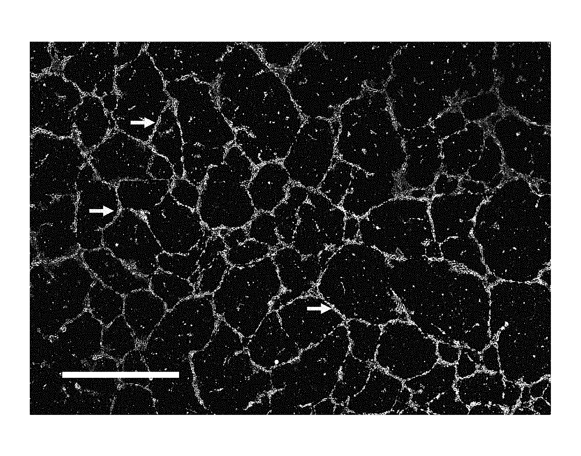
FIG. 1 shows cultured human umbilical vein endothelial cells (HUVECs) after 7 days of culture using the capillary fabrication device. Calcein AM was loaded for fluorescent imaging at 10 .mu.M. White arrows show segments of a viable lumenized capillary network. A typical undifferentiated HUVEC is about 30 .mu.m in, vitro. When integrated into a tubule, a typical HUVEC is about 50 to 60 .mu.m. The lumenized capillary lengths range 100 .mu.m to 1500 .mu.m. ECFCs also formed capillaries when cultured using the capillary fabrication device for at least 4 days (not shown).
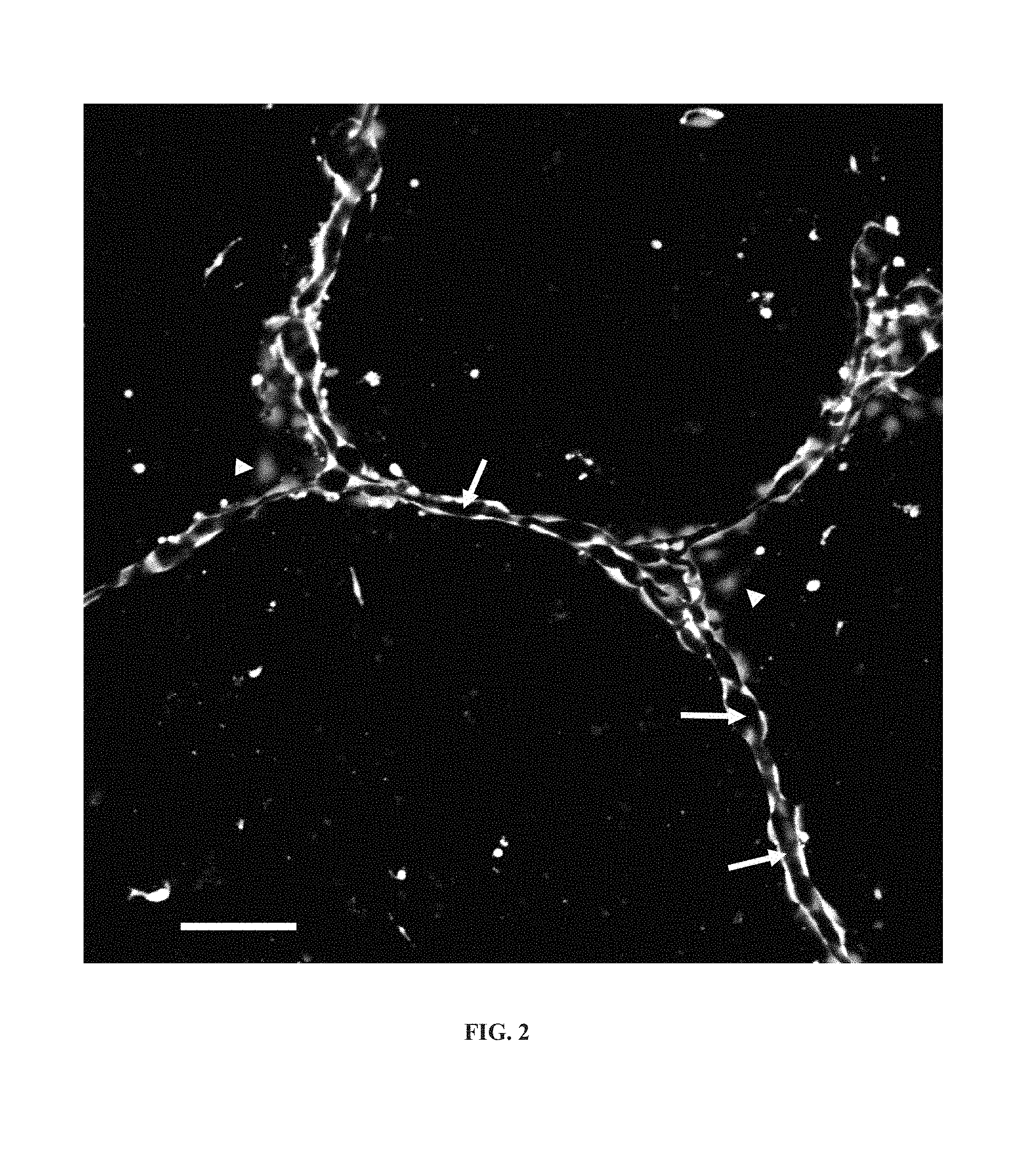
FIG. 2 shows viable lumenized capillary networks formed from HUVECs after 36 hours in the disclosed capillary fabrication device as described herein. They remained viable 7 days later. White arrows show the lumen in the disclosed capillary fabrication device as described herein. The lumenized capillaries have a diameter of 10 to 15 .mu.m. Arrow heads show endothelial cells which have not integrated into the lumenized capillary network. A typical undifferentiated HUVEC is about 30 .mu.m in vitro. When integrated into a tubule, a typical HUVEC is about 50 to 60 .mu.m. Bar, 50 .mu.m.
FIG. 3 shows the formation of support medium. After covering the cell-culture surface with support-generating medium containing dissolved gel-forming material, the support-generating medium forms/deposits a loosely-connected support medium. (A), (B), (C), (D) show phase-contrast images of the support medium at 30 minutes, 3 hours, 6 hours and 12 hours after plating. Support medium is the darker patches of material (black arrow-heads in (A) and (B)). Support medium covers the entire surface after about 6 hours (C). Support medium coverage does not change significantly after 6 hours. (D) The support medium after 12 hours. The gray background is the hydrophobic cell-culture surface. Bar, 20 .mu.m.
FIG. 4 shows components of a standard two-dimensional Matrigel.TM. capillary fabrication device. Matrigel.TM. solidifies at room temperature. Endothelial cells are plated on top of a layer (300-500 .mu.m) of solidified Matrigel.TM.. Cells are covered by about 2 mm of liquid culture medium. A quasi two-dimensional capillary-like network one cell length thick forms on top of the solidified Matrigel.TM..
FIG. 5 shows components of the capillary fabrication device. Support-generating medium which contains a dissolved gel-forming material forms a gel of support medium of about 10 to 30 .mu.m in thickness. Cells are covered by about 2 mm of support-generating medium. Lumenized capillary networks form inside the support medium.
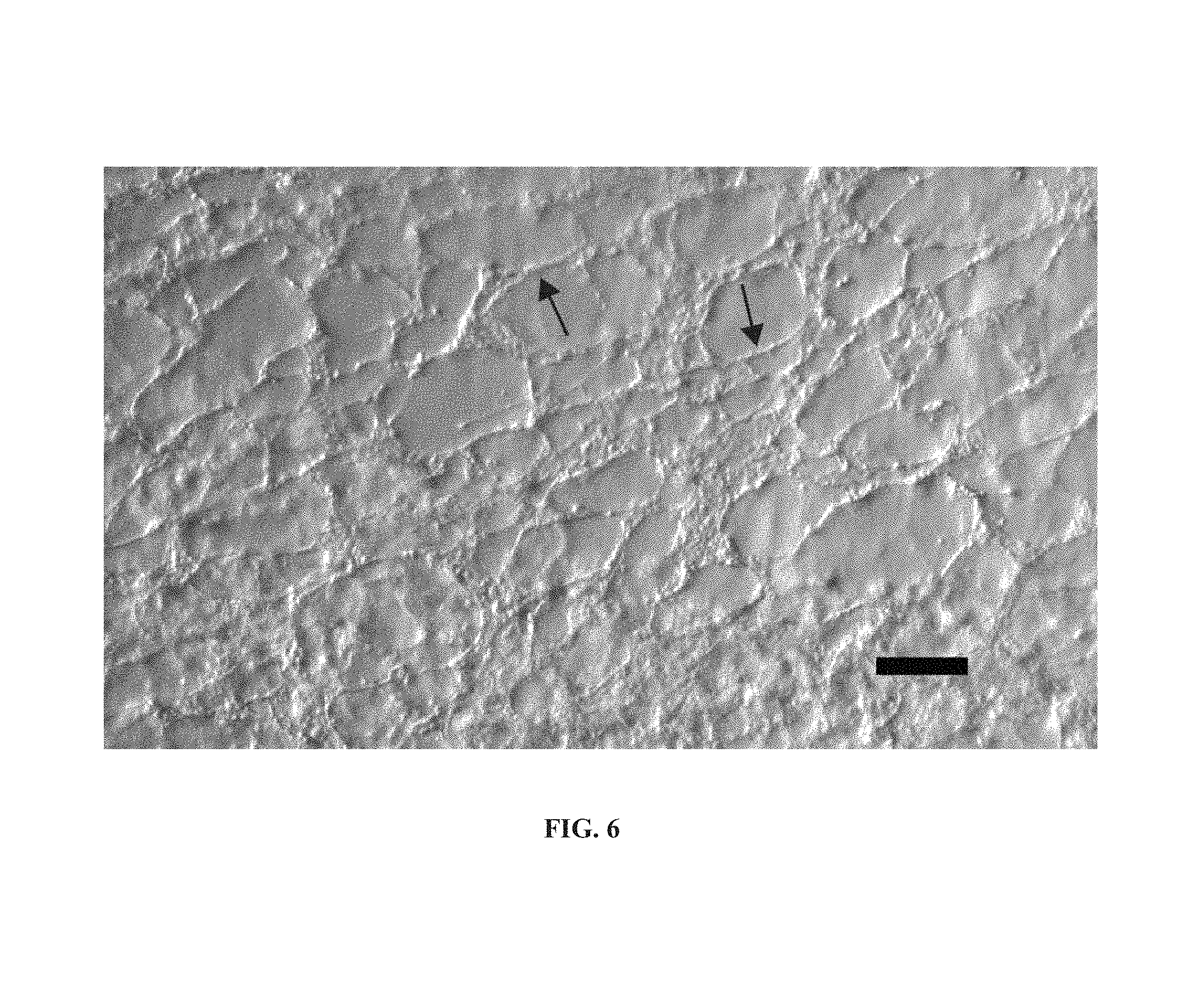
FIG. 6 shows detached ECM generated using a capillary formation device. The ECM was detached from the bottom of a polystyrene dish by making the liquid culture media slightly acidic (pH.about.6.0-6.5). Arrows show manufactured cables. Scale bar.about.250 .mu.m.
FIG. 7 shows the formation of capillaries by HUVECs cultured on a polystyrene dish having regions coated with support medium and regions coated with highly hydrophobic PDMS. HUVECs grew and formed capillary networks on the polystyrene coated with support medium but did not grow and form networks on regions coated with PDMS. The boundary between PDMS regions and polystyrene (with support medium) regions are demarcated by two black lines. The black arrow shows a formed capillary. Scale bar.about.250 .mu.m.
DETAILED DESCRIPTION OF THE ILLUSTRATIVE EMBODIMENTS
For the purposes of promoting an understanding of the principles of the disclosure, reference will now be made to illustrative embodiments. It is understood that no limitation to the scope is intended. It is further understood that any alterations and modifications to the illustrated embodiments and further applications of the principles as may occur to one skilled in the art to which this invention pertains are included within the scope of the disclosure.
As used herein, the phrase "capillary fabrication device" generally refers to a cell-culture which provides an environment for manufacturing capillary-like patterns.
As used herein, the phrase "controlled-geometry capillary fabrication device" or "custom-patterned capillary fabrication device" generally refers to a cell-culture which provides an environment for manufacturing capillary-like networks which have a predefined geometry including tubule lengths, locations and connectivity.
As used herein, the phrase "forming capillary network" generally refers to a group of cells in a capillary fabrication device which are organizing to form one or more tubules.
As used herein, the phrase "formed capillary network" generally refers to a group of cells in a capillary fabrication device which are organized into one or more tubules.
As used herein, the phrase "ECM and basement membrane" generally refers to the extracellular matrix proteins, growth factors, and other components that are associated with the forming or formed capillary networks. The ECM and basement membrane may be produced and/or modified by the cells in the forming or formed capillary network. In general, ECM includes basement membrane which is a laminar organization of extracellular proteins (and associated/other components) including collagen type IV and laminin.
As used herein, the phrase "cell-culture surface" generally refers to a solid which can physically support cells and support medium, and the resulting capillaries.
As used herein, the phrase "non-treated polystyrene" refers to a polystyrene cell culture surface that has not been pre-treated (i.e., prior to contact with the support-generating medium) with an adherence-enhancing substance, corona discharge, gas-plasma, or other like treatments. Likewise, the phrase "cell-culture-treated" refers to cell-culture surfaces providing an adhesive substrate for cell attachment and survival.
As used herein, "patterned surface" refers to a cell-culture surface which has spatially or temporally varying physical or chemical properties including but not limited to hydrophobicity and protein binding affinities. A patterned surface may include glass, biocompatible and biodegradable materials and polymers including but not limited to non-treated polystyrene and temperature sensitive polymers. A patterned surface may include coatings of proteins, polymers, or other materials. A patterned surface may also include scaffolds, meshes and other 2-dimensional or 3-dimensional structures.
As used herein, the phrase "temperature sensitive polymer" refers to a polymer which changes its hydration state depending on temperature. A temperature sensitive polymer includes but is not limited to poly(N-isopropylacrylamide).
As used herein, the phrase "gel-forming material" refers to one or more components that are capable of existing in a gelatinous or high viscosity liquid form. A gel-forming material may include one or more extracellular matrix (ECM) proteins. Extracellular matrix proteins include, but are not limited to, laminin, collagen IV, heparan sulfate proteoglycans, entactin/nidogen, TGF-.beta., epidermal growth factor, insulin-like growth factor, fibroblast growth factor, and tissue plasminogen activator, and the like. A gel-forming material of extracellular matrix proteins includes but is not limited to Matrigel.TM..
As used herein, "Matrigel.TM." refers to a commercially available gelatinous extracellular protein mixture secreted by Engelbreth-Holm-Swarm (EHS) mouse sarcoma cells. Such proteins include laminin, collagen IV, heparan sulfate proteoglycans, entactin/nidogen, TGF-.beta., epidermal growth factor, insulin-like growth factor, fibroblast growth factor, and tissue plasminogen activator, and additional proteins. Stock Matrigel.TM. commercially available currently has about 13 mg/ml of protein. Approximately, more than 80 percent of Matrigel.TM. is ECM proteins. The concentration of ECM proteins is initially about 10.4 mg/ml in Matrigel.TM. (.about.80% of 13 mg). Therefore, Matrigel.TM. stock diluted 1:30 in cell-culture medium (support-generating medium) contains about 347 .mu.g of ECM proteins per ml, and a solution diluted 1:60 contains about 173 .mu.g ECM proteins per ml.
As used herein, "Growth Factor Reduced Matrigel.TM." (GFR Matrigel.TM.) refers to a commercially available gel that is purified and characterized to a greater extent than standard Matrigel.TM.. GFR Matrigel.TM. effectively has lower levels of a variety of growth factors except for TGF-beta which may be bound to collagen IV and/or sequestered in a latent form that partitions with the major components in the purification procedure. The major components: laminin, collagen IV and entactin are conserved while the level of heparan sulfate proteoglycan is reduced by about 40-50%.
As used herein, the phrase "liquid cell-culture medium" generally refers to a liquid capable of growing cells and dissolving the gel-forming material. The liquid cell-culture medium may comprise HEPES, or a bicarbonate-based buffer, or other buffer. Optionally, it may also comprise serum albumin.
As used herein, the phrase "support-generating medium" refers to a mixture of liquid cell-culture medium and a gel-forming material at low density. The density of the gel-forming material in the support-generating medium is low enough that a substantial portion of the support-generating medium remains in liquid form (i.e. dissolved in the liquid cell-culture medium) throughout the culture period (FIG. 5).
As used herein, the phrases "support medium" or "support matrix" refers to a thin soft gel produced spontaneously by the support-generating medium or by the interaction of the support-generating medium with the cultured cells, or by the interaction of the support-generating medium with the cell-culture surface (FIG. 3).
As used herein, the phrase "endothelial lineage" refers to endothelial cells or precursor cells or their offspring that are capable of differentiating into an endothelial cell. Cells of endothelial lineage include, but are not limited to, endothelial progenitor cells, endothelial colony forming cells, vascular stem cells, embryonic and adult stem cells, circulating endothelial progenitor cells, angioblasts, microvasculature, arteries, veins, and lymphatic endothelial cells of different origin including but not limited to dermal, bladder, cardiac, and pulmonary.
As used herein, "viable" capillaries refer to lumenized capillaries comprising living endothelial cells.
Described herein are devices for fabrication of engineered capillary networks. In various embodiments, the devices comprise living cells, a cell-culture surface, a support-generating medium, and a liquid cell-culture medium. In one illustrative aspect, a gel-forming material is substantially dissolved in the liquid cell-culture medium to form a support-generating medium, which then forms a support medium on the cell-culture surface. As used herein, "substantially dissolved" gel-forming substance means that little or no gel-forming material remains in solid form after the gel forming-material is mixed with the liquid cell-culture medium. The support medium is formed by the support-generating medium or by the interaction of the support-generating medium with the cultured cells, or by the interaction of the support-generating medium with the cell-culture surface. The support medium may partially or completely coat the cell-culture surface. The support medium harbors the living cells. The support medium is not rigid. The support medium permits, growth, motility, and differentiation of living cells. The cells may permeate the support medium as they grow, differentiate, and produce their own ECM and basement membrane.
In one embodiment, the capillary fabrication device has a cell-culture surface that is custom-patterned. In one illustrative aspect, the cell-culture surface pattern is a network-like pattern that contains regions of spatially and/or temporally variable hydrophobicities. In one illustrative aspect, the cell-culture surface comprises hydrophobicity gradients. In one illustrative aspect, the cell-culture surface patterns are linear. In one illustrative aspect, the cell-culture surface patterns are nonlinear. In another illustrative aspect, the cell-culture surface comprises both linear and non-linear patterns.
The cells of the capillary fabrication device may be of human or non-human origin. In one embodiment, the cells are of endothelial lineage. In one embodiment, the cells comprise a plurality of cells types of endothelial lineage. In another embodiment, the cells comprise one or more cell types of endothelial lineage and at least one additional cell type not of endothelial lineage. In one aspect, the cells of non-endothelial lineage provide support and/or stabilize the forming or formed capillary networks. In one illustrative aspect, the additional cell type is selected from the group consisting of pericytes, smooth muscle cells, fibroblasts, and any combination thereof.
In one embodiment, the cells of endothelial lineage or non-endothelial lineage are modified. As used herein, the term "modified cells" includes, but is not limited to, genetic modifications, for example, the insertion, deletion, or change in the expression of a gene or genes in a cell. The term "modified cells" also includes cells affected by any chemical, mechanical, electromagnetic or other modifications made to the culture to modify the cells. Modifications of cells may be made continuously or intermittently at any time before culturing the cells in the capillary fabrication device, and/or during and/or after capillary network formation.
In one embodiment, the capillary fabrication device comprises a cell-culture surface having at least one hydrophobic region. In one illustrative embodiment, the hydrophobic regions form a pattern. In one illustrative aspect, the pattern comprises a hexagonal network of narrow bands of less hydrophilic cell-culture surface having a width of from about 5 microns to about 20 microns, from about 5 microns to about 15 microns, from about 5 microns to about 8 microns, from about 5 microns to about 10 microns, from about 8 microns to about 15 microns, from about 10 microns to about 15 microns, from about 10 microns to about 20 microns, or from about 12 microns to about 20 microns; and a length of from about 175 microns to about 300 microns, from about 200 microns to about 300 microns, from about 175 to about 250 microns, from about 200 to about 250 microns, from about 225 to about 300 microns, or from about 225 to about 25 microns. These various ranges of width and length of the bands are also contemplated where the term "about" is not included. The custom-patterned/controlled-geometry capillaries form on top of the predefined hexagonal network pattern of less hydrophilic cell-culture surface.
In one embodiment, the capillary fabrication devices have a cell-culture surface that is modified to change its hydrophobicity in certain pre-defined regions by application of a hydrophobic substance capable of attracting the cells. Illustratively, the hydrophobic substance may be applied by etching, stamping, contact printing, and the like.
In one embodiment, the cell-culture surface is pre-coated with a layer of a temperature sensitive polymer. The temperature sensitive polymer may become significantly dehydrated at about 37.degree. C. and significantly hydrated at temperatures lower than about 32.degree. C. Illustratively, the polymer coating is more hydrophobic in its hydrated state than in its dehydrated state.
In one embodiment, the temperature sensitive coating is applied to the cell-culture surface in a pre-formed pattern. In another illustrative embodiment, the temperature sensitive coating is applied to the cell-culture surface uniformly and then UV laser ablation is used to pattern the coated surface by reducing the thickness of the coating, thereby reducing the hydrophobicity of that portion of the cell culture-surface. In one illustrative aspect, the temperature sensitive polymer is poly(N-isopropylacrylamide).
In one embodiment of the capillary fabrication devices, the cell culture surface is flat. For example, a flat surface may be a petri dish, well-plate, microscope slide, coverslip, and the like. In one embodiment, the cell-culture surface is comprised of a material selected from the group consisting of non-treated polystyrene, glass, a temperature sensitive polymer, and any combination thereof. Optionally, the cell-culture surface may contain meshes or scaffolds to further pattern the capillary networks in two or three dimensions.
The capillary fabrication devices may comprise any suitable liquid medium for cell culture. Several cell-culture media are known to those of skill in the art. In one embodiment, the cell-culture medium is a defined medium. In one illustrative aspect, the cell-culture medium is bicarbonate based. In one illustrative aspect, the cell-culture medium is HEPES. In one embodiment, the cell-culture medium contains one or more serum proteins. In one illustrative aspect, the cell-culture medium contains serum albumin.
The gel-forming material of the capillary fabrication devices is substantially dissolved in the liquid cell-culture medium to form the support-generating medium. Gel-forming materials for use in the capillary fabrication devices are known to those of skill in the art. In one embodiment, the gel-forming material comprises one or more ECM proteins. Extracellular matrix proteins include, but are not limited to, laminin, collagen IV, heparan sulfate proteoglycans, entactin/nidogen, TGF-.beta., epidermal growth factor, insulin-like growth factor, fibroblast growth factor, and tissue plasminogen activator, and the like. In one illustrative embodiment, the gel-forming material of the capillary fabrication devices is Matrigel.TM..
In one embodiment, the support-generating medium comprises Matrigel.TM. diluted from about 20 to about 80 times, from about 20 to about 60 times, from about 20 to about 40 times, from about 30 to about 80 times, from about 30 to about 60 times, from about 30 to about 45 times, or from about 45 to about 60 times in the liquid cell-culture medium. These various dilutions are also contemplated where the word "about" is not included.
In one embodiment, the support-generating medium of the capillary fabrication devices comprises from about 125 .mu.g to about 500 .mu.g, from about 125 .mu.g to about 170 .mu.g, from about 125 .mu.g to about 250 .mu.g, from about 125 .mu.g to about 350 .mu.g, from about 170 .mu.g to about 350 .mu.g, from about 225 .mu.g to about 350 .mu.g, or from about 170 .mu.g to about 225 .mu.g of ECM proteins per ml of liquid cell-culture medium. These various concentrations are also contemplated where the word "about" is not included.
In the capillary fabrication devices described herein, the support-generating medium forms a support medium after contacting the cell-culture surface and/or the living cells. In one embodiment, the living cells permeate the support medium and reside within the support medium soon after cell plating.
In one embodiment, the capillary fabrication devices comprise a support medium that is less than 150 microns thick. In various other embodiments, the support medium has a thickness of from about 10 microns to about 150 microns, about 10 microns to about 125 microns, about 10 microns to about 100 microns, about 10 microns to about 75 microns, about 10 microns to about 60 microns, about 10 microns to about 50 microns, about 10 microns to about 40 microns, about 20 microns to about 75 microns, about 20 microns to about 60 microns, about 20 microns to about 50 microns, about 30 microns to about 75 microns, about 30 microns to about 60 microns, about 30 to about 50 microns, about 40 microns to about 70 microns, or about 40 microns to about 60 microns. In another embodiment, the support medium has a thickness of from about 20 microns to about 40 microns. In another embodiment, the support medium has a thickness of from about 50 microns to about 100 microns. These various thicknesses are also contemplated where the word "about" is not included. It is appreciated that the thickness of the support medium may not be uniform at all portions of the cell-culture surface.
In one embodiment, a formed capillary network is described made using the capillary fabrication devices described herein. In one illustrative aspect, the formed capillary network comprises tubules and is substantially lumenized. In one illustrative embodiment, the capillary network has a pre-patterned geometry. In one illustrative embodiment, the capillary network comprises tubules having a diameter of at least 5 microns. In another illustrative embodiment, the capillary network comprises tubules having a diameter of at least 10 microns. In one illustrative embodiment, the capillary network comprises tubules having a diameter of at least 15 microns. In one illustrative embodiment, the capillary network comprises tubules having a diameter of at least 20 microns.
In various embodiments, the capillary fabrication devices are used to generate forming or formed capillary networks. In one embodiment, the capillary fabrication devices are used to assess the effects of one or more test substances on angiogenesis. In one illustrative embodiment, the plated cells, or the cells of the forming or formed capillary network are contacted with a test substance suspected to promote or inhibit angiogenesis.
In various embodiments, the ECM and basement membrane produced by the living cells of the capillary fabrication device are detached. The ECM and basement membrane may be detached from the forming or formed capillary networks by various methods. In one illustrative embodiment, the ECM and basement membrane are detached from the capillary network by changing the pH of the cell-culture medium, changing the temperature of the cell-culture medium, contacting the capillary networks and/or ECM and basement membrane with a stream of culture medium, or any combination thereof.
The capillary networks and/or the detached ECM and basement membrane may be used in in vitro tissue engineering applications, for example, cell-sheet engineering, bioprinting, and the like.
The capillary networks and/or detached ECM and basement membrane may also be used in in vivo tissue engineering applications, for example, tissue-repair, tissue regeneration, and the like.
EXAMPLES
Example 1: Sample Capillary Fabrication Devices Using Matrigel.TM. in the Support-Generating Medium to Culture HUVECs or ECFCs
Materials.
Matrigel.TM. is purchased from BD Biosciences. Single-donor human umbilical cord endothelial cells (HUVECs) are cultured according to protocols provided by PromoCell.TM.. It is recommended to use HUVECs passaged less than 4 times. Alternatively, endothelial colony forming cells (ECFCs) are used. ECFCs may be extracted from human peripheral and umbilical cord blood.
A stock of 1.times. Matrigel.TM. is diluted about 30 to about 60 times with about 4.degree. C. cell-culture medium (from PromoCell) to produce a homogeneous support-generating medium. The support-generating medium is plated in non-treated polystyrene petri dishes as the cell-culture surface which has a hydrophobic surface. Components of the mixture settle/stick to the bottom of the dish and form a loosely connected low density environment. About 3 ml of the support-generating medium is enough for 35 mm dishes. The support medium thickness formed on the dish is about 20 to about 40 microns or when Matrigel.TM. is diluted about 60 times. The support medium thickness formed on the dish is about 50 to about 100 microns or when Matrigel.TM. is diluted about 30 times. The dishes are incubated for about 30 to about 60 minutes in 5% CO.sub.2 at 37.degree. C. in a cell-culture incubator. The culture dishes containing cultured HUVECs and 35 mm dishes of support-generating medium are transferred from the incubator to the cell-culture hood at the same time to minimize heat shock.
After harvesting the cultured HUVECs, the cells are added to the dishes containing the support-generating medium at about 50 to about 300 cells/mm.sup.2 to complete the capillary fabrication device. The capillary fabrication device is then incubated in 5% CO.sub.2 at 37.degree. C. To produce a fully connected network, cell densities higher than 150 cells/mm.sup.2 are used. To distribute the plated HUVECs uniformly, the dishes are gently swirled alternately clockwise and counterclockwise. The support-generating medium is exchanged about every 48 hours with fresh pre-warmed support-generating medium, prepared as above. If the support-generating medium is removed (for example, after about 60 minutes of incubation at 37.degree. C.), and not replenished with support-generating medium, then the HUVECs do not form lumenized capillary networks. After formation of capillary networks, replacing the support-generating medium with regular liquid cell-culture medium may keep the capillaries viable, but formation of new capillaries from cells not integrated in capillaries, may be significantly reduced.
Optionally, the suspended cells may be mixed with the support-generating medium immediately before cell plating (keeping the dilution ratio of Matrigel.TM. in the support-generating medium between about 1:30 and about 1:60). This procedure eliminates the step of incubating the dishes for about 30 to about 60 minutes and is suited to high-throughput experiments.
Other sources of modified or unmodified cells may be used, including those from human and non-human sources. Endothelial lineage cells may also be used.
Optionally, the cell culture surface may be a flat surface, including but not limited to a well-plate, slide, or coverslip.
A glass culture surface, such as a slide or coverslip, allows for improved high-resolution imaging of the lumenized capillary networks using an inverted or upright microscope for assaying chemical treatments to promote or inhibit vascularization or other purposes.
Optionally, other support/stabilizing cells can be added to the cell culture, including but not limited to pericytes, smooth muscle cells, fibroblasts, myoblasts or any other cell types capable of interacting with or stabilizing the forming or formed lumenized capillary network.
Optionally, modifications to the cells, support-generating medium, or cell-culture surface can be made continuously or intermittently at any time before culturing the cells in the capillary fabrication device, during culturing in the capillary fabrication device and during and after capillary formation and during any subsequent applications. Such modifications are useful in tissue engineering and tissue repair applications which require functional lumenized capillary networks.
Example 2: Sample Custom-Patterned/Controlled-Geometry Capillary Fabrication Devices
In this example, the cell-culture surface in the capillary fabrication device has hydrophobicity gradients. For example, the non-treated polystyrene petri dish in Example 1 can be replaced with a patterned ultra low adhesive polystyrene dish before plating support-generating medium. The ultra low adhesive polystyrene dish provides a cell-culture surface which is more hydrophilic than regular non-treated polystyrene petri dishes. The pattern on the ultra low adhesive polystyrene dish comprises a hexagonal network of narrow bands of 5 to 15 microns in width and 200 to 250 microns in length of less hydrophilic cell-culture surface. Cells of endothelial lineage can be used. The custom-patterned/controlled-geometry capillaries form on top of the predefined hexagonal network pattern of less hydrophilic cell-culture surface.
Optionally, the non-treated polystyrene cell-culture surface in Example 1 can be pre-coated with a layer of temperature sensitive polymers, like poly(N-isopropylacrylamide). The temperature sensitive polymer is dehydrated at 37.degree. C. and hydrated at temperatures lower than 32.degree. C. The polymer coating is more hydrophobic in its hydrated state. The coating hydrophobicity also depends on the thickness of the coating. A coating thickness of more than about 3 nm results in a hydrophobic cell-culture surface at 37.degree. C. The non-treated polystyrene is coated uniformly and then UV laser ablation used to pattern the coated surface by reducing the thickness of the coating according to a customized pattern. The cells in the custom-patterned capillary fabrication device form lumenized capillaries over areas with thinner coating. The cell-culture surface may also be modified by methods including, but not limited to, etching, stamping, and contact printing.
Use of a temperature sensitive polymer in this protocol allows detachment of the formed capillary network and the associated ECM and basement membrane with minimal damage. The cell-culture temperature can be adjusted to facilitate detachment. The detached capillary network and the associated ECM and basement membrane can be used in tissue engineering and tissue repair applications.
The custom-patterned capillary fabrication device is capable of manufacturing capillary networks of specified pattern using cell-culture surfaces with network patterns consisting of narrow bands of 5 to 15 microns in width and specified length and connectivity. The device is capable of producing replicas of 2-dimensional capillary networks like choroidal capillaries in the retina.
Example 3: Sample Capillary Assay Devices
Capillary networks are grown as described in Example 1 using the capillary fabrication device or Example 2 using the custom-patterned capillary fabrication device combined with one or modifications with the aim of determining the effect of those modifications on the cultured cells or on the resulting capillary networks.
The capillary assay device can be used for determining the proangiogenic effects of different isoforms of vascular endothelial growth factor (VEGF) including soluble and ECM-bound isoforms. The cells in the capillary assay device are cultured inside the support medium, thus the device provides quasi-in vivo conditions for the interaction of VEGF isoforms and the cells. Cells in the 2-dimensional standard Matrigel.TM. capillary fabrication device are cultured on top of a thick solidified gel which differs significantly from in vivo conditions.
The capillary assay device also can be used for determining the effects of various toxic materials such as, for example, lead, arsenic, and the like on angionesis. Further, the capillary assay device may be used to study cellular mechanosignaling related to angiogenesis, or the pro/anti angiogenic effects of mechanical stressors such as, for example, ultrasonic waves or altered pressure. Additionally, the capillary assay device may be used to study the pro/anti angiogenic effects of magnetic or electric fields, or electromagnetic waves.
Example 4: Engineered Capillary Networks with Modified Cells
Cells used in this example can be modified before, during or after growth of capillary network using the capillary fabrication device (Example 1) or the custom-patterned capillary fabrication device (Example 2). After a sufficient period of growth, the capillaries are detached in a manner causing minimal or no damage to the capillary network and can be used for in vivo or in vitro implantation.
The capillary networks formed using custom-patterned capillary fabrication device (Example 2) have predefined geometry which makes them suitable for tissue engineering and repair applications with specified geometries and metabolic rates.
In vitro modification of cells used to grow the capillary networks may be useful in minimizing the chance of rejection in allotransplantation or xenotransplantation.
Example 5: Engineered ECM and/or Basement Membrane
Capillary networks are grown as described in Example 1 using the capillary fabrication device or Example 2 using the custom-patterned capillary fabrication device. After a sufficient period of growth, the cells are destroyed, for example, by washing the capillary network with a liquid, for example, distilled water, while retaining the ECM and basement membrane. Then the patterned ECM and basement membrane are detached in a manner causing minimal or no damage to the capillary network, for example, by using a stream of culture media, or by using temperature sensitive polymers. Adjusting the temperature or pH to modify hydrophobicity can facilitate detachment. For example, adjusting the pH of the culture media to slightly acidic (pH.about.6-6.5) is found to facilitate detachment of ECM.
The thickness of the ECM may be controlled by the concentration of the support-generating medium. For example, using diluted Matrigel.TM. as a gel-forming material, the thickness of manufactured ECM is about 20 to 40 microns when Matrigel.TM. is diluted at a ratio of 1:60 and about 50 to 100 microns when diluted at a ratio of 1:30. The ECM has a net-work like morphology. The support matrix is organized in cables that are about 50 to 500 microns long when cultured for 5 days using a 1:30 dilution of Matrigel.TM.. The width of the cables is about 15 to 100 microns. The length of the cables increases over the duration of the culture period. For example, cables about 1.5 mm long are observed after 10 days of culture. At more dilute concentrations (e.g. Matrigel.TM. at 1:60) the cables elongate faster. Thus, the dilution of the gel forming material and duration of culture can be controlled to generate ECM with customized morphology.
The cell-remodeled basement membrane and ECM may then be used to enhance growth/regrowth of vasculature in vivo or in vitro. In vivo implantation can be examined.
After in vivo implantation of the cultured ECM and/or basement membrane, regrowth of capillaries and pericyte recruitment occurs as cells invade the empty sleeves of basement membrane and/or ECM left behind by the destroyed capillaries. Since extracellular components are highly conserved, this organically-manufactured basement membrane/ECM is useful for medical tissue-repair applications with minimal transfer of external factors and thus significant reduction of the likelihood of rejection after implant.
The engineered ECM and basement membrane using custom-patterned capillary fabrication devices (Example 2) have predefined geometry which makes them suitable for growth/regrowth of vasculature in tissue engineering and tissue repair applications with specified geometries and metabolic rates.
Example 6: Use of Engineered ECM and Basement Membrane in Cell-Sheet Engineering and Bioprinting
In this example quasi-two-dimensional layers of engineered ECM and basement membrane (Example 5) are placed in horizontal layers separated by 100 to 200 micron layers of engineered tissue in cell-sheet engineering and bioprinting applications. The resulting enhanced growth of capillary networks in cell-sheet engineering and bioprinting applications results in a functional vascular network to provide blood flow to all volumes of the engineered tissue, enhancing the likelihood of integration of engineered 3-dimensional tissues.
Example 7: Use of Implantable Capillary Networks in Cell-Sheet Engineering and Bioprinting
In this example quasi-two-dimensional layers of formed or forming capillary networks and associated ECM and basement membrane (Example 4) formed using the capillary fabrication device or custom-patterned fabrication device are placed in horizontal layers separated by 100 to 200 micron layers of engineered tissue in cell-sheet engineering and bioprinting applications. Integration of the capillary networks with existing or engineered larger vessels in these applications results in a functional vascular network to provide blood flow to all volumes of the engineered tissue, enhancing the likelihood of integration of engineered 3-dimensional tissues.
Example 8: Formation and Patterning of Hydrophobic Domains on the Cell Culture Surface
Highly hydrophobic domains in the culture surface coating are generated with polydimethylsiloxane (PDMS) which the cells do not populate. Moderately hydrophobic domains, which cells invade and populate, are generated with long carbon chain silanes, for example trichloro(octyl)silane, trichloro(decyl)silane, trichloro(dodecyl)silane, trichloro(octadecyl)silane, trimethoxy(octyl)silane, trimethoxy(decyl)silane, trimethoxy(dodecyl)silane, trimethoxy(octadecyl)silane, triethoxy(octyl)silane, triethoxy(decyl)silane, triethoxy(dodecyl)silane, or triethoxy(octadecyl)silane. Photoresist masking may be used to generate patterns of moderately hydrophobic and highly hydrophobic domains on the cell culture surface.
For example, a clean glass slide is functionalized with one or more long carbon silanes (moderately hydrophobic) to create a carbon chain monolayer. The slide is exposed in a 0.2-2% silane in organic solvents (e.g. methanol, acetone, toluene, heptane, carbon tetrachloride, xylene, and the like) followed by a methanol rinse and then dried under nitrogen gas. The surface is then spin-coated with a thin, approximately 5-10 .mu.m layer of positive photoresist (e.g. SPR220, PMMA, and the like). After softbaking the substrate is exposed to UV light through a mask with a spatial pattern to be replicated by the cells of the functionalized glass substrates. After removing the exposed photoresist a thin layer of PDMS is spin-coated on. The PDMS is then etched by tetrabutylammonium fluoride (TBAF) solution in tetrahydrofuran (THF) until the layer becomes 2-5 .mu.m thick. The substrates are then briefly exposed to UV light without any mask. The photoresist domains masked during the prior exposure are removed, revealing the silane coated glass domains surrounded by PDMS domains.
Example 9: HUVECs do not Invade Highly Hydrophobic PDMS Domains
A capillary fabrication device is utilized to culture HUVECs as described in Example 1. A PDMS coating is applied as described in Example 8 to a polystyrene surface. The HUVECs grow and form capillaries on a polstyrene surface containing a support medium, but do not grow on regions of the surface coated with PDMS (FIG. 7).
Example 10: Implantation of Viable Capillary Networks
Capillary networks formed using the capillary fabrication devices are implanted ex ovo. Chicken eggs are incubated for 3 days at 37.degree. C. On day 3, eggs are broken and embryos are transferred to 10 cm dishes. The embryos are grown for 3-4 more days. On day 6-7, capillary networks formed by HUVECs are implanted in up to four regions of chorioallantoic membrane (CAM). The CAMs are scratched with a sharp needle until a small amount of blood appears. The capillaries are then injected into the CAM using a micro-pipette.
The capillary networks are labeled with Calcein AM to examine their viability. Calcein AM is used as an indicator of viability because it diffuses out of cells when they die. Implanted capillary networks are viewed under microscope after implantation. Viable implanted HUVEC cells are observed 2 days after implantation.
While the invention has been illustrated and described in detail in the foregoing description, such an illustration and description is to be considered as exemplary and not restrictive in character, it being understood that only the illustrative embodiments have been described and that all changes and modifications that come within the scope of the invention are desired to be protected. Those of ordinary skill in the art may readily devise their own implementations that incorporate one or more of the features described herein, and thus fall within the scope of the present invention.
* * * * *
D00000
D00001
D00002
D00003
D00004
D00005
D00006
XML
uspto.report is an independent third-party trademark research tool that is not affiliated, endorsed, or sponsored by the United States Patent and Trademark Office (USPTO) or any other governmental organization. The information provided by uspto.report is based on publicly available data at the time of writing and is intended for informational purposes only.
While we strive to provide accurate and up-to-date information, we do not guarantee the accuracy, completeness, reliability, or suitability of the information displayed on this site. The use of this site is at your own risk. Any reliance you place on such information is therefore strictly at your own risk.
All official trademark data, including owner information, should be verified by visiting the official USPTO website at www.uspto.gov. This site is not intended to replace professional legal advice and should not be used as a substitute for consulting with a legal professional who is knowledgeable about trademark law.