Anti-vascular endothelial growth factor receptor 2 (VEGFR2) antibody and methods of use thereof for detecting VEGFR2 and for inhibiting tumor growth, tumor angiogenesis and/or inducing cancer cell cytotoxicity
Wu , et al. Fe
U.S. patent number 10,196,447 [Application Number 15/563,950] was granted by the patent office on 2019-02-05 for anti-vascular endothelial growth factor receptor 2 (vegfr2) antibody and methods of use thereof for detecting vegfr2 and for inhibiting tumor growth, tumor angiogenesis and/or inducing cancer cell cytotoxicity. This patent grant is currently assigned to Academia Sinica. The grantee listed for this patent is Academia Sinica. Invention is credited to Yu-Ling Chang, Chiung-Yi Chiu, I-Ju Liu, Ruei-Min Lu, Han-Chung Wu.

| United States Patent | 10,196,447 |
| Wu , et al. | February 5, 2019 |
Anti-vascular endothelial growth factor receptor 2 (VEGFR2) antibody and methods of use thereof for detecting VEGFR2 and for inhibiting tumor growth, tumor angiogenesis and/or inducing cancer cell cytotoxicity
Abstract
An isolated antibody or an antigen-binding fragment thereof having a specific binding affinity to an epitope located within the domain 1 or domain 3 of human vascular endothelial growth factor receptor 2 (VEGFR2; SEQ ID NO: 74) is disclosed. The epitope within the domain 3 of the VEGFR2 is located between amino acid residues 250 and 270 of SEQ ID NO: 74. Use of the antibody or antigen-binding fragment thereof in the manufacture of a medicament for inhibiting tumor growth, tumor angiogenesis, and/or inducing cancer cell cytotoxicity in a subject in need thereof is also disclosed. Also disclosed is a method of detecting the presence of VEGFR2 in a tumor vascular endothelial cell or a cancer cell in a biological sample.
| Inventors: | Wu; Han-Chung (Taipei, TW), Lu; Ruei-Min (New Taipei, TW), Chiu; Chiung-Yi (Douliu, TW), Liu; I-Ju (Taipei, TW), Chang; Yu-Ling (New Taipei, TW) | ||||||||||
|---|---|---|---|---|---|---|---|---|---|---|---|
| Applicant: |
|
||||||||||
| Assignee: | Academia Sinica (Taipei,
TW) |
||||||||||
| Family ID: | 57126972 | ||||||||||
| Appl. No.: | 15/563,950 | ||||||||||
| Filed: | April 12, 2016 | ||||||||||
| PCT Filed: | April 12, 2016 | ||||||||||
| PCT No.: | PCT/US2016/027057 | ||||||||||
| 371(c)(1),(2),(4) Date: | October 03, 2017 | ||||||||||
| PCT Pub. No.: | WO2016/168159 | ||||||||||
| PCT Pub. Date: | October 20, 2016 |
Prior Publication Data
| Document Identifier | Publication Date | |
|---|---|---|
| US 20180127504 A1 | May 10, 2018 | |
Related U.S. Patent Documents
| Application Number | Filing Date | Patent Number | Issue Date | ||
|---|---|---|---|---|---|
| 62147344 | Apr 14, 2015 | ||||
| Current U.S. Class: | 1/1 |
| Current CPC Class: | C12N 15/1138 (20130101); G01N 33/57492 (20130101); C07K 16/2863 (20130101); C07K 16/30 (20130101); C07K 16/22 (20130101); A61P 35/02 (20180101); A61K 39/3955 (20130101); A61K 31/337 (20130101); C07K 16/3069 (20130101); A61P 35/00 (20180101); C07K 16/3061 (20130101); A61K 39/3955 (20130101); A61K 2300/00 (20130101); C07K 2317/55 (20130101); C07K 2317/21 (20130101); C12N 2310/14 (20130101); C07K 2317/76 (20130101); C07K 2317/92 (20130101); C07K 2317/622 (20130101); C07K 2317/73 (20130101); C12N 2310/531 (20130101); C07K 2317/56 (20130101); C07K 2317/34 (20130101); C07K 2317/64 (20130101); C07K 2317/567 (20130101); C07K 2317/565 (20130101); C07K 2317/33 (20130101); C07K 2317/732 (20130101); A61K 2039/505 (20130101); C07K 2317/734 (20130101); G01N 2333/71 (20130101) |
| Current International Class: | A61K 39/00 (20060101); A61P 35/02 (20060101); A61P 35/00 (20060101); C07K 16/22 (20060101); C07K 16/28 (20060101); G01N 33/574 (20060101); C12N 15/113 (20100101); C07K 16/30 (20060101); A61K 31/337 (20060101); A61K 39/395 (20060101) |
| 2002/020008 | Sep 2002 | WO | |||
| 2014/055998 | Apr 2014 | WO | |||
Other References
|
Paul, WE. Fundamental Immunology, 3rd ed. Raven Press, NY, Chap. 9, pp. 292-295, 1993. cited by examiner . Rudikoff S. et al. Proc. Natl. Acad. Sci. USA, 79:1979-1983, 1982. cited by examiner . Colman, PM. Research in Immunology, Elsevier, NY, 145(1):33-36, 1994. cited by examiner . International Search Report for PCT/US2016/027057, dated Sep. 19, 2016. cited by applicant . Written Opinion of International Search Authority for PCT/US2016/027057, dated Sep. 19, 2016. cited by applicant . Franklin et al. "The Structural Basis for the Function of Two Anti-VEGF Receptor 2 Antibodies" Structure 19,1097-1107, Aug. 10, 2011. cited by applicant . Lu et al. "Selection of High Affinity Human Neutralizing Antibodies to 3 VEGFR2 From a Large Antibody Phage Display Library for Antiangiogenesis Therapy" (Int. J. Cancer: 97, 393-399 (2002)). cited by applicant. |
Primary Examiner: Landsman; Robert S
Attorney, Agent or Firm: Saunders; Hsiu-Ming Intellectual Property Connections, Inc.
Parent Case Text
REFERENCE TO RELATED APPLICATION
This application is a national stage application (under 35 U.S.C. 371) of PCT/US2016/027057 filed on 12 Apr. 2016, which claims priority to U.S. provisional application 62/147,344 filed on 14 Apr. 2015, all of which are herein incorporated by reference in their entireities.
Claims
What is claimed is:
1. An isolated antibody or an antigen-binding fragment thereof, which comprises a heavy chain variable region (V.sub.H) and a light chain variable region (V.sub.L), the V.sub.H comprising V.sub.H CDR1, V.sub.H CDR2, and V.sub.H CDR3, and the V.sub.L comprising V.sub.L CDR1, V.sub.L CDR2, and V.sub.L CDR3, wherein: the V.sub.H CDR1, V.sub.H CDR2, V.sub.H CDR3 comprise the amino acid sequence of SEQ NO: 6, SEQ ID NO: 7, and SEQ ID NO: 8, respectively; and the V.sub.L CDR1, V.sub.L CDR2, and V.sub.L CDR3 comprise the amino acid sequence of SEQ ID NO: 9, Asp Ala Ser, and SEQ ID NO: 10 or 73, respectively.
2. The antibody or antigen-binding fragment thereof of claim 1, wherein: the V.sub.L CDR3 comprises the amino acid sequence of SEQ ID NO: 73.
3. The antibody or antigen-binding fragment thereof of claim 1, wherein the V.sub.H comprises the amino acid sequence of SEQ ID NO: 76; and the V.sub.L comprises the amino acid sequence of SEQ ID NO: 77 or 78.
4. The antibody or antigen-binding fragment thereof of claim 1, which is a single-chain variable fragment, a Fab fragment or a Fv fragment.
5. The antibody or antigen-binding fragment thereof of claim 1, wherein the antibody is a fully human antibody.
6. A composition comprising a therapeutically effective amount of the antibody or antigen-binding fragment thereof of claim 5 and a pharmaceutically acceptable vehicle or carrier.
7. The composition of claim 6, further comprising a therapeutically effective amount of a chemotherapeutic agent.
8. The antibody or antigen-binding fragment thereof of claim 3, wherein the antibody is a fully human antibody.
9. The antibody or antigen-binding fragment thereof of claim 4, wherein the antibody is a fully human antibody.
10. The antibody or antigen-binding fragment thereof of claim 2, wherein the antibody is a fully human antibody.
11. An isolated antibody or an antigen-binding fragment thereof, or a single-chain variable fragment, which comprises: (a) a heavy chain variable region (V.sub.H) comprising the amino acid sequence of SEQ ID NO: 76; and (b) a light chain variable region (V.sub.L) comprising the amino acid sequence of SEQ ID NO: 77 or 78.
12. A method of detecting the presence of VEGFR2 on tumor vascular endothelial cells or cancer cells in a biological sample, comprising: (i) admixing the antibody or antigen-binding fragment thereof of claim 1 with the biological sample; (ii) allowing the antibody or antigen-binding fragment thereof and the VEGFR2 on the tumor vascular endothelial cells or cancer cells in the biological sample to interact and form a complex; and (iii) detecting the presence of the VEGFR2 on the tumor vascular endothelial cells or cancer cells in the complex.
13. A method of detecting the presence of VEGFR2 on tumor vascular endothelial cells or cancer cells in a biological sample, comprising: (i) admixing the antibody or antigen-binding fragment thereof of claim 2 with the biological sample; (ii) allowing the antibody or antigen-binding fragment thereof and the VEGFR2 on the tumor vascular endothelia cells or cancer cells in the biological sample to interact and from a complex; and (iii) detecting the presence of the VEGFR2 on the tumor vascular endothelial cells or cancer cells in the complex.
14. A method for inhibiting tumor growth, tumor angiogenesis, and/or inducing cytotoxicity in cancer cells, comprising: administering to a subject in need thereof a composition comprising a therapeutically effective amount of the isolated antibody or antigen-binding fragment thereof of claim 1 and a pharmaceutically acceptable carrier, wherein the tumor and/or cancer cell expresses VEGFR2.
15. A method for inhibiting tumor growth, tumor angiogenesis, and/or inducing cancer cell cytotoxicity in a subject in need thereof, comprising: administering to the subject in need thereof a therapeutically effective amount of the antibody or antigen-binding fragment thereof of claim 2, wherein the antibody is a fully human antibody.
16. The method of claim 15, wherein the tumor or cancer is at least one selected from the group consisting of pancreatic, breast, lung, leukemia, prostate and ovary cancer.
17. A method for inhibiting tumor growth, tumor angiogenesis, and/or inducing cancer cell cytotoxicity in a subject in need thereof, comprising: administering to the subject in need thereof a therapeutically effective amount of the antibody or antigen-binding fragment thereof of claim 5.
18. The method of claim 17, wherein the tumor or cancer is at least one selected from the group consisting of pancreatic, breast, lung, leukemia, prostate and ovary cancer.
19. A method for inhibiting tumor growth, tumor angiogenesis, and/or inducing cancer cell cytotoxicity in a subject in need thereof, comprising: administering to the subject in need thereof a therapeutically effective amount of the antibody or antigen-binding fragment thereof of claim 8.
20. The method of claim 19, wherein the tumor or cancer is at least one selected from the group consisting of pancreatic, breast, lung, leukemia, prostate and ovary cancer.
Description
FIELD OF THE INVENTION
The present invention relates generally to antibodies with anti-cancer activities, and more specifically to anti-VEGFR2 antibodies.
BACKGROUND OF THE INVENTION
Angiogenesis rarely occurs in adult healthy tissues. Vascular endothelial growth factor receptor 2 (VEGFR2) is expressed infrequently and at low levels in normal endothelial cells, as compared to tumor-associated endothelial cells. VEGFR2 expression is 3- to 5-folds higher in tumor vessels than that in normal vessels. Immunohistochemistry in biopsies of cancer patients further confirmed that VEGFR2 expression is significantly elevated in tumor vessels when compared with the vascular endothelium in normal tissues adjacent to the tumor region. Notably, expression of VEGFR2 is greater in high-metastatic tumor vessels than in low-metastatic tumor vessels.
VEGFR2 expression was originally shown to be restricted to the vessels of tumor tissues. However, recent studies have provided evidence that VEGFR2 is also present in malignant tumor cells. Circulating tumor epithelial cells in the blood of breast cancer patients were found to express VEGFR2, and thus such expression is associated with tumor metastasis and prognosis. Therefore, blocking VEGFR2-mediated signaling transduction to concomitantly inhibit tumor endothelial and malignant cells is considered an excellent strategy for the development of anticancer therapeutics.
The results from clinical studies indicate that fully human therapeutic antibodies against VEGFR2 are safe and well-tolerated. They show promise as an emerging therapy for cancer by blocking tumor angiogenesis. Therefore, development of a novel anti-VEGFR2 human antibody with enhanced therapeutic efficacy will benefit cancer patients.
SUMMARY OF THE INVENTION
In one aspect, the invention relates to an isolated antibody or an antigen-binding fragment thereof that has a specific binding affinity to an epitope located within the domain 1 or domain 3 of human vascular endothelial growth factor receptor 2 (VEGFR2; SEQ ID NO: 74), wherein the epitope within the domain 3 of the VEGFR2 is located between amino acid residues 250 and 270 of SEQ ID NO: 74.
In one embodiment of the invention, the epitope comprises the amino acid sequence of NWEYPS (SEQ ID NO: 66). The epitope does not comprise the amino acid sequence of GID, KH, or GLMTK (SEQ ID NO: 75).
In another embodiment of the invention, the antibody or antigen-binding fragment thereof of the invention exhibits a specific binding affinity to tumor vascular endothelial cells.
The antibody or antigen-binding fragment thereof of the invention comprises a heavy chain variable region (V.sub.H) and a light chain variable region (V.sub.L), the V.sub.H, comprising V.sub.H CDR1, V.sub.H CDR2, and V.sub.H CDR3, and the V.sub.L comprising V.sub.L CDR1, V.sub.L CDR2, and V.sub.L CDR3, (i) wherein: the V.sub.H CDR1, V.sub.H CDR2, and V.sub.H CDR3 comprise the amino acid sequence of SEQ ID NO: 1, SEQ ID NO: 2, and SEQ ID NO: 3, respectively; and the V.sub.L CDR1, V.sub.L CDR2, and V.sub.L CDR3 comprise the amino acid sequence of SEQ ID NO: 4, Ala Ala Ser, and SEQ ID NO: 5, respectively; or (ii) wherein: the V.sub.H CDR1, V.sub.H CDR2, V.sub.H CDR3 comprise the amino acid sequence of SEQ ID NO: 6, SEQ ID NO: 7, and SEQ ID NO: 8, respectively; and the V.sub.L CDR1, V.sub.L CDR2, and V.sub.L CDR3 comprise the amino acid sequence of SEQ ID NO: 9, Asp Ala Ser, and SEQ ID NO: 10 or 73, respectively; or (iii) wherein: the V.sub.H CDR1, V.sub.H CDR2, and V.sub.H CDR3 comprise the amino acid sequence of SEQ ID NO: 11, SEQ ID NO: 12, and SEQ ID NO: 13, respectively; and the V.sub.L CDR1, V.sub.L CDR2, and V.sub.L CDR3 comprise the amino acid sequence of SEQ ID NO: 14, Asp Ala Ser, and SEQ ID NO: 15, respectively;
or (iv) wherein:
the V.sub.H CDR1, V.sub.H CDR2, and V.sub.H CDR3 comprise the amino acid sequence of SEQ ID) NO: 16, SEQ ID NO: 17, and SEQ ID NO: 18, respectively; and the V.sub.L CDR1, V.sub.L CDR2, and V.sub.L CDR3 comprise the amino acid sequence of SEQ ID NO: 19, Gly Ala Ser, and SEQ ID NO: 20, respectively;
or (v) wherein:
the V.sub.H CDR1, V.sub.H CDR2, and V.sub.H CDR3 comprise the amino acid sequence of SEQ ID NO: 21, SEQ ID NO: 22, and SEQ ID NO: 23, respectively; and the V.sub.L CDR1, V.sub.L CDR2, and V.sub.L CDR3 comprise the amino acid sequence of SEQ ID NO: 24, Asp Ala Ser, and SEQ ID NO: 25, respectively.
In another embodiment of the invention, the V.sub.H CDR1, V.sub.H CDR2, and V.sub.H CDR3 comprise the amino acid sequence of SEQ ID NO: 6, SEQ ID NO: 7, and SEQ ID NO: 8, respectively; and the V.sub.L CDR1, V.sub.L CDR2, and V.sub.L CDR3 comprise the amino acid sequence of SEQ ID NO: 9, Asp Ala Ser, and SEQ ID NO: 10 or 73, respectively.
In another embodiment of the invention, the antibody or antigen-binding fragment thereof comprises: (a) a heavy chain variable region (V.sub.H) comprising the amino acid sequence of SEQ ID NO: 76; and (b) a light chain variable region (V.sub.L) comprising the amino acid sequence of SEQ ID NO: 77 or 78.
In another embodiment of the invention, the antibody or antigen-binding fragment thereof is a single-chain variable fragment, a Fab fragment, or a Fv fragment.
In another embodiment of the invention, the antibody or antigen-binding fragment thereof is a fully human antibody.
In another embodiment of the invention, the antibody or antigen-binding fragment thereof is labeled with a detectable compound or an enzyme.
In another aspect, the invention relates to a composition comprising a therapeutically effective amount of an antibody or antigen-binding fragment thereof of the invention and a pharmaceutically acceptable vehicle or carrier.
In one embodiment of the invention, the composition further comprises a chemotherapeutic agent. In one embodiment of the invention, the chemotherapeutic agent is docetaxel.
Further in another aspect, the invention relates to use of an antibody or antigen-binding fragment thereof or a composition of the invention in the manufacture of a medicament for inhibiting tumor growth, tumor angiogenesis, and/or inducing cancer cell cytotoxicity in a subject in need thereof.
In one embodiment of the invention, the tumor and/or cancer cell express VEGFR2. The use of the antibody or antigen-binding fragment thereof may further comprise use of an additional chemotherapeutic agent such as docetaxel in the manufacture of a medicament for inhibiting tumor growth, tumor angiogenesis, and/or inducing cancer cell cytotoxicity in the subject in need thereof.
Alternatively, the invention relates to an antibody or antigen-binding fragment thereof or a composition of the invention for use in inhibiting tumor growth, tumor angiogenesis, and/or inducing cancer cell cytotoxicity in a subject in need thereof.
The invention also relates to a method for inhibiting tumor growth, tumor angiogenesis, and/or inducing cancer cell cytotoxicity, comprising:
administering to a subject in need thereof a composition comprising a therapeutically effective amount of an isolated antibody or antigen-binding fragment thereof of the invention and a pharmaceutically acceptable carrier, and thereby inhibiting tumor growth, tumor angiogenesis, and/or inducing cancer cell cytotoxicity in the subject in need thereof.
The method of the invention may further comprise administering to the subject in need thereof a composition comprising a therapeutically effective amount of a chemotherapeutic agent such as docetaxel. In one embodiment of the invention, the chemotherapeutic agent is simultaneously administered to the subject in need thereof.
In one embodiment of the invention, the tumor or cancer is at least one selected from the group consisting of pancreatic, breast, lung, leukemia, prostate and ovary cancer.
Yet in another aspect, the invention relates to a method of detecting the presence of VEGFR2 on tumor vascular endothelial cells or cancer cells in a biological sample, comprising: (i) admixing the antibody or antigen-binding fragment thereof of the invention with the biological sample, (ii) allowing the antibody or antigen-binding fragment thereof and the VEGFR2 on the tumor vascular endothelial cells or cancer cells in the biological sample to interact and form a complex; and (iii) detecting the presence of the VEGFR2 on the tumor vascular endothelial cells or cancer cells in the complex.
In one embodiment of the invention, the biological sample is a tissue specimen from a patient.
In another embodiment of the invention, the presence of the VEGFR2 on the tumor vascular endothelial cells or cancer cells in the complex is detected by immunoassay.
These and other aspects will become apparent from the following description of the preferred embodiment taken in conjunction with the following drawings, although variations and modifications therein may be affected without departing from the spirit and scope of the novel concepts of the disclosure.
The accompanying drawings illustrate one or more embodiments of the invention and, together with the written description, serve to explain the principles of the invention. Wherever possible, the same reference numbers are used throughout the drawings to refer to the same or like elements of an embodiment.
BRIEF DESCRIPTION OF THE DRAWINGS
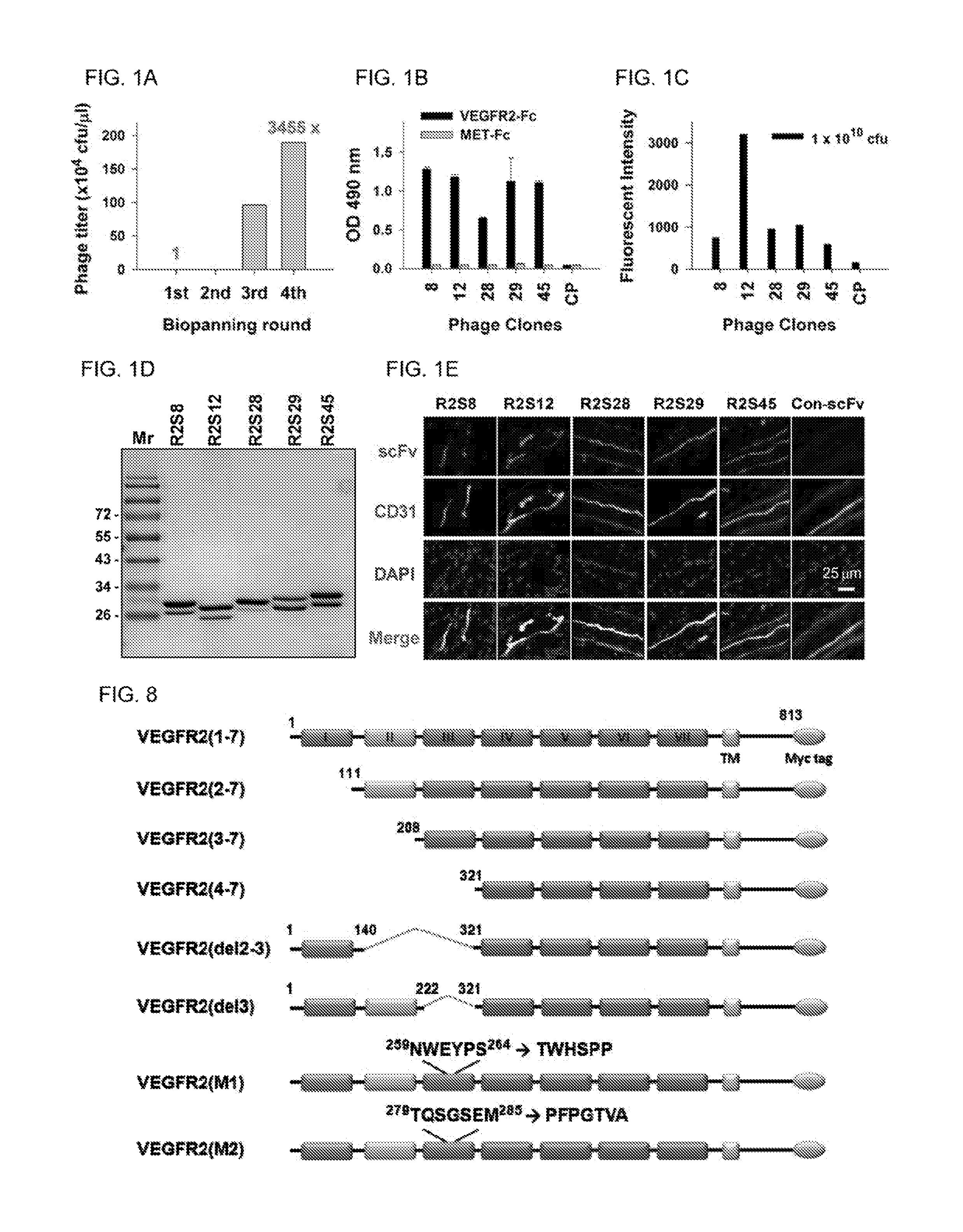
FIGS. 1A-E show the results of selection and identification of phage-displayed scFvs against VEGFR2. (A) Phage display biopanning for VEGFR2-Fc recombinant protein. After four rounds of biopanning, the recovery rate of the phages was increased by 3,455-fold over that of the first round. cfu, colony-forming units. (B) Comparison of the binding of selected phage clones to VEGFR2-Fc protein by ELISA with a 1.times.10.sup.9 cfu phage titer. (C) Cellular VEGFR2 binding affinity of phage clones were evaluated on HUVECs by flow cytometry with 1.times.10.sup.10 cfu.(D) Soluble anti-VEGFR2 scFvs were purified and analyzed by SDS-PAGE with Coormassie blue staining. (E) Immunofluorescent staining for human tumor vasculature, Frozen sections of surgical specimens of lung cancer patients were probed with anti-V.EGFR2 scFvs, followed by anti-E, tag antibody and rhodamine-conjugated secondary antibody staining. Vascular endothelium was stained with anti-human CD31 antibody, and then incubated with FITC-conjugated secondary antibody. Nuclei were stained with DAPI; Con-scFv, control scFv.
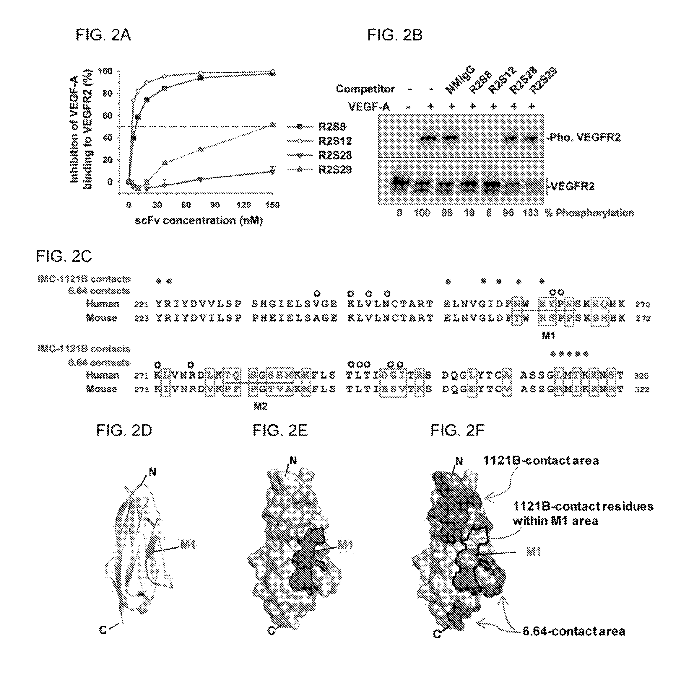
FIGS. 2A-F show that anti-VEGFR2 scFv suppresses VEGF-A binding and activation of VEGFR2 in HUVECs. (A) Analysis of the competition ability of anti-VEGFR2 scFv with VEGF-A by ELISA. The amount of VEGF-A binding to immobilized VEGFR2 in the absence of competitors was considered to be 100%. (B) Phosphorylated VEGFR2 (Pho. VEGFR2) expression in HUVECs treated with VEGF-A and scFv competitors was detected by Westernblot. Quantification of phosphorylated VEGFR2 was based on luminescence intensity, and normalized to total VEGFR2. (C to F) Epitope mapping of R2S12. (C) Sequence alignment of VEGFR2 domain 3 (VEGFR2-D3) of human (from 221 a.a. to 320 a.a. of SEQ ID NO: 74) and mouse (from 223 a.a. to 322 a.a. of SEQ ID NO: 80). Residues that differ between the two species are boxed. The residues involved in mutants M1 and M2 are underlined. The filled and open circles are used to indicate human VEGFR2-D3 residues in contact with 1121B and 6.64 antibodies, respectively. (D) Graphic depicting the VEGFR2-D3 backbone; NWEYPS (SEQ ID NO: 66) residues (M1) responsible for R2S12 binding are highlighted. (E) Model of the surface of VEGFR2-D3. NWEYPS (SEQ ID NO: 66) residues, i.e., the M1 area, is delineated by the black line. (F) The residues that make contact with 1121B and 6.64 on the surface of VEGFR2-D3 are indicated. The contacting residues of 1121B, which are localized in the M1 area, are also indicated. (N, N terminus; C, C terminus.)
FIGS. 3A-D show affinity maturation of anti-VEGFR2 hAb, and analysis of anti-VEGFR2-AF hAb activity. (A) Amino acids of the light chain variable domain of CDR3 (V.sub.L-CDR3) of R2S12 (SEQ ID NO: 10) and R2S12AF (SEQ ID NO: 73). Residues that differ between R2S12 and R2S12AF are boxed. (B) Kinetic constants of anti-VEGFR2 and anti-VEGFR2-AF hAb, as determined using purified IgG and a BIACORE T100.TM.. The K.sub.d value was calculated using BIACORE T100.TM. evaluation software. (C) Competitive ELISA was performed to examine dose-dependent inhibition of VEGF-A binding to VEGFR2 by human antibody. A value of 100% was attributed to the binding of 4 nM VEGF-A to immobilized VEGFR2 in the absence of competitors. Error bar, SD; n=4. (D) Determination of the binding activity of anti-VEGFR2 antibody to HUVEC by flow cytometry analysis. Antibody concentration: 0.1 .mu.g/ml.
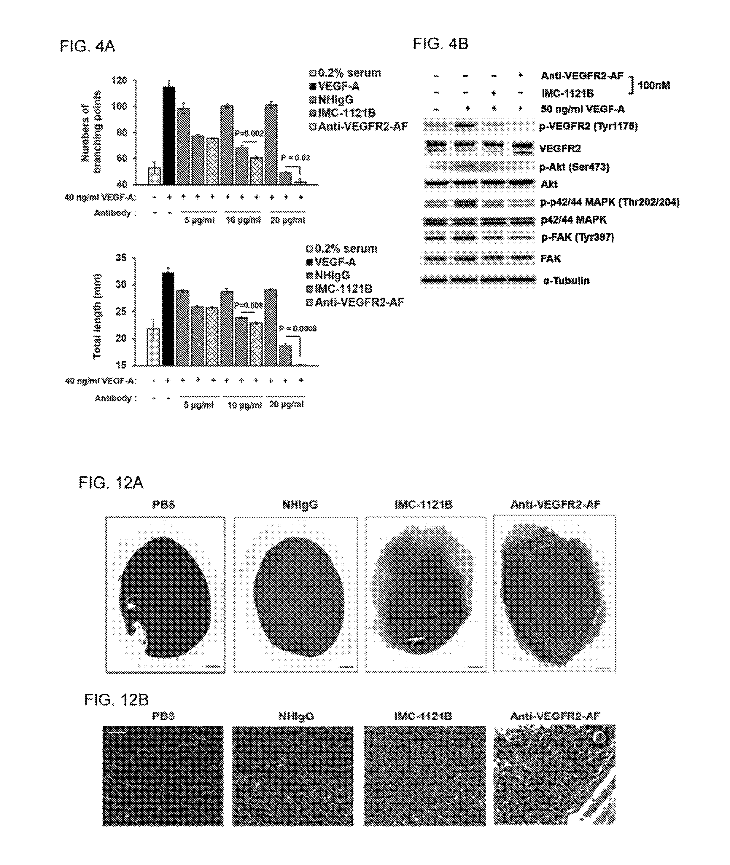
FIGS. 4A-B show that anti-VEGFR2-AF hAb inhibits the VEGFR2 signaling pathway and disrupts capillary structure formation in HUVECs. (A) Capillary structure formation assays were performed using MATRIGEL.RTM.-coated .mu.-Slides. HUVECs (4.times.10.sup.4 cells per well) were incubated with 0.2% FBS and treated with 40 ng/ml VEGF-A, or 40 ng/ml VEGF-A together with NHIgG. IMC-1121B, or anti-VEGFR2-AF antibody for 5 hours at 37.degree. C. Tubular structures were observed under phase contrast, and the relative sprout length (lower panel) and branching points (upper panel) were quantitatively measured with ImageJ software. All data were obtained from three independent experiments. (B) HUVECs were treated with 50 ng VEGF-A or 100 nM anti-VEGFR2-AF or IMC-1121B for 10 min at 37.degree. C. Total protein was prepared from treated HUVECs and examined by Western blot analysis. .alpha.-tubulin was used as a loading control.
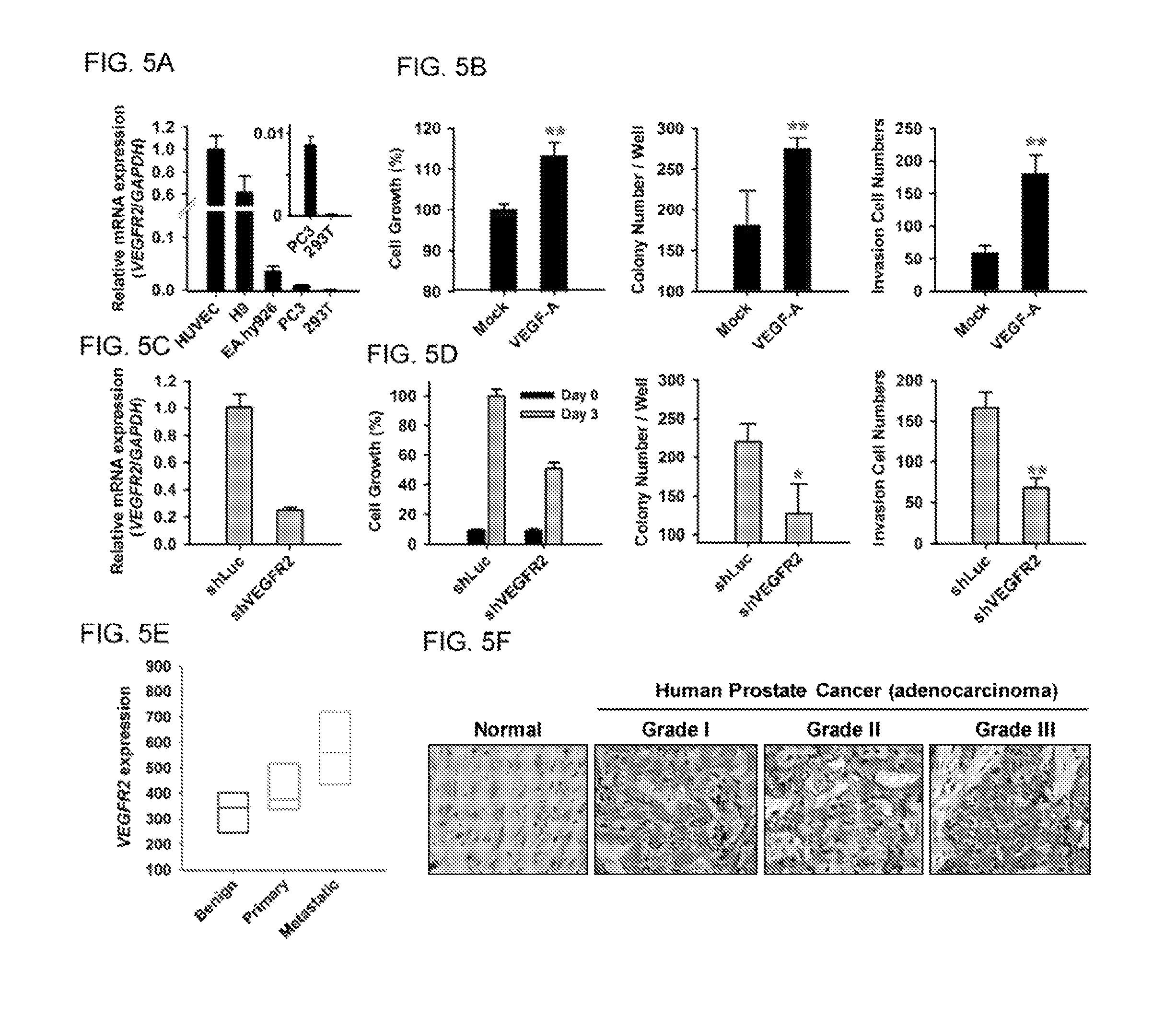
FIGS. 5A-F show characterization of VEGFR2 activity in human prostate cancer cells. (A) Analysis of VEGFR2 expression in the indicated cell lines by quantitative RT-PCR. 293T cells were used as a negative control. Expression of VEGFR2 was normalized to that of GAPDH. (B) PC-3 cells treated with VEGF-A were subjected to colony formation, MTT, and invasion assays. n=6 in each group. (C) PC-3 cells were treated with VEGFR2-targeted shRNA (shVEGFR2), and VEGFR2 expression was analyzed by quantitative RT-PCR. Luciferase shRNA (shLuc) was used as a negative control. (D) MTT, colony formation, and Transwell invasion assays were performed to analyze shVEGFR2-PC-3 cells treated with VEGF-A. (E) Box plots showing relative VEGFR2 expression in metastatic prostate tumors as compared with benign and primary tumors, as determined using a public microarray database. (F) Immunohistochemical staining of a human prostate cancer tissue array (PRC481, Pantomics) using anti-VEGFR2 antibody (55B11, Cell Signaling) to analyze VEGFR2 protein expression in human normal and tumor prostate tissues.
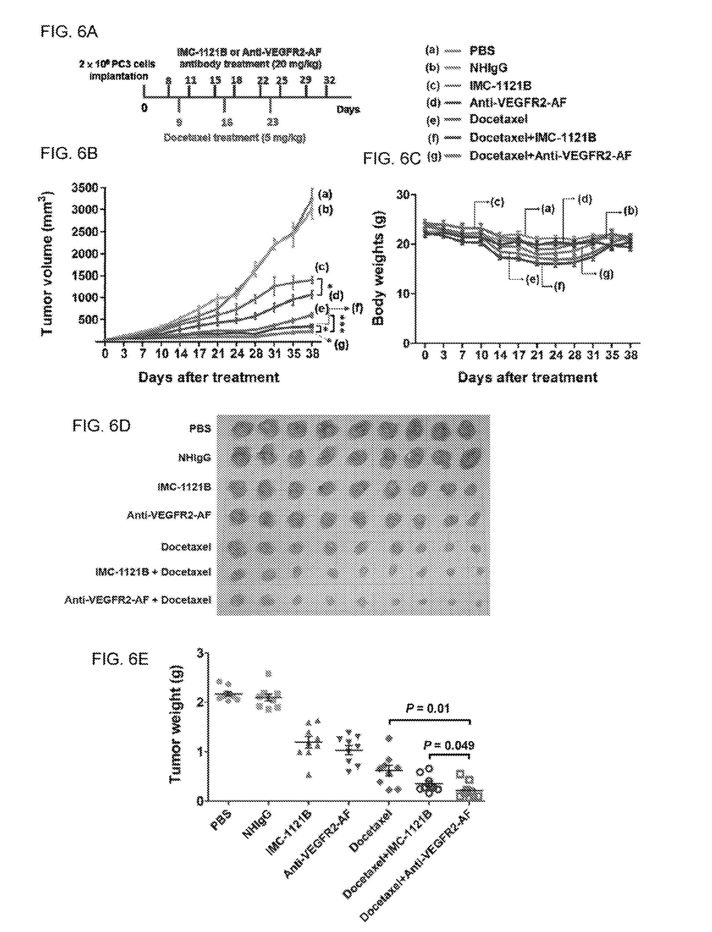
FIGS. 6A-E show analysis of the therapeutic efficacy of anti-VEGFR2-AF hAb in a PC-3 mouse xenograft model. (A) The treatment schedule. (B) The tumor growth profiles of mice of each group. (C) Body weight of each group. (D) At the end of the treatment period, tumor mass was dissected from each mouse. (E) Tumor weight was measured at the end of the treatment period. All data are shown as the mean of nine mice per group; bars, SE; *, P<0.05.
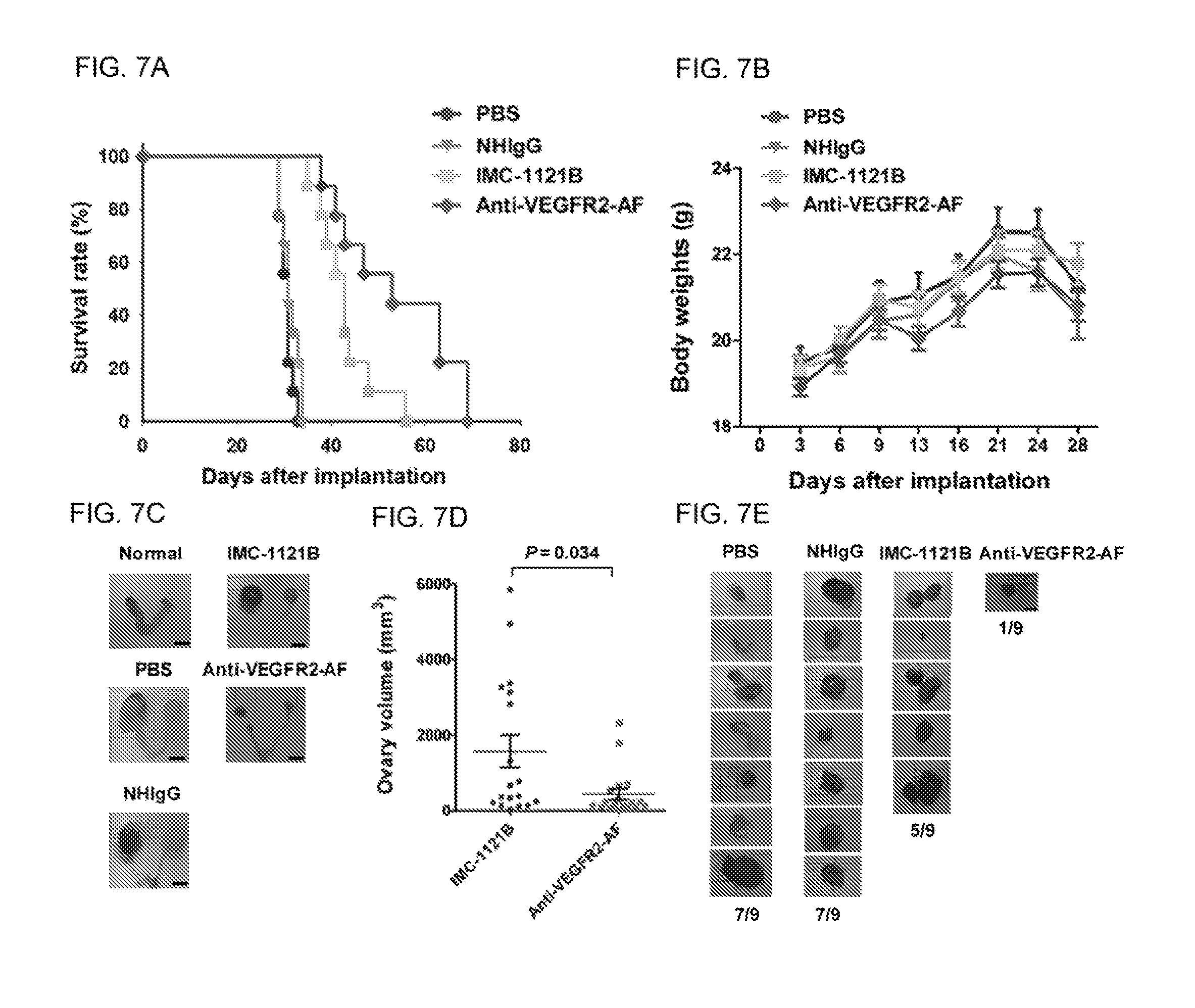
FIGS. 7A-E show that anti-VEGFR2-AF hAb exhibits greater antitumor activity than IMC-1121B in a HL60 mouse xenograft model. (A) Kaplan-Meier survival analysis of mice of each group. Survival was significantly prolonged in the anti-VEGFR2-AF hAb group compared with the IMC-1121B group based on log-rank test (P=0.0284). (B) Body weight of mice of each group. (C) Ovaries were dissected from each mouse after death. The ovaries from a NSG mouse without leukemia were used as a normal control. (D) Ovary volume in mice treated with IMC-1121B or anti-VEGFR2-AF. (E) Morphometric analysis of lymph node (LN) changes in leukemia-tumor bearing mice. Leukemia cells that had metastasized to lymph nodes were dissected from mice of the indicated groups (n=9 for each group).
FIG. 8 shows construction of VEGFR2-expressing vectors. Schematic presentation of constructs expressing deletion or substitution mutants of human VEGFR2 domains. There are seven immunogiobulin-like domains in the extracellular region of VEGFR2, which are labeled I to VII. TM: transmembrane domain. The sequence identifiers of .sup.259NWEYPS.sup.264, .sup.261TWHSPP.sup.266, .sup.279TQSGSEM.sup.285, and .sup.281PFPGTVA.sup.287 are SEQ ID NO: 66, SEQ ID NO: 68, SEQ ID NO: 67, and SEQ ID NO: 69, respectively.
FIGS. 9A-B show the results of selection and identification of scFvs against VEGFR2 using a phage-displayed synthetic scFv library. (A) Affinity maturation for R2S12. The phage-displayed R2S12-V.sub.L-CDR3 mutagenic scFv library was incubated for 1 hour at 4.degree. C. with 0.1 .mu.g VEGFR2-Fc immobilized on Protein G DYNABEADS.RTM.. Subsequently, the beads were washed four times with PBS containing 1% TWEEN.TM. 20. After four rounds of biopanning, the recovery rate of the phages was increased by 929-fold over that of the first round. (B) Comparison of the binding activity of the indicated concentrations of R2S12 and R2S12-AF scFv to VEGFR2-Fc protein, as assayed by ELISA. Error bar, SE.
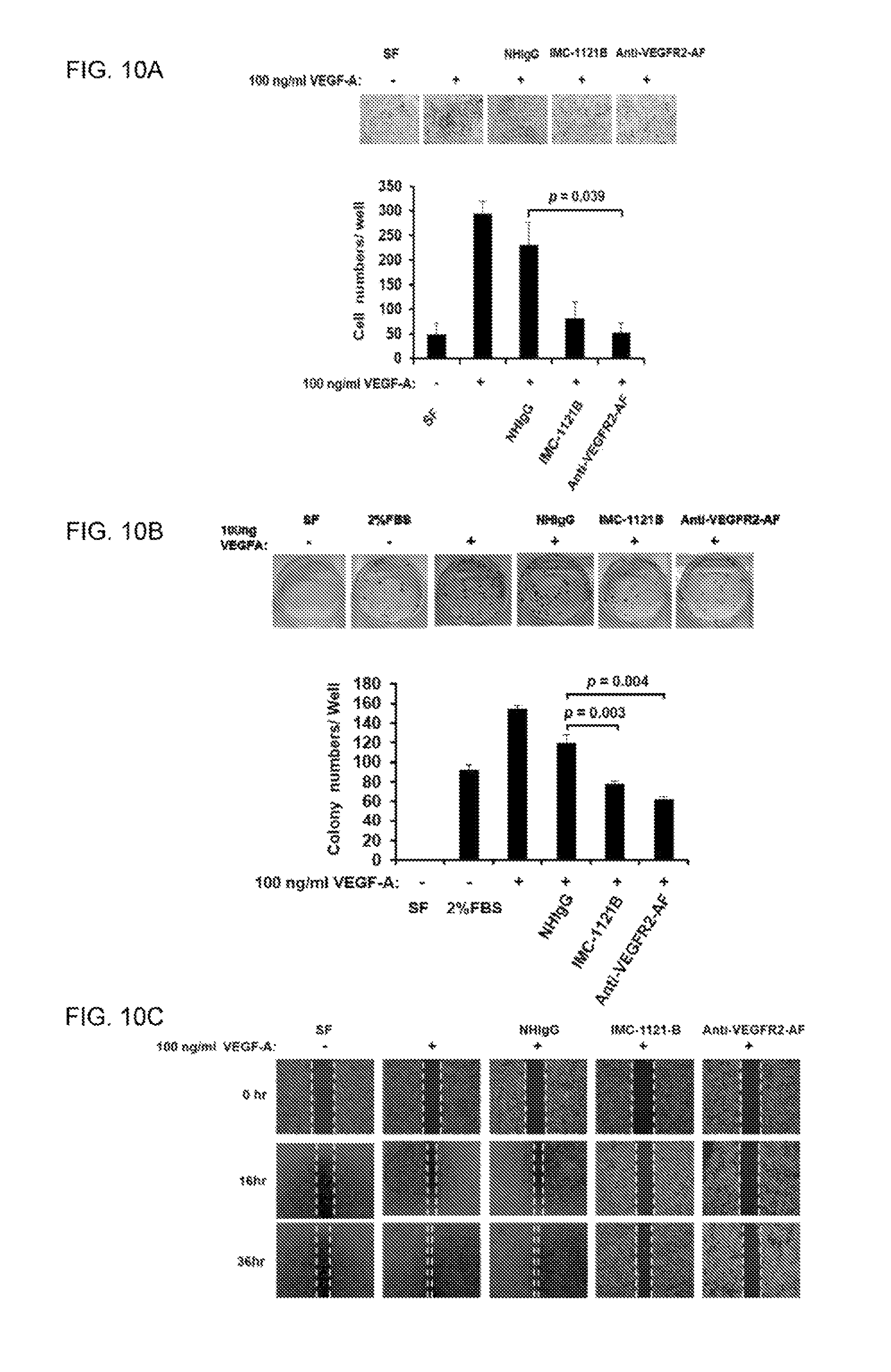
FIGS. 10A-C show that anti-VEGFR2-AF hAb antagonized VEGF-A mediated cellular activity in PC-3 cells. (A) Transwell assays were carried out to examine the invasion capacity of PC-3 cells subjected to the indicated treatments. Upper panel: Giemsa staining of invasive cells. 100.times. magnification; n=3 in each group; scale bar, 150 .mu.m. (B) A total of 1.times.10 PC-3 cells were seeded in a six-well plate, and treated with or without 100 ng/ml VEGF-A and 10 .mu.g/ml of NHIgG IMC-1121B. or anti-VEGFR2-AF antibody. The plate was incubated for 7 days to allow colony formation. Cell colonies were visualized by crystal violet staining. The relative number of colonies was calculated in each well after elution of crystal violet solution. n=3 in each group. (C) Wound healing assay. PC-3 cells were incubated in RPMI with 2% FBS, and stimulated by treatment with 100 ng/ml VEGF-A in the presence or absence of 10 .mu.g/ml NHIgG IMC-1121B, or anti-VEGFR2-AF, individually. Images were taken after 0, 16, and 36 hours of incubation. Scale bar, 150 .mu.m. n=3 in each group. Error bar, SE.
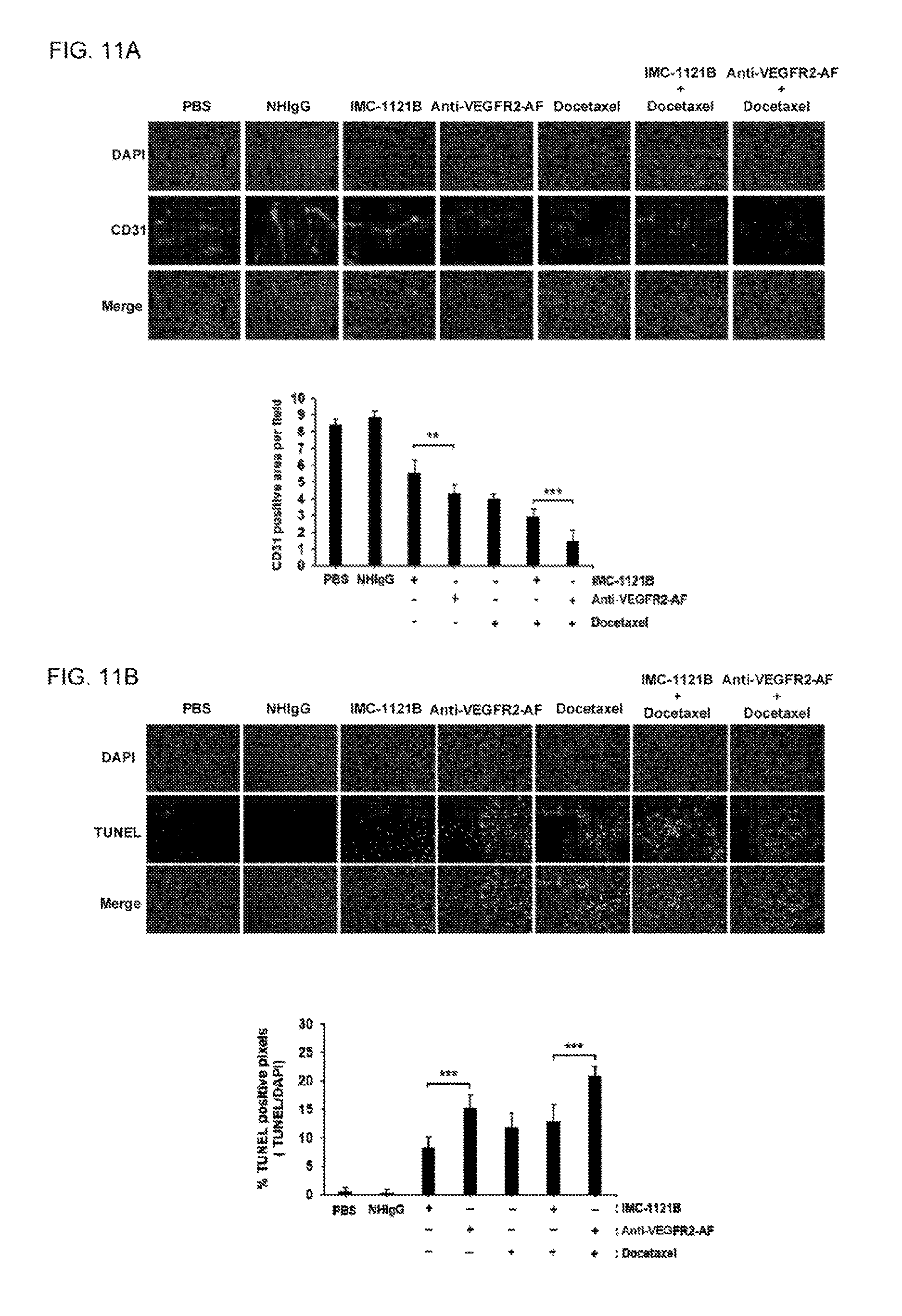
FIGS. 11A-B show investigation of vascular endothelium and apoptotic cells in tumor tissue after drug treatment. Frozen tumor sections were prepared from mice of each group at the end of the treatment period. (A) Sections were stained with anti-CD31 antibody to visualize tumor blood vessels. CD31-positive endothelium was quantitatively measured using ImageJ software. (B) Apoptotic cells in frozen tumor sections were analyzed using TUNEL assay. The apoptotic cells were quantified using ImageJ software. The sections were stained with DAPI for indication of all cells. n=5; Scale bar, 100 .mu.m; 200.times. magnification; Error bar, SE; **, P<0.01; ***, P<0.001.
FIGS. 12A-B show histopathological phenotype of ovaries of human leukemia xenograft mice following antibody treatment. Ovaries were harvested from HL-60 tumor-bearing mice after treatment with saline, NHIgG, IMC-1121B, or anti-VEGFR2-AF hAb. The tissues were sliced and stained with hematoxylin and eosin (H&E), revealing infiltration with leukemia cells. The group treated with Anti-VEGFR2-AF showed fewer blood vessels, and retained some primary oocytes. n=9 in each group. (A) Ovary at 200.times. magnification. Scale bar, 500 .mu.m. (B) Scale bar, 150 .mu.m.
DETAILED DESCRIPTION OF THE INVENTION
Various embodiments of the invention are now described in detail. Referring to the drawings, like numbers indicate like components throughout the views. As used in the description herein and throughout the claims that follow, the meaning of "a", "an", and "the" includes plural reference unless the context clearly dictates otherwise. Also, as used in the description herein and throughout the claims that follow, the meaning of "in" includes "in" and "on" unless the context clearly dictates otherwise. Additionally, some terms used in this specification are more specifically defined below.
Definitions
The terms used in this specification generally have their ordinary meanings in the art, within the context of the invention, and in the specific context where each term is used. Certain terms that are used to describe the invention are discussed below, or elsewhere in the specification, to provide additional guidance to the practitioner regarding the description of the invention. For convenience, certain terms may be highlighted, for example using italics and/or quotation marks. The use of highlighting has no influence on the scope and meaning of a term; the scope and meaning of a term is the same, in the same context, whether or not it is highlighted. It will be appreciated that same thing can be said in more than one way. Consequently, alternative language and synonyms may be used for any one or more of the terms discussed herein, nor is any special significance to be placed upon whether or not a term is elaborated or discussed herein. Synonyms for certain terms are provided. A recital of one or more synonyms does not exclude the use of other synonyms. The use of examples anywhere in this specification including examples of any terms discussed herein is illustrative only, and in no way limits the scope and meaning of the invention or of any exemplified term. Likewise, the invention is not limited to various embodiments given in this specification.
Unless otherwise defined, all technical and scientific terms used herein have the same meaning as commonly understood by one of ordinary skill in the art to which this invention pertains. In the case of conflict, the present document, including definitions will control.
The term "treating" or "treatment" refers to administration of an effective amount of the compound to a subject in need thereof, who has cancer, or a symptom or predisposition toward such a disease, with the purpose of cure, alleviate, relieve, remedy, ameliorate, or prevent the disease, the symptoms of it, or the predisposition towards it. Such a subject can be identified by a health care professional based on results from any suitable diagnostic method.
"An effective amount" refers to the amount of an active compound that is required to confer a therapeutic effect on the treated subject. Effective doses will vary, as recognized by those skilled in the art, depending on rout of administration, excipient usage, and the possibility of co-usage with other therapeutic treatment.
The term "chemotherapeutic agent" refers to a pharmacological agent that is known to be of use in the treatment of cancer.
The "Guidance for Industry and Reviewers Estimating the Safe Starting Dose in Clinical Trials for Therapeutics in Adult Healthy Volunteers" published by the U.S. Department of Health and Human Services Food and Drug Administration discloses a "therapeutically effective amount" may be obtained by calculations from the following formula: HED=animal dose in mg/kg.times.(animal weight in kg/human weight in kg).sup.0.33.
Ramucirumab (i.e., IMC-1121B) trade name is CYRAMZA.RTM..
Human vascular endothelial growth factor receptor 2 (VEGFR2) domain 1 region is from 45 a.a. to 110 a.a., and domain 3 is from 224 a.a. to 320 a.a.
Sequence Identifiers:
QQLDDIPIT (R2S12AF variable light chain CDR3; SEQ ID NO: 73); VGFR2 (HUMAN Vascular endothelial growth factor receptor 2; SEQ ID NO: 74); GLMTKK (SEQ ID NO: 75);
The V.sub.H region of R2S12 and R2S12-AF are the same (SEQ ID NO: 76): QVNLRESGGGLVKPGGSLRLSCAASGFTFGSYTMNWVRQAPGKGLEWVASITSGSSYIFYTDSVKGRFIISRD- NSRSSLFLQMNSLRAEDTAIYYCARGSASAFDIWGQGTMVTVSS;
The V.sub.L region of R2S12 (SEQ ID NO: 77): DIQMTQSPSSLSASVGDRVTITCKASDDIINYLNWYQQKPGEAPKLLIYDASILETGVPSRFSGSGSGTDFTF- TISSLQPEDIATYYCQQYDILPLTFGGGTKLEIK;
The V.sub.L region of R2S12AF (SEQ ID NO: 78): DIQMTQSPSSLSASVGDRVTITCKASDDIINYLNWYQQKPGEAPKLLIYDASILETGVPSRFSGSGSGTDFTF- TISSLQPEDIATYYCQQLDDIPITFGGGTKLEIK; The amino acid residues that are different between R2S12 V.sub.L and R2S12AF V.sub.L are underlined above.
Human IgG1 constant region (SEQ ID NO: 79); Mouse VEGFR2 amino acid sequence (SEQ ID NO: 80).
Abbreviation: complementarity determining regions (CDRs); Fab (fragment, antigen-binding region); F.sub.V region (variable domain).
EXAMPLES
Without intent to limit the scope of the invention, exemplary instruments, apparatus, methods and their related results according to the embodiments of the present invention are given below. Note that titles or subtitles may be used in the examples for convenience of a reader, which in no way should limit the scope of the invention. Moreover, certain theories are proposed and disclosed herein; however, in no way they, whether they are right or wrong, should limit the scope of the invention so long as the invention is practiced according to the invention without regard for any particular theory or scheme of action.
Materials and Methods
Isolation of Phages Binding to VEGFR2 from a Phage-displayed scFv Library
A human naive phage-displayed scFv library with 6.times.10.sup.10 complexity previously established in our laboratory was used for selection. The scFv library was subtracted non-specific binding with protein G DYNABEADS.RTM. (Invitrogen), and subsequently incubated with VEGFR2-Fc recombinant protein (R&D Systems)-immobilized DYNABEADS.RTM.. After washing with PBS containing 0.1% TWEEN.TM. 20 (PBST0.1), phages bound to VEGFR2-Fc were recovered by infection with E. coli TG1 cells. After determination of phage titer, the next round of biopanning was performed.
Competitive VEGF Binding Assay
Various concentrations of anti-VEGFR2 scFvs were mixed with 3 nM human VEGF-A (Peprotech), and added to 96-well plates coated with 1 .mu.g/ml of VEGFR2-Fc and pre-blocked in 1% BSA. After incubation for 1 hr at RT and washes with PBST, the bound VEGF molecules were detected using anti-VEGF mAb (GeneTex) and HRP-labeled goat anti-mouse IgG. The reaction was developed with a mixture of OPD and H.sub.2O.sub.2, and subsequently terminated with 3 N HCl. The absorbance was determined using a microplate reader at 490 nm.
Human Tumor Vasculature Staining with Anti-VEGFR2 scFvs
Human lung cancer surgical specimens were obtained from the Department of Pathology, National Taiwan University Hospital. Frozen section slides were washed with PBS and then fixed with paraformaldehyde. After washing with PBS, slides were blocked with normal horse serum (Vector), and incubated with the scFv. After washing with PBST, a mixture of rabbit anti-E tag antibody (Bethyl Laboratories) and mouse anti-human CD31 mAb (BD) was added, and the slides were incubated for 1 hr. The coverslips were stained for 1 hour with FITC-labeled anti-mouse IgG, rhodamine-labeled goat anti-rabbit IgG, and DAPI, and captured using an Inverted Fluorescence Microscope (Zeiss, Axiovert 200M).
Tube Formation Assay
MATRIGEL.RTM. (BD Biosciences) was thawed at 4.degree. C. overnight, and 10 .mu.l MATRIGEL.RTM. was added to each well of a pre-chilled .mu.-Slide Angiogenesis (Ibidi); the slide was then incubated at 37.degree. C. for 15 minutes. Starved HUVECs (4.times.10.sup.4 cells) were added to EBM-2 containing 0.2% serum with or without 40 ng/ml VEGF-A and anti-VEGFR2 antibodies. After 24 hours of incubation, endothelial cell tube formation was assessed with an OLYMPUS inverted microscope and digital camera (OLYMPUS, DP-12). Tubular lengths and branching points were quantitatively evaluated with ImageJ software. Inhibition percentage by antibodies was expressed as a percentage of that in VEGF-A-treated wells without competitor.
Clinical Data Set Analysis
Raw microarray data were downloaded from the Gene Expression Omnibus at the National Center for Biotechnology Information (NCBI) website. Raw data were normalized. GEO profile GDS2545/1954_at/KDR was used for metastatic prostate cancer analysis.
Construction and Expression of Anti-VEGFR2 Human Antibody
The V.sub.H region of R2S12, R2S12-AF, and IMC-1121B (Lu et al., (2003) "Tailoring in vitro selection for a picomolar affinity human antibody directed against vascular endothelial growth factor receptor 2 for enhanced neutralizing activity" J Biol Chem 278, 43496-43507) were cloned separately into modified expression vector pcDNA5-FRT-Gammal with a signal peptide and human IgG1 constant region, using AgeI and NheI sites. In addition, the V.sub.L region of R2S12, R2S12-AF, and IMC-1121B were separately cloned into modified expression vector p-Kappa-HuGs, using AgeI and EcoRV sites. Both heavy and light chain gene-containing plasmids were combined into a biscistronic vector to generate a single vector system. The plasmids were transfected into FLPIN.TM.-CHO cells (Invitrogen). The transfected cells were selected using hygromycin B after 2-3 weeks to establish stable clones; these clones were cultured in SFM4CHO media (Thermo Scientific) to produce human antibodies. After 2 weeks of incubation, cultured media of stable clones was collected, centrifuged, and filtered through a 0.45 .mu.m membrane. The supernatant was then subjected to protein G column chromatography (GE healthcare) for purification of anti-VEGFR2 human IgG. After dialysis of eluents with PBS, the concentration of antibody was assessed using Bradford reagent (Thermo Scientific) and spectrophotometry.
Affinity Maturation of Anti-VEGFR2 Human IgG
Affinity maturation was performed as previously described. Briefly, we constructed a synthetic phage-displayed scFv library comprised of the V.sub.H and V.sub.L gene repertoire of R2S12, with random mutations introduced at seven amino acid residues of V.sub.L-CDR3. This synthetic library was used to perform biopanning for VEGFR2-Fc-immobilized DYNABEADS.RTM.. After four to five rounds of stringent in vitro biopanning, positive clones were screened and identified by ELISA. Superior VEGFR2-binding clones were identified through comparison to the respective parental clone.
Measurement of Binding Kinetics
The affinity and kinetics of anti-VEGFR2 antibodies were measured by surface plasmon resonance in a BIACORE T100.TM. (GE healthcare). VEGFR2-Fc protein was coupled to an EDC- and NHS-activated CM5 sensor chip in a BIACORE flow cell, and then blocked with ethanolamine according to the manufacturer's directions. Associated and dissociated phases were monitored under continuous flow of 30 .mu.l/min, using antibody concentrations ranging from 0.1 to 100 nM for 5 min. Regeneration was performed by injection of regenerate buffer (0.2 M NaCl, 10 mM glycine, pH 2.7). To determine binding constants, the sensorgrams were fit globally to a sample 1:1 interaction model using BIAevaluation software (GE healthcare).
Animal Models
Procedures involving animals and their care were conducted according to the guidelines of the Academia Sinica Institutional Animal Care and Utilization Committee in compliance with national and international laws and policies. Non-obese diabetic-severe combined immunodeficiency (NOD/SCID) mice were purchased from the National Laboratory Animal Center (Taiwan). The human prostate cancer xenograft tumor model was developed by subcutaneously injecting 2.times.10.sup.6 PC-3 cells into the dorsal flank of a six-week old male mouse. Animals were monitored daily for general health, and body weights were measured twice weekly. Tumor size was measured with slide calipers and calculated as length.times.width.sup.2.times.0.52. Mice with size-matched tumors (50 mm.sup.3) were randomly assigned to different treatment groups (n=9) and intravenously injected with normal human IgG (NHIgG; Jackson ImmunoResearch), IMC-1121B, anti-VEGFR2-AF antibodies, or an equivalent volume of PBS through the tail vein. An antibody dose of 20 mg/kg was injected twice a week for four weeks. For combination therapy, docetaxel (ScinoPharm Taiwan) was also intravenously administered at a dose of 5 mg/kg once a week for three weeks. At the end of the experiment, tumor tissue and visceral organs were removed and fixed for histological analysis.
For systemic leukemia engraftment studies, NOD/SCID/IL2R.gamma.-/- (NSG) mice were obtained from the Animal Center of the Institute of Cellular and Organismic Biology, Academia Sinica. Six-week old females were intravenously injected with 5.times.10.sup.6 HL-60 cells through the tail vein. Animals were monitored daily for general health, and body weights were measured twice weekly. At 3 days after tumor inoculation, mice were randomly selected (n=9) for intravenous injection with 20 mg/kg NHIgG, IMC-1121B, anti-VEGFR2-AF antibodies, or an equivalent volume of PBS, twice weekly. Mice were observed daily for signs of toxicity, and the survival times were recorded. At the end-point of treatment, the visceral organs of each mouse were removed and fixed for further histological examination.
Cell Culture
HUVEC (human umbilical vascular endothelial cells) were purchased from LONZA. HL-60 (human promyelocytic leukemia), PC-3 (human prostate cancer), EA.hy926 (human umbilical vein cell line), and 293T (human embryonic kidney cell) cell lines were obtained from the American Type Culture Collection (ATCC.RTM.). The hESC-H9 (human embryonic stem cell) line was purchased from WiCell, and the FLP-IN.TM.-CHO cell line was obtained from Invitrogen. HUVECs were cultured in endothelial growth medium (EBM-2, LONZA). HL-60 and PC-3 cells were cultured in RPMI 1640 medium (GIBCO.TM.). EA.hy926 and 293T cells were cultured in DMEM (GIBCO.TM.). FLP-IN.TM.-CHO cells were maintained in Ham's F12 medium. The hESC-H9 line was cultured as previously described. All cell lines were maintained in conditioned media supplemented with 10% fetal bovine serum (FBS; GIBCO.TM.) and 100 .mu.g/ml Penicillin/Streptomycin (P/S; GIBCO.TM.) in a humidified incubator with 5% CO.sub.2 at 37.degree. C.
Screening of Anti-VEGFR2 Phage Clones by ELISA
The selected phages were further examined by ELISA screening. The 96-well plates were coated with 1 .mu.g/ml of VEGFR2-Fc, Met-Fc (R&D), or BSA (Sigma) protein in 0.1 M sodium bicarbonate overnight at 4.degree. C. After blocking with 1% BSA in PBS (w/v) for 2 hr at room temperature, 70-randomly selected phage clones were added to the plates at a 1:2 dilution in 1% BSA, and incubated for 1 hr at room temperature. Following washes with PBST, the plates were incubated with a 1:2000 dilution of horseradish peroxidase (HRP)-conjugated mouse anti-M13 phage antibody (GE) for 1 hr. After washing with PBST, the colorimetric reaction was developed with the peroxidase substrate ortho-phenylenediamine (OPD; Sigma) plus H.sub.2O.sub.2 for 15 min, and then terminated by the addition of 3 N HCl. The absorbance at 490 nm was determined using a microplate reader (SpectraMax, Molecular Devices). Plasmid DNA of positive clones were isolated and sequenced using the pCANTAB5 sequencing primer set.
Plasmid Construction
Human cDNA clone encoding the full-length VEGFR2 sequence (NM_002253.2) was purchased from Thermo Seientifics, and used as a PCR template for the following constructs. Various lengths of the VEGFR2 extracellular region with signal peptide, transmembrane domain, and truncated cytoplasmic domain were constructed, as follows (see also FIG. 8): VEGER2(1-7), full-length extracellular region of VEGFR2, comprised of domains 1-7 from residues Met.sup.1 to Leu.sup.813; VEGFR2(2-7), containing domains 2-7 from residues Ala.sup.111 to Leu.sup.813; VEGFR2(3-7), containing domains 3-7 from residues Ser.sup.208 to Leu.sup.813; VEGFR2(4-7), containing domains 4-7 from residues Phe.sup.321 to Leu.sup.813; VEGFR2(del2-3), in which domains 2 and 3 of VEGFR2 were deleted by ligation of two fragments encoding domain 1 (Met.sup.1 to Glu.sup.140) and domains 4-7 (Phe.sup.321 to Leu.sup.813); VEGFR2(del3), in which domain 3 of VEGFR2 was deleted by ligation of two fragments encoding domains 1-2 (Met.sup.1 to Arg.sup.222) and 4-7 (Phe.sup.321 to Leu.sup.813); VEGFR2(M1), a mutagenic construct containing all seven domains of the full-length extracellular region a VEGFR2 (Met.sup.1 to Leu.sup.813), in which .sup.259NWEYPS.sup.264 (SEQ ID NO:66) in domain 3 is replaced with .sup.261TWYHSPP.sup.266 (SEQ ID NO; 68) of the mouse homolog; and VEGFR2 (M2), amutagenic construct containing all seven domains of the full-length extracellular region of VEGFR2 (Met.sup.1 to Leu.sup.813), in which .sup.279TQSGSEM.sup.285 (SEQ ID NO: 67) in domain 3 is replaced by .sup.281PFPGTVA.sup.287 (SEQ ID NO: 69) of the mouse homolog.
Expression and Purification of Soluble scFv
E. coli strain HB2151 was infected with anti-VEGFR2 scFv phage clone PC-8, 12, 28, 29, or 45, and periplasmic extracts of bacteria were prepared. Soluble scFv was purified in periplasmic extracts using protein L agarose columns (Thermo Scientific) according to the manufacturer's instructions. Purified scFvs were completely dialyzed with PBS, and analyzed by reducing SDS-PAGE followed by Coomassie blue staining.
Proliferation Assay
A total of 1.times.10.sup.4 HUVECs were seeded onto 96-well plates overnight. The cells were then starved in serum-free EBM-2 overnight. Subsequently, 8 .mu.g/ml of the selected scFv, together with 40 ng/ml VEGF in low-serum EBM-2 (0.2%), was added to the wells, and then incubated for 48 hours. Cell proliferation was assessed using MTT reagent (Invitrogen), according to the manufacturer's instructions.
Flow Cytometry Analysis
About 1.times.10.sup.4 HUVEC were incubated with the selected anti-VEGFR2 phage clones at 4.degree. C. for 1 hour in FACS buffer (PBS containing 1% fetal bovine serum). After the cells were washed with FACS buffer, they were first incubated with mouse anti-M13 phage Ab for 1 hr at 4.degree. C., and then with R-phycoerythrin-conjugated goat anti-mouse IgG (Jackson Immuno Research) for 30 min at 4.degree. C. Flow cytometry was performed with a FACSCantoII (BD), and emission fluorescence intensity was measured with FACS Diva software (BD) to quantitatively compare binding affinities.
Lentivirus-mediated Short Hairpin RNA (shRNA) Knock-down
The lentiviral vector pLKO_TRCN0000199129, which encodes shRNA sequence, ccggcgctgacatgtacggtc tat gctcgagca tag accgta cat gtcagcgttttttg (SEQ ID NO: 70), and targets human VEGFR2, was obtained from the National RNAi Core Facility (Academia Sinica, Taiwan). The pLKO_TRCN0000072249 vector encoding shRNA against firefly luciferase was used as a negative control. For virus production, pLKO vector, the envelope plasmid pMD.G, and the packing plasmid pCMV-.DELTA.R8.91 were co-transfected at a ratio of 10:1:9 into 293T cells using LIPOFECTAMINE.RTM. 2000 (Invitrogen). At 18 hours post-transfection, culture media were re-placed with fresh DMEM plus 10% FBS and 1% BSA. The supernatant containing virus particles was harvested after incubation for 24 and 48 hours.
PC-3 cells were seeded at a density of 1.times.10.sup.6 cells in a 60-mm dish one day before lentivirus transduction. Virus-containing media supplemented with 8 .mu.g/ml polybrene (Sigma-Aldrich) was added to PC-3cells, and incubated for 24 hours. Subsequently, the transduced cells were selected by incubation in growth media containing 2 .mu.g/ml puromycin for 3 days.
Imnunohistochemical Staining
Iminunohistochemical staining was carried out as previously described. Briefly, sections were deparaffmized and rehydrated, and antigen retrieval was performed concomitantly using Trilogy buffer (Cell Marque). Endogenous peroxidase activity was then blocked by incubation in 3% H.sub.2O.sub.2 in methanol for 30 minutes. After washes with PBS, sections were incubated with 1% BSA for 30 min to block non-specific binding. Sections were then incubated with anti-VEGFR2 antibody (55B11, Cell Signaling) for 1 hr at room temperature. After washing with PBST0.1, sections were treated with the polymer-based Super Sensitive detection system (Biogenex, San Ramon) according to the manufacturer's instructions. Horseradish peroxidase activity was detected by the development of color with chromogenic substrate diaminobenzidine hydrochloride (DAB) (0.02%). The slides were lightly counterstained with hematoxylin (Sigma-Aldrich), mounted with Permount (Fisher Scientific), and examined by light microscopy.
Western Blot Analysis
Western blots were pertbrmed using standard protocols, as previously described. The primary antibodies were purchased from Cell Signaling Technology and used at a 1,000-fold dilution for protein detection; the antibodies used are as follows: anti-VEGFR2 (clone 55B11), anti-phospho-VEGFR2 (Tyr1175; clone 19A10), anti-FAK, anti-phospho-FAK (Tyr397; cloneD20B1), anti-p42/44 MAPK, anti-phospho-p42/44 MAPK (Thr202/Tyr204; cloneD13.14.4E), anti-Akt, and anti-phospho-Akt (Ser473).
Immunofluorescence Assay of Frozen Tissue Sections
For tumor blood vessel studies, samples were fixed in OCT. Frozen blocks were cut into 50 .mu.m sections, and frozen tumor tissue sections were fixed with 1% paraformaldehyde, permeabilized with 0.1% TRITON.TM.-X 100, blocked with normal horse serum (Vector), and then incubated for 1 hour at room temperature (RT) with a 1:100 dilution of the primary antibody (rat anti-mouse CD31 (PECAM-1); BD Bioscience). Subsequently, the tissue sections were incubated with Alexa 549-conjugated goat anti-rat antibody (Invitrogen) at room temperature for 1 hour. Nuclei were stained with DAPI, and sections were then mounted with fluorescent mounting solution. Immunofluorescent images were acquired using a Zeiss Axiovert 200M microscope. Positive areas of CD31 endothelial cells were quantified by pixel area counting and normalized with DAPI staining using ImageJ software under low power magnification.
Terminal Deoxynucleotidyltransferase-mediateddUTP Nick End Labeling (TUNEL)
The frozen tumor tissue sections were fixed with 1% paratbrmaldehyde and permeabilized with 0.1% TRITON.TM.-X 100, before being incubated with terminal deoxynucleotidyltransferase-mediated dUTP nick end labeling reaction mixture (Roche Diagnostics) at 37.degree. C. for 1 hour. After washing three times with PBS, the slides were incubated with FITC-anti-DIG antibody (1:2000) and DAPI (1:500). Slides were mounted with mounting solution and visualized under a fluorescent microscope. Slides were independently examined by three individuals. Areas with TUNEL-positive cells were quantified by pixel area counting, and normalized to DAPI staining using ImageJ software.
Hematoxylin and Eosin (H&E) Staining
Tumors and indicated organs were dissected from mice and fixed in 4% paraformaldehyde overnight. Fixation and processing of specimens were performed in accordance with standard procedures. The specimens were embedded in paraffin and cut into 50 .mu.m sections. Rehydrated paraffin-embedded tissue sections were stained with Mayer's hematoxylin solution (Wako) for 5 minutes and washed with water for 1-2 minutes. The slides were then stained with eosin solution (Wako) for 10 minutes. Tissues were visualized with Tissue Gnostics microscopes.
Quantitative RT-PCR
Total RNA extractions were performed using TrizolRNA isolation reagent (Invitrogen). Subsequently, cDNA was synthesized using oligo(dT) primers (Fermentas) and Super Script III reverse transcriptase (Invitrogen), according to the manufacturer's instructions. The forward and reverse primers used to amplify VEGFR2 cDNA through PCR are as follows: VEGFR2-F: gaacatttgggaaatctcttgc (SEQE ID NO: 71); VEGFR2-R: cggaagaacaatgtagtctttgc (SEQ ID NO: 72). Quantitative PCR was performed using the LightCycler480 System (Roche Applied Science). The transcript levels of VEGFR2 were normalized to those of GAPDH in the same sample. The ratio values were calculated accordingly for each sample. The reactions were performed in triplicate.
Results
Identification of Phage-displayed scFv that Binds to VEGFR2
A phage-displayed human naive scFv library was used to isolate phages that bind to VEGFR2 recombinant protein. After four rounds of affinity selection (biopanning), the titer of bound phage increased by as much as 3,455-fold (FIG. 1A). Through ELISA screening and DNA sequencing, we identified five distinct phage clones (R2PC8, R2PC12, R2PC28, R2PC29, R2PC45; Table 1) that bind highly to VEGFR2-Fc, but not to c-Met-Fe control protein (FIG. 1B). We then used FACS assay of human umbilical vein endothelial cells (HUVEC) to confirm that all five clones have the ability to bind to VEGFR2 on the cell surface; of the five clones, R2PC12 exhibited the greatest reactivity (FIG. 1C). Table 1 shows the amino acid sequence of V.sub.H and V.sub.L domains of anti-VEGFR2 scFvs.
TABLE-US-00001 TABLE 1 V.sub.H domains FR1 CDR1 FR2 CDR2 (SEQ ID NO: ) (SEQ ID NO: ) (SEQ ID NO: ) (SEQ ID NO: ) R2PC8 QVQLVQSGGGLVKPGGSL GFITSSYS MSWAIRQAPGK ISSSSSYI RLSCAAS (26) (1) GLEWVSS (27) (2) R2PC12 QVNLRESGGGLVKPGGSL GFTFGSYT MNWVRQAPOK ITSGSSYI RLSCAAS (34) (6) GLEWVAS (35) (7) R2PC28 EVQLVESGGALVQPGGSL EFTFSHYN LHWVRQAPGK ISDDGRNK RLSCVGS (42) (11) GLEWLAV (43) (12) R2PC29 QVQLQQSGAEMKKSGSSV GGNFISKG ISWVRQAPGQG IIPLFGTG KVSCKAS (50) (16) LEWMGG (51) (17) R2PC45 QVNLRESGGGVVQPGRSL GFTFSSYA MHWVRQAPGK ISYDGSNK RLSCAAS (58) (21) GLEWVAV (59) (22) FR3 CDR3 FR4 Family R2PC8 YYADSVKGRFTISRDNAK ARSTDAFDI WGQGTMVTVSS V.sub.H3 NSLYLQMNSLRAEDTAVY (3) (29) YC (28) R2PC12 FYTDSVKGRFTISRDNSRSS ARGSASAFDI WGQGTMVTVS V.sub.H3 LFLQMNSLRAEDTAIYYC (8) S (37) (36) R2PC28 YYGDSVKGRFTISRDNSKN ARVPTVWRG WGQGTMVTVS V.sub.H3 TLYLQMNGLRAEDTAVYY GVYDI S (45) C (44) (13) R2PC29 NYAQKFQGRVTITADESTT ATADVDYSDS WGQGTMVTVS V.sub.H1 TVYLQLTSLTPEDTAMYFC LEAFDM S (52) (18) (53) R2PC45 YYADSVKGRFTISRDNSKN AREQDYGSSS WGQGTMVTVS V.sub.H3 TLYLQMNSLRAEDTAVYY GDAFDI S (61) C (60) (23) V.sub.L domains FR1 CDR1 FR2 CDR2 R2PC8 DIVMTQSPSSLSASVGDRVTI QRISNY LNWYQHKSGE AAS TCRAS (30) (4) DPKLLIY (31) (Ala Ala Ser) R2PC12 DIQMTQSPSSLSASVGDRVTI DDIINY LNWYQQKPGE DAS TCKAS (38) (9) APKLLIY (39) (Asp Ala Ser) R2PC28 EIVLTQSPATLSLSPGERATL QSVGSY LAWYQQRPGQP DAS SCRAS (46) (14) PRLLIY (47) (Asp Ala Ser) R2PC29 DIVMTQSPSSLSASVGDRVTI QSINNY LNWYQQKPGK GAS TCRAS (54) (19) APNLLIY (55) (Gly Ala Ser) R2PC45 DIQMTQSPSSLSASVGDRVTI QRISSY LNWYQQKPGK DAS TCRAS (62) (24) APKLLIY (63) (Asp Ala Ser) FR3 CDR3 FR4 Family R2PC8 SLQSGVPSRFSGSGSGTDFTL QQYDRYPPT FGQGTKLEIK V.sub..kappa.1 TISSLQPEDFATYYC (32) (5) (33) R2PC12 ILETGVPSRFSGSGSGTDFTF QQYDILPLT FGGGTKLEIK V.sub..kappa.1 TISSLQPEDIATYYC (40) (10) (41) R2PC28 NRATGVAARFSGSGSGTDFT HQSSSLPRT FGQGTKLEIK V.sub..kappa.3 LTIDSLEAEDAATYYC (48) (15) (49) R2PC29 SLQSGVPSRFRGSGSGTDFTL QQSYSTPL FGQGTKLEIK V.sub..kappa.1 TISSLQPEDFATYYC (56) (20) (57) R2PC45 NLQSGVPSRFSGSGSGTDFTL HQSYSAPPT FGQGTKVEIK V.sub..kappa.1 TINGLQPDDFAIYFC (64) (25) (65) Complementarity-determining regions 1-3 (CDR1-3) and framework regions 1-4 (FR1-4) for both the V.sub.H and V.sub.L domains are shown. The V domain families were aligned by IMGT database
Subsequently, we generated soluble scFv proteins from the five VEGFR2-binding phage clones, which were designated as R2S8, R2S12, R2S28, R2S29, and R2S45 (FIG. 1D). The binding ability of the anti-VEGFR2 scFvs to tumor vascular endothelium in human lung cancer surgical specimens was investigated through the use of immunofluorescence staining assays. We observed that the fluorescent signals of anti-VEGFR2 scFvs apparently colocalize with endothelial cell marker CD31 (FIG. 1E), suggesting that these scFvs are able to specifically recognize tumor vasculature.
Anti-VEGFR2 scFvs Antagonized the VEGF-A/VEGFR2 Interaction and VEGF-A-induced VEGFR2 Phosphorylation
To determine whether the anti-VEGFR2 scFvs can block VEGF-A binding to VEGFR2, we performed a plate-based competition binding assay in which increasing concentrations of scFvs competed with VEGF-A for binding to the immobilized VEGFR2. The interaction of VEGF-A with VEGFR2 was strongly suppressed by R2S8 and R2S12, with half-maximal inhibitory concentrations (IC.sub.50) of 7.03 and 3.26 nM, respectively, whereas R2S28 and R2S29 exhibited comparatively weak competitive ability (FIG. 2A). We next investigated whether the scFvs could antagonize VEGF-A-mediated activation of VEGFR2 in HUVECs; R2S8 and R2S12 apparently inhibited tyrosine phosphorylation of VEGFR2 by VEGF-A, and R2S12 exhibited the strongest inhibition activity (FIG. 2B).
Identification of Binding Epilopes of Anti-VEGFR2 scFv
To map the binding domain responsible for anti-VEGFR2 scFv, we generated a series of VEGFR2 deletion mutants, which consist of signal peptide and transmembrane domain (FIG. 8). These protein mutants were ectopically expressed in 293T cells, and examined by immunofluorescent staining with R2S8, R2S12, and R2S28 (Table 2). We found that R2S28 bound to cells expressing VEGFR2(1-7), but not to cells expressing VEGFR2(2-7) or VEGFR2(4-7), suggesting that domain 1 of VEGFR2 is necessary for R2S28 binding. R2S8 and R2S12 bound to 293T cells expressing VEGFR2(1-7) and VEGFR2(2-7), but not to cells expressing constructs lacking domain 3, e.g., VEGFR2(4-7), VEGFR2(del2-3), or VEGFR2(del3), further indicating that their binding epitopes are located within domain 3. Table 2 shows epitope mapping of anti-VEGFR2 scFv.
TABLE-US-00002 TABLE 2 R2S8 R2S12 R2S28 VEGFR2(1-7) + + + VEGFR2(2-7) + + - VEGFR2(3-7) nd* nd* nd* VEGFR2(4-7) - - - VEGFR2(del2-3) - - + VEGFR2(del3) - - + VEGFR2(M1) - - + VEGFR2(M2) + + + *nd, binding not determined because the construct was not expressed.
Furthermore, we found that both neutralizing scFvs, R2S8 and R2S12, did not cross-react with murine VEGFR2 protein. Amino acid sequence alignment between human and murine VEGFR2 revealed that domain 3, which has only 67% identity, is the most diverse of the seven domains of the extracellular region of VEGFR2. Thus, we speculated that the distinct epitopes of R2S8 and R2S12 in domain 3 of human VEGFR2 are not displayed in murine VEGFR2. Comparing domain 3 of human and mouse revealed that thirty residues are different, and that these residues are grouped into clusters. To identify the amino acid residues in domain 3 critical for R2S8 and R2S12 binding, we selected two major clusters (M1 and M2) for mutagenesis, as follows: the human NWEYPS (SEQ ID NO: 66) and TQSGSEM (SEQ ID NO: 67) residues were substituted with mouse TWHSPP (SEQ ID NO: 68) and PFPGTVA (SEQ ID) NO: 69) residues, respectively (FIG. 2C). The results of immunofluorescent staining show that R2S8 and R2S12 do not recognize 293T cells expressing VEGFR2-M1 mutant protein. In contrast, mutations in the M2 region of VEGFR2 had no effect on binding to R2S8 and R2S12 (Table 2).
We built a molecular model of VEGFR2 domain 3 from previously reported crystal structural information and our mutagenesis data. The ribbon and surface models show that the NWEYPS (SEQ ID NO: 66) residues (M1 region) localize to a .beta.-strand and middle surface of VEGFR2 domain 3 (FIGS. 2D and 2E). The contacting residues and binding surface of the neutralizing anti-VEGFR2 antibodies, IMC-1121B and 6.64, are located on VEGFR2 domain 3 (FIGS. 2C and 2F). We observed that the M1 region is close to the binding epitopes of the IMC-121B and 6.64 antibodies. Two binding residues, Asn259 and Glu261, of IMC-1121B antibody were found to be situated in the M1 region. Therefore, our results suggest that the binding epitopes of R2S8 and R2S12 are most likely different from those of IMC-1121B and 6.64 antibodies.
Affinity Maturation of R2S12 was Performed to Generate an Anti-VEGFR2-AF Human Antibody with Higher Binding Activity
Neutralizing antibodies with high affinity are important for therapeutic efficacy. As R2S12 was demonstrated to exhibit the greatest binding and antagonizing activity of the examined scFvs, we further improved its binding activity using phage display-based affinity maturation. After four rounds of stringent in vitro selection, a clone (R2S12AF) with superior binding activity was identified (FIGS. 9A-B). Four residues within V.sub.L-CDR3 of R2S12-AF are different from its parental clone, R2S12 (FIG. 3A). The scFv format has been shown to be of limited clinical use on account of their short serum half-life (approximately 3.5 hours) and inability to trigger human effector functions. To overcome these challenges, we molecularly engineered the coding sequences of R2S12 and R2S12-AF scFv into a human IgG1 backbone to create anti-VEGFR2 and anti-VEGFR2-AF fully human antibody (hAb), respectively.
We analyzed the affinity of both human antibodies to VEGFR2 using a BIACORE T100.TM.. Measurement of the kinetic parameters of antibody-antigen showed that anti-VEGFR2-AF hAb possesses a sub-nmol/L affinity constant (K.sub.d=0.264 nM) and an 8-fold increase in binding affinity to VEGFR2 over its parental clone anti-VEGFR2 hAb (K.sub.d=2.1 nM; FIG. 3B). We performed solid-phase competitive assays to quantitatively evaluate the disruption of VEGF-A/VEGFR2 binding by individual antibodies. FIG. 3C shows that the IC.sub.50 value of VEGF-A binding to VEGFR2 was 0.88 nM for anti-VEGFR2-AF hAb, 1.87 nM for anti-VEGFR2 hAb, and 1.42 nM for IMC-1121B. These data indicate that anti-VEGFR2-AF hAb is superior to IMC-1121B at blocking the VEGF/VEGFR2 interaction. The results of FACS analysis of HUVECs further demonstrate that anti-VEGFR2-AF hAb exhibits stronger binding than IMC-1121B to cell-surface VEGFR2 (FIG. 3D).
Anti-VEGFR2-AF hAb Inhibits Activation of the VEGFR2-mediated Signaling Pathway and Disrupts Capillary Structure Formation in HUVECs
To elucidate the anti-angiogenic potential of anti-VEGFR2-AF hAb, we analyzed the impact of anti-VEGFR2-AF hAb on HUVEC growth, migration, and tube formation. We found that anti-VEGFR2-AF hAb suppressed VEGF-A-induced HUVEC proliferation and migration using MTT and wound-healing assays, respectively. To investigate the effect of anti-VEGFR2-AF hAb on endothelial cell tube formation (a critical step in angiogenesis), we examined HUVECs on MATRIGEL.RTM. layers in the absence or presence of VEGF-A with anti-VEGFR2-AF hAb (FIG. 4A). The ability of endothelial cells to migrate and organize into capillary-like structures was assessed and quantified using an inverted photomicroscope. We verified that anti-VEGFR2-AF hAb is more effective than IMC-1121B in suppressing VEGF-A-triggered capillary-like structures, based on measurements of tubule length and branch point number (FIG. 4A).
To investigate the molecular mechanism underlying the anti-angiogenic properties of anti-VEGFR2-AF hAb, we examined the signaling molecules and pathways by Western blotting (FIG. 4B). We found that anti-VEGFR2-AF hAb efficiently diminished VEGF-A-induced phosphorylation of VEGFR-2 and their downstream signaling molecules, such as Akt, MAPK, and FAK. In particular, the levels of phosphorylated VEGFR2, Akt, and MAPK in the anti-VEGFR2-AF-treated group were lower than those in IMC-1121B-treated group, whereas FAK phosphorylation was only marginally reduced by treatment with either antibody (FIG. 4B). The results indicate that anti-VEGFR2-AF hAb significantly inhibits several essential steps of vascular endothelial cell angiogenesis, suggesting that this antibody may have anti-angiogenic potential in vivo.
Anti-VEGFR2-AF hAb Inhibits VEGF-A-induced Cellular Function in VEGFR2-expressing Human Prostate Cancer Cells
We chose the human prostate cancer cell line PC-3 as a model to study the therapeutic efficacy of anti-VEGFR2-AF hAb in inhibiting tumor growth. We performed quantitative reverse transcription-PCR (qRT-PCR) analysis to investigate the endogenous expression of VEGFR2 in PC-3 cells (FIG. 5A). HUVECs, EA.hy926, and hESC-H9 cells are known to exhibit high expression of VEGFR2 mRNA, and were thus used as positive controls. We found that VEGFR2 mRNA was readily detectable in PC-3 cells, but was barely detectable in negative control 293T cells.
To characterize the functional significance of VEGF-A/VEGFR2 on PC-3 cells, we analyzed PC-3 cell activity upon VEGF-A treatment. We found that treatment with VEGF-A was able to enhance various cellular activities of PC-3 cells, including proliferation, colony formation, and invasive ability (FIG. 5B). We established VEGFR2-knock down PC-3 cells with lentivirus-mediated shRNA (shVEGFR2; FIG. 5C), and found that knockdown of VEGFR2 reduced proliferation, colony formation, and invasive ability of PC-3 cells as compared to PC-3 cells infected with control shRNA (shLuc; FIG. 5D). We treated PC-3 cells with anti-VEGFR2-AF hAb and demonstrated that VEGF-A-induced cellular activities can be suppressed by anti-VEGFR2-AF hAb (FIGS. 10A-C). Hence, these data show that the VEGF/VEGFR2 axis is crucial for clonogenic and tumorigenic capabilities of PC-3 cells.
We investigated VEGFR2 expression patterns in relevant microarray data sets which are publicly available. We found that the amount of VEGFR2 transcripts in metastatic prostate tumor was higher than that in primary tumor (FIG. 5E). We used a commercial anti-VEGFR2 antibody to stain a human prostate cancer tissue array, and revealed that VEGFR2 is detectable in tumor cells, and that its expression level is elevated in Grade III prostate adenocarcinoma as compared to Grade I (FIG. 5F). In normal prostate tissue specimens, VEGFR2 is present in vascular endothelium, but not in normal prostate cells.
Therapeutic Efficacy of Anti-VEGFR2-AF in Human Prostate Cancer Xenografts
We used the PC-3 xenograll prostate tumor model to elucidate the in vivo antitumor activity of anti-VEGFR2-AF hAb versus IMC-1121B, Docetaxel is a first-line chemotherapeutic agent for patients with metastatic castration-resistant prostate cancer. Thus, we also investigated the therapeutic effects of combining docetaxel and anti-VEGFR2-AF hAb or IMC-1121B. NOD/SCID mice bearing PC-3 xenografts were administrated with IMC-1121B, anti-VEGFR2-AF hAb, docetaxel, anti-VEGFR2-AF hAb plus docetaxel, sir IMC-1121B plus docetaxel (FIG. 6A). By day 38, tumor growth reduction reached 90% for mice treated with the combination of anti-VEGFR2-AF hAb plus docetaxel, 82% for mice treated with the combination of IMC-1121B plus docetaxel, 70% for mice treated with docetaxel, 52% for mice treated with anti-VEGFR2-AF hAb, and 45% for mice treated with IMC-1121B (FIG. 6B). Body weight was used as a surrogate indicator of the health status of the mice (FIG. 6C). The anti-VEGFR2-AF hAb and IMC-1121B groups exhibited no significant changes in body weight during the treatment period as compared to the NHIgG group. Treatment with docetaxel alone caused a marked loss of body weight (about 20%). Mice treated with docetaxel in combination with other antibodies lost a similar amount of body weight to mice treated with docetaxel alone, which indicates that anti-VEGFR2-AF hAb and IMC-1121B do not enhance docetaxel-induced toxicity.
Tumor weights were measured and found to be consistent with tumor volume (FIGS. 6D and 6E). We further examined tumor tissues in each group by using anti-CD31 antibody to detect tumor blood vessels and TUNEL assay to identify apoptotic cells. We found that anti-VEGFR2-AF hAb is superior to IMC-1121B at reducing tumor vascular density and enhancing cancer cell apoptosis (FIGS. 11A and 11B). These results show that anti-VEGFR2-AF hAb is more effective than IMC-1121B at attenuating tumor growth, and that anti-VEGFR2-AF hAb significantly enhances the effectiveness of docetaxel in the treatment of human prostate tumors in mice.
Anti-VEGFR2-AF hAlb Prolonged the Survival of Mice Bearing HL-60 Leukemia Xenografts
The VEGF-A/VEGFR2 pathway has crucial functions not only in solid tumors, but also in liquid tumors, such as leukemia or lymphoma. Previous studies provided evidence that IMC-1121B inhibits HL-60 leukemia growth, and prolongs survive in a mouse model. To compare the anti-leukemia effect of IMC-1121B with that of anti-VEGFR2-AF hAb, we developed a HL-60 leukemia xenograft model in NSG mice. Mice received intravenous injections of 5.times.10.sup.6 HL-60 cells, and were treated 3 days later with IMC-1121B, anti-VEGFR2-AF, NHIgG, or PBS. As shown in FIG. 7A, all PBS- or NHIgG-treated mice died within 36 days; however, leukemia-bearing mice treated with antibodies against VEGFR2 exhibited a marked extension in survival time. Mice treated with anti-VEGFR2-AF hAb survived longer (70 days; median=53 days) than those treated with IMC-1121B (56 days; median=43 days). No significant changes in body weight were observed between groups (FIG. 7B).
Post-mortem histopathological examinations of all mice showed that no obvious pathological changes were found in the livers, spleens, heart, or kidneys of mice in each group. The ovaries of leukemia-bearing mice were swollen as compared to normal ovaries (FIG. 7C). H&E staining revealed that the ovaries had been infiltrated by metastatic leukemia cells (FIGS. 12A and 12B). The average ovarian volume in the anti-VEGFR2-AF hAb-treated group was significantly smaller than that in the IMC-1121B-treated group (FIG. 7D).
Furthermore, lymph nodes with leukemia infiltration were identified in one of nine mice treated with anti-VEGFR2-AF hAb, whereas lymph nodes with leukemia infiltration were present in five of nine mice treated with IMC-1121B (FIG. 7E).
Anti-VEGFR2-AF hAb-mediated targeting of VEGFR2 on tumor endothelium not only disrupts VEGF-A-induced signaling, but also triggers antibody-dependent cell-mediated cytotoxicity (ADCC) or complement-dependent cytotoxicity (CDC) to directly kill the targeted cells, which may enhance current anti-angiogenesis therapy.
Anti-VEGFR2-AF hAb may be able to exert dual targeting and inhibition effects on both tumor vascular and malignant cells, as tumor cells also express VEGFR2. The dual targeting ability may have synergistic effects on cancer therapy. We produced a fully human antibody, anti-VEGFR2-AF, which exhibited superior binding to VEGFR2, antagonizing the activity of this receptor. Similar to IMC-1121B, anti-VEGFR2-AF hAb specifically bound to human VEGFR2, but not to murine VEGFR2. Compared to IMC-1121B, anti-VEGFR2-AF hAb presented with greater antitumor efficacy in vitro and in vivo, by interrupting VEGF-A/VEGFR2 axis-mediated signaling. We are the first to demonstrate that anti-VEGFR2 antibody can enhance the therapeutic efficacy of docetaxel in the treatment of prostate cancer. The findings suggest that anti-VEGFR2-AF hAb may be potentially used as a therapeutic antibody for cancer treatment by simultaneously and directly inhibiting angiogenesis and VEGFR2-expressing tumor cells.
In summary, compared to FDA-approved anti-VEGFR2 human antibody IMC-1121B (Ramucirumab), anti-VEGFR2-AF hAb possessed significantly superior activity, and suppressed VEGF-A-mediated capillary structure formation in vitro. We observed VEGFR2 expression in human prostate cancer cell line (PC-3) and leukemia cell line (HL-60), and demonstrated that VEGFR2 expression is associated with malignancy and metastasis of human prostate cancer. In PC-3-derived xenograft mouse models, treatment with anti-VEGFR2-AF hAb (monotherapy or combined with docetaxel) suppressed tumor growth and angiogenesis more effectively than treatment with IMC-1121B. In mice with HL-60-derived leukemia, anti-VEGFR2-AF hAb exhibited more significant efficacy than IMC-1121B in prolonging survival and reducing metastasis of leukemia cells to ovaries and lymph nodes.
The foregoing description of the exemplary embodiments of the invention has been presented only for the purposes of illustration and description and is not intended to be exhaustive or to limit the invention to the precise forms disclosed. Many modifications and variations are possible in light of the above teaching. All references cited and discussed in this specification are incorporated herein by reference in their entireties and to the same extent as if each reference was individually incorporated by reference.
SEQUENCE LISTINGS
1
8018PRTArtificial SequenceR2PC8 VH CDR1 1Gly Phe Thr Phe Ser Ser Tyr Ser 1 5 28PRTArtificial SequenceR2PC8 VH CDR2 2Ile Ser Ser Ser Ser Ser Tyr Ile 1 5 39PRTArtificial SequenceR2PC8 VH CDR3 3Ala Arg Ser Thr Asp Ala Phe Asp Ile 1 5 46PRTArtificial SequenceR2PC8 VL CDR1 4Gln Arg Ile Ser Asn Tyr 1 5 59PRTArtificial SequenceR2PC8 V L CDR3 5Gln Gln Tyr Asp Arg Tyr Pro Pro Thr 1 5 68PRTArtificial SequenceR2PC12 VH CDR1 6Gly Phe Thr Phe Gly Ser Tyr Thr 1 5 78PRTArtificial SequenceR2PC12VH CDR2 7Ile Thr Ser Gly Ser Ser Tyr Ile 1 5 810PRTArtificial SequenceR2PC12 VH CDR3 8Ala Arg Gly Ser Ala Ser Ala Phe Asp Ile 1 5 10 96PRTArtificial SequenceR2PC12 VL CDR1 9Asp Asp Ile Ile Asn Tyr 1 5 109PRTArtificial SequenceR2PC12 V L CDR3 10Gln Gln Tyr Asp Ile Leu Pro Leu Thr 1 5 118PRTArtificial SequenceR2PC28 VH CDR1 11Glu Phe Thr Phe Ser His Tyr Asn 1 5 128PRTArtificial SequenceR2PC28 VH CDR2 12Ile Ser Asp Asp Gly Arg Asn Lys 1 5 1314PRTArtificial SequenceR2PC28 VH CDR3 13Ala Arg Val Pro Thr Val Trp Arg Gly Gly Val Tyr Asp Ile 1 5 10 146PRTArtificial SequenceR2PC28 VL CDR1 14Gln Ser Val Gly Ser Tyr 1 5 159PRTArtificial SequenceR2PC28 V L CDR3 15His Gln Ser Ser Ser Leu Pro Arg Thr 1 5 168PRTArtificial SequenceR2PC29 VH CDR1 16Gly Gly Asn Phe Ile Ser Lys Gly 1 5 178PRTArtificial SequenceR2PC29 VH CDR2 17Ile Ile Pro Leu Phe Gly Thr Gly 1 5 1816PRTArtificial SequenceR2PC29 VH CDR3 18Ala Thr Ala Asp Val Asp Tyr Ser Asp Ser Leu Glu Ala Phe Asp Met 1 5 10 15 196PRTArtificial SequenceR2PC29 VL CDR1 19Gln Ser Ile Asn Asn Tyr 1 5 208PRTArtificial SequenceR2PC29 V L CDR3 20Gln Gln Ser Tyr Ser Thr Pro Leu 1 5 218PRTArtificial SequenceR2PC45 VH CDR1 21Gly Phe Thr Phe Ser Ser Tyr Ala 1 5 228PRTArtificial SequenceR2PC45 VH CDR2 22Ile Ser Tyr Asp Gly Ser Asn Lys 1 5 2316PRTArtificial SequenceR2PC45 VH CDR3 23Ala Arg Glu Gln Asp Tyr Gly Ser Ser Ser Gly Asp Ala Phe Asp Ile 1 5 10 15 246PRTArtificial SequenceR2PC45 VL CDR1 24Gln Arg Ile Ser Ser Tyr 1 5 259PRTArtificial SequenceR2PC45 V L CDR3 25His Gln Ser Tyr Ser Ala Pro Pro Thr 1 5 2625PRTArtificial SequenceR2PC8 VH FR1 26Gln Val Gln Leu Val Gln Ser Gly Gly Gly Leu Val Lys Pro Gly Gly 1 5 10 15 Ser Leu Arg Leu Ser Cys Ala Ala Ser 20 25 2717PRTArtificial SequenceR2PC8 VH FR2 27Met Ser Trp Val Arg Gln Ala Pro Gly Lys Gly Leu Glu Trp Val Ser 1 5 10 15 Ser 2838PRTArtificial SequenceR2PC8 VH FR3 28Tyr Tyr Ala Asp Ser Val Lys Gly Arg Phe Thr Ile Ser Arg Asp Asn 1 5 10 15 Ala Lys Asn Ser Leu Tyr Leu Gln Met Asn Ser Leu Arg Ala Glu Asp 20 25 30 Thr Ala Val Tyr Tyr Cys 35 2911PRTArtificial SequenceR2PC8 VH FR4 29Trp Gly Gln Gly Thr Met Val Thr Val Ser Ser 1 5 10 3026PRTArtificial SequenceR2PC8 VL FR1 30Asp Ile Val Met Thr Gln Ser Pro Ser Ser Leu Ser Ala Ser Val Gly 1 5 10 15 Asp Arg Val Thr Ile Thr Cys Arg Ala Ser 20 25 3117PRTArtificial SequenceR2PC8 VL FR2 31Leu Asn Trp Tyr Gln His Lys Ser Gly Glu Asp Pro Lys Leu Leu Ile 1 5 10 15 Tyr 3236PRTArtificial SequenceR2PC8 VL FR3 32Ser Leu Gln Ser Gly Val Pro Ser Arg Phe Ser Gly Ser Gly Ser Gly 1 5 10 15 Thr Asp Phe Thr Leu Thr Ile Ser Ser Leu Gln Pro Glu Asp Phe Ala 20 25 30 Thr Tyr Tyr Cys 35 3310PRTArtificial SequenceR2PC8 VL FR4 33Phe Gly Gln Gly Thr Lys Leu Glu Ile Lys 1 5 10 3425PRTArtificial SequenceR2PC12 VH FR1 34Gln Val Asn Leu Arg Glu Ser Gly Gly Gly Leu Val Lys Pro Gly Gly 1 5 10 15 Ser Leu Arg Leu Ser Cys Ala Ala Ser 20 25 3517PRTArtificial SequenceR2PC12 VH FR2 35Met Asn Trp Val Arg Gln Ala Pro Gly Lys Gly Leu Glu Trp Val Ala 1 5 10 15 Ser 3638PRTArtificial SequenceR2PC12 VH FR3 36Phe Tyr Thr Asp Ser Val Lys Gly Arg Phe Thr Ile Ser Arg Asp Asn 1 5 10 15 Ser Arg Ser Ser Leu Phe Leu Gln Met Asn Ser Leu Arg Ala Glu Asp 20 25 30 Thr Ala Ile Tyr Tyr Cys 35 3711PRTArtificial SequenceR2PC12 VH FR4 37Trp Gly Gln Gly Thr Met Val Thr Val Ser Ser 1 5 10 3826PRTArtificial SequenceR2PC12 VL FR1 38Asp Ile Gln Met Thr Gln Ser Pro Ser Ser Leu Ser Ala Ser Val Gly 1 5 10 15 Asp Arg Val Thr Ile Thr Cys Lys Ala Ser 20 25 3917PRTArtificial SequenceR2PC12 VL FR2 39Leu Asn Trp Tyr Gln Gln Lys Pro Gly Glu Ala Pro Lys Leu Leu Ile 1 5 10 15 Tyr 4036PRTArtificial SequenceR2PC12 VL FR3 40Ile Leu Glu Thr Gly Val Pro Ser Arg Phe Ser Gly Ser Gly Ser Gly 1 5 10 15 Thr Asp Phe Thr Phe Thr Ile Ser Ser Leu Gln Pro Glu Asp Ile Ala 20 25 30 Thr Tyr Tyr Cys 35 4110PRTArtificial SequenceR2PC12 VL FR4 41Phe Gly Gly Gly Thr Lys Leu Glu Ile Lys 1 5 10 4225PRTArtificial SequenceR2PC28 VH FR1 42Glu Val Gln Leu Val Glu Ser Gly Gly Ala Leu Val Gln Pro Gly Gly 1 5 10 15 Ser Leu Arg Leu Ser Cys Val Gly Ser 20 25 4317PRTArtificial SequenceR2PC28 VH FR2 43Leu His Trp Val Arg Gln Ala Pro Gly Lys Gly Leu Glu Trp Leu Ala 1 5 10 15 Val 4438PRTArtificial SequenceR2PC28 VH FR3 44Tyr Tyr Gly Asp Ser Val Lys Gly Arg Phe Thr Ile Ser Arg Asp Asn 1 5 10 15 Ser Lys Asn Thr Leu Tyr Leu Gln Met Asn Gly Leu Arg Ala Glu Asp 20 25 30 Thr Ala Val Tyr Tyr Cys 35 4511PRTArtificial SequenceR2PC28 VH FR4 45Trp Gly Gln Gly Thr Met Val Thr Val Ser Ser 1 5 10 4626PRTArtificial SequenceR2PC28 VL FR1 46Glu Ile Val Leu Thr Gln Ser Pro Ala Thr Leu Ser Leu Ser Pro Gly 1 5 10 15 Glu Arg Ala Thr Leu Ser Cys Arg Ala Ser 20 25 4717PRTArtificial SequenceR2PC28 VL FR2 47Leu Ala Trp Tyr Gln Gln Arg Pro Gly Gln Pro Pro Arg Leu Leu Ile 1 5 10 15 Tyr 4836PRTArtificial SequenceR2PC28 VL FR3 48Asn Arg Ala Thr Gly Val Ala Ala Arg Phe Ser Gly Ser Gly Ser Gly 1 5 10 15 Thr Asp Phe Thr Leu Thr Ile Asp Ser Leu Glu Ala Glu Asp Ala Ala 20 25 30 Thr Tyr Tyr Cys 35 4910PRTArtificial SequenceR2PC28 VL FR4 49Phe Gly Gln Gly Thr Lys Leu Glu Ile Lys 1 5 10 5025PRTArtificial SequenceR2PC29 VH FR1 50Gln Val Gln Leu Gln Gln Ser Gly Ala Glu Met Lys Lys Ser Gly Ser 1 5 10 15 Ser Val Lys Val Ser Cys Lys Ala Ser 20 25 5117PRTArtificial SequenceR2PC29 VH FR2 51Ile Ser Trp Val Arg Gln Ala Pro Gly Gln Gly Leu Glu Trp Met Gly 1 5 10 15 Gly 5238PRTArtificial SequenceR2PC29 VH FR3 52Asn Tyr Ala Gln Lys Phe Gln Gly Arg Val Thr Ile Thr Ala Asp Glu 1 5 10 15 Ser Thr Thr Thr Val Tyr Leu Gln Leu Thr Ser Leu Thr Pro Glu Asp 20 25 30 Thr Ala Met Tyr Phe Cys 35 5311PRTArtificial SequenceR2PC29 VH FR4 53Trp Gly Gln Gly Thr Met Val Thr Val Ser Ser 1 5 10 5426PRTArtificial SequenceR2PC29 VL FR1 54Asp Ile Val Met Thr Gln Ser Pro Ser Ser Leu Ser Ala Ser Val Gly 1 5 10 15 Asp Arg Val Thr Ile Thr Cys Arg Ala Ser 20 25 5517PRTArtificial SequenceR2PC29 VL FR2 55Leu Asn Trp Tyr Gln Gln Lys Pro Gly Lys Ala Pro Asn Leu Leu Ile 1 5 10 15 Tyr 5636PRTArtificial SequenceR2PC29 VL FR3 56Ser Leu Gln Ser Gly Val Pro Ser Arg Phe Arg Gly Ser Gly Ser Gly 1 5 10 15 Thr Asp Phe Thr Leu Thr Ile Ser Ser Leu Gln Pro Glu Asp Phe Ala 20 25 30 Thr Tyr Tyr Cys 35 5710PRTArtificial SequenceR2PC29 VL FR4 57Phe Gly Gln Gly Thr Lys Leu Glu Ile Lys 1 5 10 5825PRTArtificial SequenceR2PC45 VH FR1 58Gln Val Asn Leu Arg Glu Ser Gly Gly Gly Val Val Gln Pro Gly Arg 1 5 10 15 Ser Leu Arg Leu Ser Cys Ala Ala Ser 20 25 5917PRTArtificial SequenceR2PC45 VH FR2 59Met His Trp Val Arg Gln Ala Pro Gly Lys Gly Leu Glu Trp Val Ala 1 5 10 15 Val 6038PRTArtificial SequenceR2PC45 VH FR3 60Tyr Tyr Ala Asp Ser Val Lys Gly Arg Phe Thr Ile Ser Arg Asp Asn 1 5 10 15 Ser Lys Asn Thr Leu Tyr Leu Gln Met Asn Ser Leu Arg Ala Glu Asp 20 25 30 Thr Ala Val Tyr Tyr Cys 35 6111PRTArtificial SequenceR2PC45 VH FR4 61Trp Gly Gln Gly Thr Met Val Thr Val Ser Ser 1 5 10 6226PRTArtificial SequenceR2PC45 VL FR1 62Asp Ile Gln Met Thr Gln Ser Pro Ser Ser Leu Ser Ala Ser Val Gly 1 5 10 15 Asp Arg Val Thr Ile Thr Cys Arg Ala Ser 20 25 6317PRTArtificial SequenceR2PC45 VL FR2 63Leu Asn Trp Tyr Gln Gln Lys Pro Gly Lys Ala Pro Lys Leu Leu Ile 1 5 10 15 Tyr 6436PRTArtificial SequenceR2PC45 VL FR3 64Asn Leu Gln Ser Gly Val Pro Ser Arg Phe Ser Gly Ser Gly Ser Gly 1 5 10 15 Thr Asp Phe Thr Leu Thr Ile Asn Gly Leu Gln Pro Asp Asp Phe Ala 20 25 30 Ile Tyr Phe Cys 35 6510PRTArtificial SequenceR2PC45 VL FR4 65Phe Gly Gln Gly Thr Lys Val Glu Ile Lys 1 5 10 666PRTHomo sapiens 66Asn Trp Glu Tyr Pro Ser 1 5 677PRTHomo sapiens 67Thr Gln Ser Gly Ser Glu Met 1 5 686PRTMus musculus 68Thr Trp His Ser Pro Pro 1 5 697PRTMus musculus 69Pro Phe Pro Gly Thr Val Ala 1 5 7059DNAArtificial SequenceAnti-VEGFR2 shRNA sequence 70ccggcgctga catgtacggt ctatgctcga gcatagaccg tacatgtcag cgttttttg 597122DNAArtificial SequenceVEGFR2-F primer 71gaacatttgg gaaatctctt gc 227223DNAArtificial SequenceVEGFR2-R primer 72cggaagaaca atgtagtctt tgc 23739PRTArtificial SequenceR2S12AF variable light chain CDR3 73Gln Gln Leu Asp Asp Ile Pro Ile Thr 1 5 741356PRTHomo sapiens 74Met Gln Ser Lys Val Leu Leu Ala Val Ala Leu Trp Leu Cys Val Glu 1 5 10 15 Thr Arg Ala Ala Ser Val Gly Leu Pro Ser Val Ser Leu Asp Leu Pro 20 25 30 Arg Leu Ser Ile Gln Lys Asp Ile Leu Thr Ile Lys Ala Asn Thr Thr 35 40 45 Leu Gln Ile Thr Cys Arg Gly Gln Arg Asp Leu Asp Trp Leu Trp Pro 50 55 60 Asn Asn Gln Ser Gly Ser Glu Gln Arg Val Glu Val Thr Glu Cys Ser 65 70 75 80 Asp Gly Leu Phe Cys Lys Thr Leu Thr Ile Pro Lys Val Ile Gly Asn 85 90 95 Asp Thr Gly Ala Tyr Lys Cys Phe Tyr Arg Glu Thr Asp Leu Ala Ser 100 105 110 Val Ile Tyr Val Tyr Val Gln Asp Tyr Arg Ser Pro Phe Ile Ala Ser 115 120 125 Val Ser Asp Gln His Gly Val Val Tyr Ile Thr Glu Asn Lys Asn Lys 130 135 140 Thr Val Val Ile Pro Cys Leu Gly Ser Ile Ser Asn Leu Asn Val Ser 145 150 155 160 Leu Cys Ala Arg Tyr Pro Glu Lys Arg Phe Val Pro Asp Gly Asn Arg 165 170 175 Ile Ser Trp Asp Ser Lys Lys Gly Phe Thr Ile Pro Ser Tyr Met Ile 180 185 190 Ser Tyr Ala Gly Met Val Phe Cys Glu Ala Lys Ile Asn Asp Glu Ser 195 200 205 Tyr Gln Ser Ile Met Tyr Ile Val Val Val Val Gly Tyr Arg Ile Tyr 210 215 220 Asp Val Val Leu Ser Pro Ser His Gly Ile Glu Leu Ser Val Gly Glu 225 230 235 240 Lys Leu Val Leu Asn Cys Thr Ala Arg Thr Glu Leu Asn Val Gly Ile 245 250 255 Asp Phe Asn Trp Glu Tyr Pro Ser Ser Lys His Gln His Lys Lys Leu 260 265 270 Val Asn Arg Asp Leu Lys Thr Gln Ser Gly Ser Glu Met Lys Lys Phe 275 280 285 Leu Ser Thr Leu Thr Ile Asp Gly Val Thr Arg Ser Asp Gln Gly Leu 290 295 300 Tyr Thr Cys Ala Ala Ser Ser Gly Leu Met Thr Lys Lys Asn Ser Thr 305 310 315 320 Phe Val Arg Val His Glu Lys Pro Phe Val Ala Phe Gly Ser Gly Met 325 330 335 Glu Ser Leu Val Glu Ala Thr Val Gly Glu Arg Val Arg Ile Pro Ala 340 345 350 Lys Tyr Leu Gly Tyr Pro Pro Pro Glu Ile Lys Trp Tyr Lys Asn Gly 355 360 365 Ile Pro Leu Glu Ser Asn His Thr Ile Lys Ala Gly His Val Leu Thr 370 375 380 Ile Met Glu Val Ser Glu Arg Asp Thr Gly Asn Tyr Thr Val Ile Leu 385 390 395 400 Thr Asn Pro Ile Ser Lys Glu Lys Gln Ser His Val Val Ser Leu Val 405 410 415 Val Tyr Val Pro Pro Gln Ile Gly Glu Lys Ser Leu Ile Ser Pro Val 420 425 430 Asp Ser Tyr Gln Tyr Gly Thr Thr Gln Thr Leu Thr Cys Thr Val Tyr 435 440 445 Ala Ile Pro Pro Pro His His Ile His Trp Tyr Trp Gln Leu Glu Glu 450 455 460 Glu Cys Ala Asn Glu Pro Ser Gln Ala Val Ser Val Thr Asn Pro Tyr 465 470 475 480 Pro Cys Glu Glu Trp Arg Ser Val Glu Asp Phe Gln Gly Gly Asn Lys 485 490 495 Ile Glu Val Asn Lys Asn Gln Phe Ala Leu Ile Glu Gly Lys Asn Lys 500 505 510 Thr Val Ser Thr Leu Val Ile Gln Ala Ala Asn Val Ser Ala Leu Tyr 515 520 525 Lys Cys Glu Ala Val Asn Lys Val Gly Arg Gly Glu Arg Val Ile Ser 530 535 540 Phe His Val Thr Arg Gly Pro Glu Ile Thr Leu Gln Pro Asp Met Gln 545 550 555 560 Pro Thr Glu Gln Glu Ser Val Ser Leu Trp Cys Thr Ala Asp Arg Ser 565 570 575 Thr Phe Glu Asn Leu Thr Trp Tyr Lys Leu Gly Pro Gln Pro Leu Pro 580 585 590 Ile His Val Gly Glu Leu Pro Thr Pro Val Cys Lys Asn Leu Asp Thr 595 600 605 Leu Trp Lys Leu Asn Ala Thr Met Phe Ser Asn Ser Thr Asn Asp Ile 610 615 620 Leu Ile Met Glu Leu Lys Asn Ala Ser Leu Gln Asp Gln Gly Asp Tyr 625 630 635 640 Val Cys Leu Ala Gln Asp Arg Lys Thr Lys Lys Arg His Cys Val Val 645 650 655 Arg Gln Leu Thr Val Leu Glu Arg Val Ala Pro Thr Ile Thr Gly Asn 660 665 670 Leu Glu Asn Gln Thr Thr Ser Ile Gly Glu Ser Ile Glu Val Ser Cys 675 680 685 Thr Ala Ser Gly Asn Pro Pro Pro Gln Ile Met Trp Phe Lys Asp Asn 690 695 700 Glu Thr Leu Val Glu Asp Ser Gly Ile Val Leu Lys Asp Gly Asn Arg 705
710 715 720 Asn Leu Thr Ile Arg Arg Val Arg Lys Glu Asp Glu Gly Leu Tyr Thr 725 730 735 Cys Gln Ala Cys Ser Val Leu Gly Cys Ala Lys Val Glu Ala Phe Phe 740 745 750 Ile Ile Glu Gly Ala Gln Glu Lys Thr Asn Leu Glu Ile Ile Ile Leu 755 760 765 Val Gly Thr Ala Val Ile Ala Met Phe Phe Trp Leu Leu Leu Val Ile 770 775 780 Ile Leu Arg Thr Val Lys Arg Ala Asn Gly Gly Glu Leu Lys Thr Gly 785 790 795 800 Tyr Leu Ser Ile Val Met Asp Pro Asp Glu Leu Pro Leu Asp Glu His 805 810 815 Cys Glu Arg Leu Pro Tyr Asp Ala Ser Lys Trp Glu Phe Pro Arg Asp 820 825 830 Arg Leu Lys Leu Gly Lys Pro Leu Gly Arg Gly Ala Phe Gly Gln Val 835 840 845 Ile Glu Ala Asp Ala Phe Gly Ile Asp Lys Thr Ala Thr Cys Arg Thr 850 855 860 Val Ala Val Lys Met Leu Lys Glu Gly Ala Thr His Ser Glu His Arg 865 870 875 880 Ala Leu Met Ser Glu Leu Lys Ile Leu Ile His Ile Gly His His Leu 885 890 895 Asn Val Val Asn Leu Leu Gly Ala Cys Thr Lys Pro Gly Gly Pro Leu 900 905 910 Met Val Ile Val Glu Phe Cys Lys Phe Gly Asn Leu Ser Thr Tyr Leu 915 920 925 Arg Ser Lys Arg Asn Glu Phe Val Pro Tyr Lys Thr Lys Gly Ala Arg 930 935 940 Phe Arg Gln Gly Lys Asp Tyr Val Gly Ala Ile Pro Val Asp Leu Lys 945 950 955 960 Arg Arg Leu Asp Ser Ile Thr Ser Ser Gln Ser Ser Ala Ser Ser Gly 965 970 975 Phe Val Glu Glu Lys Ser Leu Ser Asp Val Glu Glu Glu Glu Ala Pro 980 985 990 Glu Asp Leu Tyr Lys Asp Phe Leu Thr Leu Glu His Leu Ile Cys Tyr 995 1000 1005 Ser Phe Gln Val Ala Lys Gly Met Glu Phe Leu Ala Ser Arg Lys 1010 1015 1020 Cys Ile His Arg Asp Leu Ala Ala Arg Asn Ile Leu Leu Ser Glu 1025 1030 1035 Lys Asn Val Val Lys Ile Cys Asp Phe Gly Leu Ala Arg Asp Ile 1040 1045 1050 Tyr Lys Asp Pro Asp Tyr Val Arg Lys Gly Asp Ala Arg Leu Pro 1055 1060 1065 Leu Lys Trp Met Ala Pro Glu Thr Ile Phe Asp Arg Val Tyr Thr 1070 1075 1080 Ile Gln Ser Asp Val Trp Ser Phe Gly Val Leu Leu Trp Glu Ile 1085 1090 1095 Phe Ser Leu Gly Ala Ser Pro Tyr Pro Gly Val Lys Ile Asp Glu 1100 1105 1110 Glu Phe Cys Arg Arg Leu Lys Glu Gly Thr Arg Met Arg Ala Pro 1115 1120 1125 Asp Tyr Thr Thr Pro Glu Met Tyr Gln Thr Met Leu Asp Cys Trp 1130 1135 1140 His Gly Glu Pro Ser Gln Arg Pro Thr Phe Ser Glu Leu Val Glu 1145 1150 1155 His Leu Gly Asn Leu Leu Gln Ala Asn Ala Gln Gln Asp Gly Lys 1160 1165 1170 Asp Tyr Ile Val Leu Pro Ile Ser Glu Thr Leu Ser Met Glu Glu 1175 1180 1185 Asp Ser Gly Leu Ser Leu Pro Thr Ser Pro Val Ser Cys Met Glu 1190 1195 1200 Glu Glu Glu Val Cys Asp Pro Lys Phe His Tyr Asp Asn Thr Ala 1205 1210 1215 Gly Ile Ser Gln Tyr Leu Gln Asn Ser Lys Arg Lys Ser Arg Pro 1220 1225 1230 Val Ser Val Lys Thr Phe Glu Asp Ile Pro Leu Glu Glu Pro Glu 1235 1240 1245 Val Lys Val Ile Pro Asp Asp Asn Gln Thr Asp Ser Gly Met Val 1250 1255 1260 Leu Ala Ser Glu Glu Leu Lys Thr Leu Glu Asp Arg Thr Lys Leu 1265 1270 1275 Ser Pro Ser Phe Gly Gly Met Val Pro Ser Lys Ser Arg Glu Ser 1280 1285 1290 Val Ala Ser Glu Gly Ser Asn Gln Thr Ser Gly Tyr Gln Ser Gly 1295 1300 1305 Tyr His Ser Asp Asp Thr Asp Thr Thr Val Tyr Ser Ser Glu Glu 1310 1315 1320 Ala Glu Leu Leu Lys Leu Ile Glu Ile Gly Val Gln Thr Gly Ser 1325 1330 1335 Thr Ala Gln Ile Leu Gln Pro Asp Ser Gly Thr Thr Leu Ser Ser 1340 1345 1350 Pro Pro Val 1355 755PRTHomo sapiens 75Gly Leu Met Thr Lys 1 5 76117PRTArtificial SequenceVH region of R2S12 or R2S12-AF 76Gln Val Asn Leu Arg Glu Ser Gly Gly Gly Leu Val Lys Pro Gly Gly 1 5 10 15 Ser Leu Arg Leu Ser Cys Ala Ala Ser Gly Phe Thr Phe Gly Ser Tyr 20 25 30 Thr Met Asn Trp Val Arg Gln Ala Pro Gly Lys Gly Leu Glu Trp Val 35 40 45 Ala Ser Ile Thr Ser Gly Ser Ser Tyr Ile Phe Tyr Thr Asp Ser Val 50 55 60 Lys Gly Arg Phe Thr Ile Ser Arg Asp Asn Ser Arg Ser Ser Leu Phe 65 70 75 80 Leu Gln Met Asn Ser Leu Arg Ala Glu Asp Thr Ala Ile Tyr Tyr Cys 85 90 95 Ala Arg Gly Ser Ala Ser Ala Phe Asp Ile Trp Gly Gln Gly Thr Met 100 105 110 Val Thr Val Ser Ser 115 77107PRTArtificial SequenceVL region of R2S12 77Asp Ile Gln Met Thr Gln Ser Pro Ser Ser Leu Ser Ala Ser Val Gly 1 5 10 15 Asp Arg Val Thr Ile Thr Cys Lys Ala Ser Asp Asp Ile Ile Asn Tyr 20 25 30 Leu Asn Trp Tyr Gln Gln Lys Pro Gly Glu Ala Pro Lys Leu Leu Ile 35 40 45 Tyr Asp Ala Ser Ile Leu Glu Thr Gly Val Pro Ser Arg Phe Ser Gly 50 55 60 Ser Gly Ser Gly Thr Asp Phe Thr Phe Thr Ile Ser Ser Leu Gln Pro 65 70 75 80 Glu Asp Ile Ala Thr Tyr Tyr Cys Gln Gln Tyr Asp Ile Leu Pro Leu 85 90 95 Thr Phe Gly Gly Gly Thr Lys Leu Glu Ile Lys 100 105 78107PRTArtificial SequenceVL region of R2S12AF 78Asp Ile Gln Met Thr Gln Ser Pro Ser Ser Leu Ser Ala Ser Val Gly 1 5 10 15 Asp Arg Val Thr Ile Thr Cys Lys Ala Ser Asp Asp Ile Ile Asn Tyr 20 25 30 Leu Asn Trp Tyr Gln Gln Lys Pro Gly Glu Ala Pro Lys Leu Leu Ile 35 40 45 Tyr Asp Ala Ser Ile Leu Glu Thr Gly Val Pro Ser Arg Phe Ser Gly 50 55 60 Ser Gly Ser Gly Thr Asp Phe Thr Phe Thr Ile Ser Ser Leu Gln Pro 65 70 75 80 Glu Asp Ile Ala Thr Tyr Tyr Cys Gln Gln Leu Asp Asp Ile Pro Ile 85 90 95 Thr Phe Gly Gly Gly Thr Lys Leu Glu Ile Lys 100 105 79330PRTHomo sapiens 79Ala Ser Thr Lys Gly Pro Ser Val Phe Pro Leu Ala Pro Ser Ser Lys 1 5 10 15 Ser Thr Ser Gly Gly Thr Ala Ala Leu Gly Cys Leu Val Lys Asp Tyr 20 25 30 Phe Pro Glu Pro Val Thr Val Ser Trp Asn Ser Gly Ala Leu Thr Ser 35 40 45 Gly Val His Thr Phe Pro Ala Val Leu Gln Ser Ser Gly Leu Tyr Ser 50 55 60 Leu Ser Ser Val Val Thr Val Pro Ser Ser Ser Leu Gly Thr Gln Thr 65 70 75 80 Tyr Ile Cys Asn Val Asn His Lys Pro Ser Asn Thr Lys Val Asp Lys 85 90 95 Lys Val Glu Pro Lys Ser Cys Asp Lys Thr His Thr Cys Pro Pro Cys 100 105 110 Pro Ala Pro Glu Leu Leu Gly Gly Pro Ser Val Phe Leu Phe Pro Pro 115 120 125 Lys Pro Lys Asp Thr Leu Met Ile Ser Arg Thr Pro Glu Val Thr Cys 130 135 140 Val Val Val Asp Val Ser His Glu Asp Pro Glu Val Lys Phe Asn Trp 145 150 155 160 Tyr Val Asp Gly Val Glu Val His Asn Ala Lys Thr Lys Pro Arg Glu 165 170 175 Glu Gln Tyr Asn Ser Thr Tyr Arg Val Val Ser Val Leu Thr Val Leu 180 185 190 His Gln Asp Trp Leu Asn Gly Lys Glu Tyr Lys Cys Lys Val Ser Asn 195 200 205 Lys Ala Leu Pro Ala Pro Ile Glu Lys Thr Ile Ser Lys Ala Lys Gly 210 215 220 Gln Pro Arg Glu Pro Gln Val Tyr Thr Leu Pro Pro Ser Arg Asp Glu 225 230 235 240 Leu Thr Lys Asn Gln Val Ser Leu Thr Cys Leu Val Lys Gly Phe Tyr 245 250 255 Pro Ser Asp Ile Ala Val Glu Trp Glu Ser Asn Gly Gln Pro Glu Asn 260 265 270 Asn Tyr Lys Thr Thr Pro Pro Val Leu Asp Ser Asp Gly Ser Phe Phe 275 280 285 Leu Tyr Ser Lys Leu Thr Val Asp Lys Ser Arg Trp Gln Gln Gly Asn 290 295 300 Val Phe Ser Cys Ser Val Met His Glu Ala Leu His Asn His Tyr Thr 305 310 315 320 Gln Lys Ser Leu Ser Leu Ser Pro Gly Lys 325 330 801340PRTMus musculus 80Met Glu Ser Lys Ala Leu Leu Ala Val Ala Leu Trp Phe Cys Val Glu 1 5 10 15 Thr Arg Ala Ala Ser Val Gly Leu Pro Gly Asp Phe Leu His Pro Pro 20 25 30 Lys Leu Ser Thr Gln Lys Asp Ile Leu Thr Ile Leu Ala Asn Thr Thr 35 40 45 Leu Gln Ile Thr Cys Arg Gly Gln Arg Asp Leu Asp Trp Leu Trp Pro 50 55 60 Asn Ala Gln Arg Asp Ser Glu Glu Arg Val Leu Val Thr Glu Cys Gly 65 70 75 80 Gly Gly Asp Ser Ile Phe Cys Lys Thr Leu Thr Ile Pro Arg Val Val 85 90 95 Gly Asn Asp Thr Gly Ala Tyr Lys Cys Ser Tyr Arg Asp Val Asp Ile 100 105 110 Ala Ser Thr Val Tyr Val Tyr Val Arg Asp Tyr Arg Ser Pro Phe Ile 115 120 125 Ala Ser Val Ser Asp Gln His Gly Ile Val Tyr Ile Thr Glu Asn Lys 130 135 140 Asn Lys Thr Val Val Ile Pro Cys Arg Gly Ser Ile Ser Asn Leu Asn 145 150 155 160 Val Ser Leu Cys Ala Arg Tyr Pro Glu Lys Arg Phe Val Pro Asp Gly 165 170 175 Asn Arg Ile Ser Trp Asp Ser Glu Ile Gly Phe Thr Leu Pro Ser Tyr 180 185 190 Met Ile Ser Tyr Ala Gly Met Val Phe Cys Glu Ala Lys Ile Asn Asp 195 200 205 Glu Thr Tyr Gln Ser Ile Met Tyr Ile Val Val Val Val Gly Val Ile 210 215 220 Leu Ser Pro Pro His Glu Ile Glu Leu Ser Ala Gly Glu Lys Leu Val 225 230 235 240 Leu Asn Cys Thr Ala Arg Thr Glu Leu Asn Val Gly Leu Asp Phe Thr 245 250 255 Trp His Ser Pro Pro Ser Lys Ser His His Lys Lys Ile Val Asn Arg 260 265 270 Asp Val Lys Pro Phe Pro Gly Thr Val Ala Lys Met Phe Leu Ser Thr 275 280 285 Leu Thr Ile Glu Ser Val Thr Lys Ser Asp Gln Gly Glu Tyr Thr Cys 290 295 300 Val Ala Ser Ser Gly Arg Met Ile Lys Arg Asn Arg Thr Phe Val Arg 305 310 315 320 Val His Thr Lys Pro Phe Ile Ala Phe Gly Ser Gly Met Lys Ser Leu 325 330 335 Val Glu Ala Thr Val Gly Ser Gln Val Arg Ile Pro Val Lys Tyr Leu 340 345 350 Ser Tyr Pro Ala Pro Asp Ile Lys Trp Tyr Arg Asn Gly Arg Pro Ile 355 360 365 Glu Ser Asn Tyr Thr Met Ile Val Gly Asp Glu Leu Thr Ile Met Glu 370 375 380 Val Thr Glu Arg Asp Ala Gly Asn Tyr Thr Val Ile Leu Thr Asn Pro 385 390 395 400 Ile Ser Met Glu Lys Gln Ser His Met Val Ser Leu Val Val Asn Val 405 410 415 Pro Pro Gln Ile Gly Glu Lys Ala Leu Ile Ser Pro Met Asp Ser Tyr 420 425 430 Gln Tyr Gly Thr Met Gln Thr Leu Thr Cys Thr Val Tyr Ala Asn Pro 435 440 445 Pro Leu His His Ile Gln Trp Tyr Trp Gln Leu Glu Glu Ala Cys Ser 450 455 460 Tyr Arg Pro Gly Gln Thr Ser Pro Tyr Ala Cys Lys Glu Trp Arg His 465 470 475 480 Val Glu Asp Phe Gln Gly Gly Asn Lys Ile Glu Val Thr Lys Asn Gln 485 490 495 Tyr Ala Leu Ile Glu Gly Lys Asn Lys Thr Val Ser Thr Leu Val Ile 500 505 510 Gln Ala Ala Asn Val Ser Ala Leu Tyr Lys Cys Glu Ala Ile Asn Lys 515 520 525 Ala Gly Arg Gly Glu Arg Val Ile Ser Phe His Val Ile Arg Gly Pro 530 535 540 Glu Ile Thr Val Gln Pro Ala Ala Gln Pro Thr Glu Gln Glu Ser Val 545 550 555 560 Ser Leu Leu Cys Thr Ala Asp Arg Asn Thr Phe Glu Asn Leu Thr Trp 565 570 575 Tyr Lys Leu Gly Ser Gln Ala Thr Ser Val His Met Gly Glu Ser Leu 580 585 590 Thr Pro Val Cys Lys Asn Leu Asp Ala Leu Trp Lys Leu Asn Gly Thr 595 600 605 Met Phe Ser Asn Ser Thr Asn Asp Ile Leu Ile Val Ala Phe Gln Asn 610 615 620 Ala Ser Leu Gln Asp Gln Gly Asp Tyr Val Cys Ser Ala Gln Asp Lys 625 630 635 640 Lys Thr Lys Lys Arg His Cys Leu Val Lys Gln Leu Ile Ile Leu Glu 645 650 655 Arg Met Ala Pro Met Ile Thr Gly Asn Leu Glu Asn Gln Thr Thr Thr 660 665 670 Ile Gly Glu Thr Ile Glu Val Thr Cys Pro Ala Ser Gly Asn Pro Thr 675 680 685 Pro His Ile Thr Trp Phe Lys Asp Asn Glu Thr Leu Val Glu Asp Ser 690 695 700 Gly Ile Val Leu Arg Asp Gly Asn Arg Asn Leu Thr Ile Arg Arg Val 705 710 715 720 Arg Lys Glu Asp Gly Gly Leu Tyr Thr Cys Gln Ala Cys Asn Val Leu 725 730 735 Gly Cys Ala Arg Ala Glu Thr Leu Phe Ile Ile Glu Gly Ala Gln Glu 740 745 750 Lys Thr Asn Leu Glu Val Ile Ile Leu Val Gly Thr Ala Val Ile Ala 755 760 765 Met Phe Phe Trp Leu Leu Leu Val Ile Val Leu Arg Thr Val Lys Arg 770 775 780 Ala Asn Glu Gly Glu Leu Lys Thr Gly Tyr Leu Ser Ile Val Met Asp 785 790 795 800 Pro Asp Glu Leu Pro Leu Asp Glu Arg Cys Glu Arg Leu Pro Tyr Asp 805 810 815 Ala Ser Lys Trp Glu Phe Pro Arg Asp Arg Leu Lys Leu Gly Lys Pro 820 825 830 Leu Gly Arg Gly Ala Phe Gly Gln Val Ile Glu Ala Asp Ala Phe Gly 835 840 845 Ile Asp Lys Thr Ala Thr Cys Lys Thr Val Ala Val Lys Met Leu Lys 850 855 860 Glu Gly Ala Thr His Ser Glu His Arg Ala Leu Met Ser Glu Leu Lys 865 870 875 880 Ile Leu Ile His Ile Gly His His Leu Asn Val Val Asn Leu Leu Gly 885 890 895 Ala Cys Thr Lys Pro Gly Gly Pro Leu Met Val Ile Val Glu Phe Cys 900 905 910 Lys Phe Gly Asn Leu Ser Thr Tyr Leu Arg Gly Lys Arg Asn Glu Phe 915 920 925 Val Pro Tyr Lys Ser Lys Gly Ala Arg Phe Arg Gln Gly Lys Asp Tyr 930 935 940 Val Gly Glu Leu Ser Val
Asp Leu Lys Arg Arg Leu Asp Ser Ile Thr 945 950 955 960 Ser Ser Gln Ser Ser Ala Ser Ser Gly Phe Val Glu Glu Lys Ser Leu 965 970 975 Ser Asp Val Glu Glu Glu Glu Ala Ser Glu Glu Leu Tyr Lys Asp Phe 980 985 990 Leu Thr Leu Glu His Leu Ile Cys Tyr Ser Phe Gln Val Ala Lys Gly 995 1000 1005 Met Glu Phe Leu Ala Ser Arg Lys Cys Ile His Arg Asp Leu Ala 1010 1015 1020 Ala Arg Asn Ile Leu Leu Ser Glu Lys Asn Val Val Lys Ile Cys 1025 1030 1035 Asp Phe Gly Leu Ala Arg Asp Ile Tyr Lys Asp Pro Asp Tyr Val 1040 1045 1050 Arg Lys Gly Asp Ala Arg Leu Pro Leu Lys Trp Met Ala Pro Glu 1055 1060 1065 Thr Ile Phe Asp Arg Val Tyr Thr Ile Gln Ser Asp Val Trp Ser 1070 1075 1080 Phe Gly Val Leu Leu Trp Glu Ile Phe Ser Leu Gly Ala Ser Pro 1085 1090 1095 Tyr Pro Gly Val Lys Ile Asp Glu Glu Phe Cys Arg Arg Leu Lys 1100 1105 1110 Glu Gly Thr Arg Met Arg Ala Pro Asp Tyr Thr Thr Pro Glu Met 1115 1120 1125 Tyr Gln Thr Met Leu Asp Cys Trp His Glu Asp Pro Asn Gln Arg 1130 1135 1140 Pro Ser Phe Ser Glu Leu Val Glu His Leu Gly Asn Leu Leu Gln 1145 1150 1155 Ala Asn Ala Gln Gln Asp Gly Lys Asp Tyr Ile Val Leu Pro Met 1160 1165 1170 Ser Glu Thr Leu Ser Met Glu Glu Asp Ser Gly Leu Ser Leu Pro 1175 1180 1185 Thr Ser Pro Val Ser Cys Met Glu Glu Glu Glu Val Cys Asp Pro 1190 1195 1200 Lys Phe His Tyr Asp Asn Thr Ala Gly Ile Ser His Tyr Leu Gln 1205 1210 1215 Asn Ser Lys Arg Lys Ser Arg Pro Val Ser Val Lys Thr Phe Glu 1220 1225 1230 Asp Ile Pro Leu Glu Glu Pro Glu Val Lys Val Ile Pro Asp Asp 1235 1240 1245 Ser Gln Thr Asp Ser Gly Met Val Leu Ala Ser Glu Glu Leu Lys 1250 1255 1260 Thr Leu Glu Asp Arg Asn Lys Leu Ser Pro Ser Phe Gly Gly Met 1265 1270 1275 Met Pro Ser Lys Ser Arg Glu Ser Val Ala Ser Glu Gly Ser Asn 1280 1285 1290 Gln Thr Ser Gly Tyr Gln Ser Gly Tyr His Ser Asp Asp Thr Asp 1295 1300 1305 Thr Thr Val Tyr Ser Ser Asp Glu Ala Gly Leu Leu Lys Met Val 1310 1315 1320 Asp Ala Ala Val His Ala Asp Ser Gly Thr Thr Leu Arg Ser Pro 1325 1330 1335 Pro Val 1340
* * * * *
D00001
D00002
D00003
D00004
D00005
D00006
D00007
D00008
D00009
S00001
XML
uspto.report is an independent third-party trademark research tool that is not affiliated, endorsed, or sponsored by the United States Patent and Trademark Office (USPTO) or any other governmental organization. The information provided by uspto.report is based on publicly available data at the time of writing and is intended for informational purposes only.
While we strive to provide accurate and up-to-date information, we do not guarantee the accuracy, completeness, reliability, or suitability of the information displayed on this site. The use of this site is at your own risk. Any reliance you place on such information is therefore strictly at your own risk.
All official trademark data, including owner information, should be verified by visiting the official USPTO website at www.uspto.gov. This site is not intended to replace professional legal advice and should not be used as a substitute for consulting with a legal professional who is knowledgeable about trademark law.