Anti-PHG-tau antibodies and their uses
Alderfer , et al. Fe
U.S. patent number 10,196,440 [Application Number 15/984,772] was granted by the patent office on 2019-02-05 for anti-phg-tau antibodies and their uses. This patent grant is currently assigned to Janssen Biotech, Inc.. The grantee listed for this patent is Janssen Biotech, Inc.. Invention is credited to Christopher Alderfer, Dariusz Janecki, Xuesong Liu, Thomas Malia, Marc Mercken, Melissa Murdock, Marc Vandermeeren, Sheng-Jiun Wu.


| United States Patent | 10,196,440 |
| Alderfer , et al. | February 5, 2019 |
Anti-PHG-tau antibodies and their uses
Abstract
The present invention relates to anti-PHF-tau antibodies and methods of making and using them.
| Inventors: | Alderfer; Christopher (Spring House, PA), Janecki; Dariusz (Kobenhavn O, DK), Liu; Xuesong (Spring House, PA), Murdock; Melissa (Spring House, PA), Wu; Sheng-Jiun (Spring House, PA), Mercken; Marc (Beerse, BE), Vandermeeren; Marc (Beerse, BE), Malia; Thomas (Spring House, PA) | ||||||||||
|---|---|---|---|---|---|---|---|---|---|---|---|
| Applicant: |
|
||||||||||
| Assignee: | Janssen Biotech, Inc. (Horsham,
PA) |
||||||||||
| Family ID: | 48669693 | ||||||||||
| Appl. No.: | 15/984,772 | ||||||||||
| Filed: | May 21, 2018 |
Prior Publication Data
| Document Identifier | Publication Date | |
|---|---|---|
| US 20180305445 A1 | Oct 25, 2018 | |
Related U.S. Patent Documents
| Application Number | Filing Date | Patent Number | Issue Date | ||
|---|---|---|---|---|---|
| 15646865 | Jul 11, 2017 | 10000559 | |||
| 15138635 | Aug 29, 2017 | 9745371 | |||
| 14363888 | Jun 21, 2016 | 9371376 | |||
| PCT/US2012/070486 | Dec 19, 2012 | ||||
| 61577817 | Dec 20, 2011 | ||||
| Current U.S. Class: | 1/1 |
| Current CPC Class: | A61P 25/00 (20180101); C07K 16/18 (20130101); A61K 2039/505 (20130101); C07K 2317/34 (20130101); C07K 2317/92 (20130101); C07K 2317/24 (20130101); C07K 2317/565 (20130101); C07K 2317/56 (20130101); C07K 2317/76 (20130101) |
| Current International Class: | C07K 16/18 (20060101); A61K 39/00 (20060101) |
References Cited [Referenced By]
U.S. Patent Documents
| 4816567 | March 1989 | Cabilly et al. |
| 5225539 | July 1993 | Winter |
| 7657380 | February 2010 | Lazar et al. |
| 2007/0048785 | March 2007 | Lin et al. |
| 2009/0142261 | June 2009 | Hsu et al. |
| 2009/0169547 | July 2009 | Sahin et al. |
| 2010/0261620 | October 2010 | Almagro et al. |
| 2011/0059093 | March 2011 | Bohrmann et al. |
| 2011/0077224 | March 2011 | Pandey et al. |
| 2011/0092372 | April 2011 | Almagro et al. |
| 2011/0118299 | May 2011 | Lovell et al. |
| 2011/0143443 | June 2011 | Mercken et al. |
| 2012/0087861 | April 2012 | Nitsch et al. |
| 2012/0276009 | November 2012 | Pfeifer et al. |
| 199201047 | Jan 1992 | WO | |||
| 199308302 | Apr 1993 | WO | |||
| 199517429 | Jun 1995 | WO | |||
| 199604309 | Feb 1996 | WO | |||
| 2004006955 | Jan 2004 | WO | |||
| 2009017161 | Feb 2009 | WO | |||
| 2010144711 | Dec 2010 | WO | |||
Other References
|
Abhinandan, et al., Analysis and improvements to Kabat and Structurally Correct Numbering of antibody Variable domains., Molelcular Immunology, Jul. 9, 2008, pp. 3832-3839, vol. 45. cited by applicant . Asuni, et al., Imnlunotherapy Targeting Pathological Tau Conformers in a Tangle Mouse Model Reduces Brain Pathology with Associated Functional Improvements, The Journal of Neuroscience, Aug. 22, 2007, pp. 9115-9129, vol. 27 Issue 34. cited by applicant . Boutajangout, et al., Immunotherapy Targeting Pathological Tau Prevents Cognitive Decline in a New Tangle Mouse Model, The Journal of Neuroscience, Dec. 8, 2010, pp. 16559-16566, vol. 30 Issue 49. cited by applicant . Boutajangout, et al., Passive immunization targeting pathological phospho-tau protein in a mouse model reduces functional decline and clears tau aggregates from the brain, Journal of Neurochemistry, Jun. 1, 2011, pp. 658-667, vol. 118. cited by applicant . Brion, et al., Neurofilament Monoclonal Antibodies RT97 and 8D8 Recognize Different Modified Epitopes in Paired Helical Filament-? In Alzheimer's Disease, Journal of Neurochemistry, 1993, pp. 1372-1382, vol. 60 Issue 4. cited by applicant . Brunden, et al., Advances in tau-focused drug discovery for Alzheimer's disease and related tauopathies, Nature Reviews | Drug Discovery, 2009, pp. 783-793, vol. 8. cited by applicant . Chai, et al., Passive Immunization with Anti-Tau Antibodies in Two Transgenic Models Reduction of Tau Pathology and Delay of Disease Progression, Journal of Biological Chemistry, Sep. 30, 2011, pp. 34457-34467, vol. 286 Issue 39. cited by applicant . Chothia, et al., Canonical Structures for the Hypervariable Regions of Immunoglobulins, J. Mol. Biol.., Apr. 23, 1987, pp. 901-917, vol. 196. cited by applicant . Clavaguera, et al., Transmission and spreading of tauopathy in transgenic mouse brain, Nature Cell Biology, Jun. 7, 2009, pp. 909-913, vol. 11 Issue 7. cited by applicant . Condamines, et al., New immunoassay for the mapping of neurofibrillary degeneration in Alzheimer's disease using two monoclonal antibodies against human paired helical filament tau proteins, Neuroscience Letters, May 5, 1995, pp. 81-84, vol. 192 Issue 2. cited by applicant . Eduardo A. Padlan, a Possible Procedure for Reducing the Immunogenicity of Antibody Variable Domains While Preserving Theirligand-Binding Properties, Molecular Immunology, 1991, pp. 489-498, vol. 28 Issue 4/5. cited by applicant . Fishwild, et al., High-avidity human IgGK monoclonal antibodies from a novel strain of minilocus transgenic mice, Nature Biotechnology, May 1, 1996, pp. 845-851, vol. 14. cited by applicant . Frost, et al., Propagation of Tau Misfolding from the Outside to the Inside of a Cell, The Journal of Biological Chemistry, May 8, 2009, pp. 12845-12852, vol. 284 Issue 19. cited by applicant . Goedert, et al., Epitope Mapping of Monoclonal Antibodies to the Paired Helical Filaments of Alzheimer's Disease:Identification of Phosphorylationsites in Tau Protein, Biochemical Journal, Aug. 1, 1994, pp. 871-877, vol. 301 Issue part 03. cited by applicant . Greenberg, et al., A preparation of Alzheimer paired helical filaments that displays distinct T proteins by polyacrylamide gel electrophoresis, Proc. Natl. Acad. Sci, Apr. 27, 1990, pp. 5827-5831, vol. 87. cited by applicant . Hanger, et al., Tau phosphorylation: the therapeutic challenge for neurodegenerative disease, Cell Press, Feb. 24, 2009, pp. 112-119, vol. 15 Issue 3. cited by applicant . Hasegawa, et al., Characterization of MAB AP422, A Novel Phosphorylation-Dependent Monoclonal Antibody Against Tau Protein, FEBS Letters, Elsevier, Amsterdam, NL., Mar. 1, 1996, pp. 25-30, vol. 384. cited by applicant . Hasegawa, et al., Characterization of Two Distinct Monocolonal Antibodies to Paired Helical Filaments: Further Evidence for Fetal-Type Phosphorylation of the? In Paired Helical Filaments, Journal of Neurochemistry, Jun. 1, 1993, pp. 2068-2077, vol. 60 Issue 6. cited by applicant . Hoffmann, et al., Unique Alzheimer's disease paired helical filament specific epitopes involve double phosphorylation at specific sites, Biochemistry., Jul. 1, 1997, pp. 8114-8124, vol. 36 Issue 26. cited by applicant . Jicha, et al., A conformation-and phosphorylation-dependent antibody recognizing the paired helical filaments of Alzheimer's disease, Journal of Neurochemistry, 1997, pp. 2087-2095, vol. 69 Issue 5. cited by applicant . Juan C. Almagro., Identification of differences in the specificity-determining residues of antibodies that recognize antigens of different size: implications for the rational design of antibody repertoires, Journal of Yiolecular Recognition, Dec. 17, 2003, pp. 132-143, vol. 17. cited by applicant . Knappik, et al., Fully Synthetic Human Combinatorial Antibody Libraries (HuCAL) Based on Modular Consensus Frameworks and CDRs Randomized with Trinucleotides, J. Mol. Biol., 2000, pp. 57-86, vol. 296. cited by applicant . Knight, et al., Pharmacodynamic enhancement of the anti-platelet antibody Fab abciximab by site-specific pegylation, Platelets, 2004, pp. 409-418, vol. 15 Issue 7. cited by applicant . Kohler, et al., Continuous cultures of fused cells secreting antibody of predefined specificity, Nature, Aug. 7, 1975, pp. 495-497, vol. 256. cited by applicant . Krebs, et al., High-throughput generation and engineering of recombinant human antibodies, Journal of Immunological Methods, Apr. 6, 2001, pp. 67-84, vol. 254. cited by applicant . Lefranc, et al., IMGT unique numbering for immunoglobulin and T cell receptor variable domains and Ig superfamily V-like domains, Developmental and Comparative Immunology, May 29, 2002, pp. 55-77, vol. 27. cited by applicant . Leong, et al., Adapting Pharmacokinetic Properties of a Humanized Anti-Interleukin-8 Antibody for Therapeutic Applications Using Site-Specific Pegylation, Cytokine, 2001, pp. 106-119, vol. 16 Issue 3. cited by applicant . Lonberg, et al., Antigen-specific human antibodies from mice comprising four distinct genetic modifications, Nature, Apr. 28, 1994, pp. 856-859, vol. 368. cited by applicant . Mendez, et al., Functional transplant of megabase human immunoglobulin loci recapitulates human antibody response in mice, nature genetics, 1997, pp. 146-156, vol. 15. cited by applicant . Mercken, et al., Affinity Purification of Human T Proteins and the Construction of a Sensitive Sandwich Enzyme-Linked Immunosorbent Assay for Hilman T Detection, J. Neurochem., Jun. 25, 1991, pp. 548-553, vol. 58. cited by applicant . Mercken, et al., Monoclonal antibodies with selective specificity for Alzheimer Tau are directed against phosphatase-sensitive epitopes, Acta Neuropathol, Mar. 17, 1992, pp. 265-272, vol. 84. cited by applicant . Morris, et al., The Many Faces of Tau, Neuron, May 12, 2011, pp. 410-426, vol. 70. cited by applicant . Petry, et al., Specificity of Anti-Tau Antibodies when Analyzing Mice Models of Alzheimer's Disease: Problems and Solutions, Plos One, May 2, 2014, pp. e94251-, vol. 9 Issue 5. cited by applicant . Porzig, et al., Epitope mapping of mAbs AT8 and Tau5 directed against hyperphosphorylated regions of the human tau protein, Biochemical and Biophysical Research Communications, Jun. 29, 2007, pp. 644-649, vol. 358 Issue 2. cited by applicant . Queen, et al., A humanized antibody that binds to the interleukin 2 receptor, Proc. Natl. Acad. Sci., Aug. 30, 1989, pp. 10029-10033, vol. 86. cited by applicant . Shi, et al., De Novo Selection of High-Affinity Antibodies from Synthetic Fab Libraries Displayed on Phage as pIX Fusion Proteins, J. Mol. Biol., Jan. 28, 2010, pp. 385-396, vol. 397 Issue 2. cited by applicant . Singer, et al., Neighbored phosphorylation sites as PHF-tau specific markers in Alzheimer's disease, Biochemical and Biophysical Research Communications, Aug. 4, 2006, pp. 819-828, vol. 346 Issue 3. cited by applicant . Spillantini, et al., Tau protein pathology in neurodegenerative diseases, Trends Neurosci, 1998, pp. 428-433, vol. 21 Issue 10. cited by applicant . William R Strohl., Optimization of Fc-mediated effector functions of monoclonal antibodies, Current Opinion in Biotechnology, Nov. 4, 2009, pp. 685-691, vol. 20. cited by applicant . Wischik, et al., Structural characterization of the core of the paired helical filament of Alzheimer disease, Proc. N (JI/. Acad. Sci., Mar. 1, 1988, pp. 4884-4888, vol. 85. cited by applicant . Wu, et al., An Analysis of the Sequences of the Variable Regions of Bence Jones Proteins and Myeloma Light Chainsand Their Implications for Antibody Complementarity, Bence Jones Proteins and Myeloma Light Chains, Mar. 26, 1970, pp. 211-250, Page Number. cited by applicant . Yang, et al., Tailoring structure-function and pharmacokinetic properties of single-chain Fv proteins by site-specific PEGylation, Protein Engineering, 2003, pp. 761-770, vol. 16 Issue 10. cited by applicant. |
Primary Examiner: Weidner; Adam
Parent Case Text
This application is a continuation application of U.S. patent application Ser. No. 15/646,865, filed Jul. 11, 2017, which claims priority to U.S. patent application Ser. No. 15/138,635, filed Apr. 26, 2016 (now U.S. Pat. No. 9,745,371), which claims priority to Ser. No. 14/363,888, filed Jun. 9, 2014 (now U.S. Pat. No. 9,371,376), which is a 371 National Stage Application of PCT/US2012/070486, with an international filing date of Dec. 19, 2012, and claims the benefit of U.S. Provisional Application No. 61/577,817 filed Dec. 20, 2011.
Claims
We claim:
1. A method of reducing paired helical filament tau (PHF-tau) accumulation in a patient comprising administering to a patient in need thereof a therapeutically effective amount of an isolated antibody or fragment thereof that binds paired helical filament tau (PHF-tau) comprising a heavy chain complementarity determining region (HCDR) 1, a heavy chain complementarity determining region (HCDR) 2 and a heavy chain complementarity determining region (HCDR) 3 of a heavy chain variable region (VH) of SEQ ID NO: 37, and a light chain complementarity determining region (LCDR) 1, a light chain complementarity determining region (LCDR) 2 and a light chain complementarity determining region (LCDR) 3 of a light chain variable region (VL) of SEQ ID NO: 38.
2. The method of claim 1 wherein, wherein the HCDR1, the HCDR2and the HCDR3 comprise amino acid sequences of SEQ ID NOs:13, 14 and 15, respectively, and the LCDR1, the LCDR2 and the LCDR3 comprise amino acid sequences of SEQ ID NOs: 16, 17 and 18, respectively.
3. The method of claim 2 wherein, wherein the HCDR1, the HCDR2and the HCDR3 consist of amino acid sequences of SEQ ID NOs:13, 14 and 15, respectively, and the LCDR1, the LCDR2 and the LCDR3 consists of amino acid sequences of SEQ ID NOs: 16, 17 and 18, respectively.
4. The method of claim 1, wherein the HCDR1, the HCDR2 and the HCDR3 comprise amino acid sequences of SEQ ID NOs:25, 26 and 27, respectively, and the LCDR1, the LCDR2 and the LCDR3 comprise amino acid sequences of SEQ ID NOs: 28, 29 and 30, respectively.
5. The method of claim 4, wherein the HCDR1, the HCDR2 and the HCDR3 consist of amino acid sequences of SEQ ID NOs:25, 26 and 27, respectively, and the LCDR1, the LCDR2 and the LCDR3 consist of amino acid sequences of SEQ ID NOs: 28, 29 and 30, respectively.
6. The method of claim 1, wherein the VH comprises SEQ ID NO:37and the VL comprises SEQ ID NO:38.
7. The method of claim 6, wherein the antibody is humanized.
8. The method of claim 1, wherein the patient is suffering from Alzheimer's Disease.
Description
FIELD OF THE INVENTION
The present invention relates to anti-PHF-tau antibodies, and methods of making and using them.
BACKGROUND OF THE INVENTION
Alzheimer's Disease (AD) is a degenerative brain disorder characterized clinically by progressive loss of memory, cognition, reasoning, judgment and emotional stability that gradually leads to profound mental deterioration and ultimately death. AD is a very common cause of progressive mental failure (dementia) in aged humans and is believed to represent the fourth most common medical cause of death in the United States. AD has been observed in ethnic groups worldwide and presents a major present and future public health problem.
The brains of individuals with AD exhibit characteristic lesions termed senile (or amyloid) plaques, amyloid angiopathy (amyloid deposits in blood vessels) and neurofibrillary tangles. Large numbers of these lesions, particularly amyloid plaques and neurofibrillary tangles of paired helical filaments, are generally found in several areas of the human brain important for memory and cognitive function in patients with AD.
The main protein component of the neurofibrillary degeneration in AD and several other neurodegenerative diseases is a hyperphosphorylated form of the microtubule associated protein tau. Developing therapeutics preventing or clearing tau aggregation has been of interest for many years but candidate drugs, including anti-aggregation compounds and kinase inhibitors, have only just entered in clinical testing (Brunden, et al. Nat Rev Drug Discov 8:783-93, 2009)
Recently, preclinical evidence has been produced in transgenic tau mouse models that active and passive immunization for tau can have therapeutic potential (Chai, et al. J Biol Chem 286:34457-67, 2011, Boutajangout, et al. J Neurochem 118:658-67, 2011, Boutajangout, et al. J Neurosci 30:16559-66, 2010, Asuni, et al. J Neurosci 27:9115-29, 2007). A tauopathy transmission and spreading hypothesis has recently been described and is based on the Braak stages of tauopathy progression in human brain and tauopathy spreading after tau aggregate injections in preclinical tau models (Frost, et al. J Biol Chem 284:12845-52, 2009, Clavaguera, et al. Nat Cell Biol 11:909-13, 2009). Thus, there is a need for therapeutics to prevent tau aggregation and tauopathy progression to treat AD and other neurodegenerative diseases.
BRIEF DESCRIPTION OF THE FIGURES
FIG. 1 shows competition of labeled AT8 by various anti-tau antibodies.
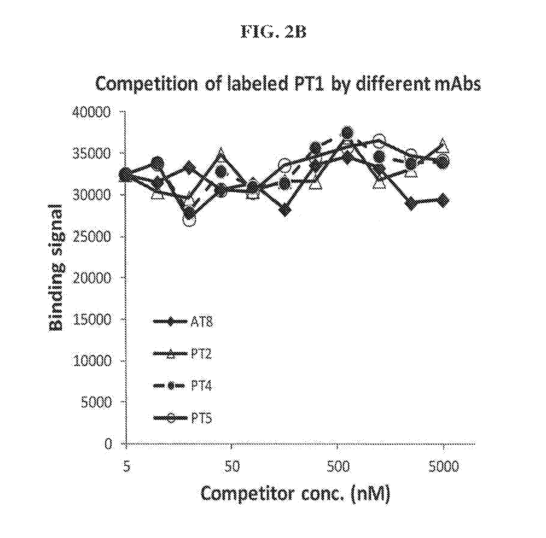
FIGS. 2A-2B show competition of labeled PT1 by various anti-tau antibodies. (A) PT1, PT3 and AT100 and (B)AT8, PT2, PT4 and PT5.
FIGS. 3A-3B show competition of labeled PT3 by various anti-tau antibodies. (A) PT1, PT3 and AT100 and (B)AT8, PT2, PT4 and PT5.
FIG. 4 shows competition of labeled AT100 by various anti-tau antibodies.
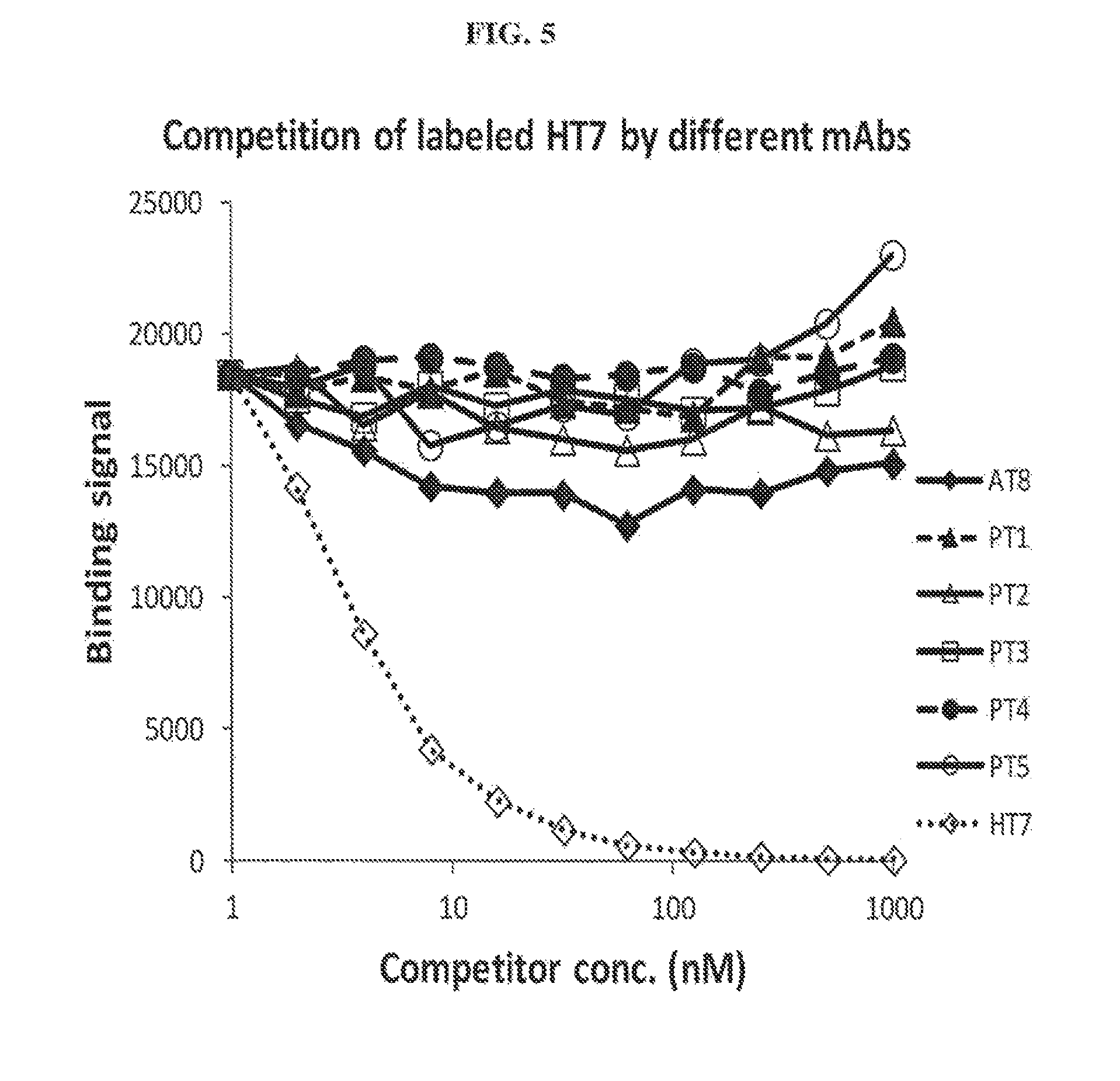
FIG. 5 shows competition of labeled HT7 by various anti-tau antibodies.
FIGS. 6A-6C show an analysis of phosphorylated tau in brainstem homogenates (fraction P1) of 5 month old female P301L transgenic animals treated with saline, mouse IgG1, PT3 or AT8 as indicated in the figure, or from non-treated non-transgenic animals (B6). ELISA was done using AT8 (A) or AT100 (B) as capture antibodies followed by biotinylated-HT7 and avidin-HRP. ELISA signals are plotted as a relative amount of AD brain homogenate (ng/ml) providing the same ELISA signal as an average samples from a non-transgenic animal (B6). Data are plotted individually together with mean+/-S.D. p values for differences between PT3- and IgG1-treated animals are indicated. (C) Western blot of brainstem homogenates fraction P1 from IgG1- or PT3-treated animals using AT100. Signal from homogenates of 10 animals treated with IgG1 (IgG1-1 to IgG1-10) and 7 animals treated with PT3 (PT3-1, PT3-2, PT3-7 to PT3-10 are shown. Actin was used as a loading control.
FIGS. 7A-7C show the levels of total tau in (A) sarcosyl soluble (pT4 soluble), (B) total tau in insoluble (pT4 insoluble) and (C) phosphorylated tau in insoluble (AT8 insoluble) cortex homogenates derived from 5-month old female P301L transgenic mice treated with PT3 or isotype control (IgG) as indicated in the figure. Levels are shown as a measure of a signal from ELISA plotted individually together with mean+/-SD. The sample 1 m inj is a positive control sample derived from a P301L mouse brain injected with a tau aggregate.
SUMMARY OF THE INVENTION
One aspect of the invention is an isolated antibody that binds PHF-tau comprising an antigen-binding site of a heavy chain variable region (VH) of SEQ ID NO:35 or 37, and an antigen-binding site of a light chain variable region (VL) of SEQ ID NO:36 or 38.
Another aspect of the invention is an isolated antibody that binds PHF-tau comprising certain heavy chain and light chain complementarity determining regions.
Another aspect of the invention is an isolated antibody that binds PHF-tau comprising an antigen-binding site of a VH of SEQ ID NO:35 and an antigen-binding site of a VL of SEQ ID NO: 36.
Another aspect of the invention is an isolated antibody that binds PHF-tau comprising an antigen-binding site of a VH of SEQ ID NO:37 and an antigen-binding site of a VL of SEQ ID NO: 38.
Another aspect of the invention is an isolated antibody or fragment that competes for PHF-tau binding with a monoclonal antibody comprising an antigen-binding site of a VH of SEQ ID NO: 35 and an antigen-binding site of a VL of SEQ ID NO: 36, or an antigen-binding site of a VH of SEQ ID NO: 37 and an antigen-binding site of a VL of SEQ ID NO: 38.
Another aspect of the invention is polynucleotides encoding the antibodies of the invention or fragments thereof.
Another aspect of the invention is a vector comprising the polynucleotides of the invention.
Another aspect of the invention is a host cell comprising the vector of the invention.
Another aspect of the invention is a method of making an antibody that binds PHF-tau comprising culturing the host cell of the invention and recovering the antibody produced by the host cell.
DETAILED DESCRIPTION OF THE INVENTION
The term "antibodies" as used herein is meant in a broad sense and includes immunoglobulin or antibody molecules including polyclonal antibodies, monoclonal antibodies including murine, human, human-adapted, humanized and chimeric monoclonal antibodies and antibody fragments.
In general, antibodies are proteins or peptide chains that exhibit binding specificity to a specific antigen. Antibody structures are well known. Immunoglobulins can be assigned to five major classes, namely IgA, IgD, IgE, IgG and IgM, depending on the heavy chain constant domain amino acid sequence. IgA and IgG are further sub-classified as the isotypes IgA1, IgA2, IgG1, IgG2, IgG3 and IgG4. Antibody light chains of any vertebrate species can be assigned to one of two clearly distinct types, namely kappa (.kappa.) and lambda (.lamda.), based on the amino acid sequences of their constant domains.
The term "antibody fragments" means a portion of an intact antibody. Examples of antibody fragments include Fab, Fab', F(ab').sub.2 and Fv fragments, CDR, antigen-binding site, heavy or light chain variable region, diabodies, single chain antibody molecules and multispecific antibodies formed from at least two intact antibodies or fragments thereof.
An immunoglobulin light or heavy chain variable region consists of a "framework" region interrupted by "antigen-binding sites". The antigen-binding sites are defined using various terms as follows: (i) Complementarity Determining Regions (CDRs) are based on sequence variability (Wu and Kabat J Exp Med 132:211-50, 1970). Generally, the antigen-binding site has three CDRs in each variable region (HCDR1, HCDR2 and HCDR3 in heavy chain variable region (VH) and LCDR1, LCDR2 and LCDR3 in light chain variable region (VL)) (Kabat et al., Sequences of Proteins of Immunological Interest, 5th Ed. Public Health Service, National Institutes of Health, Bethesda, Md., 1991). (ii) The term "hypervariable region", "HVR", or "HV" refers to the regions of an antibody variable domain which are hypervariable in structure as defined by Chothia and Lesk (Chothia and Lesk J Mol Biol 196:901-17, 1987). Generally, the antigen-binding site has three hypervariable regions in each VH (H1, H2, H3) and VL (L1, L2, L3). Chothia and Lesk refer to structurally conserved HVs as "canonical structures". Numbering systems as well as annotation of CDRs and HVs have recently been revised by Abhinandan and Martin (Abhinandan and Martin Mol Immunol 45:3832-9, 2008). (iii) Another definition of the regions that form the antigen-binding site has been proposed by Lefranc (Lefranc, et al. Dev Comp Immunol 27:55-77, 2003) based on the comparison of V domains from immunoglobulins and T-cell receptors. The International ImMunoGeneTics (IMGT) database (http:_//www_imgt_org) provides a standardized numbering and definition of these regions. The correspondence between CDRs, HVs and IMGT delineations is described in Lefranc et al., supra. (iv) The antigen-binding site can also be delineated based on Specificity Determining Residue Usage (SDRU) (Almagro J Mol Recognit 17:132-43, 2004), where Specificity Determining Residues (SDR), refers to amino acid residues of an immunoglobulin that are directly involved in antigen contact.
"Framework" or "framework sequence" are the remaining sequences within the variable region of an antibody other than those defined to be antigen-binding site sequences. Because the exact definition of an antigen-binding site can be determined by various delineations as described above, the exact framework sequence depends on the definition of the antigen-binding site.
The term "monoclonal antibody" (mAb) as used herein means an antibody (or antibody fragment) obtained from a population of substantially homogeneous antibodies. Monoclonal antibodies are highly specific, typically being directed against a single antigenic determinant.
The term "epitope" as used herein means a portion of an antigen to which an antibody specifically binds. Epitopes usually consist of chemically active (such as polar, non-polar or hydrophobic) surface groupings of moieties such as amino acids, phosphorylated amino acids or polysaccharide side chains and can have specific three-dimensional structural characteristics, as well as specific charge characteristics. An epitope can be linear in nature or can be a discontinuous epitope, e.g., a conformational epitope, which is formed by a spatial relationship between non-contiguous amino acids of an antigen rather than a linear series of amino acids. A conformational epitope includes epitopes resulting from folding of an antigen, where amino acids from differing portions of the linear sequence of the antigen come in close proximity in 3-dimensional space.
Tau is an abundant central and peripheral nervous system protein having multiple well known isoforms. In the human CNS, six major tau isoforms ranging in size from 352 to 441 exist due to alternative splicing (Hanger, et al. Trends Mol Med 15:112-9, 2009). These isoforms differ from each other by the regulated inclusion of 0-2 N-terminal inserts, and 3 or 4 tandemly arranged microtubule-binding repeats, and are referred to as 0N3R (SEQ ID NO: 1), 1N3R (SEQ ID NO:2), 2N3R (SEQ ID NO:3), 0N4R (SEQ ID NO:4), 1N4R (SEQ ID NO:5) and 2N4R (SEQ ID NO:6). The term "control tau" as used herein refers to the tau isoform of SEQ ID NO: 6 that is devoid of phosphorylation and other post-translational modifications.
Tau binds microtubules and regulates transport of cargo through cells, a process that can be modulated by tau phosphorylation. In AD and related disorders abnormal phosphorylation of tau is prevalent and thought to precede and/or trigger aggregation of tau into fibrils, termed paired helical filaments (PHF). The major constituent of PHF is hyperphosphorylated tau. The term "paired helical filament-tau" or "PHF-tau" as used herein refers to well known tau aggregates in paired helical filaments. Two major regions in PHF structure are evident in electron microscopy, the fuzzy coat and the core filament; the fuzzy coat being sensitive to proteolysis and located outside of the filaments, and the protease resistant core of filaments forming the backbone of PHFs (Wischik, et al. Proc Natl Acad Sci USA 85:4884-8, 1988).
"Antibodies that bind PHF-tau" as used herein refers to antibodies that bind PHF-tau as assessed on western blot. Typically, antibody binding to PHF-tau can be assessed after Coomassie stain of about 500 ng of PHF-tau after 1 hour blocking in 5% (w/v) nonfat dry milk (NFDM) TBS-T, 0.05% Tween-20. Antibodies that bind PHF-tau optionally do not bind control tau (SEQ ID NO:6) as measured by western blot when tested under antigen loading condition where both control tau and PHF-tau is detected equally by tau antibodies that have no preference for PHF-tau epitopes (e.g. antibody HT7, (ThermoScientific, Rockford, Ill.) (Mercken, et al. J Neurochem 58:548-53, 1992). Such exemplary antigen loading conditions are 500 ng PHF-tau and 200 ng control tau.
Conventional well known one and three-letter amino acid codes are used herein.
Compositions of Matter
The present invention relates to anti-PHF-tau antibodies and uses of such antibodies. Such anti-PHF-tau antibodies may have the properties of binding a phosphorylated epitope on PHF-tau or binding to a non-phosphorylated epitope on PHF-tau. Anti-PHF-tau antibodies may be useful as therapeutics, and as research or diagnostic reagents to detect PHF-tau in biological samples, for example in tissues or cells.
One embodiment of the invention is an isolated antibody that binds PHF-tau comprising an antigen-binding site of a heavy chain variable region (VH) of SEQ ID NO:35 or 37, or an antigen-binding site of a light chain variable region (VL) of SEQ ID NO:36 or 38. Table 1 shows antigen-binding site residues of exemplary antibodies of the invention defined according to Kabat or Chothia as well as exemplary heavy and light chain variable regions.
In another embodiment, the antigen-binding site of the VH of the antibodies of the invention comprises heavy chain complementarity determining regions (CDRs) 1 (HCDR1), 2 (HCDR2) and 3 (HCDR3) of SEQ ID NOs:7, 8 and 9 or 13, 14 and 15, respectively, or the antigen-binding site of the VL of the antibodies of the invention comprises light chain CDRs 1 (LCDR1), 2 (LCDR2) and 3 (LCDR3) of SEQ ID NOs: 10, 11 and 12 or 16, 17 and 18, respectively.
Another embodiment of the invention is an isolated antibody that binds PHF-tau comprising an antigen-binding site of a heavy chain variable region (VH) of SEQ ID NO:35 or 37, and an antigen-binding site of a light chain variable region (VL) of SEQ ID NO:36 or 38.
In another embodiment, the antigen-binding site of the VH of the antibodies of the invention comprises heavy chain complementarity determining regions (CDRs) 1 (HCDR1), 2 (HCDR2) and 3 (HCDR3) of SEQ ID NOs:7, 8 and 9 or 13, 14 and 15, respectively, and the antigen-binding site of the VL of the antibodies of the invention comprises light chain CDRs 1 (LCDR1), 2 (LCDR2) and 3 (LCDR3) of SEQ ID NOs:10, 11 and 12 or 16, 17 and 18, respectively.
TABLE-US-00001 TABLE 1 SEQ ID Sequence name NO: Sequence PT1 HCDR1, Kabat 7 SSWMG PT1 HCDR2, Kabat 8 DILPGSGGTNYNERFKG PT1 HCDR3, Kabat 9 SYYDYDRFAN PT1 LCDR1, Kabat 10 RSSESLLHSNGNTYLY PT1 LCDR2, Kabat 11 RMSNLAS PT1 LCDR3, Kabat 12 MQYLEYPLT PT3 HCDR1, Kabat 13 SYAMS PT3 HCDR2, Kabat 14 SISKGGNTYYPNSVKG PT3 HCDR3, Kabat 15 GWGDYGWFAY PT3 LCDR1, Kabat 16 KASQDINRYLN PT3 LCDR2, Kabat 17 RANRLLD PT3 LCDR3, Kabat 18 LQYDEFPLT PT1 HCDR1, Chothia 19 GYTFSSS PT1 HCDR2, Chothia 20 LPGSGG PT1 HCDR3, Chothia 21 SYYDYDRFA PT1 LCDR1, Chothia 22 SESLLHSNGNTY PT1 LCDR2, Chothia 23 RMS PT1 LCDR3, Chothia 24 YLEYPL PT3 HCDR1, Chothia 25 GFTFSSY PT3 HCDR2, Chothia 26 SKGGN PT3 HCDR3, Chothia 27 GWGDYGWFA PT3 LCDR1, Chothia 28 SQDINRY PT3 LCDR2, Chothia 29 RAN PT3 LCDR3, Chothia 30 YDEFPL PT1 VH 35 QVQLQQSGTELMKPGASVKISCKATGYT FSSSWMGWVKQRPGHGLEWIGDILPGSG GTNYNERFKGKASFTAETSSNTAYMQLS SLTSEDSAVYYCVRSYYDYDRFANWGQG TLVTVSA PT1 VL 36 DIVMTQAAPSVPVTPGESVSISCRSSES LLHSNGNTYLYWFLQRPGQSPQLLIYRM SNLASGVPDRFSGSGSGTAFTLRISRVE AEDVGVYYCMQYLEYPLTFGAGTKLELK PT3 VH 37 EVKLVESGGDLVKPGGSLKLSCAASGFT FSSYAMSWVRQNPEKRLEWVASISKGGN TYYPNSVKGRFTISRDNARNILYLQMSS LRSEDTALYYCARGWGDYGWFAYWGQVT LVTVSA PT3 VL 38 DIKMTQSPSSMYASLGERVTITCKASQD INRYLNWFQQKPGKSPKTLIYRANRLLD GVPSRFSGSGSGQDYSLTISSLDYEDMG IYYCLQYDEFPLTFGDGTKLELK
Although the embodiments illustrated in the Examples comprise pairs of variable regions, one from a heavy and one from a light chain, a skilled artisan will recognize that alternative embodiments may comprise single heavy or light chain variable regions. The single variable region can be used to screen for variable domains capable of forming a two-domain specific antigen-binding fragment capable of, for example, binding to PHF-tau. The screening may be accomplished by phage display screening methods using for example hierarchical dual combinatorial approach disclosed in PCT Publ. No. WO92/01047. In this approach, an individual colony containing either a H or L chain clone is used to infect a complete library of clones encoding the other chain (L or H), and the resulting two-chain specific antigen-binding domain is selected in accordance with phage display techniques as described.
Another embodiment of the invention is an isolated antibody that binds PHF-tau comprising an antigen-binding site of a heavy chain variable region (VH) of SEQ ID NO:35 and an antigen-binding site of a light chain variable region (VL) of SEQ ID NO: 36.
Another embodiment of the invention is an isolated antibody that binds PHF-tau comprising an antigen-binding site of a heavy chain variable region (VH) of SEQ ID NO:37 and an antigen-binding site of a light chain variable region (VL) of SEQ ID NO: 38.
Another embodiment of the invention is an isolated antibody that binds PHF-tau comprising heavy chain complementarity determining regions (CDRs) 1 (HCDR1), 2 (HCDR2) and 3 (HCDR3) of SEQ ID NOs:7, 8 and 9, respectively, and light chain CDRs 1 (LCDR1), 2 (LCDR2) and 3 (LCDR3) of SEQ ID NOs: 10, 11 and 12, respectively.
Another embodiment of the invention is an isolated antibody that binds PHF-tau comprising heavy chain complementarity determining regions (CDRs) 1 (HCDR1), 2 (HCDR2) and 3 (HCDR3) of SEQ ID NOs: 13, 14 and 15, respectively, and light chain CDRs 1 (LCDR1), 2 (LCDR2) and 3 (LCDR3) of SEQ ID NOs:16, 17 and 18, respectively.
In any of the preceding embodiments, the isolated antibody that binds PHF-tau may be humanized.
Antibodies of the present invention can be produced by a variety of techniques, for example by the hybridoma method (Kohler and Milstein Nature 256:495-7, 1975). Chimeric mAbs containing a light chain and heavy chain variable region derived from a donor antibody (typically murine) in association with light and heavy chain constant regions derived from an acceptor antibody (typically another mammalian species such as human) can be prepared by the method disclosed in U.S. Pat. No. 4,816,567. CDR-grafted mAbs having CDRs derived from a non-human donor immunoglobulin (typically murine) and the remaining immunoglobulin-derived parts of the molecule being derived from one or more human immunoglobulins can be prepared by techniques known to those skilled in the art such as that disclosed in U.S. Pat. No. 5,225,539. Fully human mAbs lacking any non-human sequences can be prepared from human immunoglobulin transgenic mice by techniques referenced in (Lonberg, et al. Nature 368:856-9, 1994, Fishwild, et al. Nat Biotechnol 14:845-51, 1996, Mendez, et al. Nat Genet 15:146-56, 1997). Human mAbs can also be prepared and optimized from phage display libraries (Knappik, et al. J Mol Biol 296:57-86, 2000, Krebs, et al. J Immunol Methods 254:67-84, 2001, Shi, et al. J Mol Biol 397:385-96, 2010).
Antibody humanization can be accomplished using well known methods, such as specificity determining residues resurfacing (SDRR) (U.S. Publ. No. 2010/0261620), resurfacing (Padlan et al. Mol. Immunol. 28:489-98, 1991), super humanization (Int. Pat. Publ. No. WO04/006955) and human string content optimization (U.S. Pat. No. 7,657,380). Human framework sequences useful for grafting/humanization can be selected from relevant databases by those skilled in the art. The selected frameworks may further be modified to preserve or enhance binding affinity by techniques such as those disclosed in Queen et al. (Queen, et al. Proc Natl Acad Sci USA 86:10029-33, 1989) or in U.S. Publ. No. 2011/0092372.
Preparation of PHF-tau to be used as an antigen for immunization or isolating antibodies from phage display libraries can be done using any suitable technique. In an exemplary method, PHF-tau is isolated from brains of patients having AD using well know protocols, such as described in Greenberg and Davies (Greenberg and Davies Proc Natl Acad Sci USA 87:5827-31, 1990). PHF-tau may be isolated from the postmortem cortex of an Alzheimer patient. The isolated PHF-tau is characterized for its purity and hyperphosphorylation status with antibodies known to react with PHF-tau. In a typical PHF-tau preparation, the hyperphosphorylated bands migrating at about 60, 64, 68 and 72 kDa in western blot (Spillantini and Goedert Trends Neurosci 21:428-33, 1998) are detected by an AT8 antibody that specifically binds hyperphosphorylated PHF-tau but not dephoshporylated PHF-tau.
Antibodies of the present invention may have the characteristics of not binding control tau of SEQ ID NO:6. Such antibodies may be generated using methods described above and testing the antibodies for their lack of binding to control tau in western blots followed by Coomassie stain as described above. Control tau may be recombinantly expressed and purified using standard methods. Exemplary antibodies binding PHF-tau but not control tau are antibodies PT1 and PT3. The antibodies of the invention may further be evaluated for their specificity for example using immunohistochemistry on control and AD brain slices.
The antibodies of the invention may have affinities towards PHF-tau with a dissociation constant (K.sub.D) less than or equal to about 10.sup.-7, 10.sup.-8, 10.sup.-9, 10.sup.-10, 10.sup.-11 or 10.sup.-12 M. The affinity of a given molecule for PHF-tau can be determined experimentally using any suitable method. Such methods may utilize Biacore, ProteOn or KinExA instrumentation, ELISA or competitive binding assays known to those skilled in the art.
Another aspect of the invention is an isolated antibody or fragment that competes for PHF-tau binding with a monoclonal antibody comprising an antigen-binding site of a heavy chain variable region (VH) of SEQ ID NO:35 and an antigen-binding site of a light chain variable region (VL) of SEQ ID NO:36, or an antigen-binding site of a heavy chain variable region (VH) of SEQ ID NO:37 and an antigen-binding site of a light chain variable region (VL) of SEQ ID NO:38.
Competition between binding to PHF-tau can be assayed in vitro using well known methods. For example, binding of MSD Sulfo-Tag.TM. NHS-ester-labeled antibody to PHF-tau in the presence of an unlabeled antibody can be assessed using immunoassay followed by electrochemiluminescence detection.
Several well known methodologies in addition to competitive binding can be employed to determine the binding epitope of the antibodies of the invention. For example, when the structures of both individual components are known, in silico protein-protein docking can be carried out to identify compatible sites of interaction. Hydrogen-deuterium (H/D) exchange can be carried out with the antigen and antibody complex to map regions on the antigen that may be bound by the antibody. Segment and point mutagenesis of the antigen can be used to locate amino acids important for antibody binding. Co-crystal structure of antibody-antigen complex is used to identify residues contributing to the epitope and paratope.
Antibodies of the invention may be monoclonal antibodies of the IgG, IgD, IgA or IgM isotypes. Antibodies of the invention may be bispecific or multispecific. An exemplary bispecific antibody may bind two distinct epitopes on PHF-tau or may bind PHF-tau and amyloid beta (A.beta.). Another exemplary bispecific antibody may bind PHF-tau and an endogenous blood-brain barrier transcytosis receptor such as insulin receptor, transferring receptor, insulin-like growth factor-1 receptor, and lipoprotein receptor. An exemplary antibody is of IgG1 type.
Immune effector properties of the antibodies of the invention may be enhanced or silenced through Fc modifications by techniques known to those skilled in the art. For example, Fc effector functions such as C1q binding, complement dependent cytotoxicity (CDC), antibody-dependent cell-mediated cytotoxicity (ADCC), phagocytosis, down regulation of cell surface receptors (e.g., B cell receptor; BCR), etc. can be provided and/or controlled by modifying residues in the Fc responsible for these activities. Pharmacokinetic properties could also be enhanced by mutating residues in the Fc domain that extend antibody half-life (Strohl Curr Opin Biotechnol 20:685-91, 2009).
Additionally, antibodies of the invention can be post-translationally modified by processes such as glycosylation, isomerization, deglycosylation or non-naturally occurring covalent modification such as the addition of polyethylene glycol moieties (pegylation) and lipidation. Such modifications may occur in vivo or in vitro. For example, the antibodies of the invention can be conjugated to polyethylene glycol (PEGylated) to improve their pharmacokinetic profiles. Conjugation can be carried out by techniques known to those skilled in the art. Conjugation of therapeutic antibodies with PEG has been shown to enhance pharmacodynamics while not interfering with function (Knight, et al. Platelets 15:409-18, 2004, Leong, et al. Cytokine 16:106-19, 2001, Yang, et al. Protein Eng 16:761-70, 2003).
Another embodiment of the invention is an isolated polynucleotide encoding the antibodies of the invention or their complement, or fragments thereof. Exemplary isolated polynucleotides are polynucleotides encoding polypeptides comprising an immunoglobulin heavy chain CDRs HCDR1, HCDR2 and HCDR3 shown in SEQ ID NOs:7, 8 and 9 or 13, 14 and 15, respectively, or polypeptides comprising an immunoglobulin light chain CDRs LCDR1, LCDR2 and LCDR3 shown in SEQ ID NOs:10, 11 and 12 or 16, 17 and 18, respectively, and polynucleotides having a sequence shown in SEQ ID NOs:31-34, encoding antibody variable regions of the invention. Other polynucleotides which, given the degeneracy of the genetic code or codon preferences in a given expression system, encode the antibodies of the invention are also within the scope of the invention. The isolated nucleic acids of the present invention can be made using well known recombinant or synthetic techniques. DNA encoding the monoclonal antibodies is readily isolated and sequenced using methods known in the art. Where a hybridoma is produced, such cells can serve as a source of such DNA. Alternatively, using display techniques wherein the coding sequence and the translation product are linked, such as phage or ribosomal display libraries, the selection of the binder and the nucleic acid is simplified. After phage selection, the antibody coding regions from the phage can be isolated and used to generate whole antibodies, including human antibodies, or any other desired antigen binding fragment, and expressed in any desired host, including mammalian cells, insect cells, plant cells, yeast, and bacteria.
Another embodiment of the invention is a vector comprising at least one polynucleotide of the invention. Such vectors may be plasmid vectors, viral vectors, transposon based vectors or any other vector suitable for introduction of the polynucleotides of the invention into a given organism or genetic background by any means.
Another embodiment of the invention is a host cell comprising any of the polynucleotides of the invention. Such host cells may be eukaryotic cells, bacterial cells, plant cells or archeal cells. Exemplary eukaryotic cells may be of mammalian, insect, avian or other animal origins. Mammalian eukaryotic cells include immortalized cell lines such as hybridomas or myeloma cell lines such as SP2/0 (American Type Culture Collection (ATCC), Manassas, Va., CRL-1581), NSO (European Collection of Cell Cultures (ECACC), Salisbury, Wiltshire, UK, ECACC No. 85110503), FO (ATCC CRL-1646) and Ag653 (ATCC CRL-1580) murine cell lines. An exemplary human myeloma cell line is U266 (ATTC CRL-TIB-196). Other useful cell lines include those derived from Chinese Hamster Ovary (CHO) cells such as CHO-K1SV (Lonza Biologics), CHO-K1 (ATCC CRL-61, Invitrogen) or DG44.
Another embodiment of the invention is a method of making an antibody that binds PHF-tau comprising culturing a host cell of the invention and recovering the antibody produced by the host cell. Methods of making antibodies and purifying them are well known in the art.
Methods of Treatment
Anti-PHF-tau antibodies of the invention or fragments thereof, including Fab, (Fab')2, scFv fragments, or antibodies comprising antigen-binding sites of the antibodies of the invention can be used to treat, reduce or prevent symptoms in patients having a neurodegenerative disease that involves pathological aggregation of tau within the brain, such as patients suffering from AD or any other tauopathy. While not wishing to be bound by any particular theory, the antibodies of the invention may exert their beneficial effect by reducing pathological tau aggregation and hence the amount of PHF-tau in the brain. The antibodies of the invention may be used to treat an animal patient belonging to any classification. Examples of such animals include mammals such as humans, rodents, dogs, cats and farm animals. For example, the antibodies of the invention are useful in the preparation of a medicament for treatment of AD wherein the medicament is prepared for administration in dosages defined herein.
Another embodiment of the invention is a method of reducing aggregation of tau in patients in need thereof comprising administering to the patient a therapeutically effective amount of the isolated antibody of the invention for a time sufficient to reduce the aggregation of tau.
Another embodiment of the invention is a method of treating or reducing symptoms of a neurodegenerative disease that involves aggregation of tau in a patient comprising administering to the patient a therapeutically effective amount of the isolated antibody of the invention for a time sufficient to treat or reduce symptoms of the neurodegenerative disease.
In any of the embodiments above, the neurodegenerative disease that involves aggregation of tau is a tauopathy.
In any of the embodiments above, the isolated antibody comprises an antibody that binds PHF-tau comprising an antigen-binding site of a VH of SEQ ID NO:35 and an antigen-binding site of a VL of SEQ ID NO:36.
In any of the embodiments above, the isolated antibody comprises an antibody that binds PHF-tau comprising an antigen-binding site of a VH of SEQ ID NO:37 and an antigen-binding site of a VL of SEQ ID NO: 38.
As used herein a "tauopathy" encompasses any neurodegenerative disease that involves the pathological aggregation of tau within the brain. In addition to familial and sporadic AD, other exemplary tauopathies are frontotemporal dementia with parkinsonism linked to chromosome 17 (FTDP-17), progressive supranuclear palsy, corticobasal degeneration, Pick's disease, progressive subcortical gliosis, tangle only dementia, diffuse neurofibrillary tangles with calcification, argyrophilic grain dementia, amyotrophic lateral sclerosis parkinsonism-dementia complex, Down syndrome, Gerstmann-Striussler-Scheinker disease, Hallervorden-Spatz disease, inclusion body myositis, Creutzfeld-Jakob disease, multiple system atropy, Niemann-Pick disease type C, prion protein cerebral amyloid angiopathy, subacute sclerosing panencephalitis, myotonic dystrophy, non-guanamian motor neuron disease with neurofibrillary tangles, postencephalitic parkinsonism, and chronic traumatic encephalopathy, such as dementia pugulistica (boxing disease). (Morris, et al. Neuron 70:410-26, 2011).
A tauopathy-related behavioral phenotype includes cognitive impairments, early personality change and disinhibition, apathy, abulia, mutism, apraxia, perseveration, stereotyped movements/behaviors, hyperorality, disorganization, inability to plan or organize sequential tasks, selfishness/callousness, antisocial traits, a lack of empathy, halting, agrammatic speech with frequent paraphasic errors but relatively preserved comprehension, impaired comprehension and word-finding deficits, slowly progressive gait instability, retropulsions, freezing, frequent falls, non-levodopa responsive axial rigidity, supranuclear gaze palsy, square wave jerks, slow vertical saccades, pseudobulbar palsy, limb apraxia, dystonia, cortical sensory loss, and tremor.
Patients amenable to treatment include asymptomatic individuals at risk of AD or other tauopathy, as well as patients presently showing symptoms. Patients amenable to treatment include individuals who have a known genetic risk of AD, such as a family history of AD or presence of genetic risk factors in the genome. Exemplary risk factors are mutations in the amyloid precursor protein (APP), especially at position 717 and positions 670 and 671 (Hardy and Swedish mutations, respectively). Other risk factors are mutations in the presenilin genes, PS1 and PS2, and ApoE4, family history of hypercholesterolemia or atherosclerosis. Individuals presently suffering from AD can be recognized from characteristic dementia by the presence of risk factors described above. In addition, a number of diagnostic tests are available to identify individuals who have AD. These include measurement of cerebrospinal fluid tau and A.beta.42 levels. Elevated tau and decreased A.beta.42 levels signify the presence of AD. Individuals suffering from AD can also be diagnosed by AD and Related Disorders Association criteria.
Administration/Pharmaceutical Compositions
Anti-PHF-tau antibodies of the invention are suitable both as therapeutic and prophylactic agents for treating or preventing neurodegenerative diseases that involves pathological aggregation of tau, such as AD or other tauopathies. In asymptomatic patients, treatment can begin at any age (e.g., at about 10, 15, 20, 25, 30 years). Usually, however, it is not necessary to begin treatment until a patient reaches about 40, 50, 60, or 70 years. Treatment typically entails multiple dosages over a period of time. Treatment can be monitored by assaying antibody, or activated T-cell or B-cell responses to the therapeutic agent over time. If the response falls, a booster dosage is indicated.
In prophylactic applications, pharmaceutical compositions or medicaments are administered to a patient susceptible to, or otherwise at risk of, AD in an amount sufficient to eliminate or reduce the risk, lessen the severity, or delay the outset of the disease, including biochemical, histologic and/or behavioral symptoms of the disease, its complications and intermediate pathological phenotypes presented during development of the disease. In therapeutic applications, compositions or medicaments are administered to a patient suspected of, or already suffering from, such a disease in an amount sufficient to reduce, arrest, or delay any of the symptoms of the disease (biochemical, histologic and/or behavioral). Administration of a therapeutic may reduce or eliminate mild cognitive impairment in patients that have not yet developed characteristic Alzheimer's pathology. An amount adequate to accomplish therapeutic or prophylactic treatment is defined as a therapeutically- or prophylactically-effective dose. In both prophylactic and therapeutic regimes, compositions or medicaments are usually administered in several dosages until a sufficient immune response has been achieved.
Anti-PHF-tau antibodies or fragments thereof of the invention may be administered in combination with other agents that are effective for treatment of related neurodegenerative diseases. In the case of AD, antibodies of the invention may be administered in combination with agents that reduce or prevent the deposition of amyloid-beta (A.beta.). It is possible that PHF-tau and A.beta. pathologies are synergistic. Therefore, combination therapy targeting the clearance of both PHF-tau and A.beta. and A.beta.-related pathologies at the same time may be more effective than targeting each individually.
In the case of Parkinson's Disease and related neurodegenerative diseases, immune modulation to clear aggregated forms of the .alpha.-synuclein protein is also an emerging therapy. A combination therapy which targets the clearance of both tau and .alpha.-synuclein proteins simultaneously may be more effective than targeting either protein individually.
In the methods of the invention, the "therapeutically effective amount" of the antibody in the treatment or ameliorating symptoms of a tauopathy can be determined by standard research techniques. For example, the dosage of the antibody can be determined by administering the agent to relevant animal models well known in the art.
In addition, in vitro assays can optionally be employed to help identify optimal dosage ranges. Selection of a particular effective dose can be determined (e.g., via clinical trials) by those skilled in the art based upon the consideration of several factors. Such factors include the disease to be treated or prevented, the symptoms involved, the patient's body mass, the patient's immune status and other factors known by the skilled artisan. The precise dose to be employed in the formulation will also depend on the route of administration, and the severity of disease, and should be decided according to the judgment of the practitioner and each patient's circumstances. Effective doses can be extrapolated from dose-response curves derived from in vitro or animal model test systems.
The mode of administration for therapeutic use of the antibodies of the invention may be any suitable route that delivers the agent to the host. Pharmaceutical compositions of these antibodies are useful for parenteral administration, e.g., intradermal, intramuscular, intraperitoneal, intravenous, subcutaneous, intranasal or intracranial or they can be administered into the cerebrospinal fluid of the brain or spine.
The antibodies of the invention may be prepared as pharmaceutical compositions containing an effective amount of the antibody as an active ingredient in a pharmaceutically acceptable carrier. The term "carrier" refers to a diluent, adjuvant, excipient, or vehicle with which the antibody is administered. Such pharmaceutical vehicles can be liquids, such as water and oils, including those of petroleum, animal, vegetable or synthetic origin, such as peanut oil, soybean oil, mineral oil, sesame oil and the like. For example, 0.4% saline and 0.3% glycine can be used. These solutions are sterile and generally free of particulate matter. They may be sterilized by conventional, well-known sterilization techniques (e.g., filtration). The compositions may contain pharmaceutically acceptable auxiliary substances as required to approximate physiological conditions such as pH adjusting and buffering agents, stabilizing, thickening, lubricating and coloring agents, etc. The concentration of the antibodies of the invention in such pharmaceutical formulation can vary widely, i.e., from less than about 0.5%, usually at or at least about 1% to as much as 15 or 20% by weight and will be selected primarily based on required dose, fluid volumes, viscosities, etc., according to the particular mode of administration selected.
The treatment may be given in a single dose schedule, or as a multiple dose schedule in which a primary course of treatment may be with 1-10 separate doses, followed by other doses given at subsequent time intervals required to maintain and or reinforce the response, for example, at 1-4 months for a second dose, and if needed, a subsequent dose(s) after several months. Examples of suitable treatment schedules include: (i) 0, 1 month and 6 months, (ii) 0, 7 days and 1 month, (iii) 0 and 1 month, (iv) 0 and 6 months, or other schedules sufficient to elicit the desired responses expected to reduce disease symptoms, or reduce severity of disease.
Thus, a pharmaceutical composition of the invention for intramuscular injection could be prepared to contain 1 ml sterile buffered water, and between about 1 ng to about 100 mg, about 50 ng to about 30 mg or about 5 mg to about 25 mg of an antibody of the invention. Similarly, a pharmaceutical composition of the invention for intravenous infusion could be made up to contain about 250 ml of sterile Ringer's solution, and about 1 mg to about 30 mg or about 5 mg to about 25 mg of an antibody of the invention. Actual methods for preparing parenterally administrable compositions are well known and are described in more detail in, for example, "Remington's Pharmaceutical Science", 15th ed., Mack Publishing Company, Easton, Pa.
The antibodies of the invention can be lyophilized for storage and reconstituted in a suitable carrier prior to use. This technique has been shown to be effective with antibody and other protein preparations and art-known lyophilization and reconstitution techniques can be employed.
Diagnostic Methods and Kits
Antibodies of the invention may be used in methods of diagnosing AD or other tauopathy in a subject. This method involves detecting, in the subject, the presence of PHF-tau using a diagnostic reagent such as an antibody or a fragment thereof of the present invention.
PHF-tau may be detected in a biological sample from a subject (e.g., blood, urine, cerebral spinal fluid) by contacting the biological sample with the diagnostic antibody reagent, and detecting binding of the diagnostic antibody reagent to PHF-tau in the sample from the subject. Assays for carrying out the detection include well known methods such as ELISA, immunohistochemistry, western blot, or in vivo imaging. Exemplary diagnostic antibodies are antibodies PT1 and PT3 of the invention, and are of IgG1,.kappa. type.
Diagnostic antibodies or similar reagents can be administered by intravenous injection into the body of the patient, or directly into the brain by any suitable route that delivers the agent to the host as exemplified above. The dosage of antibody should be within the same ranges as for treatment methods. Typically, the antibody is labeled, although in some methods, the primary antibody with affinity for PHF-tau is unlabelled and a secondary labeling agent is used to bind to the primary antibody. The choice of label depends on the means of detection. For example, a fluorescent label is suitable for optical detection. Use of paramagnetic labels is suitable for tomographic detection without surgical intervention. Radioactive labels can also be detected using PET or SPECT.
Diagnosis is performed by comparing the number, size, and/or intensity of labeled PHF-tau, tau aggregates, and/or neurofibrillary tangles in a sample from the subject or in the subject, to corresponding baseline values. The baseline values can represent the mean levels in a population of undiseased individuals. Baseline values can also represent previous levels determined in the same subject.
The diagnostic methods described above can also be used to monitor a subject's response to therapy by detecting the presence of PHF-tau in a subject before, during or after the treatment. A decrease in values relative to baseline signals a positive response to treatment. Values can also increase temporarily in biological fluids as pathological tau is being cleared from the brain.
The present invention is further directed to a kit for performing the above described diagnostic and monitoring methods. Typically, such kits contain a diagnostic reagent such as the antibodies of the invention, and optionally a detectable label. The diagnostic antibody itself may contain the detectable label (e.g., fluorescent molecule, biotin, etc.) which is directly detectable or detectable via a secondary reaction (e.g., reaction with streptavidin). Alternatively, a second reagent containing the detectable label may be utilized, where the second reagent has binding specificity for the primary antibody. In a diagnostic kit suitable for measuring PHF-tau in a biological sample, the antibodies of the kit may be supplied prebound to a solid phase, such as to the wells of a microtiter dish.
The contents of all cited references (including literature references, issued patents, published patent applications, and co-pending patent applications) cited throughout this application are hereby expressly incorporated by reference.
EXAMPLE 1
Purification of Paired Helical Filament-Tau (PHF-Tau)
PHF-tau was partially purified by a modified method of Greenberg and Davies (Greenberg and Davies Proc Natl Acad Sci U S A 87:5827-31, 1990). Briefly, postmortem tissue from the cortex obtained from a histologically confirmed Alzheimer patient was partially purified. Typically, 5 mg of frontal cortex was homogenized in 10 vol of cold buffer Buffer H (10 mM Tris, 800 mM NaCl, 1 mM EGTA and 10% sucrose/pH 7.4) using a glass/Teflon Potter tissue homogenizer (IKA Works, Inc; Staufen, Germany) at 1000 rpm. The homogenized material was centrifuged at 27000 g for 20 min in a Sorvall rotor SS34. The pellet was discarded and the supernatant was adjusted to a final concentration of 1% (w/v)N-lauroylsarcosine and 1% (v/v) 2-mercaptoethanol and incubated for 2 h at 37.degree. C. Subsequently the supernatant was centrifuged at 108000 g for 35 min at 20.degree. C. in a Beckman 60Ti rotor. The pellet was carefully washed in PBS and suspended in PBS. The supernatant was centrifuged a second time as described and the final pellet was dissolved, aliquoted and frozen at -80.degree. C. The quality of the PHF-tau preparations were evaluated on a 12% SDS-PAGE and western blot with anti-tau antibodies AT8 and HT7 (ThermoScientific, Rockford, Ill.). AT8 detects PHF-tau phosphorylated at 5202/T205, but does not bind to non-phosphorylated PHF-tau nor to the wild type tau. HT7 binds to a non-phosphorylated epitope at Tau amino acids 159-163 (of SEQ ID NO: 6), and recognizes both tau and PHF-tau. A good quality PHF-tau preparation is composed of 4 bands having molecular weights of about 60, 64, 66 and 72 kDa on a Western blot detected with an antibody reactive with hyperphosphorylated PHF-tau such as AT8. Two separate PHF-tau preparations with comparable quality and purity were made from the same brain sample. Preparation 1 was used for immunization.
EXAMPLE 2
Generation of Monoclonal Antibodies Against PHF-Tau
Anti-PHF-tau antibodies were generated using standard hybridoma technology in normal Balb/c mice (Kohler and Milstein Nature 256:495-7, 1975). Obtained hybridomas were seeded in 96-well plates and screened after 10 days in a direct ELISA on 25 ng/well coated PHF-tau as described below. Positive cells were tested for cross-reactivity on 10 ng/well coated with control tau (SEQ ID NO:6) expressed in E. Coli BL21 cells and purified by heat treatment and ammonium sulphate precipitation, and were immediately subcloned and positive clones were frozen in liquid nitrogen. All hybridomas were grown in Dulbecco's modified Eagle's medium supplemented with 10% foetal calf serum (Hyclone, Europe), Hybridoma Fusion Cloning Supplement (2%) (Roche, Brussels, Belgium) 2% HT (Sigma, USA), 1 mM sodium pyruvate, 2 mM L-glutamine and penicillin (100 U/ml) and Streptomycin (50 mg/ml).
Antibody variable regions were cloned from select hybridoma cells onto mouse IgG1/IgG2/.kappa. background and expressed and purified using routine methods.
Direct ELISA for Antibody Selection
25 ng/well PHF-tau was coated overnight at 4.degree. C. in NUNC Maxisorp (Life Technologies) flat-bottom high-binding 96-well micro titer plates in 50 .mu.l/well coating buffer (10 mM Tris, 10 mM NaCl, and 10 mM NaN3, pH 8.5). The next day, the plates were blocked with 75 .mu.l/well of 0.1% casein in PBS for 60 min at room temperature. Next, 50 .mu.l hybridoma supernatant was added and incubated for 1 h at 37.degree. C. After washing, the bound monoclonal antibodies were detected with 50 .mu.l/well of Sheep-anti-mouse IgG conjugated with horseradish peroxidase for 1 hr at 37.degree. C. (Amersham-Pharmacia Biotech). Both reagents were diluted in 0.1% Casein/PBS. The plates were washed and 50 .mu.l of a solution of 0.42 mM 3,5,3',5'-tetramethyl-benzidine, 0.003% (v/v) H.sub.2O.sub.2 in 100 mM citric acid and 100 mM disodium hydrogen phosphate (pH 4.3) was added as the substrate. The reaction was allowed to proceed for maximum 15 min on a plate shaker at room temperature, after which the color development was stopped with 2 N H.sub.2SO.sub.4, 50 .mu.l/well and the plates, were read on a micro titer plate reader at 450 nm (Thermomax, Molecular Devices).
Specificity of the Monoclonal Antibodies
Select antibodies obtained from the hybridoma screen were tested for their cross-reactivity with recombinantly expressed control tau (SEQ ID NO:6). 500 ng of PHF-tau and 200 ng of control tau were loaded on a NuPAGE.RTM. Novex.RTM. Bis-Tris 4-12% gel and blotted on a nitrocellulose membrane by use of an iBlot system (Invitrogen), according to the manufacturer's instructions. Membranes were blocked for 1 hour with Tris-Buffered Saline Tween-20 (TBS-T; 1M Tris, 150 mM NaCl and 0.05% Tween-20, pH 8.5) containing non fat dry milk (NFDM) (5% w/v; Biorad) and washed three times in TBS-T. Incubation with the primary control antibodies (1 .mu.g/ml) HT7, AT8, AT100 (ThermoScientific, Rockford, Ill.), and BT2 diluted in TBS-T containing NFDM (5% w/v) was overnight at 4.degree. C. PT1, PT2, PT3, PT4 and PT5 monoclonal antibodies selected from the hybridoma screen were added in culture supernatant containing 10% FCS. Primary antibodies were detected using Sheep-anti-mouse Ig conjugated with HRPO (1:20000 in TBS-T, Amersham Biosciences) via West Dura.RTM. enhanced chemiluminescence (Pierce, Thermoscientific). Signals were captured by the Lumi-imaging system (Roche Diagnostic). PT1 and PT3 reacted with PHF-tau and did not react with control tau in western blots. PT2 was reactive with both proteins. Binding profiles of HT7 and AT8 are described above. AT100 binds to phosphorylated Ser212/Thr214 and binds to PHF-tau but not wild type tau. BT2 recognizes a non-phosphorylated epitope comprising S199/S202, and thus recognizes wild type tau but not PHF-tau.
Competitive Epitope Binding
Monoclonal antibodies PT1, PT2, PT3, PT4, PT5, AT8 (ThermoScientific, Rockford, Ill.), AT100 (ThermoScientific, Rockford, Ill.) and HT7 (MN1000) (Thermo Scientific, Rockford, Ill.) were evaluated for competitive binding to PHF-tau or phosphorylated tau peptides. Antibodies were labeled using MSD.RTM. SULFO-TAG NHS Ester (Meso Scale Discovery) according to the manufacturer's instruction
For competition with labeled PT1, PT3, AT100 and HT7, 5 .mu.L (50 .mu.g/mL)/well of enriched PHF-Tau proteins (purified as described above) was coated on MSD HighBind plate (Meso Scale Discovery, Gaithersburg, Md.) for 2 hr at room temperature (RT). For competition with AT8, 25 .mu.L of 0.1 mg/well synthetic biotinylated and pegylated peptide RSGYSSPG(pS)PG(pT)PGSRSR-OH (New England Peptide, LLC., Gardner, Mass.) (SEQ ID NO:39) corresponding to residues 194-211 of control tau (SEQ ID NO:6) phosphorylated at residues corresponding to 5202/T205 in control tau was coated on Streptavidin-charged plates (Meso Scale Discovery, Gaithersburg, Md.). After coating, wells were blocked with 150 .mu.L of 5% MSD Blocker A buffer at RT for 2 hr, and washed three times with 0.1 M HEPES buffer, pH 7.4. 25 .mu.L of a mixture of labeled individual anti-tau antibody (10 nM or 50 nM) and serial dilutions of various unlabeled competitor antibodies (1 nM to 2 .mu.M) was added on each well. The plates were incubated for 2 hours at RT with gentle shaking and washed 3 times as above. 150 .mu.L/well of diluted MSD Read Buffer T was added and the plates were read in a SECTOR Imager 6000 (Meso Scale Discovery, Gaithersburg, Md.).
Non-overlapping epitopes of anti-Tau mAbs HT7, AT100 and AT8 are described above. Based on the published data, these antibodies were not expected to compete with each other in binding to Tau proteins or peptides.
Based on competition assays performed, none of the antibodies competed with each other for binding, indicating that they all bind to different epitopes. In each experiment, only self-inhibition was observed. FIGS. 1-5 show results of competition assays with labeled AT8, PT1, PT3, AT100, and HT7, respectively.
EXAMPLE 3
Anti-PHF Tau Antibodies Reduce PHF-Tau Accumulation In Vivo
5-month old female P301L mice (Taconic, cat#002508) were treated once weekly with mouse IgG1, saline, PT3 (500 .mu.g/mouse) or AT8 (expressed from hybridoma ECACC, deposit number 9110086) for 5 months. Mice were anesthetized, perfused with cold PBS and the brains dissected on ice. For each mouse, one brain hemisphere was homogenized in 10 volume of H-buffer, followed by centrifugation at 21,000 g for 20 min at 4.degree. C. The resulting supernatant was further centrifuged at 100,000 g for 60 min. After centrifugation, the pellet (fraction P1) was recovered and re-suspended with lysis buffer for Western and ELISA analyses, as described by Chai et al. J Biol Chem 286:34457-67, 2011. For cortex samples, the fraction P1 was further treated with 1% (w/v)N-lauroylsarcosine and ultracentrifuged to further enrich PHF-tau in the pellet. Male P301L mice were found to have low transgene expression and were not included in the analyses.
Phosphorylated tau was measured in brainstem homogenates (fraction P1) with sandwich ELISA using antibodies AT8 and AT100 as capture antibodies followed by detection by biotinylated HT7 and avidin-HRP (FIGS. 6A and 6B), and with AT100 in a western blot (FIG. 6C) essentially as described in Chai et al. J Biol Chem 286:34457-67, 2011. Briefly, the P1 pellet was resuspended with lysis buffer (Cell Signaling). P1 pellet samples were incubated in AT8 or AT100 (Thermo Scientific) pre-coated wells and the biotin labeled HT-7 antibody. Samples were then washed 5 times with buffer, followed by incubation with avidin-HRP for one hour. Following this, samples were incubated with one step TMB substrate (Thermo Scientific) for 30 min, followed by 2N H.sub.2SO.sub.4. Finally, the reaction was read at 450 nm and the quantity of AT8- or AT100-reactive tau in brain was determined using a standard curve derived from human AD brain homogenates, and plotted as a relative amount of AD brain homogenate (ng/ml) providing the same ELISA signal as an average samples from a non-transgenic animal (B6).
A statistically significant decrease or trend towards significance in phosphorylated tau was seen in P301L transgenic animals treated with PT3 when compared to the isotype control administered animals in ELISA assays using either AT100 (p=0.057) or AT8 (p=0.0475) to detected phosphorylation (ELISA signal: Saline group (1135.+-.228.8); IgG1 group (1344.+-.245.6); PT3 group (660.5.+-.134.5); AT8 group (1271.+-.274)).
To confirm the data obtained with ELISA, brainstem homogenates (fraction P1) from IgG1- or PT3-treated animals were analyzed on western blot by detecting PHF-tau using AT100 antibody. The filters were blotted using anti-actin antibody as a loading control (FIG. 6B). Western blots showed attenuated PHF-tau amount detected with AT100 antibody when compared to the animals treated with IgG1.
For cortex analysis, both sarcosyl soluble (representing soluble tau) and insoluble (representing PHF-tau) cortex fractions were analyzed by sandwich ELISA using a pan-tau antibody (PT4) or phospho-tau antibody (AT8) for capturing followed by a biotin-pan-tau antibody (hTau10) followed by HRP-avidin. Animals treated with PT3 had similar levels of total tau in comparison to animals treated with the isotype control IgG1. A trend towards lower PHF-tau levels were evident in animals treated with PT3 when compared to the isotype control in the N-lauroylsarcosine insoluble fractions (ELISA capture with PT4: IgG group: 851026.+-.261198 and PT3 group: 585639.+-.120498; ELISA, capture with AT8: IgG group: 1125886.+-.286240 (N=10) and pT3 group: 746582.+-.124970 (N=7)).
EXAMPLE 4
Characterization of Anti-PHF-Tau Antibodies
Affinity Determination
The interactions of monoclonal antibodies PT1 and PT3 with recombinant human soluble tau or PHF-tau were studied by ProteOn. All interactions were studied at 25.degree. C. using PBS pH 7.4, supplemented with 3 mM EDTA, and 0.005% Tween-20 as running or system buffer. Two different experiment formats were used, one for the interaction with recombinantly expressed control tau and another for the interaction with PHF-tau. In these experiments HT7 (Pierce, cat #MN1000), a mouse anti-tau antibody was used as a positive control.
To study the interaction with recombinantly expressed control tau a biosensor surface was prepared by coupling an anti-human or anti-mouse IgG Fc.gamma. fragment specific antibody (Ab) to the surface of a GLC (ProteOn) sensor chip using each manufacturer's instructions for amine-coupling chemistry (.about.5000 response units (RU)). The coupling buffer was 10 mM sodium acetate, pH 4.5. The anti-PHF-tau antibodies were diluted in the running buffer and injected to obtain a capture of 60-130 RUs. Capture of anti-PFH-Tau mAbs was followed by injection of recombinantly expressed control tau (Tau-441, Sigma catalog# T0576-50 ug) in solution (0.1 to 75 nM in 5-fold dilutions). The association was monitored for 2 minutes (80 .mu.L injected at 40 .mu.L/min). The dissociation was monitored for 10 minutes. Regeneration of the sensor surface was obtained with 0.85% H.sub.3PO.sub.4, or 0.85% H.sub.3PO.sub.4 followed by 50 mM NaOH. The data were fit using a 1:1 binding model. The data were fit using a 1:1 binding model.
TABLE-US-00002 TABLE 2 *K.sub.D mAb Antigen k.sub.on (M.sup.-1s.sup.-1) k.sub.off (s.sup.-1) (pM) PT1 Control Tau ** PT1 PHF-Tau 2.01E+05 6.47E-05 322 PT3 Control Tau *** PT3 PHF-Tau (1.23 .+-. 0.06)E+06 (2.18 .+-. 0.28)E-05 18 .+-. 2 *For PHF-tau this is the apparent intrinsic affinity where KD is obtained as the ratio of kff1/kon-1 derived from a fit performed using a bivalent binding model. ** No significant binding *** No binding in 4 of 5 experiments
To study the interaction with PHF-tau a biosensor surface was prepared by capture-coupling PHF-tau using HT7 as the capture reagent. Additional preparation of the PHF-tau as described earlier was required for ProteOn to limit the amount of insoluble material entering the fluidics. PHF-tau as described above was additionally prepared by 2-times centrifugation at 5000 g, 5.degree. C., 10 min where the supernatant from the second centrifugation was then diluted 1/20 or 1/40 in running buffer. To prepare the chip, HT7 was covalently immobilized to the surface of a GLC (ProteOn) sensor chip using each manufacturer's instructions for amine-coupling chemistry (.about.3000 response units (RU). The coupling buffer was 10 mM sodium acetate, pH 4.5. After HT7 immobilization PHF-tau was injected and captured (.about.300 RU) by HT7. After capture, PHF-tau was covalently immobilized to the sensor chip by activation of the chip using each manufacturer's instructions for amine-coupling chemistry. Remaining reactive sites were finally blocked by injection of ethanolamine. After preparation and stabilization of the PHF-tau-modified surface and reference surface (containing no antigen), the anti-PHF-tau antibodies were diluted in the running buffer and injected in solution (0.1-75 nM in 5-fold dilutions). The association was monitored for 3 minutes (120 .mu.L injected at 40 .mu.L/min). The dissociation was monitored for 10 or 15 minutes. Regeneration of the sensor surface was obtained with 10 mM Gly pH 2. The data were fit using a bivalent binding model where the apparent intrinsic affinity was reported as the ratio of koff-1/kon-1.
Based on the ProteOn experiments PT1 bound PHF-tau with 322 pM affinity and showed no binding to control tau under the conditions tested (Table 2). PT3 bound PHF-tau with 18.+-.2 pM affinity and showed no binding to control tau in 4 out of 5 measurements under the conditions tested. One out of the 5 ProteOn measurements showed weak binding which could be used to estimate affinity >75 nM on the basis of the highest concentration of control tau used.
The present invention now being fully described, it will be apparent to one of ordinary skill in the art that many changes and modifications can be made thereto without departing from the spirit or scope of the appended claims.
SEQUENCE LISTINGS
1
391352PRTHomo sapiens 1Met Ala Glu Pro Arg Gln Glu Phe Glu Val Met Glu Asp His Ala Gly 1 5 10 15 Thr Tyr Gly Leu Gly Asp Arg Lys Asp Gln Gly Gly Tyr Thr Met His 20 25 30 Gln Asp Gln Glu Gly Asp Thr Asp Ala Gly Leu Lys Ala Glu Glu Ala 35 40 45 Gly Ile Gly Asp Thr Pro Ser Leu Glu Asp Glu Ala Ala Gly His Val 50 55 60 Thr Gln Ala Arg Met Val Ser Lys Ser Lys Asp Gly Thr Gly Ser Asp 65 70 75 80 Asp Lys Lys Ala Lys Gly Ala Asp Gly Lys Thr Lys Ile Ala Thr Pro 85 90 95 Arg Gly Ala Ala Pro Pro Gly Gln Lys Gly Gln Ala Asn Ala Thr Arg 100 105 110 Ile Pro Ala Lys Thr Pro Pro Ala Pro Lys Thr Pro Pro Ser Ser Gly 115 120 125 Glu Pro Pro Lys Ser Gly Asp Arg Ser Gly Tyr Ser Ser Pro Gly Ser 130 135 140 Pro Gly Thr Pro Gly Ser Arg Ser Arg Thr Pro Ser Leu Pro Thr Pro 145 150 155 160 Pro Thr Arg Glu Pro Lys Lys Val Ala Val Val Arg Thr Pro Pro Lys 165 170 175 Ser Pro Ser Ser Ala Lys Ser Arg Leu Gln Thr Ala Pro Val Pro Met 180 185 190 Pro Asp Leu Lys Asn Val Lys Ser Lys Ile Gly Ser Thr Glu Asn Leu 195 200 205 Lys His Gln Pro Gly Gly Gly Lys Val Gln Ile Val Tyr Lys Pro Val 210 215 220 Asp Leu Ser Lys Val Thr Ser Lys Cys Gly Ser Leu Gly Asn Ile His 225 230 235 240 His Lys Pro Gly Gly Gly Gln Val Glu Val Lys Ser Glu Lys Leu Asp 245 250 255 Phe Lys Asp Arg Val Gln Ser Lys Ile Gly Ser Leu Asp Asn Ile Thr 260 265 270 His Val Pro Gly Gly Gly Asn Lys Lys Ile Glu Thr His Lys Leu Thr 275 280 285 Phe Arg Glu Asn Ala Lys Ala Lys Thr Asp His Gly Ala Glu Ile Val 290 295 300 Tyr Lys Ser Pro Val Val Ser Gly Asp Thr Ser Pro Arg His Leu Ser 305 310 315 320 Asn Val Ser Ser Thr Gly Ser Ile Asp Met Val Asp Ser Pro Gln Leu 325 330 335 Ala Thr Leu Ala Asp Glu Val Ser Ala Ser Leu Ala Lys Gln Gly Leu 340 345 350 2381PRTHomo sapiens 2Met Ala Glu Pro Arg Gln Glu Phe Glu Val Met Glu Asp His Ala Gly 1 5 10 15 Thr Tyr Gly Leu Gly Asp Arg Lys Asp Gln Gly Gly Tyr Thr Met His 20 25 30 Gln Asp Gln Glu Gly Asp Thr Asp Ala Gly Leu Lys Glu Ser Pro Leu 35 40 45 Gln Thr Pro Thr Glu Asp Gly Ser Glu Glu Pro Gly Ser Glu Thr Ser 50 55 60 Asp Ala Lys Ser Thr Pro Thr Ala Glu Ala Glu Glu Ala Gly Ile Gly 65 70 75 80 Asp Thr Pro Ser Leu Glu Asp Glu Ala Ala Gly His Val Thr Gln Ala 85 90 95 Arg Met Val Ser Lys Ser Lys Asp Gly Thr Gly Ser Asp Asp Lys Lys 100 105 110 Ala Lys Gly Ala Asp Gly Lys Thr Lys Ile Ala Thr Pro Arg Gly Ala 115 120 125 Ala Pro Pro Gly Gln Lys Gly Gln Ala Asn Ala Thr Arg Ile Pro Ala 130 135 140 Lys Thr Pro Pro Ala Pro Lys Thr Pro Pro Ser Ser Gly Glu Pro Pro 145 150 155 160 Lys Ser Gly Asp Arg Ser Gly Tyr Ser Ser Pro Gly Ser Pro Gly Thr 165 170 175 Pro Gly Ser Arg Ser Arg Thr Pro Ser Leu Pro Thr Pro Pro Thr Arg 180 185 190 Glu Pro Lys Lys Val Ala Val Val Arg Thr Pro Pro Lys Ser Pro Ser 195 200 205 Ser Ala Lys Ser Arg Leu Gln Thr Ala Pro Val Pro Met Pro Asp Leu 210 215 220 Lys Asn Val Lys Ser Lys Ile Gly Ser Thr Glu Asn Leu Lys His Gln 225 230 235 240 Pro Gly Gly Gly Lys Val Gln Ile Val Tyr Lys Pro Val Asp Leu Ser 245 250 255 Lys Val Thr Ser Lys Cys Gly Ser Leu Gly Asn Ile His His Lys Pro 260 265 270 Gly Gly Gly Gln Val Glu Val Lys Ser Glu Lys Leu Asp Phe Lys Asp 275 280 285 Arg Val Gln Ser Lys Ile Gly Ser Leu Asp Asn Ile Thr His Val Pro 290 295 300 Gly Gly Gly Asn Lys Lys Ile Glu Thr His Lys Leu Thr Phe Arg Glu 305 310 315 320 Asn Ala Lys Ala Lys Thr Asp His Gly Ala Glu Ile Val Tyr Lys Ser 325 330 335 Pro Val Val Ser Gly Asp Thr Ser Pro Arg His Leu Ser Asn Val Ser 340 345 350 Ser Thr Gly Ser Ile Asp Met Val Asp Ser Pro Gln Leu Ala Thr Leu 355 360 365 Ala Asp Glu Val Ser Ala Ser Leu Ala Lys Gln Gly Leu 370 375 380 3410PRTHomo sapiens 3Met Ala Glu Pro Arg Gln Glu Phe Glu Val Met Glu Asp His Ala Gly 1 5 10 15 Thr Tyr Gly Leu Gly Asp Arg Lys Asp Gln Gly Gly Tyr Thr Met His 20 25 30 Gln Asp Gln Glu Gly Asp Thr Asp Ala Gly Leu Lys Glu Ser Pro Leu 35 40 45 Gln Thr Pro Thr Glu Asp Gly Ser Glu Glu Pro Gly Ser Glu Thr Ser 50 55 60 Asp Ala Lys Ser Thr Pro Thr Ala Glu Asp Val Thr Ala Pro Leu Val 65 70 75 80 Asp Glu Gly Ala Pro Gly Lys Gln Ala Ala Ala Gln Pro His Thr Glu 85 90 95 Ile Pro Glu Gly Thr Thr Ala Glu Glu Ala Gly Ile Gly Asp Thr Pro 100 105 110 Ser Leu Glu Asp Glu Ala Ala Gly His Val Thr Gln Ala Arg Met Val 115 120 125 Ser Lys Ser Lys Asp Gly Thr Gly Ser Asp Asp Lys Lys Ala Lys Gly 130 135 140 Ala Asp Gly Lys Thr Lys Ile Ala Thr Pro Arg Gly Ala Ala Pro Pro 145 150 155 160 Gly Gln Lys Gly Gln Ala Asn Ala Thr Arg Ile Pro Ala Lys Thr Pro 165 170 175 Pro Ala Pro Lys Thr Pro Pro Ser Ser Gly Glu Pro Pro Lys Ser Gly 180 185 190 Asp Arg Ser Gly Tyr Ser Ser Pro Gly Ser Pro Gly Thr Pro Gly Ser 195 200 205 Arg Ser Arg Thr Pro Ser Leu Pro Thr Pro Pro Thr Arg Glu Pro Lys 210 215 220 Lys Val Ala Val Val Arg Thr Pro Pro Lys Ser Pro Ser Ser Ala Lys 225 230 235 240 Ser Arg Leu Gln Thr Ala Pro Val Pro Met Pro Asp Leu Lys Asn Val 245 250 255 Lys Ser Lys Ile Gly Ser Thr Glu Asn Leu Lys His Gln Pro Gly Gly 260 265 270 Gly Lys Val Gln Ile Val Tyr Lys Pro Val Asp Leu Ser Lys Val Thr 275 280 285 Ser Lys Cys Gly Ser Leu Gly Asn Ile His His Lys Pro Gly Gly Gly 290 295 300 Gln Val Glu Val Lys Ser Glu Lys Leu Asp Phe Lys Asp Arg Val Gln 305 310 315 320 Ser Lys Ile Gly Ser Leu Asp Asn Ile Thr His Val Pro Gly Gly Gly 325 330 335 Asn Lys Lys Ile Glu Thr His Lys Leu Thr Phe Arg Glu Asn Ala Lys 340 345 350 Ala Lys Thr Asp His Gly Ala Glu Ile Val Tyr Lys Ser Pro Val Val 355 360 365 Ser Gly Asp Thr Ser Pro Arg His Leu Ser Asn Val Ser Ser Thr Gly 370 375 380 Ser Ile Asp Met Val Asp Ser Pro Gln Leu Ala Thr Leu Ala Asp Glu 385 390 395 400 Val Ser Ala Ser Leu Ala Lys Gln Gly Leu 405 410 4383PRTHomo sapiens 4Met Ala Glu Pro Arg Gln Glu Phe Glu Val Met Glu Asp His Ala Gly 1 5 10 15 Thr Tyr Gly Leu Gly Asp Arg Lys Asp Gln Gly Gly Tyr Thr Met His 20 25 30 Gln Asp Gln Glu Gly Asp Thr Asp Ala Gly Leu Lys Ala Glu Glu Ala 35 40 45 Gly Ile Gly Asp Thr Pro Ser Leu Glu Asp Glu Ala Ala Gly His Val 50 55 60 Thr Gln Ala Arg Met Val Ser Lys Ser Lys Asp Gly Thr Gly Ser Asp 65 70 75 80 Asp Lys Lys Ala Lys Gly Ala Asp Gly Lys Thr Lys Ile Ala Thr Pro 85 90 95 Arg Gly Ala Ala Pro Pro Gly Gln Lys Gly Gln Ala Asn Ala Thr Arg 100 105 110 Ile Pro Ala Lys Thr Pro Pro Ala Pro Lys Thr Pro Pro Ser Ser Gly 115 120 125 Glu Pro Pro Lys Ser Gly Asp Arg Ser Gly Tyr Ser Ser Pro Gly Ser 130 135 140 Pro Gly Thr Pro Gly Ser Arg Ser Arg Thr Pro Ser Leu Pro Thr Pro 145 150 155 160 Pro Thr Arg Glu Pro Lys Lys Val Ala Val Val Arg Thr Pro Pro Lys 165 170 175 Ser Pro Ser Ser Ala Lys Ser Arg Leu Gln Thr Ala Pro Val Pro Met 180 185 190 Pro Asp Leu Lys Asn Val Lys Ser Lys Ile Gly Ser Thr Glu Asn Leu 195 200 205 Lys His Gln Pro Gly Gly Gly Lys Val Gln Ile Ile Asn Lys Lys Leu 210 215 220 Asp Leu Ser Asn Val Gln Ser Lys Cys Gly Ser Lys Asp Asn Ile Lys 225 230 235 240 His Val Pro Gly Gly Gly Ser Val Gln Ile Val Tyr Lys Pro Val Asp 245 250 255 Leu Ser Lys Val Thr Ser Lys Cys Gly Ser Leu Gly Asn Ile His His 260 265 270 Lys Pro Gly Gly Gly Gln Val Glu Val Lys Ser Glu Lys Leu Asp Phe 275 280 285 Lys Asp Arg Val Gln Ser Lys Ile Gly Ser Leu Asp Asn Ile Thr His 290 295 300 Val Pro Gly Gly Gly Asn Lys Lys Ile Glu Thr His Lys Leu Thr Phe 305 310 315 320 Arg Glu Asn Ala Lys Ala Lys Thr Asp His Gly Ala Glu Ile Val Tyr 325 330 335 Lys Ser Pro Val Val Ser Gly Asp Thr Ser Pro Arg His Leu Ser Asn 340 345 350 Val Ser Ser Thr Gly Ser Ile Asp Met Val Asp Ser Pro Gln Leu Ala 355 360 365 Thr Leu Ala Asp Glu Val Ser Ala Ser Leu Ala Lys Gln Gly Leu 370 375 380 5441PRTHomo sapiens 5Met Ala Glu Pro Arg Gln Glu Phe Glu Val Met Glu Asp His Ala Gly 1 5 10 15 Thr Tyr Gly Leu Gly Asp Arg Lys Asp Gln Gly Gly Tyr Thr Met His 20 25 30 Gln Asp Gln Glu Gly Asp Thr Asp Ala Gly Leu Lys Glu Ser Pro Leu 35 40 45 Gln Thr Pro Thr Glu Asp Gly Ser Glu Glu Pro Gly Ser Glu Thr Ser 50 55 60 Asp Ala Lys Ser Thr Pro Thr Ala Glu Asp Val Thr Ala Pro Leu Val 65 70 75 80 Asp Glu Gly Ala Pro Gly Lys Gln Ala Ala Ala Gln Pro His Thr Glu 85 90 95 Ile Pro Glu Gly Thr Thr Ala Glu Glu Ala Gly Ile Gly Asp Thr Pro 100 105 110 Ser Leu Glu Asp Glu Ala Ala Gly His Val Thr Gln Ala Arg Met Val 115 120 125 Ser Lys Ser Lys Asp Gly Thr Gly Ser Asp Asp Lys Lys Ala Lys Gly 130 135 140 Ala Asp Gly Lys Thr Lys Ile Ala Thr Pro Arg Gly Ala Ala Pro Pro 145 150 155 160 Gly Gln Lys Gly Gln Ala Asn Ala Thr Arg Ile Pro Ala Lys Thr Pro 165 170 175 Pro Ala Pro Lys Thr Pro Pro Ser Ser Gly Glu Pro Pro Lys Ser Gly 180 185 190 Asp Arg Ser Gly Tyr Ser Ser Pro Gly Ser Pro Gly Thr Pro Gly Ser 195 200 205 Arg Ser Arg Thr Pro Ser Leu Pro Thr Pro Pro Thr Arg Glu Pro Lys 210 215 220 Lys Val Ala Val Val Arg Thr Pro Pro Lys Ser Pro Ser Ser Ala Lys 225 230 235 240 Ser Arg Leu Gln Thr Ala Pro Val Pro Met Pro Asp Leu Lys Asn Val 245 250 255 Lys Ser Lys Ile Gly Ser Thr Glu Asn Leu Lys His Gln Pro Gly Gly 260 265 270 Gly Lys Val Gln Ile Ile Asn Lys Lys Leu Asp Leu Ser Asn Val Gln 275 280 285 Ser Lys Cys Gly Ser Lys Asp Asn Ile Lys His Val Pro Gly Gly Gly 290 295 300 Ser Val Gln Ile Val Tyr Lys Pro Val Asp Leu Ser Lys Val Thr Ser 305 310 315 320 Lys Cys Gly Ser Leu Gly Asn Ile His His Lys Pro Gly Gly Gly Gln 325 330 335 Val Glu Val Lys Ser Glu Lys Leu Asp Phe Lys Asp Arg Val Gln Ser 340 345 350 Lys Ile Gly Ser Leu Asp Asn Ile Thr His Val Pro Gly Gly Gly Asn 355 360 365 Lys Lys Ile Glu Thr His Lys Leu Thr Phe Arg Glu Asn Ala Lys Ala 370 375 380 Lys Thr Asp His Gly Ala Glu Ile Val Tyr Lys Ser Pro Val Val Ser 385 390 395 400 Gly Asp Thr Ser Pro Arg His Leu Ser Asn Val Ser Ser Thr Gly Ser 405 410 415 Ile Asp Met Val Asp Ser Pro Gln Leu Ala Thr Leu Ala Asp Glu Val 420 425 430 Ser Ala Ser Leu Ala Lys Gln Gly Leu 435 440 6441PRTHomo sapiens 6Met Ala Glu Pro Arg Gln Glu Phe Glu Val Met Glu Asp His Ala Gly 1 5 10 15 Thr Tyr Gly Leu Gly Asp Arg Lys Asp Gln Gly Gly Tyr Thr Met His 20 25 30 Gln Asp Gln Glu Gly Asp Thr Asp Ala Gly Leu Lys Glu Ser Pro Leu 35 40 45 Gln Thr Pro Thr Glu Asp Gly Ser Glu Glu Pro Gly Ser Glu Thr Ser 50 55 60 Asp Ala Lys Ser Thr Pro Thr Ala Glu Asp Val Thr Ala Pro Leu Val 65 70 75 80 Asp Glu Gly Ala Pro Gly Lys Gln Ala Ala Ala Gln Pro His Thr Glu 85 90 95 Ile Pro Glu Gly Thr Thr Ala Glu Glu Ala Gly Ile Gly Asp Thr Pro 100 105 110 Ser Leu Glu Asp Glu Ala Ala Gly His Val Thr Gln Ala Arg Met Val 115 120 125 Ser Lys Ser Lys Asp Gly Thr Gly Ser Asp Asp Lys Lys Ala Lys Gly 130 135 140 Ala Asp Gly Lys Thr Lys Ile Ala Thr Pro Arg Gly Ala Ala Pro Pro 145 150 155 160 Gly Gln Lys Gly Gln Ala Asn Ala Thr Arg Ile Pro Ala Lys Thr Pro 165 170 175 Pro Ala Pro Lys Thr Pro Pro Ser Ser Gly Glu Pro Pro Lys Ser Gly 180 185 190 Asp Arg Ser Gly Tyr Ser Ser Pro Gly Ser Pro Gly Thr Pro Gly Ser 195 200 205 Arg Ser Arg Thr Pro Ser Leu Pro Thr Pro Pro Thr Arg Glu Pro Lys 210 215 220 Lys Val Ala Val Val Arg Thr Pro Pro Lys Ser Pro Ser Ser Ala Lys 225 230 235 240 Ser Arg Leu Gln Thr Ala Pro Val Pro Met Pro Asp Leu Lys Asn Val 245 250 255 Lys Ser Lys Ile Gly Ser Thr Glu Asn Leu Lys His Gln Pro Gly Gly 260 265 270 Gly Lys Val Gln Ile Ile Asn Lys Lys Leu Asp Leu Ser Asn Val Gln 275 280 285 Ser Lys Cys Gly Ser Lys Asp Asn Ile Lys His Val Pro Gly Gly Gly 290 295 300 Ser Val Gln Ile Val Tyr Lys Pro Val Asp Leu Ser Lys Val Thr Ser 305 310 315 320 Lys Cys Gly Ser Leu
Gly Asn Ile His His Lys Pro Gly Gly Gly Gln 325 330 335 Val Glu Val Lys Ser Glu Lys Leu Asp Phe Lys Asp Arg Val Gln Ser 340 345 350 Lys Ile Gly Ser Leu Asp Asn Ile Thr His Val Pro Gly Gly Gly Asn 355 360 365 Lys Lys Ile Glu Thr His Lys Leu Thr Phe Arg Glu Asn Ala Lys Ala 370 375 380 Lys Thr Asp His Gly Ala Glu Ile Val Tyr Lys Ser Pro Val Val Ser 385 390 395 400 Gly Asp Thr Ser Pro Arg His Leu Ser Asn Val Ser Ser Thr Gly Ser 405 410 415 Ile Asp Met Val Asp Ser Pro Gln Leu Ala Thr Leu Ala Asp Glu Val 420 425 430 Ser Ala Ser Leu Ala Lys Gln Gly Leu 435 440 75PRTMus musculus 7Ser Ser Trp Met Gly 1 5 817PRTMus musculus 8Asp Ile Leu Pro Gly Ser Gly Gly Thr Asn Tyr Asn Glu Arg Phe Lys 1 5 10 15 Gly 910PRTMus musculus 9Ser Tyr Tyr Asp Tyr Asp Arg Phe Ala Asn 1 5 10 1016PRTMus musculus 10Arg Ser Ser Glu Ser Leu Leu His Ser Asn Gly Asn Thr Tyr Leu Tyr 1 5 10 15 117PRTMus musculus 11Arg Met Ser Asn Leu Ala Ser 1 5 129PRTMus musculus 12Met Gln Tyr Leu Glu Tyr Pro Leu Thr 1 5 135PRTMus musculus 13Ser Tyr Ala Met Ser 1 5 1416PRTMus musculus 14Ser Ile Ser Lys Gly Gly Asn Thr Tyr Tyr Pro Asn Ser Val Lys Gly 1 5 10 15 1510PRTHomo sapiens 15Gly Trp Gly Asp Tyr Gly Trp Phe Ala Tyr 1 5 10 1611PRTMus musculus 16Lys Ala Ser Gln Asp Ile Asn Arg Tyr Leu Asn 1 5 10 177PRTMus musculus 17Arg Ala Asn Arg Leu Leu Asp 1 5 189PRTMus musculus 18Leu Gln Tyr Asp Glu Phe Pro Leu Thr 1 5 197PRTMus musculus 19Gly Tyr Thr Phe Ser Ser Ser 1 5 206PRTMus musculus 20Leu Pro Gly Ser Gly Gly 1 5 219PRTMus musculus 21Ser Tyr Tyr Asp Tyr Asp Arg Phe Ala 1 5 2212PRTMus musculus 22Ser Glu Ser Leu Leu His Ser Asn Gly Asn Thr Tyr 1 5 10 233PRTMus musculus 23Arg Met Ser 1 246PRTMus musculus 24Tyr Leu Glu Tyr Pro Leu 1 5 257PRTMus musculus 25Gly Phe Thr Phe Ser Ser Tyr 1 5 265PRTMus musculus 26Ser Lys Gly Gly Asn 1 5 279PRTMus musculus 27Gly Trp Gly Asp Tyr Gly Trp Phe Ala 1 5 287PRTMus musculus 28Ser Gln Asp Ile Asn Arg Tyr 1 5 293PRTMus musculus 29Arg Ala Asn 1 306PRTMus musculus 30Tyr Asp Glu Phe Pro Leu 1 5 31357DNAMus musculus 31caggttcagc tgcagcagtc tggaactgag ctgatgaagc ctggggcctc agtgaagata 60tcctgcaagg ctactggcta cacattcagt agctcctgga tggggtgggt taagcagagg 120cctggacatg gccttgagtg gattggagac attttacctg gaagtggtgg tactaactac 180aatgagaggt tcaagggcaa ggcctcattc actgcagaaa catcctccaa cacagcctac 240atgcaactca gcagcctgac atctgaggac tctgccgtct attactgtgt aagaagctac 300tatgattacg accgctttgc taactggggc caagggactc tggtcactgt ctctgca 35732336DNAMus musculus 32gatattgtga tgactcaggc tgcaccctct gtacctgtca ctcctggaga gtcagtttcc 60atctcctgca ggtctagtga gagtctcctg catagtaatg gcaacactta cttgtattgg 120ttcctgcaga ggccaggcca gtctcctcag ctcctgatat atcggatgtc caaccttgcc 180tcaggagtcc cagacaggtt cagtggcagt gggtcaggaa ctgctttcac actgagaatc 240agtagagtgg aggctgagga tgtgggtgtt tattactgta tgcaatatct agaatatccg 300ctcacgttcg gtgctgggac caagctggag ctgaaa 33633354DNAMus musculus 33gaagtgaagc tggtggagtc tgggggagac ttagtgaagc ctggagggtc cctgaaactc 60tcctgtgcag cctctggatt cactttcagt agctatgcca tgtcttgggt tcgccagaat 120ccagagaaga ggctggagtg ggtcgcatcc attagtaagg gtggtaacac ctactatcca 180aacagcgtga agggccgatt caccatctcc agagataatg ccaggaacat cctgtacctg 240caaatgagca gtctgaggtc tgaggacacg gccctttatt actgtgcaag aggctggggt 300gattacgggt ggtttgctta ctggggccaa gtgactctgg tcactgtctc tgca 35434321DNAMus musculus 34gacatcaaga tgacccagtc tccatcttcc atgtatgcat ctctaggaga gagagtcact 60atcacttgca aggcgagtca ggacattaat aggtatttaa actggttcca gcagaaacca 120gggaaatctc ctaagaccct gatctatcgt gcaaacagat tgctagatgg ggtcccatca 180aggttcagtg gcagtggatc tgggcaagat tactctctca ccatcagcag cctggattat 240gaagatatgg gaatttatta ttgtctacag tatgatgagt ttccgctcac gttcggtgat 300gggaccaagc tggagctgaa a 32135119PRTMus musculus 35Gln Val Gln Leu Gln Gln Ser Gly Thr Glu Leu Met Lys Pro Gly Ala 1 5 10 15 Ser Val Lys Ile Ser Cys Lys Ala Thr Gly Tyr Thr Phe Ser Ser Ser 20 25 30 Trp Met Gly Trp Val Lys Gln Arg Pro Gly His Gly Leu Glu Trp Ile 35 40 45 Gly Asp Ile Leu Pro Gly Ser Gly Gly Thr Asn Tyr Asn Glu Arg Phe 50 55 60 Lys Gly Lys Ala Ser Phe Thr Ala Glu Thr Ser Ser Asn Thr Ala Tyr 65 70 75 80 Met Gln Leu Ser Ser Leu Thr Ser Glu Asp Ser Ala Val Tyr Tyr Cys 85 90 95 Val Arg Ser Tyr Tyr Asp Tyr Asp Arg Phe Ala Asn Trp Gly Gln Gly 100 105 110 Thr Leu Val Thr Val Ser Ala 115 36112PRTMus musculus 36Asp Ile Val Met Thr Gln Ala Ala Pro Ser Val Pro Val Thr Pro Gly 1 5 10 15 Glu Ser Val Ser Ile Ser Cys Arg Ser Ser Glu Ser Leu Leu His Ser 20 25 30 Asn Gly Asn Thr Tyr Leu Tyr Trp Phe Leu Gln Arg Pro Gly Gln Ser 35 40 45 Pro Gln Leu Leu Ile Tyr Arg Met Ser Asn Leu Ala Ser Gly Val Pro 50 55 60 Asp Arg Phe Ser Gly Ser Gly Ser Gly Thr Ala Phe Thr Leu Arg Ile 65 70 75 80 Ser Arg Val Glu Ala Glu Asp Val Gly Val Tyr Tyr Cys Met Gln Tyr 85 90 95 Leu Glu Tyr Pro Leu Thr Phe Gly Ala Gly Thr Lys Leu Glu Leu Lys 100 105 110 37118PRTMus musculus 37Glu Val Lys Leu Val Glu Ser Gly Gly Asp Leu Val Lys Pro Gly Gly 1 5 10 15 Ser Leu Lys Leu Ser Cys Ala Ala Ser Gly Phe Thr Phe Ser Ser Tyr 20 25 30 Ala Met Ser Trp Val Arg Gln Asn Pro Glu Lys Arg Leu Glu Trp Val 35 40 45 Ala Ser Ile Ser Lys Gly Gly Asn Thr Tyr Tyr Pro Asn Ser Val Lys 50 55 60 Gly Arg Phe Thr Ile Ser Arg Asp Asn Ala Arg Asn Ile Leu Tyr Leu 65 70 75 80 Gln Met Ser Ser Leu Arg Ser Glu Asp Thr Ala Leu Tyr Tyr Cys Ala 85 90 95 Arg Gly Trp Gly Asp Tyr Gly Trp Phe Ala Tyr Trp Gly Gln Val Thr 100 105 110 Leu Val Thr Val Ser Ala 115 38107PRTMus musculus 38Asp Ile Lys Met Thr Gln Ser Pro Ser Ser Met Tyr Ala Ser Leu Gly 1 5 10 15 Glu Arg Val Thr Ile Thr Cys Lys Ala Ser Gln Asp Ile Asn Arg Tyr 20 25 30 Leu Asn Trp Phe Gln Gln Lys Pro Gly Lys Ser Pro Lys Thr Leu Ile 35 40 45 Tyr Arg Ala Asn Arg Leu Leu Asp Gly Val Pro Ser Arg Phe Ser Gly 50 55 60 Ser Gly Ser Gly Gln Asp Tyr Ser Leu Thr Ile Ser Ser Leu Asp Tyr 65 70 75 80 Glu Asp Met Gly Ile Tyr Tyr Cys Leu Gln Tyr Asp Glu Phe Pro Leu 85 90 95 Thr Phe Gly Asp Gly Thr Lys Leu Glu Leu Lys 100 105 3918PRTArtificial Sequencephosphorylated tau peptide corresponding to residues 194-211 of control tau 39Arg Ser Gly Tyr Ser Ser Pro Gly Ser Pro Gly Thr Pro Gly Ser Arg 1 5 10 15 Ser Arg
* * * * *
D00001
D00002
D00003
D00004
D00005
D00006
D00007
D00008
D00009
D00010
S00001
XML
uspto.report is an independent third-party trademark research tool that is not affiliated, endorsed, or sponsored by the United States Patent and Trademark Office (USPTO) or any other governmental organization. The information provided by uspto.report is based on publicly available data at the time of writing and is intended for informational purposes only.
While we strive to provide accurate and up-to-date information, we do not guarantee the accuracy, completeness, reliability, or suitability of the information displayed on this site. The use of this site is at your own risk. Any reliance you place on such information is therefore strictly at your own risk.
All official trademark data, including owner information, should be verified by visiting the official USPTO website at www.uspto.gov. This site is not intended to replace professional legal advice and should not be used as a substitute for consulting with a legal professional who is knowledgeable about trademark law.