Conjugates for imaging
Vlahov , et al. Ja
U.S. patent number 10,188,759 [Application Number 14/989,498] was granted by the patent office on 2019-01-29 for conjugates for imaging. This patent grant is currently assigned to Endocyte, Inc.. The grantee listed for this patent is Endocyte, Inc.. Invention is credited to Christopher P. Leamon, Hari Krishna R. Santhapuram, Iontcho R. Vlahov.


View All Diagrams
| United States Patent | 10,188,759 |
| Vlahov , et al. | January 29, 2019 |
Conjugates for imaging
Abstract
The invention described herein relates to conjugates and compositions for imaging, diagnosing, and/or monitoring diseases using radionuclide-based imaging. In particular, the invention described herein relates to conjugates and compositions for imaging, diagnosing, and/or monitoring diseases using positron emission tomography.
| Inventors: | Vlahov; Iontcho R. (West Lafayette, IN), Leamon; Christopher P. (West Lafayette, IN), Santhapuram; Hari Krishna R. (West Lafayette, IN) | ||||||||||
|---|---|---|---|---|---|---|---|---|---|---|---|
| Applicant: |
|
||||||||||
| Assignee: | Endocyte, Inc. (West Lafayette,
IN) |
||||||||||
| Family ID: | 56285917 | ||||||||||
| Appl. No.: | 14/989,498 | ||||||||||
| Filed: | January 6, 2016 |
Prior Publication Data
| Document Identifier | Publication Date | |
|---|---|---|
| US 20160193371 A1 | Jul 7, 2016 | |
Related U.S. Patent Documents
| Application Number | Filing Date | Patent Number | Issue Date | ||
|---|---|---|---|---|---|
| 62100677 | Jan 7, 2015 | ||||
| Current U.S. Class: | 1/1 |
| Current CPC Class: | A61K 51/0459 (20130101) |
| Current International Class: | A61K 51/04 (20060101); A61K 51/08 (20060101) |
References Cited [Referenced By]
U.S. Patent Documents
| 4691024 | September 1987 | Kunikatsu et al. |
| 4713249 | December 1987 | Schroder |
| 5103018 | April 1992 | Motomichi et al. |
| 5266333 | November 1993 | Cady et al. |
| 5418982 | May 1995 | Kishi |
| 5627165 | May 1997 | Glazier |
| 5795877 | August 1998 | Jackson et al. |
| 5863536 | January 1999 | Jackson et al. |
| 5866679 | February 1999 | DeFeo-Jones et al. |
| 5902817 | May 1999 | Jackson et al. |
| 5948750 | September 1999 | Garsky et al. |
| 5962237 | October 1999 | Ts'o et al. |
| 5962521 | October 1999 | Jackson et al. |
| 5968915 | October 1999 | Jackson et al. |
| 5998362 | December 1999 | Feng et al. |
| 6054444 | April 2000 | Jackson et al. |
| 6127333 | October 2000 | Brady et al. |
| 6174858 | January 2001 | Brady et al. |
| 6177404 | January 2001 | DeFeo-Jones et al. |
| 6232287 | May 2001 | Ruoslahti et al. |
| 6368598 | April 2002 | D'Amico et al. |
| 6391305 | May 2002 | Feng et al. |
| 6428785 | August 2002 | Gocken |
| 6479470 | November 2002 | Kozikowski et al. |
| 6528499 | March 2003 | Kozikowski et al. |
| 6875886 | April 2005 | Frangioni |
| 6946133 | September 2005 | Schlom et al. |
| 7008765 | March 2006 | Bussemakers et al. |
| 7128893 | October 2006 | Leamon et al. |
| 7129254 | October 2006 | Berger et al. |
| 7147837 | December 2006 | Lauffer et al. |
| 7192586 | March 2007 | Bander |
| 7232805 | June 2007 | Weinshenker et al. |
| 7361338 | April 2008 | Jakobovits et al. |
| 7381745 | June 2008 | Kozikowski et al. |
| 7408079 | August 2008 | Pomper et al. |
| 7485299 | February 2009 | Afar et al. |
| 7514078 | April 2009 | Bander et al. |
| 7534580 | May 2009 | Reeves et al. |
| 7601332 | October 2009 | Vlahov et al. |
| 7635682 | December 2009 | Denmeade et al. |
| 7638122 | December 2009 | Yu et al. |
| 7659395 | February 2010 | Pajouhesh et al. |
| 7662795 | February 2010 | Rodriguez et al. |
| 7696185 | April 2010 | Berkman |
| 7713944 | May 2010 | Kinberger et al. |
| 7740847 | June 2010 | Allan et al. |
| 7767202 | August 2010 | Pardoll et al. |
| 7767803 | August 2010 | Diener et al. |
| 7794929 | September 2010 | Baylin et al. |
| 7872235 | January 2011 | Rousso et al. |
| 7875586 | January 2011 | Kovbasnjuk et al. |
| 7879981 | February 2011 | Obata |
| RE42275 | April 2011 | Berkman |
| 7990533 | August 2011 | Maier et al. |
| 8000773 | August 2011 | Rousso et al. |
| 8101369 | January 2012 | Nam et al. |
| 8101713 | January 2012 | Cuello et al. |
| 8105568 | January 2012 | Vlahov et al. |
| 8153595 | April 2012 | Chen |
| 8211402 | July 2012 | Babich et al. |
| 8211473 | July 2012 | Troiano et al. |
| 8211635 | July 2012 | Barton |
| 8258111 | September 2012 | Shen et al. |
| 8404817 | March 2013 | Sherman et al. |
| 8414898 | April 2013 | Afar et al. |
| 8445851 | May 2013 | Rousso et al. |
| 8450290 | May 2013 | Worm et al. |
| 8465725 | June 2013 | Babich et al. |
| 8507434 | August 2013 | Popel et al. |
| 8557772 | October 2013 | Popel et al. |
| 8606349 | December 2013 | Rousso et al. |
| 8644910 | February 2014 | Rousso et al. |
| 8685891 | April 2014 | Muraca |
| 8703918 | April 2014 | Colombatti et al. |
| 8772226 | June 2014 | Denmeade et al. |
| 8772459 | June 2014 | Ho et al. |
| 8816095 | August 2014 | Brown et al. |
| 8852630 | October 2014 | Spiegel et al. |
| 8859509 | October 2014 | Spiegel et al. |
| 8877970 | November 2014 | Zimmerman et al. |
| 8940871 | January 2015 | Wu et al. |
| 8946388 | February 2015 | Sahin et al. |
| 8987319 | March 2015 | Miller |
| 9044468 | June 2015 | Pomper et al. |
| 9242012 | January 2016 | Ma et al. |
| 9278067 | March 2016 | Boulikas |
| 9636413 | May 2017 | Vlahov et al. |
| 2001/0031252 | October 2001 | Low et al. |
| 2002/0001782 | January 2002 | Watanabe et al. |
| 2002/0055121 | May 2002 | Vielkind |
| 2002/0103136 | August 2002 | Feng |
| 2002/0115596 | August 2002 | Garsky et al. |
| 2002/0132983 | September 2002 | Junghans |
| 2003/0035804 | February 2003 | D'Amico et al. |
| 2003/0086900 | May 2003 | Low et al. |
| 2003/0133927 | July 2003 | DeFeo-Jones et al. |
| 2003/0138432 | July 2003 | Glazier |
| 2003/0207808 | November 2003 | Savitzky et al. |
| 2003/0215456 | November 2003 | Yao et al. |
| 2003/0220241 | November 2003 | DeFeo-Jones et al. |
| 2003/0232760 | December 2003 | Garsky et al. |
| 2004/0001846 | January 2004 | Israeli et al. |
| 2004/0002478 | January 2004 | Kozikowski et al. |
| 2004/0018203 | January 2004 | Pastan et al. |
| 2004/0029778 | February 2004 | Isaacs |
| 2004/0033195 | February 2004 | Leamon et al. |
| 2004/0052727 | March 2004 | Dalton et al. |
| 2004/0054190 | March 2004 | Pomper et al. |
| 2004/0058857 | March 2004 | Yao |
| 2004/0110723 | June 2004 | Frangioni |
| 2004/0146516 | July 2004 | Roben et al. |
| 2004/0213791 | October 2004 | Bander et al. |
| 2004/0242582 | December 2004 | Green et al. |
| 2005/0002942 | January 2005 | Vlahov et al. |
| 2005/0107325 | May 2005 | Manoharan et al. |
| 2005/0119166 | June 2005 | Brady et al. |
| 2005/0158780 | July 2005 | Lupold et al. |
| 2005/0234247 | October 2005 | Klar et al. |
| 2005/0239138 | October 2005 | Hess et al. |
| 2005/0239739 | October 2005 | Matulic-Adamic |
| 2005/0245486 | November 2005 | Frangioni |
| 2006/0024317 | February 2006 | Boyd et al. |
| 2006/0052312 | March 2006 | Erhardt et al. |
| 2006/0062793 | March 2006 | Webb et al. |
| 2006/0105975 | May 2006 | Pendergrast et al. |
| 2006/0106047 | May 2006 | Jiang et al. |
| 2006/0140871 | June 2006 | Sillerud |
| 2006/0148718 | July 2006 | Brady et al. |
| 2006/0155021 | July 2006 | Lenges et al. |
| 2006/0155146 | July 2006 | Lenges et al. |
| 2007/0010014 | January 2007 | Wood et al. |
| 2007/0020327 | January 2007 | Fikes et al. |
| 2007/0031326 | February 2007 | Shirvan et al. |
| 2007/0031438 | February 2007 | Junghans |
| 2007/0041901 | February 2007 | Diener et al. |
| 2007/0117153 | May 2007 | Bieniarz et al. |
| 2007/0128670 | June 2007 | Klatzmann et al. |
| 2007/0134332 | June 2007 | Turnell et al. |
| 2007/0142296 | June 2007 | McBride et al. |
| 2007/0148662 | June 2007 | Israeli et al. |
| 2007/0160617 | July 2007 | Ma et al. |
| 2007/0172422 | July 2007 | Glazier |
| 2007/0179100 | August 2007 | Manoharan |
| 2007/0219165 | September 2007 | Berkman et al. |
| 2007/0225213 | September 2007 | Kosak |
| 2007/0244055 | October 2007 | Brady et al. |
| 2007/0254316 | November 2007 | Rodriguez et al. |
| 2007/0254317 | November 2007 | Busseret-Michel et al. |
| 2008/0008719 | January 2008 | Bowdish et al. |
| 2008/0089869 | April 2008 | Denmeade et al. |
| 2008/0114153 | May 2008 | Steeves et al. |
| 2008/0175789 | July 2008 | Frangioni |
| 2008/0176821 | July 2008 | Kozikowski et al. |
| 2008/0193381 | August 2008 | Babich et al. |
| 2008/0214436 | September 2008 | Yu et al. |
| 2008/0248052 | October 2008 | Vlahov et al. |
| 2008/0269105 | October 2008 | Taft et al. |
| 2008/0311037 | December 2008 | Heston et al. |
| 2009/0117042 | May 2009 | Pomper et al. |
| 2009/0123467 | May 2009 | Bedi et al. |
| 2009/0180951 | July 2009 | Zimmerman et al. |
| 2009/0247614 | October 2009 | Manoharan et al. |
| 2009/0258002 | October 2009 | Barrett et al. |
| 2009/0274625 | November 2009 | Denmeade et al. |
| 2010/0048490 | February 2010 | Vlahov et al. |
| 2010/0092496 | April 2010 | Boyd et al. |
| 2010/0178246 | July 2010 | Babich et al. |
| 2010/0183509 | July 2010 | Babich et al. |
| 2010/0183517 | July 2010 | Berkman |
| 2010/0209343 | August 2010 | Bander et al. |
| 2010/0240701 | September 2010 | Vlahov et al. |
| 2011/0008253 | January 2011 | Babich et al. |
| 2011/0027180 | February 2011 | Magnani |
| 2011/0027274 | February 2011 | Cheng et al. |
| 2011/0064657 | March 2011 | Pomper et al. |
| 2011/0142760 | June 2011 | Pomper et al. |
| 2011/0176998 | July 2011 | Pomper et al. |
| 2011/0200677 | August 2011 | Chandran et al. |
| 2011/0288152 | November 2011 | Low et al. |
| 2012/0009121 | January 2012 | Pomper et al. |
| 2012/0276162 | November 2012 | Zale et al. |
| 2012/0322741 | December 2012 | Low |
| 2014/0107316 | April 2014 | Vlahov et al. |
| 2014/0140925 | May 2014 | Leamon et al. |
| 2015/0110814 | April 2015 | Olson |
| 2016/0067341 | March 2016 | Low et al. |
| 2016/0151508 | June 2016 | Low et al. |
| 2016/0361376 | December 2016 | Vlahov et al. |
| 2016/0361432 | December 2016 | Vlahov et al. |
| 2016/0361433 | December 2016 | Vlahov et al. |
| 2606138 | Oct 2005 | CA | |||
| 0116208 | Aug 1984 | EP | |||
| 2002-506204 | Feb 2002 | JP | |||
| 2004-536034 | Dec 2004 | JP | |||
| 2005-274569 | Oct 2005 | JP | |||
| 2006-501149 | Jan 2006 | JP | |||
| 2007-521803 | Aug 2007 | JP | |||
| WO 88/01622 | Mar 1988 | WO | |||
| WO 99/45374 | Sep 1999 | WO | |||
| WO 2002/062398 | Aug 2002 | WO | |||
| WO 2002/098885 | Dec 2002 | WO | |||
| WO 2003/060523 | Jul 2003 | WO | |||
| WO 2003/092742 | Nov 2003 | WO | |||
| WO 2003/097647 | Nov 2003 | WO | |||
| WO 2004/010957 | Feb 2004 | WO | |||
| WO 2004/069159 | Aug 2004 | WO | |||
| WO 2005/082023 | Sep 2005 | WO | |||
| WO 2006/012527 | Feb 2006 | WO | |||
| WO 2006/093991 | Sep 2006 | WO | |||
| WO 2006/096754 | Sep 2006 | WO | |||
| WO 2006/136564 | Dec 2006 | WO | |||
| WO 2007/022494 | Feb 2007 | WO | |||
| WO 2007/106869 | Sep 2007 | WO | |||
| WO 2008/057437 | May 2008 | WO | |||
| WO 2008/058192 | May 2008 | WO | |||
| WO 2008/121949 | Oct 2008 | WO | |||
| WO 2009/026177 | Feb 2009 | WO | |||
| WO 2009/082606 | Feb 2009 | WO | |||
| WO 2009/002993 | Dec 2009 | WO | |||
| WO 2010/014933 | Feb 2010 | WO | |||
| WO 2010/065902 | Jun 2010 | WO | |||
| WO 2011/106639 | Sep 2011 | WO | |||
| WO 2014/078484 | May 2014 | WO | |||
| WO 2014/134543 | Sep 2014 | WO | |||
Other References
|
Wang et al., Synthesis, Purification, and Tumor Cell Uptake of 67Ga-Deferoxamine-Folate, a Potential Radiopharmaceutical for Tumor Imaging, Bioconjugate Chemistry (1996), 7(1), 56-62. cited by examiner . Baur et al., Synthesis and labelling of Df-DUPA-Pep with gallium-68 and zirconium-89 as new PSMA ligandsJournal of Radioanalytical and Nuclear Chemistry (2014), 299(3), 1715-1721. cited by examiner . Vosjan et al., nature protocols, vol. 5 No. 4, 2010, pp. 739-743. cited by examiner . Banerjee, S. et al., "Sequential SPECT and Optical Imaging of Experimental Models of Prostate Cancer with a Dual Modality Inhibitor of the Prostate-Specific Membrane Antigen," Angewandte Chemie International Edition, 2011, 50, 9167-9170. cited by applicant . Banerjee, S.R. et al. "Synthesis and Evaluation of Technetium-99m- and Rhenium-Labeled Inhibitors of the Prostate-Specific Membrane Antigen (PSMA)," J Med Chem. Aug. 14, 2008; 51(15): 4504-4517. cited by applicant . Bennett, V.J.," Analysis of fluorescently labeled substance P analogs: binding, imaging and receptor activation," BMC Chemical Biology, 2001, 1:1. doi:10.1186/1472-6769-1-1. cited by applicant . Chen, Ying, et al., "Radiohalogenated Prostate-Specific Membrane Antigen (PSMA)-Based Ureas as Imaging Agents for Prostate Cancer," J. Med. Chem., 2008, 51 (24), pp. 7933-7943. cited by applicant . Cole et al., "Cancer theranostics: the rise of targeted magnetic nanoparticles," Trends in Biotechnology, 2011, 29, 323-332. cited by applicant . Davis, Mindy I., et al., "Crystal Structure of Prostate-Specific Membrane Antigen, A Tumor Marker and Peptidase", Apr. 26, 2005, PNAS, vol. 102, No. 17, pp. 5981-5986. cited by applicant . Definition of ligand, Random House Kernerman Webster's College Dictionary, downloaded on Jan. 25, 2014 from http://www.thefreedictionary.com/ligand, 1 page. cited by applicant . Eder et al., 68Ga-complex lipophilicity and the targeting property of a urea-based PSMA inhibitor for PET imaging, Bioconjugate Chemistry, 2012; 23:688-697. cited by applicant . Farokhzad, et al., "Nanoparticle-Aptamer Bioconjugates: A New Approach for Targeting Prostate Cancer Cells," Cancer Research 64, pp. 7668-7672 (2004). cited by applicant . Foss, Catherine A., et al, "Radiclabeled small-molecule ligands for prostate-specific membrane antigen: in vivo imaging in experimental models of prostate cancer." Clinical cancer research11.11 (2005): 4022-4028. cited by applicant . Gomez-Hens et al., "Long wavelength fluorophores: new trends in their analytical use," Trends in Analytical Chemistry, 2004; 23:127-136. cited by applicant . Henne, et al., "Synthesis and activity of a folate peptide camptothecin prodrug," ScienceDirect, Bioorganic & Medical Chemistry Letters 16 (2006) 5350-5355. cited by applicant . Hillier, Shawn M., et al., "Preclinical Evaluation of Novel Glutamate-Urea-Lysine Analogues That Target Prostate-Specific Membrane Antigen as Molecular Imaging Pharmaceuticals for Prostate Cancer," Cancer Res. Sep. 1, 2009;69(17):6932-40. cited by applicant . Jackson, Paul F., et al., "Design of NAALADase Inhibitors: A Novel Neuroprotective Strategy", 2001, Current Medicinal Chemistry, vol. 8, No. 8, pp. 949-957. cited by applicant . Jayaprakash, Sarva, et al. "Design and synthesis of a PSMA inhibitor--doxorubicin conjugate for targeted prostate cancer therapy." ChemMedChem 1.3 (2006): 299-302. cited by applicant . Kaur, G. et al., "Biological evaluation of tubulysin A: a potential anticancer and antiangiogenic natural product," Biochem. J., 2006, 396, 235-242. cited by applicant . Kozikowski, Alan P., et al., "Design of Remarkably Simple, Yet Potent Urea-Based Inhibitors of Glutamate Carbozypeptidase II (NAALADase)"2, 001, Journal of Medicinal Chemistry, vol., 44, No. 3, pp. 298-301. cited by applicant . Kozikowski, Alan P., et al., "Synthesis of Urea-Based Inhibitors as Active Site Probes of Glutamate Carboxypeptodase II: Efficacy as Analgesic Agents", 2004, Journal of Medicinal Chemistry, vol., 47, No. 7, pp. 1729-1738. cited by applicant . Kularatne et al., "Prostate-specific membrane antigen targeted imaging and therapy of prostate cancer using a PSMA inhibitor as a homing ligand," Molecular Pharmaceutics, 6(3): 780-789 (2009). cited by applicant . Kularatne, S., "Synthesis and Biological Analysis of Prostate-Specific Membrane Antigen-Targeted Anticancer Prodrugs," J. Med. Chem, 2010, 53(21), 7767-7777. cited by applicant . Larock, "Comprehensive Organic Transformations, a guide to functional group preparations," VCH Publishers, Inc. New York (1989). cited by applicant . Lu, G. et al., "Synthesis and SAR of .sup.99mTc/Re-labeled small molecule prostate specific membrane antigen inhibitors with novel polar chelates," Bioorganic and Medicinal Chemistry Letters, 2013, 23, 1557-1563. cited by applicant . Lupold, et al., "Identification and characterization of nuclease-stabilized RNA molecules that bind human prostate cancer cells via the prostate-specific membrane antigen." Cancer Res. 2002; 62:4029-4033. cited by applicant . Majer, Pavel., et al., "Synthesis and Biological Evaluation of Thiol-Based Inhibitors of Glutamate Carboxypeptodase II: Discovery of an Orally Active GCP II Inhibitor", 2003, Journal of Medicinal Chemistry, vol., 46, No. 10, pp. 1989-1996. cited by applicant . Maresca, K. P., "A Series of Halogenated Heterodimeric Inhibitors of Prostate Specific Membrane Antigen (PSMA) as Radiolabeled Probes for Targeting Prostate Cancer," J. Med. Chem., 2009, 52 (2), pp. 347-357. cited by applicant . Maresca, K., et al., "Molecular targeting of prostate cancer with small molecule inhibitors of prostate specific membrane antigen (PSMA)," J. Nucl. Med. 2007, 48 (Supplement 2):25P. cited by applicant . Martin, et al., Helv. Chim. Acta, 78, 486-504 (1995) and Abstract. cited by applicant . McNamara et al, Cell type specific delivery of siRNAs with aptamer-siRNA chimeras, Nature Biotechnolgy, 2006; 24: 1005-1015. cited by applicant . Melby, et at., Cancer Research 53(8), pp. 1755-1760 (1993). cited by applicant . Mesters, et al., et al., "Structure of Glutamate Carboxypeptidase II, a Drug Target in Neuronal Damage and Prostate Cancer"2, 006, The EMBO Journal, vol. 25, No. 6, pp. 1375-1384. cited by applicant . Olsnes, S., et al., Immunology Today, 10, pp. 291-295 (1989). cited by applicant . Paranjpe, et al., "Tumor-targeted bioconjugate based delivery of camptothecin: design, synthesis and in vitro evaluation," ScienceDirect Journal of Controlled Release 100 (2004) 275-292. cited by applicant . Pathalk et al., Enzymic protecting group techniques in organic synthesis, Stereosel. Biocatal. 775-797 (2000). cited by applicant . Peltier et al., "The Total Synthesis of Tubulysin D," J. Am. Chem. Soc. 128:16018-19 (2006). cited by applicant . Pubchem, Compound summary for: CID 58099954, Aug. 19, 2012. cited by applicant . Radioisotopes in Medicine, from http://www.word-nuclear.org/information-library/non-power-nuclear applications/radioisotopes-research/radioisotopes-in-medicine.aspx, Dec. 28, 2016, pp. 1-20. cited by applicant . Ranasinghe, M. G., et al., "Facile Synthesis of Unsymmetrical Thiolsulfonates via Sulfonylation of Mercaptans", 1988, Synthetic Communications, vol. 18, No. 3, pp. 227-232. cited by applicant . Reddy et al., "PSMA-specific anti-tumor activity of the targeted-tubulysin conjugate, EC1169," American Association for Cancer Research Annual Meeting(Apr. 8, 2013) Poster. cited by applicant . Reddy et al., "PSMA-specific anti-tumor activity of the targeted-tubulysin conjugate, EC1169," American Association for Cancer Research Annual Meeting(Apr. 8, 2013) Presentation Abstract. cited by applicant . Roy, et al., "DUPA Conjugation of a Cytotoxic Indenoisoquinoline Topoisomerase I Inhibitor for Selective Prostate Cancer Cell Targeting," J. Med. Chem. 58 (2015) 3094-3103. cited by applicant . Theodora E. Greene & Peter G.M. Wuts, "Protective Groups ion Organic Synthesis," 2d edition, John Wiley & Sons, Inc. New York (1991). cited by applicant . Truffert, et al., Tetrahedron, 52:3005 (1996). cited by applicant . Vlahov, et al., "Design and regioselective synthesis of a new generation of targeted chemotherapeutics. Part 1: EC145, a folic acid conjugate of desacetylvinblastine monohydrazide," ScienceDirect, Bioorganic & Medical Chemistry Letters 16 (2006) 5093-5096. cited by applicant . Wang et al., "Prostate-Specific Membrane Antigen Targeted Tubulysin Conjugates for Cancer Therapy," 246th ACS National Meeting and Exposition(Sep. 8, 2013) Poster. cited by applicant . Yang, et al., "Characterization of the pH of Folate Receptor-Containing Endosomes and the Rate of Hydrolysis of Internalized Acid-Labile Folate-Drug Conjugates," JPET 321: 462-468, 2007. cited by applicant. |
Primary Examiner: Katakam; Sudhakar
Attorney, Agent or Firm: Barnes & Thornburg LLP
Parent Case Text
CROSS-REFERENCE TO RELATED APPLICATIONS
This application claims the benefit of priority under 35 U.S.C. .sctn. 119(e) to U.S. Provisional Patent Application Ser. No. 62/100,677, filed Jan. 7, 2015, the entirety of which is hereby expressly incorporated by reference.
Claims
What is claimed is:
1. A conjugate, or a pharmaceutically acceptable salt thereof, of the formula ##STR00021## ##STR00022## ##STR00023##
2. The conjugate of claim 1 of the formula ##STR00024## or a pharmaceutically acceptable salt thereof.
3. The conjugate of claim 1 of the formula ##STR00025## or a pharmaceutically acceptable salt thereof.
4. The conjugate of claim 1 of the formula ##STR00026## or a pharmaceutically acceptable salt thereof.
5. The conjugate of claim 1 wherein the conjugate, or pharmaceutically acceptable salt thereof, is complexed with .sup.89Zr.
6. A composition comprising a conjugate, or a pharmaceutically acceptable salt thereof, of claim 1.
7. A kit comprising a conjugate, or a pharmaceutically acceptable salt thereof, of claim 1.
Description
TECHNICAL FIELD
The invention described herein relates to conjugates and compositions for imaging, diagnosing, and/or monitoring diseases using radionuclide-based imaging. In particular, the invention described herein relates to conjugates and compositions for imaging, diagnosing, and/or monitoring diseases using positron emission tomography.
BACKGROUND
Positron emission tomography (PET) is a nuclear imaging methodology that detects pairs of gamma rays emitted indirectly by a positron-producing radionuclide. Because the two emitted gamma rays travel in exactly opposite directions, it is possible to locate their site of origin and thereby reconstruct a three-dimensional image of all positron emitters from a computer analysis of the origins of emitted gamma rays.
Vitamin receptors are overexpressed on certain cells, including many cancer cell types, activated macrophages, and activated monocytes. In particular, folate receptors are overexpressed on many cancers. The folate receptor, a 38 KD GPI-anchored protein that binds the vitamin folic acid with high affinity (<1 nM), is overexpressed on many malignant tissues, including ovarian, breast, bronchial, and brain cancers. It is estimated that 95% of all ovarian carcinomas overexpress the folate receptor. In contrast, with the exception of kidney, choroid plexus, and placenta, normal tissues express low or non-detectable levels of the folate receptor. Most cells also use an unrelated reduced folate carrier to acquire the necessary folic acid.
Following receptor binding of vitamins to vitamin receptors, such as folic acid and analogs and derivatives of folic acid to folate receptors, rapid endocytosis delivers the vitamin into the cell, where it is unloaded in an endosomal compartment at lower pH. Importantly, covalent conjugation of small molecules, proteins, and even liposomes to vitamins and other vitamin receptor binding ligands does not block the ability of the ligand to bind to its receptor, and therefore, such conjugates can readily be delivered to and can enter cells by receptor-mediated endocytosis. Accordingly, imaging agents can be targeted to vitamin receptors, including the folate receptor, for delivery into vitamin receptor expressing cells.
The prostate is a male reproductive organ that functions to produce and store seminal fluid, which provides nutrients and fluids for the survival of sperm introduced into the vagina during reproduction. Like other tissues, the prostate gland may develop either malignant (cancerous) or benign (non-cancerous) tumors. Prostate cancer is reportedly one of the most common male cancers in western societies, and is the second leading form of malignancy among American men.
Prostate-specific membrane antigen (PSMA) is a biomarker that is overexpressed on prostate cancer cells. PSMA is over-expressed in malignant prostate tissues when compared to other organs in the human body such as kidney, proximal small intestine, and salivary glands. PSMA is also expressed on the neovasculature within many non-prostate solid tumors, including lung, colon, breast, renal, liver and pancreatic carcinomas, but not on normal vasculature. PSMA is a type II cell surface membrane-bound glycoprotein with .about.110 kD molecular weight, including an intracellular segment (amino acids 1-18), a transmembrane domain (amino acids 19-43), and an extensive extracellular domain (amino acids 44-750). Though the functions of the intracellular segment and the transmembrane domains are currently reported to be insignificant, the extracellular domain is involved in several distinct activities. For example, PSMA plays a role in the central nervous system, where it metabolizes N-acetyl-aspartyl glutamate (NAAG) into glutamic and N-acetyl aspartic acid. PSMA also plays a role in the proximal small intestine where it removes .gamma.-linked glutamate from poly-.gamma.-glutamated folate and .alpha.-linked glutamate from peptides and small molecules.
Though the particular function of PSMA on prostate cancer cells remains unresolved, PSMA is known to undergo rapid internalization into the cell, similar to cell surface bound receptors like vitamin receptors. PSMA is internalized through clathrin-coated pits and subsequently can either recycle to the cell surface or enter lysosomes. Accordingly, imaging agents can be targeted to PSMA for delivery into PSMA expressing cells, such as prostate cancer cells.
SUMMARY
It has been discovered herein that the conjugates and compositions described herein, comprising folate or a PSMA ligand, are useful for targeting and delivering radionuclides for diagnosing, imaging, and/or monitoring various diseases using PET imaging.
Several illustrative embodiments are described by the following clauses:
1. A conjugate of the formula
##STR00001## or a pharmaceutically acceptable salt thereof.
2. A conjugate of the formula
##STR00002## or a pharmaceutically acceptable salt thereof.
3. A conjugate of the formula
##STR00003## or a pharmaceutically acceptable salt thereof.
4. A conjugate of the formula
##STR00004## or a pharmaceutically acceptable salt thereof.
5. A conjugate of the formula
##STR00005## or a pharmaceutically acceptable salt thereof.
6. The conjugate, or pharmaceutically acceptable salt thereof, of any of the preceding clauses wherein the conjugate, or pharmaceutically acceptable salt thereof, is complexed with a radionuclide.
7. The conjugate, or pharmaceutically acceptable salt thereof, of clause 6 wherein the radionuclide is a positron emitting radionuclide.
8. The conjugate, or pharmaceutically acceptable salt thereof, of clause 6 or 7 wherein the radionuclide is a metal ion.
9. The conjugate, or pharmaceutically acceptable salt thereof, of clause 8 wherein the metal ion is selected from the group consisting of .sup.89Zr, .sup.45Ti, .sup.51Mn, .sup.64Cu, .sup.62Cu, .sup.61Cn, .sup.60Cu, .sup.63Zn, .sup.82Rb, .sup.86Y, .sup.68Ga, and .sup.66Ga ions.
10. The conjugate, or pharmaceutically acceptable salt thereof, of any one of clause 8 to 9 wherein the metal ion is a gallium ion.
11. The conjugate, or pharmaceutically acceptable salt thereof, of any one of clauses 8 to 10 wherein the metal ion is a .sup.66Ga ion.
12. The conjugate, or pharmaceutically acceptable salt thereof, of any one of clauses 8 to 10 wherein the metal ion is a .sup.68Ga ion.
13. The conjugate, or pharmaceutically acceptable salt thereof, of any one of clauses 8 to 9 wherein the metal ion is a zirconium ion.
14. The conjugate, or pharmaceutically acceptable salt thereof, of clause 13 wherein the metal ion is an .sup.89Zr ion.
15. The conjugate, or pharmaceutically acceptable salt thereof, of any one of clauses 8 to 9 wherein the metal ion is a copper ion.
16. The conjugate, or pharmaceutically acceptable salt thereof, of clause 15 wherein the metal ion is a .sup.64Cu ion.
17. A composition comprising the conjugate, or a pharmaceutically acceptable salt thereof, of any one of clauses 1 to 16, and a pharmaceutically acceptable carrier therefor.
18. A kit comprising the conjugate, or a pharmaceutically acceptable salt thereof, of any one of clauses 1 to 17.
DETAILED DESCRIPTION OF ILLUSTRATIVE EMBODIMENTS
In accordance with the disclosure herein, the embodiments of the enumerated clauses provided in the Summary above, or any combination thereof, are contemplated for combination with any of the embodiments described in the Detailed Description section of this patent application.
In one illustrative and non-limiting embodiment described herein, conjugates and compositions described herein are used for diagnosing, imaging, and/or monitoring various diseases. In another embodiment, uses of conjugates and compositions are described herein for manufacturing medicaments for imaging, diagnosing, and/or monitoring various diseases. In another embodiment, uses of the conjugates and compositions described herein for imaging, diagnosing, and/or monitoring various diseases are provided. In another embodiment, kits are described herein for preparing and/or using the conjugates and compositions described herein for imaging, diagnosing, and/or monitoring various diseases.
The conjugates and compositions described herein are used to image, diagnose, and/or monitor various diseases, such as cancer. In one embodiment, the conjugates or compositions described herein can be used for both human clinical medicine and veterinary applications. Thus, a "patient" can be administered the conjugates or compositions described herein, and the patient can be human or, in the case of veterinary applications, can be a laboratory, agricultural, domestic, or wild animal. In one aspect, the patient can be a human, a laboratory animal such as a rodent (e.g., mice, rats, hamsters, etc.), a rabbit, a monkey, a chimpanzee, a domestic animal such as a dog or a cat, an agricultural animal such as a cow, a horse, a pig, a sheep, a goat, and a wild animal in captivity such as a bear, a panda, a lion, a tiger, a leopard, an elephant, a zebra, a giraffe, a gorilla, a dolphin, and a whale.
In various embodiments, the cancers described herein can be cancers that are tumorigenic, including benign tumors and malignant tumors, or the cancer can be non-tumorigenic. In another embodiment, the cancer can arise spontaneously or by such processes as mutations present in the germline of the patient or by somatic mutations, or the cancer can be chemically-, virally-, or radiation-induced. Exemplary cancers include, but are not limited to, a carcinoma, a sarcoma, a lymphoma, a melanoma, a mesothelioma, a nasopharyngeal carcinoma, a leukemia, an adenocarcinoma, and a myeloma.
In some aspects, the cancer can be lung cancer, bone cancer, pancreatic cancer, skin cancer, cancer of the head, cancer of the neck, cutaneous melanoma, intraocular melanoma uterine cancer, ovarian cancer, endometrial cancer, rectal cancer, stomach cancer, colon cancer, breast cancer, triple negative breast cancer, carcinoma of the fallopian tubes, carcinoma of the endometrium, carcinoma of the cervix, Hodgkin's Disease, cancer of the esophagus, cancer of the small intestine, cancer of the endocrine system, cancer of the thyroid gland, cancer of the parathyroid gland, non-small cell lung cancer, cancer of the adrenal gland, sarcoma of soft tissue, cancer of the urethra, prostate cancer, leukemia, lymphoma, pleural mesothelioma, cancer of the bladder, Burkitt's lymphoma, cancer of the ureter, cancer of the kidney, neoplasms of the central nervous system, brain cancer, pituitary adenoma, or adenocarcinoma of the gastroesophageal junction.
In various embodiments, the conjugates used for imaging, diagnosing and/or monitoring diseases, such as cancer, can be a conjugate of the formula
##STR00006## or a pharmaceutically acceptable salt thereof,
##STR00007##
or a pharmaceutically acceptable salt thereof,
##STR00008##
or a pharmaceutically acceptable salt thereof,
##STR00009##
or a pharmaceutically acceptable salt thereof, or
##STR00010## or a pharmaceutically acceptable salt thereof.
In each of the conjugate and composition embodiments described herein, the formulae may include not only all pharmaceutically acceptable salts of the conjugates, but also may include any and all hydrates and/or solvates of the conjugates. In another embodiment, certain functional groups, such as the hydroxy, amino, and like groups form complexes and/or coordination compounds with water and/or various solvents, in the various physical forms of the conjugates described herein. Accordingly, in some embodiments, the above formulae are to be understood to be a description of such hydrates and/or solvates, including pharmaceutically acceptable solvates.
In each of the foregoing and each of the following embodiments, the conjugates described herein may include each possible isomer, such as stereoisomers and geometric isomers, both individually and in any and all possible mixtures, of the formulae described herein. In each of the foregoing and each of the following embodiments, the conjugates may include any and all crystalline forms, partially crystalline forms, and non-crystalline and/or amorphous forms of the conjugates.
As used herein, the term "solvates" refers to conjugates described herein complexed with a solvent molecule. In one embodiment, the conjugates described herein may form such complexes with solvents by simply mixing the conjugates with a solvent, or dissolving the conjugates in a solvent. In the embodiment where the conjugates are to be used as pharmaceuticals, such solvents can be pharmaceutically acceptable solvents. In another embodiment, where the conjugates are to be used as pharmaceuticals, the relative amount of solvent that forms the solvate should be less than established guidelines for such pharmaceutical uses, such as less than International Conference on Harmonization (ICH) Guidelines. In yet another embodiment, the solvates may be isolated from excess solvent by evaporation, precipitation, and/or crystallization. In some embodiments, the solvates are amorphous, and in other embodiments, the solvates are crystalline.
In the conjugates described herein, the imaging moiety for producing, for example, a PET image may include one or more positron-emitting radionuclides, such as, but not limited to, radionuclides selected from the group consisting of .sup.89Zr, .sup.45Ti, .sup.51Mn, .sup.64Cu, .sup.62CU, .sup.61CU, .sup.60Cu, .sup.63Zn, .sup.82Rb, .sup.86Y, .sup.68Ga, and .sup.66Ga. In another embodiment, the radionuclide is a metal ion, such as a positron-emitting metal ion. In another embodiment, the radionuclide is a gallium ion, such as a positron-emitting gallium ion. In another embodiment, the radionuclide is selected from the group consisting of .sup.89Zr, .sup.64Cu, .sup.68Ga, and .sup.66Ga. In another illustrative embodiment, the radionuclide is selected from the group consisting of .sup.89Zr, .sup.64Cu, and .sup.68Ga. In another embodiment, the radionuclide is .sup.68Ga or .sup.89Zr. In another embodiment in each of the foregoing and following embodiments described herein, the radionuclide is .sup.68Ga. In another embodiment in each of the foregoing and following embodiments described herein, the radionuclide is .sup.89Zr. In another embodiment in each of the foregoing and following embodiments described herein, the radionuclide is .sup.64Cu. In one aspect, factors that may influence selection of a suitable radionuclide include sufficient half-life of the positron-emitting radionuclide to permit preparation of a diagnostic composition in a pharmaceutically acceptable carrier prior to administration to the patient, and sufficient remaining half-life to yield sufficient activity to permit extra-corporeal imaging by a PET scan. In yet another aspect, a suitable radionuclide should have a sufficiently short half-life to limit patient exposure to unnecessary radiation.
Illustrative positron-decaying radionuclides having suitable half-lives include .sup.45Ti, half-life about 3 hours; .sup.61Cu, half-life about 3.4 hours; .sup.63Zn, half-life about 38 minutes; .sup.82Rb, half-life about 2 minutes; .sup.68Ga, half-life about 68 minutes, .sup.66Ga, half-life about 9.5 hours; and .sup.89Zr, half-life about 78.4 hours.
In other embodiments, pharmaceutically acceptable salts of the conjugates are described. In one aspect, pharmaceutically acceptable salts of the conjugates described herein include acid addition and base salts thereof. Suitable acid addition salts are formed from acids which form non-toxic salts. Illustrative examples include the acetate, aspartate, benzoate, besylate, bicarbonate/carbonate, bisulphate/sulphate, borate, camsylate, citrate, edisylate, esylate, formate, fumarate, gluceptate, gluconate, glucuronate, hexafluorophosphate, hibenzate, hydrochloride/chloride, hydrobromide/bromide, hydroiodide/iodide, isethionate, lactate, malate, maleate, malonate, mesylate, methylsulphate, naphthylate, 2-napsylate, nicotinate, nitrate, orotate, oxalate, palmitate, pamoate, phosphate/hydrogen phosphate/dihydrogen phosphate, saccharate, stearate, succinate, tartrate, tosylate and trifluoroacetate salts. Suitable base salts of the conjugates described herein are formed from bases which form non-toxic salts. Illustrative examples include the arginine, benzathine, calcium, choline, diethylamine, diolamine, glycine, lysine, magnesium, meglumine, olamine, potassium, sodium, tromethamine and zinc salts. Hemisalts of acids and bases may also be formed, for example, hemisulphate and hemicalcium salts.
In one embodiment, the conjugates described herein may be administered as a formulation in association with one or more pharmaceutically acceptable carriers. In one illustrative aspect, the carriers can be excipients. In one embodiment, the choice of carrier will to a large extent depend on factors such as the particular mode of administration, the effect of the carrier on solubility and stability, and the nature of the dosage form. In one illustrative aspect, pharmaceutically acceptable carriers for the delivery of the conjugates described herein and methods for their preparation will be readily apparent to those skilled in the art. Such compositions and methods for their preparation may be found, for example, in Remington: The Science & Practice of Pharmacy, 21th Edition (Lippincott Williams & Wilkins, 2005), incorporated herein by reference. In some embodiments, the carrier is suitable for parenteral administration and can be in a sterile aqueous solution.
In one embodiment, a kit is described comprising any of the conjugates, or a pharmaceutically acceptable salt thereof, described herein. In one aspect, such a kit can comprise one or more separate pharmaceutical compositions, at least one of which contains a conjugate, or a pharmaceutically acceptable salt thereof, as described herein. In another embodiment, the kit can comprise a conjugate, or a pharmaceutically acceptable salt thereof, as described herein and one or more separate compositions for labeling the conjugate, or pharmaceutically acceptable salt thereof, with, for example, a metal ion. In another embodiment, means for separately retaining the compositions, such as a container, divided bottle, or divided foil packet are included in the kit. In another embodiment, compositions comprising one or more conjugates described herein, in containers having labels that provide instructions for use of the conjugates are described. In another embodiment, the compositions in the kit are in the form of reconstitutable lyophilizates. In another embodiment, the compositions are in liquid form. In yet another embodiment, the compositions are each in a sterile vial or container.
The following examples further illustrate specific embodiments of the invention; however, the following illustrative examples should not be interpreted in any way to limit the invention.
CONJUGATE EXAMPLES
Procedures for Synthesis of Pet Imaging Agents
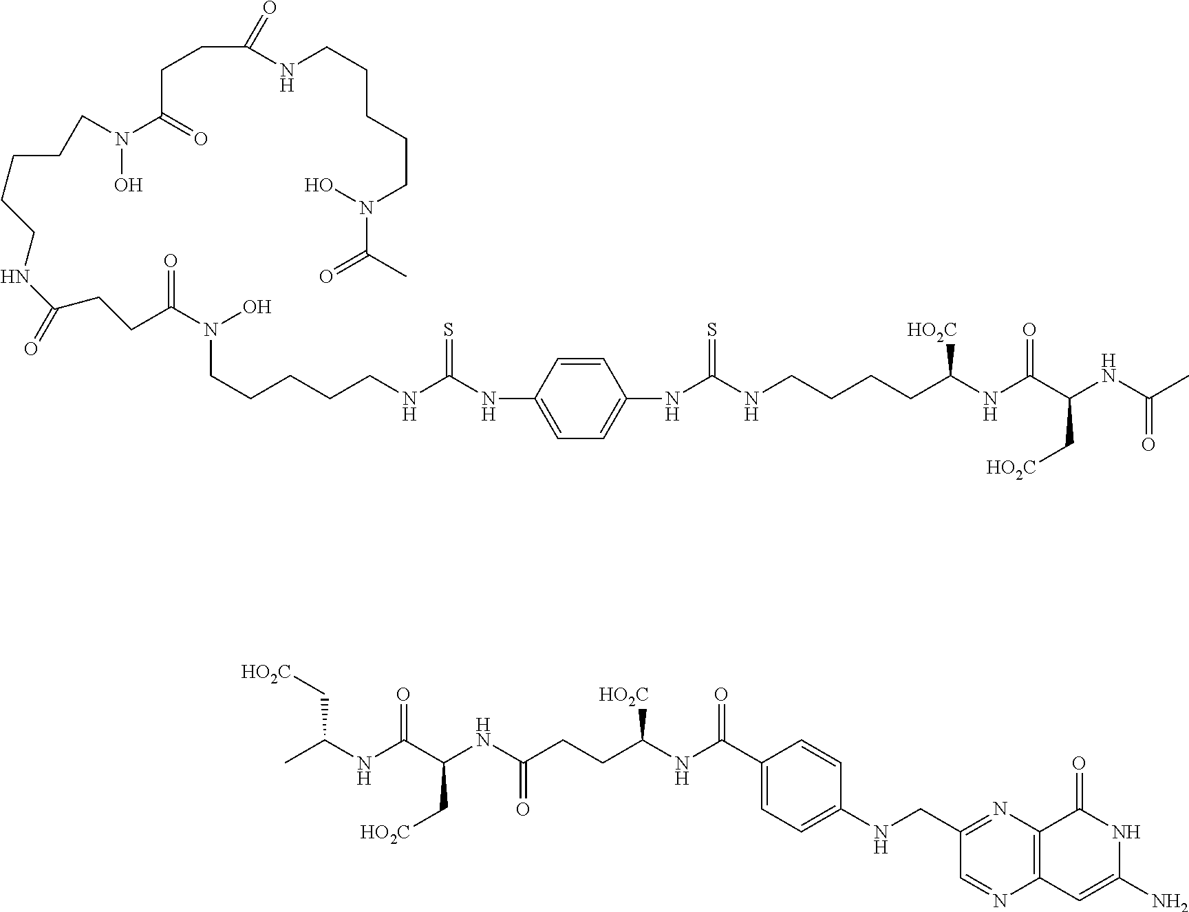
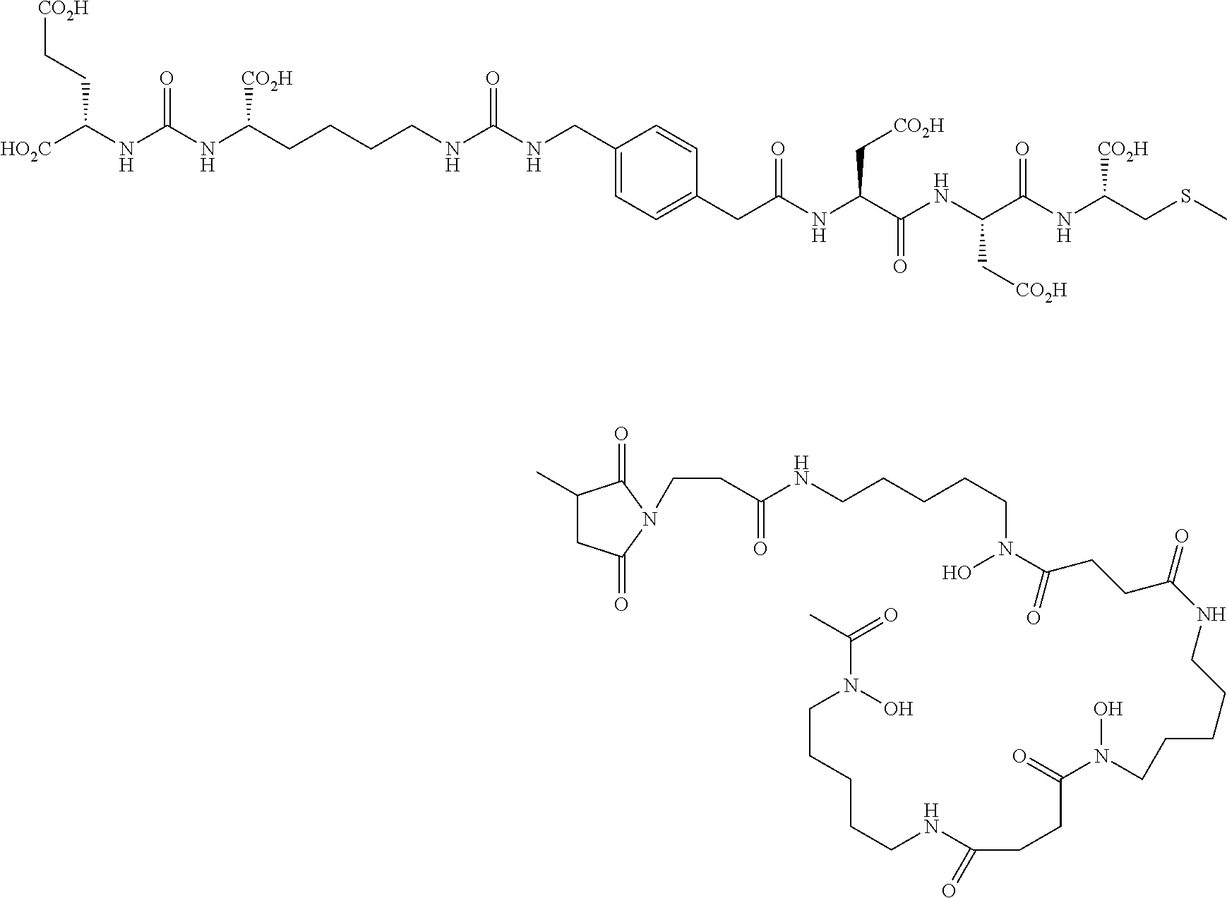
Synthesis of EC2418:
##STR00011##
TABLE-US-00001 TABLE Reagents for peptide synthesis MW Reagents mmol equivalent (g/mol) Amount Fmoc-Lys(MTT)-Resin 1.0 2.632 g (0.38 mmol/g) Fmoc-Asp(Ot-Bu)-OH 2.0 2 411.5 0.822 g Fmoc-Asp(Ot-Bu)-OH 2.0 2 411.5 0.822 g Fmoc-Glu-Ot-Bu 2.0 2 425.5 0.850 g N.sup.10TFA-Pteroic Acid 1.5 1.5 408 0.612 g (dissolve in 10 ml DMSO) DIPEA 4.0 4 129.25 0.697 mL (d = 0.742) PyBOP 2.0 2 520 1.040 g
Coupling Steps: Initial Peptide Synthesis on-Resin:
Commercially available 100-200 mesh peptide-loaded resin was utilized in an AAPPTec-sourced peptide synthesizer equipped with DMF, DMF-Peptide, DMF-PyBOP, DMF-DIPEA, and DMF-piperidine solutions. The desired peptide sequence was programmed into the software interface and run in an automated fashion. Upon completion of the sequence, the peptide-loaded resin was removed from the instrument's reaction flask. Analysis of the resin-peptide was conducted by taking a small quantity of beads, cleaving with TFA and analyzing the filtered solution by LCMS (1-50% ACN/10 mM NH4OAc, pH 5).
Cleavage of Peptide from Resin and Purification:
Peptide was cleaved from the loaded resin by a mixture of 95% TFA, 2.5% TIPS, 2.5% H.sub.2O. Resin was subjected to cleavage mixture under Argon for 35 min, drained, followed by treatment with fresh cleavage mixture for 5 min and drained (2.times.). The combined peptide-TFA solution was diluted with ether to precipitate the peptide and collected by centrifuge. Peptide cake was washed with ether and dried. Crude peptide was suspended in water and Na.sub.2CO.sub.3 was added and maintained at pH 9-10 for 1 h. The reactions mixture was acidified with 1N HCl to pH 4.0 and purified using a Biotage reverse-phase C18 column (Mobile phase A=0.1% TFA buffer and B=ACN). Product fractions were collected, combined, acetonitrile was removed and the resulting solution freeze-dried to yield EC2418 (496 mg, 62%). LCMS (ESI): [M+H].sup.+=Calculated for C.sub.33H.sub.41N.sub.11O.sub.13, 800.29; found 800.36.
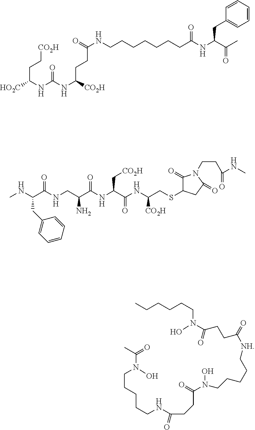
Synthesis of EC2419:
##STR00012##
To a solution of EC1919 (213 mg, 0.23 mM) in DMSO (3.0 mL) and DIPEA (0.88 mL) was added P-SCN-Bn-Deferoxamine (175 mg, 0.23 mM) in DMSO (4.0 mL). The solution was stirred at ambient temperature under argon for 3 h. Reaction mixture was loaded directly onto a Biotage column (mobile phase A=50 mM ammonium bicarbonate buffer, pH=7.0. B=ACN) for purification. Fractions containing the desired product were collected, combined, acetonitrile was removed and the resulting solution freeze-dried to afford the EC2419 (308 mg, 80.3%) as a light yellow solid. LCMS (ESI): [M+H].sup.+=Calculated for C.sub.70H.sub.98N.sub.20O.sub.24S.sub.2, 1667.65; found 1667.79.
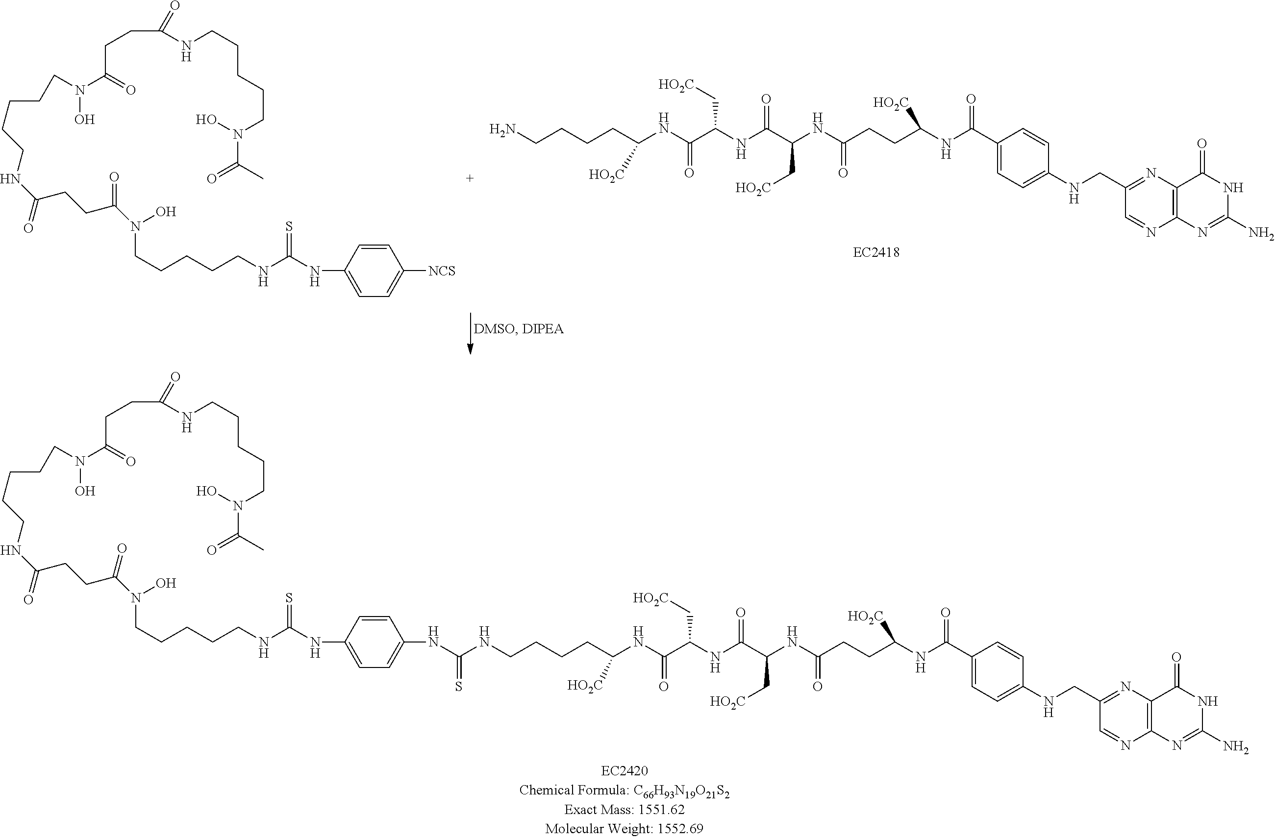
Synthesis of EC2420:
##STR00013##
To a solution of EC2418 (133.9 mg, 0.167 mM) in DMSO (1.0 mL) and DIPEA (0.58 mL) was added P-SCN-Bn-deferoxamine (105 mg, 0.14 mM) in DMSO (3.0 mL) and stirred at ambient temperature under argon for 3 h. Reaction mixture was loaded directly onto a Biotage column (mobile phase A=50 mM ammonium bicarbonate buffer, pH=7.0. B=ACN) for purification. Fractions containing the desired product were collected, combined, acetonitrile was removed and the resulting solution freeze-dried to afford the EC2420 (165 mg, 75.9%) as a light yellow solid. LCMS (ESI): [M+H].sup.+=Calculated for C.sub.66H.sub.93N.sub.19O.sub.21S.sub.2, 1552.62; found 1552.71.
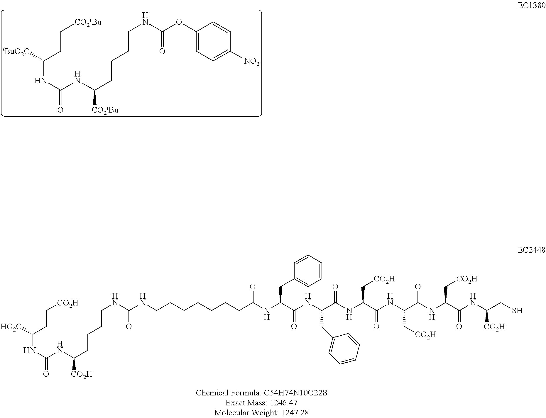
Synthesis of EC2448:
##STR00014##
TABLE-US-00002 TABLE Reagents for peptide synthesis MW Reagents mmol equivalent (g/mol) Amount Fmoc-Cys(trt)-Resin 0.5 0.833 g (0.60 mmol/g) Fmoc-Asp(Ot-Bu)-OH 1.0 2 411.5 0.411 g Fmoc-Asp(Ot-Bu)-OH 1.0 2 411.5 0.411 g Fmoc-Asp(Ot-Bu)-OH 1.0 2 411.5 0.411 g Fmoc-Phe-OH 1.0 2 387.4 0.387 g Fmoc-Phe-OH 1.0 2 387.4 0.387 g Fmoc-8-aminocaprylic Acid 1.0 2 381.4 0.381 g EC1380 1.0 2 652.7 0.653 g DIPEA 2.0 4 129.25 0.348 mL (d = 0.742) PyBOP 1.0 2 520 0.520 g
Coupling Steps: Initial Peptide Synthesis on-Resin:
Commercially-available 100-200 mesh peptide-loaded resin was utilized in an AAPPTec-sourced peptide synthesizer equipped with DMF, DMF-Peptide, DMF-PyBOP, DMF-DIPEA, and DMF-piperidine solutions. The desired peptide sequence, except EC1380, was programmed into the software interface and run in an automated fashion. Upon completion of the sequence, the peptide-loaded resin was removed from the instrument's reaction flask. Analysis of the resin-peptide was conducted by taking a small quantity of beads, cleaving with TFA and analyzing the filtered solution by LCMS (1-50% ACN/10 mM NH.sub.4OAc, pH5).
Addition of Ec1380 to Resin-Bound Peptide:
Resin-bound Peptide obtained through automated synthesis was placed in a traditional bench top solid-phase reaction vessel. N-Fmoc protection was removed using 20% piperidine in DMF under argon for 10 minutes (3.times.). The resin was then rinsed with DMF (3.times.), and IPA (3.times.). The removal of Fmoc was confirmed by Kaiser Test. The resin was then rinsed with DMF (3.times.) and suspended in DMF, with the addition of 2eq of EC1380, 2eq of PyBOP, and 4eq of DIPEA. After 1-2 h of argon bubbling, the solvent was drained and the resin rinsed with DMF (3.times.), and IPA (3.times.). Analysis of the resin-peptide was conducted by taking a small quantity of beads, cleaving with TFA and analyzing the filtered solution by LCMS (1-50% ACN/10 mM NH.sub.4OAc, pH5).
Cleavage of Peptide from Resin and Purification:
Peptide was cleaved from the loaded resin by a mixture of 92.5% TFA, 2.5% TIPS, 2.5% H.sub.2O, and 2.5% EDT. Resin was subjected to cleavage mixture under Argon for 35 min, drained, followed by treatment with fresh cleavage mixture for 5 min and drained (2.times.). The resulting peptide-TFA solution was diluted with ether to precipitate the peptide and collected by centrifuge. Peptide cake was washed with ether and dried. Crude peptide was purified using a Biotage reverse-phase C18 column (Mobile phase A=0.1% TFA buffer and B=ACN). Product fractions were collected, combined, acetonitrile was removed and freeze-dried to yield EC2448 (240 mg, 38.5%) LCMS (ESI): [M+H].sup.+=Calculated for C.sub.54H.sub.74N.sub.10O.sub.22S, 1247.47; found 1247.51.
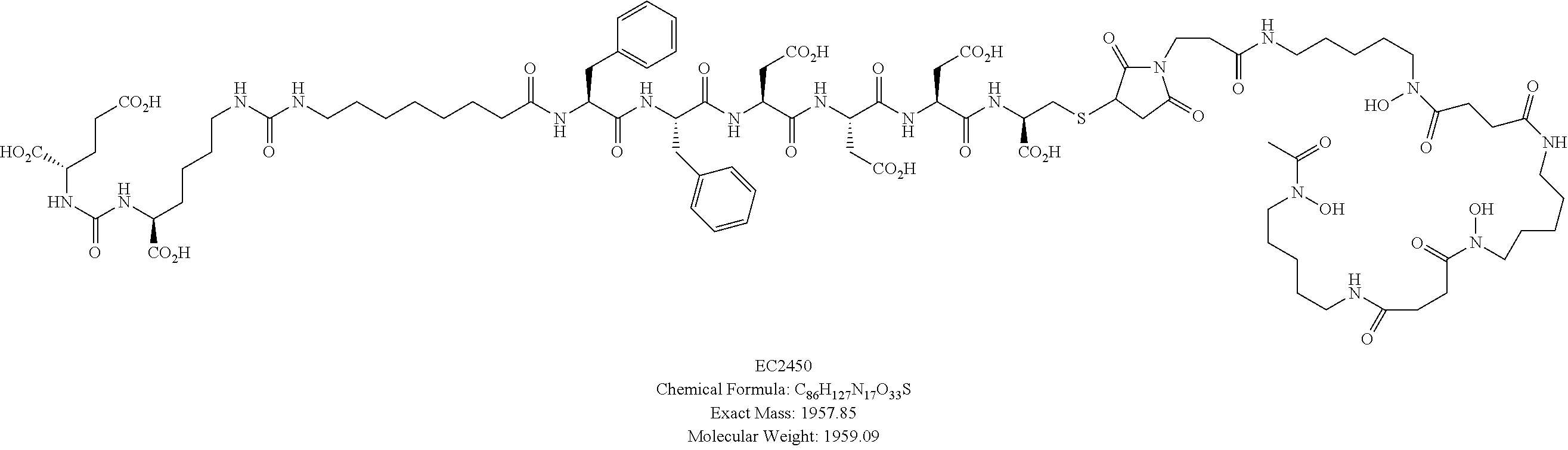
Synthesis of EC2450:
##STR00015## ##STR00016##
To a solution of deferoxamine mesylate (65.7 mg, 0.1 mM) in DMSO (0.3 mL) and DIPEA (0.087 mL) was added .beta.-maleimido-propionic acid N-hydroxysuccinimide ester (26.6 mg, 0.1 mM) in DMSO (0.3 mL) and stirred at ambient temperature under argon for 1 h. Solution of EC2448 (118.5 mg, 0.095 mM) in DMSO (0.5 mL) and DIPEA (0.26 mL) were added and stirred for additional 30 min. Reaction mixture was loaded directly onto a Biotage column (mobile phase A=50 mM ammonium bicarbonate buffer, pH=7.0. B=ACN) for purification. Fractions containing the desired product were collected, combined, acetonitrile was removed and freeze-dried to afford the EC2450 (56 mg, 30.1%, over two steps) as a white solid. LCMS (ESI): [M-2H].sup.2-=Calculated for C.sub.86H.sub.127N.sub.17O.sub.33S, 978.54; found 978.55.
Synthesis of EC2458:
##STR00017## ##STR00018##
To a solution of deferoxamine mesylate (65.7 mg, 0.1 mM) in DMSO (0.3 mL) and DIPEA (0.087 mL) was added .beta.-maleimido-propionic acid N-hydroxysuccinimide ester (26.6 mg, 0.1 mM) in DMSO (0.3 mL) and stirred at ambient temperature under argon for 1 h. Solution of EC1167 (92.8 mg, 0.11 mM) in DMSO (1.0 mL) was added and stirred for additional 3 h. Reaction mixture was loaded directly onto a Biotage column (mobile phase A=50 mM ammonium bicarbonate buffer, pH=7.0. B=ACN) for purification. Fractions containing the desired product were collected, combined, acetonitrile was removed and the resulting solution freeze-dried to afford the EC2458 as a white solid. LCMS (ESI): [M-2H].sup.2-=Calculated for C.sub.65H.sub.98N.sub.14O.sub.28S, 776.81; found 776.67.
Synthesis of EC2460:
##STR00019## ##STR00020##
To a solution of deferoxamine mesylate (65.7 mg, 0.1 mM) in DMSO (0.3 mL) and DIPEA (0.087 mL) was added .beta.-maleimido-propionic acid N-hydroxysuccinimide ester (26.6 mg, 0.1 mM) in DMSO (0.3 mL) and stirred at ambient temperature under argon for 1 h. Solution of EC0652 (116.6 mg, 0.11 mM) in DMSO (0.5 mL) was added and stirred for additional 30 min. Reaction mixture was loaded directly onto a Biotage column (mobile phase A=50 mM ammonium bicarbonate buffer, pH=7.0. B=ACN) for purification. Fractions containing the desired product were collected, combined, acetonitrile was removed and the resulting solution freeze-dried to afford the EC2460 (116 mg, 65.4%, over two steps) as a white solid. LCMS (ESI): [M-2H].sup.2- Calculated for C.sub.79H.sub.118N.sub.16O.sub.28S, 884.97; found 884.86.
The deferoxamine conjugates described above may be complexed to a positron emitting metal ion by any of the procedures known to those skilled in the art of producing PET-imaging conjugates and/or compounds.
* * * * *
References
C00001
C00002
C00003
C00004
C00005
C00006
C00007
C00008
C00009
C00010
C00011
C00012
C00013
C00014
C00015

C00016
C00017

C00018

C00019

C00020

C00021

C00022

C00023
C00024

C00025
C00026

XML
uspto.report is an independent third-party trademark research tool that is not affiliated, endorsed, or sponsored by the United States Patent and Trademark Office (USPTO) or any other governmental organization. The information provided by uspto.report is based on publicly available data at the time of writing and is intended for informational purposes only.
While we strive to provide accurate and up-to-date information, we do not guarantee the accuracy, completeness, reliability, or suitability of the information displayed on this site. The use of this site is at your own risk. Any reliance you place on such information is therefore strictly at your own risk.
All official trademark data, including owner information, should be verified by visiting the official USPTO website at www.uspto.gov. This site is not intended to replace professional legal advice and should not be used as a substitute for consulting with a legal professional who is knowledgeable about trademark law.