Bioprosthetic tissue with reduced calcification
Dove , et al. Ja
U.S. patent number 10,188,511 [Application Number 15/138,123] was granted by the patent office on 2019-01-29 for bioprosthetic tissue with reduced calcification. This patent grant is currently assigned to Edwards Lifesciences Corporation. The grantee listed for this patent is Edwards Lifesciences Corporation. Invention is credited to James A. Davidson, Darin P. Dobler, Jeffrey S. Dove, Gregory A. Wright.


| United States Patent | 10,188,511 |
| Dove , et al. | January 29, 2019 |
Bioprosthetic tissue with reduced calcification
Abstract
A treatment for bioprosthetic tissue used in implants or for assembled bioprosthetic heart valves to reduce in vivo calcification. The method includes applying a calcification mitigant such as a capping agent or an antioxidant to the tissue to specifically inhibit oxidation in tissue. Also, the method can be used to inhibit oxidation in dehydrated tissue. The capping agent suppresses the formation of binding sites in the tissue that are exposed or generated by the oxidation and otherwise would, upon implant, attract calcium, phosphate, immunogenic factors, or other precursors to calcification. In one method, tissue leaflets in assembled bioprosthetic heart valves are pretreated with an aldehyde capping agent prior to dehydration and sterilization.
| Inventors: | Dove; Jeffrey S. (Santa Ana, CA), Dobler; Darin P. (Aliso Viejo, CA), Davidson; James A. (San Juan Capistrano, CA), Wright; Gregory A. (Orange, CA) | ||||||||||
|---|---|---|---|---|---|---|---|---|---|---|---|
| Applicant: |
|
||||||||||
| Assignee: | Edwards Lifesciences
Corporation (Irvine, CA) |
||||||||||
| Family ID: | 40789552 | ||||||||||
| Appl. No.: | 15/138,123 | ||||||||||
| Filed: | April 25, 2016 |
Prior Publication Data
| Document Identifier | Publication Date | |
|---|---|---|
| US 20160235528 A1 | Aug 18, 2016 | |
Related U.S. Patent Documents
| Application Number | Filing Date | Patent Number | Issue Date | ||
|---|---|---|---|---|---|
| 14707970 | Apr 26, 2016 | 9320830 | |||
| 14139367 | May 12, 2015 | 9029418 | |||
| 13745496 | Jun 10, 2014 | 8748490 | |||
| 12338872 | Jan 22, 2013 | 8357387 | |||
| 61016263 | Dec 21, 2007 | ||||
| Current U.S. Class: | 1/1 |
| Current CPC Class: | A61F 2/2415 (20130101); A61L 27/3629 (20130101); A61F 2/2412 (20130101); A61F 2/24 (20130101); A61L 27/3604 (20130101); A61L 27/3691 (20130101); A61L 27/3687 (20130101); A61L 27/507 (20130101); A61F 2/0095 (20130101); A61L 27/3625 (20130101); A61F 2/2418 (20130101); A61L 2400/02 (20130101); A61L 2430/20 (20130101) |
| Current International Class: | A61F 2/24 (20060101); A61L 27/50 (20060101); A61F 2/00 (20060101); A61L 27/36 (20060101) |
References Cited [Referenced By]
U.S. Patent Documents
| 2393580 | January 1946 | Weiskopf |
| 2484813 | October 1949 | Bower |
| 2567929 | September 1951 | Fessenden |
| 4067091 | January 1978 | Backman |
| 4120649 | October 1978 | Schechter |
| 4120991 | October 1978 | Ornstein et al. |
| 4197658 | April 1980 | Fraser |
| 4207689 | June 1980 | Romera-Sierra et al. |
| 4244992 | January 1981 | von Hagens |
| 4294753 | October 1981 | Urist |
| 4323358 | April 1982 | Lentz et al. |
| 4328256 | May 1982 | Romero-Sierra et al. |
| 4347671 | September 1982 | Dias et al. |
| 4350492 | September 1982 | Wright et al. |
| 4372743 | February 1983 | Lane |
| 4378224 | March 1983 | Nimni et al. |
| 4402697 | September 1983 | Pollock et al. |
| 4405327 | September 1983 | Pollock |
| 4481009 | November 1984 | Nashef |
| 4553974 | November 1985 | Dewanjee |
| 4599084 | July 1986 | Nashef |
| 4624822 | November 1986 | Arru et al. |
| 4647283 | March 1987 | Carpentier et al. |
| 4648881 | March 1987 | Carpentier et al. |
| 4655773 | April 1987 | Grassi |
| 4676070 | June 1987 | Linner |
| 4729139 | March 1988 | Nashef |
| 4753652 | June 1988 | Langer et al. |
| 4755593 | July 1988 | Lauren |
| 4770665 | September 1988 | Nashef |
| 4776853 | October 1988 | Klement et al. |
| 4786287 | November 1988 | Nashef et al. |
| 4831065 | May 1989 | Pietsch et al. |
| 4838888 | June 1989 | Nashef |
| 4865871 | September 1989 | Livesey et al. |
| 4885005 | December 1989 | Nashef et al. |
| 4891319 | January 1990 | Roser |
| 4958008 | September 1990 | Petite et al. |
| 4975526 | December 1990 | Kuberasampath et al. |
| 4976733 | December 1990 | Girardot |
| 4994030 | February 1991 | Glowczewskie, Jr. et al. |
| 4994237 | February 1991 | Login et al. |
| 4996054 | February 1991 | Pietsch et al. |
| 5002566 | March 1991 | Carpentier et al. |
| 5011913 | April 1991 | Benedict et al. |
| 5024830 | June 1991 | Linner |
| 5044165 | September 1991 | Linner et al. |
| 5051401 | September 1991 | Sikes |
| 5068100 | November 1991 | McClanahan |
| 5080670 | January 1992 | Imamura et al. |
| 5094661 | March 1992 | Levy et al. |
| 5104405 | April 1992 | Nimni |
| 5108923 | April 1992 | Benedict et al. |
| 5116564 | May 1992 | Jansen et al. |
| 5147514 | September 1992 | Mechanic |
| 5149621 | September 1992 | McNally et al. |
| 5149653 | September 1992 | Roser |
| 5154007 | October 1992 | Piunno et al. |
| 5200399 | April 1993 | Wettlaufer et al. |
| 5215541 | June 1993 | Nashef et al. |
| 5279612 | January 1994 | Eberhardt |
| 5290558 | March 1994 | O'Leary et al. |
| 5296583 | March 1994 | Levy |
| 5329846 | July 1994 | Bonutti |
| 5332475 | July 1994 | Mechanic |
| 5336616 | August 1994 | Livesey et al. |
| 5368608 | November 1994 | Levy et al. |
| 5397353 | March 1995 | Oliver et al. |
| 5424047 | June 1995 | Zwingenberger et al. |
| 5436291 | July 1995 | Levy et al. |
| 5437287 | August 1995 | Phillips et al. |
| 5447536 | September 1995 | Girardot et al. |
| 5447724 | September 1995 | Helmus et al. |
| 5460962 | October 1995 | Kemp |
| 5476516 | December 1995 | Seifter et al. |
| 5507813 | April 1996 | Dowd et al. |
| 5509932 | April 1996 | Keogh et al. |
| 5556379 | September 1996 | Wolfinbarger |
| 5558875 | September 1996 | Wang |
| 5595571 | January 1997 | Jaffe et al. |
| 5613982 | March 1997 | Goldstein |
| 5632778 | May 1997 | Goldstein |
| 5645587 | July 1997 | Chanda et al. |
| 5674298 | October 1997 | Levy et al. |
| 5679112 | October 1997 | Levy et al. |
| 5695729 | December 1997 | Chow et al. |
| 5695820 | December 1997 | Davis et al. |
| 5697972 | December 1997 | Kim et al. |
| 5713953 | February 1998 | Vallana et al. |
| 5720777 | February 1998 | Jaffe et al. |
| 5720894 | February 1998 | Neev et al. |
| 5733339 | March 1998 | Girardot et al. |
| 5746775 | May 1998 | Levy et al. |
| 5762600 | June 1998 | Bruchman et al. |
| 5766520 | June 1998 | Bronshtein |
| 5769780 | June 1998 | Hata et al. |
| 5776182 | July 1998 | Bruchman et al. |
| 5782914 | July 1998 | Schankereli |
| 5782915 | July 1998 | Stone |
| 5782931 | July 1998 | Yang et al. |
| 5843180 | December 1998 | Jaffe et al. |
| 5843181 | December 1998 | Jaffe et al. |
| 5843182 | December 1998 | Goldstein |
| 5855620 | January 1999 | Bishopric et al. |
| 5856102 | January 1999 | Bierke-Nelson et al. |
| 5856172 | January 1999 | Greenwood et al. |
| 5862806 | January 1999 | Cheung |
| 5865849 | February 1999 | Stone |
| 5873812 | February 1999 | Ciana et al. |
| 5879383 | March 1999 | Bruchman et al. |
| 5882850 | March 1999 | Khor et al. |
| 5899936 | May 1999 | Goldstein |
| 5902338 | May 1999 | Stone |
| 5904718 | May 1999 | Jefferies |
| 5911951 | June 1999 | Girardot et al. |
| 5913900 | June 1999 | Stone |
| 5919472 | July 1999 | Trescony et al. |
| 5921980 | July 1999 | Kim |
| 5922027 | July 1999 | Stone |
| 5931969 | August 1999 | Carpentier et al. |
| 5935168 | August 1999 | Yang et al. |
| 5945319 | August 1999 | Keogh |
| 5977153 | November 1999 | Camiener |
| 5987720 | November 1999 | Yamamoto |
| 5993844 | November 1999 | Abraham et al. |
| 6008292 | December 1999 | Lee et al. |
| 6017741 | January 2000 | Keogh |
| 6024735 | February 2000 | Wolfinbarger, Jr. |
| 6063120 | May 2000 | Stone |
| 6066160 | May 2000 | Colvin et al. |
| 6093204 | July 2000 | Stone |
| 6093530 | July 2000 | McIlroy et al. |
| 6106555 | August 2000 | Yang |
| 6117979 | September 2000 | Hendriks et al. |
| 6121041 | September 2000 | Mirsch, II et al. |
| 6132473 | October 2000 | Williams et al. |
| 6132986 | October 2000 | Pathak et al. |
| 6156030 | December 2000 | Neev |
| 6156531 | December 2000 | Pathak et al. |
| 6165215 | December 2000 | Rottenberg et al. |
| 6166184 | December 2000 | Hendriks et al. |
| 6174331 | January 2001 | Moe et al. |
| 6177514 | January 2001 | Pathak et al. |
| 6190407 | February 2001 | Ogle et al. |
| 6193749 | February 2001 | Schroeder et al. |
| 6203755 | March 2001 | Odland |
| 6206917 | March 2001 | Williams et al. |
| 6210957 | April 2001 | Carpentier et al. |
| 6214054 | April 2001 | Cunanan et al. |
| 6214055 | April 2001 | Simionescu et al. |
| 6231608 | May 2001 | Stone |
| 6231614 | May 2001 | Yang |
| 6251579 | June 2001 | Moore et al. |
| 6254635 | July 2001 | Schroeder et al. |
| 6258320 | July 2001 | Persing et al. |
| 6267786 | July 2001 | Stone |
| 6277555 | August 2001 | Duran et al. |
| 6287338 | September 2001 | Samowski et al. |
| 6290991 | September 2001 | Roser et al. |
| 6293970 | September 2001 | Wolfinbarger, Jr. et al. |
| 6302909 | October 2001 | Ogle et al. |
| 6312474 | November 2001 | Francis et al. |
| 6322593 | November 2001 | Pathak et al. |
| 6322994 | November 2001 | Reid |
| 6328762 | December 2001 | Anderson et al. |
| 6334873 | January 2002 | Lane et al. |
| 6352708 | March 2002 | Duran et al. |
| 6364905 | April 2002 | Simpson et al. |
| 6372228 | April 2002 | Gregory |
| 6375680 | April 2002 | Carlyle |
| 6376244 | April 2002 | Atala |
| 6383732 | May 2002 | Stone |
| 6391538 | May 2002 | Vyavahare et al. |
| 6394096 | May 2002 | Constantz |
| 6448076 | September 2002 | Dennis et al. |
| 6468660 | October 2002 | Ogle |
| 6471723 | October 2002 | Ashworth et al. |
| 6479079 | November 2002 | Pathak et al. |
| 6482199 | November 2002 | Neev |
| 6506339 | January 2003 | Girardot et al. |
| 6509145 | January 2003 | Torrianni |
| 6527979 | March 2003 | Constantz |
| 6531310 | March 2003 | Mirsch, II et al. |
| 6534004 | March 2003 | Chen et al. |
| 6547827 | April 2003 | Carpentier et al. |
| 6561970 | May 2003 | Carpentier et al. |
| 6569200 | May 2003 | Wolfinbarger, Jr. et al. |
| 6582464 | June 2003 | Gabbay |
| 6586006 | July 2003 | Roser et al. |
| 6586573 | July 2003 | Besman et al. |
| 6589591 | July 2003 | Mansouri et al. |
| 6605667 | August 2003 | Badejo et al. |
| 6613278 | September 2003 | Mills et al. |
| 6617142 | September 2003 | Keogh et al. |
| 6630001 | October 2003 | Duran et al. |
| 6652594 | November 2003 | Francis et al. |
| 6653062 | November 2003 | DePablo et al. |
| 6682559 | January 2004 | Myers et al. |
| 6685940 | February 2004 | Andya et al. |
| 6696074 | February 2004 | Dai et al. |
| 6734018 | May 2004 | Wolfinbarger, Jr. et al. |
| 6753181 | June 2004 | Atala |
| 6790229 | September 2004 | Berreklouw |
| 6797000 | September 2004 | Simpson et al. |
| 6828310 | December 2004 | Barresi et al. |
| 6872226 | March 2005 | Cali et al. |
| 6878168 | April 2005 | Carpentier et al. |
| 6893666 | May 2005 | Spievack |
| 6908591 | June 2005 | MacPhee et al. |
| 6911043 | June 2005 | Myers et al. |
| 6919172 | July 2005 | DePablo et al. |
| 6933326 | August 2005 | Griffey et al. |
| 7008763 | March 2006 | Cheung |
| 7029434 | April 2006 | Carpentier et al. |
| 7037333 | May 2006 | Myers et al. |
| 7063726 | June 2006 | Crouch et al. |
| 7087723 | August 2006 | Besman et al. |
| 7141064 | November 2006 | Scott et al. |
| 7143769 | December 2006 | Stoltz et al. |
| 7214344 | May 2007 | Carpentier et al. |
| 7238200 | July 2007 | Lee et al. |
| 7318998 | January 2008 | Goldstein et al. |
| 7338757 | March 2008 | Wolfinbarger, Jr. et al. |
| 7354749 | April 2008 | Fisher et al. |
| 7358284 | April 2008 | Griffey et al. |
| 7367969 | May 2008 | Stoltz et al. |
| RE40570 | November 2008 | Carpentier et al. |
| 7498565 | March 2009 | Silberberg et al. |
| 7579381 | August 2009 | Dove |
| 7594974 | September 2009 | Cali et al. |
| 7648676 | January 2010 | Mills et al. |
| 7682304 | March 2010 | Heyninck-Jantz et al. |
| 7914569 | March 2011 | Nguyen et al. |
| 7919112 | April 2011 | Pathak et al. |
| 7972376 | July 2011 | Dove |
| 8007992 | August 2011 | Tian |
| 8043450 | October 2011 | Cali et al. |
| 8075615 | December 2011 | Eberhardt et al. |
| 8105375 | January 2012 | Navia et al. |
| 8136218 | March 2012 | Millwee et al. |
| 8308797 | November 2012 | Paniagua et al. |
| 8357387 | January 2013 | Dove |
| 8361144 | January 2013 | Fish et al. |
| 8377143 | February 2013 | Hamby et al. |
| 8475827 | July 2013 | Hamby et al. |
| 8748490 | June 2014 | Dove |
| 9029418 | May 2015 | Dove |
| 9066785 | June 2015 | Tran |
| 9320830 | April 2016 | Dove |
| 9320930 | April 2016 | Abels |
| 2001/0000804 | May 2001 | Goldstein et al. |
| 2001/0020191 | September 2001 | Williams et al. |
| 2001/0025196 | September 2001 | Chinn et al. |
| 2001/0027344 | October 2001 | Bonutti |
| 2001/0032024 | October 2001 | Cunanan et al. |
| 2001/0039459 | November 2001 | Stone |
| 2002/0001834 | January 2002 | Keogh et al. |
| 2002/0091441 | July 2002 | Guzik |
| 2002/0111532 | August 2002 | Pathak et al. |
| 2003/0035843 | February 2003 | Livesey et al. |
| 2003/0125805 | July 2003 | Johnson et al. |
| 2003/0135284 | July 2003 | Crouch et al. |
| 2003/0167089 | September 2003 | Lane |
| 2003/0212454 | November 2003 | Scott et al. |
| 2004/0030381 | February 2004 | Shu |
| 2004/0086543 | May 2004 | Keogh et al. |
| 2004/0158320 | August 2004 | Simionescu et al. |
| 2004/0193259 | September 2004 | Gabbay |
| 2005/0010773 | January 2005 | Lapstun et al. |
| 2005/0119736 | June 2005 | Zilla et al. |
| 2005/0136510 | June 2005 | Hendriks et al. |
| 2005/0211680 | September 2005 | Li et al. |
| 2006/0084957 | April 2006 | Delfyett et al. |
| 2006/0099326 | May 2006 | Keogh et al. |
| 2006/0110370 | May 2006 | Pathak et al. |
| 2006/0159641 | July 2006 | Girardot et al. |
| 2006/0177426 | August 2006 | Gibson et al. |
| 2006/0193885 | August 2006 | Leonard Neethling et al. |
| 2006/0210960 | September 2006 | Livesey et al. |
| 2006/0217804 | September 2006 | Dove |
| 2006/0217805 | September 2006 | Dove |
| 2007/0050014 | March 2007 | Johnson |
| 2007/0073392 | March 2007 | Heyninck-Jantz et al. |
| 2007/0203576 | August 2007 | Lee et al. |
| 2007/0254005 | November 2007 | Pathak et al. |
| 2007/0292459 | December 2007 | Cooper et al. |
| 2008/0302372 | December 2008 | Davidson et al. |
| 2008/0319166 | December 2008 | Shen |
| 2009/0041729 | February 2009 | Wolfinbarger, Jr. et al. |
| 2009/0130162 | May 2009 | Pathak et al. |
| 2009/0137999 | May 2009 | Silberberg et al. |
| 2009/0188900 | July 2009 | Cali et al. |
| 2009/0326524 | December 2009 | Cali et al. |
| 2010/0036484 | February 2010 | Hariton et al. |
| 2011/0092966 | April 2011 | Guo et al. |
| 2011/0177150 | July 2011 | Pathak et al. |
| 2011/0214398 | September 2011 | Liburd et al. |
| 2011/0238167 | September 2011 | Dove et al. |
| 2011/0295363 | December 2011 | Girard et al. |
| 2011/0300625 | December 2011 | Paniagua et al. |
| 2011/0306124 | December 2011 | Strasly et al. |
| 2011/0311493 | December 2011 | Dove et al. |
| 2012/0035720 | February 2012 | Cali et al. |
| 2012/0059487 | March 2012 | Cunanan et al. |
| 2012/0067855 | March 2012 | Guo et al. |
| 2012/0078356 | March 2012 | Fish et al. |
| 2012/0095551 | April 2012 | Navia et al. |
| 2012/0123557 | May 2012 | Carpentier et al. |
| 2012/0185038 | July 2012 | Fish et al. |
| 2012/0328905 | December 2012 | Guo et al. |
| 2013/0122583 | May 2013 | Neethling |
| 2013/0238088 | September 2013 | Navia et al. |
| 0169259 | Jan 1986 | EP | |||
| 2394673 | Dec 2011 | EP | |||
| 84/01894 | May 1984 | WO | |||
| 95/11047 | Apr 1995 | WO | |||
| 95/022361 | Aug 1995 | WO | |||
| 95/034332 | Dec 1995 | WO | |||
| 96/013227 | May 1996 | WO | |||
| 98007452 | Feb 1998 | WO | |||
| 98043556 | Oct 1998 | WO | |||
| 00/032252 | Jun 2000 | WO | |||
| 03/037227 | May 2003 | WO | |||
| 2004/082536 | Sep 2004 | WO | |||
| 2006/026325 | Mar 2006 | WO | |||
| 2006/099334 | Sep 2006 | WO | |||
| 2013009581 | Jan 2013 | WO | |||
Other References
|
Carpentier, A., et al., "Biological Factors Affecting Long-Term Results of Valvular Heterografts," Forty-ninth Meeting of the American Association for Thoracic Surgery, San Francisco, CA, Mar. 31-Apr. 2, 1969. cited by applicant . Chanda, J., et al., "Heparin in Calcification Prevention of Porcine Pericardial Bioprostheses," Biomaterials, Elsevier Science Publishers,vol. 18, No. 16, ISSN: 0142-9612, Aug. 1, 1997. cited by applicant . Chvapil, M., et al., "Use of Chemically Purified and Cross-Linked Bovine Pericardium as a Ligament Substitute," Journal of Biomedical Materials Research, vol. 21, No. 12, pp. 1383-1394, 1987, University of Arizona health Science Center, Tucson, AZ. cited by applicant . Dahm, Manfred, et al., "Effects of Surface Seeding with Vital Cells on the Calcium Uptake of Biological Materials for Heart Valve Replacement," J Heart Valve Dis, vol. 5, No. 2, Mar. 1996, 148-151. cited by applicant . Fahner, P., et al., "Systematic Review of Preservation Methods and Clinical Outcome of Infrainguinal Vascular Allografts," Journal of Vascular Surgery, vol. 44, No. 3 , pp. 518-524, 2006. cited by applicant . Fumoto, H., et al., "Performance of Bioprosthetic Valves after Glycerol Dehydration, Ethylene Oxide Steilization, and Rehydration," Innovations, vol. 6, No. 1, Jan./ Feb. 2011. cited by applicant . Grabenwoger, M., et al., "Decreased Tissue Reaction to Bioprosthetic Heart Valve Material after L-glutamic acid Treatment. A Morphological Study," J. Biomed Mater. Res. Sep. 1992; 26(9): 1231-40. cited by applicant . Grant, R.A., et al., The Effects of Irradiation With High Energy Electrons on the Structure and Reactivity of Native and Cross-Linked Collagen Fibres, 1970, pp. 387-405, J. Cell Sci 7. cited by applicant . Hauschka, P., et al., Direct Identification of the Calcium-Binding Amino Acid .gamma.-Carboxyglutamate, in Mineralized Tissue, Oct. 1975, pp. 3295-3929, Proc. Nat. Acad. Sci, vol. 72, No. 10. cited by applicant . Jayakrishnan, A., et al., "Glutaraldehyde as a Fixative in Bioprostheses and Drug Delivery Matrices," Biomaterial, vol. 17, Issue 5, 1996, pp. 471-484. cited by applicant . Khora, Eugene, "Methods for the Treatment of Collagenous Tissues for Bioprostheses," Biomaterials, vol. 18, Issue. 2, Jan. 1997, pp. 95-105. cited by applicant . Liao, K., et al., "Mechanical Stress: An Independent Determinant of Early Bioprosthetic Calcification in Humans," Annals of Thoracic Surgery 2008;86: 491-495. cited by applicant . Nieethling, W., et al., "Enhanced Biostability and Biocompatibility of Decellularized Bovine Pericardium, Crosslinked with an Ultra-Low Concentration Monomeric Aldehyde and Treated with ADAPT.RTM.," J. Heart Valve Dis. 2008; 17: 456-464. cited by applicant . Ohan, M.P., et al., Glucose Stabilizes Collagen Sterilized With Gamma Irradiation, 2003, pp. 1188-1195, Wiley Periodicals, Inc., J Biomed Mater Res 67A. cited by applicant . Olde Damink, L.H.H., et al., Influence of Ethylene Oxide Gas Treatment on the in Vitro Degradation Behavior of Dermal Sheep Collagen, 1995, Journal of Biomedical Materials Research, vol. 29, 149-155, John Wiley & Sons, Inc. cited by applicant . PCT/US2008/013879 International Search Report Dated Mar. 11, 2009. cited by applicant . R Parker, et al. Storage of Heart Valve Allografts in Glycerol With Subsequent Antibiotic Sterilisation, Thorax, 1978, 538-645, vol. 33:5, British Thoracic Society, London, UK. cited by applicant . Saegeman, V., et al, "Short and long term bacterial inhibiting effect of high concentrations of glycerol used in the prevention of skin allografts," Science Direct, Burns, No. 34, Mar. 2008. cited by applicant . Schmidt, C., et al., "Acellular Vascular Tissues: Natural Biomaterials for Tissue Repair and Tissue Engineering," Biomaterial, vol. 21, pp. 2215-2231, 2000. cited by applicant . Trantina-Yates, A.E., et al., "Detoxification of Top Enhanced, Diamine-Extended Glutaraldehyde Fixation Significantly Reduces Bioprosthetic Root Calcification in the Sheep Model," J. Heart Valve Dis. Jan. 2003; 12(1): 93-100. cited by applicant . Zilla, P., et al., "Carbodiimide Treatment Dramatically Potentiates the Anticalcific Effect of Alpha-Amino Oleic Acid on Glutaraldehyde-Fixed Aortic Wall Tissue," The Annals of Thoracic Surgery, Elsevier, vol. 79, No. 3, ISSN: 0003-4975; Mar. 1, 2005. cited by applicant. |
Primary Examiner: Wax; Robert A
Assistant Examiner: Mercier; Melissa S
Attorney, Agent or Firm: Sheppard Mullin Kim; Michelle
Parent Case Text
CROSS-REFERENCE TO RELATED APPLICATIONS
This application is a divisional of U.S. patent application Ser. No. 14/707,970, filed May 8, 2015, now U.S. Pat. No. 9,320,830, which is a continuation of U.S. patent application Ser. No. 14/139,367, filed Dec. 23, 2013, now U.S. Pat. No. 9,029,418, which is a continuation of U.S. patent application Ser. No. 13/745,496, filed Jan. 18, 2013, now U.S. Pat. No. 8,748,490, which is a continuation of U.S. patent application Ser. No. 12/338,872 filed Dec. 18, 2008, now U.S. Pat. No. 8,357,387, which claims the benefit of U.S. Patent Application No. 61/016,263, filed Dec. 21, 2007, the entire disclosures of which are incorporated by reference herein.
Claims
What is claimed is:
1. A packaged bioprosthetic device comprising: an at least partially cross-linked pericardial tissue comprising functional groups that have been blocked, removed or altered by reacting the functional groups with capping agents, wherein the functional groups are one or both of aldehyde groups and carboxylic acid groups; and a packaging storing the at least partially cross-linked biological tissue without a liquid preservative solution.
2. The packaged bioprosthetic device of claim 1, wherein the bioprosthetic device is a bioprosthetic heart valve comprising a stent coupled to leaflets made from the at least partially cross-linked biological tissue.
3. The packaged bioprosthetic device of claim 1, wherein the biological tissue is at least partially cross-linked with glutaraldehyde or other aldehyde-containing agents.
4. The packaged bioprosthetic device of claim 1, wherein the at least a portion of the functional groups are blocked, removed or altered by exposing the at least partially cross-linked biological tissue to a reducing agent.
5. The packaged bioprosthetic device of claim 1, wherein the at least partially cross-linked biological tissue comprises glycerol.
6. The packaged bioprosthetic device of claim 5, wherein the at least partially cross-linked biological tissue and the packaging is sterilized by ethylene oxide, gamma irradiation or electron beam irradiation.
7. The packaged bioprosthetic device of claim 1, wherein the at least partially cross-linked biological tissue is at least partially dehydrated.
8. The packaged bioprosthetic device of claim 7, wherein the at least partially cross-linked biological tissue and the packaging is sterilized by ethylene oxide, gamma irradiation or electron beam irradiation.
9. The packaged bioprosthetic device of claim 1, wherein the capping agent is one or more selected from the group consisting of: an amine, an alkyl amine, an amino alcohol, an ethanolamine, an amino acid, a lysine, a hydroxylysine, an amino sulfonate, a taurine, an amino sulfate, a dextran sulfate, a chondroitin sulfate, a hydrophilic multifunctional polymer, a polyvinyl alcohol, a polyethyleneimine, a hydrophobic multifunctional polymer, an .alpha.-dicarbonyl, a methylglyoxal, a 3-deoxyglucose, a glyoxal, a hydrazide, an adipic hydrazide, a heparin, a mono-epoxy alkane, a di-epoxy alkane, and a polyepoxyalkane.
10. The packaged bioprosthetic device of claim 9, wherein the functional groups have been reacted with a reducing agent.
11. The packaged bioprosthetic device of claim 10, wherein the reducing agent is selected from the group consisting of: a sodium borohydride and a sodium cyanoborohydride.
12. The packaged bioprosthetic device of claim 11, wherein the bioprosthetic tissue comprises glycerol.
13. The packaged bioprosthetic device of claim 1, wherein the biological tissue is at least partially cross-linked with glutaraldehyde or an aldehyde-containing agent.
14. The packaged bioprosthetic device of claim 1, wherein the capping agent is one or more selected from the group consisting of: an amine, an alkyl amine, an amino alcohol, an ethanolamine, an amino acid, a lysine, a hydroxylysine, an amino sulfonate, a taurine, an amino sulfate, a dextran sulfate, a chondroitin sulfate, a hydrophilic multifunctional polymer, a polyvinyl alcohol, a polyethyleneimine, a hydrophobic multifunctional polymer, an .alpha.-dicarbonyl, a methylglyoxal, a 3-deoxyglucose, a glyoxal, a hydrazide, an adipic hydrazide, a heparin, a mono-epoxy alkane, a di-epoxy alkane, and a polyepoxyalkane.
15. The packaged bioprosthetic device of claim 1, wherein the functional groups have been reacted with a reducing agent.
16. The packaged bioprosthetic device of claim 15, wherein the reducing agent is selected from the group consisting of: a sodium borohydride and a sodium cyanoborohydride.
17. The packaged bioprosthetic device of claim 16, wherein the bioprosthetic tissue comprises glycerol.
Description
FIELD OF THE INVENTION
The present invention relates generally to methods for treating bioprosthetic tissue, materials, and devices to reduce post-implantation calcification, and reducing post-implantation calcification, in particular for bioprosthetic tissue used in heart valves.
BACKGROUND OF THE INVENTION
Heart valve replacement may be indicated when there is a narrowing of the native heart valve, commonly referred to as stenosis, or when the native valve leaks or regurgitates, such as when the leaflets are calcified. In one therapeutic solution, the native valve may be excised and replaced with either a biologic or a mechanical valve. Certain medical conditions may require grafting or suturing a tissue patch to repair physiological abnormalities. These include, but are not limited to hernia repair, vascular wounds, congenital heart defect repair and reconstruction, and bladder wall repair.
Tissue-type or "bioprosthetic" valves have flexible tissue leaflets supported by a base structure that project into the flow stream and function much like those of a natural human heart valve by coapting against each other to ensure one-way blood flow. In tissue-type valves, a whole xenograft valve (e.g., porcine) or a plurality of xenograft leaflets (e.g., bovine or equine pericardium) typically provide fluid occluding surfaces. Synthetic tissue leaflets have also been proposed. One or more flexible leaflets mount within a peripheral support structure, for example as seen in the CARPENTIER-EDWARDS Porcine Heart Valve and PERIMOUNT Pericardial Heart Valve available from Edwards Lifesciences of Irvine, Calif.
Implantable biological tissues can be formed of human tissues preserved by freezing (i.e., cryopreservation) the homograft tissues, or of animal tissues preserved by chemically fixing (i.e., tanning) the xenograft tissues. The type of biological tissues used as bioprostheses include cardiac valves, blood vessels, skin, dura mater, pericardium, small intestinal submucosa ("SIS tissue"), ligaments and tendons. These biological tissues typically contain connective tissue proteins (i.e., collagen and elastin) that act as the supportive framework of the tissue. The pliability or rigidity of each biological tissue is largely determined by the relative amounts of collagen and elastin present within the tissue and/or by the physical structure and configuration of its connective tissue framework. Collagen is the most abundant connective tissue protein present in most tissues. Each collagen molecule is made up of three (3) polypeptide chains intertwined in a coiled helical configuration.
The techniques used for chemical fixation of biological tissues typically involve exposing the biological tissue to one or more chemical fixatives (i.e., tanning agents) which form cross-links between the polypeptide chains within a given collagen molecule (i.e., intramolecular cross-linkages), or between adjacent collagen molecules (i.e., intermolecular cross-linkages). Examples of chemical fixative agents that have been used to cross-link collagenous tissues include: formaldehyde, glutaraldehyde, dialdehyde starch, hexamethylene diisocyanate and certain polyepoxy compounds.
One problem associated with the implantation of many bioprosthetic materials is that the connective tissue proteins (i.e., collagen and elastin) within them can become calcified following implantation in the body. Such calcification can result in undesirable stiffening or degradation of the bioprosthesis. This damage to the collagenous tissue leads to valve failure.
Of the various chemical fixatives available, glutaraldehyde (also referred to as simply "glut") has been the most widely used since the discovery of its anti-immunological and anti-degenerative effects by Dr. Alain Carpentier in 1968. See Carpentier, A., J. Thorac. Cardiovascular Surgery, 58: 467-69 (1969). In addition, glutaraldehyde is one of the most common sterilization agents. Glutaraldehyde is therefore used as the preferred fixative and sterilant for many commercially available bioprosthetic products, such as in the bioprosthetic heart valves available from Edwards Lifesciences of Irvine, Cali. Glutaraldehyde creates potential calcium binding sites within the tissue that can lead to calcification in vivo, such as residual aldehydes, acids, Schiff bases, etc. These groups can contribute to calcification unless mitigated via capping. Mitigating such calcification is particularly important during storage, especially when the tissue is not being stored in aqueous solution.
Various techniques have been proposed for mitigating the in vivo calcification of glutaraldehyde-fixed bioprostheses or for otherwise improving the glutaraldehyde fixation process. Among these are methods described in U.S. Pat. No. 4,729,139 (Nashef); U.S. Pat. No. 4,885,005 (Nashef et al.); U.S. Pat. No. 4,648,881 (Carpentier et al.); U.S. Pat. No. 5,002,566 (Carpentier); EP 103947 (Pollock et al.), U.S. Pat. No. 5,476,516 (Seifter et al.), and U.S. Pat. No. 5,215,541 (Nashef et al.). The techniques in U.S. Pat. No. 5,862,806 (Cheung) include dehydration using an organic solution (i.e. ethanol, but no glycerol) of glutaraldehyde-treated tissues, prior to the application of a chemical reducing agent such as sodium cyanoborohydride or sodium borohydride in an organic solvent. This process involves only the addition of a reducing agent without any capping agents, such as proteins, amines or amino acids. The calcification mitigation techniques disclosed in U.S. Pat. No. 6,471,723 and U.S. Pat. No. 4,786,287 involve the addition of a variety of amines to detoxify the aldehyde groups in glutaraldehyde-fixed tissue. These chemicals are not permanently attached to the tissue (e.g., by addition of a reducing agent), and so diffuse out of the tissue over time, which dramatically lowers the calcium mitigation efficacy of these treatments. The techniques in U.S. Pat. No. 5,476,516 involve the addition of polyols (e.g., glycerol) and alcohols to bioprosthetic tissues as a calcification mitigation treatment alone, but do not address any oxidation mitigation (i.e., capping) strategies. U.S. Pat. No. 6,509,145 and U.S. Pat. No. 7,078,163 address oxidation of bioprosthetic tissue for the purpose of calcification mitigation. U.S. Pat. No. 6,630,001 and U.S. Pat. No. 6,277,555 discuss the use of glycerol preservation and lyophilization of tissue, but do not discuss chemical methods to prevent oxidation. U.S. Pat. No. 6,352,708 includes glycerol preservation of fresh, "non-fixed" tissue, and treatments with glycerol and heparin, but does not include combinations of chemical treatments to prevent oxidation or reduce calcification with a glycerol drying step.
Recently a new technique of calcium mitigation by elevated-temperature fixation of the tissue in glutaraldehyde was described in U.S. Pat. No. 6,561,970 (Carpentier et al.) and in combination with relative tissue/fluid movement in U.S. Pat. No. 5,931,969 (Carpentier et al.). Another technique, involving adjusting the pH of a glutaraldehyde fixation solution, is disclosed in U.S. Pat. No. 6,878,168 (Carpentier et al.) The Edwards Lifesciences XenoLogiX.TM. Tissue Treatment eliminates up to 98% of phospholipids in an attempt to reduce calcium binding sites. In the Carpentier-Edwards ThermaFix.TM. Advanced Heart Valve Tissue Process, also from Edwards Lifesciences, both thermal and chemical treatments are used to remove unstable glutaraldehyde molecules and thus reduce calcium binding sites, resulting in a marked reduction in calcium uptake versus glutaraldehyde-only controls.
Bioprosthetic valves are generally stored in glutaraldehyde or formaldehyde solution, and must be rinsed prior to implantation. Glutaraldehyde is widely used as a storage solution due to its sterilant properties but is known to contribute to calcification. Strategies to minimize glutaraldehyde content in the final product have been demonstrated to mitigate in vivo calcification. Studies have shown that storage solutions without gluaraldehyde reduce in vivo calcification compared to those with glutaraldehyde. (Mirzaie, et al. Ann Thorac Cardiovasc Surg 2007 13:102).
One such strategy to avoid glutaraldehyde as a storage solution is to dehydrate the bioprosthetic tissue in a glycerol/ethanol mixture, sterilize with ethylene oxide, and package the final product "dry". This process circumvents the potential toxicity and calcification effects of glutaraldehyde as a sterilant and storage solution. There have been several methods proposed to use glycerine, alcohols, and combinations thereof as post-glut processing methods so that the resulting tissue is in a "dry" state rather than a wet state with excess glut. These approaches avoid the use of aqueous liquid aldehyde, or liquid sterilant as storage solutions for tissue and devices. Glycerol-based methods can be used for such storage as described in the following examples. The storage of heart valve tissue in glycerol was described by Parker et al. (Thorax 1978 33:638), but does not include any calcification mitigation techniques and does not describe any advantages. Also, U.S. Pat. No. 6,534,004 (Chen et al.) describes the storage of bioprosthetic tissue in polyhydric alcohols such as glycerol. However, neither of these addresses mitigating potential oxidation of the tissue.
In processes where the tissue is dehydrated in an ethanol/glycerol solution, the tissue may be sterilized by ethylene oxide, gamma irradiation, or electron beam irradiation. Ethylene oxide sterilization requires exposing the tissue to increased temperatures and water vapor which will generate oxidative damage in the tissue (Olde Damink, L H. et al. J Biomed Mater Res 1995 29:149). Gamma irradiation is known to generate significant reactive oxygen species in collagenous substrates which causes backbone scission and breakage of collagen fibrils (Ohan, M P et al. J Biomed Mater Res A 2003 67:1188). This damage will lead to decreased mechanical and biochemical functionality in the tissue. Electron beam irradiation will also cleave the collagen backbone and lead to deterioration of the tissue structure and reactivity (Grant, R A et al. J Cell Sci 1970 7:387). Damage from oxidation during sterilization and/or storage will contribute to valve deterioration and structural failure. U.S. Pat. No. 6,605,667 discusses the addition of various antioxidant stabilizers to polymerizable adhesives, but does not address damage mitigation to bioprosthetic tissue by ionizing radiation or oxidation during storage.
Although these glycerol-based methods are useful as alternatives to storage in aqueous, liquid-type solutions, they do not address the fact that the post-process functional groups (i.e. aldehydes) can oxidize over time and thus increase the potential for calcification. The present invention describes a capping method such that oxidation and other changes are dramatically reduced with storage time. The prior art does not address the changes within dehydrated bioprosthetic tissue during storage that occur as a result of in vitro oxidation by air or in vivo oxidation. The high aldehyde content in glutaraldehyde-fixed tissue is highly susceptible to oxidation, which results in calcification and tissue failure. Thus, the present invention teaches an improved tissue treatment method for implantable tissue devices.
The present invention addresses certain detrimental changes within dehydrated tissue that can occur as a result of oxidation either from sterilization, atmospheric exposure during storage and handling, or from in vivo oxidation. Storage of bioprosthetic tissue in glutaraldehyde provides some antioxidant effect and helps to prevent oxidation of the aldehyde functions in the tissue that are likely to contribute to increased calcification. In processes where the tissue is dehydrated and stored in air, the tissue is not protected from oxidation and will lead to biochemical damage from reactive oxygen species. The resulting oxidative biomarkers, such as carboxylic acids, are likely to promote calcium binding and proceed to failure of the bioprosthesis due to calcification. The permanent capping of the aldehyde groups in the tissue (reductive amination) will prevent significant oxidation of the tissue and lead to longer service lifetimes of the material. The present invention involves the chemical capping of aldehydes (and other species) or otherwise neutralizing of the tissue to prevent oxidation in dehydrated tissue.
The invention also describes the addition of chemicals (e.g. antioxidants) to the dehydration solution (ethanol/glycerol) to prevent oxidation of the tissue during sterilization (ethylene oxide, gamma irradiation, electron beam irradiation, etc.) and storage. Dehydrated bioprosthetic tissue is particularly susceptible to oxidation during sterilization and storage. The prior art does not discuss the chemical prevention of this damage for this type of bioprosthetic material.
SUMMARY OF THE INVENTION
One object of the invention is to provide a method of mitigating calcification in bioprosthetic implant tissue, comprising: a) treating bioprosthetic implant tissue with a capping agent that reacts with functional groups on said tissue, and b) dehydrating the capped tissue with a non-aqueous solution.
Another object is to provide calcification-resistant tissue, comprising: a) bioprosthetic implant tissue that has been treated with a capping agent which reacts with functional groups on said tissue, and b) dehydrated with a non-aqueous solution.
A further understanding of the nature and advantages of the present invention are set forth in the following description and claims, particularly when considered in conjunction with the accompanying drawings in which like parts bear like reference numerals.
BRIEF DESCRIPTION OF THE DRAWINGS
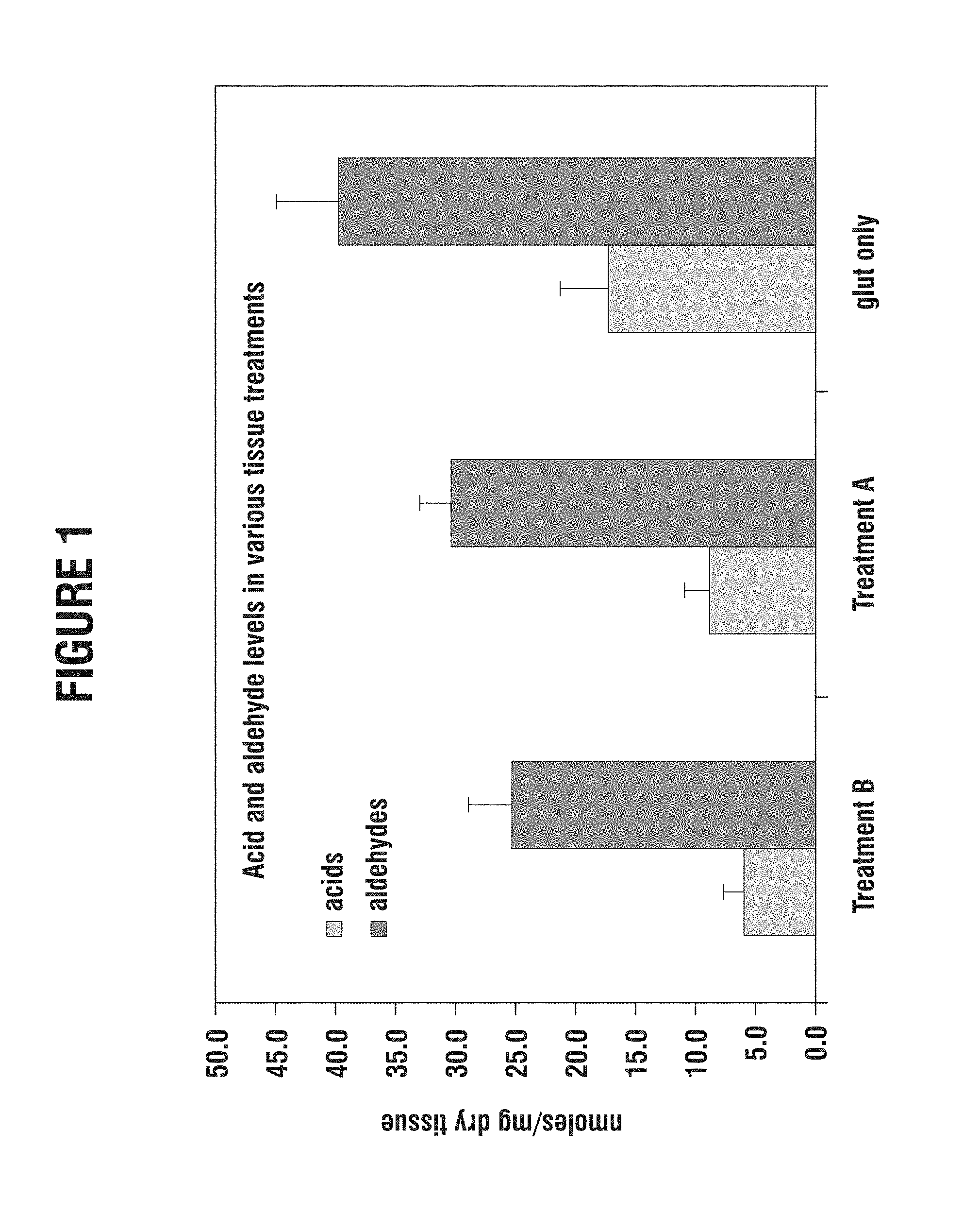
FIG. 1 is a graph showing the aldehyde and acid content in bovine pericardial tissue after several different chemical treatments;
FIG. 2 is a graph correlating the calcium content of in vivo tissue with the corresponding acid and aldehyde content for tissue treated three different ways;
FIG. 3 is a graph illustrating the acid and aldehyde content of various tissue treatments;
FIG. 4 is a chart showing the decrease in calcification by capping, reduction and drying (GLX process);
FIG. 5 is a chart that indicates the reduction in calcification variability by capping, reduction and drying (GLX process);
FIG. 6 is a chart showing the decrease in calcification by capping, reduction and drying (GLX process) after 80 days of real time shelf life;
FIG. 7 is a box and whisker plot of 35 Day Rabbit Intramuscular Study.
FIG. 8 is a box and whisker plot of 35 Day Rabbit Intramuscular Study, Short Term Shelf Life and Calcification.
DESCRIPTION OF THE PREFERRED EMBODIMENTS
The present invention provides an improved bioprosthetic tissue treatment process that greatly reduces the potential for calcification after implantation by blocking free aldehyde groups prior to a dehydration step and/or the addition of chemical agents to prevent oxidative damage during sterilization. "Bioprosthetic tissue" includes, without limitation, bovine pericardium and porcine tissue which are commonly used in bioprosthetic heart valves, and blood vessels, skin, dura mater, pericardium, small intestinal submucosa ("SIS tissue"), tissue heart valves, ligaments and tendons. "Implants" in the present application refers not only to heart valves, including transcatheter heart valves, but also to vascular prostheses and grafts, tissue grafts, bone grafts, and orbital implant wraps, among others.
A "bioprosthetic heart valve" refers to a fully assembled prosthetic valve made at least partly from bioprosthetic tissue. Some whole porcine valves are used in so-called "stentless" bioprosthetic valves in which there is very little if any synthetic material added for support or anchoring purposes. A "stented" bioprosthetic valve typically has some kind of synthetic (e.g., polymer or metallic) support for the leaflets, which may be the leaflets of a whole porcine xenograft or separate bovine pericardial leaflets. Heart valves contemplated herein include surgical heart valves, transapical heart valves, transfemoral heart valves and other types of heart valves.
Prior art tissue treatments address crosslinking, microbes, and other aspects of the tissue in a "static" setting, and typically involve immersion of the tissue in glutaraldehyde, Tween (polyoxyethylene 20 sorbitan monooleate), ethanol, formaldehyde, and other agents to mitigate post-implant calcification. Some prior art processes include the addition of various chemicals to reduce the toxicity of the crosslinked tissue or mitigate calcification via the use of metal ions (i.e., Al.sup.3+ or Fe.sup.3+ see U.S. Pat. No. 5,746,775, Levy) or bulk blocking agents (i.e., 2-amino oleic acid--see U.S. Pat. No. 4,976,733, Giradot). But each of these methods is only applied to initially processed tissue, not to dehydrated tissue or tissue devices to prevent oxidative damage. The prior art processes are limited to the addition of chemical or biological agents to crosslinked tissue that are temporarily attached to the tissue, or they are limited to reduction or oxidation of the tissue alone without any addition of "capping agents" (e.g., U.S. Pat. No. 5,862,806, Cheung).
Unlike the various prior art tissue processes, where the goal is to fix (i.e. crosslink, etc.) the tissue, this invention describes an additional process whereby acids and other potential binding sites formed from the prior art fixation processes are "capped." It also involves "capping" the binding sites and potential binding sites that are generated from oxidation of fixed tissue. Tissue treatment with glutaraldehyde, Tween (polyoxyethylene 20 sorbitan monooleate), ethanol, formaldehyde, and other agents can provide useful fixation of the tissue. However, it will also generate binding sites capable of interacting with or attracting calcium, phosphate, immunogenic factors, or other precursors to calcification. For example, there are many negatively charged carboxylic acid groups formed after glutaraldehyde fixation of the tissue, and these groups are capable of attracting calcium ions (due to their negative charge and electrostatic interactions with positively charged ions) leading to calcification of the tissue or adverse cellular interactions. Carboxylic acid groups like those in glutamic acid or gamma carboxy glutamic acid are known to bind calcium atoms (Hauschka et al. PNAS 1975 72:3925). Calcium binding proteins such as bone sialoprotein contain carboxylic acid-rich domains designed to attract and bind calcium, leading to hydroxyapatite formation (calcification). The overall level and location of acid groups in these proteins determines the ability of the protein to efficiently bind calcium and form hydroxyapatite. The term "acid potential" of the tissue refers to the level of these chemical functional groups within the fixed tissue which may eventually form acid groups or "binding sites" by oxidation, dehydration, hydration, or similar processes.
The inventors have discovered that such binding, causes significant post-implant damage in bioprosthetic materials, especially tissues used for heart valve leaflets. For example, the oxidative damage that occurs during storage and handling of dehydrated or "dry" tissue can create carboxylic acid groups that will bind calcium and lead to tissue failure. This progressive leaflet damage process can create new binding sites or potential binding sites that are precursors to calcification and immunogenic related pathways. The present invention describes a method to cap these newly formed binding sites prior to implantation of the tissue for tissue-based bioprosthetic into the body. The inventors have also discovered that bioprosthetic tissue exposed to oxidation from the atmosphere when not submersed in a glutaraldehyde solution or during sterilization is likely to contain more acid groups that contribute to calcification and inflammation. In dry storage, the dehydrated tissue is sterilized and stored "dry" without the protective effect of the glutaraldehyde solution. The ease of handling and storage of this new product is greatly facilitated due to the absence of the glutaraldehyde storage solution. This technology can be improved by treating such bioprosthetic tissue with a capping agent and/or adding a chemical protectant during the dehydration phase.
One chemical target within the invention is the permanent "capping" of the acid groups which dramatically reduces their ability to attract calcium, phosphate, immunogenic factors, or other groups. The term "capping" refers to the blocking, removal, or alteration of a functional group that would have an adverse effect on the bioprosthesis properties. For example, the addition of 1-ethyl-3-[3-dimethylaminopropyl]carbodiimide hydrochloride (EDC), N-hydroxysulfosuccinimide sulfo-NHS), and ethanolamine will effectively cap the acid groups with a non-reactive alcohol group.
In addition to acid binding sites, tissues treated with glutaraldehyde or other aldehyde-containing agents also yield tissue with many free aldehyde groups that cause increased toxicity, higher calcification, and involvement in immunogenic responses. These aldehyde groups can easily oxidize into carboxylic acid groups via air oxidation, in vivo blood oxidation, macrophage oxidation, and other similar oxidation pathways. Thus, an additional target of the invention includes the permanent capping of aldehyde groups, which are potential binding sites, in a way that would prevent or mitigate their ability to transform into acids or other groups and thus further mitigate the potential for calcification in the body (in vivo). In addition to acids and aldehydes there are other possible binding sites such as immunogenic and antigenic factors, capping which is included within the scope of this invention.
The present capping process includes chemical reduction of the tissue, which, when applied to the tissue in the presence of a capping agent, will permanently connect the capping agent to the target group. For example, the addition of ethanolamine to the tissue will cap the aldehyde groups, while the reducing agent (e.g., sodium borohydride) reduces any Schiff base created by reaction of the aldehyde with the amine group of ethanolamine. Thus an aldehyde is ultimately replaced by a hydroxyl group, which may be beneficial for tissue hydration, flexibility, and cell interactions. Of course, other capping agents can be used instead of ethanolamine and other reducing agents other than sodium borohydride and are known by those skilled in the art and which are included in the scope of this patent. Another preferred strategy is to oxidize the tissue aldehydes to acids, and then cap the acid groups. This may involve the addition of 1-ethyl-3-[3-dimethylaminopropyl]carbodiimide hydrochloride (EDC), N-hydroxysulfosuccinimide (sulfo-NHS), and ethanolamine. These new "capped" groups will reduce the attraction of calcium, phosphate, immunogenic factors, or other similar agents.
In one specific embodiment, the invention provides a method of treating bioprosthetic implant tissue to reduce in vivo calcification of comprising at least partially cross-linking bioprosthetic implant tissue, then treating the cross-linked tissue with an aldehyde (or acid) capping solution to mitigate calcification, and dehydrating the tissue in an ethanol/glycerol solution. The glycerol solution may include an antioxidant treatment and may contain a water-soluble wax. The tissue is then allowed to dry and then subjected to final sterilization (e.g., ethylene oxide, gamma irradiation, or electron beam irradiation). The following steps describe an implementation of this process in the manufacture of prosthetic heart valves.
Aldehyde Capping. After the valve leak and flow inspection, the valves are briefly rinsed in 20% ethane to remove any excess glutaraldehyde adhering to the tissue. This is thought to enhance the capping process by ensuring that the capping solution can attach to aldehydes on the tissue rather than free glutaraldehyde in solution. The valves are then exposed to a capping solution of ethanolamine and sodium borohydride, at room temperature under agitation for 4 hours. Valves are removed from the capping solution, and rinsed for a few minutes at room temperature with the same solution used in the final bioburden reduction process to remove excess capping solution.
Glycerol Treatment. After the valves have been processed through a standard final bioburden reduction step, they undergo the glycerol treatment in a solution of 75 wt % glycerol and 25 wt % ethanol. Valves are soaked in this solution for one hour at room temperature. During this time, most of the water molecules present in the pericardial tissue are replaced with glycerol. Valves are removed from the solution and placed in a clean hood to allow any excess solution to evaporate or drip off the valves.
EO Sterilization. The dehydrated valves are then ready for packaging. They are packaged in double sterile barrier packaging consisting of a rigid tray (PETG) with a Tyvek lid. The package is sealed in a cleanroom, and sterilized in 100% ethylene oxide.
The calcification mitigant preferably contains a capping agent selected from: an amine, an amino acid, an amino sulfonate, a hydrophilic multifunctional polymer, a hydrophobic multifunctional polymer, .alpha.-dicarbonyl, a hydrazides, a N,N-disuccinimidyl carbonate, a carbodiimide, 2-chloro-1-methylpyridinium iodide (CMPI), an antibiotic, a cell recruiting agent, a hemocompatibility agent, an antiinflamatory agent, an antiproliferative agent, an immunogenic suppressing agent, a reducing agent, and a mono-, di- or polyepoxy alkane.
The chemical anti-oxidant is desirably selected from:
a water soluble antioxidant such as ascorbic acid,
a fat soluble antioxidant such as tocopherols,
a carbohydrate such as fructose, sucrose, or mannitol
a hindered phenol such as butylated hydroxytoluene (BHT),
a hindered amine light stabilizer (HALS) such as p-phenylamine diamine, trimethyl dihydrodquinoline, or alkylated diphenyl amines
a phosphite/phosphonite such as triphenyl phosphine,
and a thioester such as a thiocinnamate
The calcification mitigant (capping agent) and/or the chemical oxidation protectant is desirably delivered in one or a combination of the following selected solutions:
an aqueous solution such as an aqueous buffered solution, water, short chain alcohols, glycerol, or plasticizers,
an organic solvent, and
an organic buffered solution.
The tissue is preferably fully cross-linked prior to the capping process. In one embodiment, the tissue comprises precut heart valve leaflets mounted and treated in a suitable apparatus. Alternatively, the tissue may be bulk sheets of tissue treated in a suitable apparatus.
Examples of capping agents, provided in species and subspecies, are: amines, alkyl amines, amino alcohols, ethanolamine,
amino acids, lysine, hydroxylysine,
amino sulfonates, taurine, amino sulfates, dextran sulfate, chondroitin sulfate,
hydrophilic multifunctional polymer, polyvinyl alcohol, polyethyleneimine,
hydrophobic multifunctional polymer,
.alpha.-dicarbonyls methylglyoxal 3-deoxyglucosone glyoxal
hydrazides adipic hydrazide
N,N-disuccinimidyl carbonate
carbodiimides 1-ethyl-3-[3-dimethylaminopropyl]carbodiimide hydrochloride (EDC) N-cyclohexyl-N'-(2-morpholinoethyl)carbodiimide (CMC) 1,3-dicyclohexyl carbodiimide (DCC)
2-chloro-1-methylpyridinium iodide (CMPI)
an antibiotic,
a cell recruiting agent,
a hemocompatibility agent, heparin,
an anti-inflammatory agent,
an antiproliferative agent,
an immunogenic suppressing agent,
a reducing agent, sodium cyanoborohydride, sodium borohydride, sodium bisulfite+acetylacetone, formic acid+formaldehyde, and
mono-, di- or polyepoxy alkanes.
The effect of preferred capping agents is to not only block functional groups that will attract calcium, phosphate, immunogenic factors, or other similar agents, but to replace those groups with a superior biological functionality. The term "biological functionality" is defined as the effect of tissue components on the local biology of the implanted material. Improved biological functionality of a tissue treatment may include reduction in overall charge of the tissue, better hemocompatibility, increased tissue hydration, better cell interactions, better flexibility, etc. For example, capping aldehyde functions with ethanolamine blocks the aldehyde group from oxidizing into an acid and replaces it with a hydroxyl group, which may be beneficial for tissue hydration, flexibility, and cell interactions. The desired biological functionality of the capped tissue will determine the type of capping compounds employed.
The capping strategy is also designed to block the biological functionality of components of the tissue that may contribute to adverse cellular reactions. Some of these targets include, but are not limited to .alpha.-gal, MHC-1 associated proteins, HLA antigens and the like. The invention addresses the capping or blocking of proteins, carbohydrates, lipids, and other components of the tissue that may contribute to cellular reactions. For example, the .alpha.-gal carbohydrate may be blocked by treatment with 2-chloro-1-methylpyridinium iodide (CMPI) and other agents that neutralize the hydroxyl groups which are known by those skilled in the art. Another example includes MHC-1 associated proteins that may be capped or effectively neutralized by treatment with 1-ethyl-3-[3-dimethylaminopropyl]carbodiimide hydrochloride (EDC) and ethanolamine. Also included in the invention's capping process is the targeting of proteins, carbohydrates or lipids associated with cell and vessel remnants. For example, fibronectin may be capped or blocked by the addition of methylglyoxal to the tissue. This dicarbonyl is known to bind critical arginine functions within proteins and impairs the biological functionality of these proteins.
Another aspect of the invention includes the activation of capping technology upon sterilization. For example, the treatment of tissue with specific capping agents (e.g. glucose and ethanolamine or taurine) prior to gamma irradiation sterilization would produce activation of the capping agents upon irradiation. The capping agents added to the tissue would effectively cap targets within the tissue immediately, but sterilization (i.e. ethylene oxide, electron beam irradiation, or gamma irradiation) would generate new binding sites that would then be capped by the residual capping agents within the tissue. Gamma irradiation of collagen is known to cleave peptide bonds within the backbone and produce new functional groups that may have adverse effects on the tissue. These new functional groups are included in the targets or binding sites described herein and may be capped or blocked by the capping agents listed herein.
Immunogenic factors are defined as anything causing or involved in stimulating an immunogenic response. These include any chemical or biological agent capable of inducing an immune type response. For example, vessel and cell membrane components left in the tissue may cause some type of immunogenic or antigenic response from the body's natural immune system. The invention includes capping agents capable of masking, replacing, or blocking these immunogenic elements in the tissue either statically or dynamically. For example, a whole valve could be fixed, then capped with a non-immunogenic or more hemocompatible capping agent such as heparin prior to dehydration and sterilization. This is different from prior art processes that add heparin to fixed tissue without any dehydration of the valve or any consideration of the post-process oxidation conditions. The invention process can be supplemented with a decellularization process to reduce immunologic or antigenic binding sites and potential binding sites and is also within the scope of this invention.
To better understand the principles underlying the treatment techniques of the present invention, a number of graphs in (see the Figures) are presented based on actual testing. As mentioned above, the invention generally comprises treating tissue so that calcium or phosphate binding sites, or other such sites which could trigger calcification, are capped. The correlation between acid binding sites and tissue calcification can be seen in (FIG. 2 see also Hauschka et al. PNAS 1975 72:3925.) and it appears that acid templating directs mineralization in a variety of species. Thus, the amount of free acids and/or aldehydes in the tissue at the time of implantation correlates with the number of such binding sites and, therefore, increases the potential for calcification. The amount of free acids and aldehydes present in tissue can be measured by known methods, for example, a standard spectrophotometric assay.
FIG. 1 is a graph showing both the free aldehyde and free acid content in bovine pericardial tissue as measured by the aforementioned technique. It should be understood that all of the tests referred to herein involve glutaraldehyde-fixed bovine pericardial tissue. The tissues studied have all been chemically treated, one with glutaraldehyde only, and two others with tissue treatments that have been used by Edwards Lifesciences to prepare commercial bioprosthetic tissue for implant. However, other cross-linking and tissue processing methods can be used and are within the scope of this invention.
The aldehyde and acid content of the tissues is measured subsequent to the chemical treatments, and without any damage or stress imparted to the tissue. On the right side of the graph of FIG. 1, the tissue samples have been treated in glutaraldehyde only, in particular in a solution of 0.625% glutaraldehyde for a period of 14 days. A total of 10 samples were treated and subsequently tested. The measurements showed an average level of about 40 nanomoles of aldehydes and about 17 nanomoles of acids per milligram of dry weight of the tissue.
The middle of the graph of FIG. 1 shows the results from testing a total of 10 tissue samples subjected to Treatment A, which is commercially known as the XenoLogiX.TM. tissue treatment process from Edwards Lifesciences of Irvine, Calif. Treatment A involves first fixing with glutaraldehyde then treating twice with a sterilant including a cross-linking agent such as formaldehyde, a surfactant such as Tween-80 (Polyoxyethylene sorbitan monooleate), and a denaturant such as ethyl alcohol. Both the aldehyde and acid content of the tissue subjected to Treatment A were less than that of tissue treated with glutaraldehyde alone, with the aldehyde content decreasing by about 25% and the acid content by about 50%. This reduction has been attributed to the further reduction of phospholipids which are sources of acid binding sites as well as hemiacetal formation from alcohol and aldehyde groups.
The left side of FIG. 1 shows the results from testing a total of 10 samples subjected to Treatment B, which is commercially known as the Carpentier-Edwards ThermaFix.TM. tissue treatment process from Edwards Lifesciences. Treatment B is essentially the same as Treatment A, with the addition of a heat treating step after fixing and prior to sterilizing. Both the aldehyde and acid content of the tissue subjected to Treatment B were less than that of tissue treated glutaraldehyde alone, with the aldehyde content decreasing by about 33% and the acid content by more than 50%. In addition, Treatment B reduces both the aldehyde and acid content relative to Treatment A by between 10-20%.
FIG. 2 is a graph that repeats the results of aldehyde/acid content measurements for the three tissue treatments shown in FIG. 1, and also superimposes measurements of calcium uptake from like tissue samples implanted subcutaneously in rabbits from a separate study. These acid levels are measured in the tissue prior to implant and are likely to increase in vivo. FIG. 2 reveals that the amount of calcium found in the implanted tissues correlates with the levels of aldehydes/acids from the three tissue treatments. That is, as the level of free aldehydes and free acids in the various tissue samples decreases, the amount of calcium absorbed upon implant also decreases. Again, it is understood that a number of factors contribute to calcium uptake, but the availability of certain calcium and phosphate binding sites, among others, is a prime indicator of future calcification. The graph of FIG. 2 therefore suggests that decreasing the levels of aldehydes/acids in the tissue will reduce the propensity for the tissue to calcify.
As mentioned above, it is now understood that oxidation of the aldehyde groups in tissue to carboxylic acid groups produces an increase in calcification. Evidence of this phenomenon is provided in the graph of FIG. 3. Specifically, as explained above, the level of acids in the tissue correlates directly with the propensity to calcify after implant. FIG. 3 indicates the acid levels in various tissue samples. The types of tissue treatments are fresh untreated tissue, glutaraldehyde-fixed tissue, XenoLogiX (XLX), ThermaFix (TFX), ThermaFix tissue treated with glycerol and ethanol only, and the GLX process, which includes treatment with glutaraldehyde, capped with ethanolamine while being reduced with sodium borohydride, and dehydrated with glycerol and ethanol.
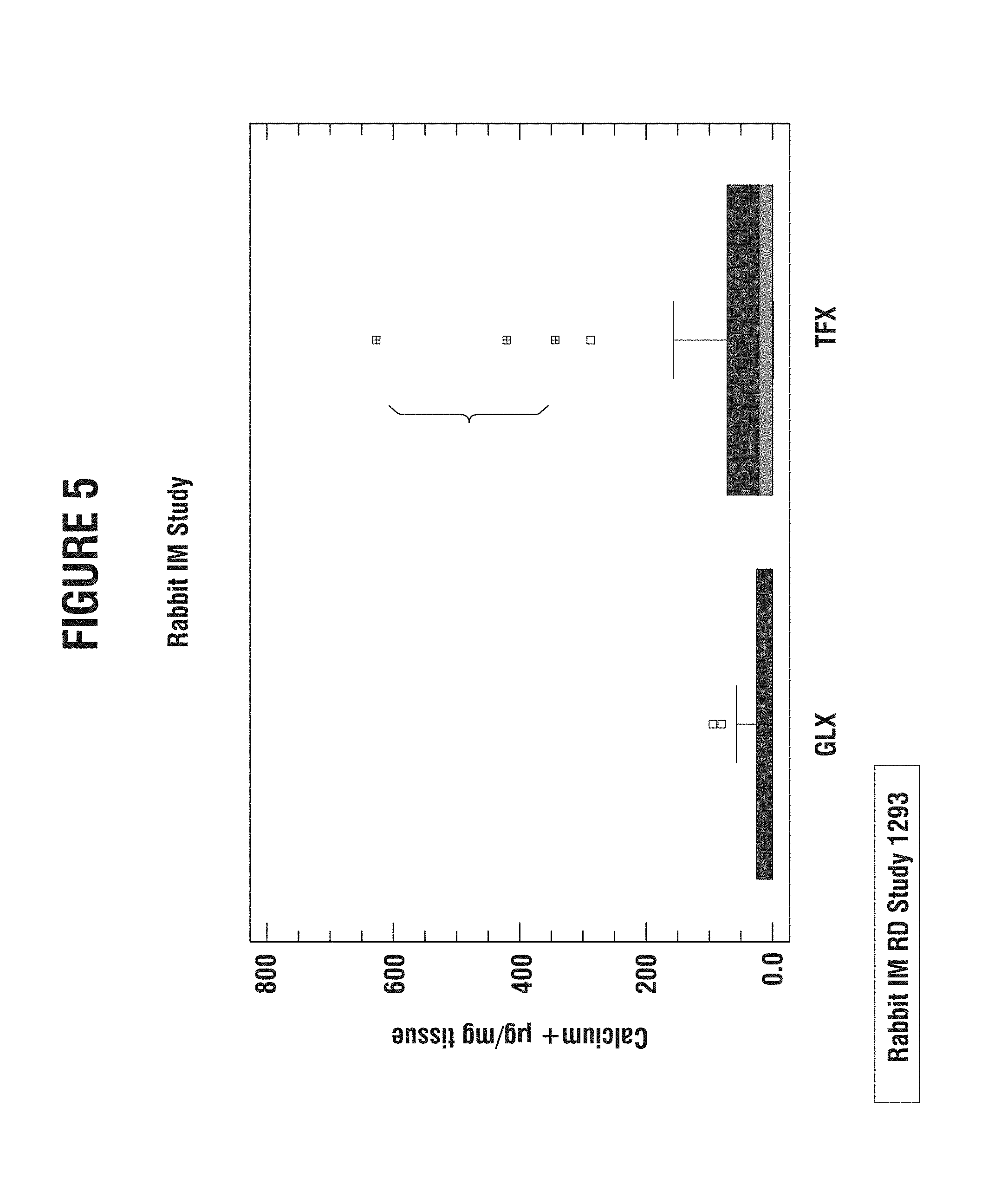
FIGS. 4 and 5 are the results of a rabbit intramuscular implant study indicating that the GLX process (described in this invention) significantly reduces calcification over current technology (TFX) and over simple glycerol treatment (GLY-treated). These data also indicate that the GLX process reduces variability in calcification. All calcification measurements were measured by atomic absorption spectroscopy and normalized to dry weight of lyophilized tissue.
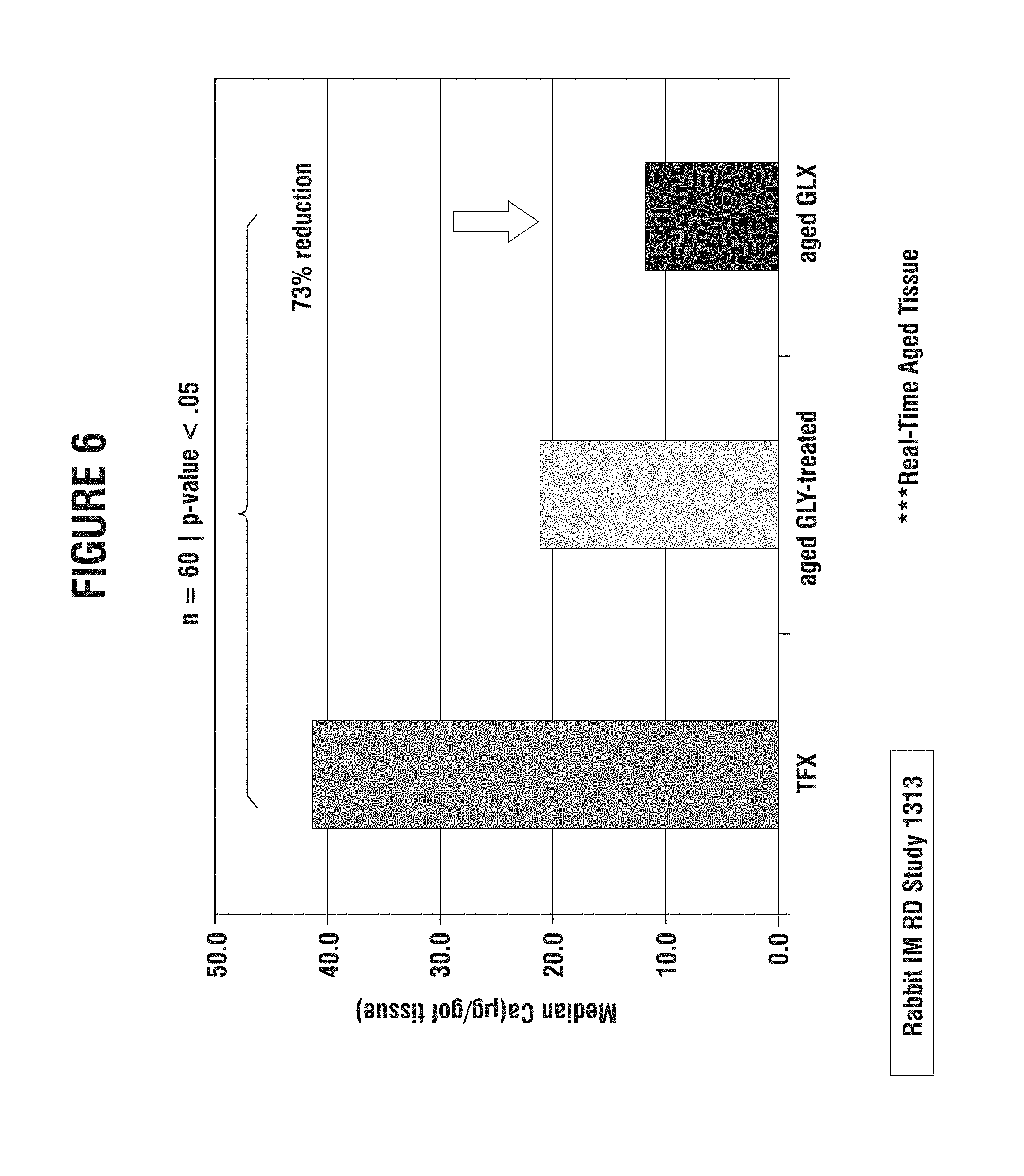
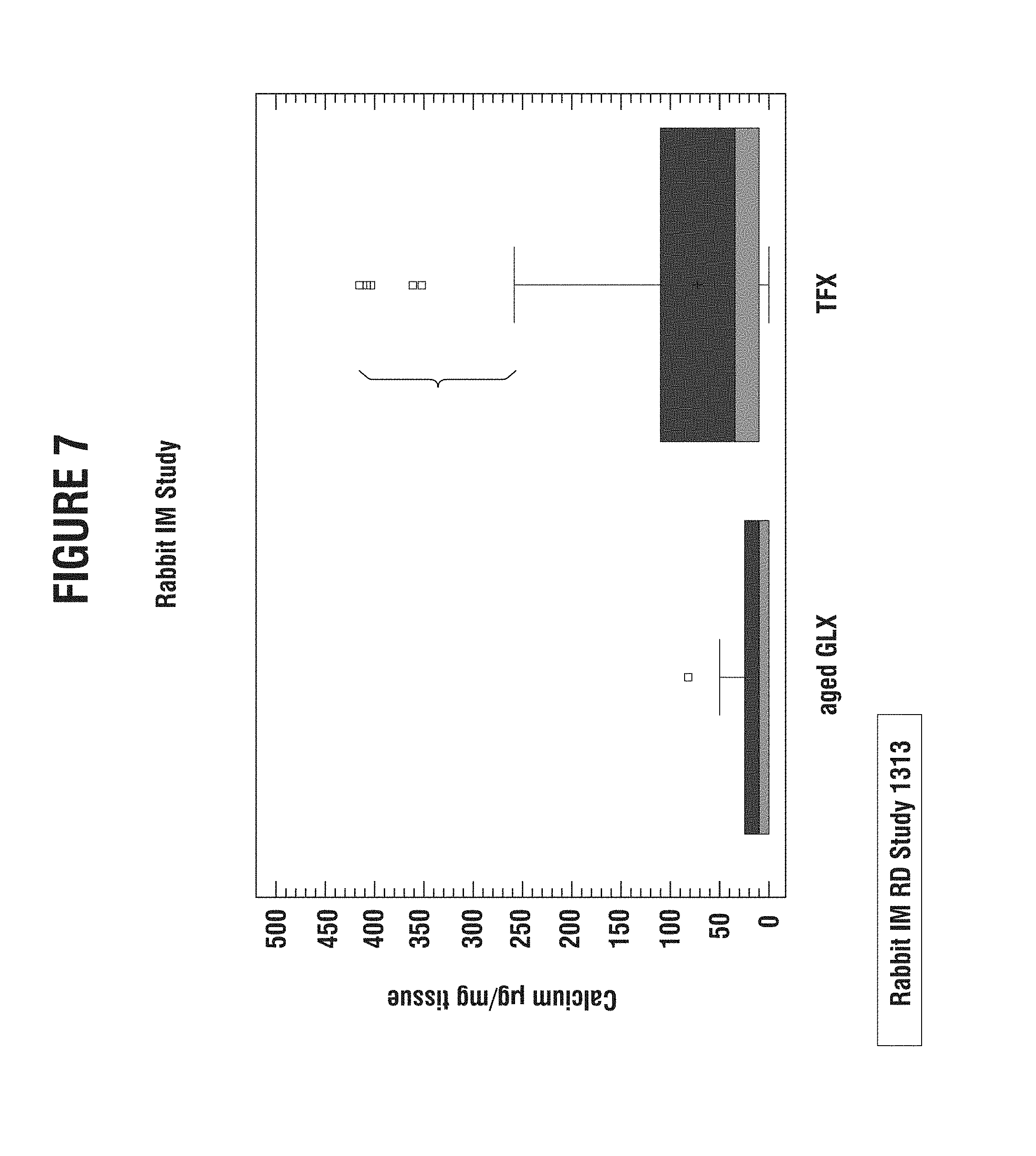
FIGS. 6 and 7 illustrate that after 80 days of real time shelf life, the GLX treated tissue shows significantly less calcification than TFX or the glycerol treatment alone. The GLX process also reduces variability after 80 days of shelf life. All calcification measurements were measured by atomic absorption spectroscopy and normalized to dry weight of lyophilized tissue.
Based on the foregoing empirical results, the inventors believe that the oxidative damage of dehydrated tissue imparted on bioprosthetic tissue greatly contributes to the propensity for calcification of tissue. In particular, heart valve leaflets not stored in glutaraldehyde are subjected to significant oxidative damage. This deleterious tissue damage process can create new binding sites not previously detected or recognized, as potential attachment sites of calcium and phosphate ions, thereby initiating calcification.
To help prevent this post-implant damage-calcification process, the present invention involves mitigating oxidation by capping the numerous aldehydes that are susceptible to oxidation and increased calcification initiation.
The preferred embodiments include, but are not limited to: 1. The fixed tissue valve or tissue sheet is treated in a solution containing an aldehyde capping agent. 2. Embodiment 1, but where a sterilization step is added during or after the capping process. 3. Embodiments 1 and 2, but where the processing is agitated. 4. Embodiments 1, 2, and 3, but where the capping agent is for the other binding sites such as acids or biological-immune related sites. 5. The aldehyde capping solution may contain an amine (10 mM ethanolamine) and a reducing agent (132 mM sodium borohydride) in 50 mM phosphate buffer at pH 7.4. 6. The tissue or valve is then dehydrated in a glycerol solution 7. The tissue or valve is then sterilized with ethylene oxide
Example 1
Aldehyde capping using ethanolamine and sodium borohydride of glutaraldehyde-fixed tissue. Bioprosthetic tissue was removed from 0.625% glutaraldehyde just after heat treatment step and rinsed in ethanol:saline (20%/80%) for 2 minutes. One liter of capping solution was prepared containing 10 mM ethanolamine (0.06%), and 110 mM sodium borohydride (0.42%) in 50 mM phosphate buffer (pH 7.3-7.8)
The capping solution was placed on an orbital shaker, then tissues (leaflets or valves) were placed in the solution so that they were completely submerged. The ratio of tissue to solution was 3 leaflets per 100 ml or one valve per 100 ml. The container was partly covered but not completely sealed because hydrogen gas liberated by the chemical reaction with water could cause the container to explode. The orbital shaker was operated at between 80-100 rpm for 4 hours at room temperature. The tissue was removed and rinsed in FET solution (formaldehyde, ethanol, tween-80) for three minutes.
Example 2
Glycerol dehydration process for pericardial valve bioprosthesis. Pericardial valves were dehydrated by holding each valve with forceps on the sewing ring of the valve and placing the valve in a glycerol/ethanol (75%/25%) mixture. Beakers containing the valves were placed on an orbital shaker operating between 50-60 RPM for at least one (1) hour but not more than four (4) hours then immediately treated to remove excess glycerol. This was done by holding them with forceps on the sewing ring of the valve, taking the valve out of the glycerol/ethanol mixture and then placing it on an absorbent towel in a wide mouth jar. After being allowed to dry for at least 5 minutes at room temperature the jar was attached to a lyophilizer and dried for 2 hours. Valves were then transferred to ethylene oxide gas permeable packages and sterilized with ethylene oxide.
Example 3
Calcification Mitigation--Small Animal Model. In order to evaluate the calcification mitigation properties of GLX treated and EO sterilized pericardial tissue, two small animal feasibility studies were conducted. These studies demonstrate that, 1) GLX is superior to TFX in mitigating the occurrence of calcification in tissue, and 2) real time aged GLX tissue is also superior to TFX in mitigating calcification. In both studies, GLX valves demonstrated reduced variability in calcification data when compared to TFX valves. Test methods and results of each are summarized below.
Example 3A
Rabbit Study #1. Calcification Potential of Ethylene Oxide Sterilization on GLX Processed Pericardial Tissue in a 35 Day Rabbit Intramuscular Study. A study utilizing sixty (60) rabbits was conducted to look at the effects on calcification of GLX treated pericardium sterilized using 100% ethylene oxide. The control group was ThermaFix (TFX) processed bovine pericardium and the test group was GLX processed pericardium. Using a sign-rank test, GLX tissue was found to be significantly different (p=0.0004) when compared to TFX, and demonstrated a 93% calcification reduction over TFX. GLX data also showed reduced outliers and reduced variability. Box and Whisker plots show an appreciable reduction in variability with the GLX process. Data are presented in FIG. 7, with the y-axis measuring .mu.g calcium/mg dry weight tissue.
Example 3B
Rabbit Study #2. Effects of a Short Term Shelf Life on Calcification of GLX Processed Pericardial Tissue in a 35 Day Rabbit Intramuscular Study. A second study utilizing sixty (60) additional rabbits was conducted to look at the effects on calcification of short term shelf life of GLX processed pericardium. The control group was ThermaFix (TFX) processed bovine pericardium and the test group was GLX processed pericardium. The GLX tissue samples were packaged in Tyvek pouches and sterilized via 100% ethylene oxide. GLX samples were stored in a controlled steady state chamber at 25.degree. C. and 60% humidity for a period of 83 days. The TFX samples had not been aged. In this study the GLX processed tissue demonstrated significantly reduced levels of calcification, 73% compared (p=0.009) to TFX as well as reduced outliers and reduced variability in the data. Data are presented in FIG. 8, with the y-axis measuring .mu.g calcium/mg dry weight tissue.
While the invention has been described in its preferred embodiments, it is to be understood that the words which have been used are words of description and not of limitation. Therefore, changes may be made within the appended claims without departing from the true scope of the invention.
* * * * *
D00000
D00001
D00002
D00003
D00004
D00005
D00006
D00007
D00008
XML
uspto.report is an independent third-party trademark research tool that is not affiliated, endorsed, or sponsored by the United States Patent and Trademark Office (USPTO) or any other governmental organization. The information provided by uspto.report is based on publicly available data at the time of writing and is intended for informational purposes only.
While we strive to provide accurate and up-to-date information, we do not guarantee the accuracy, completeness, reliability, or suitability of the information displayed on this site. The use of this site is at your own risk. Any reliance you place on such information is therefore strictly at your own risk.
All official trademark data, including owner information, should be verified by visiting the official USPTO website at www.uspto.gov. This site is not intended to replace professional legal advice and should not be used as a substitute for consulting with a legal professional who is knowledgeable about trademark law.