Method of analysing a blood sample of a subject for the presence of a disease marker
Wurdinger , et al. J
U.S. patent number 10,174,365 [Application Number 14/006,089] was granted by the patent office on 2019-01-08 for method of analysing a blood sample of a subject for the presence of a disease marker. This patent grant is currently assigned to STICHTING VU-VUMC, VERENIGING VOOR CHRISTELIJK HOGER ONDERWIJS, WETENSCHAPPELIJK ONDERZOEK EN PATIENTENZORG. The grantee listed for this patent is Rolf Jonas Nilsson, Thomas Wurdinger. Invention is credited to Rolf Jonas Nilsson, Thomas Wurdinger.


| United States Patent | 10,174,365 |
| Wurdinger , et al. | January 8, 2019 |
Method of analysing a blood sample of a subject for the presence of a disease marker
Abstract
The present invention relates to a method of analyzing a blood sample of a subject for the presence of a disease marker, said method comprising the steps of a) extracting nucleic acid from anucleated blood cells in said blood sample to provide an anucleated blood cells-extracted nucleic acid fraction, and b) analyzing said anucleated blood cells-extracted nucleic acid fraction for the presence of a disease marker, wherein said disease marker is a disease-specific mutation in a gene of a cell of said subject, or wherein said disease marker is a disease-specific expression profile of genes of a cell of said subject.
| Inventors: | Wurdinger; Thomas (Amsterdam, NL), Nilsson; Rolf Jonas (Amsterdam, NL) | ||||||||||
|---|---|---|---|---|---|---|---|---|---|---|---|
| Applicant: |
|
||||||||||
| Assignee: | STICHTING VU-VUMC (Amsterdam,
NL) VERENIGING VOOR CHRISTELIJK HOGER ONDERWIJS, WETENSCHAPPELIJK ONDERZOEK EN PATIENTENZORG (Amsterdam, NL) |
||||||||||
| Family ID: | 45562414 | ||||||||||
| Appl. No.: | 14/006,089 | ||||||||||
| Filed: | January 16, 2012 | ||||||||||
| PCT Filed: | January 16, 2012 | ||||||||||
| PCT No.: | PCT/NL2012/050025 | ||||||||||
| 371(c)(1),(2),(4) Date: | March 26, 2014 | ||||||||||
| PCT Pub. No.: | WO2012/128616 | ||||||||||
| PCT Pub. Date: | September 27, 2012 |
Prior Publication Data
| Document Identifier | Publication Date | |
|---|---|---|
| US 20140199693 A1 | Jul 17, 2014 | |
Foreign Application Priority Data
| Mar 18, 2011 [EP] | 11158912 | |||
| May 27, 2011 [EP] | 11167973 | |||
| Jul 15, 2011 [WO] | PCT/NL2011/050518 | |||
| Current U.S. Class: | 1/1 |
| Current CPC Class: | C12Q 1/6886 (20130101); C12Q 1/6834 (20130101); C12Q 2600/158 (20130101); C12Q 2600/156 (20130101) |
| Current International Class: | C12Q 1/68 (20180101); C12Q 1/6886 (20180101); C12Q 1/6834 (20180101) |
References Cited [Referenced By]
U.S. Patent Documents
| 5185244 | September 1993 | Wallace |
| 2003/0119003 | June 2003 | Simons |
| 2004/0014059 | January 2004 | Liew |
| 2004/0137470 | July 2004 | Dhallan |
| 2005/0069875 | March 2005 | Schuh et al. |
| 2006/0288432 | December 2006 | Vainchenker et al. |
| 2007/0059774 | March 2007 | Grisham et al. |
| 2007/0059781 | March 2007 | Kapur et al. |
| 2008/0038764 | February 2008 | Yin |
| 2010/0184069 | July 2010 | Fernando et al. |
| 2013/0196873 | August 2013 | Wurdinger et al. |
| 2014/0256590 | September 2014 | Wurdinger et al. |
| 103168235 | Jun 2013 | CN | |||
| 2380194 | Apr 2003 | GB | |||
| WO 2000 63441 | Oct 2000 | WO | |||
| WO 2002 070738 | Sep 2002 | WO | |||
| 2005/043121 | May 2005 | WO | |||
| 2007/035585 | Mar 2007 | WO | |||
| 2008/156858 | Dec 2008 | WO | |||
| WO2010/139811 | Dec 2010 | WO | |||
| 2012008839 | Jan 2012 | WO | |||
| 2012/128616 | Sep 2012 | WO | |||
| 2013/022342 | Feb 2013 | WO | |||
Other References
|
Healy et al. Circulation. 2006;113:2278-2284. cited by examiner . Claverley et al. (Clin Transl Sci 2010 3(5) 227-232). cited by examiner . Girardot et al. (Blood, 2010, vol. 116, No. 3, pp. 437-445. cited by examiner . Affymetrix Netaffx search results for PCA3 on human exon 1.0 ST array. Obtained from https://www.affymetrix.com/analysis/netaffx/showresults.affx on Jul. 17, 2017, 1 page. cited by examiner . Affymetrix Netaffx search results for KLK3 on human exon 1.0 ST array. Obtained from https://www.affymetrix.com/analysis/netaffx/exon/transcript.affx?pk=1:383- 9538 on Jul. 17, 2017, 3 pages. cited by examiner . Schutte M, Elstrodt F, Bralten LBC, Nagel JHA, Duijm E, et al. (2008) Exon Expression Arrays as a Tool to Identify New Cancer Genes. PLoS One 3(8): e3007. doi:10.1371/journal.pone.0003007 (Year: 2008). cited by examiner . Affymetrix results, Details for Cluster 3002640, EGFR, obtained from https://www.affymetrix.com/analysis/netaffx/exon/transcript.affx?pk=1:300- 2640 on Jan. 3, 2018, 13 pages (Year: 2018). cited by examiner . Calverley et al., "Significant Downregulation of Platelet Gene Expression in Metastatic Lung Cancer," Clinical Transl. Sci., 3(5): 227-232 (2010). cited by applicant . Skog et al., "Glioblastoma Microvesicles Transport RNA and Proteins that Promote Tumour Growth and Provide Diagnostic Biomarkers," Nature Cell Biology, 10(12):1470-1476 (2008). cited by applicant . Lood et al., "Platelet Transcriptional Profile and Protein Expression in Patients with Systemic Lupus Erythematosus: Up-Regulation of the Type I Interferon System is Strongly Associated with Vascular Disease,"116:1951-1957 (2010). cited by applicant . International Search Report from International Application No. PCT/NL2011/050518, dated Mar. 9, 2012. cited by applicant . International Search Report from International Application No. PCT/NL2012/050025, dated Jul. 2, 2012. cited by applicant . International Search Report from International Application No. PCT/NL2012/050550 dated Oct. 31, 2012. cited by applicant . Sidransky "Emerging Molecular Markers of Cancer," Nature Reviews Cancer 2(3): 210-219 (2002). cited by applicant . Giradot et al., "miR-28 is a Thrombopoietin Receptor Targeting MicroRNA Detected in a Fraction of Myeloproliferative Neoplasm Patient Platelets," Blood 116(3): 437-445 (2010). cited by applicant . Gnatenko et al., "Class Prediction Models of Thrombocytosis Using Genetic Biomarkers," Blood 115(1):7-14 (2010). cited by applicant . Al-Mufti et al., "Detection of Fetal Messenger Ribonucleic Acid in Maternal Blood to Determine Fetal RhD Status as a Strategy for Noninvasive Prenatal Diagnosis," Am. J. Obstet. Gynecol., 179:210-214 (1998). cited by applicant . Hahn et al., "Determination of Fetal Chromosome Aberrations from Fetal DNA in Maternal Blood: Has the Challenge Finally Been Met?," Expert Reviews in Molecular Medicine, 13:1-14 (2011). cited by applicant . Kolialexi et al., "Noninvasive Prenatal Diagnosis of beta-Thalassaemia Using Individual Fetal Erythroblasts Isolated from Maternal Blood After Enrichment," Prenatal Diagnosis, 27:1228-1232 (2007). cited by applicant . Lee et al., "Separation of Model Mixtures of Epsilon-Globin Positive Fetal Nucleated Red Blood Cells and Anucleate Erythrocytes Using a Microfluidic Device," Journal of Chromatography, 1217:1862-1866 (2010). cited by applicant . Nagalla et al., "Platelet RNA Chips Dip Into Thrombocytosis", Blood, 115:2-3 (2010). cited by applicant . Ramaswamy et al., "Multiclass Cancer Diagnosis Using Tumor Gene Expression Signatures", PNAS, 98(26): 15149-15154 (2001). cited by applicant . Su et al., "Molecular Classification of Human Carcinomas by Use of Gene Expression Signatures," Cancer Research, 61: 7388-7393 (2001). cited by applicant . Lu et al., "MicroRNA Expression Profiles Classify Human Cancers," Nature, 435:834-838 (2005). cited by applicant . Campbell, et al., Definition of subtypes of essential thrombocyhaemia and relation to polycythaemia vera based on JAK2 V617 mutation status: a prostective study, The Lancet, Lancet Limited, Dec. 5, 2005, vol. 366, No. 9501, pp. 1945-1953, London, GB. cited by applicant . Ozeki, et al., A family having type 2B von Willebrand disease with an R1306W mutation: Severe thrombocytopenia leads to the normalization of high molecular weight multimers, Feb. 1, 2010, vol. 125, No. 2, Tarrytown, NY. cited by applicant . Raghavachari, et al., Amplified expression profiling of platelet transcriptome reveals changes in arginine metabolic pathways in patients with sickle cell disease, Circulation, Mar. 27, 2007, vol. 115, No. 12, pp. 1-22. cited by applicant . Salven, et al., Leukocytes and platelets of patients with cancer contain high levels of vascular endothelial growth factor, Clinical Cancer Research, The American Association for Cancer Research, Mar. 1, 1999, vol. 5, No. 3, US. cited by applicant . Nilsson, et al., "Blood platelets contain tumor-derived RNA biomarkers", Blood, American Society of Hematology, 2011, vol. 118, No. 13, pp. 3680-3683. cited by applicant . Go, et al., "C21ORF105, A chromosome 21-encoded mRNA, is not a discriminative marker gene for prediction of Down syndrome in maternal plasma", Prenatal Diagnosis, 2007, vol. 27, pp. 146-149. cited by applicant . Nong et al., "Nitric Oxide Inhalation Inhibits Platelet Aggregation and Platelet-Mediated Pulmonary Thrombosis in Rats", Circulation Research, 1997, 81:865-869. cited by applicant . Tuttle et al., "Placental Lactogen is Expressed but is not Translated into Protein in Breast Cancer", PLOS One, 2014, 9(1): e87325. cited by applicant . Ng et al., "mRNA of Placental origin is Readily Detectable in Maternal Plasma", PNAS, 2003, 100:4748-4753. cited by applicant . Yanabu et al., "Tyrosine Phosphorylation and p72syk Activation by an Anti-glycoprotein Ib Monoclonal Antibody", Blood, 1997, 89(5): 1590-1598. cited by applicant . Alevizos, I., et al., "MicroRNAs in Sjogren's Syndrome as a Prototypic Autoimmune Disease," Autoimmun Rev., Jul. 2010, v. 9, pp. 681-621. cited by applicant . Hill, A., "Exosomes in Neurological Disease," Neurology (2009), v. 25, pp. 27-32. cited by applicant . Free Dictionary Definition for `Device`. Available via url:<thefreedictionary.com/device>, printed on Jul. 31, 2017. cited by applicant . Dittrich et al., "Analysis of SAGE data in human platelets: Features of the transcriptome in an anucleate cell". Throm Haemost 95 (4): 643-651, 2006. cited by applicant . Croix et al., MicroSAGE Manual version 1.0e, obtained from www.sagenet.org/protocol/ year: 2000. cited by applicant . Ji et al., "Epidermal growth factor receptor variant III mutations in lung tumorigenesis and sensitivity to tyrosine kinase inhibitors", PNAS, 2006, 103, 7817-7822. cited by applicant . Zschocke et al., "A fluorescent multiplex ARMS method for rapid mutation analysis", Molecular and Cellular Probes, 1995, 9, 447-451. cited by applicant . Yoshimoto et al., "Development of a real-time RT-PCR assay for detecting EGFRvIII in glioblastoma samples", Clin Cancer Res 2008, 14 (2):488-493. cited by applicant. |
Primary Examiner: Switzer; Juliet C
Attorney, Agent or Firm: McDermott Will & Emery LLP
Claims
The invention claimed is:
1. A method of quantifying RNA levels in a blood sample of a subject, said method comprising the steps of: a) isolating thrombocytes from the blood sample; b) extracting messenger ribonucleic acid (mRNA) from said thrombocytes to provide an extracted nucleic acid fraction from said thrombocytes; c) analyzing said extracted nucleic acid fraction by reverse transcribing extracted mRNA into cDNA, quantifying said cDNA to determine the amount of mRNA of at least three genes by conducting an amplification reaction which employs gene specific primers, wherein the at least three genes comprise genes encoding prostate cancer antigen 3, prostate-specific antigen, and epidermal growth factor receptor variant III.
2. The method of claim 1, wherein the primers for prostate specific antigen comprise SEQ ID NO: 3 and SEQ ID NO: 4.
3. The method of claim 1, wherein the primers for prostate cancer antigen 3 comprise SEQ ID NO: 5 and SEQ ID NO: 6.
4. The method of claim 1, wherein the primers for prostate cancer antigen 3 comprise SEQ ID NO: 7 and SEQ ID NO: 8.
5. The method of claim 1, wherein the primers for epidermal growth factor receptor variant III comprise SEQ ID NO: 9 and SEQ ID NO: 10.
6. The method of claim 1, wherein the primers for epidermal growth factor receptor variant III comprise SEQ ID NO: 11 and SEQ ID NO: 12.
Description
RELATED APPLICATIONS
This application is the U.S. National Stage of International Application No. PCT/NL2012/050025, filed Jan. 16, 2012, published in English, and claims the benefit of European Application No. 11158912.3, filed on Mar. 18, 2011, European Application No. 11167973.4, filed on May 27, 2011, and International Application No. PCT/NL2011/050518, filed on Jul. 15, 2011, the entire teachings of the above applications are incorporated herein by reference.
SEQUENCE LISTING
The instant application contains a Sequence Listing which has been submitted in ASCII format via EFS-Web and is hereby incorporated by reference in its entirety. Said ASCII copy, created on Nov. 25, 2013, is named P91312US10 sequence list_ST25.txt and is 8 KB in size.
FIELD OF THE INVENTION
The invention is in the field of medical diagnostics, in particular in the field of disease diagnostics and monitoring. The invention is directed to markers for the detection of disease, to methods for detecting disease, and to a method for determining the efficacy of a disease treatment.
BACKGROUND OF THE INVENTION
In clinical practice there is a strong need to be able to detect disease in its earliest stages, to predict disease progression, and to implement patient-tailored therapy. Early detection of in particular neoplastic disease (cancer) is critical to ensure favourable treatment of the disease. In spite of numerous advances in medical research, cancer remains a major cause of death worldwide. When patients seek treatment, they are generally exhibiting symptoms of distant metastases, meaning that too often the cancer is detected too late.
Lung, prostate, breast, and colon cancer are the most common tumours, and in order to facilitate appropriate remedial action by surgical resection, radiotherapy, chemotherapy, or other known treatment methods there is a need for rapid and simple methods for the early diagnoses of cancer. The availability of good diagnostic methods for cancer is also important to assess patient responses to treatment, or to assess recurrence due to re-growth at the original site or metastasis.
Several types of cancer markers, such as, for example, oncogene products, growth factors and growth factor receptors, angiogenic factors, proteases, adhesion factors and tumour suppressor gene products, etc, are presently known and are not only considered essential for early diagnosis, but also for differential diagnosis of patients with uncertain clinical abnormalities such as for distinguishing malignant from benign abnormalities; for predicting the likelihood of response in a particular patient with established malignancy to a selected therapeutic method of treatment; and for providing information concerning the risk, presence, status, or future behaviour of the malignancy in a human or animal subject. Currently, the ability to detect and diagnose cancer through the detection of tumour or cancer markers is an area of widespread interest and as a consequence the need exists for reproducible and reliable methods of identifying new and more useful cancer markers in patient specimens.
Glioblastoma is the most common and most aggressive type of primary brain tumor in humans. The disease is difficult to diagnose and even harder to treat due, in part, to the blood-brain barrier that hinders the delivery of therapeutic agents and detection of potentially important diagnostic markers. Diagnostic markers for glioblastoma are available, but are specific for the tumour tissue itself and require a tumour sample.
Improved screening and detection methods are needed in order to detect cancer in an early phase and to follow the progression of the disease. In the case of cancer we are at a state where we do not only need to detect the tumour, but also need to detect it before it has reached a point of no return, where the treatment becomes palliative instead of curative. People at risk, as well as patients with recurring cancer, should be monitored extensively. Furthermore, since tumours can respond differently to different therapeutics, patient stratification is becoming of importance.
Genetic analysis using tumour biopsies has allowed the identification of many mutations that are useful for diagnosis of the cancer as well as for emerging patient stratification strategies. However, a disadvantage of current genetic analysis of tumours is the need for tumour biopsies, which are often impossible to dissect from patients. Furthermore, the use of biopsies is static and does not allow genetic monitoring of tumour progression or recurrence over time. Moreover, many tumours are heterogeneous, resulting in potential false-positive or false-negative genetic characterization of biopsies of such tumours.
Recently, the use of circulating tumour cells for diagnosis and monitoring of tumour progression or recurrence showed the use of blood as a source of tumour-derived material, notably tissue fragments in the form of cells. However, the use of circulating tumour cells is inefficient for most cancers.
Calverley et al (Clinical and Translational Science vol 3, issue 5, 2010) disclose a down-regulation of gene expression of platelets in metastatic lung cancer. The authors identified 200 genes that appeared to be differentially expressed between healthy persons and patients. According to the authors, the platelet proteome is mirrored in the transcriptome of the platelet. The gene expression as measured was correlated to genes from megakaryocytes. No disclosure is made that RNA/DNA derived from other cells than megakaryocytes was measured when testing the thrombocytes, and no indication that circulating RNA/DNA derived from other cells can be taken up by thrombocytes.
In general, a disease marker is defined as a compound of which the concentration is altered, preferably elevated, in a biological fluid from a diseased patient when compared to a normal healthy subject, and which may subsequently be used as a marker compound indicative of a disease. Yet, the identification of specific compounds, for instance proteins, in various body fluids as markers of disease, such as cancer, has been hampered by the lack of suitable techniques therefore.
Also in case of diseases other than cancer, markers may be available that are difficult to detect. This hampers early diagnosis of the disease.
Lood et al (Blood vol 116, no 11, 2010) disclose that the gene expression of IFN-I regulated genes in platelets in patients with SLE is increased. The authors hypothesize that IFNalpha influences gene expression in megakaryocytes, resulting in an increased level of IFN-I regulated proteins in platelets. The expressions of genes from megakaryocytes are thus correlated to SLE or vascular disease. No disclosure is made that RNA/DNA from diseased cells may be taken up by platelets.
The present invention aims to overcome the problem of the prior art that not all diseased tissues or disease types (e.g. tumours) result in circulating disease cells (e.g. circulating tumour cells). The present invention also aims to overcome the problem that protein markers for detecting diseases such as cancer are difficult to detect. Further, the present invention aims to provide methods that do not require biopsies, and allow extensive monitoring of patients.
SUMMARY OF THE INVENTION
The present invention in a first aspect provides a method of analysing a blood sample of a subject for the presence of a disease marker, said method comprising the steps of a) extracting nucleic acid from anucleated blood cells, preferably thrombocytes, in said blood sample to provide an anucleated blood cell-extracted nucleic acid fraction, and b) analysing said anucleated blood cell-extracted nucleic acid fraction for the presence of a disease marker, wherein said disease marker is a disease-specific mutation in a gene of a nucleated cell of said subject, or wherein said disease marker is a disease-specific expression profile of genes of a nucleated cell of said subject.
It was surprisingly found that nucleic acids from nucleated cells are present in anucleated blood cells such as thrombocytes. It may be that nucleated cells excrete nucleic acids into the blood stream and these excreted nucleic acids are then taken up from the blood stream by anucleated cells such as thrombocytes or that in some other way of transport nucleic acids from nucleated cells are transferred to anucleated blood cells. The inventors realized for the first time that disease markers may be used on the nucleic acids extracted from anucleated blood cells to identify diseases from nucleated cells.
In a preferred embodiment of the method of the invention said anucleated blood cell-extracted nucleic acid fraction comprises nucleic acid originating from a nucleated cell. In a preferred embodiment of the present invention and embodiments thereof, the anucleated blood cell-extracted nucleic acid fraction is not megakaryocyte-derived nucleic acid or megakaryocyte-derived RNA, i.e. the nucleic acid fraction to be tested is not of megakaryocyte-lineage or megakaryocyte genomic origin.
The term "anucleated blood cell" as used herein refers to a cell that lacks a nucleus. The term includes reference to both erythrocyte and thrombocyte. Preferred embodiments of anucleated cells in aspects of this invention are thrombocytes. The term "anucleated blood cell" preferably does not include reference to cells that lack a nucleus as a result of faulty cell division.
The term "nucleated cell" as used herein refers to a cell having a nucleus. The term includes reference to somatic cells, germ cells and stem cells, and may include cells from colon, pancreas, brain, bladder, breast, prostate, lung, breast, ovary, uterus, liver, kidney, spleen, thymus, thyroid, nerve tissue, connective tissue, blood, epithelial tissue, lymph node, bone, muscle and skin tissues. The nucleated cell is preferably a cell from a diseased tissue. In a preferred embodiment, the nucleated cell is not a megakaryocyte.
Thus, the present invention is generally aimed at analysing nucleic acids that have been transferred from cells that have a nucleus into cells that have no nucleus, wherein the cells that have no nucleus can be easily isolated from the blood stream and contain nucleic acid from the nucleated cells. The term "nucleus" refers to the membrane-enclosed organelle found in eukaryotic cells that contains most of the cell's genetic material organized in the form of chromosomes. The genes within these chromosomes are the cell's nuclear genome. The interior of the nucleus contains a number of subnuclear bodies including the RNA-comprising nucleolus, which is mainly involved in the assembly of RNA-comprising ribosomes. After being produced in the nucleolus, ribosomes are exported to the cytoplasm where they translate mRNA.
An anucleated blood cell-extracted nucleic acid fraction preferably refers to a fraction comprising chromosomal DNA, ribosomal RNA, nucleolus RNA, and/or messenger RNA.
The term "gene" as used herein, and in particular in the phrasing "mutation in a gene of a nucleated cell" is meant to refer to any nucleic acid sequence, both chromosomal and extra-chromosomal, of a nucleated (somatic) cell, preferably a nuclear nucleic acid sequence, and may include transcribed and non-transcribed sequences as well as ribosomal RNA sequences, most preferably chromosomal sequences that are transcribed into RNA.
In a preferred embodiment of a method of the invention said disease-specific mutation is in a chromosomal gene.
In another preferred embodiment, said gene is not a gene from an anucleated blood cell. In another preferred embodiment, said gene is not a gene from a megakaryocyte. In yet another preferred embodiment said gene is not CD109.
In a preferred embodiment of a method of the invention said disease-specific expression profile is the expression profile of chromosomal genes. In particular of chromosomal genes from a nucleated cell the mRNA of which is present in a thrombocyte.
In another preferred embodiment of a method of the invention said nucleic acid is ribonucleic acid (RNA), more preferably messenger ribonucleic acid (mRNA).
In a preferred embodiment of a method of the invention said nucleic acid is not mtDNA. Hence, mitochondrial nucleic acid is preferably not an aspect of the present invention.
In another preferred embodiment of a method of analysing a blood sample according to the invention said step b) of analysing said anucleated blood cell-extracted nucleic acid fraction for the presence of a disease marker comprises the selective amplification of:
i) said mutation by reverse transcriptase polymerase chain reaction amplification using at least one nucleic acid mutation-specific amplification primer or probe, or
ii) a plurality of mRNAs by reverse transcriptase polymerase chain reaction amplification to determine the expression level of the chromosomal genes encoding said mRNAs to thereby provide an expression profile for said genes and comparing said expression profile to a reference profile.
The blood sample is preferably outside the body.
In a preferred embodiment of a method of the invention the disease is selected from the group consisting of cancer, autoimmune disease, skin diseases, eye disease, endocrine diseases, neurological disorders, and cardiovascular diseases.
In another preferred embodiment of a method of the invention said disease is selected from the group consisting of autoimmune disease, skin diseases, eye disease, endocrine diseases, neurological disorders, and cardiovascular diseases.
In another preferred embodiment of a method of the invention said disease is cancer.
In yet another preferred embodiment of a method of the invention said cancer is a solid tumour cancer, preferably selected from colon, pancreas, brain, bladder, breast, prostate, lung, breast, ovary, uterus, liver, kidney, spleen, thymus, thyroid, nerve tissue, epithelial tissue, lymph node, bone, muscle and skin.
In another preferred embodiment of a method of the invention said disease is not cancer.
In another preferred embodiment of a method of the invention said disease is not a vascular disease.
In another preferred embodiment of a method of the invention said disease is not systemic lupus erythematosus.
In another preferred embodiment of a method of the invention said disease is not sickle cell disease.
In another preferred embodiment of a method of the invention said disease is not Alzheimer's disease.
In another preferred embodiment of a method of the invention said disease is not a disease associated with pathological megakaryocyte function.
In another preferred embodiment of a method of the invention said disease is not a disease associated with pathological platelet function.
The above embodiments that are disclaimed in preferred embodiments may be combined in any combination.
In another preferred embodiment of a method of the invention said disease is selected from the group consisting of autoimmune disease, skin diseases, eye disease, endocrine diseases, and neurological disorders.
In preferred embodiments of aspects of the invention the auto-immune disease is selected from the group consisting of Achlorhydra Autoimmune Active Chronic Hepatitis; Acute Disseminated Encephalomyelitis; Acute hemorrhagic leukoencephalitis; Addison's Disease; Agammaglobulinemia; Alopecia greata; Amyotrophic Lateral Sclerosis; Ankylosing Spondylitis; Anti-GBM/TBM Nephritis; Antiphospholipid syndrome; Antisynthetase syndrome; polyarticular Arthritis; Atopic allergy; Atopic Dermatitis; Autoimmune Aplastic Anemia; Autoimmune cardiomyopathy; Autoimmune enteropathy; Autoimmune hemolytic anemia; Autoimmune hepatitis; Autoimmune inner ear disease; Autoimmune lymphoproliferative syndrome; Autoimmune peripheral neuropathy; Autoimmune pancreatitis; Autoimmune polyendocrine syndrome; Autoimmune progesterone dermatitis; Autoimmune thrombocytopenic purpura; Autoimmune uveitis; Balo disease/Balo concentric sclerosis; Bechets Syndrome; Berger's disease; Bickerstaffs encephalitis; Blau syndrome; Bullous Pemphigoid; Castleman's disease; Celiac disease; Chagas disease; Chronic Fatigue Immune Dysfunction Syndrome; Chronic inflammatory demyelinating polyneuropathy; Chronic recurrent multifocal osteomyelitis; Chronic lyme disease; Chronic obstructive pulmonary disease; Churg-Strauss syndrome; Cicatricial Pemphigoid; Coeliac Disease; Cogan syndrome; Cold agglutinin disease; Complement component 2 deficiency; Cranial arteritis; CREST syndrome; Crohns Disease; Cushing's Syndrome; Cutaneous leukocytoclastic angiitis; Dego's disease; Dercum's disease; Dermatitis herpetiformis; Dermatomyositis; Diabetes mellitus type 1; Diffuse cutaneous systemic sclerosis; Dressler's syndrome; Discoid lupus erythematosus; Eczema; Endometriosis; Enthesitis-related arthritis; Eosinophilic fasciitis; Eosinophilic gastroenteritis; Epidermolysis bullosa acquisita; Erythema nodosum; Essential mixed cryoglobulinemia; Evan's syndrome; Fibrodysplasia ossificans progressiva; Fibromyalgia/Fibromyositis; Fibrosing aveolitis; Gastritis; Gastrointestinal pemphigoid; Giant cell arteritis; Glomerulonephritis; Goodpasture's syndrome; Graves' disease; Guillain-Barre syndrome; Hashimoto's encephalitis; Hashimoto's thyroiditis; Haemolytic anaemia; Henoch-Schonlein purpura; Herpes gestationis; Hidradenitis suppurativa; Hughes syndrome; Hyp ogammaglobulinemia; Idiopathic Inflammatory Demyelinating Diseases; Idiopathic pulmonary fibrosis; Idiopathic thrombocytopenic purpura; IgA nephropathy; Inclusion body myositis; Inflammatory demyelinating polyneuopathy; Interstitial cystitis; Irritable Bowel Syndrome (IBS); Juvenile idiopathic arthritis; Juvenile rheumatoid arthritis; Kawasaki's Disease; Lambert-Eaton myasthenic syndrome; Leukocytoclastic vasculitis; Lichen planus; Lichen sclerosus; Linear IgA disease; Lou Gehrig's Disease; Lupoid hepatitis; Lupus erythematosus; Majeed syndrome; Meniere's disease; Microscopic polyangiitis; Miller-Fisher syndrome; Mixed Connective Tissue Disease; Morphea; Mucha-Habermann disease; Muckle-Wells syndrome; Multiple Myeloma; Multiple Sclerosis; Myasthenia gravis; Myositis; Narcolepsy; Neuromyelitis optica; Neuromyotonia; Occular cicatricial pemphigoid; Opsoclonus myoclonus syndrome; Ord thyroiditis; Palindromic rheumatism; PANDAS; Paraneoplastic cerebellar degeneration; Paroxysmal nocturnal hemoglobinuria; Parry Romberg syndrome; Parsonnage-Turner syndrome; Pars planitis; Pemphigus; Pemphigus vulgaris; Pernicious anaemia; Perivenous encephalomyelitis; POEMS syndrome; Polyarteritis nodosa; Polymyalgia rheumatica; Polymyositis; Primary biliary cirrhosis; Primary sclerosing cholangitis; Progressive inflammatory neuropathy; Psoriasis; Psoriatic Arthritis; Pyoderma gangrenosum; Pure red cell aplasia; Rasmussen's encephalitis; Raynaud phenomenon; Relapsing polychondritis; Reiter's syndrome; Restless leg syndrome; Retroperitoneal fibrosis; Rheumatoid arthritis; Rheumatoid fever; Sarcoidosis; Schizophrenia; Schmidt syndrome; Schnitzler syndrome; Scleritis; Scleroderma; Sjogren's syndrome; Spondyloarthropathy; Sticky blood syndrome; Still's Disease; Stiff person syndrome; Subacute bacterial endocarditis (SBE); Susac's syndrome; Sweet syndrome; Sydenham Chorea; Sympathetic ophthalmia; Takayasu's arteritis; Temporal arteritis; Tolosa-Hunt syndrome; Transverse Myelitis; Ulcerative Colitis; Undifferentiated connective tissue disease; Undifferentiated spondyloarthropathy; Vasculitis; Vitiligo; Wegener's granulomatosis; Wilson's syndrome; and Wiskott-Aldrich syndrome.
In other preferred embodiments of aspects of the invention the skin disease is selected from the group consisting of Acneiform eruptions; Autoinflammatory syndromes; Chronic blistering; Conditions of the mucous membranes; Conditions of the skin appendages; Conditions of the subcutaneous fat; Congenital anomalies; Connective tissue diseases (such as Abnormalities of dermal fibrous and elastic tissue); Dermal and subcutaneous growths; Dermatitis (including Atopic Dermatitis, Contact Dermatitis, Eczema, Pustular Dermatitis, and Seborrheic Dermatitis); Disturbances of pigmentation; Drug eruptions; Endocrine-related skin disease; Eosinophilic; Epidermal nevi, neoplasms, cysts; Erythemas; Genodermatoses; Infection-related skin disease; Lichenoid eruptions; Lymphoid-related skin disease; Melanocytic nevi and neoplasms (including Melanoma); Monocyte- and macrophage-related skin disease; Mucinoses; Neurocutaneous; Noninfectious immunodeficiency-related skin disease; Nutrition-related skin disease; Papulosquamous hyperkeratotic (including Palmoplantar keratodermas); Pregnancy-related skin disease; Pruritic; Psoriasis; Reactive neutrophilic; Recalcitrant palmoplantar eruptions; Resulting from errors in metabolism; Resulting from physical factors (including Ionizing radiation-induced); Urticaria and angioedema; Vascular-related skin disease.
In other preferred embodiments of aspects of the invention the endocrine disease is selected from the group consisting of Adrenal disorders; Glucose homeostasis disorders; Thyroid disorders; Calcium homeostasis disorders and Metabolic bone disease; Pituitary gland disorders; and Sex hormone disorders.
In other preferred embodiments of aspects of the invention the eye disease is selected from the group consisting of H00-H06 Disorders of eyelid, lacrimal system and orbit; H10-H13 Disorders of conjunctiva; H15-H22 Disorders of sclera, cornea, iris and ciliary body; H25-H28 Disorders of lens; H30-H36 Disorders of choroid and retina (including H30 Chorioretinal inflammation, H31 Other disorders of choroid, H.sub.32Chorioretinal disorders in diseases classified elsewhere, H33 Retinal detachments and breaks, H34 Retinal vascular occlusions, H35 Other retinal disorders, and H36 Retinal disorders in diseases classified elsewhere); H40-H42 Glaucoma; H43-H45 Disorders of vitreous body and globe; H46-H48 Disorders of optic nerve and visual pathways; H49-H52 Disorders of ocular muscles, binocular movement, accommodation and refraction; H53-H54.9 Visual disturbances and blindness; and H55-H59 Other disorders of eye and adnexa.
In other preferred embodiments of aspects of the invention the neurological disorder is selected from the group consisting of Abarognosis; Acquired Epileptiform Aphasia; Acute disseminated encephalomyelitis; Adrenoleukodystrophy; Agenesis of the corpus callosum; Agnosia; Aicardi syndrome; Alexander disease; Alien hand syndrome; Allochiria; Alpers' disease; Alternating hemiplegia; Alzheimer's disease; Amyotrophic lateral sclerosis (see Motor Neurone Disease); Anencephaly; Angelman syndrome; Angiomatosis; Anoxia; Aphasia; Apraxia; Arachnoid cysts; Arachnoiditis; Arnold-Chiari malformation; Arteriovenous malformation; Ataxia Telangiectasia; Attention deficit hyperactivity disorder; Auditory processing disorder; Autonomic Dysfunction; Back Pain; Batten disease; Behcet's disease; Bell's palsy; Benign Essential Blepharospasm; Benign Intracranial Hypertension; Bilateral frontoparietal polymicrogyria; Binswanger's disease; Blepharospasm; Bloch-Sulzberger syndrome; Brachial plexus injury; Brain abscess; Brain damage; Brain injury; Brain tumor; Brown-Sequard syndrome; Canavan disease; Carpal tunnel syndrome; Causalgia; Central pain syndrome; Central pontine myelinolysis; Centronuclear myopathy; Cephalic disorder; Cerebral aneurysm; Cerebral arteriosclerosis; Cerebral atrophy; Cerebral gigantism; Cerebral palsy; Cerebral vasculitis; Cervical spinal stenosis; Charcot-Marie-Tooth disease; Chiari malformation; Chorea; Chronic fatigue syndrome; Chronic inflammatory demyelinating polyneuropathy (CIDP); Chronic pain; Coffin Lowry syndrome; Coma; Complex regional pain syndrome; Compression neuropathy; Congenital facial diplegia; Corticobasal degeneration; Cranial arteritis; Craniosynostosis; Creutzfeldt-Jakob disease; Cumulative trauma disorders; Cushing's syndrome; Cytomegalic inclusion body disease (CIBD); Cytomegalovirus Infection; Dandy-Walker syndrome; Dawson disease; De Morsier's syndrome; Dejerine-Klumpke palsy; Dejerine-Sottas disease; Delayed sleep phase syndrome; Dementia; Dermatomyositis; Developmental dyspraxia; Diabetic neuropathy; Diffuse sclerosis; Dravet syndrome; Dysautonomia; Dyscalculia; Dysgraphia; Dyslexia; Dystonia; Empty sella syndrome; Encephalitis; Encephalocele; Encephalotrigeminal angiomatosis; Encopresis; Epilepsy; Erb's palsy; Erythromelalgia; Essential tremor; Fabry's disease; Fahr's syndrome; Fainting; Familial spastic paralysis; Febrile seizures; Fisher syndrome; Friedreich's ataxia; Fibromyalgia; Gaucher's disease; Gerstmann's syndrome; Giant cell arteritis; Giant cell inclusion disease; Globoid Cell Leukodystrophy; Gray matter heterotopia; Guillain-Barre syndrome; HTLV-1 associated myelopathy; Hallervorden-Spatz disease; Head injury; Headache; Hemifacial Spasm; Hereditary Spastic Paraplegia; Heredopathia atactica polyneuritiformis; Herpes zoster oticus; Herpes zoster; Hirayama syndrome; Holoprosencephaly; Huntington's disease; Hydranencephaly; Hydrocephalus; Hypercortisolism; Hypoxia; Immune-Mediated encephalomyelitis; Inclusion body myositis; Incontinentia pigmenti; Infantile phytanic acid storage disease; Infantile Refsum disease; Infantile spasms; Inflammatory myopathy; Intracranial cyst; Intracranial hypertension; Joubert syndrome; Karak syndrome; Kearns-Sayre syndrome; Kennedy disease; Kinsbourne syndrome; Klippel Feil syndrome; Krabbe disease; Kugelberg-Welander disease; Kuru; Lafora disease; Lambert-Eaton myasthenic syndrome; Landau-Kleffner syndrome; Lateral medullary (Wallenberg) syndrome; Learning disabilities; Leigh's disease; Lennox-Gastaut syndrome; Lesch-Nyhan syndrome; Leukodystrophy; Lewy body dementia; Lissencephaly; Locked-In syndrome; Lou Gehrig's disease (See Motor Neurone Disease); Lumbar disc disease; Lumbar spinal stenosis; Lyme disease-Neurological Sequelae; Machado-Joseph disease (Spinocerebellar ataxia type 3); Macrencephaly; Macropsia; Megalencephaly; Melkersson-Rosenthal syndrome; Menieres disease; Meningitis; Menkes disease; Metachromatic leukodystrophy; Microcephaly; Micropsia; Migraine; Miller Fisher syndrome; Mini-stroke (transient ischemic attack); Mitochondrial myopathy; Mobius syndrome; Monomelic amyotrophy; Motor Neurone Disease; Motor skills disorder; Moyamoya disease; Mucopolysaccharidoses; Multi-infarct dementia; Multifocal motor neuropathy; Multiple sclerosis; Multiple system atrophy; Muscular dystrophy; Myalgic encephalomyelitis; Myasthenia gravis; Myelinoclastic diffuse sclerosis; Myoclonic Encephalopathy of infants; Myoclonus; Myopathy; Myotubular myopathy; Myotonia congenita; Narcolepsy; Neurofibromatosis; Neuroleptic malignant syndrome; Neurological manifestations of AIDS; Neurological sequelae of lupus; Neuromyotonia; Neuronal ceroid lipofuscinosis; Neuronal migration disorders; Niemann-Pick disease; Non 24-hour sleep-wake syndrome; Nonverbal learning disorder; O'Sullivan-McLeod syndrome; Occipital Neuralgia; Occult Spinal Dysraphism Sequence; Ohtahara syndrome; Olivopontocerebellar atrophy; Opsoclonus myoclonus syndrome; Optic neuritis; Orthostatic Hypotension; Overuse syndrome; Palinopsia; Paresthesia; Parkinson's disease; Paramyotonia Congenita; Paraneoplastic diseases; Paroxysmal attacks; Parry-Romberg syndrome; Pelizaeus-Merzbacher disease; Periodic Paralyses; Peripheral neuropathy; Persistent Vegetative State; Pervasive developmental disorders; Photic sneeze reflex; Phytanic acid storage disease; Pick's disease; Pinched nerve; Pituitary tumors; PMG; Polio; Polymicrogyria; Polymyositis; Porencephaly; Post-Polio syndrome; Postherpetic Neuralgia (PHN); Postinfectious Encephalomyelitis; Postural Hypotension; Prader-Willi syndrome; Primary Lateral Sclerosis; Prion diseases; Progressive hemifacial atrophy; Progressive multifocal leukoencephalopathy; Progressive Supranuclear Palsy; Pseudotumor cerebri; Rabies; Ramsay-Hunt syndrome (Type I and Type II); Rasmussen's encephalitis; Reflex neurovascular dystrophy; Refsum disease; Repetitive motion disorders; Repetitive stress injury; Restless legs syndrome; Retrovirus-associated myelopathy; Rett syndrome; Reye's syndrome; Rhythmic Movement Disorder; Romberg syndrome; Saint Vitus dance; Sandhoff disease; Schizophrenia; Schilder's disease; Schizencephaly; Sensory integration dysfunction; Septo-optic dysplasia; Shaken baby syndrome; Shingles; Shy-Drager syndrome; Sjogren's syndrome; Sleep apnea; Sleeping sickness; Snatiation; Sotos syndrome; Spasticity; Spina bifida; Spinal cord injury; Spinal cord tumors; Spinal muscular atrophy; Spinocerebellar ataxia; Steele-Richardson-Olszewski syndrome; Stiff-person syndrome; Stroke; Sturge-Weber syndrome; Subacute sclerosing panencephalitis; Subcortical arteriosclerotic encephalopathy; Superficial siderosis; Sydenham's chorea; Syncope; Synesthesia; Syringomyelia; Tarsal tunnel syndrome; Tardive dyskinesia; Tarlov cyst; Tay-Sachs disease; Temporal arteritis; Tetanus; Tethered spinal cord syndrome; Thomsen disease; Thoracic outlet syndrome; Tic Douloureux; Todd's paralysis; Tourette syndrome; Toxic encephalopathy; Transient ischemic attack; Transmissible spongiform encephalopathies; Transverse myelitis; Traumatic brain injury; Tremor; Trigeminal neuralgia; Tropical spastic paraparesis; Trypanosomiasis; Tuberous sclerosis; Von Hippel-Lindau disease; Viliuisk Encephalomyelitis; Wallenberg's syndrome; Werdnig-Hoffman disease; West syndrome; Whiplash; Williams syndrome; Wilson's disease; and Zellweger syndrome.
In other preferred embodiments of aspects of the invention the cardiovascular disease is selected from the group consisting of Aneurysm; Angina; Atherosclerosis; Cerebrovascular Accident (Stroke); Cerebrovascular disease; Congestive Heart Failure; Coronary Artery Disease; Myocardial infarction (Heart Attack); and Peripheral vascular disease.
In other preferred embodiments of aspects of the invention the cardiovascular disease is not systemic lupus erythematosus.
In another aspect, the present invention provides a method of diagnosing disease in a subject using the method of analysing a blood sample according to the invention. Hence, in another preferred embodiment of a method of the invention, said method of analysing a blood sample according to the invention is part of a method of diagnosing disease in a subject, and wherein the presence of said disease marker in said anucleated blood cell-extracted nucleic acid fraction is indicative of said subject suffering from said disease.
In another aspect, the present invention provides a method for determining the efficacy of a disease treatment in a subject, comprising the steps of: analysing a blood sample of a subject for the presence of a disease marker using the method of analysing a blood sample according to the invention at a first time point to thereby provide a first value for the level of said disease marker in said subject; analysing a blood sample of said subject for the presence of a disease marker using the method of analysing a blood sample according to the invention at a second time point that is earlier or later, preferably later, than said first time point, to thereby provide a second value for the level of said disease marker in said subject, wherein said subject has been subjected to a disease treatment between said first and second time point, and comparing said first and second value to determine the efficacy of said disease treatment in said subject.
The skilled artisan will understand that treatment prior to the first time point and subsequent measurements at a second, later, time point without any disease treatment having occurred between said time points, is included in aspects of the invention for determining the efficacy of a disease treatment.
In another aspect, the present invention provides a method for determining the stage of disease. In order to determine the stage of disease, it is beneficial to correlate disease marker values as determined by methods of this invention to disease stages. A single measurement of the disease marker may than be compared to one or more reference values to obtain an indication of the stage of the disease.
In another aspect, the present invention provides a method for determining the stage of disease in a subject, comprising the steps of: analysing a blood sample of a subject for the presence of a disease marker using the method of analysing a blood sample of a subject for the presence of a disease marker according to the present invention to thereby provide a test value for the level of said disease marker in said subject, providing a reference value for the level of said disease marker wherein said reference value is correlated to a particular stage of disease, and comparing said test and reference value to determine the stage of disease in said subject.
In yet another aspect, the present invention provides a kit of parts adapted for performing a method of the invention as described herein above, the kit comprising a packaging material which comprises at least one of: a container for holding anucleated blood cells, preferably thrombocytes, separated from a blood sample; an agent for extracting nucleic acids from said anucleated blood cells; an agent for selectively amplifying from said nucleic acids extracted from said anucleated blood cells a disease-specific marker as described herein above, such as a disease-specific mutation in a gene of a nucleated cell of a subject or a disease-specific expression profile of nucleic acid from a nucleated cell of said subject, for instance by reverse transcriptase polymerase chain reaction amplification, and a printed or electronic instruction for performing a method of the invention as described herein above, the kit further comprising: a reference for said disease marker, wherein said reference is indicative for the presence or absence of said disease marker in said anucleated blood cells-extracted nucleic acid fraction.
In a preferred embodiment of a kit according to the present invention said reference is a reference value for the level of nucleic acids comprising said disease-specific mutation in anucleated blood cells in a healthy control subject or in a control subject suffering from disease, or wherein said reference is a reference expression profile, for instance for a plurality of mRNAs in anucleated blood cells from a healthy control subject or from a control subject suffering from disease.
In another preferred embodiment of a kit according to the present invention said agent or instruction is selected from a particle or fluorescent marker-labeled anti-anucleated blood cell antibody (preferably a fluorescent marker-labeled anti-thrombocyte antibody), an instruction for bead-based anucleated blood cells isolation (preferably thrombocyte isolation), an instruction for FACS sorting of anucleated blood cells (preferably of thrombocytes), an instruction for anucleated blood cell (preferably thrombocyte) recovery by centrifugation, or negative selection of non-anucleated blood cell components (preferably non-thrombocyte components).
In yet another aspect, the present invention provides a device for diagnosing disease, the device comprising a support and at least one agent for specifically determining a level and/or activity of at least one nucleic acid mutant in a anucleated blood cells sample of the subject, said agent being attached to said support, and a computer-readable medium having computer-executable instructions for performing a method of the invention as described herein above.
In a preferred embodiment of a device according to the present invention, said at least one agent is an oligonucleotide probe or sequencing primer.
In a preferred embodiment of a device according to the present invention, the device comprises a lateral flow device, a dipstick or a cartridge for performing a nucleic acid hybridization reaction between an anucleated blood cells-extracted nucleic acid and at least one nucleic acid mutation-specific amplification primer or oligonucleotide probe, or between an anucleated blood cells-extracted nucleic acid and a plurality of gene-specific amplification primers or oligonucleotide probes for providing an disease-specific gene expression profile.
DESCRIPTION OF THE DRAWINGS
FIG. 1 displays RNA profiles as analyzed using an Agilent Bioanalyzer Picochip (Agilent Technologies, Inc.), with the length of the RNA (in number of nucleotides) on the X-axis, and the amount of RNA (in fluorescence units) on the Y-axis. Here depicted, RNA derived from microvesicles in the blood serum fraction (1A), RNA derived from microvesicles in the blood plasma fraction (1B) or RNA derived from thrombocytes (1C). It is shown that 1) RNA is present in microvesicles in serum and plasma and in thrombocytes, 2) microvesicles isolated from plasma samples contain less RNA than microvesicles isolated from serum samples, and 3) thrombocytes isolated from plasma samples contain RNAs of various sizes, including important fractions of relatively long RNA chains (>200 nucleotides (nt), and even >1000 nucleotides).
FIG. 2 displays the findings of tumour derived genetic material found in thrombocytes from patients with brain tumours. Blood samples from patients (P1-14) were taken (whole blood tube (serum (S)) and anticoagulant-EDTA blood (plasma (P)). From the plasma tube, thrombocytes (T) were collected by centrifugation protocol. As controls, thrombocytes were collected from healthy individuals (C.sub.1-6). Some patients lack the serum sample, indicated by X in FIG. 2, and some have pooled serum and plasma samples indicated by SP in FIG. 2. Using nested PCR for RNA detection, the mutant EGFRvIII (V3) could be detected in thrombocytes of 4 glioblastoma patients out of 15 (27%) (P4, P5, P9, P10). This is in line with the published literature where mutant EGFRvIII is found in 20% of high grade gliomas (Liu et al. 2005). These experiments do provide the proof of principle that thrombocytes can be used as a biomarker source for the diagnosis of cancer by the identification of tumour-derived nucleic acids.
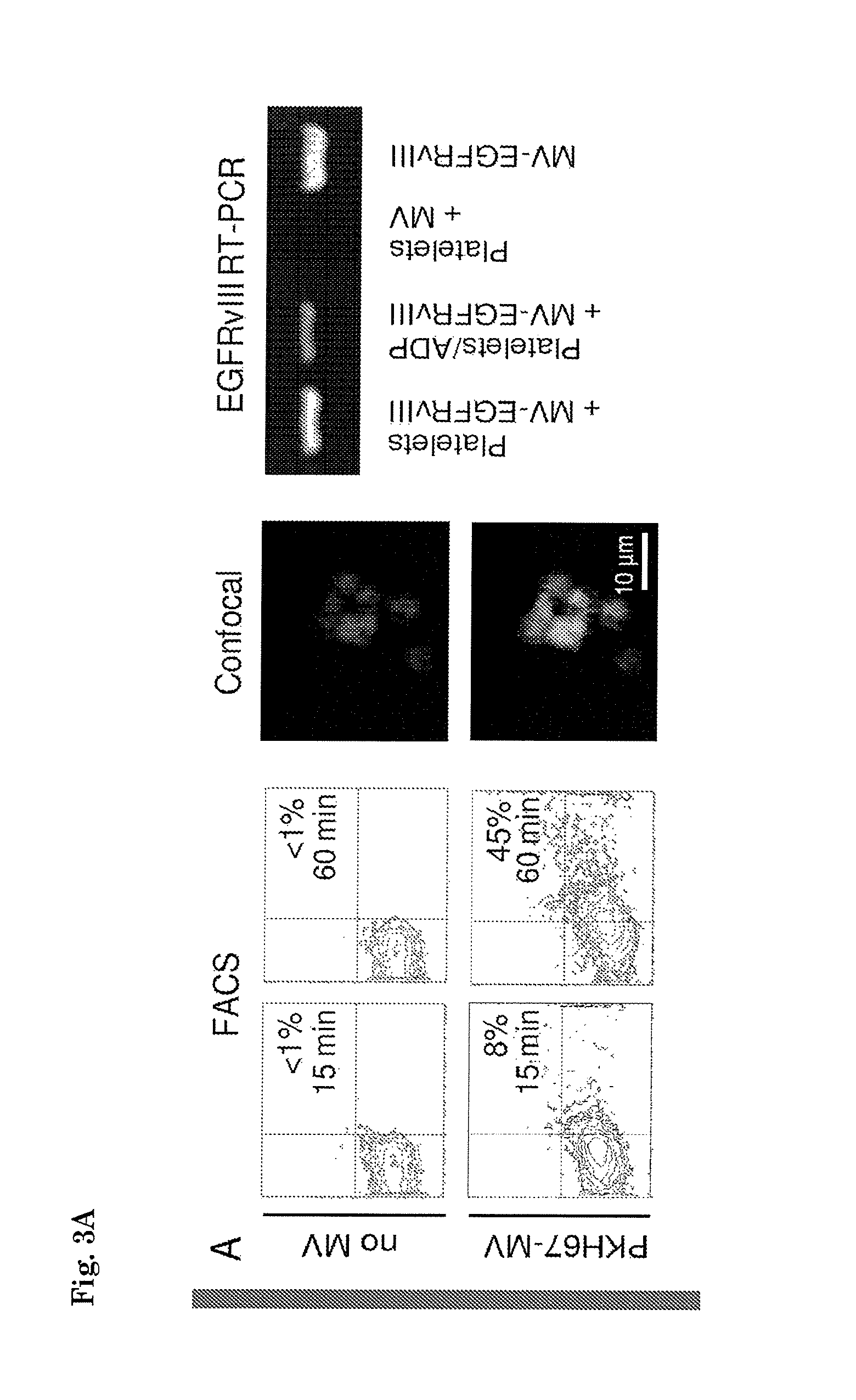
FIG. 3. (A) U87 glioma-derived microvesicles were labelled with PKH67 green fluorescent dye and incubated with isolated platelets. After 15 and 60 min of incubation in the presence and absence of microvesicles the platelets were washed and subjected to FACS analysis of PKH67 fluorescence. In addition, the platelets were stained and analyzed by confocal microscopy to determine microvesicle uptake. RNA was isolated from RNase-treated platelets after incubation with microvesicles under different conditions. RT-PCR was performed to detect EGFRvIII RNA. MV/MVEGFRvIII: microvesicles isolated from U87/U87-EGFRvIII cells. (B) RNA was isolated from platelets from healthy control subjects or glioma patients and subjected to RT-PCR analysis. Corresponding glioma tissue biopsies served as control. PC=U87-EGFRvIII RNA; NC=H20; nd=not determined; * indicates positive signal. (C) RNA as in (B) was subjected to gene expression arrays. Heat map of top-30 glioma biomarkers is shown on the left. Individual expression levels for the top-10 RNAs depicted on the right. Dashed line=BG (background).
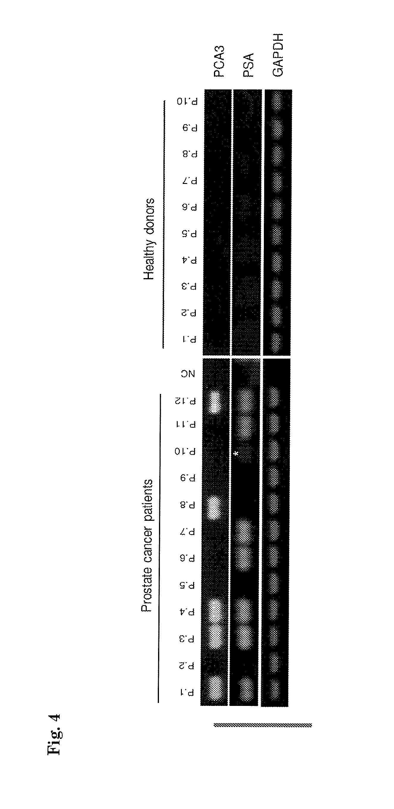
FIG. 4. RNA was isolated from platelets from healthy control subjects (n=8) and prostate cancer patients (n=12) and subjected to PCA3, PSA, and GAPDH RT-PCR analysis. * indicates weak positive signal.
FIG. 5 shows the probe sequences used for the detection of the genes displayed in FIG. 3C.
DETAILED DESCRIPTION OF THE INVENTION
As used herein, the term "cancer" refers to a disease or disorder resulting from the proliferation of oncogenically transformed cells. "Cancer" shall be taken to include any one or more of a wide range of benign or malignant tumours, including those that are capable of invasive growth and metastasis through a human or animal body or a part thereof, such as, for example, via the lymphatic system and/or the blood stream. As used herein, the term "tumour" includes both benign and malignant tumours or solid growths, notwithstanding that the present invention is particularly directed to the diagnosis or detection of malignant tumours and solid cancers. Cancers further include but are not limited to carcinomas, lymphomas, or sarcomas, such as, for example, ovarian cancer, colon cancer, breast cancer, pancreatic cancer, lung cancer, prostate cancer, urinary tract cancer, uterine cancer, acute lymphatic leukaemia, Hodgkin's disease, small cell carcinoma of the lung, melanoma, neuroblastoma, glioma (e.g. glioblastoma), and soft tissue sarcoma, lymphoma, melanoma, sarcoma, and adenocarcinoma. In preferred embodiments of aspects of the present invention, thrombocyte cancer is disclaimed.
The term "cancer-derived" as used herein refers to origination from a cancer or cancer cell.
The term "cancer-derived nucleic acid" shall be taken to mean any nucleic acid that is indicative of cancer in the subject, specifically and in most preferred embodiments a mutant DNA or RNA indicating the presence in the cancer of a mutant gene that is expressed by or is present in a cancer cell of the subject, of which mutant gene the nucleic acid sequence is altered relative to the normal gene of a healthy control subject. The term "cancer-derived nucleic acid" shall also be taken to include (i) a nucleic acid that is produced by, expressed by, or present in a cancer cell but not in a normal healthy (non-cancerous) cell, or whose production or expression is altered (enhanced or reduced) by or in a cancer cell compared to a normal cell; or (ii) a nucleic acid that is produced by, expressed by, or present in a normal cell but not by or in a cancer cell. Hence, the nucleic acid need not be a mutant nucleic acid having a mutated sequence but may be a normal nucleic acid having a wild-type (non-cancer) sequence, but whose profile or expression level is altered in a cancer cell relative to a normal cell. In one preferred embodiment, the cancer-derived nucleic acid is a mutant nucleic acid (DNA, cDNA, or RNA) specific for the cancer, preferably an RNA transcript. In another very preferred embodiment, the cancer-derived nucleic acid is a nucleic acid expression profile indicative of being cancer-derived or cancer-specific, as explained in detail herein.
As used herein the term "cancer marker" refers to in particular to a cancer marker gene or a cancer marker gene expression profile. As used herein, the term "cancer marker gene" refers to a gene whose sequence or expression level, alone or in combination with other genes, is correlated with cancer or prognosis of cancer. The correlation may relate to either an increased or decreased expression of the gene reflected in an increased or decreased presence of the RNA expression product of said gene in the nucleic acid fraction obtainable from thrombocytes. For example, the expression of the gene may be indicative of cancer, or lack of expression of the gene may be correlated with poor prognosis in a cancer patient. In the case of prostate cancer AMACR, PCA3 and PSA are suitable cancer markers. In the case of colorectal cancer KRAS mutations are suitable cancer markers. In the case of lung carcinoma EGFR mutations are suitable cancer markers. In the case of melanoma BRAF mutations are suitable cancer markers. In the case of glioma EGFRvIII mutations are suitable cancer markers. Other suitable cancer markers may be derived from Tables 1 and 2 as provided herein or from the Examples or Figures. The skilled person will understand that many other cancer markers may be employed in aspects and embodiments of this invention.
As used herein, the term "stage of cancer" refers to a qualitative or quantitative assessment of the level of advancement of a cancer. Criteria used to determine the stage of a cancer include, but are not limited to, the size of the tumor, whether the tumor has spread to other parts of the body and where the cancer has spread (e.g., within the same organ or region of the body or to another organ).
The term "cancer" in the terms "cancer derived", "cancer marker", "cancer marker gene", and/or "stage of cancer" may be generalized to the term "disease" as the definitions for cancer are generally applicable to all diseases as indicated herein.
The term "disease-derived" as used herein refers to origination from a disease or diseased cell.
The term "disease-derived nucleic acid" shall be taken to mean any nucleic acid that is indicative of a disease in the subject, specifically and in most preferred embodiments a mutant DNA or RNA indicating the presence in the disease of a mutant gene that is expressed by or is present in a diseased cell of the subject, of which mutant gene the nucleic acid sequence is altered relative to the normal gene of a healthy control subject. The term "disease-derived nucleic acid" shall also be taken to include (i) a nucleic acid that is produced by, expressed by, or present in a diseased cell but not in a normal healthy (non-diseased) cell, or whose production or expression is altered (enhanced or reduced) by or in a diseased cell compared to a normal cell; or (ii) a nucleic acid that is produced by, expressed by, or present in a normal cell but not by or in a diseased cell. Hence, the nucleic acid need not be a mutant nucleic acid having a mutated sequence but may be a normal nucleic acid having a wild-type (non-disease) sequence, but whose profile or expression level is altered in a diseased cell relative to a normal cell. In one preferred embodiment, the disease-derived nucleic acid is a mutant nucleic acid (DNA, cDNA, or RNA) specific for the disease, preferably an RNA transcript. In another very preferred embodiment, the disease-derived nucleic acid is a nucleic acid expression profile indicative of being disease-derived or disease-specific, as explained in detail herein. In a preferred embodiment disease-derived nucleic acid does not include cancer-derived nucleic acid. In yet another preferred embodiment, the disease derived nucleic acid does not include vascular disease derived nucleic acid, and/or systemic lupus erythematosus derived nucleic acid. In a preferred embodiment disease-derived nucleic acid does not include sickle cell disease derived nucleic acid. In a preferred embodiment disease-derived nucleic acid does not include Alzheimer's disease derived nucleic acid. In a preferred embodiment of the present invention and embodiments thereof the disease-derived nucleic acid does not include CD109 nucleic acid. In yet another preferred embodiment of the present invention and embodiments thereof, the disease-derived nucleic acid does not comprise megakaryocyte derived nucleic acid. In yet another preferred embodiment of the present invention and embodiments thereof, the disease-derived nucleic acid does not comprise nucleic acid derived from disease associated with pathological megakaryocyte and/or platelet function.
As used herein the term "disease marker" refers to in particular to a disease marker gene or a disease marker gene expression profile. As used herein, the term "disease marker gene" refers to a gene whose sequence or expression level, alone or in combination with other genes, is correlated with disease or prognosis of the disease. The correlation may relate to either an increased or decreased expression of the gene reflected in an increased or decreased presence of the RNA expression product of said gene in the nucleic acid fraction obtainable from thrombocytes. For example, the expression of the gene may be indicative of a disease, or lack of expression of the gene may be correlated with poor prognosis in a patient. In a preferred embodiment said disease marker gene is not a CD109 gene.
As used herein, the term "stage of disease" refers to a qualitative or quantitative assessment of the level of advancement of a disease. Criteria used to determine the stage of a disease include, but are not limited to, whether the disease has spread to other parts of the body and where the disease has spread to (e.g., within the same organ or region of the body or to another organ).
The term "disease" as used herein may refer to cancer, autoimmune disease, skin diseases, eye disease, endocrine diseases, neurological disorders, and cardiovascular diseases.
The term "disease" as used herein may refer to autoimmune disease, skin diseases, eye disease, endocrine diseases, neurological disorders, and/or cardiovascular diseases.
The term "disease" as used herein may refer to autoimmune disease, skin diseases, eye disease, endocrine diseases, and/or neurological disorders.
The term "disease" as used herein may, in some preferred embodiments, not refer to cancer, cardiovascular disease, systemic lupus erythematosus, sickle cell disease, Alzheimer's disease, diseases associated with pathological platelet function, and/or diseases associated with pathological megakaryocyte function.
Thus, diseases that in addition to or instead of cancer can be detected using the means and methods of the present invention include for instance the following auto-immune diseases: Achlorhydra Autoimmune Active Chronic Hepatitis; Acute Disseminated Encephalomyelitis; Acute hemorrhagic leukoencephalitis; Addison's Disease; Agammaglobulinemia; Alopecia greata; Amyotrophic Lateral Sclerosis; Ankylosing Spondylitis; Anti-GBM/TBM Nephritis; Antiphospholipid syndrome; Antisynthetase syndrome; polyarticular Arthritis; Atopic allergy; Atopic Dermatitis; Autoimmune Aplastic Anemia; Autoimmune cardiomyopathy; Autoimmune enteropathy; Autoimmune hemolytic anemia; Autoimmune hepatitis; Autoimmune inner ear disease; Autoimmune lymphoproliferative syndrome; Autoimmune peripheral neuropathy; Autoimmune pancreatitis; Autoimmune polyendocrine syndrome; Autoimmune progesterone dermatitis; Autoimmune thrombocytopenic purpura; Autoimmune uveitis; Balo disease/Balo concentric sclerosis; Bechets Syndrome; Berger's disease; Bickerstaffs encephalitis; Blau syndrome; Bullous Pemphigoid; Castleman's disease; Celiac disease; Chagas disease; Chronic Fatigue Immune Dysfunction Syndrome; Chronic inflammatory demyelinating polyneuropathy; Chronic recurrent multifocal osteomyelitis; Chronic lyme disease; Chronic obstructive pulmonary disease; Churg-Strauss syndrome; Cicatricial Pemphigoid; Coeliac Disease; Cogan syndrome; Cold agglutinin disease; Complement component 2 deficiency; Cranial arteritis; CREST syndrome; Crohns Disease; Cushing's Syndrome; Cutaneous leukocytoclastic angiitis; Dego's disease; Dercum's disease; Dermatitis herpetiformis; Dermatomyositis; Diabetes mellitus type 1; Diffuse cutaneous systemic sclerosis; Dressler's syndrome; Discoid lupus erythematosus; Eczema; Endometriosis; Enthesitis-related arthritis; Eosinophilic fasciitis; Eosinophilic gastroenteritis; Epidermolysis bullosa acquisita; Erythema nodosum; Essential mixed cryoglobulinemia; Evan's syndrome; Fibrodysplasia ossificans progressiva; Fibromyalgia/Fibromyositis; Fibrosing aveolitis; Gastritis; Gastrointestinal pemphigoid; Giant cell arteritis; Glomerulonephritis; Goodpasture's syndrome; Graves' disease; Guillain-Barre syndrome; Hashimoto's encephalitis; Hashimoto's thyroiditis; Haemolytic anaemia; Henoch-Schonlein purpura; Herpes gestationis; Hidradenitis suppurativa; Hughes syndrome; Hypogammaglobulinemia; Idiopathic Inflammatory Demyelinating Diseases; Idiopathic pulmonary fibrosis; Idiopathic thrombocytopenic purpura; IgA nephropathy; Inclusion body myositis; Inflammatory demyelinating polyneuopathy; Interstitial cystitis; Irritable Bowel Syndrome (IBS); Juvenile idiopathic arthritis; Juvenile rheumatoid arthritis; Kawasaki's Disease; Lambert-Eaton myasthenic syndrome; Leukocytoclastic vasculitis; Lichen planus; Lichen sclerosus; Linear IgA disease; Lou Gehrig's Disease; Lupoid hepatitis; Lupus erythematosus; Majeed syndrome; Meniere's disease; Microscopic polyangiitis; Miller-Fisher syndrome; Mixed Connective Tissue Disease; Morphea; Mucha-Habermann disease; Muckle-Wells syndrome; Multiple Myeloma; Multiple Sclerosis; Myasthenia gravis; Myositis; Narcolepsy; Neuromyelitis optica; Neuromyotonia; Occular cicatricial pemphigoid; Opsoclonus myoclonus syndrome; Ord thyroiditis; Palindromic rheumatism; PANDAS; Paraneoplastic cerebellar degeneration; Paroxysmal nocturnal hemoglobinuria; Parry Romberg syndrome; Parsonnage-Turner syndrome; Pars planitis; Pemphigus; Pemphigus vulgaris; Pernicious anaemia; Perivenous encephalomyelitis; POEMS syndrome; Polyarteritis nodosa; Polymyalgia rheumatica; Polymyositis; Primary biliary cirrhosis; Primary sclerosing cholangitis; Progressive inflammatory neuropathy; Psoriasis; Psoriatic Arthritis; Pyoderma gangrenosum; Pure red cell aplasia; Rasmussen's encephalitis; Raynaud phenomenon; Relapsing polychondritis; Reiter's syndrome; Restless leg syndrome; Retroperitoneal fibrosis; Rheumatoid arthritis; Rheumatoid fever; Sarcoidosis; Schizophrenia; Schmidt syndrome; Schnitzler syndrome; Scleritis; Scleroderma; Sjogren's syndrome; Spondyloarthropathy; Sticky blood syndrome; Still's Disease; Stiff person syndrome; Subacute bacterial endocarditis (SBE); Susac's syndrome; Sweet syndrome; Sydenham Chorea; Sympathetic ophthalmia; Takayasu's arteritis; Temporal arteritis; Tolosa-Hunt syndrome; Transverse Myelitis; Ulcerative Colitis; Undifferentiated connective tissue disease; Undifferentiated spondyloarthropathy; Vasculitis; Vitiligo; Wegener's granulomatosis; Wilson's syndrome; and Wiskott-Aldrich syndrome.
Apart from the above diseases, aspects of the present invention are also applicable to the prognosis and diagnosis of the following skin diseases: Acneiform eruptions; Autoinflammatory syndromes; Chronic blistering; Conditions of the mucous membranes; Conditions of the skin appendages; Conditions of the subcutaneous fat; Congenital anomalies; Connective tissue diseases (such as Abnormalities of dermal fibrous and elastic tissue); Dermal and subcutaneous growths; Dermatitis (including Atopic Dermatitis, Contact Dermatitis, Eczema, Pustular Dermatitis, and Seborrheic Dermatitis); Disturbances of pigmentation; Drug eruptions; Endocrine-related skin disease; Eosinophilic; Epidermal nevi, neoplasms, cysts; Erythemas; Genodermatoses; Infection-related skin disease; Lichenoid eruptions; Lymphoid-related skin disease; Melanocytic nevi and neoplasms (including Melanoma); Monocyte- and macrophage-related skin disease; Mucinoses; Neurocutaneous; Noninfectious immunodeficiency-related skin disease; Nutrition-related skin disease; Papulosquamous hyperkeratotic (including Palmoplantar keratodermas); Pregnancy-related skin disease; Pruritic; Psoriasis; Reactive neutrophilic; Recalcitrant palmoplantar eruptions; Resulting from errors in metabolism; Resulting from physical factors (including Ionizing radiation-induced); Urticaria and angioedema; Vascular-related skin disease.
Apart from the above diseases, aspects of the present invention are also applicable to the prognosis and diagnosis of the following endocrine diseases: Adrenal disorders; Glucose homeostasis disorders; Thyroid disorders; Calcium homeostasis disorders and Metabolic bone disease; Pituitary gland disorders; and Sex hormone disorders.
Apart from the above diseases, aspects of the present invention are also applicable to the prognosis and diagnosis of the following eye diseases: H00-H06 Disorders of eyelid, lacrimal system and orbit; H10-H13 Disorders of conjunctiva; H15-H22 Disorders of sclera, cornea, iris and ciliary body; H25-H28 Disorders of lens; H30-H36 Disorders of choroid and retina (including H30 Chorioretinal inflammation, H31 Other disorders of choroid, H.sub.32Chorioretinal disorders in diseaseas classified elsewhere, H33 Retinal detachments and breaks, H34 Retinal vascular occlusions, H35 Other retinal disorders, and H36 Retinal disorders in diseases classified elsewhere); H40-H42 Glaucoma; H43-H45 Disorders of vitreous body and globe; H46-H48 Disorders of optic nerve and visual pathways; H49-H52 Disorders of ocular muscles, binocular movement, accommodation and refraction; H53-H54.9 Visual disturbances and blindness; and H55-H59 Other disorders of eye and adnexa.
Apart from the above diseases, aspects of the present invention are also applicable to the prognosis and diagnosis of the following neurological disorders: Abarognosis; Acquired Epileptiform Aphasia; Acute disseminated encephalomyelitis; Adrenoleukodystrophy; Agenesis of the corpus callosum; Agnosia; Aicardi syndrome; Alexander disease; Alien hand syndrome; Allochiria; Alpers' disease; Alternating hemiplegia; Alzheimer's disease; Amyotrophic lateral sclerosis (see Motor Neurone Disease); Anencephaly; Angelman syndrome; Angiomatosis; Anoxia; Aphasia; Apraxia; Arachnoid cysts; Arachnoiditis; Arnold-Chiari malformation; Arteriovenous malformation; Ataxia Telangiectasia; Attention deficit hyperactivity disorder; Auditory processing disorder; Autonomic Dysfunction; Back Pain; Batten disease; Behcet's disease; Bell's palsy; Benign Essential Blepharospasm; Benign Intracranial Hypertension; Bilateral frontoparietal polymicrogyria; Binswanger's disease; Blepharospasm; Bloch-Sulzberger syndrome; Brachial plexus injury; Brain abscess; Brain damage; Brain injury; Brain tumor; Brown-Sequard syndrome; Canavan disease; Carpal tunnel syndrome; Causalgia; Central pain syndrome; Central pontine myelinolysis; Centronuclear myopathy; Cephalic disorder; Cerebral aneurysm; Cerebral arteriosclerosis; Cerebral atrophy; Cerebral gigantism; Cerebral palsy; Cerebral vasculitis; Cervical spinal stenosis; Charcot-Marie-Tooth disease; Chiari malformation; Chorea; Chronic fatigue syndrome; Chronic inflammatory demyelinating polyneuropathy (CIDP); Chronic pain; Coffin Lowry syndrome; Coma; Complex regional pain syndrome; Compression neuropathy; Congenital facial diplegia; Corticobasal degeneration; Cranial arteritis; Craniosynostosis; Creutzfeldt-Jakob disease; Cumulative trauma disorders; Cushing's syndrome; Cytomegalic inclusion body disease (CIBD); Cytomegalovirus Infection; Dandy-Walker syndrome; Dawson disease; De Morsier's syndrome; Dejerine-Klumpke palsy; Dejerine-Sottas disease; Delayed sleep phase syndrome; Dementia; Dermatomyositis; Developmental dyspraxia; Diabetic neuropathy; Diffuse sclerosis; Dravet syndrome; Dysautonomia; Dyscalculia; Dysgraphia; Dyslexia; Dystonia; Empty sella syndrome; Encephalitis; Encephalocele; Encephalotrigeminal angiomatosis; Encopresis; Epilepsy; Erb's palsy; Erythromelalgia; Essential tremor; Fabry's disease; Fahr's syndrome; Fainting; Familial spastic paralysis; Febrile seizures; Fisher syndrome; Friedreich's ataxia; Fibromyalgia; Gaucher's disease; Gerstmann's syndrome; Giant cell arteritis; Giant cell inclusion disease; Globoid Cell Leukodystrophy; Gray matter heterotopia; Guillain-Barre syndrome; HTLV-1 associated myelopathy; Hallervorden-Spatz disease; Head injury; Headache; Hemifacial Spasm; Hereditary Spastic Paraplegia; Heredopathia atactica polyneuritiformis; Herpes zoster oticus; Herpes zoster; Hirayama syndrome; Holoprosencephaly; Huntington's disease; Hydranencephaly; Hydrocephalus; Hypercortisolism; Hypoxia; Immune-Mediated encephalomyelitis; Inclusion body myositis; Incontinentia pigmenti; Infantile phytanic acid storage disease; Infantile Refsum disease; Infantile spasms; Inflammatory myopathy; Intracranial cyst; Intracranial hypertension; Joubert syndrome; Karak syndrome; Kearns-Sayre syndrome; Kennedy disease; Kinsbourne syndrome; Klippel Feil syndrome; Krabbe disease; Kugelberg-Welander disease; Kuru; Lafora disease; Lambert-Eaton myasthenic syndrome; Landau-Kleffner syndrome; Lateral medullary (Wallenberg) syndrome; Learning disabilities; Leigh's disease; Lennox-Gastaut syndrome; Lesch-Nyhan syndrome; Leukodystrophy; Lewy body dementia; Lissencephaly; Locked-In syndrome; Lou Gehrig's disease (See Motor Neurone Disease); Lumbar disc disease; Lumbar spinal stenosis; Lyme disease--Neurological Sequelae; Machado-Joseph disease (Spinocerebellar ataxia type 3); Macrencephaly; Macropsia; Megalencephaly; Melkersson-Rosenthal syndrome; Menieres disease; Meningitis; Menkes disease; Metachromatic leukodystrophy; Microcephaly; Micropsia; Migraine; Miller Fisher syndrome; Mini-stroke (transient ischemic attack); Mitochondrial myopathy; Mobius syndrome; Monomelic amyotrophy; Motor Neurone Disease; Motor skills disorder; Moyamoya disease; Mucopolysaccharidoses; Multi-infarct dementia; Multifocal motor neuropathy; Multiple sclerosis; Multiple system atrophy; Muscular dystrophy; Myalgic encephalomyelitis; Myasthenia gravis; Myelinoclastic diffuse sclerosis; Myoclonic Encephalopathy of infants; Myoclonus; Myopathy; Myotubular myopathy; Myotonia congenita; Narcolepsy; Neurofibromatosis; Neuroleptic malignant syndrome; Neurological manifestations of AIDS; Neurological sequelae of lupus; Neuromyotonia; Neuronal ceroid lipofuscinosis; Neuronal migration disorders; Niemann-Pick disease; Non 24-hour sleep-wake syndrome; Nonverbal learning disorder; O'Sullivan-McLeod syndrome; Occipital Neuralgia; Occult Spinal Dysraphism Sequence; Ohtahara syndrome; Olivopontocerebellar atrophy; Opsoclonus myoclonus syndrome; Optic neuritis; Orthostatic Hypotension; Overuse syndrome; Palinopsia; Paresthesia; Parkinson's disease; Paramyotonia Congenita; Paraneoplastic diseases; Paroxysmal attacks; Parry-Romberg syndrome; Pelizaeus-Merzbacher disease; Periodic Paralyses; Peripheral neuropathy; Persistent Vegetative State; Pervasive developmental disorders; Photic sneeze reflex; Phytanic acid storage disease; Pick's disease; Pinched nerve; Pituitary tumors; PMG; Polio; Polymicrogyria; Polymyositis; Porencephaly; Post-Polio syndrome; Postherpetic Neuralgia (PHN); Postinfectious Encephalomyelitis; Postural Hypotension; Prader-Willi syndrome; Primary Lateral Sclerosis; Prion diseases; Progressive hemifacial atrophy; Progressive multifocal leukoencephalopathy; Progressive Supranuclear Palsy; Pseudotumor cerebri; Rabies; Ramsay-Hunt syndrome (Type I and Type II); Rasmussen's encephalitis; Reflex neurovascular dystrophy; Refsum disease; Repetitive motion disorders; Repetitive stress injury; Restless legs syndrome; Retrovirus-associated myelopathy; Rett syndrome; Reye's syndrome; Rhythmic Movement Disorder; Romberg syndrome; Saint Vitus dance; Sandhoff disease; Schizophrenia; Schilder's disease; Schizencephaly; Sensory integration dysfunction; Septo-optic dysplasia; Shaken baby syndrome; Shingles; Shy-Drager syndrome; Sjogren's syndrome; Sleep apnea; Sleeping sickness; Snatiation; Sotos syndrome; Spasticity; Spina bifida; Spinal cord injury; Spinal cord tumors; Spinal muscular atrophy; Spinocerebellar ataxia; Steele-Richardson-Olszewski syndrome; Stiff-person syndrome; Stroke; Sturge-Weber syndrome; Subacute sclerosing panencephalitis; Subcortical arteriosclerotic encephalopathy; Superficial siderosis; Sydenham's chorea; Syncope; Synesthesia; Syringomyelia; Tarsal tunnel syndrome; Tardive dyskinesia; Tarlov cyst; Tay-Sachs disease; Temporal arteritis; Tetanus; Tethered spinal cord syndrome; Thomsen disease; Thoracic outlet syndrome; Tic Douloureux; Todd's paralysis; Tourette syndrome; Toxic encephalopathy; Transient ischemic attack; Transmissible spongiform encephalopathies; Transverse myelitis; Traumatic brain injury; Tremor; Trigeminal neuralgia; Tropical spastic paraparesis; Trypanosomiasis; Tuberous sclerosis; Von Hippel-Lindau disease; Viliuisk Encephalomyelitis; Wallenberg's syndrome; Werdnig-Hoffman disease; West syndrome; Whiplash; Williams syndrome; Wilson's disease; and Zellweger syndrome.
Apart from the above diseases, aspects of the present invention are also applicable to the prognosis and diagnosis of the following cardiovascular diseases: Aneurysm; Angina; Atherosclerosis; Cerebrovascular Accident (Stroke); Cerebrovascular disease; Congestive Heart Failure; Coronary Artery Disease; Myocardial infarction (Heart Attack); and Peripheral vascular disease. In a preferred embodiment of the method of the invention and embodiments thereof, and in preferred embodiments of other aspects of the invention, the disease or the cardiovascular disease is not systemic lupus erythematosus.
In a preferred embodiment of the method of the invention and embodiments thereof, and in preferred embodiments of other aspects of the invention, the disease is not a disease selected from the group comprising cancer, cardiovascular disease, systemic lupus erythematosus, sickle cell disease, Alzheimer's disease, diseases associated with pathological platelet function, and/or diseases associated with pathological megakaryocyte function.
As used herein, "nucleic acid" includes reference to a deoxyribonucleotide or ribonucleotide polymer in either single- or double-stranded form, and unless otherwise limited, encompasses known analogues having the essential nature of natural nucleotides in that they hybridize to single-stranded nucleic acids in a manner similar to naturally occurring nucleotides (e.g., peptide nucleic acids).
The term "RNA" refers to ribonucleic acid, a molecule of RNA encoding for a protein product or non-coding for a protein product (such as miRNAs but not excluding other non-coding RNAs). RNA is transcribed from a DNA template.
As used herein the term "mutant" refers to a nucleic acid compound, protein, molecule, vector or cell resulting from mutation of the native wild type coding sequence or subunits thereof.
As used herein the term "mutation" refers to any change that alters a native coding sequence either by displacement, addition, deletion, insertion, cross-linking, or other destruction or substitution of one or more nucleotides of the native coding sequence, including naturally occurring splice variants. In particular, the mutation provides a gene that causes the cell to be a cancer cell. Such mutations include inherited and acquired mutations of tumor suppressor genes and/or oncogenes.
By "amplified" is meant the construction of multiple copies of a nucleic acid sequence or multiple copies complementary to the nucleic acid sequence using at least one of the nucleic acid sequences as a template. Amplification systems include the polymerase chain reaction (PCR) system, ligase chain reaction (LCR) system, nucleic acid sequence based amplification (NASBA, Cangene, Mississauga, Ontario), Q-Beta Replicase systems, transcription-based amplification system (TAS), and strand displacement amplification (SDA). See, e.g., Diagnostic Molecular Microbiology. Principles and Applications, D. H. Persing et al., Ed., American Society for Microbiology, Washington, D.C. (1993). The product of amplification is termed an amplicon.
The term "hybrid" refers to a double-stranded nucleic acid molecule, or duplex, formed by hydrogen bonding between complementary nucleotides. The terms "hybridise" or "anneal" refer to the process by which single strands of nucleic acid sequences form double-helical segments through hydrogen bonding between complementary nucleotides.
The term "oligonucleotide" refers to a short sequence of nucleotide monomers (usually 6 to 100 nucleotides) joined by phosphorous linkages (e.g., phosphodiester, alkyl and aryl-phosphate, phosphorothioate), or non-phosphorous linkages (e.g., peptide, sulfamate and others). An oligonucleotide may contain modified nucleotides having modified bases (e.g., 5-methyl cytosine) and modified sugar groups (e.g., 2'-O-methyl ribosyl, 2'-O-methoxyethyl ribosyl, 2'-fluoro ribosyl, 2'-amino ribosyl, and the like). Oligonucleotides may be naturally-occurring or synthetic molecules of double- and single-stranded DNA and double- and single-stranded RNA with circular, branched or linear shapes and optionally including domains capable of forming stable secondary structures (e.g., stem-and-loop and loop-stem-loop structures).
The term "primer" as used herein refers to an oligonucleotide which is capable of annealing to the amplification target allowing a DNA polymerase to attach thereby serving as a point of initiation of DNA synthesis when placed under conditions in which synthesis of primer extension product which is complementary to a nucleic acid strand is induced, i.e., in the presence of nucleotides and an agent for polymerization such as DNA polymerase and at a suitable temperature and pH. The (amplification) primer is preferably single stranded for maximum efficiency in amplification. Preferably, the primer is an oligodeoxy ribonucleotide. The primer must be sufficiently long to prime the synthesis of extension products in the presence of the agent for polymerization. The exact lengths of the primers will depend on many factors, including temperature and source of primer. A "pair of bi-directional primers" as used herein refers to one forward and one reverse primer as commonly used in the art of DNA amplification such as in PCR amplification.
The term "probe" refers to a single-stranded oligonucleotide sequence that will recognize and form a hydrogen-bonded duplex with a complementary sequence in a target nucleic acid sequence analyte or its cDNA derivative.
The terms "stringency" or "stringent hybridization conditions" refer to hybridization conditions that affect the stability of hybrids, e.g., temperature, salt concentration, pH, formamide concentration and the like. These conditions are empirically optimised to maximize specific binding and minimize non-specific binding of primer or probe to its target nucleic acid sequence. The terms as used include reference to conditions under which a probe or primer will hybridise to its target sequence, to a detectably greater degree than other sequences (e.g. at least 2-fold over background). Stringent conditions are sequence dependent and will be different in different circumstances. Longer sequences hybridise specifically at higher temperatures. Generally, stringent conditions are selected to be about 5.degree. C. lower than the thermal melting point (T.sub.m) for the specific sequence at a defined ionic strength and pH. The T.sub.m is the temperature (under defined ionic strength and pH) at which 50% of a complementary target sequence hybridises to a perfectly matched probe or primer. Typically, stringent conditions will be those in which the salt concentration is less than about 1.0 M Na.sup.+ ion, typically about 0.01 to 1.0 M Na.sup.+ ion concentration (or other salts) at pH 7.0 to 8.3 and the temperature is at least about 30.degree. C. for short probes or primers (e.g. 10 to 50 nucleotides) and at least about 60.degree. C. for long probes or primers (e.g. greater than 50 nucleotides). Stringent conditions may also be achieved with the addition of destabilizing agents such as formamide. Exemplary low stringent conditions or "conditions of reduced stringency" include hybridization with a buffer solution of 30% formamide, 1 M NaCl, 1% SDS at 37.degree. C. and a wash in 2.times.SSC at 40.degree. C.
Exemplary high stringency conditions include hybridization in 50% formamide, 1 M NaCl, 1% SDS at 37.degree. C., and a wash in 0.1.times.SSC at 60.degree. C. Hybridization procedures are well known in the art and are described in e.g. Ausubel et al, Current Protocols in Molecular Biology, John Wiley & Sons Inc., 1994.
"Subject" as used herein includes, but is not limited to, mammals, including, e.g., a human, a non-human primate, a mouse, a pig, a cow, a goat, a cat, a rabbit, a rat, a guinea pig, a hamster, a degu, a horse, a monkey, a sheep, or other non-human mammal; and non-mammal animals, including, e.g., a non-mammalian vertebrate, such as a bird (e.g., a chicken or duck) or a fish, and an invertebrate. The subject may be a healthy animal or human subject undergoing a routine well-being check up. Alternatively, the subject may be at risk of having a disease (e.g., a genetically predisposed subject, a subject with medical and/or family history of cancer, a subject who has been exposed to carcinogens, occupational hazard, environmental hazard] and/or a subject who exhibits suspicious clinical signs of a disease [e.g., blood in the stool or melena, unexplained pain, sweating, unexplained fever, unexplained loss of weight up to anorexia, changes in bowel habits (constipation and/or diarrhoea), tenesmus (sense of incomplete defecation, for rectal cancer specifically), anaemia and/or general weakness). According to another embodiment, the subject may be a patient diagnosed with the disease and is performing a routine check-up, in-between treatments.
The term "thrombocyte", as used herein, refers to blood platelets, i.e. the small, irregularly-shaped cell fragments that do not have a nucleus containing DNA, and that circulate in the blood of mammals. Thrombocytes are 2-3 .mu.m in diameter, and are derived from fragmentation of precursor megakaryocytes. Platelets or thrombocytes lack nuclear DNA, although they retain some megakaryocyte-derived mRNAs as part of their lineal origin. The average lifespan of a thrombocyte is 5 to 9 days. Thrombocytes are involved and play an essential role in haemostasis, leading to the formation of blood clots. In a preferred embodiment of the present invention and embodiments thereof, the anucleated blood cell-extracted nucleic acid fraction is not megakaryocyte-derived nucleic acid or megakaryocyte-derived RNA.
The term "blood" as used herein refers to whole blood (including plasma and cells) and includes arterial, capillary and venous blood.
The term "nucleated cell" as used herein preferably refers to a Bartholin's gland cell; Salivary gland mucous cell; Salivary gland serous cell; Von Ebner's gland cell; Mammary gland cell; Lacrimal gland cell; Ceruminous gland cell; Eccrine sweat gland cell; Apocrine sweat gland cell; Gland of Moll cell; Sebaceous gland cell; Bowman's gland cell; Brunner's gland cell; Seminal vesicle cell; Prostate gland cell; Bulbourethral gland cell; Gland of Littre cell; Uterus endometrium cell; Isolated goblet cell; Stomach lining mucous cell; Gastric gland zymogenic cell; Gastric gland oxyntic cell; Pancreatic acinar cell; Paneth cell; Type II pneumocyte; Clara cell; Anterior pituitary cell; Intermediate pituitary cell; Magnocellular neurosecretory cell; Thyroid gland cell; Parathyroid gland cells; Adrenal gland cells; Leydig cell; Theca interna cell; Corpus luteum cell; Juxtaglomerular cell; Macula densa cell; Peripolar cell; Mesangial cell; Blood vessel and lymphatic vascular endothelial fenestrated cell; Blood vessel and lymphatic vascular endothelial continuous cell; Blood vessel and lymphatic vascular endothelial splenic cell; Synovial cell; Serosal cell; Squamous cell; Columnar cell; Dark cell; Vestibular membrane cell; Stria vascularis basal cell; Stria vascularis marginal cell; Cell of Claudius; Cell of Boettcher; Choroid plexus cell; Pia-arachnoid squamous cell; Pigmented ciliary epithelium cell; Nonpigmented ciliary epithelium cell; Corneal endothelial cell; Peg cell; Respiratory tract ciliated cell; Oviduct ciliated cell; Uterine endometrial ciliated cell; Rete testis ciliated cell; Ductulus efferens ciliated cell; Ciliated ependymal cell; Epidermal keratinocyte; Epidermal basal cell; Keratinocyte; Nail bed basal cell; Medullary hair shaft cell; Cortical hair shaft cell; Cuticular hair shaft cell; Cuticular hair root sheath cell; External hair root sheath cell; Hair matrix cell; Surface epithelial cell; basal cell; Urinary epithelium cell; Auditory inner hair cell; Auditory outer hair cell; Primary sensory neurons; Merkel cell; Olfactory receptor neuron; Photoreceptor cells; Carotid body cell (blood pH sensor); Hair cell; Taste bud cell; Cholinergic neural cell; Adrenergic neural cell; Peptidergic neural cell; Inner pillar cell; Outer pillar cell; Inner phalangeal cell; Outer phalangeal cell; Border cell; Hensen cell; Vestibular apparatus supporting cell; Taste bud supporting cell; Olfactory epithelium supporting cell; Schwann cell; Satellite cell; Enteric glial cell; Astrocyte; Neuron cells; Oligodendrocyte; Spindle neuron; Anterior lens epithelial cell; Crystallin-containing lens fiber cell; Hepatocyte; Adipocytes; Liver lipocyte; Kidney glomerulus parietal cell; Kidney glomerulus podocyte; Kidney proximal tubule brush border cell; Loop of Henle thin segment cell; Kidney distal tubule cell; Kidney collecting duct cell; pneumocyte; Pancreatic duct cell; Nonstriated duct cell; Duct cell; Intestinal brush border cell; Exocrine gland striated duct cell; Gall bladder epithelial cell; Ductulus efferens nonciliated cell; Epididymal principal cell; Epididymal basal cell; Ameloblast epithelial cell; Planum semilunatum epithelial cell; Organ of Corti interdental epithelial cell; Loose connective tissue fibroblasts; Corneal fibroblasts; Tendon fibroblasts; Bone marrow reticular tissue fibroblasts; Other nonepithelial fibroblasts; Pericyte; Nucleus pulposus cell; Cementoblast/cementocyte; Odontoblast/odontocyte; Hyaline cartilage chondrocyte; Fibrocartilage chondrocyte; Elastic cartilage chondrocyte; Osteoblast/osteocyte; Osteoprogenitor cell; Hyalocyte; Stellate cell; Hepatic stellate cell; Pancreatic stelle cell; Skeletal muscle cell; Satellite cell; Heart muscle cell; Smooth muscle cell; Myoepithelial cell; Monocyte; Connective tissue macrophage; Epidermal Langerhans cell; Osteoclast; Dendritic cell; Microglial cell; Neutrophil granulocyte; Eosinophil granulocyte; Basophil granulocyte; Mast cell; Helper T cell; Suppressor T cell; Cytotoxic T cell; Natural Killer T cell; B cell; Natural killer cell; Reticulocyte; Melanocyte; Retinal pigmented epithelial cell; Oogonium/Oocyte; Spermatid; Spermatocyte; Spermatogonium cell; Spermatozoon; Ovarian follicle cell; Sertoli cell; Thymus epithelial cell; and Interstitial kidney cell.
Targeted therapy and personalized medicine are critically depending on disease profiling and the development of companion diagnostics. Mutations in disease-derived nucleic acids can be highly predictive for the response to targeted treatment. However, obtaining easily accessible high-quality nucleic acids remains a significant developmental hurdle. Blood generally contains 150,000-350,000 thrombocytes (platelets) per microliter, providing a highly available biomarker source for research and clinical use. Moreover, thrombocyte isolation is relatively simple and is a standard procedure in blood bank/haematology labs. Since platelets do not contain a nucleus, their RNA transcripts--needed for functional maintenance--are derived from bone marrow megakaryocytes during thrombocyte origination. It has now been found that thrombocytes may take up RNA and/or DNA from cells other than megakaryocytes during circulation via various transfer mechanisms. Tumor cells for instance release an abundant collection of genetic material, some of which is secreted by microvesicles in the form of mutant RNA. During circulation in the blood stream thrombocytes absorb the genetic material secreted by cancer cells and other diseased cells, serving as an attractive platform for the companion diagnostics of cancer and other diseases as indicated above e.g. in the context of personalized medicine.
In the Examples below it is shown that platelets isolated from healthy human control subjects have the ability to take up RNA from RNA-containing microvesicles derived from human brain tumor cells (glioma), after which they contain tumor-associated RNA, including for instance mutant EGFRvIII mRNA in the case of glioma patients. Hence, it was determined that circulating platelets isolated from glioma patients contain RNA biomarkers. RT-PCR was used to confirm that mutant EGFRvIII mRNA found in the thrombocytes reflects the presence of glioma tissues.
The presence of tumor and/or disease-markers messages is not unique to platelets from glioma patients but is more generally applicable for a wide range of diseases as identified herein. Messenger RNAs coding for the prostate cancer markers PCA3 and PSA could be demonstrated in platelets from prostate cancer patients, whereas these markers were absent in platelets from healthy control subjects.
Apart from detecting gene mutations associated with cancer or other diseases, the present inventors also found that gene expression arrays could be used to classify a thrombocytes nucleic acid sample as being that of a subject suffering from a specific type of (solid tumour) cancer or other disease. It was established that mRNA expression profiles obtained with nucleic acids extracted from platelets isolated from healthy control subjects or extracted from platelets isolated from glioma patients differed specifically. Distinct mRNA expression profiles were obtained and a minimal glioma biomarker signature could be detected, as shown for the Top-30 hits in FIG. 3C. The distinct profile as shown in FIG. 3C comprises a significant increase in the expression of the following genes: WFDC1, Kremenl, DEF4A, ARG1, FKBP5, ACRC, ENST0328043, A_32_P167111, MAP2, ECTL8, UNC13B, TP5313, FDXR, BX119718, SORT1, PFN4, C1QTNF5, A_24_P237896, PGLYRP1, SEC14L2, BC018626, MAOB, TCN1, AMOTL1, TSP50, A_24_P927015, THC2325987, C18orf1, and LIN28 (some of these gene names are referred to with reference to the Microarray Accession number, e.g. the oligonucleotide probe of the Agilent Chip). It will be understood that this profile is not limitative to the scope of the present invention, since the skilled person is well aware how to obtain other suitable gene expression profiles using the methods of the present invention for other cancers, and for other diseases in general.
The present inventors have now found that blood platelets contain cancer markers and disease markers in the form of tumor-derived or tumor-associated or disease-derived nucleic acids or nucleic acid expression profiles and that these platelets may serve as a diagnostic platform for the molecular profiling of cancer and other diseases as identified herein. This is highly useful in the context of personalized medicine.
The present invention provides a novel and easy-to-use method to isolate circulating disease-derived material (e.g. disease markers as used herein) for genetic analysis. The present inventors isolated tumor-derived RNA from circulating thrombocytes, yielding pure RNA and thereby providing an easy way to extract high quality RNA from low amounts of blood. Thrombocyte nucleic acid (NA) isolation and subsequent analysis presents a marked increase in the diagnostic sensitivity of circulating NA in blood.
The present inventors found that in diseased patients circulating thrombocytes contain significant amounts of disease-derived RNA and/or DNA. This disease-derived RNAs and/or DNAs presents unique genetic information about the disease, which may be used to determine disease type, extent of disease and possibly the susceptibility of the disease to therapeutic treatment. In a preferred embodiment of a method or embodiment of the invention said disease is not cancer.
In another preferred embodiment of a method or embodiment of the invention said disease is not a vascular disease.
In another preferred embodiment of a method or embodiment of the invention said disease is not systemic lupus erythematosus.
In another preferred embodiment of a method or embodiment of the invention said disease is not sickle cell disease.
In another preferred embodiment of a method or embodiment of the invention said disease is not Alzheimer's disease.
In another preferred embodiment of a method or embodiment of the invention said disease is not a disease associated with pathological megakaryocyte function.
In another preferred embodiment of a method or embodiment of the invention said disease is not a disease associated with pathological platelet function.
The above-referenced preferred embodiments wherein certain diseases are disclaimed can be combined in any manner in aspects of this invention.
In a preferred embodiment of the method of the invention and embodiments thereof the disease is not a disease selected from the group comprising cancer, cardiovascular disease, systemic lupus erythematosus, sickle cell disease, Alzheimer's disease, diseases associated with pathological platelet function, and/or diseases associated with pathological megakaryocyte function.
Diseases involving abnormal platelet function may comprise post-transfusion purpura (PTP), post transfusion platelet refractioriness (PTPR), nenotal alloimmune thrombocytopenia (NATP), thrombocytopenia, and/orthrombocythaemia.
In another preferred embodiment of a method or embodiment of the invention said disease-derived nucleic acid does not originate from a megakaryocyte. It is expressly indicated that the nucleic acid subject of this invention is taken up or accumulated by the thrombocyte from the extracellular (blood plasma) environment, and not megakaryocyte-lineage derived. The thrombocyte RNA and/or DNA can be analyzed for the presence of specific disease-derived RNAs and/or DNAs, as demonstrated herein for the EGFRvIII mutant RNA derived from glioma tumours.
The present invention describes a method of finding specific nucleic acid transcripts derived from nucleated cells of disease origin within anucleated blood cells such as thrombocytes extracted from blood. This approach is robust and easy. This is attributed to the rapid and straight forward extraction procedures and the quality of the extracted NA. Within the clinical setting, thrombocytes extraction (from blood samples) is already implemented in general biological sample collection and therefore it is foreseen that the implementation into the clinic is relatively easy.
The present invention provides a general method for analysing blood of a subject for the presence of a disease-derived nucleic acid and a method of diagnosing disease in a subject using said general method. When reference is herein made to a method of the invention, both embodiments are referred to.
A method of the invention can be performed on any suitable body sample comprising anucleated blood cells, such as for instance a tissue sample comprising blood, but preferably said sample is whole blood.
A blood sample of a subject can be obtained by any standard method, for instance by venous extraction.
The amount of blood needed is not particularly limited. Depending on the methods employed, the skilled person will be capable of establishing the amount of sample required to perform the various steps of the method of the present invention and obtain sufficient NA for genetic analysis. Generally, such amounts will comprise a volume ranging from 0.01 .mu.l to 100 ml.
The body sample may be analyzed immediately following collection of the sample. Alternatively, analysis according to the method of the present invention can be performed on a stored body sample or on a stored fraction of anucleated blood cells thereof, preferably thrombocytes. The body sample for testing, or the fraction of anucleated blood cells thereof, can be preserved using methods and apparatuses known in the art. In a collected anucleated blood cell fraction, the thrombocytes are preferably maintenance in inactivated state (i.e. in non-activated state). In that way, the cellular integrity and the disease-derived nucleic acids are best preserved.
In case the fraction of anucleated blood cells is a thrombocyte fraction, this platelet isolated fraction does preferably not include platelet poor plasma or platelet rich plasma (PRP). Further isolation of the platelets is preferred for optimal resolution.
The body sample may suitably be processed otherwise, for instance, it may be purified, or digested, or specific compounds may be extracted therefrom. Depending upon the method of characterizing the NA present in the anucleated blood cells in said sample, which method preferably involves RT-PCR, the anucleated blood cells may be extracted from the sample by methods known to the skilled person and be transferred to any suitable medium for extraction of the NA therefrom should the analysis method so require. The recipient subject's body sample may be treated to remove abundant nucleic acid degrading enzymes (like RNases, DNases) therefrom, in order to prevent early destruction of the nucleic acids.
Thrombocyte extraction from the body sample of the subject may involve any available method. In transfusion medicine, thrombocytes are often collected by apheresis, a medical technology in which the blood of a donor or patient is passed through an apparatus that separates out one particular constituent and returns the remainder to the circulation. The separation of individual blood components is done with a specialized centrifuge. Plateletpheresis (also called thrombopheresis or thrombocytapheresis) is the apheresis process of collecting thrombocytes. Modern automatic plateletpheresis allows blood donors to give a portion of their thrombocytes, while keeping their red blood cells and at least a portion of blood plasma. Although it is possible to provide the body sample comprising thrombocytes as envisioned herein by apheresis, it is often easier to collect whole blood and isolate the thrombocyte fraction therefrom by centrifugation. Generally, in such a protocol, the thrombocytes are first separated from the other blood cells by a centrifugation step of about 120.times.g for about 20 minutes at room temperature to obtain a platelet rich plasma (PRP) fraction. The thrombocytes are then washed (for instance in PBS-EDTA) to remove plasma proteins and enrich for thrombocytes. Wash steps are generally carried out at 850-1000.times.g for about 10 min at room temperature. Further enrichments can be carried out to yield more pure thrombocyte fractions.
Platelet isolation generally involves blood sample collection in Vacutainer tubes containing anticoagulant citrate dextrose (e.g. 36 ml citric acid, 5 mmol/l KCl, 90 mmol/l NaCl, 5 mmol/l glucose, 10 mmol/l EDTA pH 6.8). A suitable protocol for platelet isolation is described in Ferretti et al. (J Clin Endocrinol Metab 2002; 87:2180-2184). This method involves a preliminary centrifugation step (1,300 rpm per 10 min) to obtain platelet-rich plasma (PRP). Platelets are then washed three times in an anti-aggregation buffer (Tris-HCl 10 mmol/l; NaCl 150 mmol/l; EDTA 1 mmol/l; glucose 5 mmol/l; pH 7.4) and centrifuged as above, to avoid any contamination with plasma proteins and to remove any residual erythrocytes. A final centrifugation at 4,000 rpm for 20 min may then be performed to isolate platelets. The platelet pellet may be washed (e.g. in phosphate buffered saline For quantitative determination of disease marker levels, the protein concentration of platelet membranes may be used as internal reference. Such protein concentrations may be determined by the method of Bradford (Anal Biochem 1976; 72:248-254), using serum albumin as standard.
Following the provision of the body sample of the subject, and the extraction therefrom of the anucleated blood cells, the anucleated blood cells of the subject are screened for the presence of disease-specific nucleic acids. If disease-specific nucleic acids are encountered in the anucleated blood cells of the subject, or if disease-specific nucleic acids are encountered in the anucleated blood cells of the subject at a higher level than in the anucleated blood cells in an unaffected blood sample of a control subject, which disease-specific nucleic acids are considered to originate from a diseased cell or tissue residing in the subject, said subject is diagnosed with disease as defined herein.
Disease-specific nucleic acids (RNA and/or DNA disease markers) are defined as originating from disease cells that contain mutations or no mutations in the nucleic acid sequences that are associated with or specific to the disease, and also include disease-derived anucleated blood cells nucleic acids which are up- or down-regulated as compared to nucleic acids in anucleated blood cells from healthy donors. Hence, the terms "disease-specific nucleic acids" and "disease-derived nucleic acids" are used interchangeable herein. It will be appreciated that non-mutated genes can be identified and used for disease diagnostics. If certain genes are overexpressed in certain diseases, these nucleic acids may be transferred to anucleated blood cells. However, if these nucleic acids are already present in anucleated blood cells of healthy subjects one can expect an increase in the number of nucleic acid copies in anucleated blood cells of such diseased patients. Hence, quantification of the copy number of certain genes (by quantitative PCR or microarrays e.g.) in anucleated blood cells may be beneficial in certain embodiments of aspects of this invention for detecting the presence of a diseases overexpressing such genes. Preferably the disease marker or disease-specific nucleic acids are not derived from a megakaryocyte. In a preferred embodiment of the present invention and embodiments thereof the disease marker or disease specific nucleic acid is not a mutation at position 12027 in mitochondrial DNA. In a preferred embodiment of the present invention and embodiments thereof the disease marker or disease specific nucleic acid is not a mutation at position 11778 in mitochondrial DNA. In a preferred embodiment of the present invention and embodiments thereof the disease marker or disease specific nucleic acid is not a mutation in the CD109 gene. In a preferred embodiment of the present invention and embodiments thereof the disease marker or disease specific nucleic acid is not a mutation at position 2108 and/or position 954 of the coding region of the CD109 gene. Any of the above disclaimed embodiments may be disclaimed in any combination in aspects herein.
A further step in a method of the invention is the provision of an anucleated blood cells-extracted nucleic acid fraction. Such a nucleic acid fraction is subsequently used for the detection of a disease marker therein. An anucleated blood cells-extracted nucleic acid fraction may be obtained by any NA extraction method available. Usually RNA extraction is performed by using chaotropic reagents. The first step in isolating total RNA from cells or tissue is to break open the cells under denaturing conditions. In 1979, Chirgwin et al. (Biochemistry, 18[24]:5294-9, 1979) devised a method for the efficient isolation of total RNA by homogenization in a 4 M solution of the potent protein denaturant guanidinium thiocyanate with 0.1 M 2-mercaptoethanol to break protein disulfide bonds. RNA was then isolated by ethanol extraction or by ultracentrifugation through cesium chloride. In 1987 Chomczynski and Sacchi (Analytical Biochemistry, 162[1]:156-9, 1987) modified this method to devise a rapid single-step extraction procedure using a mixture of guanidinium thiocyanate and phenol-chloroform, a method especially useful for processing large numbers of samples or for isolation of RNA from small quantities of cells or tissue. Any commercial kit can also be used for the extraction of RNA, non-limiting examples thereof include Ambion's RNAqueous.TM. system, Bio101's RNaid Plus kit, Bioline Ltd.'s RNAce kits, CLONTECH's NucleoSpin.RTM. RNA II and NucleoTrap mRNA kits, Invitrogen Corp.'s S.N.A.P. Total RNA Isolation Kit and QIAGEN's RNeasy kits.
The detection of a disease-derived nucleic acid in the extracted nucleic acid sample may occur by any genetic analysis technique available that is suitable for the detection of nucleic acid sequence mutations or expression profiles in nucleic acids that are specific for the disease. Usually, such sequence mutations can be easily detected by selective nucleic acid hybridization, involving the formation of a duplex nucleic acid structure formed by selective hybridization with each other of two single-stranded nucleic acid sequences. Selective hybridization includes reference to hybridization, under stringent hybridization conditions, of a nucleic acid sequence to a specified nucleic acid target sequence to a detectably greater degree (e.g., at least 2-fold over background) than its hybridization to non-target nucleic acid sequences and to the substantial exclusion of non-target nucleic acids. Selectively hybridizing sequences typically have about at least 80% sequence identity, preferably 90% sequence identity, and most preferably 100% sequence identity (i.e., complementary) with each other.
Alternatively, detection of a disease-derived nucleic acid may occur through sequencing technologies such as DNA and RNA sequencing.
When detecting sequence mutations in RNA, or expression profiles of RNA, it is preferred that the RNA is transcribed into cDNA prior to the detection of sequence mutations therein or quantitation of the amount expressed.
RNA can be reverse transcribed into cDNA using RNA-dependent DNA polymerases such as, for example, reverse transcriptases from viruses, retrotransposons, bacteria, etc. These can have RNase H activity, or reverse transcriptases can be used that are so mutated that the RNase H activity of the reverse transcriptase was restricted or is not present (e.g. MMLV-RT RNase H.sup.-). RNA-dependent DNA synthesis (reverse transcription) can also be carried by enzymes that show altered nucleic acid dependency through mutation or modified reaction conditions and thus obtain the function of the RNA-dependent DNA polymerase. Commercial kits are available to reverse transcribe RNA into cDNA.
Once the RNA is reverse transcribed into cDNA, the DNA sequence can be analysed for the presence of cancer-specific mutations or expression profiles can be determined using for instance selective nucleic acid hybridization as described above. Such techniques are well known in the art and may comprise selective amplification using amplification primers that are specific for the mutation to be detected or selective hybridization to nucleic acid arrays using mRNA-specific probes. Alternatively, general primers can be used to amplify the DNA comprising the suspected mutation and the mutation can than be detected in the amplicon by selective nucleic acid hybridization using probes that are specific for the mutation. Expression profiles are generally obtained using methods of quantitative hybridization well described in the art, an illustration of which is described in the Examples.
Methods of the invention can in principle be performed by using any nucleic acid amplification method, such as the Polymerase Chain Reaction (PCR; Mullis 1987, U.S. Pat. Nos. 4,683,195, 4,683,202, en 4,800,159) or by using amplification reactions such as Ligase Chain Reaction (LCR; Barany 1991, Proc. Natl. Acad. Sci. USA 88:189-193; EP Appl. No., 320,308), Self-Sustained Sequence Replication (3SR; Guatelli et al., 1990, Proc. Natl. Acad. Sci. USA 87:1874-1878), Strand Displacement Amplification (SDA; U.S. Pat. No. 5,270,184, en U.S. Pat. No. 5,455,166), Transcriptional Amplification System (TAS; Kwoh et al., Proc. Natl. Acad. Sci. USA 86:1173-1177), Q-Beta Replicase (Lizardi et al., 1988, Bio/Technology 6:1197), Rolling Circle Amplification (RCA; U.S. Pat. No. 5,871,921), Nucleic Acid Sequence Based Amplification (NASBA), Cleavase Fragment Length Polymorphism (U.S. Pat. No. 5,719,028), Isothermal and Chimeric Primer-initiated Amplification of Nucleic Acid (ICAN), Ramification-extension Amplification Method (RAM; U.S. Pat. Nos. 5,719,028 and 5,942,391) or other suitable methods for amplification of DNA.
In order to amplify DNA with a small number of mismatches to one or more of the amplification primers, an amplification reaction may be performed under conditions of reduced stringency (e.g. a PCR amplification using an annealing temperature of 38.degree. C., or the presence of 3.5 mM MgCl.sub.2). The person skilled in the art will be able to select conditions of suitable stringency.
The primers herein are selected to be "substantially" complementary (i.e. at least 65%, more preferably at least 80% perfectly complementary) to their target regions present on the different strands of each specific sequence to be amplified. It is possible to use primer sequences containing e.g. inositol residues or ambiguous bases or even primers that contain one or more mismatches when compared to the target sequence. In general, sequences that exhibit at least 65%, more preferably at least 80% homology with the target DNA oligonucleotide sequences, are considered suitable for use in a method of the present invention. Sequence mismatches are also not critical when using low stringency hybridization conditions.
The detection of the amplification products can in principle be accomplished by any suitable method known in the art. The detection fragments may be directly stained or labelled with radioactive labels, antibodies, luminescent dyes, fluorescent dyes, or enzyme reagents. Direct DNA stains include for example intercalating dyes such as acridine orange, ethidium bromide, ethidium monoazide or Hoechst dyes.
Alternatively, the DNA fragments may be detected by incorporation of labelled dNTP bases into the synthesized DNA fragments. Detection labels which may be associated with nucleotide bases include e.g. fluorescein, cyanine dye or BrdUrd.
When using a probe-based detection system, a suitable detection procedure for use in the present invention may for example comprise an enzyme immunoassay (EIA) format (Jacobs et al., 1997, J. Clin. Microbiol. 35, 791795). For performing a detection by manner of the EIA procedure, either the forward or the reverse primer used in the amplification reaction may comprise a capturing group, such as a biotin group for immobilization of target DNA PCR amplicons on e.g. a streptavidin coated microtiter plate wells for subsequent EIA detection of target DNA amplicons (see below). The skilled person will understand that other groups for immobilization of target DNA PCR amplicons in an EIA format may be employed.
Probes useful for the detection of the target DNA as disclosed herein preferably bind only to at least a part of the DNA sequence region as amplified by the DNA amplification procedure. Those of skill in the art can prepare suitable probes for detection based on the nucleotide sequence of the target DNA without undue experimentation as set out herein. Also the complementary sequences of the target DNA may suitably be used as detection probes in a method of the invention, provided that such a complementary strand is amplified in the amplification reaction employed.
Suitable detection procedures for use herein may for example comprise immobilization of the amplicons and probing the DNA sequences thereof by e.g. southern blotting. Other formats may comprise an EIA format as described above. To facilitate the detection of binding, the specific amplicon detection probes may comprise a label moiety such as a fluorophore, a chromophore, an enzyme or a radio-label, so as to facilitate monitoring of binding of the probes to the reaction product of the amplification reaction. Such labels are well-known to those skilled in the art and include, for example, fluorescein isothiocyanate (FITC), .beta.-galactosidase, horseradish peroxidase, streptavidin, biotin, digoxigenin, .sup.35S or .sup.125I. Other examples will be apparent to those skilled in the art.
Detection may also be performed by a so called reverse line blot (RLB) assay, such as for instance described by Van den Brule et al. (2002, J. Clin. Microbiol. 40, 779-787). For this purpose RLB probes are preferably synthesized with a 5' amino group for subsequent immobilization on e.g. carboxylcoated nylon membranes. The advantage of an RLB format is the ease of the system and its speed, thus allowing for high throughput sample processing.
The use of nucleic acid probes for the detection of DNA fragments is well known in the art. Mostly these procedures comprise the hybridization of the target DNA with the probe followed by post-hybridization washings. Specificity is typically the function of post-hybridization washes, the critical factors being the ionic strength and temperature of the final wash solution. For DNA-DNA hybrids, the T.sub.m (the thermal melting point, i.e. the temperature under defined ionic strength and pH at which 50% of a complementary target sequence hybridizes to a perfectly matched probe) can be approximated from the equation of Meinkoth and Wahl (Anal. Biochem., 138: 267-284 (1984)): T.sub.m=81.5.degree. C.+16.6 (log M)+0.41 (% GC)-0.61 (% form)-500/L; where M is the molarity of monovalent cations, % GC is the percentage of guanosine and cytosine nucleotides in the DNA, % form is the percentage of formamide in the hybridization solution, and L is the length of the hybrid in base pairs. The T.sub.m is reduced by about 1.degree. C. for each 1% of mismatching; thus, the hybridization and/or wash conditions can be adjusted to hybridize to sequences of the desired identity. For example, if sequences with >90% identity are sought, the T.sub.m can be decreased 10.degree. C. Generally, stringent conditions are selected to be about 5.degree. C. lower than the T.sub.m for the specific sequence and its complement at a defined ionic strength and pH. However, severely stringent conditions can utilize a hybridization and/or wash at 1, 2, 3, or 4.degree. C. lower than T.sub.m; moderately stringent conditions can utilize a hybridization and/or wash at 6, 7, 8, 9, or 10.degree. C. lower than the T.sub.m; low stringency conditions can utilize a hybridization and/or wash at 11, 12, 13, 14, 15, or 20.degree. C. lower than T.sub.m Using the equation, hybridization and wash compositions, and desired T.sub.m, those of ordinary skill will understand that variations in the stringency of hybridization and/or wash solutions are inherently described. If the desired degree of mismatching results in a T.sub.m of less than 45.degree. C. (aqueous solution) or 32.degree. C. (formamide solution) it is preferred to increase the SSC concentration so that a higher temperature can be used. An extensive guide to the hybridization of nucleic acids is found in Tijssen, Laboratory Techniques in Biochemistry and Molecular Biology-Hybridization with Nucleic Acid Probes, Part I, Chapter 2 "Overview of principles of hybridization and the strategy of nucleic acid probe assays", Elsevier. New York (1993); and Current Protocols in Molecular Biology, Chapter 2, Ausubel, et al., Eds., Greene Publishing and Wiley-Interscience, New York (1995).
Detection probes are preferably selected to be "substantially" complementary to one of the strands of the double stranded DNA amplicons generated by an amplification reaction in a method of the invention. Preferably the probes are substantially complementary to the immobilizable (e.g. biotin labelled) antisense strands of the amplicons generated from the target DNA.
It is allowable for detection probes to contain one or more mismatches to their target sequence. In general, sequences that exhibit at least 65%, more preferably at least 80% homology with the target DNA oligonucleotide sequences are considered suitable for use in a method of the present invention.
The step of analysing the anucleated blood cell-extracted nucleic acid fraction for the presence of a disease marker can thus be performed by standard nucleic acid analysis techniques. The step of determining whether there is an alteration in the level of said nucleic acid marker in said nucleic acid fraction with respect to an unaffected blood sample will involve (semi-) quantitative measurements of the amount of disease marker in the anucleated blood cells. A much preferred protocol for the detection of disease-specific markers in the nucleic acids isolated from anucleated blood cells is therefore quantitative reverse-transcription PCR (qRT-PCR) (Freeman et al., BioTechniques 26:112-125 (1999)).
An "unaffected blood sample" as referred to above refers to the level of the disease marker in anucleated blood cells of a healthy control subject or from the same subject prior to the onset of the disease. Since anucleated blood cell characteristics and quantities of anucleated blood cell components depend on, amongst other things, species and age, it is preferable that the non-diseased control anucleated blood cells come from a subject of the same species, age and from the same sub-population (e.g. smoker/nonsmoker). Alternatively, control data may be taken from databases and literature. It will be appreciated that the control sample may also be taken from the diseased subject at a particular time-point, in order to analyze the progression of the disease.
Disease markers include cancer/specific mutations and cancer-specific mutations may include a wide variety of mutations known to be associated with cancer. A non-limiting list of examples of mutations for various cancers is provided at http://www.sanger.ac.uk/genetics/CGP/Census/ and in the Tables herein.
The invention further provides a kit for diagnosing disease in a subject, the kit comprising a packaging material which comprises at least one agent for specifically determining a level and/or activity of at least one nucleic acid mutant and/or nucleic acid profile in an anucleated blood cell sample of the subject. As used herein, the term "diagnosing" refers to determining the presence of a disease, classifying a disease, determining a severity of disease (grade or stage), monitoring disease progression, forecasting an outcome of the disease and/or prospects of recovery.
It will be appreciated that the tools necessary for detecting the disease-derived nucleic acid may be provided as a kit, such as an FDA-approved kit, which may contain one or more unit dosage form containing the active ingredient for detection of the disease-derived nucleic acid in anucleated blood cells by a method of the present invention.
Alternatively, the kit may comprise means for collecting the sample and specific amplification and/or detection primers packaged separately.
The kit may be accompanied by instructions for performing a method of the present invention.
For example, the kit may be comprised in a device such as a dipstick or a cartridge, (optionally comprised in a housing) to which a blood sample or an isolated and/or amplified anucleated blood cell nucleic acid sample may be applied and which detects a disease-derived or disease-specific nucleic acid or nucleic acid profile in said sample. The device may comprise any agent capable of specifically detecting the disease-derived nucleic acid. For example, the device may comprise one or a combination of immobilized mutation-specific hybridization probes that bind the disease-derived nucleic acid and an indicator for detecting binding. In an embodiment of this invention, supports are provided in the device to which the hybridization probes are removably or fixedly attached.
According to one embodiment, the device may be a lateral flow device comprising inlet means for flowing a blood sample or an isolated and/or amplified anucleated blood cell nucleic acid sample into contact with the agents capable of detecting the disease-derived nucleic acid. The test device can also include a flow control means for assuring that the test is properly operating. Such flow control means can include control nucleic acids bound to a support which capture detection probes added to the sample as a means of confirming proper flow of sample fluid through the test device. Alternatively, the flow control means can include capture probes in the control region which capture control nucleic acids naturally present in said sample or added thereto as control, again indicating that proper flow is taking place within the device.
In another aspect, the present invention provides the use of device of the present invention for diagnosing disease in a subject using any one of the methods described herein above. Very suitable devices for use in diagnosing disease in a subject using any one of the methods described herein above include Platelet RNA chips such as for instance described in Nagalla & Bray (2010) Blood 115 (1): 2-3 and Gnatenko et al. Blood 115 (1): 7-14.
The invention will now be exemplified by means of the following non-limiting examples.
EXAMPLES
Example 1
Thrombocytes were isolated from blood samples of 4 glioblastoma patients and 4 healthy donors by centrifugation steps. The thrombocytes were then subjected to RNA extraction using Trizol RNA isolation. The purified thrombocytic RNA samples were then converted to cDNA and analyzed by Agilent 4.times.44K expression microarrays using standard microarray protocols. This allowed the profiling of the mRNAs in the different thrombocyte preparations.
About 8500 RNA transcripts could not be detected by expression microarrays in platelets from healthy donors. These transcripts were present at levels below the detection limit of the Agilent 4.times.44K chip in thrombocytes from healthy donors. Hence, such RNAs may all be potential biomarkers for cancer diagnostics. Of the RNAs not detected by expression microarrays in thrombocytes from healthy donors, a substantial set of RNAs was detected in thrombocytes from glioblastoma patients. Table 1 summarizes unique thrombocytic RNA transcripts detected in thrombocytes from glioblastoma patients but not in thrombocytes from healthy donors by expression microarrays. Unique RNA transcripts detected in 4/4 patient samples (Table 1A) or in 3/4 patient samples (Table 1B), but not in any of the four control samples are summarized in Table 1.
TABLE-US-00001 TABLE 1 Unique thrombocytic RNA transcripts detected in thrombocytes from glioblastoma patients but not in thrombocytes from healthy donors by expression microarrays. 1A. Transcripts detected in thrombocytes in four out of four patient samples, but not detected in thrombocytes from control samples. A_23_P207233 A_23_P47546 A_24_P452024 A_24_P642240 A_24_P654255 A_24_P712193 A_24_P816073 A_32_P142521 A_32_P167111 A_32_P35839 A_32_P59532 AA594975 AF035790 AF119839 AF130062 AI138440 AK098562 ASPM AW269819 BC002534 BC024745 BC047055 BHMT BM683433 BX118161 C10orf10 C9orf138 CCL16 CENPQ CLN5 CLTCL1 COCH CPA6 CUTL2 DKFZp547H025 DLSTP DNAJC5B ENST00000303697 ENST00000315208 ENST00000382726 FILIP1 GAL GPR149 GTSE1 HAS3 HFE HOXB6 HOXD11 IGF1 IL21 LDLR LOC221710 LOC388160 LOC641999 LRRC2 LRRC4 MGC16291 MPDZ MYCL1 NPR3 OLAH OR2H1 PLK4 PNMA2 ROBO4 SEPT10 SLC14A1 SP2 SPANXB2 TAF5L TCEAL7 THC2279825 THC2334717 THC2340924 THC2412206 TIMP4 TMPRSS3 TNFAIP6 TNK1 ZNF596 1B. Transcripts detected in thrombocytes in three out of four patient samples, but not detected in thrombocytes from control samples. A_23_P72252 A_24_P195400 A_24_P195621 KRT8P23 A_24_P246777 A_24_P315255 A_24_P647965 A_24_P669822 A_24_P752208 A_24_P790361 A_24_P834066 A_24_P915245 A_24_P928453 A_24_P929126 A_24_P931713 A_24_P933278 A_24_P934497 A_24_P935492 A_32_P119949 A_32_P136427 A_32_P15328 A_32_P182135 A_32_P69993 A_32_P743731 A_32_P75311 A_32_P92274 AA420988 AA669267 AA843546 AA890136 AA918648 ABCA10 ABCB9 ACADL ACE ADAM32 AF119848 AF136408 AF217973 AF263545 AF315716 AF401032 AI291464 AI335947 AI885257 AK021897 AK057725 AK057935 AK074369 AK091028 AK096102 AK096991 AK130038 AL133089 ALDH5A1 ANKRD40 APOA1 APOA1 APOD AW385956 AY358234 BC017851 BC037882 BC038740 BC041899 BC37295_3 BCL2L11 BF376089 BF435769 BF509481 BF826743 BHMT2 BHMT2 BM476468 BM681332 BPI BQ028381 BX091616 BX647685 C15orf37 C17orf53 C18orf56 C20orf117 C3orf23 C4orf6 C4orf7 C6orf10 C6orf52 C9orf39 CART1 CC2D1A CCL7 CDC2 CES4 CF527929 CITED4 CLDN4 CNTN2 COL1A1 COL5A2 COL6A1 COX11 CPLX2 CPNE6 CRB1 DB380193 DENND1A DPPA5 DST EFEMP1 EGFR ENST00000254271 ENST00000258873 ENST00000272235 ENST00000295989 ENST00000299308 ENST00000300996 ENST00000315293 ENST00000335534 ENST000003- 54261 ENST00000354417 ENST00000355077 ENST00000355247 ENST00000356104 ENST000003- 69615 ENST00000374334 ENST00000374458 ENST00000375587 ENST00000381050 ENTPD8 EPS8L3 ERVWE1 ESX1 F8 FAM104B FAM62C FAM71B FAM9C FBXW10 FCRL4 FGFR1 FLJ25715 FLJ32312 FLJ37543 FLJ39582 FLJ39779 FNDC5 FRG2 FSIP2 FTCD GAPDHS GAS2L2 GAS8 GCKR GLRA1 GPR143 GPR98 GUCA1C HILS1 HLA-DRB6 HOXD3 HR44 IGF1 IGF1 IGSF4 INHBA ITGAV JPH1 KIAA0492 KIAA1661 KIF20A KLHL9 KREMEN1 LCE3B LENEP LIFR LOC222171 LOC339524 LOC348021 LOC388503 LOC390211 LOC440295 LOC642730 LOC643100 LOC643125 LOC648556 LOC92270 M31157 MGC39584 MGC43122 MRAP MSTO1 MYLC2PL MYO7A NDST3 NF2 NNMT NR1H4 NTN1 NTRK3 NTS NXPH3 ODAM OPCML OR8H1 PALM2-AKAP2 PAX9 PCNXL2 PHC2 PKNOX1 PLAC1 POTE2 PPCDC PPFIBP1 PPP1R14C RBMY2EP RCBTB1 RHOD RRAGB RSHL1 RSPO1 S72478 SAA4 SASS6 SDK1 SLC22A9 SLC26A9 SLCO1A2 SNAP25 SP5 SPBC25 STEAP1 SUFU SUNC1 SYCE1 SYT12 TAS2R38 TAS2R4 TBC1D3 TBC1D8B TCEB3C TEK THC2269604 THC2269920 THC2276996 THC2279230 THC2281591 THC2281747 THC2286878 THC2286962 THC2289112 THC2296760 THC2316481 THC2316929 THC2339079 THC2339904 THC2347643 THC2369034 THC2374304 THC2374505 THC2380237 THC2385918 THC2407039 THC2444579 THC2454812 THRSP TM4SF20 TNFRSF13B TREH TRPA1 TRPC7 TSC22D2 TSHZ2 TTTY6 UGT8 UNC13B USP2 USP6 VLDLR WWTR1 X87895 ZNF28 ZNF57
TABLE-US-00002 TABLE 2 Cancer-specific mutations for various cancers. Symbol GeneID Chr Band Tumour Types (Somatic Mutations) Cancer Syndrome ABL1 25 9q34.1 CML, ALL, T-ALL ABL2 27 1q24-q25 AML ACSL3 2181 2q36 prostate AF15Q14 57082 15q14 AML AF1Q 10962 1q21 ALL AF3p21 51517 3p21 ALL AF5q31 27125 5q31 ALL AKAP9 10142 7q21-q22 papillary thyroid AKT1 207 14q32.32 breast, colorectal, ovarian, NSCLC AKT2 208 19q13.1-q13.2 ovarian, pancreatic ALK 238 2p23 ALCL, NSCLC, Neuroblastoma Familial neuroblastoma ALO17 57714 17q25.3 ALCL APC 324 5q21 colorectal, pancreatic, desmoid, Adenomatous polyposis coli; hepatoblastoma, glioma, other CNS Turcot syndrome ARHGEF12 23365 11q23.3 AML ARHH 399 4p13 NHL ARNT 405 1q21 AML ASPSCR1 79058 17q25 alveolar soft part sarcoma ASXL1 171023 20q11.1 MDS, CMML ATF1 466 12q13 malignant melanoma of soft parts, angiomatoid fibrous histiocytoma ATIC 471 2q35 ALCL ATM 472 11q22.3 T-PLL, leukemia, lymphoma, Ataxia-telangiectasia medulloblastoma, glioma BCL10 8915 1p22 MALT BCL11A 53335 2p13 B-CLL BCL11B 64919 14q32.1 T-ALL BCL2 596 18q21.3 NHL, CLL BCL3 602 19q13 CLL BCL5 603 17q22 CLL BCL6 604 3q27 NHL, CLL BCL7A 605 12q24.1 BNHL BCL9 607 1q21 B-ALL BCR 613 22q11.21 CML, ALL, AML BHD 201163 17p11.2 renal, fibrofolliculomas, trichodiscomas Birt-Hogg-Dube syndrome BIRC3 330 11q22-q23 MALT BLM 641 15q26.1 leukemia, lymphoma, skin squamous Bloom Syndrome cell, other cancers BMPR1A 657 10q22.3 gastrointestinal polyps Juvenile polyposis BRAF 673 7q34 melanoma, colorectal, papillary thyroid, borderline ov, Non small-cell lung cancer (NSCLC), cholangiocarcinoma, pilocytic astrocytoma BRCA1 672 17q21 ovarian, breast, Hereditary breast/ovarian cancer BRCA2 675 13q12 breast, ovarian, pancreatic, leukemia Hereditary breast/ovarian (FANCB, FANCD1) cancer BRD3 8019 9q34 lethal midline carcinoma of young people BRD4 23476 19p13.1 lethal midline carcinoma of young people BRIP1 83990 17q22 AML, leukemia, breast Fanconi anaemia J, breast cancer susceptiblity BTG1 694 12q22 BCLL BUB1B 701 15q15 rhabdomyosarcoma Mosaic variegated aneuploidy C12orf9 93669 12q14.3 lipoma C15orf21 283651 15q21.1 prostate CANT1 124583 17q25 prostate CARD11 84433 7p22 DLBL CARS 833 11p15.5 ALCL CBFA2T1 862 8q22 AML CBFA2T3 863 16q24 AML CBFB 865 16q22 AML CBL 867 11q23.3 AML, JMML, MDS CBLB 868 3q13.11 AML CBLC 23624 19q13.2 AML CCND1 595 11q13 CLL, B-ALL, breast CCND2 894 12p13 NHL, CLL CCND3 896 6p21 MM CD74 972 5q32 NSCLC CD79A 973 19q13.2 DLBCL CD79B 974 17q23 DLBCL CDH1 999 16q22.1 lobular breast, gastric Familial gastric carcinoma CDH11 1009 16q22.1 aneurysmal bone cysts CDK4 1019 12q14 melanoma Familial malignant melanoma CDK6 1021 7q21-q22 ALL CDKN2A- 1029 9p21 melanoma, multiple other tumour types, Familial malignant melanoma p16(INK4a) pancreatic CDKN2A- 1029 9p21 melanoma, multiple other tumour types, Familial malignant melanoma p14ARF pancreatic CDKN2C 1031 1p32 glioma, MM CDX2 1045 13q12.3 AML CEBPA 1050 19q13.1 AML, MDS CEP1 11064 9q33 MPD, NHL CHCHD7 79145 8q11.2 salivary adenoma CHEK2 11200 22q12.1 breast familial breast cancer CHIC2 26511 4q11-q12 AML CHN1 1123 2q31-q32.1 extraskeletal myxoid chondrosarcoma CIC 23152 19q13.2 soft tissue sarcoma CLTC 1213 17q11-qter ALCL, renal CLTCL1 8218 22q11.21 ALCL CMKOR1 57007 2q37.3 lipoma COL1A1 1277 17q21.31-q22 dermatofibrosarcoma protuberans, aneurysmal bone cyst COPEB 1316 10p15 prostate, glioma COX6C 1345 8q22-q23 uterine leiomyoma CREB1 1385 2q34 clear cell sarcoma, angiomatoid fibrous histiocytoma CREB3L2 64764 7q34 fibromyxoid sarcoma CREBBP 1387 16p13.3 AL, AML CRLF2 64109 Xp22.3; Yp11.3 B-ALL, Downs associated ALL CRTC3 64784 15q26.1 salivary gland mucoepidermoid CTNNB1 1499 3p22-p21.3 colorectal, cvarian, hepatoblastoma, others, pleomorphic salivary adenoma CYLD 1540 16q12-q13 cylindroma Familial cylindromatosis D10S170 8030 10q21 papillary thyroid, CML DDB2 1643 11p12 skin basal cell, skin squamous cell, Xeroderma pigmentosum (E) melanoma DDIT3 1649 12q13.1-q13.2 liposarcoma DDX10 1662 11q22-q23 AML* DDX5 1655 17q21 prostate DDX6 1656 11q23.3 B-NHL DEK 7913 6p23 AML DICER1 23405 14q32.13 pleuropulmonary blastoma Familial Pleuropulmonary Blastoma DUX4 22947 4q35 soft tissue sarcoma EGFR 1956 7p12.3-p12.1 glioma, NSCLC Familial lung cancer EIF4A2 1974 3q27.3 NHL ELF4 2000 Xq26 AML ELK4 2005 1q32 prostate ELKS 23085 12p13.3 papillary thyroid ELL 8178 19p13.1 AL ELN 2006 7q11.23 B-ALL EML4 27436 2p21 NSCLC EP300 2033 22q13 colorectal, breast, pancreatic, AML EPS15 2060 1p32 ALL ERBB2 2064 17q21.1 breast, ovarian, other tumour types, NSCLC, gastric ERCC2 2068 19q13.2-q13.3 skin basal cell, skin squamous cell, Xeroderma pigmentosum (D) melanoma ERCC3 2071 2q21 skin basal cell, skin squamous cell, Xeroderma pigmentosum (B) melanoma ERCC4 2072 16p13.3-p13.13 skin basal cell, skin squamous cell, Xeroderma pigmentosum (F) melanoma ERCC5 2073 13q33 skin basal cell, skin squamous cell, Xeroderma pigmentosum (G) melanoma ERG 2078 21q22.3 Ewing sarcoma, prostate, AML ETV1 2115 7p22 Ewing sarcoma, prostate ETV4 2118 17q21 Ewing sarcoma, Prostate carcinoma ETV5 2119 3q28 Prostate ETV6 2120 12p13 congenital fibrosarcoma, multiple leukemia and lymphoma, secretory breast, MDS, ALL EVI1 2122 3q26 AML, CML EWSR1 2130 22q12 Ewing sarcoma, desmoplastic small round cell tumor, ALL, clear cell sarcoma, sarcoma, myoepithelioma EXT1 2131 8q24.11-q24.13 exostoses, osteosarcoma Multiple Exostoses Type 1 EXT2 2132 11p12-p11 exostoses, osteosarcoma Multiple Exostoses Type 2 EZH2 2146 7q35-q36 DLBCL FACL6 23305 5q31 AML, AEL FANCA 2175 16q24.3 AML, leukemia Fanconi anaemia A FANCC 2176 9q22.3 AML, leukemia Fanconi anaemia C FANCD2 2177 3p26 AML, leukemia Fanconi anaemia D2 FANCE 2178 6p21-p22 AML, leukemia Fanconi anaemia E FANCF 2188 11p15 AML, leukemia Fanconi anaemia F FANCG 2189 9p13 AML, leukemia Fanconi anaemia G FBXW7 55294 4q31.3 colorectal, endometrial, T-ALL FCGR2B 2213 1q23 ALL FEV 54738 2q36 Ewing sarcoma FGFR1 2260 8p11.2-p11.1 MPD, NHL FGFR1OP 11116 6q27 MPD, NHL FGFR2 2263 10q26 gastric. NSCLC, endometrial FGFR3 2261 4p16.3 bladder, MM, T-cell lymphoma FH 2271 1q42.1 lieomyomatosis, renal hereditary leiomyomatosis and renal cell cancer FIP1L1 81608 4q12 idiopathic hypereosinophilic syndrome FLI1 2313 11q24 Ewing sarcoma FLT3 2322 13q12 AML, ALL FNBP1 23048 9q23 AML FOXL2 668 3q23 granulosa-cell tumour of the ovary FOXO1A 2308 13q14.1 alveolar rhabdomyosarcomas FOXO3A 2309 6q21 AL FOXP1 27086 3p14.1 ALL FSTL3 10272 19p13 B-CLL FUS 2521 16p11.2 liposarcoma, AML, Ewing sarcoma, angiomatoid fibrous histiocytoma, fibromyxoid sarcoma FVT1 2531 18q21.3 B-NHL GAS7 8522 17p AML* GATA1 2623 Xp11.23 megakaryoblastic leukemia of Downs Syndrome GATA2 2624 3q21.3 AML(CML blast transformation) GATA3 2625 10p15 breast GMPS 8833 3q24 AML GNAQ 2776 9q21 uveal melanoma GNAS 2778 20q13.2 pituitary adenoma GOLGA5 9950 14q papillary thyroid GOPC 57120 6q21 glioblastoma GPC3 2719 Xq26.1 Wilms tumour Simpson-Golabi-Behmel syndrome GPHN 10243 14q24 AL GRAF 23092 5q31 AML, MDS HCMOGT-1 92521 17p11.2 JMML HEAB 10978 11q12 AML HEI10 57820 14q11.1 uterine leiomyoma HERPUD1 9709 16q12.2-q13 prostate HIP1 3092 7q11.23 CMML HIST1H4I 8294 6p21.3 NHL HLF 3131 17q22 ALL HLXB9 3110 7q36 AML HMGA1 3159 6p21 microfollicular thyroid adenoma, various benign mesenchymal tumors HMGA2 8091 12q15 lipoma HNRNPA2B1 3181 7p15 prostate HOOK3 84376 8p11.21 papillary thyroid HOXA11 3207 7p15-p14.2 CML HOXA13 3209 7p15-p14.2 AML HOXA9 3205 7p15-p14.2 AML* HOXC11 3227 12q13.3 AML HOXC13 3229 12q13.3 AML HOXD11 3237 2q31-q32 AML HOXD13 3239 2q31-q32 AML* HRAS 3265 11p15.5 infrequent sarcomas, rare other types, Costello syndrome rhadomyosarcoma, ganglioneuroblastoma, bladder HRPT2 3279 1q21-q31 parathyroid adenoma, mulitiple Hyperparathyroidism-jaw ossifying jaw fibroma tumor syndrome HSPCA 3320 14q32.31 NHL HSPCB 3326 6p12 NHL IDH1 3417 2q33.3 gliobastoma IDH2 3418 15q26.1 GBM IGH@ 3492 14q32.33 MM, Burkitt lymphoma, NHL, CLL, B-ALL, MALT, MLCLS IGK@ 50802 2p12 Burkitt lymphoma, B-NHL IGL@ 3535 22q11.1-q11.2 Burkitt lymphoma IKZF1 10320 7p12.2 ALL IL2 3558 4q26-q27 intestinal T-cell lymphoma IL21R 50615 16p11 NHL IL6ST 3572 5q11 hepatocellular ca IRF4 3662 6p25-p23 MM IRTA1 83417 1q21 B-NHL ITK 3702 5q31-q32 peripheral T-cell lymphoma JAK1 3716 1p32.3-p31.3 ALL JAK2 3717 9p24 ALL, AML, MPD, CML
JAK3 3718 19p13.1 acute megakaryocytic leukemia, JAZF1 221895 7p15.2-p15.1 endometrial stromal tumours JUN 3725 1p32-p31 sarcoma KDM5A 5927 12p11 AML KDM5C 8242 Xp11.22-p11.21 clear cell renal carcinoma KDM6A 7403 Xp11.2 renal, oesophageal SCC, MM KDR 3791 4q11-q12 NSCLC, angiosarcoma KIAA1549 57670 7q34 pilocytic astrocytoma KIT 3815 4q12 GIST, AML, TGCT, mastocytosis, Familial gastrointestinal mucosal melanoma, epithelioma stromal tumour KLK2 3817 19q13.41 prostate KRAS 3845 12p12.1 pancreatic, colorectal, lung, thyroid, AML, others KTN1 3895 14q22.1 papillary thryoid LAF4 3899 2q11.2-q12 ALL, T-ALL LASP1 3927 17q11-q21.3 AML LCK 3932 1p35-p34.3 T-ALL LCP1 3936 13q14.1-q14.3 NHL LCX 80312 10q21 AML LHFP 10186 13q12 lipoma LIFR 3977 5p13-p12 salivary adenoma LMO1 4004 11p15 T-ALL LMO2 4005 11p13 T-ALL LPP 4026 3q28 lipoma, leukemia LYL1 4066 19p13.2-p13.1 T-ALL MADH4 4089 18q21.1 colorectal, pancreatic, small intestine, Juvenile polyposis gastrointestinal polyps MAF 4094 16q22-q23 MM MAFB 9935 20q11.2-q13.1 MM MALT1 10892 18q21 MALT MAML2 84441 11q22-q23 salivary gland mucoepidermoid MAP2K4 6416 17p11.2 pancreatic, breast, colorectal MDM2 4193 12q15 sarcoma, glioma, colorectal, other MDM4 4194 1q32 GBM, bladder, retinoblastoma MDS1 4197 3q26 MDS, AML MDS2 259283 1p36 MDS MECT1 94159 19p13 salivary gland mucoepidermoid MEN1 4221 11q13 parathyroid tumors, parathyroid Multiple Endocrine Neoplasia adenoma, pituitary adenoma, pancreatic Type 1 islet cell, carcinoid MET 4233 7q31 papillary renal, head-neck squamous cell Familial Papillary Renal Cancer MHC2TA 4261 16p13 NHL MITF 4286 3p14.1 melanoma MKL1 57591 22q13 acute megakaryocytic leukemia MLF1 4291 3q25.1 AML MLH1 4292 3p21.3 colorectal, endometrial, ovarian, CNS Hereditary non-polyposis colorectal cancer, Turcot syndrome MLL 4297 11q23 AML, ALL MLLT1 4298 19p13.3 AL MLLT10 8028 10p12 AL MLLT2 4299 4q21 AL MLLT3 4300 9p22 ALL MLLT4 4301 6q27 AL MLLT6 4302 17q21 AL MLLT7 4303 Xq13.1 AL MN1 4330 22q13 AML, meningioma MPL 4352 p34 MPD Familial essential thrombocythemia MSF 10801 17q25 AML* MSH2 4436 2p22-p21 colorectal, endometrial, ovarian Hereditary non-polyposis colorectal cancer MSH6 2956 2p16 colorectal, endometrial, ovarian Hereditary non-polyposis colorectal cancer MSI2 124540 17q23.2 CML MSN 4478 Xq11.2-q12 ALCL MTCP1 4515 Xq28 T cell prolymphocytic leukemia MUC1 4582 1q21 B-NHL MUTYH 4595 1p34.3-1p32.1 colorectal Adenomatous polyposis coli MYB 4602 6q22-23 adenoid cystic carcinoma MYC 4609 8q24.12-q24.13 Burkitt lymphoma, amplified in other cancers, B-CLL MYCL1 4610 1p34.3 small cell lung MYCN 4613 2p24.1 neuroblastoma MYH11 4629 16p13.13-p13.12 AML MYH9 4627 22q13.1 ALCL MYST4 23522 10q22 AML NACA 4666 12q23-q24.1 NHL NBS1 4683 8q21 NHL, glioma, medulloblastoma, Nijmegen breakage syndrome rhabdomyosarcoma NCOA1 8648 2p23 alveolar rhadomyosarcoma NCOA2 10499 8q13.1 AML NCOA4 8031 10q11.2 papillary thyroid NF1 4763 17q12 neurofibroma, glioma Neurofibromatosis type 1 NF2 4771 22q12.2 meningioma, acoustic neuroma, renal Neurofibromatosis type 2 NFIB 4781 9p24.1 adenoid cystic carcinoma, lipoma NFKB2 4791 10q24 B-NHL NIN 51199 14q24 MPD NONO 4841 Xq13.1 papillary renal cancer NOTCH1 4851 9q34.3 T-ALL NOTCH2 4853 1p13-p11 marginal zone lymphoma, DLBCL NPM1 4869 5q35 NHL, APL, AML NR4A3 8013 9q22 extraskeletal myxoid chondrosarcoma NRAS 4893 1p13.2 melanoma, MM, AML, thyroid NSD1 64324 5q35 AML NTRK1 4914 1q21-q22 papillary thyroid NTRK3 4916 15q25 congenital fibrosarcoma, Secretory breast NUMA1 4926 11q13 APL NUP214 8021 9q34.1 AML, T-ALL NUP98 4928 11p15 AML NUT 256646 q13 lethal midline carcinoma of young people OLIG2 10215 21q22.11 T-ALL OMD 4958 9q22.31 aneurysmal bone cysts P2RY8 286530 Xp22.3; Yp11.3 B-ALL, Downs associated ALL PAFAH1B2 5049 11q23 MLCLS PALB2 79728 16p12.1 Wilms tumor, medulloblastoma, AML, Fanconi anaemia N, breast breast cancer susceptibility PAX3 5077 2q35 alveolar rhabdomyosarcoma PAX5 5079 9p13 NHL, ALL, B-ALL PAX7 5081 1p36.2-p36.12 alveolar rhabdomyosarcoma PAX8 7849 2q12-q14 follicular thyroid PBX1 5087 1q23 pre B-ALL, myoepithelioma PCM1 5108 8p22-p21.3 papillary thyroid, CML, MPD PCSK7 9159 11q23.3 MLCLS PDE4DIP 9659 1q12 MPD PDGFB 5155 22q12.3-q13.1 DFSP PDGFRA 5156 4q11-q13 GIST, idiopathic hypereosinophilic syndrome PDGFRB 5159 5q31-q32 MPD, AML, CMML, CML PER1 5187 17p13.1-17p12 AML, CMML PHOX2B 8929 4p12 neuroblastoma familial neuroblastoma PICALM 8301 11q14 TALL, AML, PIK3CA 5290 3q26.3 colorectal, gastric, gliobastoma, breast PIK3R1 5295 5q13.1 gliobastoma, ovarian, colorectal PIM1 5292 6p21.2 NHL PLAG1 5324 8q12 salivary adenoma PML 5371 15q22 APL, ALL PMS1 5378 2q31-q33 colorectal, endometrial, ovarian Hereditary non-polyposis colorectal cancer PMS2 5395 7p22 colorectal, endometrial, ovarian, Hereditary non-polyposis medulloblastoma, glioma colorectal cancer, Turcot syndrome PMX1 5396 1q24 AML PNUTL1 5413 22q11.2 AML POU2AF1 5450 11q23.1 NHL POU5F1 5460 6p21.31 sarcoma PPARG 5468 3p25 follicular thyroid PRCC 5546 1q21.1 papillary renal PRDM16 63976 1p36.23-p33 MDS, AML PRF1 5551 10q22 various leukaemia, lymphoma PRKAR1A 5573 17q23-q24 myxoma, endocrine, papillary thyroid Carney complex PRO1073 29005 11q31.1 renal cell carcinoma (childhood epithelioid) PSIP2 11168 9p22.2 AML PTCH 5727 9q22.3 skin basal cell, medulloblastoma Nevoid Basal Cell Carcinoma Syndrome PTEN 5728 10q23.3 harmartoma, glioma, prostate, Cowden Syndrome, Bannayan- endometrial Riley-Ruvalcaba syndrome PTPN11 5781 12q24.1 JMML, AML, MDS RAB5EP 9135 17p13 CMML RAD51L1 5890 14q23-q24.2 lipoma, uterine leiomyoma RAF1 5894 3p25 pilocytic astrocytoma RANBP17 64901 5q34 ALL RAP1GDS1 5910 4q21-q25 T-ALL RARA 5914 17q12 APL RB1 5925 13q14 retinoblastoma, sarcoma, breast, small Familial retinoblastoma cell lung RBM15 64783 1p13 acute megakaryocytic leukemia RECQL4 9401 8q24.3 osteosarcoma, skin basal and sqamous Rothmund-Thompson Syndrome cell REL 5966 2p13-p12 Hodgkin Lymphoma RET 5979 10q11.2 medullary thyroid, papillary thyroid, Multiple endocrine neoplasia pheochromocytoma 2A/2B ROS1 6098 6q22 glioblastoma, NSCLC RPL22 6146 1p36.31 AML, CML RPN1 6184 3q21.3-q25.2 AML RUNX1 861 21q22.3 AML, preB- ALL, T-ALL RUNXBP2 7994 8p11 AML SBDS 51119 7q11 AML, MDS Schwachman-Diamond syndrome SDH5 54949 11q12.2 paraganglioma Familial paraganglioma SDHB 6390 1p36.1-p35 paraganglioma, pheochromocytoma Familial paraganglioma SDHC 6391 1q21 paraganglioma, pheochromocytoma Familial paraganglioma SDHD 6392 11q23 paraganglioma, pheochromocytoma Familial paraganglioma SEPT6 23157 Xq24 AML SET 6418 9q34 AML SETD2 29072 3p21.31 clear cell renal carcinoma SFPQ 6421 1p34.3 papillary renal cell SFRS3 6428 6p21 follicular lymphoma SH3GL1 6455 19p13.3 AL SIL 6491 1p32 T-ALL SLC45A3 85414 1q32 prostate SMARCA4 6597 19p13.2 NSCLC SMARCB1 6598 22q11 malignant rhabdoid Rhabdoid predisposition syndrome SMO 6608 7q31-q32 skin basal cell SOCS1 8651 16p13.13 Hodgkin Lymphoma, PMBL SRGAP3 9901 3p25.3 pilocytic astrocytoma SS18 6760 18q11.2 synovial sarcoma SS18L1 26039 20q13.3 synovial sarcoma SSH3BP1 10006 10p11.2 AML SSX1 6756 Xp11.23-p11.22 synovial sarcoma SSX2 6757 Xp11.23-p11.22 synovial sarcoma SSX4 6759 Xp11.23 synovial sarcoma STK11 6794 19p13.3 NSCLC, pancreatic, jejunal Peutz-Jeghers syndrome harmartoma, ovarian, testicular STL 7955 6q23 B-ALL SUFU 51684 10q24.32 medulloblastoma Medulloblastoma predisposition SUZ12 23512 17q11.2 endometrial stromal tumours SYK 6850 9q22 MDS, peripheral T-cell lymphoma TAF15 8148 17q11.1-q11.2 extraskeletal myxoid chondrosarcomas, ALL TAL1 6886 1p32 lymphoblastic leukemia/biphasic TAL2 6887 9q31 T-ALL TCEA1 6917 8q11.2 salivary adenoma TCF1 6927 12q24.2 hepatic adenoma, hepatocellular ca Familial Hepatic Adenoma TCF12 6938 15q21 extraskeletal myxoid chondrosarcoma TCF3 6929 19p13.3 pre B-ALL TCL1A 8115 14q32.1 T-CLL TCL6 27004 14q32.1 T-ALL TET2 54790 4q24 MDS TFE3 7030 Xp11.22 papillary renal, alveolar soft part sarcoma, renal TFEB 7942 6p21 renal (childhood epithelioid) TFG 10342 3q11-q12 papillary thyroid, ALCL, NSCLC TFPT 29844 19q13 pre-B ALL TFRC 7037 3q29 NHL THRAP3 9967 1p34.3 aneurysmal bone cysts TIF1 8805 7q32-q34 APL TLX1 3195 10q24 T-ALL TLX3 30012 5q35.1 T-ALL TMPRSS2 7113 21q22.3 prostate TNFAIP3 7128 6q23 marginal zone B-cell lymphomas, Hodgkin's lymphoma, primary mediastinal B cell lymphoma TNFRSF17 608 16p13.1 intestinal T-cell lymphoma TNFRSF6 355 10q24.1 TGCT, nasal NK/T lymphoma, skin squamous cell ca -burn scar-related TOP1 7150 20q12-q13.1 AML* TP53 7157 17p13 breast, colorectal, lung, sarcoma, Li-Fraumeni syndrome adrenocortical, glioma, multiple other tumour types TPM3 7170 1q22-q23 papillary thyroid, ALCL TPM4 7171 19p13.1 ALCL
TPR 7175 1q25 papillary thyroid TRA@ 6955 14q11.2 T-ALL TRB@ 6957 7q35 T-ALL TRD@ 6964 14q11 T-cell leukemia TRIM27 5987 6p22 papillary thyroid TRIM33 51592 1p13 papillary thyroid TRIP11 9321 14q31-q32 AML TSC1 7248 9q34 hamartoma, renal cell Tuberous sclerosis 1 TSC2 7249 16p13.3 hamartoma, renal cell Tuberous sclerosis 2 TSHR 7253 14q31 toxic thyroid adenoma TTL 150465 2q13 ALL USP6 9098 17p13 aneurysmal bone cysts VHL 7428 3p25 renal, hemangioma, pheochromocytoma von Hippel-Lindau syndrome WAS 7454 Xp11.23-p11.22 lymphoma Wiskott-Aldrich syndrome WHSC1 7468 4p16.3 MM WHSC1L1 54904 8p12 AML WRN 7486 8p12-p11.2 osteosarcoma, meningioma, others Werner Syndrome WT1 7490 11p13 Wilms, desmoplastic small round cell Denys-Drash syndrome, Frasier tumor syndrome, Familial Wilms tumor WTX 139285 Xq11.1 Wilms tumour XPA 7507 9q22.3 skin basal cell, skin squamous cell, Xeroderma pigmentosum (A) melanoma XPC 7508 3p25 skin basal cell, skin squamous cell, Xeroderma pigmentosum (C) melanoma ZNF145 7704 11q23.1 APL ZNF198 7750 13q11-q12 MPD, NHL ZNF278 23598 22q12-q14 Ewing sarcoma ZNF331 55422 19q13.3-q13.4 follicular thyroid adenoma ZNF384 171017 12p13 ALL ZNF521 25925 18q11.2 ALL ZNF9 7555 3q21 aneurysmal bone cysts ZNFN1A1 10320 7p12 ALL, DLBL
Example 2
Introduction
Diagnostic platforms which are highly predictive for diagnosing, monitoring, and stratifying cancer patients are key instruments in the development of personalized medicine. In this Example, it is demonstrated that tumor cells transfer (mutant) RNA into blood platelets in vitro, and it is shown that blood platelets isolated from glioblastoma and prostate cancer patients contain the cancer-associated RNA biomarkers EGFRvIII, and PCA3 and PSA, respectively. Moreover, gene expression arrays revealed a distinct mRNA signature in platelets from glioma patients as compared to normal control subjects. Because platelets are easily accessible and isolated, they may form an attractive platform for the companion diagnostics of cancer.
Methods
Platelet Isolation and Tissue Resection.
Platelets were isolated from whole blood collected in purple-cap BD Vacutainers containing EDTA anti-coagulant by standard centrifugation, and quality (activation and aggregation) as well as purity was assessed by microscopic analysis showing less than 0.1% contamination with red or white blood cells. Next, isolated platelet pellets were snap-frozen for further use. Glioma tissue resection and whole blood harvesting from glioma and prostate cancer patients was performed at the VU University medical center and Umea University, as described elsewhere (J. Skog et al., Nat Cell Biol. 10(12), 1470-6 (2008)).
Microvesicle Isolation, Labeling, and Transfer.
Microvesicles were isolated from U87-EGFRvIII glioma cells and labeled as described previously (J. Skog et al., Nat Cell Biol. 10(12), 1470-6 (2008)). After U87-dEGFR microvesicle incubation the platelets were washed and treated with RNAse enzymes to ensure the EGFRvIII RNA was delivered into the platelets and therefore protected from RNAse-mediated degradation. For confocal microscopy analysis the platelets were stained with texas red-conjugated wheat germ agglutinin to indicate platelet structure and analyzed for microvesicle uptake by the presence of green PKH67. RNA purification. RNA was isolated using miRvana (Ambion) or miRNeasy (Qiagen) protocols according the manufacturer's instruction. RNA concentration and quality was determined using a Bioanalyzer 2100 with total RNA Pico chip (Agilent).
RT-PCR.
RT-PCR for EGFRvIII, PCA3, PSA, and GAPDH, was performed as described previously (J. Skog et al., Nat Cell Biol. 10(12), 1470-6 (2008)) using the following primer sets:
TABLE-US-00003 GAPDH primers: (SEQ ID NO: 1) forward 5'-GAAGGTGAAGGTCGGAGTC-3', (SEQ ID NO: 2) reverse: 5'-TCAGAAGATGGTGATGGGATTTC-3'. PSA primers: (SEQ ID NO: 3) forward 5'-ATGTGGGTCCCGGTTGTCTT-3', (SEQ ID NO: 4) reverse 5'-TCCCACAATCCGAGACAGGA-3'. Nested PCA3 primers: PCR1: (SEQ ID NO: 5) forward 5'-AGTCCGCTGTGAGTCT-3', (SEQ ID NO: 6) reverse 5'-CCATTTCAGCAGATGTGTGG-3'; PCR2: (SEQ ID NO: 7) forward 5'-ATCGACGGCACTTTCTGAGT-3', (SEQ ID NO: 8) reverse 5'-TGTGTGGCCTCAGATGGTAA-3'. Nested EGFRvIII primers: PCR1: (SEQ ID NO: 9) forward 5'-CCAGTATTGATCGGGAGAGC-3', (SEQ ID NO: 10) reverse 5'-TGTGGATCCAGAGGAGGAGT-3'; PCR2: (SEQ ID NO: 11) forward 5'-GAGCTCTTCGGGGAGCAG-3', (SEQ ID NO: 12) reverse 5'-GCCCTTCGCACTTCTTACAC-3'.
Gene Expression Arrays.
The mRNA expression arrays were performed at the VU University Medical Center microarray core facility using Agilent 4.times.44K gene expression arrays. Platelet RNA integrity was assessed using the Agilent 2100 Bioanalyzer (Agilent Technologies, Inc.). RNA samples were labelled using the Agilent Low RNA Input Linear Amplification Kit Plus (5188-5340) according to the manufacturer's protocol.
Briefly, 25 ng of total RNA was amplified and reverse transcribed to cDNA using T7-polymerase and subsequently labelled with Cy3 or Cy5. Dye incorporation was measured using a Nanodrop ND-1000 spectrophotometer. Subsequently, cRNA was hybridized using the Agilent Gene Expression Hybridization Kit (5188-5242), according to the manufacturer's protocol. Briefly, 825 ng of Cy3 labelled cRNA was mixed with 825 ng of Cy5 labelled cRNA, fragmented for 30 min at 60.degree. C. in the dark and hybridized on an Agilent Hybridization Chamber Gasket Slide (G2534-60011) in a rotation oven at 65.degree. C. for 17 h. Slides scanned using an Agilent Microarray Scanner (G2565BA). Image analysis and array normalization was performed using feature extraction software version 9.5 (Agilent Technologies, Inc.). The Agilent GE2-v5_95 protocol was applied using default settings.
Statistical Analysis.
The heat map (FIG. 3C) of the gene expression data was generated using median centered arrays in Excel (Microsoft Office 2007 package) with the S.A.M. analysis plug-in, with a set false discovery rate <0.5%. The top-30 significantly differentially expressed genes are depicted using Heatmap Builder v1.1 software (King et al. Physiol Genomics. Sep. 21, 2005; 23(1):103-118)).
Results
In this Example it is shown that platelets isolated from healthy human control subjects have the ability to take up RNA-containing microvesicles derived from human brain tumor cells (glioma), and contain tumor-associated RNA, including mutant EGFRvIII. Uptake of PKH67 labelled glioma-derived microvesicles is demonstrated in blood platelets by FACS analysis and confocal microscopy. In addition, it was shown that microvesicle-mediated transfer of mutant EGFRvIII RNA into platelets from healthy control subjects by RT-PCR occurs. Furthermore, it is determined that circulating platelets isolated from glioma patients, contain RNA biomarkers (see FIG. 3B). RT-PCR was used to determine whether mutant EGFRvIII mRNA was found in resected high-grade glioma tissues (n=18) and the result was compared to platelets from the same patient and to platelets from healthy control subjects (n=30). The samples were coded and RT-PCR was performed in a blind assay. Four of the 18 (22.5%) glioma samples contained the EGFRvIII transcript, as observed before. Notably, EGFRvIII could be amplified from platelets in 3 out of these 4 EGFRvIII-positive patients (75%), and in none of the platelets of the healthy donors (n=12), whereas GAPDH mRNA was detected in all platelet samples. A possible false negative signal was detected in the platelets of one patient only, which may be contributed to inadequate processing of the blood sample. Conversely, one patient with EGFRvIII-negative tissue sample was EGFRvIII-positive in the platelet sample, most likely due to heterogeneous distribution of EGFRvIII positive foci in high-grade gliomas.
To demonstrate that the presence of tumor-associated messages is not unique to platelets from glioma patients we report the presence of mRNAs coding for the prostate cancer markers PCA3 and PSA in platelets from prostate cancer patients (n=12) and their absence in platelets from healthy control subjects (n=10) (See FIG. 4). Finally, using gene expression arrays it was determined the mRNA expression profiles of platelets isolated from healthy control subjects (n=12), and glioma patients (n=8). Distinct mRNA expression profiles were obtained and a minimal glioma biomarker signature was detected (FIG. 3C, left panel). Interestingly, several of the potential biomarkers were barely detectable in control samples, whereas in the glioma samples they were highly expressed (FIG. 3C, right panel).
In conclusion, the findings of the present inventors demonstrate that blood platelets contain cancer markers in the form of tumor-derived or tumor-associated RNA and, therefore, may serve as a diagnostic platform for the molecular profiling of cancer in the context of personalized medicine.
SEQUENCE LISTINGS
1
42119DNAArtificialprimer 1gaaggtgaag gtcggagtc 19223DNAArtificialprimer 2tcagaagatg gtgatgggat ttc 23320DNAArtificialprimer 3atgtgggtcc cggttgtctt 20420DNAArtificialprimer 4tcccacaatc cgagacagga 20516DNAArtificialprimer 5agtccgctgt gagtct 16620DNAArtificialprimer 6ccatttcagc agatgtgtgg 20720DNAArtificialprimer 7atcgacggca ctttctgagt 20820DNAArtificialprimer 8tgtgtggcct cagatggtaa 20920DNAArtificialprimer 9ccagtattga tcgggagagc 201020DNAArtificialprimer 10tgtggatcca gaggaggagt 201118DNAArtificialprimer 11gagctcttcg gggagcag 181220DNAArtificialprimer 12gcccttcgca cttcttacac 201360DNAArtificialprobe 13aagcagctag gttgcaagaa cattcctcta ctttctgcta agccttggaa acagttggga 601460DNAArtificialprobe 14catcgtgggt ctcatgcacg tcaagacctt cccacatcca aactcagctt ccagcaggga 601560DNAArtificialprobe 15caattcaggg tccctggaga tcatcctaac aatgtggggc tgttaggttt tacctttgaa 601660DNAArtificialprobe 16ctggaatatt gtgaatgact agggaggtgg ggtagagcac tctccgtcct gctgctggca 601760DNAArtificialprobe 17aggaagtctg atcaccagtt tgctgaggtc tagggacaat aagtacaagc aaatgctggt 601860DNAArtificialprobe 18tcaatctaag aaatggttta gtttttctct ttagctctat ggcatttcac tcaagtggac 601960DNAArtificialprobe 19tggaatcagg agacaaagct accacatgtg gaaaggtact atgtgtccat gtcattcaaa 602060DNAArtificialprobe 20ggttaaaaag agcttggcat ctgaaaacag ggagtggtgt ggaggggaag cagcctccgt 602160DNAArtificialprobe 21cgatctaacc ccttacccat ctctctactg ctggactgtg gagggtcacc aggttgggaa 602260DNAArtificialprobe 22ggctgccatt aggaactgca tgcagttaag ccgtataacc tctctgattt ttagtttctc 602360DNAArtificialprobe 23agatcgcaat gaggagtagc agggtagctg gttgctagag ttacggtggg gatcagaaac 602460DNAArtificialprobe 24gcttgatctg acgctccttt tctaaataac ttggatggat tattcgtatt ttttggtaac 602560DNAArtificialprobe 25taggagtccc aattattttt gactagggga tgggggacag ttgacatttc tggtcctaca 602660DNAArtificialprobe 26ttttgagtcc ggaaaaacag aattccaagt caaattctgt tccaattatc ctggccatcg 602760DNAArtificialprobe 27tatctcttct tccttttgta tcctccattg tatcttcata caaaggacag tacacacttg 602860DNAArtificialprobe 28agaggcaaca cttaaacact agggctactg tggcatctat gtagacagga aagacaaacg 602960DNAArtificialprobe 29gaaaataaac tggaatgatc atctatggct tgggccgctt aggaacaaga accggagaga 603060DNAArtificialprobe 30agagctacag gaaatggttg tttctcctat actttgtcct taacatcttt cttgatccta 603160DNAArtificialprobe 31ctgtctccct gtttgtgtaa acatactaga gtatactgcg gcgtgttttc tgtctaccca 603260DNAArtificialprobe 32tcatccttcc aaaggggcca gttgttctgt gcacttgtcc acccctgtta cccccaggat 603360DNAArtificialprobe 33caaacacaga actggaaaag cagatggtct gactgtgcta tggcctcatc atcaagactt 603460DNAArtificialprobe 34tggtggcagg gccatgggag gttggaaggc acccacatcc ttaaagccat cagtagctat 603560DNAArtificialprobe 35ctgtctctgc tgcagacttg gagaagtatg aaaaatggat ggttgaattt ggatctgctt 603660DNAArtificialprobe 36tcctagaaaa cccattgtgt ctctggatct ctagcacatt actaaaagag cctctgcttt 603760DNAArtificialprobe 37gaattgtcca gagaatcaag gattttttgc ggaatcttgt acccaggaca gagtcctagt 603860DNAArtificialprobe 38gagaacgtcg ctatggaacc tgcatctacc agggaagact ctgggcattc tgctgctgag 603960DNAArtificialprobe 39ccaaaagtgt ttgttggcaa ttattcccct aggctgagcc tgctcatgta cctctgatta 604060DNAArtificialprobe 40atatttggga ccacagtatg tcgcaggcat tactaatctg aaaaagtgct caacctcccc 604160DNAArtificialprobe 41gggccatatg cacaaatatt gtaactcttg gtatctttac tgcatcatag tcaataaact 604260DNAArtificialprobe 42ttgtttttat gttcatttgc tggagcgcaa gacgtgctga cacagtgagt tttctctgat 60
* * * * *
References
D00001
D00002
D00003
D00004
D00005
D00006
D00007
D00008
S00001
XML
uspto.report is an independent third-party trademark research tool that is not affiliated, endorsed, or sponsored by the United States Patent and Trademark Office (USPTO) or any other governmental organization. The information provided by uspto.report is based on publicly available data at the time of writing and is intended for informational purposes only.
While we strive to provide accurate and up-to-date information, we do not guarantee the accuracy, completeness, reliability, or suitability of the information displayed on this site. The use of this site is at your own risk. Any reliance you place on such information is therefore strictly at your own risk.
All official trademark data, including owner information, should be verified by visiting the official USPTO website at www.uspto.gov. This site is not intended to replace professional legal advice and should not be used as a substitute for consulting with a legal professional who is knowledgeable about trademark law.