Hematological analyzer, method for analyzing body fluid and computer program product
Nagai , et al. J
U.S. patent number 10,168,320 [Application Number 14/461,469] was granted by the patent office on 2019-01-01 for hematological analyzer, method for analyzing body fluid and computer program product. This patent grant is currently assigned to SYSMEX CORPORATION. The grantee listed for this patent is SYSMEX CORPORATION. Invention is credited to Hans Kalkman, Takaaki Nagai, Noriyuki Narisada.


View All Diagrams
| United States Patent | 10,168,320 |
| Nagai , et al. | January 1, 2019 |
Hematological analyzer, method for analyzing body fluid and computer program product
Abstract
A hematological analyzer for measuring blood, sets a body fluid measurement mode; receives a measurement start instruction; irradiates a measurement sample with light and obtains optical information from cells contained in the measurement sample; and classifies at least white blood cells and nucleated cells other than white blood cells contained in the measurement sample, and counts the white blood cells and nucleated cells other than white blood cells based on the optical information obtained from the cells in the measurement sample prepared from a body fluid sample and white blood cell measuring reagent when the body fluid measurement mode has been set and the measurement start instruction has been selected, is disclosed. A method for analyzing body fluid and a computer program product are also disclosed.
| Inventors: | Nagai; Takaaki (Kobe, JP), Narisada; Noriyuki (Kobe, JP), Kalkman; Hans (Uetersen, DE) | ||||||||||
|---|---|---|---|---|---|---|---|---|---|---|---|
| Applicant: |
|
||||||||||
| Assignee: | SYSMEX CORPORATION (Kobe-shi,
JP) |
||||||||||
| Family ID: | 39273118 | ||||||||||
| Appl. No.: | 14/461,469 | ||||||||||
| Filed: | August 18, 2014 |
Prior Publication Data
| Document Identifier | Publication Date | |
|---|---|---|
| US 20140356903 A1 | Dec 4, 2014 | |
Related U.S. Patent Documents
| Application Number | Filing Date | Patent Number | Issue Date | ||
|---|---|---|---|---|---|
| 12023501 | Jan 31, 2008 | 8841117 | |||
Foreign Application Priority Data
| Feb 1, 2007 [JP] | 2007-022524 | |||
| Apr 27, 2007 [JP] | 2007-119012 | |||
| Current U.S. Class: | 1/1 |
| Current CPC Class: | G01N 33/721 (20130101); G01N 33/5094 (20130101); G01N 15/1459 (20130101); G01N 33/80 (20130101); G01N 15/12 (20130101); G01N 2015/0076 (20130101); G01N 2015/1402 (20130101); G01N 2015/0073 (20130101); G01N 2015/008 (20130101); G01N 2015/1486 (20130101) |
| Current International Class: | G01N 33/50 (20060101); G01N 15/14 (20060101); G01N 15/12 (20060101); G01N 33/80 (20060101); G01N 33/72 (20060101); G01N 15/00 (20060101) |
References Cited [Referenced By]
U.S. Patent Documents
| 5122453 | June 1992 | Martin et al. |
| 5138181 | August 1992 | Lefevre et al. |
| 5693484 | December 1997 | Nakamoto et al. |
| 5812419 | September 1998 | Chupp et al. |
| 6979550 | December 2005 | Rivas et al. |
| 2003/0143117 | July 2003 | Nagai et al. |
| 2003/0215890 | November 2003 | Ornstein et al. |
| 2003/0219850 | November 2003 | Tsuji et al. |
| 2006/0004541 | January 2006 | Miyamoto |
| 1710558 | Oct 2006 | EP | |||
| 1710558 | Dec 2015 | EP | |||
| 4-337459 | Nov 1992 | JP | |||
| 2002-207035 | Jul 2002 | JP | |||
| 2003-287491 | Oct 2003 | JP | |||
Other References
|
S Akiba, "Cerebrospinal Fluid Cell Fractionation Assay by Automated Blood Cell Measuring Apparatus," CSF Assay by ADVIA 120/120, Rinsho Kensa, 2005, pp. 393-400, vol. 49, No. 4. cited by applicant . Hoffman et al., "Automated counting of cells in cerebrospinal fluid using the CellDyn-4000 haematology Analyser", Clin Chem Lab Med, 2002, 40(11): 1168-1173. cited by applicant . D'Onofrio et al., "Simulaneous measurement of reticulocyte and red blood cell indices in healthy subjects and patient with microcytic and macrocytic anemia", Blood, 1995, 85(3):818-823. cited by applicant . Cell Dyn-4000 information sheet. cited by applicant . H. Dosogne et al., Differential Leukocyte Count Method for Bovine Low Somatic Cell Count Milk, Journal of Dairy Science, Mar. 1, 2003, p. 828-834, vol. 86, No. 3. cited by applicant. |
Primary Examiner: Riggs, II; Larry D
Attorney, Agent or Firm: Metrolexis Law Group, PLLC
Parent Case Text
This is a continuation of U.S. application Ser. No.: 12/023,501 filed Jan. 31, 2008, which claims priority from Japanese Patent Application No. 2007-022524, filed on Feb. 1, 2007, and Japanese Patent Application No. 2007-119012, filed on Apr. 27, 2007, the contents of all of which are hereby incorporated by reference into this application.
Claims
What is claimed is:
1. A hematological analyzer for measuring a measurement sample, comprising: a fluid supplying unit comprising a reaction chamber to be supplied with a fluid sample and at least a stain solution from a reagent container to form the measurement sample; a measuring unit comprising a detecting unit that detects side scattered light and fluorescent light obtained from a cell in the measurement sample passing through a sheath flow cell; an operating unit that receives input; and a controller coupled to at least the measuring unit and the operating unit, the controller programmed to analyze a measurement result output from the measuring unit; wherein the controller is programmed to: receive from the operating unit, an input designating a measuring mode from among at least a blood measuring mode and a body fluid measuring mode; analyze the measurement result according to the designation of the measuring mode; wherein analyzing the measurement result according to the blood measuring mode comprises analyzing the measurement result to classify white blood cells in the measurement sample into a plurality of subclasses and to obtain a cell count of the plurality of subclasses of the white blood cells in the measurement sample respectively, based on at least the intensity of the side scattered light and the intensity of the fluorescent light in the measurement result, wherein the cell count of the plurality of subclasses of the white blood cells is obtained based on differences of the intensity of side scattered light between the cells and differences of the intensity of the fluorescent light between the cells in the measurement result; and analyzing the measurement result according to the body fluid measuring mode comprises analyzing the measurement result to classify cells in the measurement sample into mononuclear cells, polymorphonuclear cells, and highly fluorescent nucleated cells of which the intensity of the fluorescent light is greater than that of white blood cells and to obtain a cell count of at least the classified mononuclear cells and the classified polymorphonuclear cells in the measurement sample, based on at least the intensity of the side scattered light and the intensity of the fluorescent light in the measurement result, wherein the cell count of at least the classified mononuclear cells and the classified polymorphonuclear cells in the measurement sample is obtained based on differences between the intensity of side scattered light between the cells and differences of the intensity of the fluorescent light between the cells in the measurement result.
2. The hematological analyzer of claim 1, wherein the controller is programmed to analyze the measurement result to count cells distributed in a region excluding a region in which the intensity of the fluorescent light in the measurement result is weaker and a region in which the intensity of the fluorescent light in the measurement result is greater, in the condition in which the measuring mode is designated as the body fluid measuring mode.
3. The hematological analyzer of claim 1, wherein the controller is programmed to analyze the measurement result to count particles distributed in a region excluding a region in which the intensity of the fluorescent light in the measurement result is weaker, in the condition in which the measuring mode is designated as the body fluid measuring mode.
4. The hematological analyzer of claim 1, wherein the controller is programmed to analyze the measurement result to calculate the ratio of mononuclear cells among the white blood cells, in the condition in which the measuring mode is designated as the body fluid measuring mode.
5. The hematological analyzer of claim 1, wherein the controller is programmed to analyze the measurement result to calculate the ratio of polymorphonuclear cells among the white blood cells, in the case that the controller has received the designation of the condition in which the measuring mode is designated as the body fluid measuring mode.
6. The hematological analyzer of claim 1, further comprising: an electrical resistance detecting unit comprising electrodes through which the cell in the measurement sample passes, the electrodes detecting a change in resistance based on the cell passing through electrodes the electrical resistance detecting unit producing information associated with the change in resistance, wherein the controller is programmed to analyze the information produced by the electrical resistance detection unit to count red blood cells in the measurement sample based on the change in resistance detected by the electrical resistance detecting unit, in the condition in which the measuring mode is designated as one of: the blood measuring mode and the body fluid measuring mode.
7. The hematological analyzer of claim 1, further comprising: a fluid device for preparing the measurement sample, wherein the controller is programmed to control the fluid device to prepare the measurement sample by mixing the fluid sample comprising a blood sample and a reagent for measuring white blood cells, in the condition in which the measuring mode is designated as the blood measuring mode; and the controller is programmed to control the fluid device to prepare the measurement sample by mixing the fluid sample comprising a body fluid sample and a reagent for measuring white blood cells, in the condition in which the measuring mode is designated as the body fluid measuring mode.
8. The hematological analyzer of claim 1, wherein the measuring unit is configured to measure body fluid in the measurement sample which is selected from a group consisting of cerebrospinal fluid, thoracic fluid, abdominal fluid, fluid of the cardiac sac, synovial fluid, dialysate from peritoneal dialysis, and intraperitoneal rinse, in the condition in which the measuring mode is designated as the body fluid measuring mode.
9. The hematological analyzer of claim 1, wherein the highly fluorescent nucleated cells which have higher fluorescent intensity than white blood cells are selected from a group consisting of macrophages, mesothelial cells, and tumor cells.
10. The hematological analyzer of claim 1, wherein the fluid supplying unit is configured to prepare the measurement sample by mixing the fluid sample comprising one of: a blood sample and a body fluid sample, and a reagent.
11. The hematological analyzer of claim 10, wherein the fluid supplying unit is configured to prepare the measurement sample by mixing the blood sample and the reagent, in the condition in which the measuring mode is designated as the blood measuring mode; and the fluid supplying unit is configured to prepare the measurement sample by mixing the body fluid sample and the reagent, in the condition in which the measuring mode is designated as the blood measuring mode.
12. The hematological analyzer of claim 1, wherein the controller is programmed to analyze the measurement result to classify cells in the measurement sample into mononuclear cells, polymorphonuclear cells, and highly fluorescent nucleated cells in a condition in which an intensity of the side scattered light of the highly fluorescent nucleated cells overlaps an intensity of the side scattered light of both mononucleated white cells and polynuclear cells.
13. The hematological analyzer of claim 1, wherein a reagent used for the blood measurement mode and the body fluid measurement mode is the same.
14. A hematological analyzer capable of analyzing cells in a sample containing one of: body fluids; and blood based upon an input received from an operating unit, the hematological analyzer comprising: a fluid supplying unit comprising one or more reaction chambers that can be supplied with at least the sample and a stain solution from a reagent container, the stain solution comprising at least one fluorescence stain that chemically modifies the optical properties of the sample; a detection device comprising a sheath flow cell fluidically connected to the one or more reaction chambers, and an optical system the optical system comprising: an irradiating light positioned to focus on cells from the sample passing through the interior of the sheath flow cell; a detector that detects front scattered light obtained from irradiating the cells from the sample that pass through the sheath flow cell and outputs a signal; a detector that detects side scattered light obtained from irradiating the cells from the sample that pass through the sheath flow cell that outputs a signal; a detector that detects fluorescent light obtained from irradiating the cells from the sample that pass through the sheath flow cell that outputs a signal; a controller that receives the input from the operating unit to select a measurement mode from between at least a blood measurement mode and a body fluid measurement mode, wherein the controller is programmed to perform operations comprising: setting operating conditions for the blood measurement mode and for the body fluid measurement mode selected in response to the input from the operating unit that selects the measurement mode; analyze the detector output signals according to the selected measurement mode, wherein analyzing the detector output signals according to the blood measurement mode comprises analyzing the detector output signals to classify white blood cells into a plurality of subclasses and to obtain a cell count of the plurality of subclasses of the white blood cells respectively, based on at least an intensity of side scattered light and an intensity of fluorescent light, wherein the cell count of the plurality of subclasses of the white blood cells in the sample is obtained based on differences of the intensity of side scattered light between the cells and differences of the intensity of the fluorescent light between the cells in the detector output signals; and analyzing the detector output signals according to the body fluid measurement mode comprises analyzing the detector output signals to classify body fluid cells into mononuclear cells, polymorphonuclear cells, and highly fluorescent nucleated cells of which the intensity of the fluorescent light is greater than the intensity of the fluorescent light of white blood cells and to obtain a cell count of at least the mononuclear cells, polymorphonuclear cells, based on at least the intensity of the side scattered light and the intensity of the fluorescent light in the detector output signals, wherein the cell count of at least the classified mononuclear cells and the classified polymorphonuclear cells in the sample is obtained based on differences of the intensity of side scattered light between the cells and differences of the intensity of the fluorescent light between the cells in the detector output signals.
15. A hematological analyzer comprising: a reaction chamber configured to form a blood sample and a body fluid sample respectively; a measuring unit comprising a flow cell through which the blood sample and the body fluid sample pass respectively, and a detecting unit configured to detect side scattered light and fluorescent light obtained from a cell in the flow cell to produce a measurement result; a controller programmed to: analyze the measurement result of the blood sample and the body fluid sample according to a blood measurement mode and a body fluid measurement mode respectively, wherein analyzing the measurement result according to the blood measurement mode comprises analyzing the measurement result, based on at least the intensity of the side scattered light and the intensity of the fluorescent light in the measurement result, to classify white blood cells in the blood sample into a plurality of subclasses and to obtain a cell count of the plurality of subclasses of the white blood cells in the blood sample respectively, wherein the cell count of the plurality of subclasses of the white blood cells is obtained based on differences of the intensity side scattered light between the cells and differences of the intensity of the fluorescent light between the cells in the measurement result, and analyzing the measurement result according to the body fluid measurement mode comprises analyzing the measurement result to classify cells in the body fluid sample into mononuclear cells, polymorphonuclear cells, and highly fluorescent nucleated cells of which the intensity of the fluorescent light is greater than that of white blood cells and to obtain a cell count of at least the classified mononuclear cells and the classified polymorphonuclear cells in the body fluid sample, based on at least the intensity of the side scattered light and the intensity of the fluorescent light in the measurement result, wherein the cell count of at least the classified mononuclear cells and the classified polymorphonuclear cells in the body fluid sample is obtained based on differences of the intensity of side scattered light between the cells and differences of the intensity of the fluorescent light in the measurement result.
Description
FIELD OF THE INVENTION
The present invention relates to a hematological analyzer, a method for analyzing body fluid and a computer program product capable of measuring not only blood, but also body fluids other than blood such as cerebrospinal fluid (spinal fluid), fluid of the thoracic cavity (pleural fluid), abdominal fluid and the like.
BACKGROUND
In the field of clinical examinations, blood is collected from a body and used as a sample which is measured by a hematological analyzer, and the analysis result is used to aid diagnosis and monitor treatment.
In the area of clinical examinations there is a demand for a convenient way to measure body fluids other than blood, such as cerebrospinal fluid. Although cells are not normally found in body fluids, cells such as blood cells (hemorrhage), anomalous cells, and bacteria and the like appear in cases of disease, organ-related tumors, injury and the like.
US Patent Publication No. 2003-0215890 discloses art for measuring cells in body fluids using a blood cell analyzer. In US Patent Publication No. 2003-0215890, a measurement sample is prepared by mixing cerebrospinal fluid (CSF) with a reagent composition which contains an aldehyde, surface active agent, and cyclodextrin, analyzing the prepared measurement sample using a model ADVIA 120 cytometer analyzer, and classifying and counting the cells in the cerebrospinal fluid using the cytograms shown in FIGS. 11A through 11G.
Cerebrospinal fluid (CSF) is the only body fluid actually analyzed in the art disclosed in US Patent Publication No. 2003-0215890, inasmuch as, for example, abdominal fluid and thoracic fluid and the like are not analyzed. Although cerebrospinal fluid seldom contains particles other than blood cells, mesothelial cells, macrophages, tumor cells and the like may be present in body fluids other than cerebrospinal fluid, for example, abdominal fluid and thoracic fluid, depending on the disease of the patient. When a body fluid which has a particle composition other than blood cells is analyzed by the art disclosed in US Patent Publication No. 2003-0215890, there is a possibility, for example, that particles other than blood cells may appear in cell fraction regions of any cytogram, in which case an accurate analysis result can not be obtained.
SUMMARY OF THE INVENTION
The scope of the present invention is defined solely by the appended claims, and is not affected to any degree by the statements within this summary.
A first aspect of the present invention is a hematological analyzer for measuring blood, comprising: a measurement mode setting means for setting a body fluid measurement mode; a measurement starting means for receiving a measurement start instruction; an optical information obtaining means for irradiating a measurement sample with light and obtaining optical information from cells contained in the measurement sample; and an analyzing means for classifying at least white blood cells and nucleated cells other than white blood cells contained in the measurement sample, and counting the white blood cells and nucleated cells other than white blood cells based on the optical information obtained from the cells in the measurement sample prepared from a body fluid sample and white blood cell measuring reagent when the body fluid measurement mode has been set and the measurement starting means has received the measurement start instruction.
A second aspect of the present invention is a hematological analyzer comprising: a measurement mode setting means for setting a body fluid measurement mode; a measurement starting means for receiving a measurement start instruction; a sample aspirating means for aspirating a body fluid sample; a measurement sample preparing means for preparing a measurement sample using a white blood cell measuring reagent and the body fluid sample aspirated by the sample aspirating means; an optical information obtaining means for irradiating the measurement sample with light and obtaining optical information from cells contained in the measurement sample; and analyzing means for classifying the cells contained in the measurement sample based on the obtained optical information, and counting the classified cells; wherein the measurement sample preparing means prepares the measurement sample from the white blood cell measuring reagent and the body fluid sample aspirated by the sample aspirating means when the body fluid measurement mode has been set by the measurement mode setting means and the measurement start instruction has been received by the measurement starting means, and the analyzing means classifies the cells contained in the measurement sample as white blood cells and nucleated cells other than the white blood cells based on the optical information, and counts the white blood cells and the nucleated cells other than white blood cells.
A third aspect of the present invention is a method for analyzing body fluid comprising: (a) a step of setting a measurement mode to a body fluid measurement mode; (b) a step of receiving a measurement start instruction after the body fluid measurement mode has been set; (c) a step of irradiating a measurement sample prepared from a white blood cell measuring reagent and a body fluid sample and obtaining optical information from cells contained in the measurement sample after receiving the measurement start instruction; and (d) a step of classifying the cells contained in the measurement sample at least as white blood cells and nucleated cells other than the white blood cells based on the obtained optical information, and counting the number of the white blood cells and the number of the nucleated cells other than white blood cells.
A fourth aspect of the present invention is a computer program product, comprising: a computer readable medium; and instructions, on the computer readable medium, adapted to enable a general purpose computer to perform operations, comprising (a) a step of setting a measurement mode to a body fluid measurement mode; (b) a step of receiving a measurement start instruction after the body fluid measurement mode has been set; (c) a step of irradiating a measurement sample prepared from a white blood cell measuring reagent and a body fluid sample and obtaining optical information from the cell contained in the measurement sample after the measurement start instruction has been received; and (d) a step of classifying the cells contained in the measurement sample at least as white blood cells and nucleated cells other than white blood cells based on the obtained optical information, and counting the number of the white blood cells and the number of the nucleated cells other than white blood cells.
BRIEF DESCRIPTION OF THE DRAWINGS
FIG. 1 is an exterior view of a blood cell analyzer of a first embodiment of the present invention;
FIG. 2 is a block diagram of the measuring unit of the analyzer;
FIG. 3 is a block diagram of the fluid supplying unit;
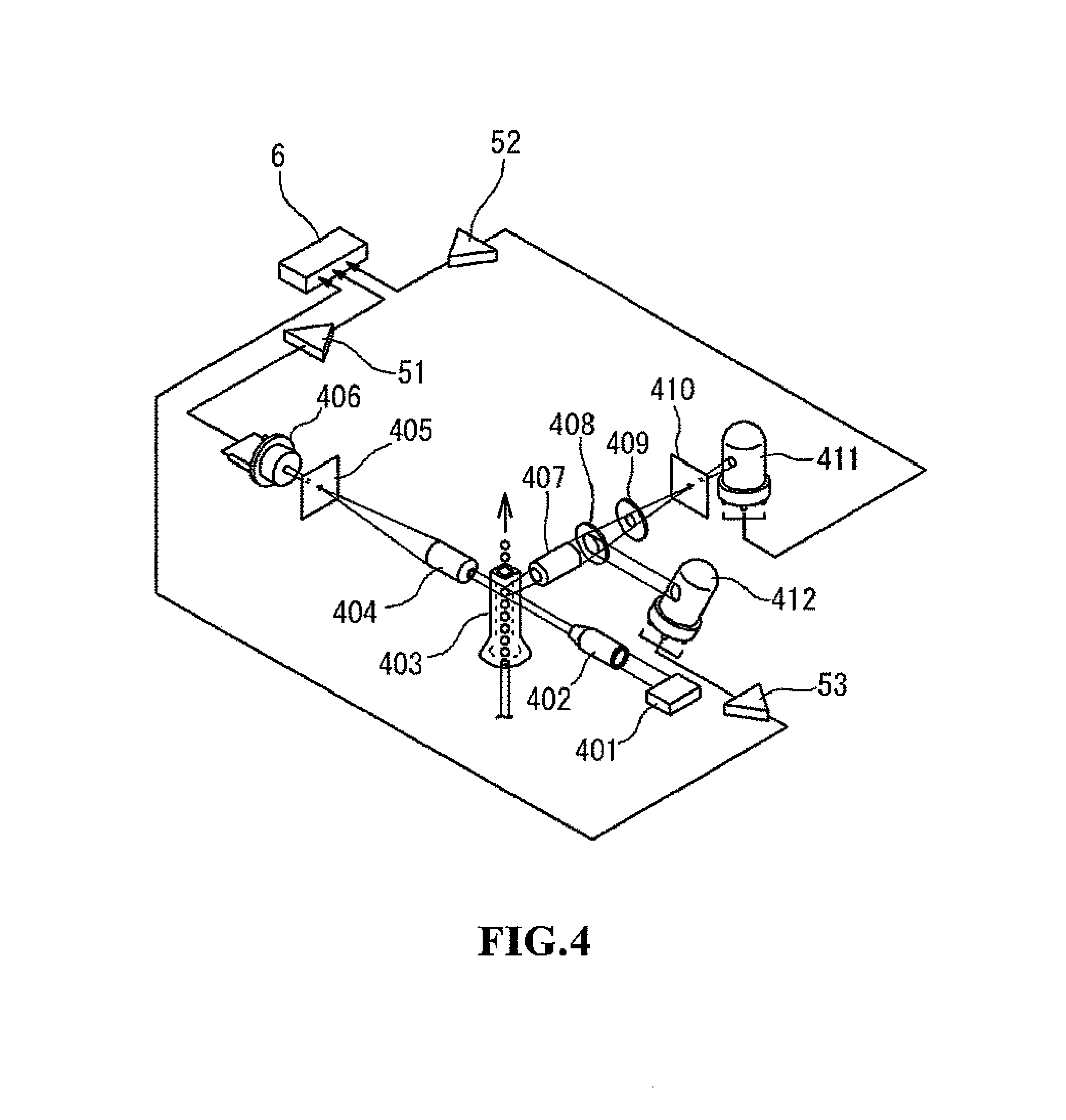
FIG. 4 shows the optical system of the white blood cell detection unit;
FIG. 5 shows the RBC/PLT detection unit;
FIG. 6 shows the HGB detection unit;
FIG. 7 is a flow chart of the sample measuring process;
FIG. 8 shows the display screen for setting the measurement mode;
FIG. 9 is a flow chart showing the pre sequence process;
FIG. 10 is a schematic view of a scattergram derived from measurements of a DIFF measurement sample prepared from body fluid;
FIG. 11 compares measurement results by the blood cell analyzer of the embodiment and measurement results by a reference method;
FIG. 12 is a schematic view of a scattergram derived from measurements of a DIFF measurement sample prepared from blood;
FIG. 13 is a display screen showing the measurement results in the blood measurement mode;
FIG. 14 is a display screen showing the measurement results in the body fluid measurement mode;
FIG. 15 is a display screen showing the measurement results in the body fluid measurement mode;
FIG. 16 is a display screen showing the measurement results in the body fluid measurement mode; and
FIG. 17 is a confirmation screen at the start of the blank check which is displayed in the body fluid measurement mode.
DETAILED DESCRIPTION OF THE PREFERRED EMBODIMENTS
The preferred embodiments of the present invention will be described hereinafter with reference to the drawings.
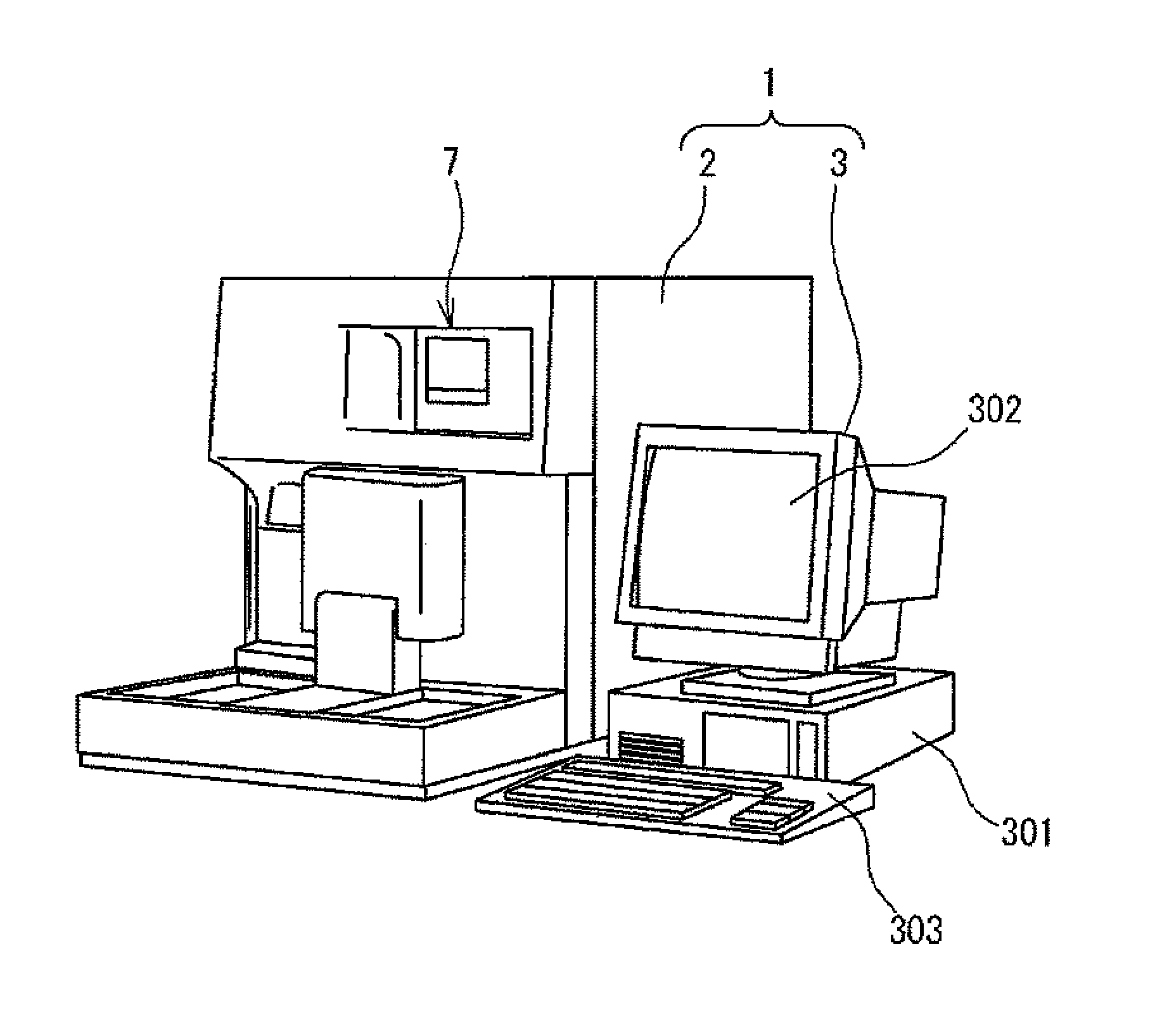
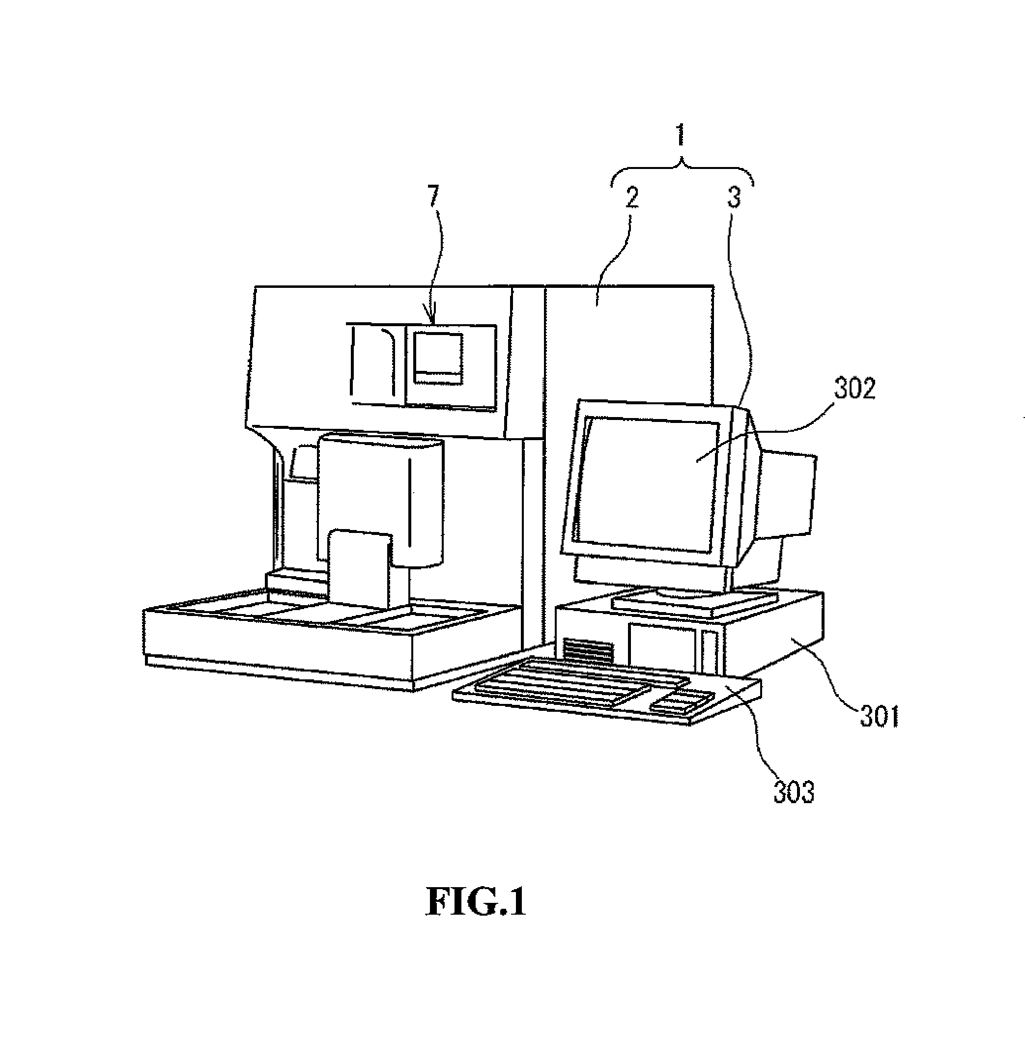
FIG. 1 shows a hematological analyzer 1. The hematological analyzer 1 is configured as an automatic multi-item blood cell analyzer which performs blood analysis by measuring blood samples held in sample containers (blood collection tubes), obtaining characteristics information representing the characteristics of the blood cells contained in the sample, and analyzing the characteristic information. The hematological analyzer 1 is also capable of analyzing body fluids. In the blood cell analyzer of the present embodiment, the body fluids used as analysis objects include, fluid within the body cavity other than blood. Specifically, cerebrospinal fluid (spinal fluid, CSF: fluid filling the ventricle or sublemmal cavity), fluid of the thoracic cavity (pleural fluid, PE: fluid collected in pleural cavity), abdominal fluid (fluid collected in the abdominal cavity), fluid of the cardiac sac (fluid collected in the cardiac sac), synovial fluid (fluid present in joints, synovial sac, peritenon) and the like. Among types of body fluid which can be analyzed are dialysate of peritoneal dialysis (CAPD), intraperitoneal rinse and the like. Cells are usually not observed in these body fluids, however, the fluids may contain blood cells, abnormal cells, and cells such as bacteria in the case of disease, tumor of related organs, or injury. For example, it is possible to clinically estimate the following from measurement results in the case of cerebrospinal fluid. For example, sub-arachnoidal hemorrhage is indicted when there is an increase of red blood cells, meningitis is indicated when there is an increase of neutrophils, infectious disease (parasitic and fungal) is indicated when there is an increase of eosinophils, tuberculous meningitis and viral meningitis are indicated when there is an increase of monocytes, and advanced meningeal tumor is indicated when there is an increase of other cells. ed In the case of abdominal and thoracic fluids, cancers may be indicated when analysis of finds nucleated cells other than blood cells, that is, the fluid contains nucleated cells of mesothelial cells, macrophages, tumor cells and the like.
The hematological analyzer 1 is provided with a measuring unit 2 which has the function of measuring blood and body fluid samples, and a data processing unit 3 which obtains analysis results by processing the measurement results output from the measurement unit 2. The data processing unit 3 is provided with a control unit 301, a display unit 302, and an input unit 303. Although the measuring unit 2 and data processing unit 3 are separate devices in FIG. 1, the both may also be integrated in a single apparatus.
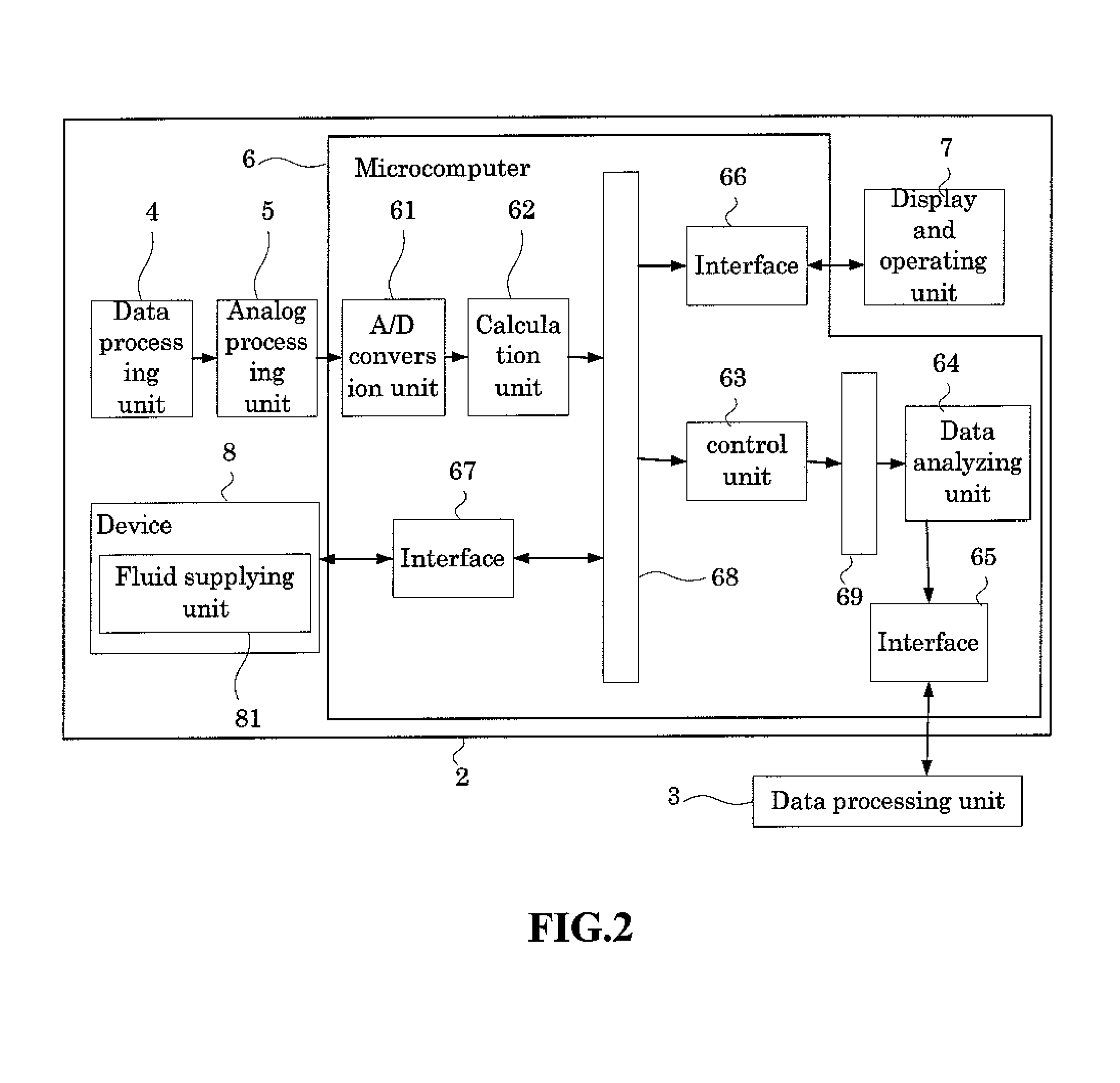
FIG. 2 is a block diagram of the measuring unit 2 of the analyzer 1. As shown in FIG. 2, the measuring unit 2 is provided with a blood cell detecting unit 4, an analog processing unit 5 which processes the output (analog signals) of the detecting unit 4, microcomputer unit 6, display and operating unit 7, and a device 8 for measuring blood and body fluids. The device 8 includes a fluid supplying unit 81 which is described below.
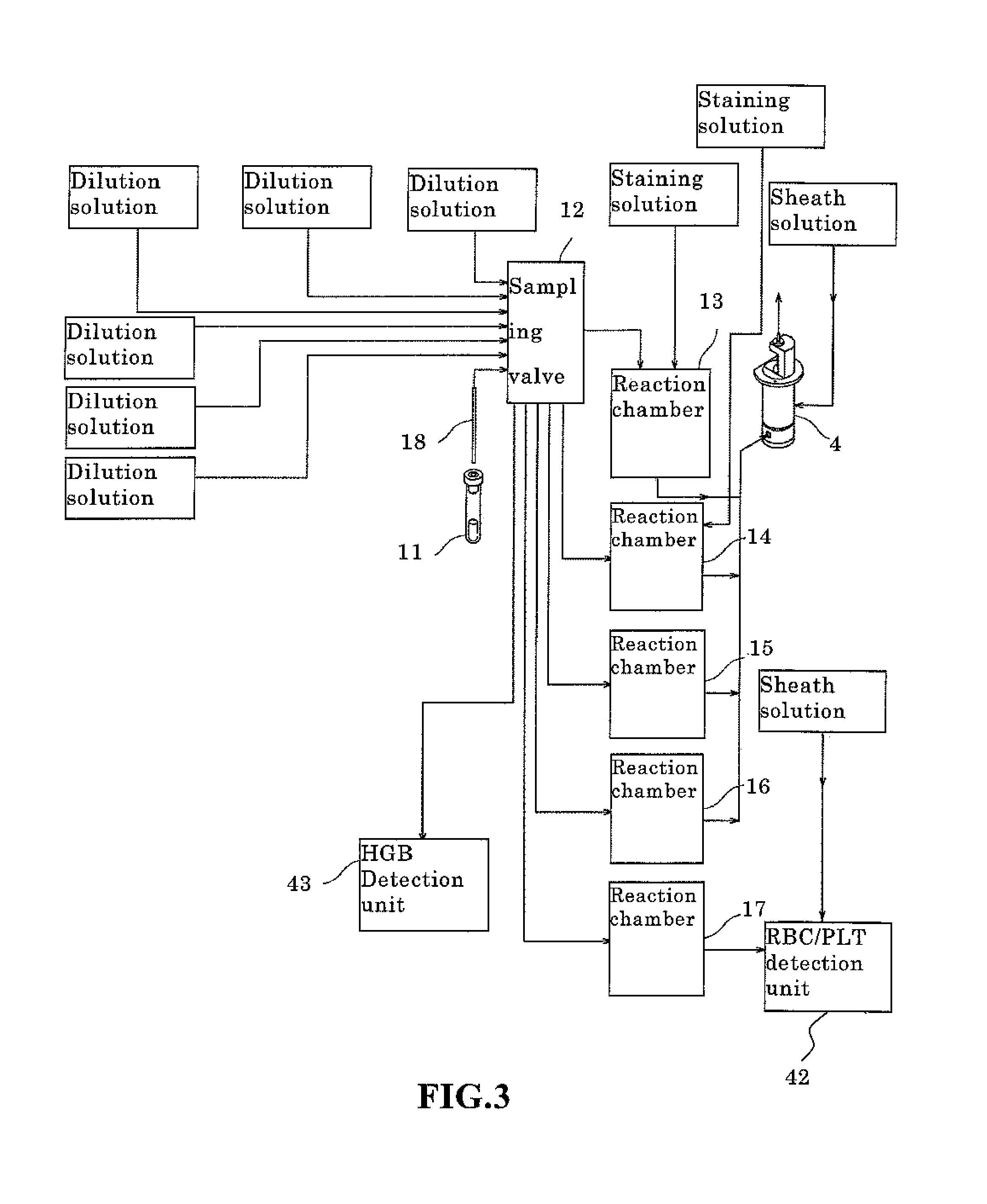
FIG. 3 is a block diagram showing the structure of the fluid supplying unit 81. As shown in FIG. 3, the fluid supplying unit 81 is provided with a sample aspiration nozzle 18, a plurality of reagent containers, a sampling valve 12, and reactions chambers 13 through 17. The sample aspiration nozzle 18 aspirates sample from a sample container, and delivers the sample to the sampling valve 12. The sampling valve 12 divides the delivered sample into several aliquots of predetermined volume. The number of divisions differs depending on the mode of measurement (discrete mode); in the CBC mode the sample is divided into three aliquots to measure the number of red blood cells, the number of white blood cells, the number of platelets, and the hemoglobin concentration. In addition to the CBC measurement items, the sample is divided into four aliquots in the CBC-DIFF mode so as to also classify five types of white blood cells. Furthermore, In addition to the measurement items of the CBC+DIFF mode, the sample is divided into five aliquots in the CBC+DIFF+RET mode so as to also measure reticulocytes.
Similarly, in addition to the measurement items of the CBC+DIFF mode, the sample is divided into five aliquots in the CBC+DIFF+NRBC mode so as to also measure nucleated red blood cells. In addition to the measurement items of the CBC+DIFF+RET mode, the sample is divided into six aliquots in the CBC+DIFF+RET+NRBC mode so as to also measure nucleated red blood cells. The above mentioned measurement modes are blood measuring modes which measure whole blood. Finally, the sample is divided into two aliquots in the body fluid measuring mode for measuring body fluid.
Reagent (dilution solution) is introduced from a reagent container to the sampling valve, and the aliquots of the divided sample are delivered together with the reagent to the reaction chambers 13 through 17 and an HGB detection unit 43, which is described later, a predetermined amount of sample (aliquot) and a predetermined amount of reagent and a predetermined amount of stain collected by the sampling valve 12 are supplied to the reaction chamber 13 by a dosage pump which is not shown in the drawing, the sample and reagent are mixed to prepare a measurement sample for four classifications of white blood cells (DIFF).
The reagent "stomatolyzer 4DL" made by Sysmex Corporation may be used as the dilution solution. This reagent contains surface active agent and induces hemolysis of red blood cells. The reagent "stomatolyzer 4DS" made by Sysmex Corporation may be used as the stain. This stain contains ethylene glycol, low molecular alcohol, and polymethene colorant; a 50.times. dilute sample is ultimately prepared by staining the blood cell component after hemolysis by the dilution agent.
When the body fluid measurement mode has been selected, a measurement sample for the classification of white blood cells is prepared from a fluid sample under the conditions of the amount of the sample and reagent used for the four classifications of white blood cells are identical, the reagents are identical, and the amounts of the reagent are identical. In the white blood cell classification of the body fluid measurement mode, the white blood cells are classified, not in four types, but two types, as shall be described later.
A predetermined amount of sample collected by the sampling valve 12, a predetermined amount of hemolytic dilution agent, and a predetermined amount of stain solution are supplied to the reaction chamber 14 by a dosage pump which is not shown in the drawing, the sample and reagents are then mixed to prepare a measurement sample for measuring nucleated red blood cells (NRBC).
A predetermined amount of sample collected by the sampling valve 12, a predetermined amount of dilution agent, and a predetermined amount of stain solution are supplied to the reaction chamber 15 by a dosage pump which is not shown in the drawing, the sample and reagents are then mixed to prepare a measurement sample for measuring reticulocytes (RET).
A predetermined amount of sample collected by the sampling valve 12, and a predetermined amount of hemolytic dilution agent are supplied to the reaction chamber 16 by a dosage pump which is not shown in the drawing, the sample and reagents are then mixed to prepare a measurement sample for measuring white blood cells and basophils (WBC/BASO).
A predetermined amount of sample collected by the sampling valve 12, and a predetermined amount of dilution solution are supplied to the reaction chamber 17 by a dosage pump which is not shown in the drawing, the sample and reagents are then mixed to prepare a measurement sample for measuring red blood cells and platelets (RBC/PLT).
A predetermined amount of sample collected by the sampling valve 12, and a predetermined amount of hemolytic dilution agent are supplied to the HGB detection unit 43 which is described later.
The detection device 4 is provided with a white blood cell detection unit 41 for detecting white blood cells. The white blood cell detection unit 41 is also used to detect nucleated red blood cells and reticulocytes. In addition to the white blood cell detection unit, the detection device 4 is also provided with an RBC/PLT detection unit 42 for measuring the number of red blood cells and the number of platelets, and an HGB detection unit 43 for measuring the amount of pigment in the blood.
The white blood cell detection unit 41 is configured as an optical detection unit, specifically, a detection unit which uses a flow cytometric method. Cytometry measures the optical properties and physical properties of cells and other biological particles, and flow cytometry measures these particles as they pass by in a narrow flow. FIG. 4 shows the optical system of the white blood cell detection unit 41. In the same drawing, the beam emitted from a laser diode 401 irradiates, via a collimator lens 402, the blood cells passing through the interior of a sheath flow cell 403. The intensity of the front scattered light, the intensity of the side scattered light, and the intensity of the side fluorescent light from the blood cells within the sheath flow cell irradiated by the light are detected by the white blood cell detection unit 41.
The scattered light is a phenomenon due to the change in the direction of travel of the light caused by particles such as blood cells and the like which are present as obstructions in the direction of travel of the light. Information on the characteristics of the particles related to the size and composition of the particles can be obtained by detecting this scattered light. The front scattered light emerges from the particles in approximately the same direction as the direction of travel of the irradiating light. Characteristic information related to the size of the particle (blood cell) can be obtained from the front scattered light. The side scattered light emerges from the particle in an approximate perpendicular direction relative to the direction of travel of the irradiating light. Characteristic information related to the interior of the particle can be obtained from the side scattered light. When a particle is irradiated by laser light, the side scattered light intensity is dependent on the complexity (that is, nucleus shape, size, density, and granularity) of the interior of the cell. therefore, the blood cells can be classified (discriminated) and the number of cells can be counted by using the characteristics of the side scattered light intensity. Although the front scattered light and side scattered light are described as the scattered light used in the present embodiment, the present invention is not limited to this configuration inasmuch as scattered light of any angle may also be used relative to the optical axis of the light emitted from a light source that passes through the sheath flow cell insofar as scattered light signals are obtained which represent the characteristics of the particles necessary for analysis.
When fluorescent material such as a stained blood cell is irradiated by light, light is given off by the particle at a wavelength which is longer than the wavelength of the irradiating light. The intensity of the fluorescent light is increased by the stain, and characteristics information can be obtained relating to the degree of staining of the blood cell by measuring the fluorescent light intensity. The classification and other measurements of the white blood cells can then be performed by the difference in the (side) fluorescent light intensity.
As shown in FIG. 4, the front scattered light from the blood cell (white blood cells and nucleated red blood cells) which pass through the sheath flow cell 403 is received by a photodiode (front scattered light receiving unit) 406 through a collective lens 404 and pinhole 405. The side scattered light is received by a photo multiplexer (side scattered light receiving unit) 411 through a collective lens 407, dichroic mirror 408, optical filter 409, and pinhole 410. The side fluorescent light is received by a photo multiplexer (side fluorescent light receiving unit) 412 through the collective lens 407 and dichroic mirror 408. The photoreception signals output from the light receiving units 406, 411, and 412 are subjected to analog processing such as amplification and waveform processing and the like by an analog processing unit 5 which is configured by amps 51, 52, 53 and the like, and the analog-processed photoreception signals are provided to the microcomputer 6.
The configuration of the RBC/PLT detection unit 42 is described below. FIG. 5 is a schematic view briefly showing the structure of the RBC/PLT detection unit 42. The RBC/PLT detection unit 42 is capable of measuring the numbers of red blood cells and platelets by a sheath flow-DC detection method. The RBC/PLT detection unit 42 has a sheath flow cell 42a as shown in FIG. 5. The sheath flow cell 42a is provided with a sample nozzle 42b which is open toward the top so that sample can be supplied from the reaction chamber 17 to the sample nozzle 42b . The sheath flow cell 42a has a tapered chamber 42c which narrows toward the top, and the sample nozzle 42b is disposed in the center part within the chamber 42c . An aperture 42d is provided at the top end of the chamber 42c , and this aperture 42d is aligned with the center position of the sample nozzle 42b . Measurement sample supplied from the sample supplying unit is sent upward from the tip of the sample nozzle 42b , and front sheath fluid is simultaneously supplied to the chamber 42c and flows upward toward the aperture 42d . The flow of the measurement sample, which is encapsulated in the front sheath fluid, is narrowly constricted by the tapered chamber 42c and the blood cells within the measurement sample pass one by one through the aperture 42d . Electrodes are provided at the aperture 42d , and a direct current is supplied between these electrodes. The change in the resistance of the direct current is detected at the aperture 42d when the measurement sample flows through the aperture 42d , and the electrical signal of the change in resistance is output to the controller 25. Since the resistance of the direct current increases when blood cells pass through the aperture 42d , the electrical signals reflect information of the passage of the blood cells through the aperture 42d so that the numbers of red blood cells and platelets can be counted by subjecting these electrical signals to signal processing.
A recovery tube 42e , which extends vertically, is provided above the aperture 42d . The recovery tube 42e is disposed within a chamber 42f which is connected to the chamber 42c through the aperture 42d . The inner wall of the chamber 42f is separated from the bottom end of the recovery tube 42e . The chamber 42f is configured to supply a back sheath, and this back sheath flows downward through the chamber 42f in a region outside the recovery tube 42e . The back sheath which flows outside the recovery tube 42e arrives at the bottom part of the chamber 42f , and thereafter flows between the inner wall of the chamber 42f and the bottom end of the recovery tube 42e so as to flow into the interior of the recovery tube 42e . The blood cells which has passed through the aperture 42d are therefore prevented from refluxing, thus preventing erroneous detection of the blood cells.
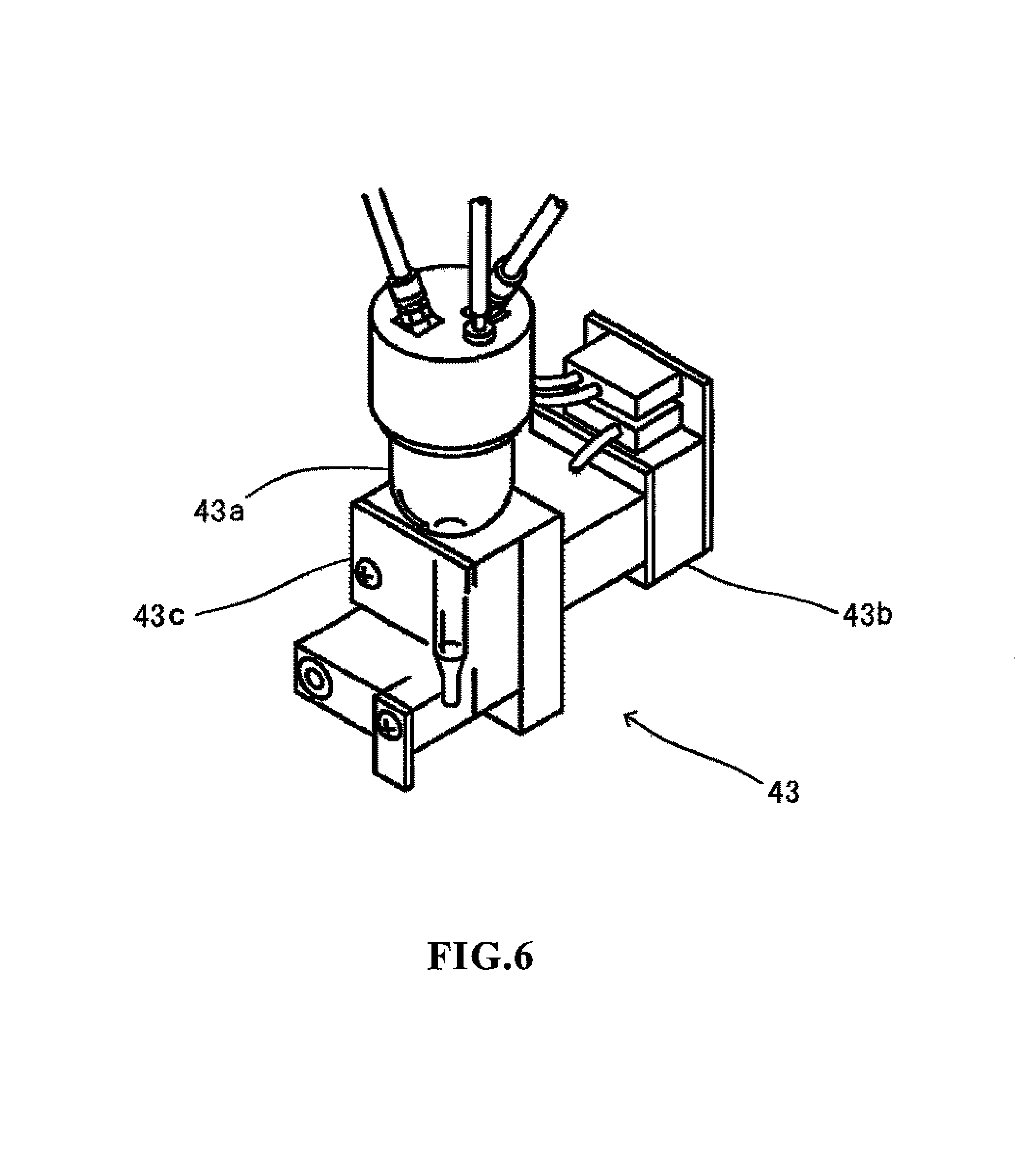
The configuration of the HGB detection unit 43 is described below. The HGB detection unit 43 is capable of measuring the amount of hemoglobin (HGB) by an SLS hemoglobin method. FIG. 6 is a perspective view of the structure of the HGB detection unit 43. The HGB detection unit 43 has a cell 43a for accommodating a diluted sample, a light-emitting diode 43b for emitting light toward the cell 43a , and a photoreceptor element 43c for receiving the transmission light that has passed through the cell 43a . A fixed amount of blood is diluted with dilution fluid and a predetermined hemolytic agent at a predetermined dilution ratio by the sampling valve 12 to prepare a dilute sample. The hemolytic agent has properties which transform the hemoglobin in the blood to SLS-hemoglobin. The dilute sample is supplied to the cell 43a and accommodated therein. In this condition, the light-emitting diode 43b emits light that passes through the cell 43a and is received by the photoreceptor element 43c which is disposed opposite the light-emitting diode 43b with the cell 43a interposed therebetween. Since the light-emitting diode 43b emits light having a wavelength that is highly absorbed by the SLS-hemoglobin, and the cell 43a is configured of plastic material which has a high light transmittancy, the photoreceptor element 43c only receives the transmission light absorbed by the dilute sample of the light emitted from the light-emitting diode 43b . The photoreceptor element 43c outputs electrical signals which correspond to the amount of received light (optical density) to the microcomputer 6, and the microcomputer 6 compares the optical density with the optical density of the dilution solution which was measured previously, then calculates the hemoglobin value.
The microcomputer 6 is provided with an A/D converter 61 for converting the analog signals received from the analog processing unit 5 to digital signals. The output of the A/D converter 61 is sent to a calculation unit 62 of the microcomputer 6, and calculations are performed for predetermined processing of the photoreception signals in the calculation unit 62. The calculation unit 62 prepares distribution data (two-dimensional scattergrams (unclassified) and unidimensional histograms) based on the output of the detection device 4.
The microcomputer 6 is provided with a controller 63 configured by a memory for the control processor and the operation of the control processor, and a data analyzing unit 64 configured by a memory for the analysis processor and the operation of the analysis processor. The controller 63 controls the device 8 configured by a sampler (not shown in the drawing) for automatically supplying blood collection tubes, and a fluid system and the like for preparing and measuring samples, as well as performing other controls. The data analyzing unit 64 executes analysis processing such as clustering and the like on the distribution data. The analysis results are sent to an external data processing device 3 through an interface 65, and the data processing device 3 processes the data for screen display, storage and the like.
The microcomputer 6 is further provided with an interface 66 which is interposed between the microcomputer 6 and the display and operating unit 7, and an interface 67 which is interposed between the microcomputer 6 and the device 8. The calculation unit 62, controller 63, and interfaces 66 and 67 are connected through a bus 68, and the controller 63 and the data analyzing unit 64 are connected through a bus 69. The display and operating unit 7 includes a start switch by which the operator specifies to start a measurement, and a touch panel type liquid crystal display for displaying various types of setting values and analysis results, and receiving input from the operator.
The operation of the hematological analyzer 1 of the present embodiment is described below. FIG. 7 is a flow chart showing the flow of the operation of the hematological analyzer of the present embodiment. The hematological analyzer 1 starts when a user turns on the power source of the hematological analyzer 1 (step S1). The hematological analyzer 1 first executes a self check during startup (step S2). In the self check, the microcomputer 6 tests and checks the operation of all operating device of the hematological analyzer 1, and performs a blank check operation which measures a blank sample that does not contain a real sample. Next, the microcomputer 6 sets an initial measurement mode (step S3). The CBC+DIFF mode is the initial setting. Specifically, in the process of step S3, parameters (operating conditions) for performing blood measurements are set, for example, which reaction chamber to use and the set time for the measurement. The blood measurement mode is thus set as the initial operating mode in the hematological analyzer 1 of the present embodiment. The hematological analyzer 1 therefore remains in a standby state waiting to receive a measurement start instruction. The microcomputer 6 displays a screen on the liquid crystal display which alerts the operator to the standby state (step S4).
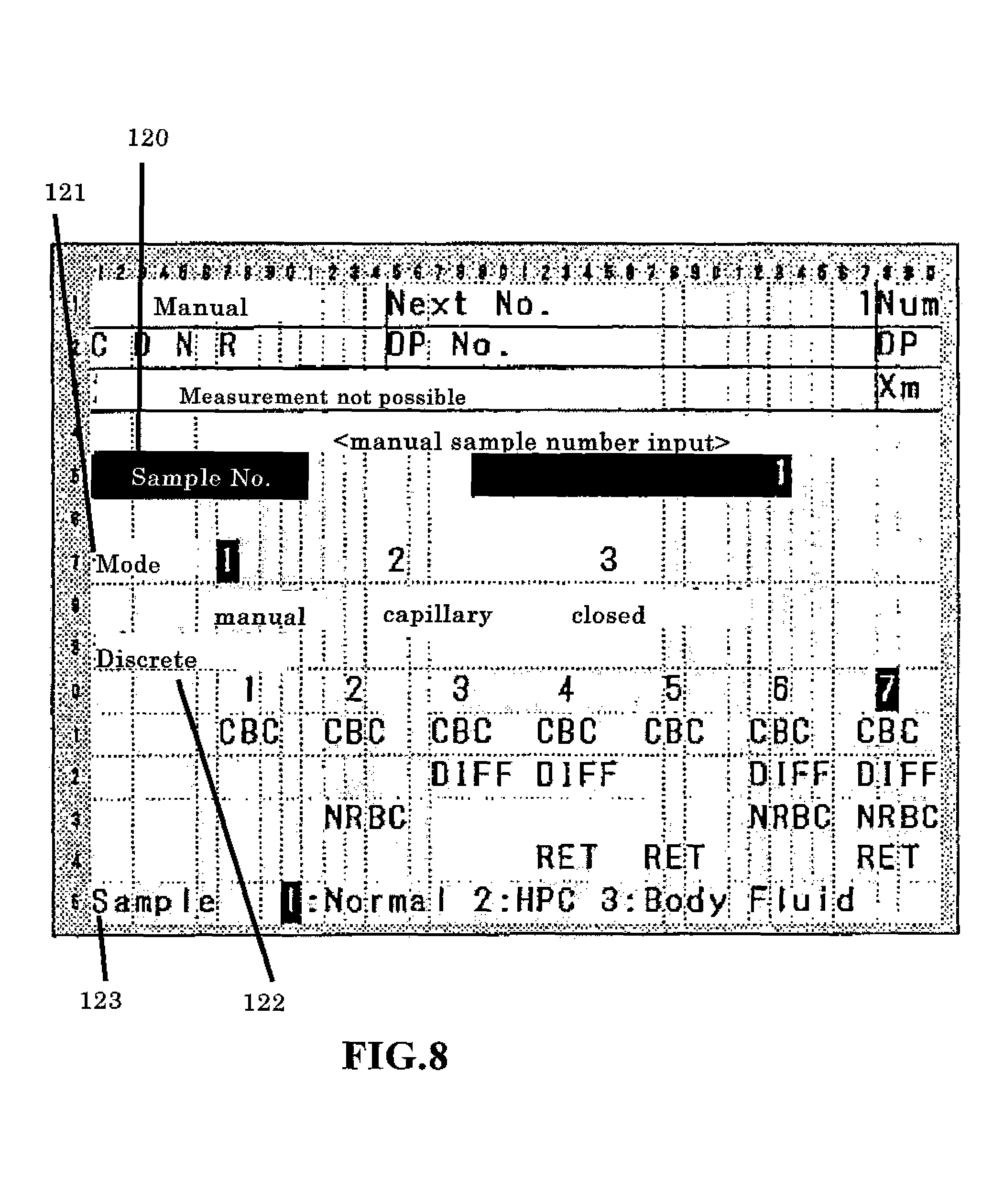
In the standby state, the operator can change the measurement mode by operating the display and operation unit 7. FIG. 8 is a schematic view of an input screen for setting the measurement mode. This screen is provided with discrete display regions including the sample number 120, type of sample uptake mode 121, type of discrete test (measurement mode) 122, and type of sample 123. The three sample uptake modes include a manual mode for aspirating a sample after the operator has manually inserted a sample container in the sample aspiration nozzle 18, a capillary mode for aspirating a measurement sample via the sample aspiration nozzle 18 after the operator has previously prepared the measurement sample by mixing a sample and reagent, and a closed mode for supplying a sample by automatically transporting a sample container using a conveyer device. The types of samples include NORMAL, which are normal blood samples; HPC, which are hematopoietic progenitor cell samples; and BODY FLUID, which are other fluids of the body. The operator can specify the sample take-up mode, measurement mode, and type of sample. When the blood measurement mode has been specified, the NORMAL sample type is specified, and an optional sample take-up mode and measurement mode are specified. When specifying the BODY FLUID measurement mode, the operator specifies MANUAL mode as the take-up mode, [CBC+DIFF], [CBC+DIFF+RET], [CBC+DIFF+NRBC], or [CBC+DIFFNRBC+RET] as the DISCRETE test, and [BODY FLUID] as the type of sample. In step S4, the operator specifies the desired mode. The operator presses the start switch to start the measurement when blood measurement is performed without changing the initially set measurement mode(step S5: N). The microcomputer 6 receives the instruction to start the measurement (step S6), and the blood sample is aspirated by the sample aspiration nozzle (step S7).
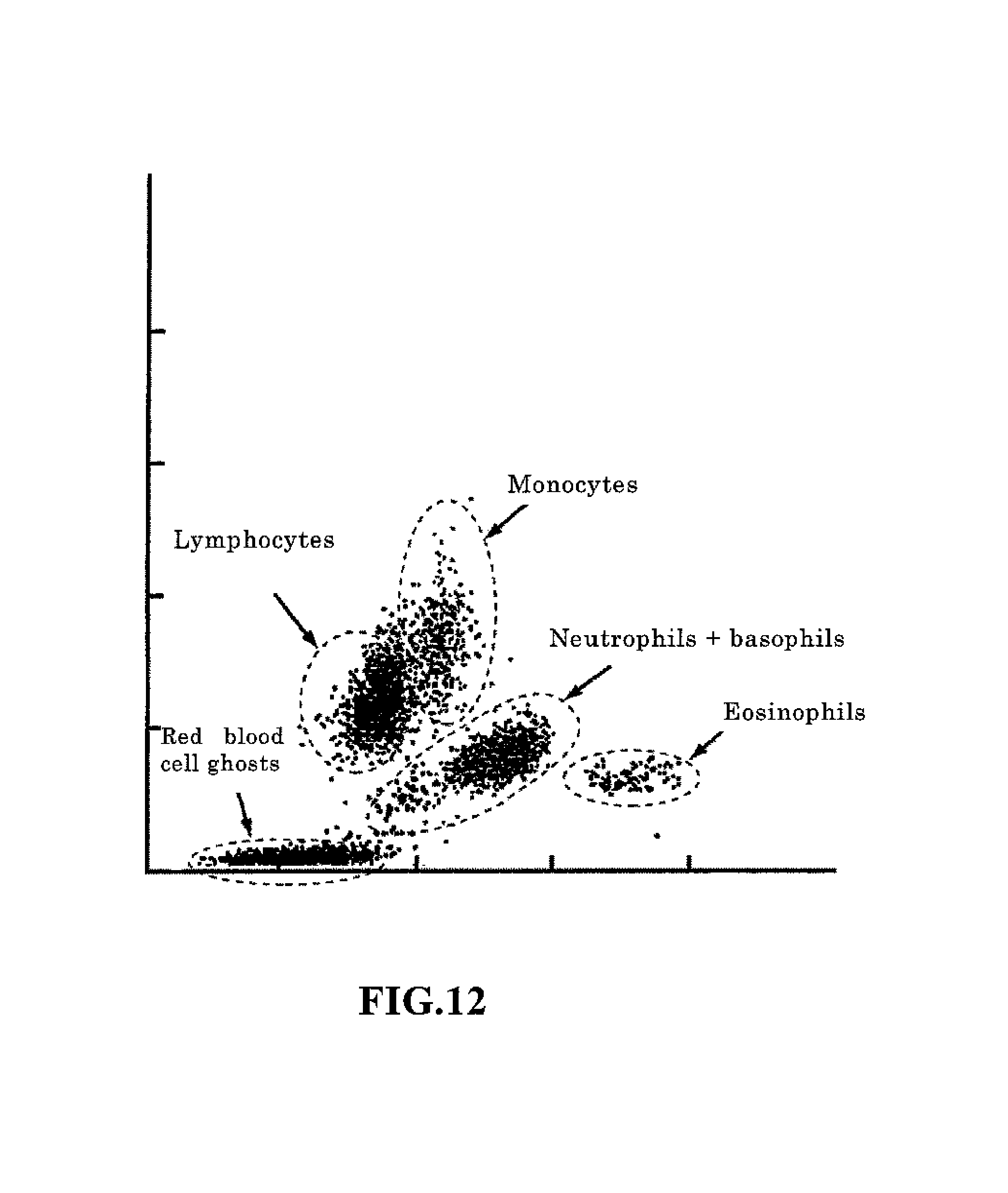
After the blood sample has been aspirated, the sample is introduced to the previously mentioned sampling valve 18, and the necessary sample preparation is performed for the measurement according to the type discrete test of the measurement mode(step S14). The measurement operation is then executed for this measurement sample (step S16). When [7] is set as the type of discrete test, for example, HGB, WBC/BASO, DIFF, RET, NRBC, and RBC/PLT measurement samples are prepared. Thereafter, the WBC/BASO, DIFF, RET, and NRBC measurement samples are measured by the white blood cell detection unit 41, the RBC/PLT measurement sample is measured by the RBC/PLT detection unit 42, and the HGB measurement sample is measured by the HGB detection unit 43. At this time, the WBC/BASO, DIFF, RET, and NRBC measurement samples are introduced to the white blood cell detection unit 41 in the order NRBC, WBC/BASO, DIFF, RET and sequentially measured since only a single white blood cell detection unit 41 is provided. In this measurement operation, the calculation unit 62 creates particle distribution maps (scattergram, histogram). The scattergram created from the optical information obtained by the DIFF measurement is described below. The calculation unit 62 generates a two-dimensional scattergram (particle distribution map) using, as characteristic parameters, the side scattered light and side fluorescent light among the photoreception signals output from the white blood cell detection unit 41 in the DIFF measurement. This scattergram (referred to as "DIFF scattergram" hereinafter) plots the side scattered light intensity on the X axis and the side fluorescent light on the Y axis; red blood cell ghost clusters, lymphocyte clusters, monocyte clusters, neutrophil+basophil clusters, and eosinophil clusters normally appear. These clusters are recognized by processing performed on the DIFF scattergram by the data analyzing unit 64.
Analysis processing is then performed based on the particle distribution maps obtained by the measurement(step S18). In the analysis processing, the data analyzing unit 64 of the microcomputer 6 classifies the four white blood cell clusters (lymphocyte cluster, monocyte cluster, neutrophil+basophil cluster, and eosinophil cluster), and the red blood cell ghost cluster as shown in FIG. 12 from the DIFF scattergram prepared by the calculation unit 62 when the DIFF measurement samples were measured by the white blood cell detection unit 41. In the analysis process of the present embodiment, each particle plotted on the scattergram and the degree of attribution of particles to each cluster at a distance from the center of gravity of each cluster is obtained. Then, each particle is attributed to a cluster according to the degree of attribution. The particle classification method is disclosed in detail in U.S. Pat. No. 5,555,196. The basophil cluster, and white blood cell clusters other than basophils, and the red blood cell ghost cluster are classified on the scattergram obtained by the WBC/BASO measurement. White blood cells are classified in five groups based on the results of the four classifications and numbers of white blood cells (refer to FIG. 12) by the analysis processing of the DIFF scattergram, and the results of the two classification and numbers of white blood cells by the analysis processing of the WBC/BASO scattergram. Specifically, the data analysis unit 64 subtracts the basophil cell count obtained by the analyzing the WBC/BASO scattergram from the neutrophil+basophil cell count obtained by analyzing the DIFF scattergram, to obtain the neutrophil cell count and the basophil cell count. Thus, five classifications of white blood cells are obtained as well as the number of blood cells in each classification. In addition, the trough is detected in the curve in the unidimensional histogram created based on the characteristic information from the detection unit 42, and the particles are classified as red blood cells and platelets in the RBC/PLT measurement. The analysis results thus obtained are output to the display unit 302 of the data processing unit 3 (step S20).
When input specifying the measurement mode is received as described above in step S5, the microcomputer 6 sets the parameters (operating conditions) for the body fluid measurement, for example, the reaction chamber to use and the set time of the measurement and the like (step S8). In the present embodiment, the measurement time is three times the time for blood measurement, as will be described later.
The measuring unit 2 starts the pre sequence (step S10) when the measurement mode has been switched from the previous measurement mode (in this instance, the blood measurement mode) to the body fluid measurement mode (step S9). The pre sequence is a process of preparing for the body fluid measurement. Since samples which have a low concentration of blood cell component are measured in the body fluid measurement, the setting is switched from the blood measurement mode ([1:NORMAL] is displayed in FIG. 8) to the body fluid measurement mode, and the lack of background influence is confirmed in the body fluid measurement results.
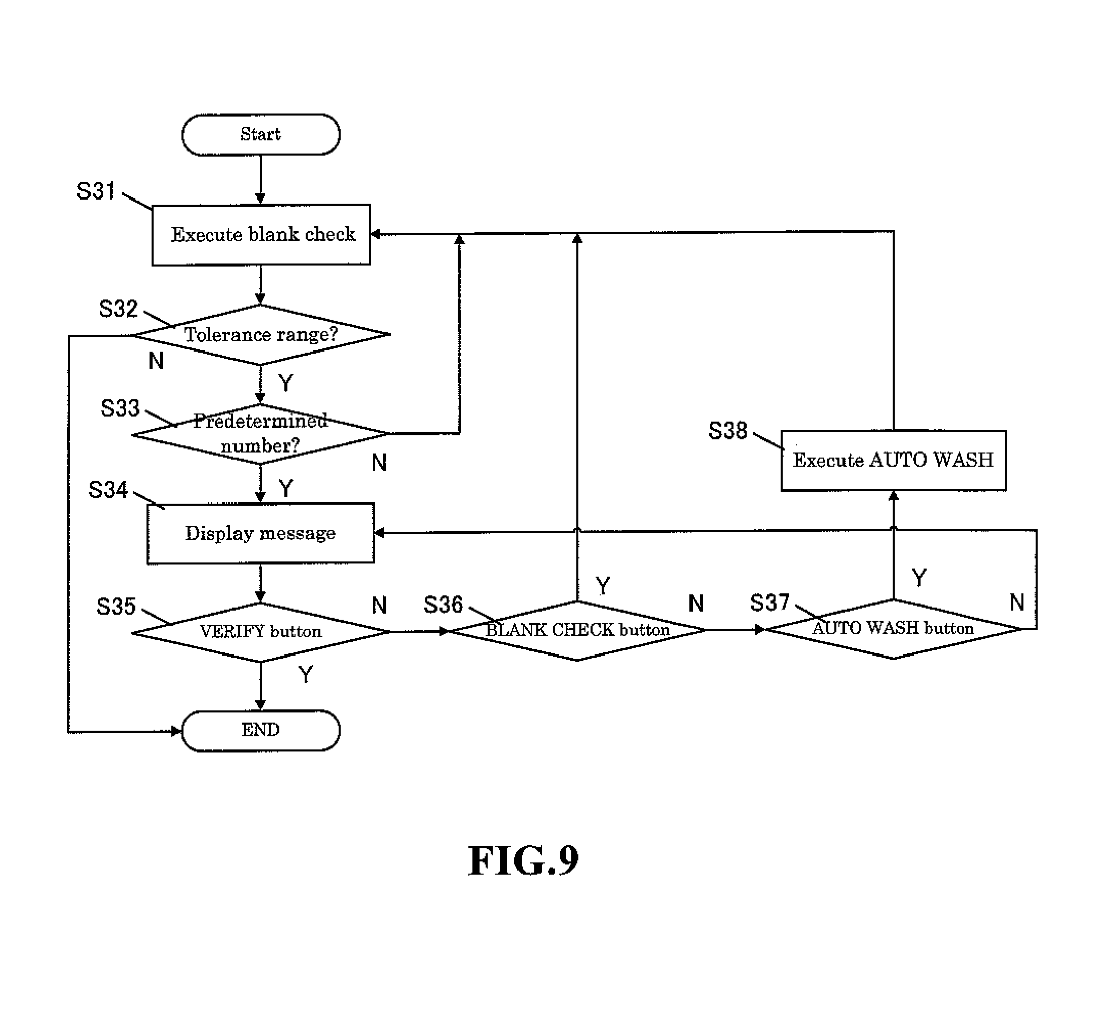
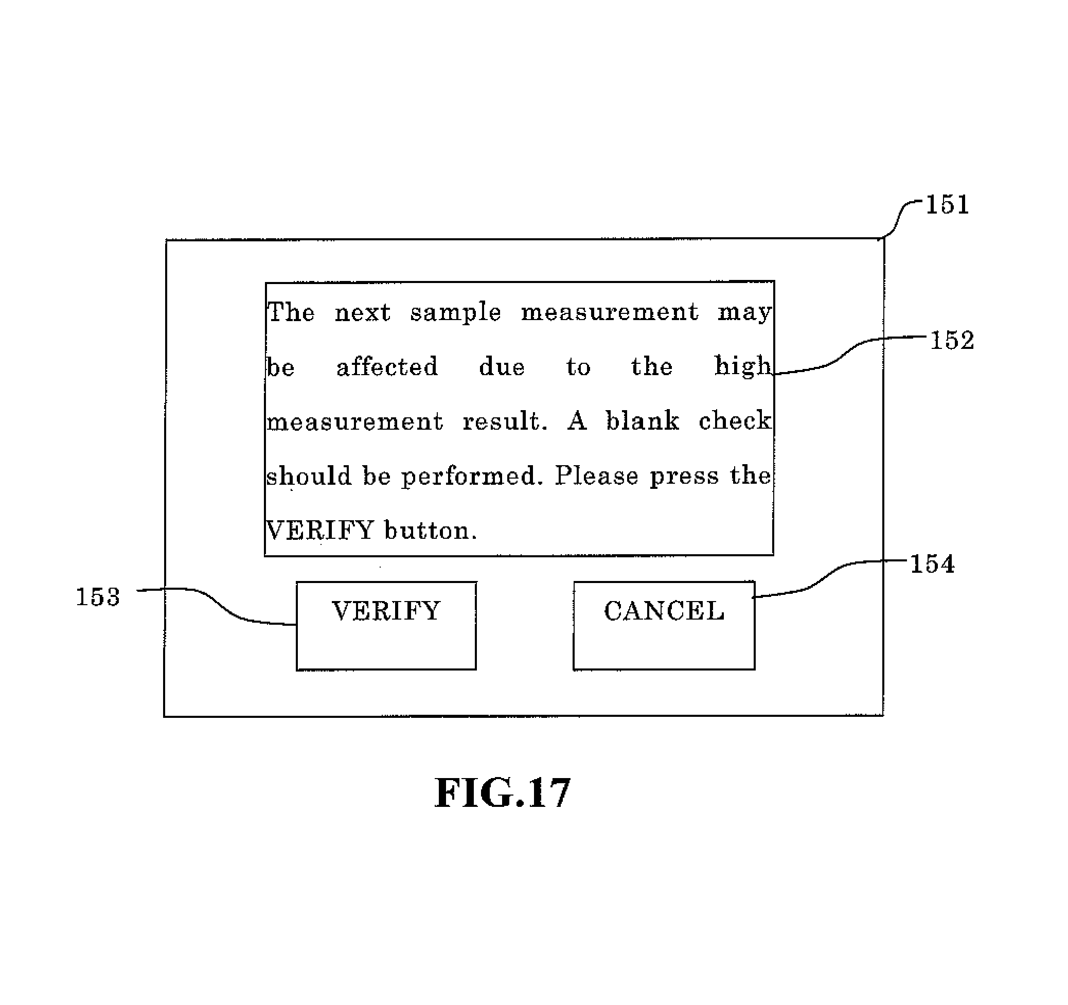
The pre sequence includes a blank check operation. The blank check determination standard of the pre sequence is set at a fraction and is more strict than the determination standard of the blank check (for example, the blank check performed after power on and automatic wash) performed in the blood measurement mode. When the setting is changed from the body fluid measurement mode to the blood measurement mode, this pre sequence is not performed since there is no background influence (carry over effect) on the normal blood measurement results. Furthermore, when body fluid samples are measured in a repeated body fluid measurement mode, this pre sequence is not performed since there is normally no background influence. There is concern, however, that the next sample measurement may be affected when the body fluid sample analysis results exceed a predetermined value due to an extremely high number of particles in the body fluid since the measurement results are high, and therefore the operator is alerted of this concern that the analysis results of the next sample may be affected. Then, the blank check measurement is performed. A configuration is desirable in which a message "please press VERIFY" is output to the screen, and the blank check is performed when the operator presses the VERIFY button. In this case, a configuration is possible in which a CANCEL button may be provided on the screen to transition to the standby screen without performing a blank check when the operator presses the CANCEL button. It is also desirable that a flag indicate the low reliability of the measurement results when a blank check is not performed. Wasted reagent and time can thus be avoided by performing an additional blank check only when needed.
FIG. 9 is a flow chart showing the sequence of the pre sequence process performed when the measurement mode is changed from the blood measurement mode to the body fluid measurement mode. The hematological analyzer 1 performs the pre sequence by measuring a blank sample using the measuring unit 2 (step S31), comparing the measurement result with predetermined tolerance values and determining whether or not the measurement results are less than the tolerance values using the microcomputer 6 (step S32). When the measurement results are less than the tolerance values, the microcomputer 6 ends the pre sequence and the process returns. When the measurement results are not less than the tolerance value, the microcomputer 6 determines whether or not the blank check was executed the set number of times (for example, three times) (step S33), and when the number of executions of the blank check is less than a predetermined number, the process returns to step S31 and the blank check is performed again for the predetermined number of times. When the measurement results of the blank check performed a predetermined number of times are not less than the tolerance values, a screen is displayed with includes a VERIFY button, BLANK CHECK button, and AUTOMATIC WASH button and the blank check measurement results are displayed on the display and operation unit 7 (step S34). When the operator has pressed the VERIFY button (step S35), the microcomputer 6 ends the pre sequence and the process returns. When the BLANK CHECK button has been pressed (step S36), the process returns to step S31 and the blank check is performed again; when the AUOMATIC WASH button has been pressed (step S37), automatic washing is performed using a special washing solution (strep S38), and thereafter the process returns to step S31 and the blank check is performed again.
When the pre sequence ends as described above, the hematological analyzer 1 enters the standby state (step S11). When the operator presses the start switch and starts the body fluid measurement, the sample aspiration nozzle 18 of the measuring unit 2 is immersed in the sample container in the same manner as for the manual measurement of the blood sample. When the instruction to start measurement is received by the microcomputer 6 (step S12), the body fluid aspiration begins (step S13).
After the body fluid sample has been aspirated, the body fluid sample is introduced to the sampling valve 91 in the same manner as the blood sample. Then, the RBC/PLT measurement sample is prepared by the reaction chamber 13 (step S15). Subsequently, the DIFF measurement sample is measured by the white blood cell detection unit 41, and the RBC/PLT measurement sample is measured by the RBC/PLT detection unit 42 (step S17). Since only the DIFF measurement sample is measured by the white blood cell detection unit 41 in the body fluid measurement mode, the measurement is completed in a shorter time than the blood measurement even though the measurement time is longer than the measurement time in the blood measurement mode. The analysis accuracy of the low particle concentration body fluid sample can therefore be improved by increasing the measurement time of the body fluid measurement to be longer than the measurement time of the blood measurement. Although the measurement accuracy can be improved due to the increased number of particles counted by lengthening the measurement time, a two to six fold increase in the measurement time is suitable because the sample processing ability is reduced when the measurement time is excessively long, and there is a limit to the performance of the syringe pump which delivers the measurement sample to the white blood cell detection unit 41. In the present embodiment, the measurement time in the body fluid measurement mode is set at three times the measurement time of the blood measurement mode.
The RBC/PLT measurement sample is introduced to the electrical resistance detection unit 41 in the same manner for all measurement modes, and measurement is performed under a fixed flow speed condition. The analysis processing is performed thereafter based on the characteristic information obtained by the measurements (step S19), and the analysis results are output to the display unit 302 of the data processing unit 3 (step S21). In the analysis processing of the blood measurement mode, the DIFF scattergram and the like are analyzed, and information is calculated for five types of white blood cell subclasses (NEUT: neutrophil, LYMPH: lymphocyte, MONO: monocyte, EO: eosinophil, and BASO: basophil), whereas in the analysis processing of the body fluid measurement mode, two subclasses (MN: mononuclear cell, PMN: polymorphonuclear cell) are classified in a partially integrated form because there are a lesser number of blood cells and these cells are sometimes damaged. The lymphocytes and monocytes belong to mononuclear cells, and neutrophils, eosinophils, and basophils belong to polymorphonuclear cells. Since the classification algorithm is the same as the algorithm described for the analysis processing in the blood measurement mode, further description is omitted.
Next, the analysis results obtained in step S19 are compared to the tolerance value (predetermined threshold value) (step S22). The tolerance value is the same value as the tolerance value used in the blank check of the pre sequence performed in step S10. When the analysis result is greater than the tolerance value (step S22: Y), the verification screen 151 at the start of the blank check is displayed, as shown in FIG. 17. A message is displayed on the verification screen 151 indicating there is concern that the measurement of the next sample may be influenced due to the high measurement result. Then, the blank check measurement is performed. A message display area 152 for displaying the message "please press the VERIFY button", a VERIFY button 153, and a CANCEL button 154 are displayed. Next, determinations are made as to whether or not the user has pressed the VERIFY button 153 or the CANCEL button 154 (step S24), and the blank check is executed when the VERIFY button has been pressed (VERIFY in step S24) (step S25). The process returns to step S5 without performing the blank check when the analysis result obtained in step S19 is less than the tolerance value (step S22: N), and the when the CANCEL button has been pressed (CANCEL in step S24).
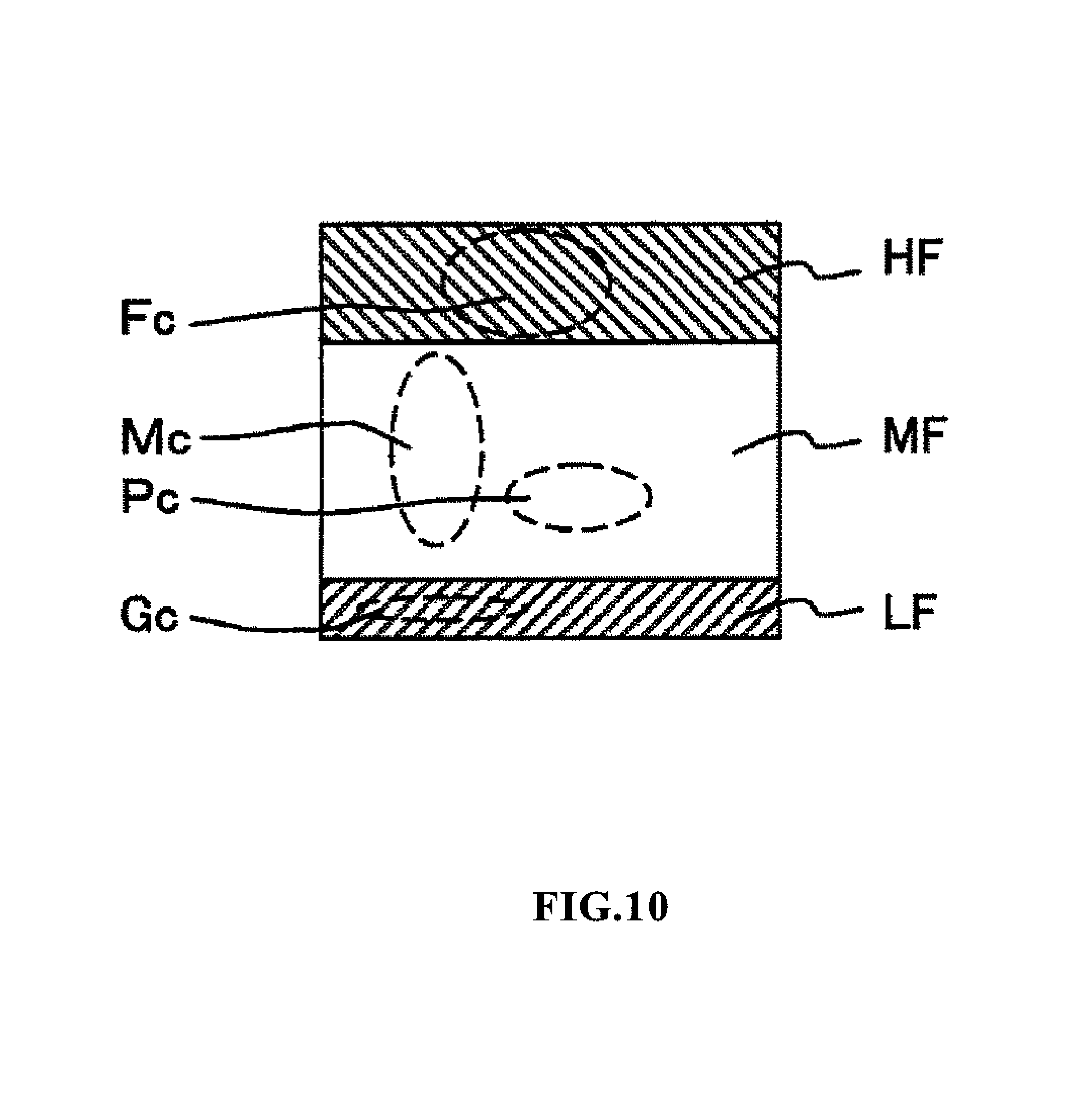
Anomalous particles (macrophages, mesothelial cells, tumor cells and the like) other than blood cells may be present in the body fluid sample. Although it is rare for such anomalous cells to be present in cerebrospinal fluid, such cells appear comparatively frequently in abdominal and thoracic fluids. The influence of these anomalous particles must be eliminated in order to obtain a high precision classification of blood cells within the body fluid regardless of the type of body fluid. White blood cells in body fluid can be measured with greater precision based on the new knowledge than anomalous particles appear in the top part of the DIFF scattergram produced by this blood cell analyzer of the present invention. This aspect was not considered in the previously mentioned conventional art.
FIG. 10 is a schematic view of a scattergram obtained by measuring and analyzing a DIFF measurement sample prepared from body fluid and white blood cell measurement reagent in the body fluid measurement mode of the blood cell analyzer 1 of the present embodiment. The vertical axis of the scattergram represents the side fluorescent light intensity (the fluorescent light intensity at the top is greatest), and the horizontal axis represents the side scattered light intensity (the scattered light intensity at the right side is greatest). A red blood cell ghost Gc caused by hemolysis is distributed in the region LF in which the fluorescent light intensity is weakest in the scattergram, anomalous particles such as mesothelial cells and the like is distributed in the region HF in which the fluorescent light intensity is greatest, and mononuclear white blood cells Mc and polynuclear white blood cells Pc are distributed in the intermediate region MF. In the analysis of the scattergram, the particle component distributed in the region MF is analyzed as white blood cells after excluding region LF and region HF, and the particles are classified and counted in two groups. Lymphocytes and monocytes are included in the mononuclear white blood cells Mc, and neutrophils, basophils, and eosinophils are included in the polynuclear white blood cells Pc.
Since fewer and damaged blood cells are contained in body fluid, white blood cells are classified and counted as mononuclear white blood cells and polynuclear white blood cells when analyzing white blood cells in body fluid.
Anomalous particles (nucleated cells such as tumor cells, macrophages, mesothelial cells) other than blood cells may also be present in body fluid. Although it is rare for such anomalous cells to be present in cerebrospinal fluid, such cells appear comparatively frequently in abdominal and thoracic fluids. In the scattergram of FIG. 10, such nucleated cells other than white blood cells are distributed in region HF. In the present embodiment, it is possible to determine accurate white blood cells counts even in body fluid which contains such nucleated cells other than white blood cells since nucleated cells other than white blood cells can be identified. The degree of occurrence of anomalous cells can be determined by counting the cells which appear in region HF. In the present embodiment, cells are demarcated in the regions LF, MF, and HF by threshold values for demarcating each region; these threshold values may also be changed manually.
FIG. 11 compares the analysis results of the blood cell analyzer 1 of the present embodiment and the count results of a reference method to show the validity of the scattergram analysis method described above. The sample material is thoracic fluid; in the drawing, "this method" refers to the white blood cell count (WBC) and anomalous particle count (Others) calculated by the blood cell analyzer 1 of the present embodiment, and "Ref" refers to the calculation result by the reference methods (Fuchs Rosenthal calculation method and site-spin method). Examples 1, 2, and 3 are the results of analysis of thoracic fluid in which anomalous particles were plentiful, and the correlation between the reference methods and the analysis results of the blood cell analyzer 1 of the present invention can be readily understood.
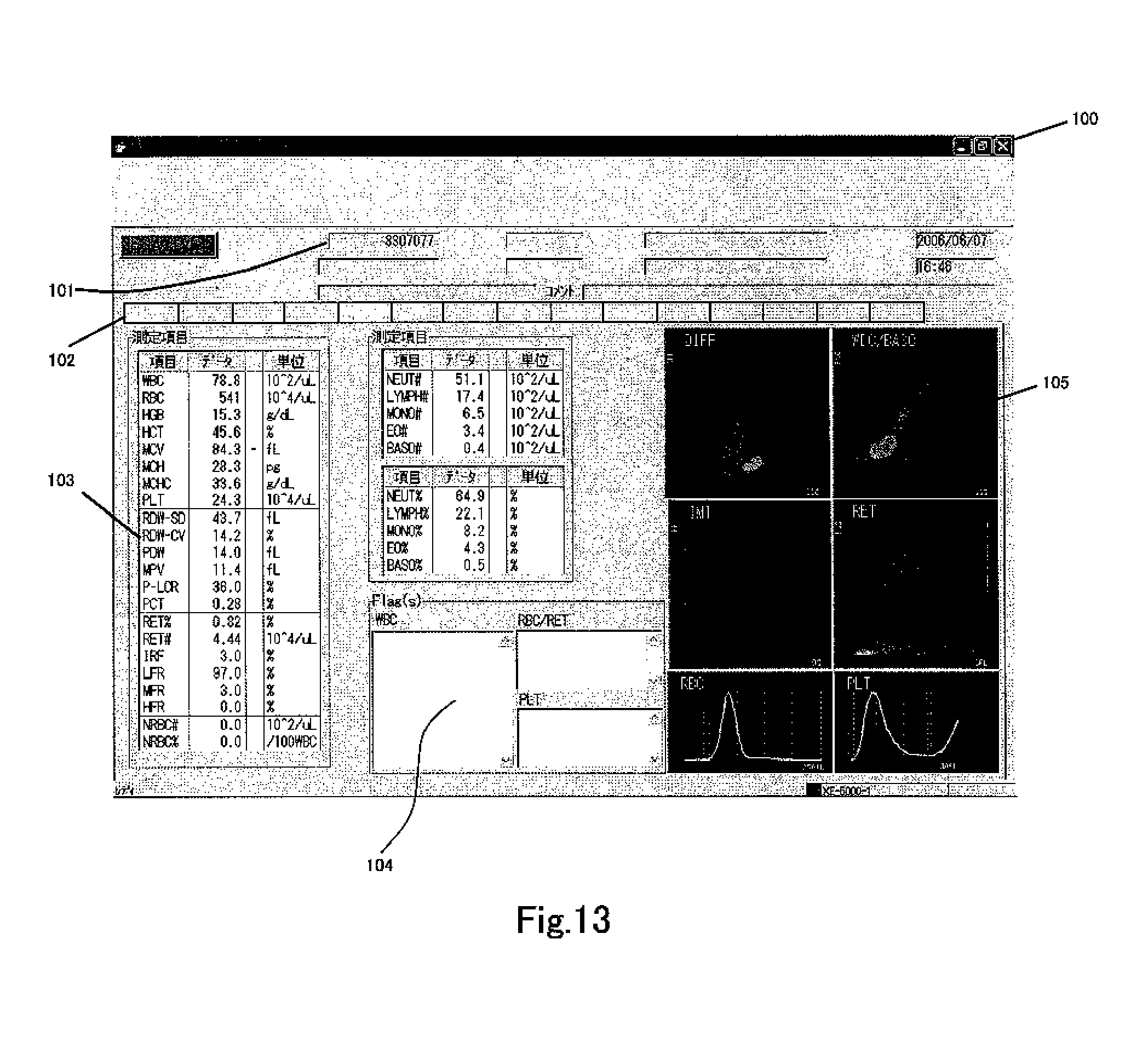
FIG. 13 shows a screen 200 which is displayed on the display unit 302 of the data processing unit 3, showing the analysis results of the DIFF measurement sample prepared from blood. A sample number display region which displays a sample number 101 is provided at the top of the screen 200, and an attribute display region which displays patient attributes is provided adjacently. The attribute display region specifically includes a patient ID, patient name, date of birth, sex, hospital department/ward, attending physician, date of measurement, time of measurement, comments and the like. A measurement result display region which displays the measurement results is provided at the bottom of the attribute display region. The measurement result display region includes several pages, and these pages can be displayed by selecting a plurality of tabs 102. Tabs have a plurality of arrangements matching the main screen, graph screen, and measurement items. FIG. 12 is a screen which is displayed when the graph screen tab has been selected. A graph display region 104 for displaying graphs and a measurement value display region 103 for displaying the measurement result values are provided in the left half of the measurement value display region , and a distribution map display region for displaying the measurement result distribution map 105 is provided in the right half. WBC, RBC, . . . , NEUT#, . . . , BASO#, . . . , NEUT#, . . . , BASO % and the like, data, and units are displayed in the measurement value display region, and flagging results representing sample anomalies and disease suspicions which are clinically useful information relating to WBC, PLT, RBC or RET are displayed in the flag display region 104.
Six distribution maps are displayed in the distribution map display region 105. The scattergram on the upper left side is a DIFF scattergram. The WBC/BASO scattergram is shown at the top right, the immature cell (IMI) scattergram is shown at mid left, and the RET scattergram is shown at mid right. The RBC scattergram is shown at the bottom left, and the PLT scattergram is shown at the bottom right.
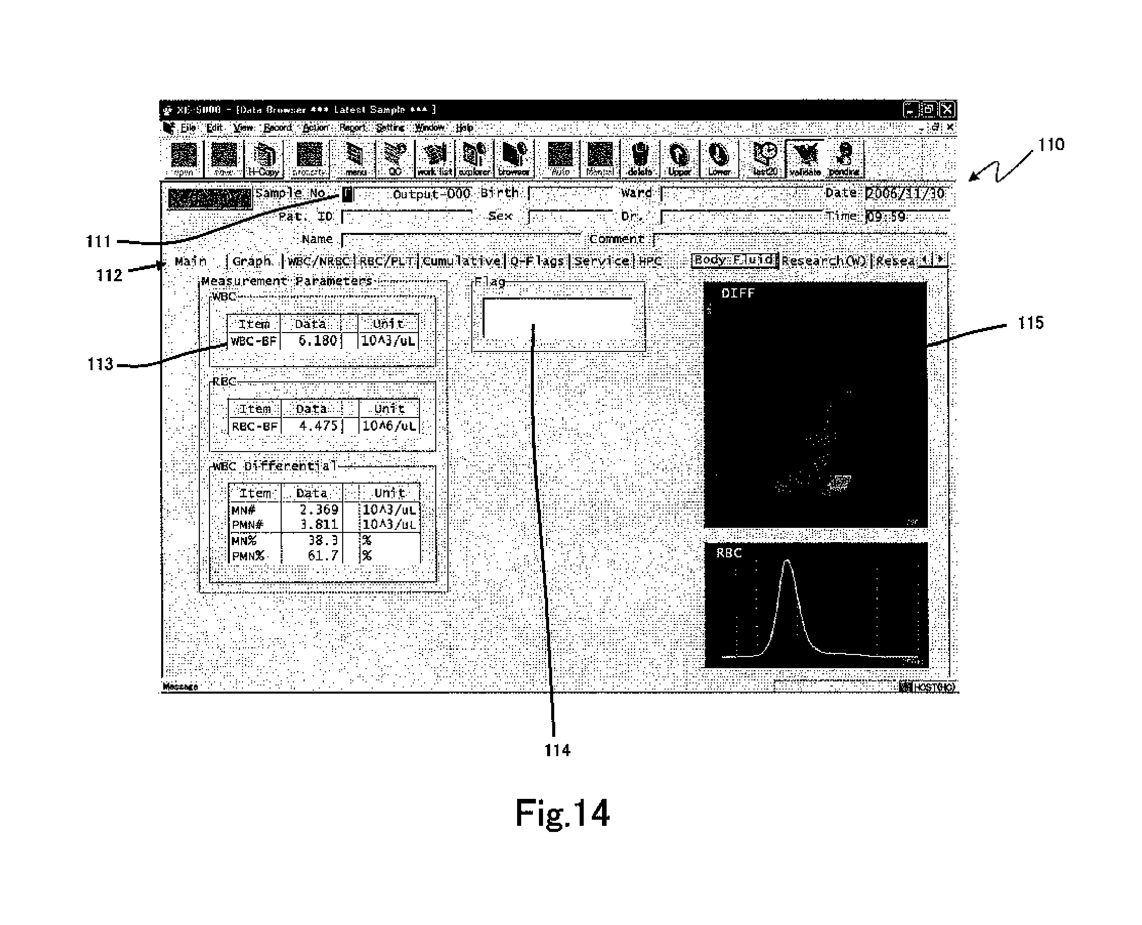
FIG. 14 shows a screen 110 displayed in the display area 302 of the data processing unit 3 as the measurement results of the DIFF measurement sample prepared from body fluid. A sample number display region 111 for displaying a sample number is provided at the top of the screen 110, and a patient attribute display region is provided adjacently. An [F], which indicates measurement has been conducted in the body fluid measurement mode, is displayed at the left end of the sample number display region 111. Thus, it can be clearly recognized that the analysis results are for body fluid measurement results. The measurement result display region includes a plurality of pages which are selectable by tab 112. In this example, the tab for body fluid measurement is selected.
The measurement value display region 113 includes the name of the measurement items for body fluid measurement rather than the measurement results of the blood measurement mode; WBC-BF (WBC count), RBC-BF (RBC count), MN# (mononuclear cell count (lymphocytes+monocytes)), PMN# (polymorphonuclear cell count (neutrophils+basophils+eosinophils)), MN % (ratio of mononuclear cells among white blood cells), PMN % (ratio of polymorphonuclear cells among white blood cells), measurement values, and units are associated and displayed. A flag display region 114 is provided in the body fluid measurement similar to the blood measurement. Two distribution maps 115 are displayed in the distribution map display region, and the top scattergram is a DIFF scattergram. The bottom scattergram is an RBC scattergram.
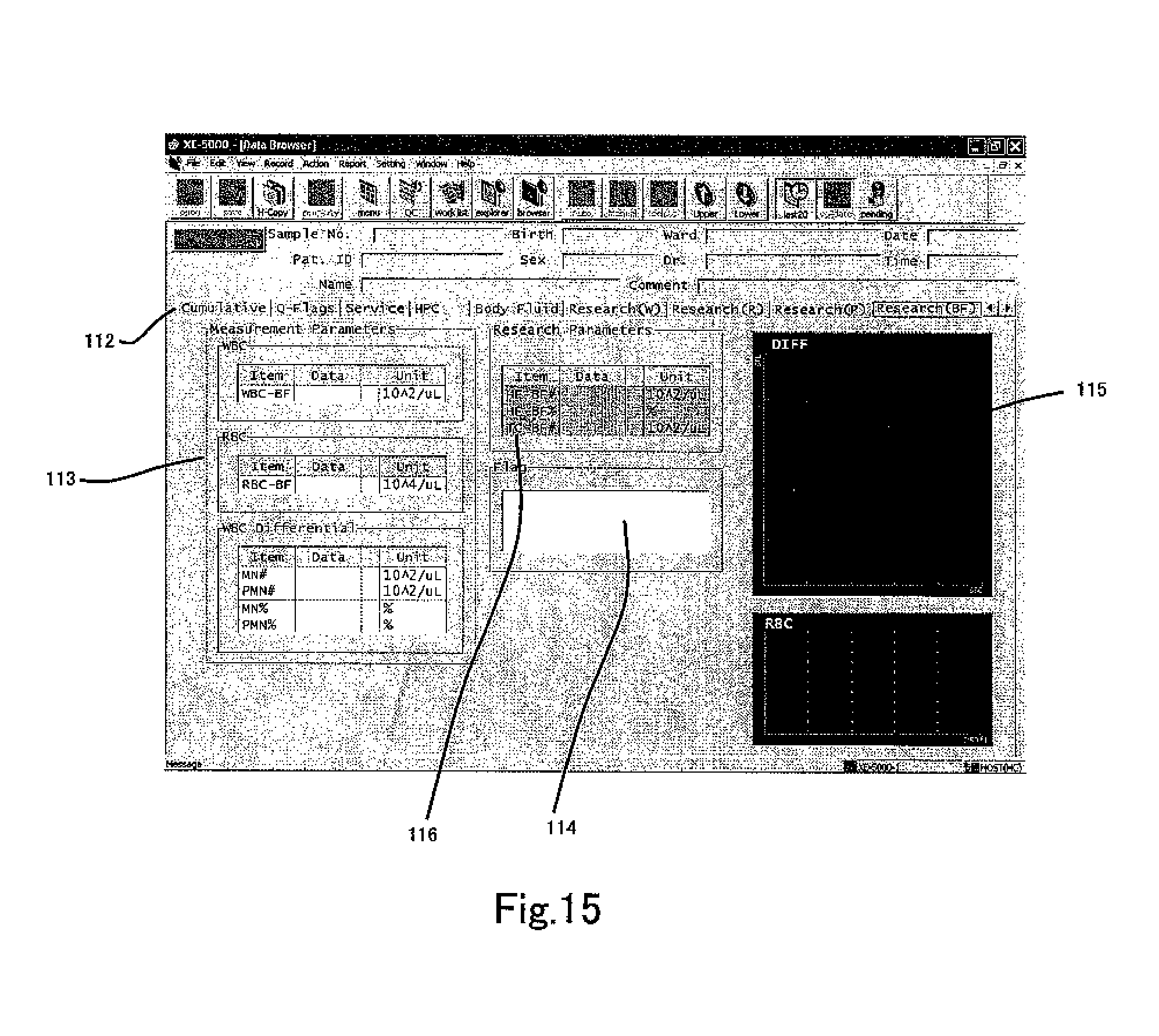
FIG. 15 shows an example in which the Research BF tab 112 is selected in the screen 110 of FIG. 14. This screen displays the same items as screen 110 with the exception that a research parameter display region 116 is also displayed. The research parameter display region 116 displays number of particles in region HF [HF-BF#], the ratio of the number of particles in the region HF relative to the number of particles in the region including both region HF and region MF [HF-BF %], and the number of particles in the region including both region HF and region MF [TC-BF#] in FIG. 10. [HF-BF %] is the percentage of HF-BF relative to TC-BF.
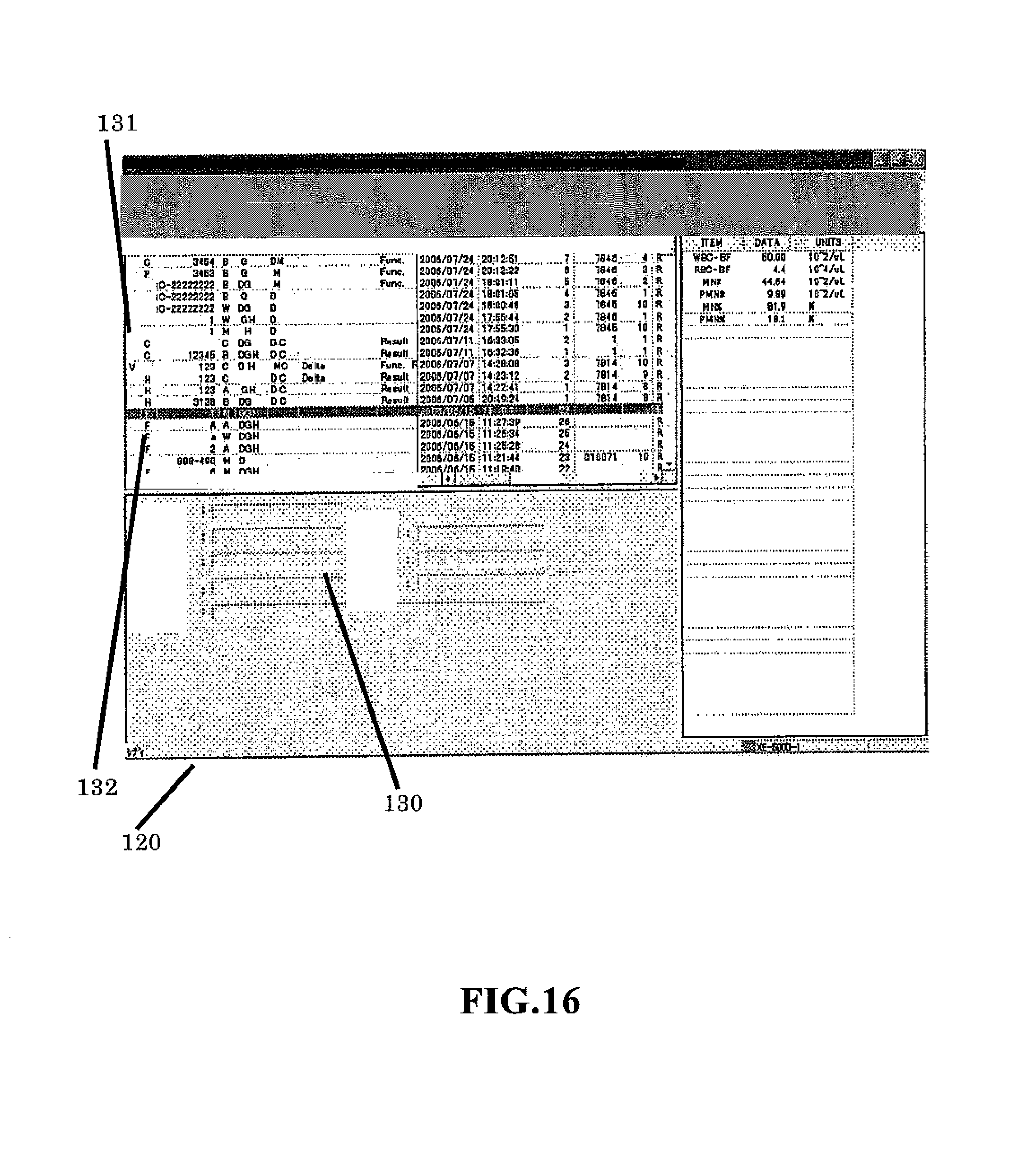
FIG. 16 shows a screen 120 showing a list of stored samples which is displayed on the display unit 302 of the data processing unit 3. Reference number 130 refers to a patient attribute display region. Provided above this region is a measurement result display region which displays the measurement result selected by a tab. A row 131 on the left end of the measurement result display region is used to indicate whether the validation operation has been performed or not for the measurement result. A "V" symbol indicates validation has been performed. A row 132 on the right indicates the measurement mode. An "F" symbol indicates the measurement results are for the body fluid mode. Although there are high value samples that require blank checking in the body fluid mode, and inverted "F" symbol can be displayed to indicate the blank check has not been performed (that is, CANCEL was selected in step S24).
Although the structure and functions of the blood cell analyzer of the present invention have been described as being pre-established in the blood cell analyzer, the same functions may be realized by a computer program so that the functions of the present invention can be realized in a conventional blood cell analyzer by installing the computer program in a conventional blood cell analyzer.
Although the amount of sample, type of reagent, and amount of reagent are the same when preparing measurement samples for the white blood cell classification measurement in the blood measurement mode and the white blood cell classification measurement in the body fluid measurement mode in the present embodiment, the present invention is not limited to this configuration inasmuch as the amount of sample and the amount of reagent used to prepare a measurement sample for white blood cell classification in the body fluid measurement mode may be greater than the amount of sample and the amount of reagent used to prepare a measurement sample for white blood cell classification in the blood measurement mode. Since the measurement time is greater and the amount of measurement sample needed for measurement is greater for white blood cell classification in the body fluid measurement mode than in the blood measurement mode, it is thereby possible to prepare suitable amounts of measurement sample for white blood cell classification in the blood measurement mode and for white blood cell classification in the body fluid measurement mode. Moreover, the type of reagent used fro white blood cell classification in the blood measurement mode may differ from the type of reagent used for white blood cell classification in the body fluid measurement mode.
Although white blood cell classification is performed in the body fluid measurement mode using scattered light and fluorescent light in the present embodiment, the present invention is not limited to this configuration inasmuch as white blood cell classification may also be performed in the body fluid measurement mode using, for example, scattered light and absorbed light. The measurement of absorbed light may be accomplished by preparing a measurement sample by mixing a staining reagent to stain the white blood cells, and other reagent together with the sample, supplying this measurement sample to a flow cell to form a sample flow within the flow cell, irradiating this sample flow with light, and receiving the light emitted from the sample flow via a photoreceptor element such as a photodiode or the like. The light is absorbed by the white blood cells when the white blood cells pass through the flow cell, and the degree of that absorption can be grasped as the amount of light received by the photoreceptor element. Such measurement of absorbed light is disclosed in U.S. Pat. Nos. 5,122,453, and 5,138,181. furthermore, electrical resistance may be measured rather than scattered light, in which case white blood cells can be classified by the electrical resistance and absorbed light.
* * * * *
D00000
D00001
D00002
D00003
D00004
D00005
D00006
D00007
D00008
D00009
D00010
D00011
D00012
D00013
D00014
D00015
D00016
D00017
XML
uspto.report is an independent third-party trademark research tool that is not affiliated, endorsed, or sponsored by the United States Patent and Trademark Office (USPTO) or any other governmental organization. The information provided by uspto.report is based on publicly available data at the time of writing and is intended for informational purposes only.
While we strive to provide accurate and up-to-date information, we do not guarantee the accuracy, completeness, reliability, or suitability of the information displayed on this site. The use of this site is at your own risk. Any reliance you place on such information is therefore strictly at your own risk.
All official trademark data, including owner information, should be verified by visiting the official USPTO website at www.uspto.gov. This site is not intended to replace professional legal advice and should not be used as a substitute for consulting with a legal professional who is knowledgeable about trademark law.