Amphiphilic Polynucleotides
Chilkoti; Ashutosh ; et al.
U.S. patent application number 16/927982 was filed with the patent office on 2021-01-14 for amphiphilic polynucleotides. The applicant listed for this patent is Duke University. Invention is credited to Ashutosh Chilkoti, Sonal Deshpande, Lei Tang, Stefan Zauscher.
| Application Number | 20210009999 16/927982 |
| Document ID | / |
| Family ID | 1000005117997 |
| Filed Date | 2021-01-14 |
View All Diagrams
| United States Patent Application | 20210009999 |
| Kind Code | A1 |
| Chilkoti; Ashutosh ; et al. | January 14, 2021 |
AMPHIPHILIC POLYNUCLEOTIDES
Abstract
Compositions and methods disclosed herein can help provide improved delivery of non-natural therapeutic nucleotides for the treatment of diseases such as cancer. An example composition includes an assembly of amphiphilic polynucleotides, where each amphiphilic polynucleotide includes an aptamer portion, a first nucleotide portion, and a second nucleotide portion.
| Inventors: | Chilkoti; Ashutosh; (Durham, NC) ; Zauscher; Stefan; (Durham, NC) ; Tang; Lei; (Durham, NC) ; Deshpande; Sonal; (Durham, NC) | ||||||||||
| Applicant: |
|
||||||||||
|---|---|---|---|---|---|---|---|---|---|---|---|
| Family ID: | 1000005117997 | ||||||||||
| Appl. No.: | 16/927982 | ||||||||||
| Filed: | July 13, 2020 |
Related U.S. Patent Documents
| Application Number | Filing Date | Patent Number | ||
|---|---|---|---|---|
| 62873306 | Jul 12, 2019 | |||
| Current U.S. Class: | 1/1 |
| Current CPC Class: | C12N 15/113 20130101; A61K 31/7105 20130101; C12N 2310/16 20130101 |
| International Class: | C12N 15/113 20060101 C12N015/113; A61K 31/7105 20060101 A61K031/7105 |
Goverment Interests
STATEMENT REGARDING FEDERALLY SPONSORED RESEARCH
[0002] This invention was made with government support under grant numbers DMR-14-11126, DMR-1121107, CBET-1033621 awarded by the National Science Foundation, and grant number 1R01EB026590-02 awarded by the National Institutes of Health. The government has certain rights in the invention.
Claims
1. A composition comprising an assembly of amphiphilic polynucleotides, each amphiphilic polynucleotide being single-stranded and comprising, in a 5' to 3' direction, an aptamer portion; a first nucleotide portion comprising a nucleotide sequence of (SEQ ID NO:9), wherein m is 100 to 2,000; and a second nucleotide portion comprising a nucleotide sequence of (SEQ ID NO:11), wherein Y.sup.1 is a non-natural hydrophobic nucleotide including a base having a Log P.gtoreq.1.95, and n is 2 to 10.
2. The composition of claim 1, wherein the aptamer portion comprises about 15 to about 100 nucleotides.
3. The composition of claim 1, wherein the aptamer portion is capable of binding to a surface protein overexpressed in a cancer cell.
4. The composition of claim 1, wherein the aptamer portion comprises an aptamer and a linker.
5. The composition of claim 4, wherein the aptamer is selected from the group consisting of (SEQ ID NO:1), (SEQ ID NO:2), (SEQ ID NO:3), (SEQ ID NO:4), (SEQ ID NO:5), and (SEQ ID NO:6).
6. The composition of claim 1, wherein m is 300 to 1,000.
7. The composition of claim 1, wherein the first nucleotide portion further comprises a nucleotide sequence of (SEQ ID NO:10), wherein Z.sup.1 is a non-natural nucleotide having an amino group, an alkynyl group, an azide group, or a combination thereof, and p is 5 to 80.
8. The composition of claim 1, wherein the non-natural hydrophobic nucleotide is selected from the group consisting of Atto-dUTP, BODIPY-dUTP, and a combination thereof.
9. The composition of claim 1, wherein the first nucleotide portion and the second nucleotide portion are included at a ratio (number of nucleotides of the first nucleotide portion: number of nucleotides of the second nucleotide portion) of about 20:1 to about 60:1.
10. The composition of claim 1, wherein the amphiphilic polynucleotide comprises about 300 to about 600 nucleotides.
11. The composition of claim 1, wherein the amphiphilic polynucleotide has a critical micelle concentration of .ltoreq.0.1 .mu.M.
12. The composition of claim 1, wherein the assembly is stable in a mixture of about 50% fetal bovine serum for about 30 minutes to about 1 day.
13. The composition of claim 1, wherein the assembly of amphiphilic polynucleotides is a nanoparticle.
14. The composition of claim 13, wherein the nanoparticle has an average hydrodynamic radius of about 20 nm to about 125 nm.
15. The composition of claim 13, wherein the nanoparticle is a micelle.
16. A method of making an amphiphilic polynucleotide that can self-assemble into a nanoparticle, the method comprising combining a terminal deoxynucleotidyl transferase (TdT), an aptamer initiator having a secondary structure, and a non-natural cytostatic deoxynucleoside triphosphate (dNTP) monomer in a buffer to provide a first reaction mixture; incubating the first reaction mixture; adding a non-natural hydrophobic dNTP monomer to the first reaction mixture to provide a second reaction mixture; and incubating the second reaction mixture to provide the amphiphilic polynucleotide of claim 1.
17. The method of claim 16, wherein the aptamer portion of the amphiphilic polynucleotide is refolded to provide a secondary structure.
18. The method of claim 16, wherein TdT is added to the first reaction mixture, the second reaction mixture, or both in multiple rounds.
19. A method of treating a disease or disorder in a subject in need thereof, the method comprising administering to the subject a therapeutically effective amount of the composition of claim 1.
20. The method of claim 19, wherein the disease or disorder is cancer.
Description
CROSS-REFERENCE TO RELATED APPLICATIONS
[0001] This application claims priority to U.S. Provisional Patent Application No. 62/873,306 filed on Jul. 12, 2019, which is incorporated fully herein by reference.
SEQUENCE LISTING
[0003] The sequence listing is filed with the application and is incorporated by reference herein. The sequence listing text file "028193-9330-US03_As_Filed_Sequence_Listing.txt" was created on Jul. 13, 2020, and is 3299 bytes in size.
BACKGROUND
[0004] Amphiphilic molecules can provide useful self-assembled structures, which can be beneficial for drug delivery applications.
SUMMARY
[0005] In one aspect, disclosed are compositions that include an assembly of amphiphilic polynucleotides, each amphiphilic polynucleotide being single-stranded and comprising, in a 5' to 3' direction, an aptamer portion, a first nucleotide portion, and a second nucleotide portion.
[0006] In another aspect, disclosed are methods of making an amphiphilic polynucleotide. The method includes combining a terminal deoxynucleotidyl transferase (TdT), an aptamer initiator having a secondary structure, and a non-natural cytostatic deoxynucleoside triphosphate (dNTP) monomer in a buffer to provide a first reaction mixture; incubating the first reaction mixture; adding a non-natural hydrophobic dNTP monomer to the first reaction mixture to provide a second reaction mixture; and incubating the second reaction mixture to provide the disclosed amphiphilic polynucleotide.
[0007] In another aspect, disclosed are methods of treating a disease or disorder in a subject in need thereof. The method includes administering to the subject a therapeutically effective amount of the disclosed composition.
BRIEF DESCRIPTION OF THE DRAWINGS
[0008] This patent or application file contains at least one drawing executed in color. Copies of this patent or patent application publication with color drawing(s) will be provided by the Office upon request and payment of the necessary fee.
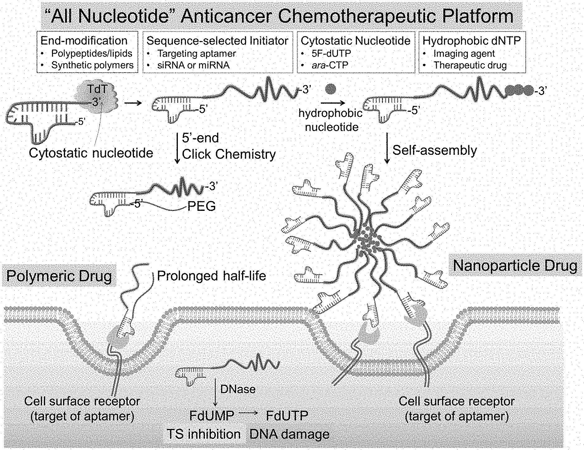
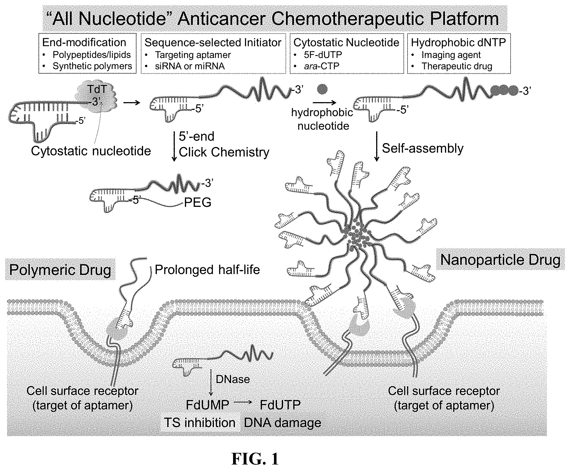
[0009] FIG. 1 shows a schematic of a synthesis of amphiphilic polynucleotides and their application in drug delivery applications.
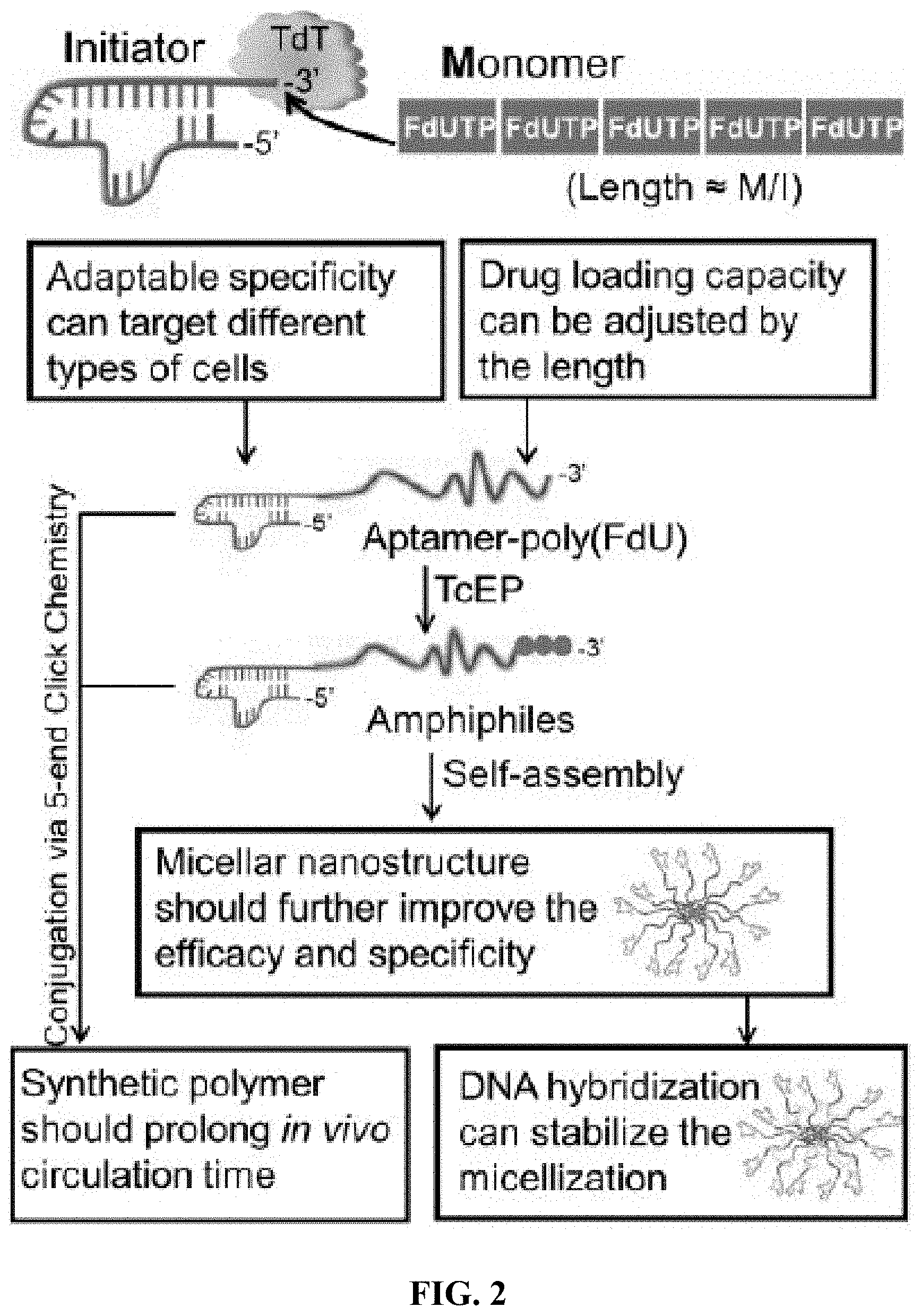
[0010] FIG. 2 shows a schematic workflow of a polydeoxyfluorouracil (which is referred to herein as polyFdU, polyFU, or polyFdUTP) amphiphilic polynucleotide.
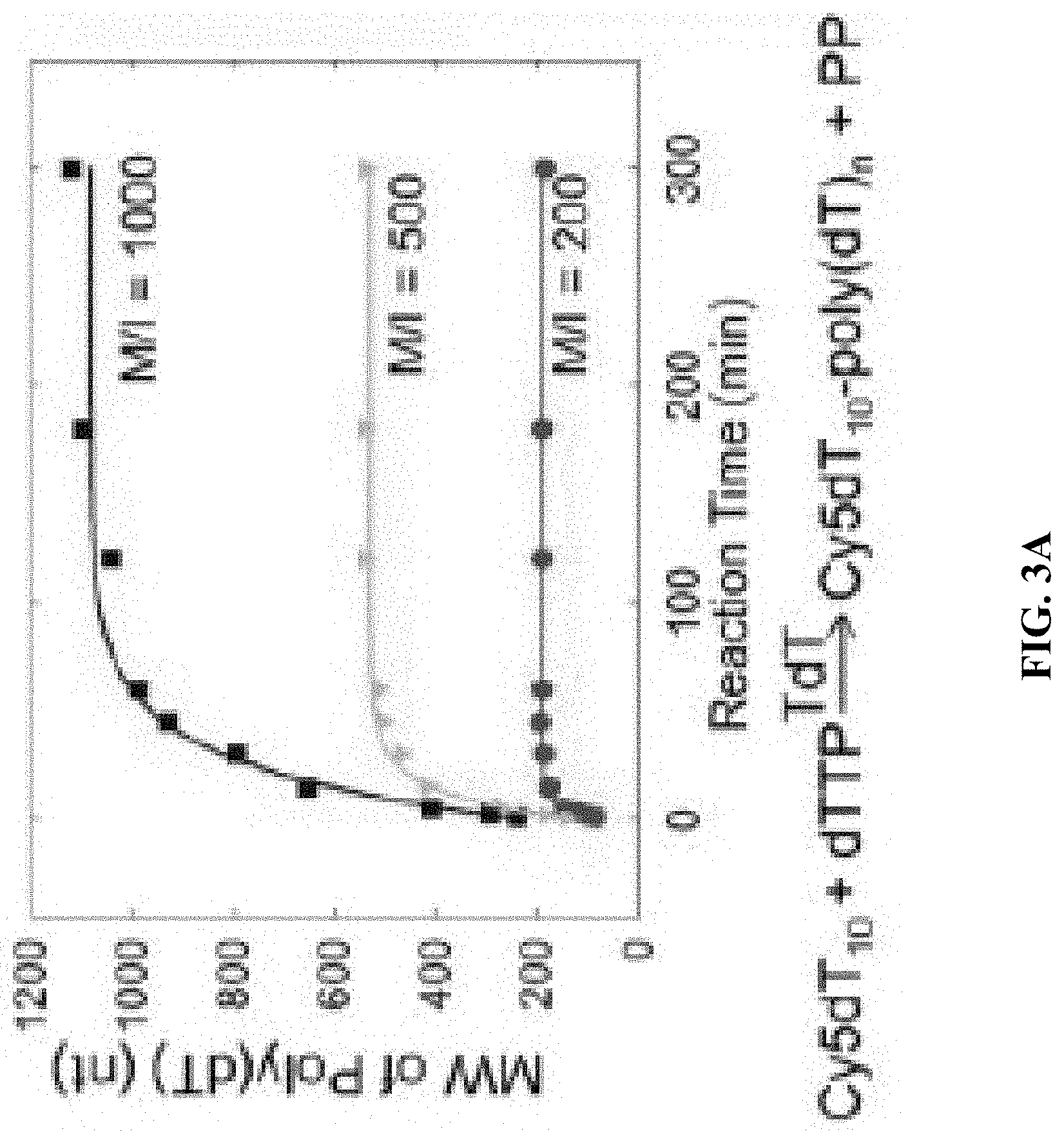
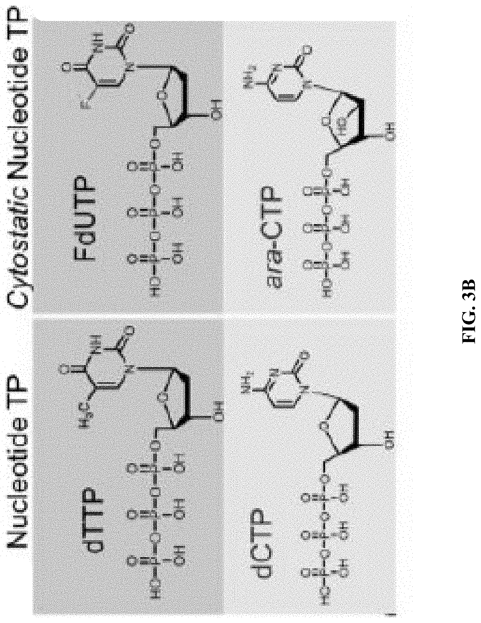
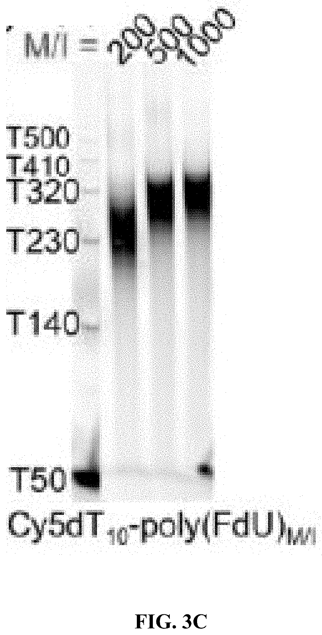
[0011] FIG. 3A shows the molecular weight (MW) vs. reaction time for different initial monomer/initiator (M/I) ratios; FIG. 3B shows chemical structures of deoxythymidine triphosphate (dTTP), deoxycytidine triphosphate (dCTP) and their analogs FdUTP, ara-CTP, respectively; and FIG. 3C shows polyacrylamide gel electrophoresis (PAGE) images showing that different MWs are generated by different M/I ratios of FdUTP to Cy5T10, ranging from 200 to 1000 at a constant [TdT] of 1 unit/.mu.l.
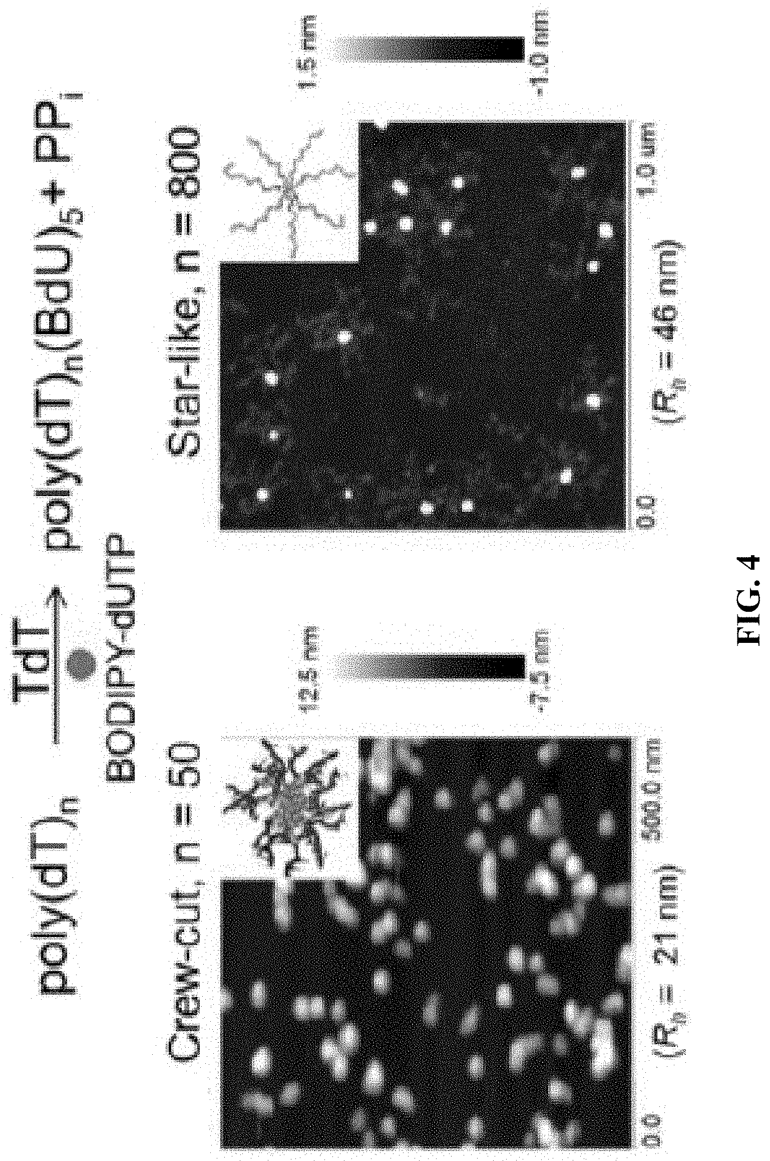
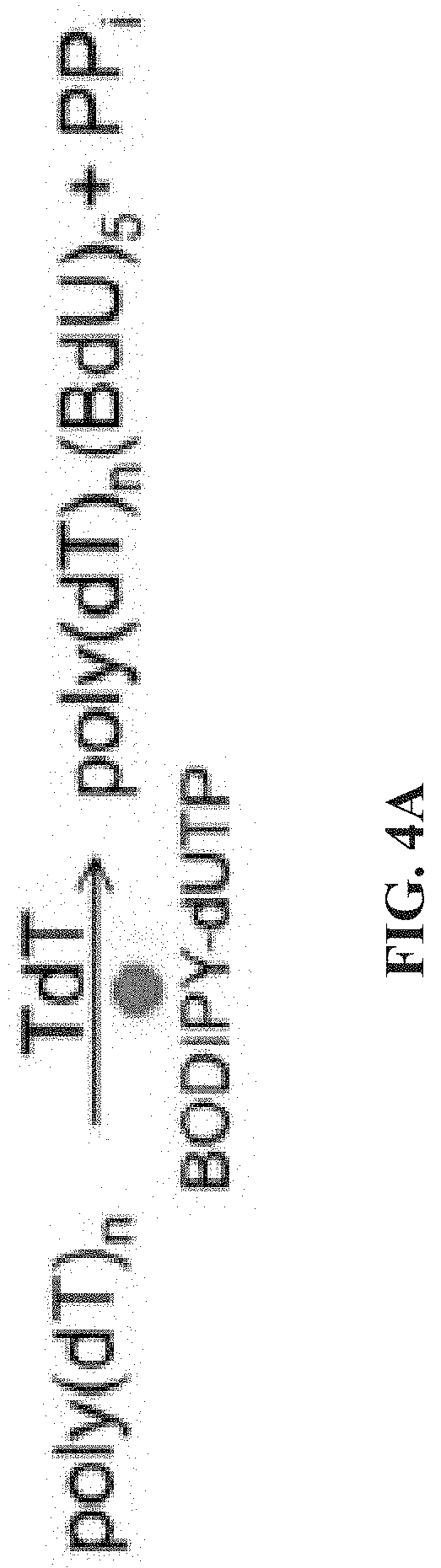
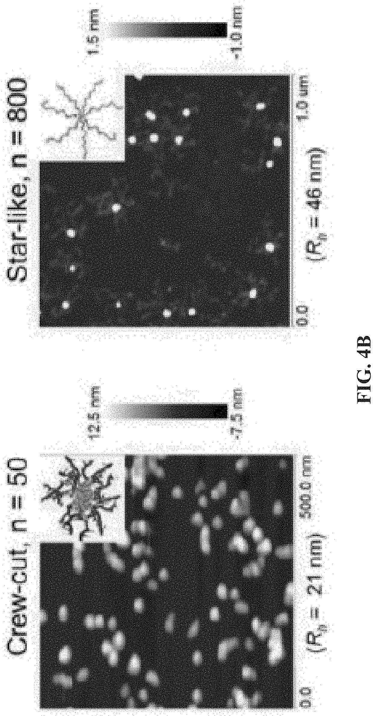
[0012] FIG. 4A shows a schematic of a TdT catalyzed enzymatic polymerization (TcEP) reaction for the synthesis of amphiphilic polynucleotides; and FIG. 4B shows atomic force microscopy (AFM) images showing self-assemblies of amphiphilic polynucleotides with different hydrophobic block lengths (n=50 and n=800).
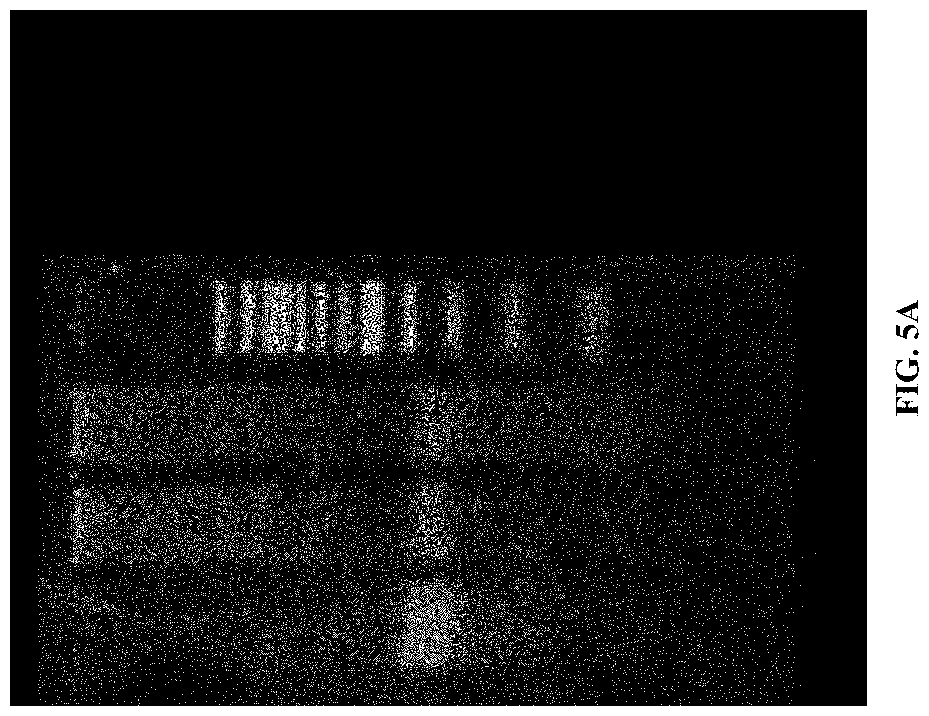
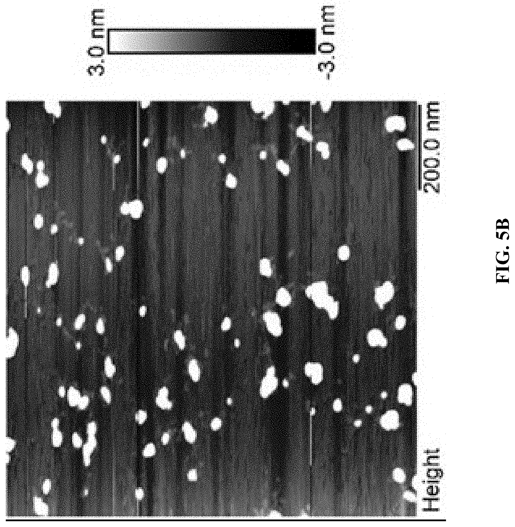
[0013] FIG. 5A shows an agarose gel electrophoresis image. From left to right: Cy5T50-polyFU, Cy5T50-polyFU-AttodU (M/I=20), Cy5T50-polyFU-AttodU (M/I=50), 100 bp ladder. FIGS. 5B and 5C show AFM images for Cy5T50-polyFU-AttodU (M/I=10) in water, sample concentration is 0.1 .mu.M. The monomer to initiator ratio (M/I) is the feed ratio of Atto-dUTP to the single-stranded (ss)DNA initiator (polyFU).
[0014] FIG. 6A is an AFM image for Cy5T50-polyFdU-AttodU with M/I=50, and FIG. 6B is an AFM image for Cy5T50-polyFdU-AttodU with M/I=300. Sample concentration is 0.1 .mu.M, solvent is water.
[0015] FIGS. 7A and 7B are a set of AFM images for Cy5T50-polyFU-AttodU (M/I=20). Sample concentrations are FIG. 7A 0.05 .mu.M and FIG. 7B 0.01 .mu.M in water.
[0016] FIG. 8A is an AFM image for A10-polyFU-AttodU (M/I=20). Sample concentration is 0.1 .mu.M in water; and FIG. 8B shows measured height of micelles from AFM images.
[0017] FIGS. 9A and 9B are a set of agarose gel electrophoresis images showing: FIG. 9A Exonuclease I degradation of Cy5T50-polyFU-AttodU (M/I=20) micelles at 0.05 .mu.M for 24 h. 5% DMSO was added to disassemble micelles. FIG. 9B FBS degradation of Cy5T50-polyFU-AttodU (M/I=20) micelles at 0.2 .mu.M concentration as a function of incubation time.
[0018] FIG. 10A is a schematic illustration of the formation of an A10-PolyFU-AttodU amphiphilic block polynucleotide; and FIGS. 10B, 10C, and 10D are a series of AFM images for A10-polyFU500-AttodU10 (FIG. 10B), A10-polyFU800-AttodU10 (FIG. 10C), and A10-polyFU1000-AttodU10 (FIG. 10D). All sample concentrations are 0.1 .mu.M in 1.times.PBS.
[0019] FIGS. 11A and 11B are a set of 5% (Tris-borate-ethylenediaminetetraacetic acid (EDTA)) (TBE)-PAGE images for 10% fetal bovine serum (FBS) degradation of A10-polyFU-AttodU10 (FIG. 11A), and A10-polyFU (FIG. 11B) at different time points. Final concentrations of samples are both 1 .mu.M.
[0020] FIGS. 12A and 12B are a set of 5% TBE-PAGE images for 50% FBS degradation of A10-polyFU-AttodU10 (FIG. 12A), and A10-polyFU (FIG. 12B) at different time points. Lanes with red asterisks stand for samples in EDTA inactivated FBS. Final concentrations of samples are both 0.7 .mu.M.
[0021] FIG. 13 is a 5% TBE-PAGE image for 10% FBS degradation of A10-polyFU-AttodU10 at different time points within a week. Red asterisk stands for sample in EDTA inactivated FBS. Final concentration of sample is 1 .mu.M.
[0022] FIGS. 14A and B are a set of AFM images for 1 day degradation of AO-polyFU-AttodU10 micelle in 10% FBS (FIG. 14A) and 10% FBS as control (FIG. 14B).
[0023] FIGS. 15A-15F are a series of 5% TBE-PAGE images for DNase I degradation at different time points for: FIG. 15A--A10-polyFU-AttodU10 micelle and A10polyFU, 1 U/mL DNase I; FIG. 15B--A10-polyFU-AttodU10 micelle, 200 U/mL DNase I; FIG. 15C-A10polyFU, 200 U/mL DNase I; FIG. 15D--A10-polyFU-AttodU10 micelle and A10polyFU, 100 U/mL DNase I; FIG. 15E--A10-polyFU-AttodU10 micelle and A10polyFU, 50 U/mL DNase I; FIG. 15F--A10-polyFU-AttodU10 micelle and A10polyFU, 60 U/mL DNase I. Final concentrations of all samples are 0.8 .mu.M.
[0024] FIGS. 16A and 16B show dynamic light scattering (DLS) measurement for: FIG. 16A--the size distribution profiles by intensity for three different batches of A10-polyFU in water, and the FIG. 16B--Z-average radius and PDI of each sample.
[0025] FIGS. 17A and 17B are a set of AFM images for two different batches of A10-polyFU ssDNA in water.
[0026] FIG. 18A is a schematic illustration of the refolding process for aptamers using PCR. 1% and 100% are the rate of temperature changes; and FIG. 18B is an AFM image for a batch of refolded A10-polyFU ssDNA in water.
[0027] FIG. 19A is a DLS measurement showing size distribution profiles by intensity and by number for three different batches of A10-polyFU and three different batches of refolded A10-polyFU; and FIG. 19B shows the Z-average radius and PDI of each sample.
[0028] FIGS. 20A and 20B are a set of agarose gel electrophoresis images showing: FIG. 20A--from left to right, 100 bp ladder, Cy5T50-polyFU, Cy5T50-poly(FdU-co-aadU) (2% aadUTP), Cy5T50-poly(FdU-co-aadU) (5% aadUTP), Cy5T50-poly(FdU-co-aadU) (10% aadUTP); and FIG. 20B--Cy5 (red) and Cy3 (green) signals for Cy3-conjugated Cy5T50-poly(FdU-co-aadU) ssDNA containing different fractions of amine groups. The yellow color is produced by overlapping of red and green signal.
[0029] FIGS. 21A and 21B are a set of agarose gel images: FIG. 21A--an agarose gel image showing molecular weight and distribution of single stranded sgc8c-poly(FdUTP); and FIG. 21B--agarose gel electrophoresis of aptamers A10, Ap1153, and sgc8c extended with FdUTP using TcEP.
DETAILED DESCRIPTION
1. Definitions
[0030] Unless otherwise defined, all technical and scientific terms used herein have the same meaning as commonly understood by one of ordinary skill in the art. In case of conflict, the present document, including definitions, will control. Preferred methods and materials are described below, although methods and materials similar or equivalent to those described herein can be used in practice or testing of the present invention. All publications, patent applications, patents and other references mentioned herein are incorporated by reference in their entirety. The materials, methods, and examples disclosed herein are illustrative only and not intended to be limiting.
[0031] The terms "comprise(s)," "include(s)," "having," "has," "can," "contain(s)," and variants thereof, as used herein, are intended to be open-ended transitional phrases, terms, or words that do not preclude the possibility of additional acts or structures. The singular forms "a," "and" and "the" include plural references unless the context clearly dictates otherwise. The present disclosure also contemplates other embodiments "comprising," "consisting of" and "consisting essentially of," the embodiments or elements presented herein, whether explicitly set forth or not.
[0032] For the recitation of numeric ranges herein, each intervening number there between with the same degree of precision is explicitly contemplated. For example, for the range of 6-9, the numbers 7 and 8 are contemplated in addition to 6 and 9, and for the range 6.0-7.0, the number 6.0, 6.1, 6.2, 6.3, 6.4, 6.5, 6.6, 6.7, 6.8, 6.9, and 7.0 are explicitly contemplated.
[0033] The modifier "about" used in connection with a quantity is inclusive of the stated value and has the meaning dictated by the context (for example, it includes at least the degree of error associated with the measurement of the particular quantity). The modifier "about" should also be considered as disclosing the range defined by the absolute values of the two endpoints. For example, the expression "from about 2 to about 4" also discloses the range "from 2 to 4." The term "about" may refer to plus or minus 10% of the indicated number. For example, "about 10%" may indicate a range of 9% to 11%, and "about 1" may mean from 0.9-1.1. Other meanings of "about" may be apparent from the context, such as rounding off, so, for example "about 1" may also mean from 0.5 to 1.4.
[0034] The term "amphiphilic," as used herein, refers to a molecule (e.g., polynucleotide) having both hydrophilic and hydrophobic properties.
[0035] The term "alkynyl," as used herein, refers to straight or branched monovalent hydrocarbyl groups having from 2 to 30 carbon atoms, such as 2 to 20, or 2 to 10 carbon atoms and having at least 1 site of triple bond unsaturation. Examples of such alkynyl groups include, but are not limited to acetylenyl (--C.ident.CH), and propargyl (--CH.sub.2C.ident.CH).
[0036] The term "amino," as used herein, refers to the functional group --NH.sub.2.
[0037] The term "azide," as used herein, refers to the functional group --N.sub.3.
[0038] The term "critical micelle concentration," as used herein, refers to the concentration of amphiphilic polynucleotides above which micelles are formed. Herein, this value was determined by AFM imaging analysis and/or fluorescence intensity analysis of micelle formation. Further details of determining CMC can be found in J. N. Phillips, Trans. Faraday 1955, 51, 561, which is incorporated by reference herein in its entirety.
[0039] The terms "effective amount" or "therapeutically effective amount," as used herein, refer to a sufficient amount of an agent or a composition or combination of compositions being administered which will relieve to some extent one or more of the symptoms of the disease or condition being treated. The result can be reduction and/or alleviation of the signs, symptoms, or causes of a disease, or any other desired alteration of a biological system. For example, an "effective amount" for therapeutic uses is the amount of the composition as disclosed herein required to provide a clinically significant decrease in disease symptoms. An appropriate "effective" amount in any individual case may be determined using techniques, such as a dose escalation study. The dose could be administered in one or more administrations. However, the precise determination of what would be considered an effective dose may be based on factors individual to each patient, including, but not limited to, the patient's age, size, type or extent of disease, stage of the disease, route of administration, the type or extent of supplemental therapy used, ongoing disease process and type of treatment desired (e.g., aggressive vs. conventional treatment).
[0040] The term "non-natural nucleotide," as used herein, refers to nucleotides that are not present in a natural biological system. Similar to a naturally occurring nucleotide (e.g., ATP, UTP, etc.), a non-natural nucleotide includes a phosphate group(s), a sugar, and a base. However, a non-natural nucleotide can differ from a naturally occurring nucleotide by having its phosphate group(s), sugar, base or combination thereof modified. Examples of non-natural nucleotides include, but are not limited to, 5-fluoro-2'-deoxyuridne-5'triphosphate, arabinofuranosylcytosine triphosphate, Atto-dUTP, and BODIPY-dUTP.
[0041] The term "polynucleotide," as used herein, refers to 300 or more nucleotides covalently linked together. Polynucleotides may be single-stranded, double-stranded, or may contain portions of both double-stranded and single-stranded sequence. A polynucleotide can include natural or non-natural nucleotides, and can contain combinations of deoxyribo- and ribo-nucleotides, and combinations of bases including, for example, uracil, adenine, thymine, cytosine, guanine, inosine, xanthine hypoxanthine, isocytosine, and isoguanine.
[0042] A polynucleotide includes a 5' end and a 3' end. Polynucleotides are said to have "5' ends" and "3' ends" because nucleotides are reacted to make polynucleotides in a manner such that the 5' phosphate of one nucleotide pentose ring is attached to the 3' oxygen of its neighbor in one direction via a phosphodiester linkage. Thus, an end of a polynucleotide can be referred to as the "5' end" if its 5' phosphate is not linked to the 3' oxygen of a nucleotide pentose ring and as the "3' end" if its 3' oxygen is not linked to a 5' phosphate of a subsequent nucleotide pentose ring. In addition, a nucleotide portion or sequence, even if internal and linked within a larger polynucleotide, also may be said to have 5' and 3' ends. For example, the aptamer portion and the aptamer can have 5' and 3' ends.
[0043] The term "subject," "patient," or "organism," as used herein, includes humans and mammals (e.g., mice, rats, pigs, cats, dogs, and horses). Typical subjects of the present disclosure may include mammals, particularly primates, and especially humans. For veterinary applications, suitable subjects may include, for example, livestock such as cattle, sheep, goats, cows, swine, and the like; poultry such as chickens, ducks, geese, turkeys, and the like, as well as domesticated animals particularly pets such as dogs and cats. For diagnostic or research applications, suitable subjects may include mammals, such as rodents (e.g., mice, rats, hamsters), rabbits, primates, and swine such as inbred pigs and the like.
[0044] The term "treatment" or "treating," as used herein when referring to protection of a subject from a disease, means preventing, suppressing, repressing, ameliorating, or completely eliminating the disease. Preventing the disease involves administering a composition of the present disclosure to a subject prior to onset of the disease. Suppressing the disease involves administering a composition of the present disclosure to a subject after induction of the disease but before its clinical appearance. Repressing or ameliorating the disease involves administering a composition of the present disclosure to a subject after clinical appearance of the disease.
2. Amphiphilic Polynucleotides
[0045] Provided herein are compositions that include an assembly of amphiphilic polynucleotides. The amphiphilic polynucleotide can be single-stranded and includes at least three different portions. These three portions can also be referred to as three different nucleotide portions. The three portions include an aptamer portion, a first nucleotide portion, and a second nucleotide portion. The three portions can be included in the amphiphilic polynucleotide in a 5' to 3' direction. For example, the aptamer portion can be coupled to the first nucleotide portion, the first nucleotide portion can be coupled to the aptamer portion and the second nucleotide portion, and the second nucleotide portion can be coupled to the first nucleotide portion. In other words, the first nucleotide portion can be in between the aptamer portion and the second nucleotide portion, where the aptamer portion and the second nucleotide portion form individual ends of the amphiphilic polynucleotide.
[0046] The amount and composition of these three portions can instill an amphiphilicity characteristic to the amphiphilic polynucleotide. This amphiphilicity characteristic can allow the amphiphilic polynucleotide to self-assemble with other amphiphilic polynucleotides to form the assembly. For example, the first nucleotide portion and the second nucleotide portion can be included in the amphiphilic polynucleotide at a ratio that can promote self-assembly. In some embodiments, the first nucleotide portion and the second nucleotide portion are included at a ratio (number of nucleotides of the first nucleotide portion: number of nucleotides of the second nucleotide portion) of about 20:1 to about 60:1, such as about 25:1 to about 60:1, or about 30:1 to about 55:1. In addition, due to the ability of the amphiphilic polynucleotides to self-assemble, the amphiphilic polynucleotide can have a critical micelle concentration (CMC). In some embodiments, the amphiphilic polynucleotide has a CMC that is less than or equal to 0.1 .mu.M, less than or equal to 0.09 .mu.M, or less than or equal to 0.08 .mu.M. In some embodiments, the amphiphilic polynucleotide has a CMC that is greater than or equal to 0.04 .mu.M, greater than or equal to 0.05 .mu.M, or greater than or equal to 0.06 .mu.M.
[0047] The amphiphilic polynucleotide can include about 300 nucleotides to about 600 nucleotides, such as about 350 nucleotides to about 550 nucleotides, or about 400 nucleotides to about 525 nucleotides. In some embodiments, the amphiphilic polynucleotide includes about 500 nucleotides. In addition, the amphiphilic polynucleotide can be made from only nucleotides. Thus, in some embodiments, the amphiphilic polynucleotide is 100% nucleotide. In other embodiments the amphiphilic polynucleotide can include molecules other than nucleotides. For example, the amphiphilic polynucleotide can be modified with polymers such as polyethylene glycol (PEG), lipids, peptides, and/or proteins.
[0048] The amphiphilic polynucleotide can self-assemble into a variety of different particulate structures and sizes. For example, the assembly can be a nanoparticle. The nanoparticle can have a varying average hydrodynamic radius depending on the structure and portions of the amphiphilic polynucleotide. The hydrodynamic radius can be measured using dynamic light scattering techniques known within the art. In addition, particle size can be measured via other techniques, such as AFM and transmission electron microscopy (TEM) image analysis. In some embodiments, the nanoparticle has an average hydrodynamic radius of about 20 nm to about 125 nm, such as about 25 nm to about 100 nm, or about 20 nm to about 60 nm. In some embodiments, the nanoparticle has an average hydrodynamic radius of about 50 nm. The assembly and nanoparticle thereof can have a micellular structure. In some embodiments, the nanoparticle is a micelle.
[0049] The amphiphilic polynucleotide can provide an assembly that is advantageously stable. Stability, as used herein, refers to the assembly being able to maintain its structural (e.g., size) and/or functional features (e.g., binding capability of aptamer portion) for a specified amount of time in a particular environment (e.g., saline, serum, blood, etc.). For example, the assembly being able to maintain its average particle size within 25% of its initial average size for 1 day corresponds to the assembly being stable for greater than or equal to 1 day. In addition, stability can also include the assembly being able to maintain a functional effect (e.g., within .+-.5% of its aptamer portion binding activity) over a period of time. In some embodiments, the assembly can be stable in serum and mixtures that include serum for extended periods of time compared to an individual amphiphilic polynucleotide not part of an assembly. This can be useful for in vivo applications by, e.g., affording the assembly an extended circulation time. In some embodiments, the assembly can be stable in a mixture of about 50% fetal bovine serum for about 30 minutes to about 1 day, such as about 1 hour to about 22 hours, about 30 minutes to about 20 hours, or about 2 hours to about 1 day.
[0050] A. Aptamer Portion
[0051] The aptamer portion of the amphiphilic polynucleotide can be used as an initiator for the synthesis of the amphiphilic polynucleotide, as well as can be used to localize the amphiphilic polynucleotide and assembly thereof to a specific target molecule, protein, cell, tissue, or the like. In some embodiments, the aptamer portion is capable of binding to a specific target protein. The target protein can be a cell surface protein or a protein present intracellularly. In addition, the target protein can be associated with a disease or disorder. For example, the target protein can be a protein that is overexpressed in a diseased cell, tissue, or the like. An overexpressed protein refers to a protein that is expressed by a diseased cell or tissue at greater than 100.times., greater than 500.times., or greater than 1000.times. the expression level of that protein in the non-diseased (e.g., healthy) cell or tissue. In some embodiments, the aptamer portion is capable of binding to a surface protein that is overexpressed in a cancer cell.
[0052] The aptamer portion can include an aptamer and a linker. The aptamer can instill the capability of the aptamer portion and amphiphilic polynucleotide (and assembly thereof) to bind to specific proteins and non-protein molecules. As used herein, "aptamer" refers to a nucleotide sequence that can bind to a target molecule. The aptamer can be DNA, RNA, or a combination thereof. The binding description in regards to the aptamer portion can also be applied to the aptamer. In some embodiments, the aptamer is selected from the group consisting of AS1411 (5'-GGTGGTGGTGGTTGTGGTGGTGGTGG-3') (SEQ ID NO:1), AIR-3 (5'-GGAAGAAAGAGGUCUGAGACAUUCUCUUAUAGGGGAGGCUGUGGUGAGGGAAUA UUAAGAGAAUUAACGGUCUAGUUCACCUCGACUUCUGGAGUUGACGUUGCUU-3') (SEQ ID NO:2), A10 (5'-GGGAGGACGAUGCGGAUCAGCCAUGUUUACGUCACUCCU-3') (SEQ ID NO:3), Sgc8c (5'-ATCTAACTGCTGCGCCGCCGGGAAAATACTGTACGGTTAGA-3') (SEQ ID NO:4), MUC1-5TR-1 (GGGAGACAAGAATAAACGCTCAAGAAGTGAAAATGACAGAACACAACATTCGACA GGAGGCTCACAACAGGC) (SEQ ID NO:5), and AP1153 (5'-CAT GGT GCA GGT GTG GCT GGG ATT CAT TTG CCG GTG CTG GTG CGT CCG CGG CCG CTA ATC CTG TTC-3') (SEQ ID NO:6). The aptamer may also include non-natural nucleotides that can be useful for nuclease resistance. Modifications that can be useful for nuclease resistance include a phosphorothioate backbone and/or sugar modification: 2' fluoro and 2'-O-methyl.
[0053] The linker can be used to decrease intermolecular hinderance between, e.g., the aptamer portion and the first nucleotide portion. The linker can be coupled to the 5' end, the 3'end, or both of the aptamer. In some embodiments, the linker includes about 2 nucleotides to about 30 nucleotides, such as about 3 nucleotides to about 20 nucleotides, or about 4 nucleotides to about 15 nucleotides. The nucleotides of the linker can be a natural nucleotide or a non-natural nucleotide. In some embodiments, the linker includes dTTP.
[0054] The aptamer portion can include about 15 nucleotides to about 100 nucleotides, such as about 20 nucleotides to about 90 nucleotides or about 25 nucleotides to about 100 nucleotides. In some embodiments, the aptamer portion includes a nucleotide sequence of (SEQ ID NO:7). In some embodiments, the aptamer portion is (SEQ ID NO:7).
[0055] B. First Nucleotide Portion
[0056] The first nucleotide portion of the amphiphilic polynucleotide can be used to provide a therapeutic nucleotide, as well as can be used to provide a hydrophilic portion of the amphiphilic polynucleotide. The first nucleotide portion can include non-natural therapeutic nucleotides, such as non-natural cytostatic nucleotides. As used herein, "non-natural cytostatic nucleotide" refers to a non-natural nucleotide that can inhibit cell growth, cell division, cell metabolism, or a combination thereof. Examples of non-natural cytostatic nucleotides include, but are not limited to, 5-fluoro-2'-deoxyuridne-5'triphosphate (FdUTP), arabinofuranosylcytosine triphosphate, 8-chloro-adenosine triphosphate, 8-amino-adenosine triphosphate, 5-aza-2'-deoxycytidine triphosphate. In some embodiments, the non-natural cytostatic nucleotide is selected from the group consisting of FdUTP, arabinofuranosylcytosine triphosphate, 8-chloro-adenosine triphosphate, 8-amino-adenosine triphosphate, 5-aza-2'-deoxycytidine triphosphate, and a combination thereof. In some embodiments, the non-natural cytostatic nucleotide is selected from FdUTP, arabinofuranosylcytosine triphosphate, and a combination thereof. In some embodiments, the non-natural cytostatic nucleotide is FdUTP.
[0057] The first nucleotide portion can include a nucleotide sequence of (X.sup.1).sub.m(SEQ ID NO:8), wherein X.sup.1 is a non-natural cytostatic nucleotide, and m is 100 to 2,000. In some embodiments, the first nucleotide portion includes a nucleotide sequence of (FdUTP).sub.m(SEQ ID NO:9), wherein m is 100 to 2,000. In some embodiments of (SEQ ID NO:8) or (SEQ ID NO:9), m is 100 to 1,000. In some embodiments, the first nucleotide portion is (SEQ ID NO:8). In some embodiments, the first nucleotide portion is (SEQ ID NO:9).
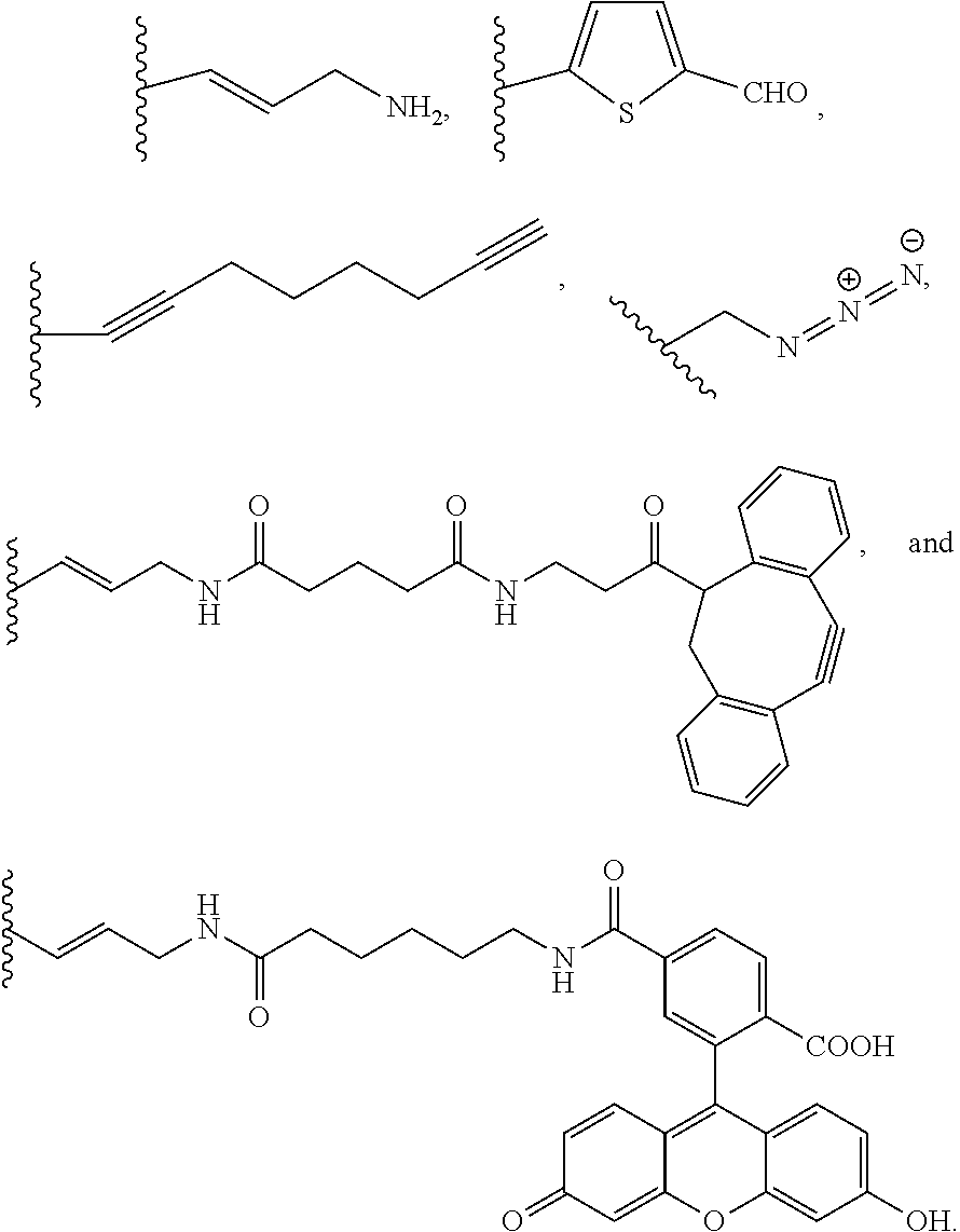
[0058] The first nucleotide portion can also include other non-natural nucleotides and sequences thereof that can be used to alter properties of the amphiphilic polynucleotide and/or the assembly thereof. For example, other non-natural nucleotides and sequences thereof can be used to cross-link the assembly or can be used to add imaging agents to the assembly. These other non-natural nucleotides and sequences thereof can include a variety of different functional groups attached to the base of the non-natural nucleotide. In some embodiments, the base that is modified is uracil. Examples of functional groups include, but are not limited to:
##STR00001##
[0059] In some embodiments, the first nucleotide portion includes a nucleotide sequence of (Z).sub.p (SEQ ID NO:10), wherein Z.sup.1 is a non-natural nucleotide having an amino group, an alkynyl group, an azide group, or a combination thereof, and p is 5 to 80. In some embodiments, the non-natural nucleotide is a modified dUTP having an amino group, an alkynyl group, an azide group, or a combination thereof. In some embodiments, the non-natural nucleotide is a base-modified dUTP having an amino group, an alkynyl group, an azide group, or a combination thereof.
[0060] The first nucleotide portion can include (SEQ ID NO:8), (SEQ ID NO:9), and/or (SEQ ID NO:10) in different arrangements. For example, the first nucleotide portion can include (SEQ ID NO:9) and (SEQ ID NO:10) as blocks. In other embodiments, the first nucleotide portion can include (SEQ ID NO:9) and (SEQ ID NO:10) randomly. In some embodiments, the first nucleotide portion includes more than one occurrence of (SEQ ID NO:8), (SEQ ID NO:9), and/or (SEQ ID NO:10). For example, the first nucleotide portion can include (SEQ ID NO:9) at more than one occurrence and can include (SEQ ID NO:10) at more than one occurrence.
[0061] C. Second Nucleotide Portion
[0062] The second nucleotide portion of the amphiphilic polynucleotide can be used to provide a hydrophobic portion of the amphiphilic polynucleotide. The second nucleotide portion can include non-natural hydrophobic nucleotides. As used herein, "non-natural hydrophobic nucleotide" refers to a non-natural nucleotide that includes a base modified with a hydrophobic molecule or hydrophobic functional group. The hydrophobicity characteristic of the second nucleotide portion can be measured via the Log P of the modified base of the non-natural hydrophobic nucleotide. Log P is the log(concentration of a compound in octanol/concentration of the compound in water). Log P=0 implies equal affinity for organic and aqueous phase; Log P<1 implies affinity towards aqueous phase; and Log P>1 implies affinity towards organic phase. Log P can be estimated, e.g., using Crippen's fragmentation method in the software ChemDraw. The second nucleotide portion can include non-natural hydrophobic nucleotides, wherein each individual non-natural hydrophobic nucleotide includes a base having a Log P.gtoreq.1.95. In some embodiments, the second nucleotide portion includes more than one type of non-natural hydrophobic nucleotide, wherein each individual non-natural hydrophobic nucleotide includes a base having a Log P.gtoreq.1.95. In some embodiments, the modified base of the non-natural hydrophobic nucleotide is uracil. In some embodiments, the non-natural hydrophobic nucleotide is a dUTP having a hydrophobic molecule or hydrophobic functional group attached to its base.
[0063] The second nucleotide portion can include a nucleotide sequence of (Y').sub.n(SEQ ID NO:11), wherein Y.sup.1 is a non-natural hydrophobic nucleotide including a base having a Log P.gtoreq.1.95, and n is 2 to 10. In some embodiments, the non-natural hydrophobic nucleotide includes a base having a Log P greater than or equal to 2, greater than or equal to 2.5, or greater than or equal to 3. In some embodiments, the non-natural hydrophobic nucleotide includes a base having a Log P less than or equal to 5, less than or equal to 4.5, less than or equal to 4.25, or less than or equal to 4.
[0064] In some embodiments, the non-natural hydrophobic nucleotide is selected from the group consisting of 5-(3-Aminoallyl)-2'-deoxyuridine-5'-triphosphate, labeled with ATTO 425 (ATTO-dUTP), 4,4-difluoro-4-bora-3a,4a-diaza-s-indacene (BODIPY)-dUTP, and a combination thereof.
[0065] In some embodiments, the second nucleotide portion is (SEQ ID NO:11).
3. Methods of Making the Amphiphilic Polynucleotides
[0066] The present disclosure also provides methods of making amphiphilic polynucleotides. The description of the compositions, amphiphilic polynucleotides, assemblies, and different portions of the amphiphilic polynucleotide can also be applied to the methods disclosed herein. The method can include combining a terminal deoxynucleotidyl transferase (TdT), an aptamer initiator having a secondary structure, and a non-natural cytostatic deoxynucleoside triphosphate (dNTP) monomer in a buffer to provide a first reaction mixture. In the method, the non-natural cytostatic dNTP monomer can be polymerized and coupled to the aptamer initiator by TdT. The aptamer initiator can also be referred to as the aptamer portion as described above. The non-natural cytostatic dNTP monomer can refer to individual non-natural cytostatic nucleotides as described above regarding the first nucleotide portion. Accordingly, the non-natural cytostatic dNTP monomer can be polymerized to provide at least a portion of the first nucleotide portion.
[0067] The different components can be added to provide the first reaction mixture at varying concentrations. The TdT can be added at a concentration of about 0.7 U/L to about 2.4 U/L. The aptamer initiator can be added at a concentration of about 0.3 .mu.M to about 1 .mu.M. The non-natural cytostatic dNTP monomer can be added at a concentration of about 90 .mu.M to about 1 mM. The buffer can have a pH of about 7.2. In addition, the buffer can include potassium cacodylate, CoCl.sub.2, and DTT. In some embodiments, the buffer includes about 100 mM potassium cacodylate, about 1 mM CoCl.sub.2, and about 0.2 mM DTT at a pH of about 7.2. Further discussion on TdT catalyzed enzymatic polymerization (TcEP) can be found in Tang et al., "High-Molecular-Weight Polynucleotides by Transferase-Catalyzed Living Chain-Growth Polycondensation," Angew. Chem., 56 (24), 6778, 2017, which is incorporated by reference herein in its entirety.
[0068] The method can also include incubating the first reaction mixture. The first reaction mixture can be incubated for about 30 minutes to about 10 hours. In some embodiments, the first reaction mixture is incubated for about 1 hour to about 3 hours. In some embodiments, the first reaction mixture is incubated at about 37.degree. C.
[0069] The method can further include adding a non-natural hydrophobic dNTP monomer to the first reaction mixture to provide a second reaction mixture. Similar to above, TdT can polymerize the non-natural hydrophobic dNTP monomer and couple it to the first nucleotide portion. The non-natural hydrophobic dNTP monomer refers to individual non-natural hydrophobic nucleotides as described above regarding the second nucleotide portion. Accordingly, the non-natural hydrophobic dNTP monomer can be polymerized to provide at least a portion of the second nucleotide portion. The non-natural hydrophobic dNTP monomer can be added at a concentration of about 3 .mu.M to about 300 .mu.M. The method can also include incubating the second reaction mixture to provide the amphiphilic polynucleotide as disclosed herein. The description of incubating the first reaction mixture can also be applied to incubating the second reaction mixture.
[0070] The method may include adding TdT to the first reaction mixture in multiple rounds (e.g., 2 rounds, 5 rounds, 10 rounds, etc.) before or after incubating. The amount of TdT added can depend on the type of dNTP monomer, the desired length of the amphiphilic polynucleotide, the length of the relevant portion, or a combination thereof. TdT can also be added in multiple rounds to the second reaction mixture for the reasons discussed for addition to the first reaction mixture. In some embodiments, the TdT is added to the first reaction mixture, the second reaction mixture, or both in multiple rounds. In some embodiments, the TdT is added to the first reaction mixture, the second reaction mixture, or both in 2 to 10 rounds.
[0071] The method may include refolding the aptamer portion of the provided amphiphilic polynucleotide. Refolding the aptamer portion can provide this portion with a secondary structure. Secondary structure, in reference to the aptamer and aptamer portion, refers to intrachain nucleobase hybridization that can provide stem structures, loop structures, and combinations thereof. Further discussion on aptamer secondary structure can be found in Sullivan et al., "Analyzing Secondary Structure Patterns in DNA Aptamers Identified via CompELS," Molecules, 2019 April; 24(8): 1572, which is incorporated by reference herein in its entirety. Providing an aptamer portion with secondary structure can aid self-assembly with decreased aggregates, as well as may aid in binding properties of the aptamer portion.
4. Uses of the Amphiphilic Polynucleotides
[0072] The present disclosure also provides methods of treating a disease or disorder in a subject in need thereof. The method may include administering to the subject a therapeutically effective amount of the disclosed compositions including the assembly of amphiphilic polynucleotides. The description of the compositions, amphiphilic polynucleotides, assemblies, and different portions of the amphiphilic polynucleotide can also be applied to the methods disclosed herein.
[0073] The disease or disorder may be cancer. Many different cancer types and subtypes may be treated by the disclosed compositions. For example, the cancer may be a carcinoma, sarcoma, lymphoma, leukemia, melanoma, mesothelioma, multiple myeloma, or seminoma. The cancer may be a cancer of the bladder, blood, bone, brain, breast, cervix, colon/rectum, endometrium, head and neck, kidney, liver, lung, muscle tissue, ovary, pancreas, prostate, skin, spleen, stomach, testicle, thyroid, or uterus. In some embodiments, the cancer is prostate cancer.
5. Administration
[0074] The disclosed compositions may be incorporated into pharmaceutical compositions suitable for administration to a subject (such as a patient, which may be a human or non-human) well known to those skilled in the pharmaceutical art. The pharmaceutical composition may be prepared for administration to a subject. Such pharmaceutical compositions can be administered in dosages and by techniques well known to those skilled in the medical arts taking into consideration such factors as the age, sex, weight, and condition of the particular subject, and the route of administration.
[0075] The pharmaceutical compositions may include pharmaceutically acceptable carriers. The term "pharmaceutically acceptable carrier," as used herein, means a non-toxic, inert solid, semi-solid or liquid filler, diluent, encapsulating material or formulation auxiliary of any type. Some examples of materials which can serve as pharmaceutically acceptable carriers are sugars such as, but not limited to, lactose, glucose and sucrose; starches such as, but not limited to, corn starch and potato starch; cellulose and its derivatives such as, but not limited to, sodium carboxymethyl cellulose, ethyl cellulose and cellulose acetate; powdered tragacanth; malt; gelatin; talc; excipients such as, but not limited to, cocoa butter and suppository waxes; oils such as, but not limited to, peanut oil, cottonseed oil, safflower oil, sesame oil, olive oil, corn oil and soybean oil; glycols, such as propylene glycol; esters such as, but not limited to, ethyl oleate and ethyl laurate; agar; buffering agents such as, but not limited to, magnesium hydroxide and aluminum hydroxide; alginic acid; pyrogen-free water; isotonic saline; Ringer's solution; ethyl alcohol, and phosphate buffer solutions, as well as other non-toxic compatible lubricants such as, but not limited to, sodium lauryl sulfate and magnesium stearate, as well as coloring agents, releasing agents, coating agents, sweetening, flavoring and perfuming agents, preservatives and antioxidants can also be present in the composition, according to the judgment of the formulator. The route by which the composition is administered and the form of the composition will dictate the type of carrier to be used.
[0076] The composition can be administered prophylactically or therapeutically. The compositions can be administered by methods well known in the art as described in Donnelly et al. (Ann. Rev. Immunol. 1997, 15, 617-648); Felgner et al. (U.S. Pat. No. 5,580,859, issued Dec. 3, 1996); Felgner (U.S. Pat. No. 5,703,055, issued Dec. 30, 1997); and Carson et al. (U.S. Pat. No. 5,679,647, issued Oct. 21, 1997), which are all incorporated by reference herein in their entirety. One skilled in the art would know that the choice of a pharmaceutically acceptable carrier, including a physiologically acceptable compound, depends, for example, on the route of administration. The compositions can be delivered via a variety of routes. Typical delivery routes include parenteral administration, e.g., intradermal, intramuscular or subcutaneous delivery. Other routes include oral administration, intranasal, intravaginal, transdermal, intravenous, intraarterial, intratumoral, intraperitoneal, and epidermal routes. In some embodiments, the composition is administered intravenously to the subject.
6. Examples
Example 1
Aptamer AS1411
[0077] Aptamer AS1411 has a strong binding affinity to nucleolin (NCL)50 and binding results in internalization of both the aptamer and attached drugs. NCL is a multifunctional protein that is overexpressed in malignant cells, including prostate cancer cells, both in the nucleus and on the cell membrane. In addition to DNA aptamers, RNA aptamers which contain a 2'-OH, provide a more diverse set of secondary structures and; unlike DNA aptamers, RNA aptamers are expediently synthesized by in vitro transcription. For example, RNA aptamer A10 binds to prostate-specific membrane antigen (PSMA) with high affinity (K.sub.d=2.1 nM) and can deliver small molecule drugs, therapeutic siRNAs, and nanoparticles to LNCaP prostate cancer cells. Thus, the aptamer AS1411 may be a useful initiator and targeting moiety for the disclosed amphiphilic polynucleotides.
Example 2
Synthesis, In Vitro, and In Vivo Evaluation of Therapeutic Polynucleotides
[0078] We hypothesize that in contrast to monomeric 5FU, the polymeric form poly(FdU) is more cytotoxic in the intracellular environment because poly(FdU) degrades directly into the active drug (FdUMP) by a one-step nuclease cleavage that circumvents the multiple steps needed to activate 5FU.
Synthesis and Characterization of Therapeutic Polynucleotides with 5'-Terminal Aptamer Motif
[0079] (i) TdT can polymerize FdUTP into a polymer prodrug (poly(FdU), (ii) TdT can initiate the polymerization from a nucleic acid aptamer which targets an overexpressed protein receptor in malignant cells, (iii) the MW of poly(FdU) is a function of the concentrations of FdUTP (monomer), aptamer (initiator) and TdT (biocatalyst), and (iv) the MW of poly(FdU) is nearly monodisperse, which is an important parameter affecting the in vivo pharmacokinetic (PK) and biodistribution profile (FIG. 1 and FIG. 2). This is supported by our findings that TdT can polymerize dTTP from a Cy5-labeled dT10 oligo (Cy5dT10) initiator by a living chain growth polycondensation mechanism. As a result, the MWs of the reaction products (Cy5dT10-poly(dT).sub.n) are nearly monodisperse, and can be adjusted by the initial molar concentrations of monomer (dTTP) to initiator (Cy5dT10), as typically observed for living polymerization reactions. By increasing the molar ratio of monomer to initiator from 200 to 1000, the polynucleotide MW grows from 185 nt to 1120 nt (FIG. 3A).
[0080] Since FdUTP possesses a minor nucleobase modification (i.e., the CH.sub.3 group at the C-5 of dTTP is substituted by F, FIG. 3B) it should not substantially affect the TcEP reaction. TdT can indeed polymerize FdUTP from a Cy5T10 initiator to generate Cy5T10-poly(FdU)n (FIG. 3C).
[0081] Synthesis of poly(FdU) by TcEP. We use TdT to polymerize FdUTP from three different initiators: (i) DNA aptamer AS1411, (ii) RNA aptamer A10, and (iii) clickable dibenzocyclooctyne (DBCO) modified oligonucleotide. To avoid intramolecular hindrance, we will a dT12 extension at both the 5'- and 3'-end of AS1411 aptamer, and a dT4 extension at the 3'-end of A10 to aid reaction initiation.
[0082] Synthesis of poly(ara-C) by TcEP. To demonstrate the therapeutic adaptability of our integrated delivery platform, we will also polymerize ara-CTP to generate poly(ara-C). The rationale to choose this nucleotide analog stems from the fact that (i) ara-C (the prodrug of ara-CTP) is widely used for acute myeloid leukemia treatment, and (ii) ara-CTP contains a cytosine base with an arabinose sugar (FIG. 3B) that is similar to cytosine deoxyribose that can be polymerized by TdT.
In Vitro Evaluation of Aptamer-Poly(FdU)
[0083] Compared to the monomeric free drug (5FU), the aptamer functionalized poly(FdU) can improve the efficacy and the specificity of poly(FdU) towards selected cancer cell lines.
[0084] Evaluate specific internalization and cytotoxicity of aptamer-poly(FdU). We will use AS1411-poly(FdU) and A10-poly(FdU) to confirm the targeting specificity to DU145 (NCL+) and LNCaP cells (PSMA+), respectively. We will use aptamers functionalized at the 5'-end with Cy5, to visualize cellular internalization on both positive and negative cell lines (i.e., MCF-10A, female non-cancerous cell line) by confocal microscopy. While we expect the presence of a poly(FdU) chain to not significantly affect aptamer targeting, its presence, particularly at large MW, may hinder effective poly(FdU) internalization due to charge repulsion between ssDNA and the cell surface. To establish the relationship between poly(FdU) MW and cellular internalization, we will synthesize aptamer-poly(FdU) drugs with different lengths and evaluate their internalization by confocal microscopy and flow cytometry. Once aptamer-poly(FdU).sub.n candidates that maximize cellular uptake have been identified, we will evaluate their cytotoxicity using a MTT assay.
[0085] Evaluate stability of aptamer-poly(FdU). The degradation kinetics of poly(FdU) will determine the release of active drug (5FdUMP) in serum and within cells. We will use HPLC to monitor the degradation of the aptamer-poly(FdU) in fetal bovine serum (FBS) and in the presence of DNAses.
Example 3
Synthesis, In Vitro, and In Vivo Evaluation of Micellar Therapeutic Micellar DNA NPs
[0086] TdT can also polymerize nucleotides that contain a hydrophobic modification on the nucleobase, such as BODIPY-dUTP. This allows the sequential synthesis of amphiphilic block copolynucleotides that self-assemble into micellar structures. By changing the hydrophilic block length we were able to tune the micellar morphologies from star-like to crew-cut micelles (FIG. 4).
[0087] The goal of this Example is to transform the polymeric drugs developed above into micellar DNA NPs by appending hydrophobic, unnatural dNTP to the 3'-terminus of poly(FdU). This formulation may provide (i) the increased drug loading capacity of the micelles, (ii) the presence of multiple aptamers on the micellar surface, and (iii) the micellar size (20-80 nm) may improve the drug efficacy, specificity, and in vivo circulation lifetime of the micellar DNA NPs.
Synthesis and Characterization of Micellar DNA NPs
[0088] The reaction mechanism of TcEP is similar to that of a living polymerization and thus allows for the seamless polymerization of a second block, simply by adding a new type of monomer (dNTP). This "one-pot" reaction obviates the need for challenging and yield limiting purifications steps, that are required in the synthesis of traditional bio-hybrid drug conjugates.
[0089] Synthesis of amphiphilic poly(FdU) copolynucleotides by TcEP. We synthesized amphiphilic DNA block co-polynucleotides by sequential polymerization of first, FdUTP from an aptamer initiator (see above), followed by the polymerization of hydrophobic, unnatural nucleotides (e.g., BODIPY-dUTP). Specifically, we will synthesize a range of amphiphilic block co-polynucleotides with different hydrophilic and hydrophobic block lengths. We will then establish the relationship between block lengths and micellar morphology and size. We will characterize the micelle size and their stability in the presence of DNases by static and dynamic light scattering techniques, and visualize the micellar morphology by atomic force microscopy.
In Vitro Evaluation of Micellar DNA NPs
[0090] Micelle formation may (i) significantly improve the cytotoxicity by delivering a concentrated dose of FdUMP, (ii) enhance specificity via display of multiple aptamer moieties on the micelle surface, and (iii) increase the in vivo circulation half-life time.
[0091] Evaluate specific internalization and cytotoxicity of micellar DNA NPs. We will evaluate the specific internalization and cytotoxicity of micellar DNA NPs with the matched aptamer/cell line pairs, used above. The fluorescent BODIPY-dUTP (BdUTP)) will allow us to monitor the cellular internalization by confocal microscopy. We will test the cytotoxicity of different micellar candidates (synthesized above) and compare their potency to that of polymeric aptamer-poly(5FdU) and monomeric 5FU drugs in the aptamer targeting cancer cells.
In Vivo Evaluation of DNA-Drug Nanocarriers
[0092] Here we will evaluate the most effective nanocarrier designs in a series of murine in vivo experiments to assess their toxicity, pharmacokinetics, pharmacodynamics, and anti-cancer efficacy. We will evaluate three drug formulations: free 5FU, aptamer-poly(FdU), and micellar aptamer-poly(FdU)B(dU), all injected intravenously. The preclinical model will include xenografts of human prostate cancer cell lines in male, athymic nude mice. AS1411-based nanocarriers will be evaluated in the DU145 model, while A10 nanocarriers will utilize LNCaP tumors. Tumor cells will be inoculated subcutaneously and grown to a target size of 75-125 mm.sup.3 in all experiments.
[0093] Evaluate toxicity of drug formulations and establish maximum tolerable dose (MTD). Prior to any other experiments, a 4-dose escalation study will be used to determine the MTD for each drug formulation. Using the reported MTD for free 5FU of 15-20 mg/kg, groups of mice (n=5) will receive 10 mg/kg incremental doses of each formulation. Mice will be monitored daily for weight loss and behavioral changes. Any weight loss >15% will be classified as an adverse event. As myelosuppression is a major side effect of 5FU, 25 .mu.L of blood will also be collected daily to monitor changes in absolute neutrophil count using flow cytometry. Counts below 10 neutrophil per L will be classified as severe neutropenia, an adverse event. The MTD will be defined as the highest dose that causes no adverse events.
[0094] Assess pharmacokinetic and pharmacodynamic profile of DNA-drug nanocarriers. Next, improvement of the pharmacokinetic behavior of the polymer and NPs drug in circulation will be assessed. Each nanocarrier drug will be assigned to a group (n=5) and injected at the lowest common MTD identified.
[0095] 10 .mu.L blood samples will be collected and processed into serum at time points of 15 min, 30 min, 1 h, 2 h, 4 h, 8 h, 12 h, 24 h, 48 h, 72 h, 96 h, and 120 h. We will use LC-ESI-MS mass spectroscopy to quantify the 5FU in each sample using a standard curve. The data will be fit to a 2-compartment model to determine the distribution phase and elimination phase half-life (t.sub.1/2) values for each construct. A subsequent biodistribution study will then examine tumor-specific accumulation compared to non-targeted tissues. Mice will be similarly injected at the lowest common MTD, sacrificed at specified time points, and dissected. Each construct will be evaluated at 4 time points (n=6) corresponding to the pharmacokinetic timeline. The first time point will occur at the end of the distribution phase, with subsequent measurements at the elimination phase t.sub.1/2, 2 t.sub.1/2, and 4 t.sub.1/2. Tissues will be homogenized and analyzed using LC-ESI-MS/MS.
[0096] Evaluate efficacy of DNA-drug nanocarriers against prostate tumor models. To evaluate the antitumor potency of our DNA-drug nanocarriers, a longitudinal regression study will be carried out. Each nanocarrier drug will be administered as a single dose, both at its own MTD and dose-matched to the MTD of free 5FU. Mice will be tracked for up to 90 days following initial treatment. Tumors will be measured using calipers and volume determined by the formula L*W2/2. Any mouse whose tumor exceeds 1650 mm.sup.3 or whose weight loss exceeds 15% will be humanely euthanized. Tumor response and overall survival will be analyzed to determine efficacy of the new nanodrug formulations.
In Vivo Animal Testing
[0097] All in vivo procedures included in this proposal involve studying targeted drug delivery to xenografted prostate tumors in a mouse model. As prostate cancer is a gender-specific disease, male athymic nu/nu mice will be purchased at 5-6 weeks of age with an approximate body weight of 22 grams. This immunocompromised mouse model was selected as it allows investigators to directly investigate the therapeutic response of human cancers to the aptamer-poly(FdU) drugs. Tumor inoculations will occur before mice reach 10 weeks of age. We have designed a sequential set of in vivo experiments to examine toxicity, pharmacokinetics, targeting specificity, and efficacy. As devised, the results of each experiment will inform the next to minimize the experimental groups required in each experiment. 198 total mice are required to complete all experiments, with an additional 48 required if pharmacokinetic performance necessitates redesigning the carriers to include a PEG. Experimental break-downs are included in the descriptions as follows:
[0098] MTD of 5-FU Nanodrugs. An in vivo experiment will include determining the maximum tolerable dose (MTD) of each different formulation of 5 FU in a 4-dose scaled escalation study in nude mice. First, a baseline MTD of free 5-FU will be verified using the reported value of 20 mg/kg. Free 5-FU will be by administered via intravenous injection at doses of 10, 20, 30, and 40 mg/kg. Mice will be monitored daily for behavioral changes and weight loss. 25 .mu.L blood samples will likewise be collected daily from the tail to assess absolute neutrophil count. The MTD will be defined as the highest dose at which no adverse events are observed in any mice at a given dose. An adverse events will include weight loss >15% of initial body weight, the onset of severe neutropenia (neutrophil count <10/L), or the development of debilitating physiological behaviors and conditions. We have calculated that 5 mice, aged 5-6 weeks old, will be necessary to assess tolerable toxicity effects at each dose level. Once a baseline MTD reference for free 5-FU is established (MTD5FU), we will then evaluate the MTD of an aptamer-poly(FdU) (e.g., AS1411-poly(FdU)) in both polymer and micelle form. A similar 4-dose scaled escalation will be conducted for each, in healthy nude mice using a 10 mg/kg dose increment starting at the MTD5FU. The same adverse event criteria will be applied to determine the MTD of each individual construct. When combined, 60 mice will be required for evaluating all MTDs (3 formulations*4=doses*n=5). In the potential situation that the pharmacokinetic performance of the polynucleotides needs to be improved, a second toxicity study will be replicated to affirm the MTD of the PEGylated versions of the aptamer-poly(FdU). As the MTD of the poly(FdU) already known, only 3-dose escalation is expected to be necessary, requiring a further 30 mice.
[0099] Pharmacokinetic and Pharmacodynamic Analysis of Aptamer-Poly(FdU) Nanocarriers. An advantage of formulating 5-FU as a polymeric nanocarrier is to improve upon the 8-14 minute half-life of the small molecule drug. Nanoparticle formulations have been widely shown to vastly extend the pharmacokinetic half-life of drugs, reduce the frequency of drug injection regimens, and thus temper the associated toxicity profile of a drug. As such, we will measure the pharmacokinetic elimination profile of each nanocarrier against free 5-FU in the same athymic nu/nu mouse model. To eliminate dose-dependence complications in comparing the pharmacokinetics of each formulation, all will be injected intravenously at the MTD of free 5-FU. 10 .mu.L of blood will be collected from mouse tail veins using a lancet at time points of 15 min, 30 min, 1 h, 2 h, 4 h, 8 h, 12 h, 24 h, 48 h, 72 h, 96 h, and 120 h. Pressure will be applied to collection site to stanch the blood flow once complete. Upon collection, blood will be stored on ice, centrifuged to separate into serum, and then quantified using LC-ESI-MS mass spectroscopy. Mass of 5-FU over time will be fitted to a 2-compartmental model to determine the duration of the distribution phase and half-life of the elimination phase. Based on our experience, a sample size of 6 mice per drug formulation is sufficient for robust pharmacokinetic characterization. This necessitates the use of 18 mice for this experiment (3 formulations*n=6). If the initial pharmacokinetic profiles of the poly(FdU) carriers are poor, alternative formulations that use PEG can be developed to enhance the circulating half-life further. In this case, an additional 18 mice will be required to repeat the pharmacokinetic evaluation after the MTD is verified. Only the best option (aptamer-poly(FdU) vs. PEGylated nanocarriers) will be used in the remaining experiments. Subsequently, a biodistribution study will be conducted to quantify the targeted accumulation of 5-FU. Unlike previous studies, athymic nu/nu mice will be inoculated with tumor cell lines. Based on our in vitro studies, either DU145 (a nucleolin expressing prostate cell line) or LNCaP (a PSMA expressing cell line), will be used for assessing AS1411 and A10 targeting, respectively. 3 groups will be tested in the identified model: free 5-FU, aptamer-poly(FdU), and micelle nanocarrier. Accumulation of the drugs, both at the tumor site and in off-target tissues, will be quantified at 4 different time points. Each time point, with an n=6, will correspond to the pharmacokinetic profile of each drug: end of the distribution phase, and at t.sub.1/2, 2 t.sub.1/2, and 4 t.sub.1/2 of the elimination phase. Mice will be sacrificed and the following tissues collected: tumor, kidneys, liver, heart, lungs, stomach, pancreas, spleen, intestines, and rectum. Tissues will be homogenized and analyzed using LC-ESI-MS/MS.
[0100] Anti-Tumor Regression Studies. In the final set of experiments of this Example, the aptamer nanocarrier formulation will be used to intravenously treat its corresponding tumor model. Tumors will be grown subcutaneously to a volume of 100 mm.sup.3 before being treated. An untreated group and free 5-FU group will be included as controls for comparison of results. The polymer and micelle formulations will be administered at 2 different doses. One dose will be matched to the MTD of free 5-FU for direct comparison. The second dose will be at the unique MTD of the polymer and micelle to illustrate the efficacious advantages of improved PK/PD characteristics. Thus, the aptamer/tumor model will require 6 groups with an n=8. This group size was determined using a 90% power calculation for achieving statistical significance between the different formulations and doses. Once treated, mice will be monitored for up to 90 days. Tumor size will be measured using calipers and body weight will be tracked. Mice whose tumors exceed 1650 mm.sup.3 or who lose more than 15% of initial body weight will be euthanized. 48 mice will be required for both aptamer tumor regression studies. Efficacy will be assessed by tumor response and overall survival.
Example 4
Enzymatic Synthesis of Nucleobase-Modified Single-Stranded DNA Offers Tunable Resistance of Nuclease Degradation
Materials & Methods
[0101] Materials. All oligonucleotides were purchased from Integrated DNA Technologies (Coralville, Iowa, U.S.A.). Recombinant terminal deoxynucleotidyl transferase was purchased from Promega (Madison, Wis., U.S.A.). Aminoallyl-dUTP (NH.sub.2-dUTP), fluorescein-12-dUTP (FITC-dUTP), dTTP, agarose, and 2% SYBR Green II were purchased from Thermo Fisher Scientific (Waltham, Mass., U.S.A.). 5-Dibenzylcyclooctyne-dUTP (DBCO-dUTP) and alkyne-dUTP were purchased from Jena Bioscience (Jena, Germany). AldehydedUTP (CHO-dUTP) was synthesized by Duke University's small molecule synthesis facility. Cyanine 3-dUTP (Cy3-dUTP) was purchased from Enzo Life Science, Inc. (Farmingdale, N.Y., U.S.A.). Sulfo-Cyanine3 N-hydroxysuccinimide (NHS) ester and dimethylformamide (labeling grade) were purchased from Lumiprobe (Hallandale Beach, Fla., U.S.A.). Exonuclease I (E. coli) and DNase I (RNasefree) were purchased from New England BioLabs Inc., (Ipswich, Mass., U.S.A.). Human serum (male AB clotted whole blood) was purchased from Sigma-Aldrich (St. Louis, Mo., U.S.A.). Illustra ProbeQuant G-50 microcolumns were purchased from GE Healthcare Life Sciences (Pittsburgh, Pa., U.S.A.). Microcon-10 kDa centrifugal filter unit was purchased from EMD Millipore (Billerica, Mass., U.S.A.). 10% Mini-PROTEAN TBE Gels, 15 well, 15 .mu.L were purchased from Bio-Rad (Hercules, Calif., U.S.A.). NANOpure water (18.2 .OMEGA.M) was used for all aqueous solution reactions.
[0102] Synthesis of ssDNA (polyT) Using TdT. The reaction mixture included 1 .mu.M Cy5-labeled oligonucleotide initiator 5'-Cy5-dT10-3', 1 mM dTTP, and 1 U/.mu.L TdT in TdT buffer (1.times., 100 mM potassium cacodylate, 1 mM CoCl.sub.2, and 0.2 mM DTT, pH 7.2). The reaction mixture was incubated at 37.degree. C. for 2 h and terminated by heating at 90.degree. C. for 3 min, followed with purification using a Microcon-10 kDa centrifugal filter unit. Product of this reaction: Cy5dT10-poly(dT).sub.n.
[0103] End-Functionalization of ssDNA Using TdT. The reaction mixture included 1 .mu.M synthesized 5'-Cy5-polyT-3', 20 .mu.MNH.sub.2-dUTP (aldehyde-dUTP, alkyne-dUTP, DBCO-dUTP, FITC-dUTP, or Cy3-dUTP), and 1 U/L TdT in TdT buffer. The reaction mixture was incubated at 37.degree. C. for 2 h and terminated by heating at 90.degree. C. for 3 min, followed with purification using a Microcon-10 kDa centrifugal filter unit. The product of this reaction: Cy5poly(dT).sub.n-(unnatural nucleotide). According to the type of nucleotide used, the sequence of the third block will vary. For example, if NH.sub.2-dUTP is used, the product will be: Cy5poly(dT).sub.n-poly(NH.sub.2dU).
[0104] Synthesis of ssDNA with Various Densities of Unnatural Nucleotides (NH.sub.2-dUTP, CHO-dUTP, alkyne-dUTP, DBCO-dUTP, FITC-dUTP, and Cy3-dUTP). The reaction mixture included 1 .mu.M 5'-Cy5-dT10-3', 1 mM nucleotides (dTTP+unnatural nucleotides) with a range of ratios of unnatural nucleotides to dTTP (0.1, 0.2, 0.5, 1.0, and 2.0 (only for NH.sub.2-dUTP)) and 1 U/L TdT in TdT buffer. The reaction mixture was incubated at 37.degree. C. for 2 h and terminated by heating at 90.degree. C. for 3 min, followed by purification using a Microcon-10 kDa centrifugal filter unit. The product of this reaction: Cy5dT10-poly(dT-co-unnatural nucleotide).
[0105] Sulfo-Cy3 NHS Ester and 3-Sulfo-N-succinimidyl Benzoate Coupling on Synthesized Poly(T-co-NH.sub.2). The reaction mixture included of 1.25 .mu.M poly(T-co-NH.sub.2) (feeding ratio of NH.sub.2-dUTP/dTTP=0.1, 0.2, 0.5, 1.0 and 2.0) and sulfo-Cy3 NHS ester or 3-sulfo-N-succinimidyl benzoate (300 .mu.M, 600 .mu.M, 1.125 mM, 1.67 mM, and 2.2 mM) in a 9/10 reaction volume of 100 mM sodium bicarbonate buffer (pH=8.5) and 1/10 reaction volume of DMF. The reaction mixture was incubated at room temperature in a shaker overnight, followed by purification using illustra ProbeQuant G-50 microcolumns and a Microcon-10 kDa centrifugal filter unit.
[0106] Copper-Catalyzed Click Reaction on Synthesized Poly(T-co-alkyne). The reaction mixture included 0.25 .mu.M poly(T-co-alkyne), 0.1 .mu.M triethylammonium acetate buffer, pH 7.0, 0.1 mM azide-Cy3, 0.5 mM ascorbic acid, and 0.5 mM copper(II)-TBTA in water and DMSO (1:1 in volume). The reaction mixture was degassed by bubbling N.sub.2 gas for 2 min and then incubated at room temperature in a shaker overnight, followed by purification using illustra ProbeQuant G-50 micro columns and a Microcon-10 kDa centrifugal filter unit.
[0107] Copper-Free Click Reaction on Synthesized Poly(T-co-DBCO). The reaction mixture included 0.35 .mu.M poly(T-co-DBCO) and 62.5 .mu.M azide-Cy3 in H2O and t-BuOH (1:1 in volume). The reaction mixture was incubated at room temperature in a shaker overnight, followed with purification using illustra ProbeQuant G-50 micro columns and a Microcon-10 kDa centrifugal filter unit.
[0108] Degradation of ssDNA in the Presence of Exo- and Endonucleases. The degradation reaction mixture included 64 ng/L (or 100 ng/.mu.L) synthesized ssDNA, 0.02 U/L exonuclease I (or 0.02 U/.mu.L DNase I) in exonuclease I reaction buffer (1.times., 67 mM Glycine-KOH, 6.7 mM MgCl.sub.2, 10 mM j-ME, pH 9.5 @ 25.degree. C.) or DNase I reaction buffer (1.times., 10 mM Tris-HCl, 2.5 mM MgCl.sub.2, 0.5 mM CaCl.sub.2), pH 7.6 @ 25.degree. C.). The mixture was incubated at 37.degree. C. At each time point, 4 .mu.L was taken and added into 2 .mu.L of 100 mM EDTA followed by heating at 90.degree. C. for 3 min. All samples were analyzed by polyacrylamide gel electrophoresis.
[0109] Stability of ssDNA in human serum. The degradation reaction mixture included 30 ng/L synthesized polyT (poly(T-co-NH.sub.2) 0.5 or poly(T-co-Cy3) 0.5 in 85% human serum. The mixture was incubated at 37.degree. C. for up to 3 days. At each time point, 1 .mu.L was taken and added to 4 .mu.L of 1.times.TBE and 5 .mu.L of 2.times. urea sample loading buffer (1.times., 89 mM Tris-HCl, 89 mM boric acid, 2 mM EDTA, 7 .mu.M urea, 12% Ficoll, pH 8.0). All samples were analyzed by agarose gel electrophoresis.
[0110] Characterization. Gel electrophoresis was conducted on a mini-PROTEAN tetra vertical electrophoresis cell by loading a 2 .mu.L sample (.about.0.2 .mu.M) and 2.times.RNA loading buffer (loading dyes were removed) into a 10% Mini-PROTEAN TBE gel purchased from Bio-Rad Laboratories, and then applying 130 V for 45 min. The gels were imaged with a Typhoon 9410 scanner (GE Healthcare Life Science, Piscataway, N.J.) at 633 nm (Cy5) or 532 nm (Cy3) laser excitation. The fluorescence intensities of CHO, FITC, Cy5, and Cy3 were measured on a Nanodrop 3300 fluorescence spectrometer (Thermo Scientific). The concentration of ssDNA was measured on a Nanodrop 1000 UV-vis spectrophotometer (Thermo Scientific).
[0111] All-Atom Molecular Dynamics (MD) Simulations. Nucleic Acid Builder (NAB) software was used to generate a 40-mer of the Bform adenine-thymine double helix. The complementary adenine strand was subsequently deleted using BIOVIA Discover Studio, to create a 40-mer single-stranded thymine DNA molecule (polyT). Using the same software, poly(T-co-NH.sub.2) and poly(T-co-Cy3) were created by replacing the methyl group of the thymine with --NH.sub.2 and --Cy3, respectively. Poly(T-co-NH.sub.2) and poly(T-co-Cy3) were formed with unnatural nucleotide densities of 5%, 7.5%, 20%, 35%, and 45% to stay consistent with experiment. The unnatural nucleotide placement was chosen randomly but kept consistent between poly(T-co-NH.sub.2) and poly(T-co-Cy3). The partial charges for poly(T-co-NH.sub.2) and poly(T-co-Cy3) were calculated using GAMESS-US and the RESP charge method (Red Server).
[0112] PolyT, poly(T-co-NH.sub.2), and poly(T-co-Cy3) were simulated with the AMBER99 force field and the Generalized Born (GB) implicit solvent model at a 50 mM salt concentration. While there has not been a systemic evaluation of DNA in implicit solvent, improvements in the GB model have been shown to accurately predict the energetics and structures of nucleic acids. The generalized amber force field (GAFF) was used for the unnatural nucleotides. All starting structures for implicit simulations were subjected to minimization for 10000 steps, followed by an unconstrained heating to 300 K, and an MD equilibration using a Berendsen thermostat. The production MD runs were performed at 300 K for 300 ns using a 1 fs time step. Our simulation setup was validated via the agreement between the simulations and Forster Resonance Energy Transfer (FRET) measurements of the polyT end-to-end distance as well as the agreement between the simulations and Small Angle X-ray Scattering (SAXS) measurements of radius of gyration (R.sub.g).
[0113] Analysis of All-Atom MD Simulations. The mass-weighted root-mean-square fluctuations (RMSF) was calculated per residue for each ssDNA strand's backbone with the CPPTRAJ 15 module in the AMBER 16 package. RMSF represents the positional standard deviation with reference to the average coordinates over the last 100 ns of the simulation trajectory. The per-residue, backbone RMSF values were summed for all modified nucleotides and their adjacent nucleotides in each ssDNA strand. A .DELTA.RMSF value is reported based on equation (1):
.DELTA. RMSF = R N RMSF PolyT M ( R N ) + RMSF PolyT M ( R N - 1 ) + RMSF PolyT M ( R N + 1 ) - R N RMSF PolyT ( R N ) + RMSF PolyT ( R N - 1 ) + RMSF PolyT ( R N + 1 ) Equation ( 1 ) ##EQU00001##
where RN represents the set of modified nucleobases, polyTM represents the modified ssDNA strands (i.e., poly(T-co-NH.sub.2) and poly(T-co-Cy3)), and polyT represents unmodified ssDNA strands. Each summation counted each residue a maximum of one time, thus, RN.noteq.RN-1.noteq.RN+1 across each set of modified nucleotides. Solvent accessible surface area (SASA) was calculated via an in-house TCL script and VMD 1.9.2.77 The SASA was calculated for the ssDNA backbones with a commonly used probe radius of 1.4 .ANG. to elucidate the steric hindrance associated with ssDNA's conformation and incorporation of unnatural nucleotides.
[0114] Further details of this Example can be found in Gu et al., "Enzymatic synthesis of nucleobase-modified single-stranded DNA offers tunable resistance to nuclease degradation," Biomacromolecules 2018, 19, 3525-3535 (including the supplemental/supporting information), which is incorporated by reference herein in its entirety.
Results & Discussion
[0115] 3' End-Functionalized ssDNA in the Presence of Exonuclease I. To investigate the effects of unnatural nucleobase size on ssDNA stability upon exposure to exonuclease, we separately modified the 3'-end of polyT with six different types of unnatural nucleotides: NH.sub.2-dUTP, CHO-dUTP, alkyne-dUTP, DBCO-dUTP, FITC-dUTP, and Cy3-dUTP using TdT enzymatic polymerization. The polyTs were successfully tail-functionalized with these base-modified nucleotides at the 3'-end. These 3'-end functionalized ssDNAs were treated with exonuclease I. Unmodified polyT was degraded fastest while ssDNA with bulky nucleobases at their 3'-end (DBCO, FITC, and Cy3 modified polyT) were degraded significantly slower. These results suggest that bulky nucleobases increase the resistance of 3' end-functionalized ssDNA to exonuclease I degradation.
[0116] Exonuclease I is 3'-5' exonuclease and, thus, has to interact with the 3'-terminal nucleotide first before sequentially cleaving subsequent nucleotides in a ssDNA chain. During the hydrolytic degradation, Exonuclease I interacts with both the ssDNA backbone and the nucleobases; more specifically, the terminal nucleobase binds to a hydrophobic pocket formed by the side chains of exonuclease I. Therefore, certain unnatural nucleobases present at the 3'-end may not fit the hydrophobic pocket of exonuclease I well. This disrupts enzyme binding with the ssDNA backbone and thus prevents the effective hydrolysis of the phosphodiester bond. Compared to the relatively small, alkyne-, CHO--, and NH.sub.2-functionalized nucleobases, the bulky, DBCO-, FITC-, and Cy3-functionalized nucleobases fit more poorly into the enzyme's hydrophobic pocket, this interferes with nuclease binding even more significantly, which is manifest in an enhanced stability of ssDNA upon exposure to exonuclease I. Although exonuclease I is not a sequence-specific nuclease, the size of the nucleobase modification still plays a significant role for the DNA cleavage by exonuclease. We note that DBCO and FITC are both hydrophobic, which may induce ssDNA self-assembly. If that were the case, the 3'-end of the ssDNA is buried in the hydrophobic core of the resulting DNA micelles, and the observed enhanced resistance to nuclease degradation could thus largely arise from the micellar self-assembly rather than from the poor fit between an unnatural nucleobase and the exonuclease. To rule out this possibility, we decreased the concentration of polyT-(DBCO).sub.n and polyT-(FITC).sub.n in the degradation system to prevent self-assembly (no assemblies were detected using dynamic light scattering). Compared to polyT control, both functionalized ssDNAs at low concentration, where the ssDNA would exist as free polymer chains, still showed significant resistance to degradation by exonuclease I. Hence, it is likely that the unnatural nucleobases present at the 3'-end disrupt DNA-exonuclease binding and retard DNA degradation rather than the lack of access of nuclease to the 3'-end of the ssDNA.
[0117] Internally Functionalized ssDNA in the Presence of Exonuclease I and DNase I. To investigate the effects of unnatural nucleobase density and size on ssDNA stability upon exposure to both exo- and endonuclease, we synthesized ssDNA with a controlled density of unnatural nucleobases along the polynucleotide chain. We refer to this type of ssDNA as "internally-functionalized ssDNA". We first studied the incorporation efficiency of six different, unnatural nucleotides. In these experiments, we kept the nucleotide (monomer)/oligonucleotide (initiator) ratio constant at 1000/1 and systematically varied the feed ratio of unnatural nucleotides/dTTP from 0.1 to 2.0. As the feed ratio of unnatural nucleotides/dTTP increases (as shown in Table 1), the MW of the synthesized ssDNA decreases dramatically, except for the incorporation of NH.sub.2-dUTP. Some bulky, unnatural nucleotides (DBCO-dUTP and FITC-dUTP) nearly prohibited enzymatic polymerization when the unnatural nucleotide/dTTP ratio reached 0.5.
TABLE-US-00001 TABLE 1 Average degree of polymerization of ssDNA with different types of unnatural nucleotides. feed ratio of unnatural nucleotide/dTTP 0.1 0.2 0.5 1.0 2.0 avg number of nucleotides per chain unnatural nucleotides (degree of polymerization) NH.sub.2-dUTP ~730 ~740 ~750 ~690 ~640 CHO-dUTP ~350 ~260 ~150 ~100 alkyne-dUTP ~640 ~480 ~200 ~130 DBCO-dUTP ~260 ~110 FITC-dUTP ~180 ~130 Cy3-dUTP ~490 ~260 ~180 ~140 no unnatural nucleotides ~800 (PolyT)
[0118] In addition, we calculated the density of incorporated unnatural nucleotides (Table 2). Compared to the other unnatural nucleotides, NH.sub.2-dUTP had the highest incorporation density, especially when the ratio of unnatural nucleotide/dTTP was equal or larger than 0.5. Differences in the incorporation efficiency between natural and unnatural nucleotides may occur. For other DNA polymerases it has been demonstrated that the modification of the nucleobase structure impacts the incorporation efficiency of such modified nucleotides into the DNA strand. In many cases, the structural change of the enzyme-substrate complex renders nucleobase-modified nucleotides a worse substrate for DNA polymerases. However, other nucleobase-modified nucleotides were tolerated or even more efficiently processed, because the interactions between modified nucleobase and amino acids near the active site stabilize the enzyme-substrate complex. A similar argument may be invoked for TdT to explain why one nucleobase-modified nucleotide is better accepted than another. However, to gain structural insights into the processing of nucleobase-modified nucleotides by TdT requires further crystal structure analysis of the TdT-substrate complex.
TABLE-US-00002 TABLE 2 Incorporation density of different types of unnatural nucleotides in ssDNA. feed ratio of unnatural nucleotide/dTTP 0.1 0.2 0.5 1.0 2.0 unnatural density of incorporated unnatural nucleotides nucleotides (per 100 nucleotides) NH.sub.2-dUTP 4.4 .+-. 0.2 7.3 .+-. 1.3 20.8 .+-. 3.9 36.2 .+-. 0.9 46.3 .+-. 2.0 CHO-dUTP 6.3 4.2 2.7 1.9 alkyne-dUTP 2.2 2.2 DBCO-dUTP 2.7 FITC-dUTP 10.2 7.2 Cy3-dUTP 4.3 7.2 4.1 2.6
[0119] Because NH.sub.2-dUTP exhibited the highest incorporation efficiency and because it can be readily functionalized to tune the size of unnatural nucleobase, we used it for the degradation study of internally functionalized ssDNA.
[0120] To investigate the effects of N.sub.12-functionalization density on ssDNA resistance to nuclease degradation, we treated the NH.sub.2-- internally functionalized ssDNA with two types of nucleases: Exonuclease I and DNase I. As shown in Tables 3 and 4, after treatment with the two nucleases, the rate and extent of degradation correlates inversely with the incorporation density of NH.sub.2-dUTP. Although more complex models for exonuclease degradation are available, we chose a simple exponential decay function fit as we are interested in comparing the half-life of different synthesized ssDNA. This simple model fits the data well and yields an estimate of the half-life which thus provides a single, useful parameter that enables the quantitative comparison across different polynucleotides.
[0121] The data for N.sub.12-- internally functionalized ssDNA in Table 3 show that the half-life increases with increasing incorporation density of NH.sub.2-dUTP, which indicates the increased resistance of ssDNA to exonuclease degradation with increasing NH.sub.2-dUTP incorporation density. This is because that for internally functionalized ssDNA, as the degradation proceeds, each incorporated NH.sub.2-dUTP along the ssDNA chain can interfere with enzyme binding and will lead to a stepwise, retarded degradation. Therefore, when the density of incorporated NH.sub.2-dUTP increases, the degradation rate of modified ssDNA will decrease.
TABLE-US-00003 TABLE 3 Half-life of polynucleotides upon exposure to Exonuclease I. t.sub.half (min) t.sub.half (min) polyT 3.2 .+-. 0.2 polyT 3.2 .+-. 0.2 poly(T-co-NH.sub.2) 0.1 23.1 .+-. 5.6 poly(T-co-Cy3) 0.1 76.4 .+-. 4.1 poly(T-co-NH.sub.2) 0.2 38.4 .+-. 11.7 poly(T-co-Cy3) 0.2 126.6 .+-. 55.2 poly(T-co-NH.sub.2) 0.5 104.9 .+-. 31.0 poly(T-co-Cy3) 0.5 265.3 .+-. 36.1 poly(T-co-NH.sub.2) 1.0 273.2 .+-. 37.5 poly(T-co-Cy3) 1.0 >360 poly(T-co-NH.sub.2) 2.0 >360 poly(T-co-Cy3) 2.0 >360 .sup.aPoly(T-co-NH.sub.2) X refers to ssDNA synthesized with a feed ratio of NH.sub.2-dUTP/dTTP = X. Poly(T-co-Cy3) X refers to ssDNA after Cy3 NHS-ester conjugation to poly(T-co-NH.sub.2) X (assuming a conjugation efficiency of 100%).
[0122] In contrast, in the endonuclease treatment, significant resistance to degradation was achieved only at high NH.sub.2-dUTP incorporation density. DNase I indiscriminately cleaves the phosphate bonds within a ssDNA chain. Previous research showed that when DNase I binds the DNA backbone, the nucleobases typically point away from the DNase I without specific interactions.
[0123] Although the nucleobases seem not to interact with DNase I directly, our DNase I degradation results show that increasing the incorporation density of NH.sub.2-dUTP enhances ssDNA resistance to endonuclease degradation significantly, especially at high incorporation density. In addition, ssDNA degradation by DNase I is likely correlated with local DNA flexibility, and several studies showed that increased conformational constraint of ssDNA can lead to enhanced resistance to DNase I degradation. Therefore, the mechanism underlying the enhanced resistance to DNase I degradation in our study likely arises from the local, conformational constraint caused by the incorporated NH.sub.2-dUTP. To elucidate the local chain flexibility of ssDNA as a function of unnatural nucleobase (--NH.sub.2) density, MD simulations were used to determine the local, mass-weighted root-mean-square fluctuations (RMSF), associated with the unnatural nucleotides in the ssDNA backbone. The .DELTA.RMSF of poly(T-co-NH.sub.2), compared to polyT, decreases with increasing unnatural nucleobase density, which means more local regions of poly(T-co-NH.sub.2) become inflexible. Therefore, when the incorporation density of NH.sub.2-dUTP is low (4.4.+-.0.2% and 7.3.+-.1.3%, as shown in Table 4), DNase I has a high probability of binding to a conformationally unconstrained ssDNA chain segment and, thus, readily cleaves the ssDNA strand into smaller ssDNA fragments. As the NH.sub.2-dUTP incorporation density increases, the increasing local conformational constraints in ssDNA make effective DNase I binding less likely and thus prevent ssDNA cleavage.
TABLE-US-00004 TABLE 4 Half-life of polynucleotides upon exposure to DNase I. t.sub.half (min) t.sub.half (min) polyT 9.3 .+-. 0.2 polyT 9.3 .+-. 0.2 poly(T-co-NH.sub.2) 0.1 9.4 .+-. 0.7 poly(T-co-Cy3) 0.1 poly(T-co-NH.sub.2) 0.2 10.6 .+-. 0.5 poly(T-co-Cy3) 0.2 17.0 .+-. 3.5 poly(T-co-NH.sub.2) 0.5 17.0 .+-. 0.2 poly(T-co-Cy3) 0.5 25.4 .+-. 3.2 poly(T-co-NH.sub.2) 1.0 34.2 .+-. 9.2 poly(T-co-Cy3) 1.0 30.4 .+-. 1.6 poly(T-co-NH.sub.2) 2.0 73.2 .+-. 13.2 poly(T-co-Cy3) 2.0 63.2 .+-. 12.9
[0124] Furthermore, to investigate the size effect of unnatural nucleobase on resistance of internally functionalized ssDNA to nuclease degradation, we conjugated Cy3 groups onto the poly(T-co-NH.sub.2) via NHS ester coupling chemistry to increase the unnatural nucleobase size. After this modification, the ssDNA resistance to both exo- and endonuclease degradation were further enhanced (Tables 3 and 4). Specifically, in exonuclease degradation, the change in the degradation rate for poly(T-co-NH.sub.2) is larger than that for poly(T-co-Cy3), which suggests that exonuclease faces additional difficulties in binding to an unnatural nucleotide with large nucleobase modification. In contrast, for endonuclease degradation, the change in the degradation rate for poly(T-co-NH.sub.2) is only slightly greater than that for poly(T-co-Cy3). This suggests that the size of the unnatural nucleobase may not play a significant role in determining the resistance of ssDNA to endonuclease degradation. This is also supported by our simulation results that show that the substantial size difference between the Cy3 moiety and N.sub.12 moiety on the nucleobase does not affect the local chain flexibility (.DELTA.RMSF) significantly.
[0125] However, we also noticed that, unlike exonuclease I, which degrades ssDNA into single nucleotides, DNase I degrades ssDNA into small fragments. During the degradation process, DNase I not only cleaves intact ssDNA, but also further cleaves degraded ssDNA fragments. Therefore, besides the fraction of residual, intact ssDNA, the fraction and size of the degraded ssDNA fragments are also an important parameter to assess the resistance of ssDNA to DNase I degradation. However, our synthesized polyT and poly(T-co-NH.sub.2) are only labeled with a Cy5 molecule at the 5' end, and thus the degraded fragments, without the Cy5 label, do not show up in the gel chromatogram. To visualize these degraded fragments by gel electrophoresis, we replaced a fraction of dTTP with other natural nucleotides (dATP, dCTP, and dGTP) in the enzymatic polymerization reaction, so that the resulting ssDNA and also the degraded fragments now have secondary structure which can trap SYBR Green staining dye. We first tested different combinations of the natural nucleotides for efficient ssDNA synthesis. We found that all ssDNA reaction products could be stained, and that the combination of dTTP, dATP, and dCTP and NH.sub.2-dUTP, dATP, and dCTP resulted in the narrowest MW distributions. These latter ssDNAs were chosen for Cy3 NHS-ester conjugation and subsequent DNase I degradation analysis.
[0126] Although, after incorporating NH.sub.2-- or Cy3-unnatural nucleobases, the amount of intact ssDNA remaining after degradation with DNase I is about the same for all three types of copolynucleotides, the degradation patterns are very different. While degraded poly(Cy3-co-Aco incorporation density of Cy3 unnatural nucleobases, the SASA of the ssDNA backbone decreases significantly. This is in contrast to ssDNA that contains NH.sub.2-dUTP. Therefore, the incorporation of Cy3 moieties makes the ssDNA backbone less accessible to endonuclease binding. Thus, both density and size of the unnatural nucleobases appear to be important factors determining the internally functionalized ssDNA resistance to endonuclease degradation.
[0127] Stability of Internally Functionalized ssDNA in Human Serum. At last, we evaluated the stability of the synthesized ssDNA in human serum mimicking the in vivo environment that contains both exonuclease and endonuclease. We chose one set of internally functionalized ssDNA (poly(T-co-NH.sub.2) 0.5 and poly(T-co-Cy3) 0.5) and incubated them in 85% human serum for up to 3 days. The half-life of polyT is .about.4 h (control), while the half-life of poly(T-co-NH.sub.2) 0.5 increases to .about.9 h. After Cy3 conjugation, the half-life of poly(T-co-Cy3) further increases to .about.15 h. Thus, the NH.sub.2-- and Cy3-internally functionalized ssDNA are significantly more stable than unfunctionalized polyT toward degradation by human serum. This result is consistent with our stability investigations of the synthesized ssDNA in the presence of Exonuclease I or DNaseI.
[0128] In this Example, different nucleotides with modified nucleobase structure were shown to be easily and effectively incorporated into growing ssDNA using TdT enzymatic polymerization. Although the nuclease degradation of ssDNA is due to the hydrolysis of the phosphate backbone, we demonstrated that the presence of unnatural nucleobases can also impede the degradation, and does not entail modification of the sugar-phosphate backbone structure. We discovered that both the size and density of unnatural nucleobases are factors that determine the half-life of ssDNA in nuclease degradation. MD simulations show that the incorporated unnatural nucleobases induce a decrease in local ssDNA chain flexibility and accessibility of nucleases to the ssDNA backbone with increasing size and incorporation density of unnatural nucleobases. Finally, unnatural nucleobase functionalized ssDNA also showed improved resistance to nuclease degradation in human serum, which contains both exonuclease and endonuclease. Therefore, the enzymatic incorporation of unnatural nucleotides with modified nucleobase structures into ssDNA can be an effective strategy to tune and prolong the lifetime of DNA-based materials for in vivo applications.
Example 5
In Situ Enzymatic Synthesis of Aptamer Targeted Polynucleotide Drug Nanoparticles for Cancer Therapy
[0129] Synthesis of Cy5-T50-poly(FU) by TdT-catalyzed enzymatic polymerization (TcEP). Upon increasing the M/I ratio to 1000, the MW remained constant at .about.300 nucleotides (nts). This MW plateau is likely caused by the deactivation of TdT when only unnatural nucleotides are present in the reaction mixture. By adding multiple rounds of TdT, we can generate polyFU with length longer than 300 nts.
[0130] Synthesis and Characterization of Micellar DNA Nanoparticles (NPs). We found that FITC-dUTP is insufficiently hydrophobic to drive self-assembly. This motivated the search for another unnatural dUTP that has the proper hydrophobicity. Although Atto 425 dUTP is a fairly bulky hydrophobe, we confirmed that TdT can successfully add multiple copies to the 3' end of ssDNA chains. The polynucleotide used was Cy5-T50-poly(FU).sub.n-poly(AttodU).
[0131] When running an agarose gel we observed that in addition to a band at the same position as Cy5T50-polyFU (e.g., disassembled strands due to electrophoresis), Cy5T50-poly(FU)-poly(AttodU) products contain smears and ladder-like bands as well as a strong signal in the wells, which both likely stem from DNA micelles (FIG. 5A). In addition, AFM images confirm the existence of micellar structures (FIG. 5B and FIG. 5C).
[0132] After confirming the ability of Atto 425 dUTP to drive self-assembly into micellar structures, we investigated the effect of changing the M/I ratios on micelle formation. With increased M/I ratio, the aggregation number increased. Furthermore, rod-like nanoparticles formed at a high M/I=300 (FIG. 6). We tried to estimate the CMC of the micelles by changing the sample concentration and imaging with AFM. From FIG. 7A, micellar structure appear to be still stable at 0.05 .mu.M (50 nM), and no free chains are visible. However, at 0.01 .mu.M (10 nM), although some micelles still form, there are free chains visible, which means that some micelles are not stable or did not form in the first place (FIG. 7B).
[0133] In addition, we synthesized A10-poly(FU).sub.n-poly(AttodU) (where Atto dUTP/A10-polyFU: M/I=20) and imaged with AFM. A10 had a dT4 extension at the 3'-end. Results show successful formation of micelles similar to those generated with the Cy5 initiator (FIG. 8). We measured an average dry-height of 13.5.+-.2.8 nm from AFM images.
[0134] To estimate the number of Atto-dUTP on each polynucleotide chain, we tried to degrade the sample with Exonuclease I, which depolymerizes a polynucleotide chain from the 3' end. Unexpectedly, we found that our products are largely resistant to Exonuclease I even at a polynucleotide concentration as low as 0.05 .mu.M (FIG. 9A). In addition, 5% DMSO is not effective in disassembling micelles. Next we tested the FBS resistance of the micelles at a concentration of 0.2 .mu.M. We found, however, that the micelles are sufficiently stable to survive in 10% FBS over a duration of 6 h (FIG. 9B).
[0135] Stability of Micelles. Since we realized that the Cy5 fluorophore interferes with the dynamic light scattering (DLS) characterization of micelles, we shifted to an A10 aptamer initiator at this stage (FIG. 10A) to synthesize A10-poly(FU).sub.n-poly(AttodU). The resulting micelles were imaged with AFM (FIG. 10B, FIG. 10C, and FIG. 10D). Once we realized that for degradation studies we should use a concentration that is much higher than the CMC of the micelles, we again investigated the nuclease resistance of our micellar structures. Briefly, immediately after adding FBS to the reaction mixture, 3 .mu.L of the solution was removed, and added to 2 .mu.L 0.5 .mu.M EDTA, the mixture was kept at -20.degree. C. The remaining solution was kept at 37.degree. C. At different time points, we removed 3 .mu.L of the reaction mixture and added it to 2 .mu.L 0.5 .mu.M EDTA, again storing the mixture at -20.degree. C. At the end of the experiments, we ran a TBE-PAGE gel for all the aliquots. Due to the lower intensity of A10-Syber Safe and the smaller pore size of the PAGE gel, the A10-polyFU-AttodU products on 5% TBE-PAGE gels appear to mostly stay in wells, producing strong signals in wells compared with A10-polyFU products (FIG. 11A and FIG. 11B). We concluded that the micellar structures are stable in 50% FBS for at least 24 hours (FIG. 12), and mostly stable in 10% FBS for at least a week (FIG. 13).
[0136] By including multiple controls, we confirmed that the strong signals in the wells arise not from FBS, but from micelles. This is further supported by a band, visible in all the lanes, at the 300-base position that is associated with FBS.
[0137] We have tried to characterize the morphology of micelles in FBS using AFM (FIG. 14) and with DLS. But both techniques could not clearly show the integrity of micelles due to the high concentration of proteins in FBS. Going forward, we studied the DNase I (DNA specific endonuclease) degradation of the micelles. The first trial experiment was done by adding 1 U/mL DNase I. However, the results show that even A10pFU could not be degraded (FIG. 15A). We then increased the DNase I concentration to as high as 200 U/mL, and our results show that micelles were degraded under this condition (FIG. 15 B and FIG. 15C). We thus investigated DNase I concentrations between the range of 1 U/mL and 200 U/mL. Under 100 U/mL DNase I, both A10pFU and micelles were degraded (FIG. 15D); under 50 U/mL DNase I, A10pFU were degraded, while micelles were stable up to 2 days (FIG. 15E). When slightly increasing the DNase I concentration to 60 U/mL, we found that micelles were largely stable for about 6 hours, and were finally degraded within 24 h (FIG. 15F).
[0138] The different abilities of serum (FBS) and DNase I to degrade A10-polyFU ssDNA and A10-polyFU-Atto10 micelles may arise from the fact that FBS not only contains endonucleases, but also exonucleases, which can degrade ssDNA but not our micellar structures. Thus, for only the DNase I system, a higher concentration of DNase I is needed to degrade ssDNA, but in turn that may also entail the degradation of a higher fraction of micelles.
[0139] Size Characterization of NPs. To measure the size of our micellar DNA NPs, we used a Zetasizer from Malvern, with a 1730 backward scatter detection. We found that the z-average size for ssDNA is much larger than the root-mean-square (RMS) length of ssDNA with .about.400 bases (FIG. 16): (rr2)1/2.apprxeq. 400.times.0.3=11 nm. AFM imaging for different batches of samples showed star-like aggregates and larger aggregates for all the batches (FIG. 17).
[0140] However, for the samples for which the A10 aptamer has been refolded by PCR (FIG. 18A), the AFM images showed a random distribution of small ssDNA chains, and much more reasonable concentrations (FIG. 18B). DLS measurements for A10pFU with refolded aptamers showed .about.20 nm radius, consistent with the calculated RMS radius (FIG. 19). Therefore, we conclude that aptamers which are not properly refolded have random interactions with each other and lead to unexpected aggregations.
[0141] In this Experiment, we successfully synthesized amphiphilic block polynucleotides that self-assemble into micellar nanoparticles by Atto-dUTP extension. The micelles have a CMC as low as 50 nM, and are stable in 50% FBS for 24 hours without chemical crosslinking. The radius of the A10-polyFU-Atto micelles with 250-450 FdU and M/I=10 of Atto-dUTP is in the range of 60-100 nm in water, and 30-60 nm in 1.times.DPBS. We also showed that longer polyFU can be produced by adding multiple rounds of TdT, and corresponding micelles with larger size can be formed.
[0142] Cross-linking of Micelles. To increase the resistance of the micelles in serum, we decided to crosslink the micelles with bis(succinimidyl) nona(ethylene glycol) (BS(PEG).sub.9), which could also help to reduce their disassembly when diluted.
[0143] By increasing the feed ratio (2%, 5%, and 10% of total monomer) of aadUTP (aminoallyl-dUTP), we obtained copolynucleotides that contain increasing amounts of amine groups (FIG. 20A). The change of band positions likely arises from the positive charges on the amine groups. By reacting with Cy3-NHS, we could confirm the existence of amino groups on the chains from the Cy3 signal on the gel (FIG. 20B).
Example 6
Aptamer-Based Polynucleotide Synthesis Via TcEP
[0144] Different aptamers were used as initiators for polynucleotide synthesis. Each of the different aptamers of A10 (with a dT4 extension at the 3'-end), Ap1153 and sgc8c were extended with FdUTP using TcEP. In FIG. 21A sgc8c was extended with (FdUTP).sub.30. In FIG. 21B, A10 was extended with (FdUTP).sub..about.257; Ap1153 was extended with (FdUTP).sub..about.84; and sgc8c was extended with (FdUTP).sub..about.80.
TABLE-US-00005 Sequences AS1411 (5'-GGTGGTGGTGGTTGTGGTGGTGGTGG-3') (SEQ ID NO: 1) AIR-3 (5'- GGAAGAAAGAGGUCUGAGACAUUCUCUUAUAGGGGAGGCUGUGGUGAGGG AAUAUUAAGAGAAUUAACGGUCUAGUUCACCUCGACUUCUGGAGUUGACG UUGCUU-3') (SEQ ID NO: 2) A10 (5'-GGGAGGACGAUGCGGAUCAGCCAUGUUUACGUCACUCCU- 3') (SEQ ID NO: 3) Sgc8c (5'-ATCTAACTGCTGCGCCGCCGGGAAAATACTGTACGGTTAG A-3') (SEQ ID NO: 4) MUC1-5TR-1 (GGGAGACAAGAATAAACGCTCAAGAAGTGAAAATGACAGAACACAACAT TCGACAGGAGGCTCACAACAGGC) (SEQ ID NO: 5) AP1153 (5'-CAT GGT GCA GGT GTG GCT GGG ATT CAT TTG CCG GTG CTG GTG CGT CCG CGG CCG CTA ATC CTG TTC-3') (SEQ ID NO: 6) A10 + Linker (5'- GGGAGGACGAUGCGGAUCAGCCAUGUUUACGUCACUCCUdTdTdTdT-3' (SEQ ID NO: 7), where dT = deoxythymidine (X.sup.1).sub.m (SEQ ID NO: 8) (FdUTP).sub.m (SEQ ID NO: 9) (Z.sup.1).sub.p (SEQ ID NO: 10) (Y.sup.1).sub.n (SEQ ID NO: 11)
Sequence CWU 1
1
11126DNAArtificial SequenceSynthetic 1ggtggtggtg gttgtggtgg tggtgg
262106RNAArtificial SequenceSynthetic 2ggaagaaaga ggucugagac
auucucuuau aggggaggcu guggugaggg aauauuaaga 60gaauuaacgg ucuaguucac
cucgacuucu ggaguugacg uugcuu 106339RNAArtificial SequenceSynthetic
3gggaggacga ugcggaucag ccauguuuac gucacuccu 39441DNAArtificial
SequenceSynthetic 4atctaactgc tgcgccgccg ggaaaatact gtacggttag a
41572DNAArtificial SequenceSynthetic 5gggagacaag aataaacgct
caagaagtga aaatgacaga acacaacatt cgacaggagg 60ctcacaacag gc
72666DNAArtificial SequenceSynthetic 6catggtgcag gtgtggctgg
gattcatttg ccggtgctgg tgcgtccgcg gccgctaatc 60ctgttc
66747DNAArtificial SequenceSynthetic 7gggaggacga ugcggaucag
ccauguuuac gucacuccud tdtdtdt 4781DNAArtificial
SequenceSyntheticmisc_feature(1)..(1)non-natural cytostatic
nucleotideREPEAT(1)..(1)Repeat unit repeating 100 to 2,000 times 8n
191DNAArtificial SequenceSyntheticmisc_feature(1)..(1)non-natural
nucleotide FdUTPREPEAT(1)..(1)Repeat unit repeating 100 to 2,000
times 9n 1101DNAArtificial
SequenceSyntheticmisc_feature(1)..(1)on-natural nucleotide having
an amino group, an alkynyl group, an azide group, or a combination
thereofREPEAT(1)..(1)Repeat unit repeats 5 to 80 times 10n
1111DNAArtificial SequenceSyntheticmisc_feature(1)..(1)non-natural
hydrophobic nucleotide including a base having a LogP great than or
equal to 1.95REPEAT(1)..(1)Repeat unit repeats 2 to 10 times 11n
1
D00000
D00001
D00002
D00003
D00004
D00005
D00006
D00007
D00008
D00009
D00010
D00011
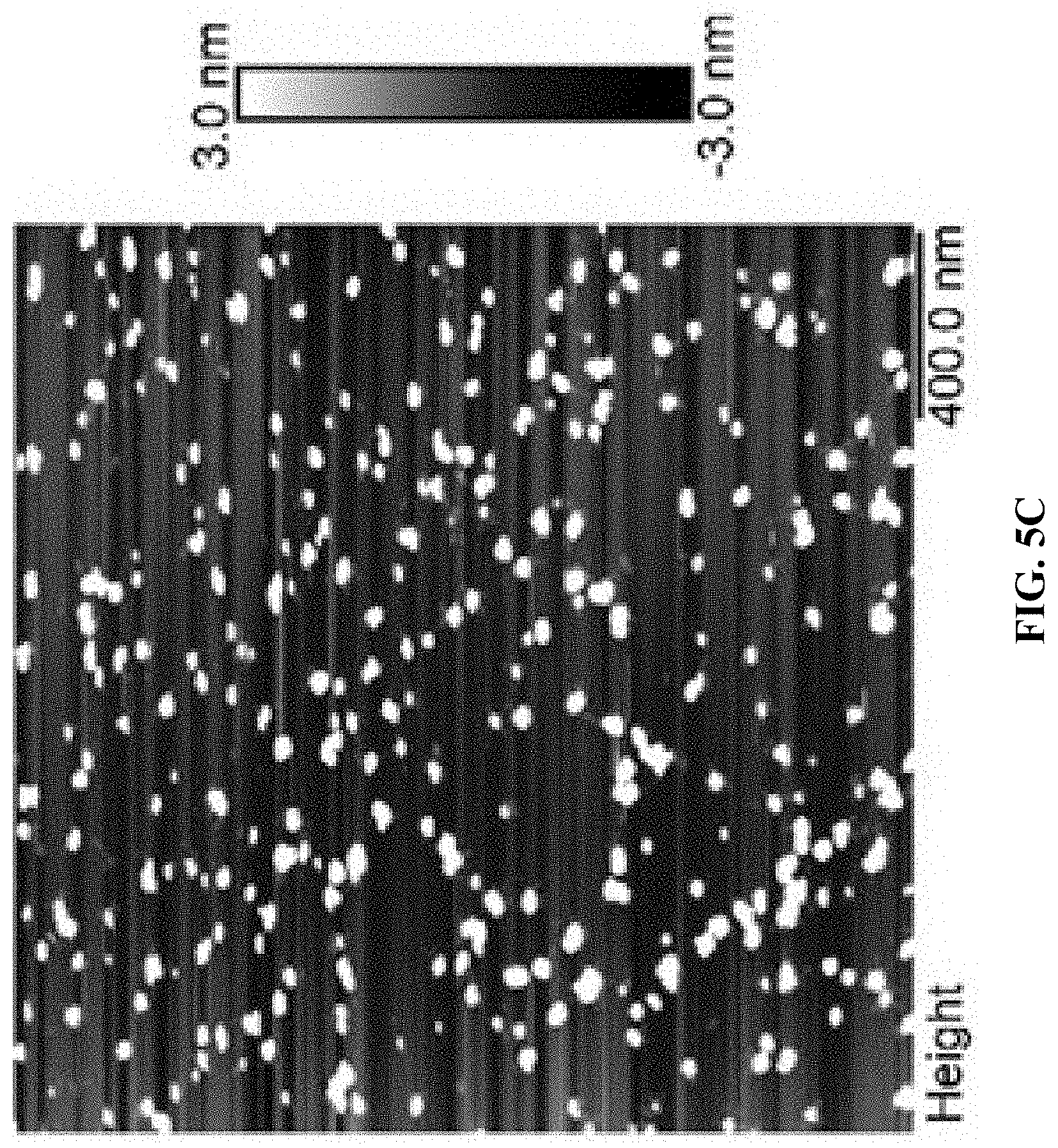
D00012
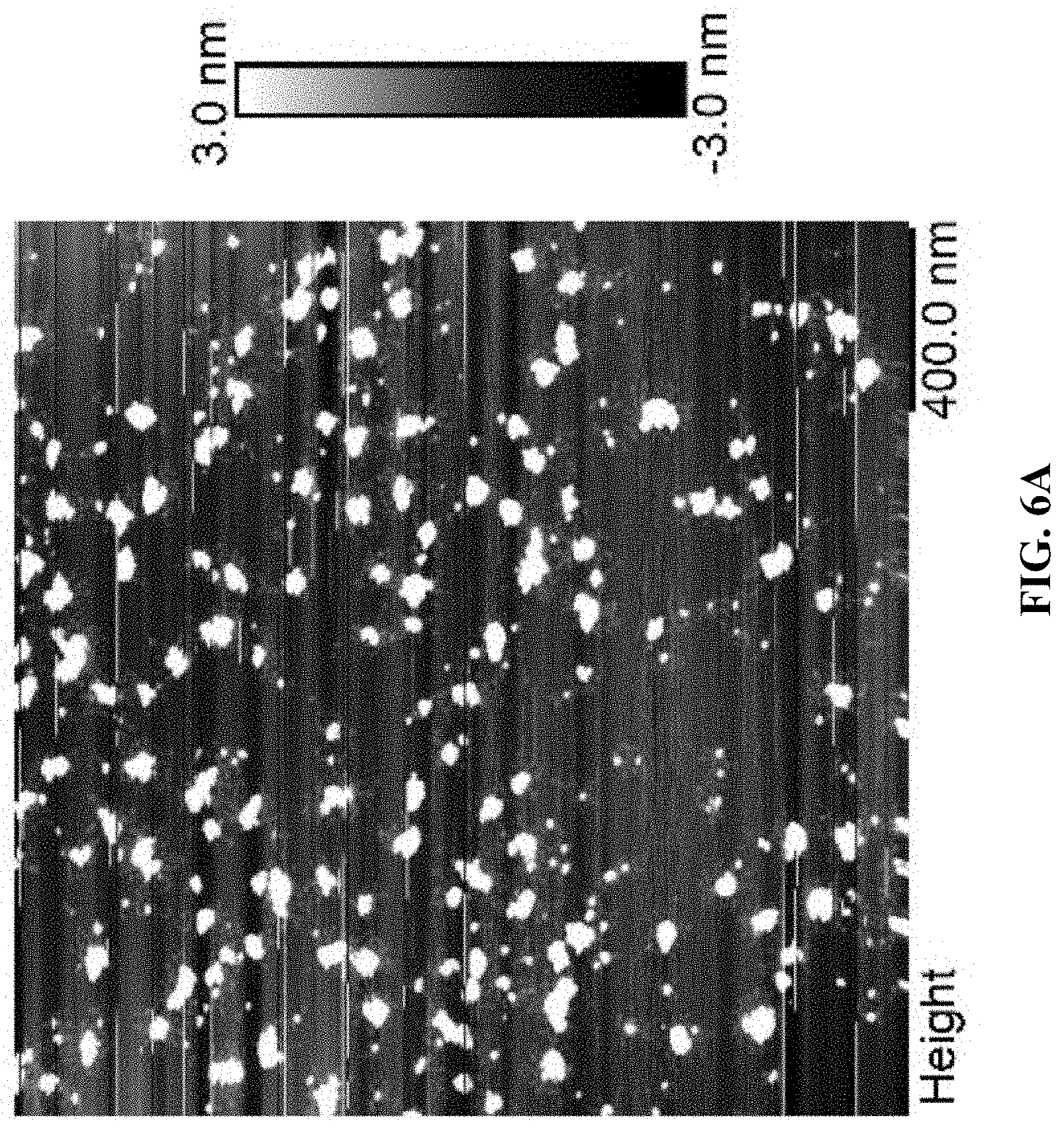
D00013
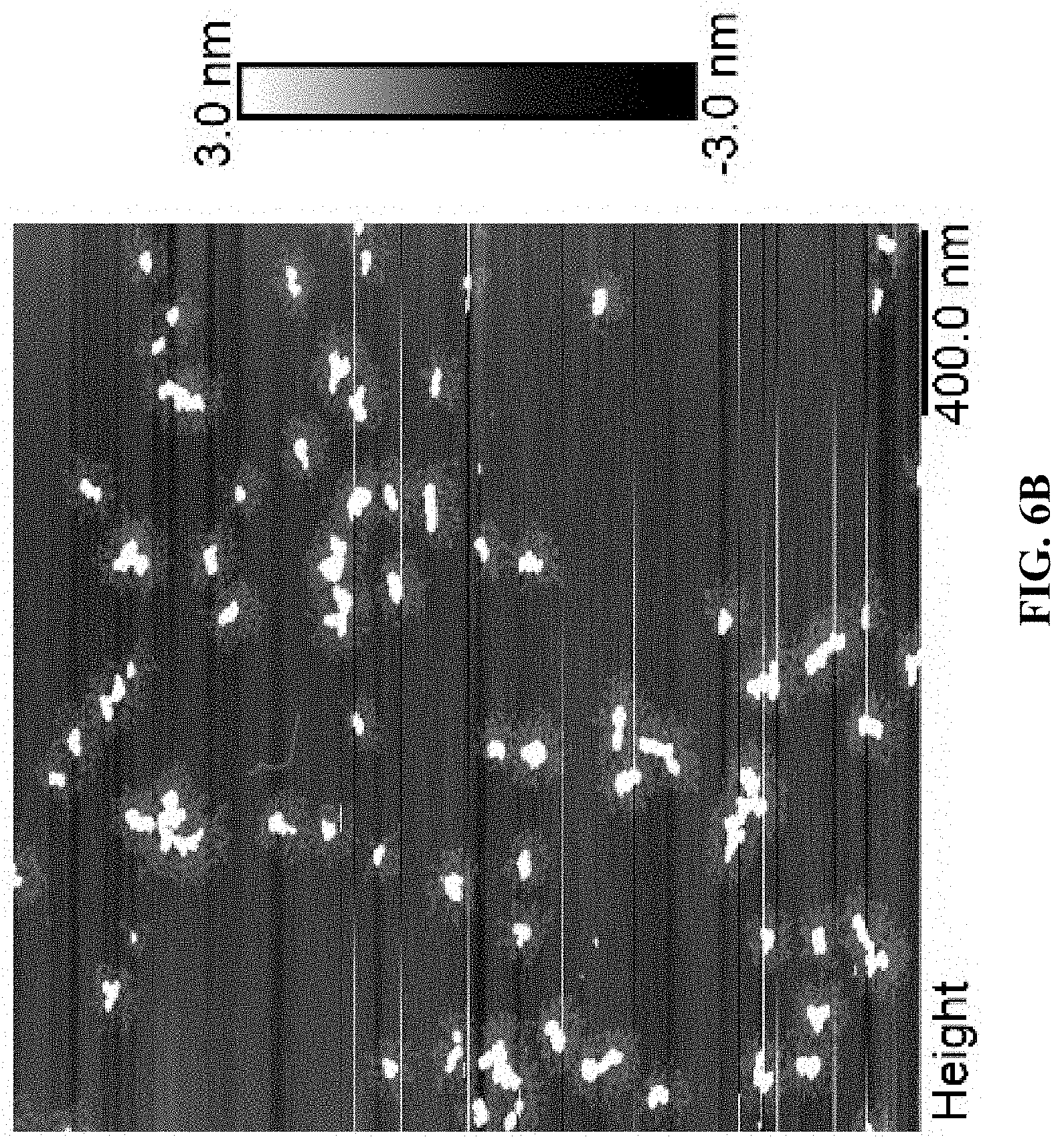
D00014
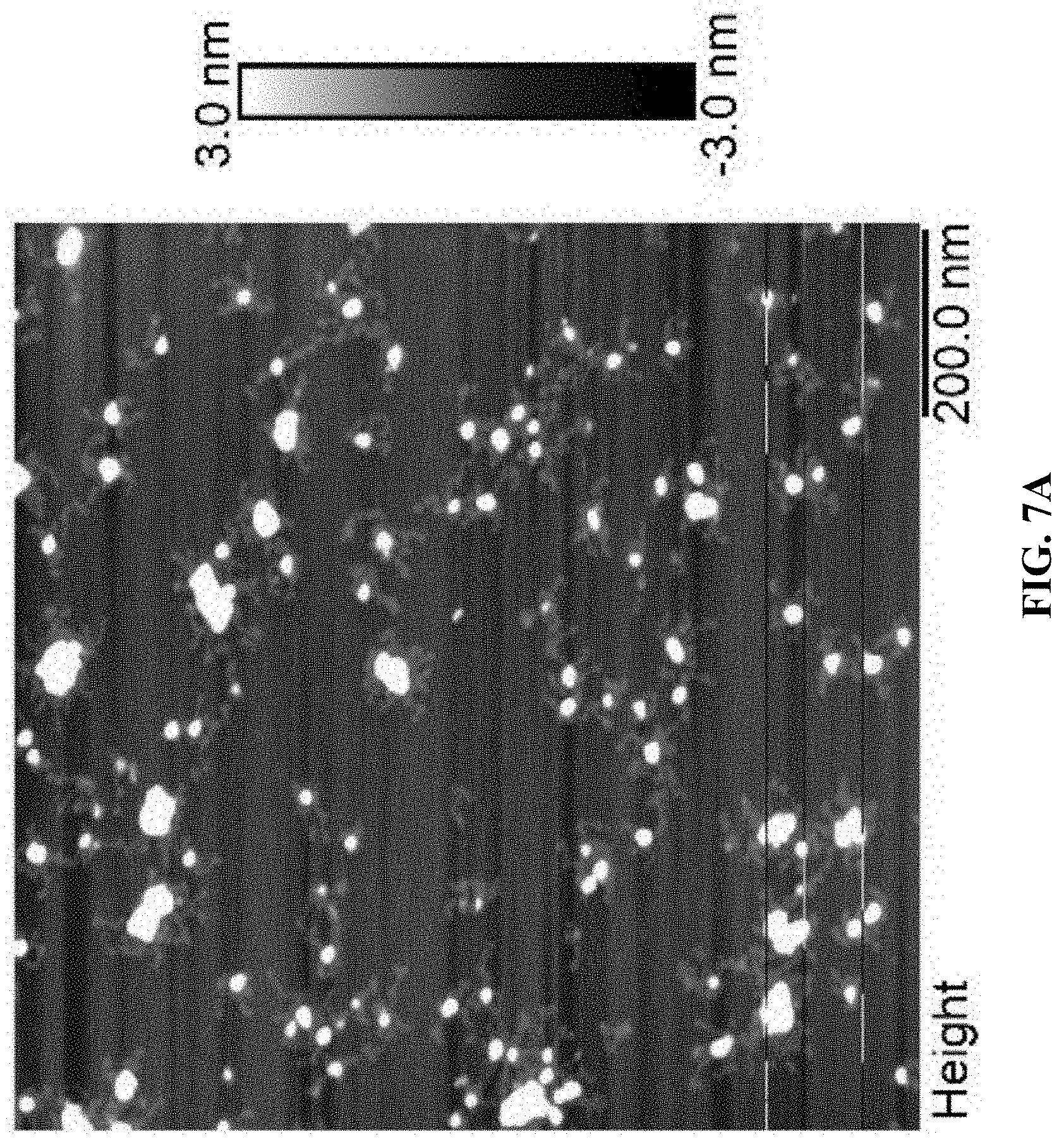
D00015
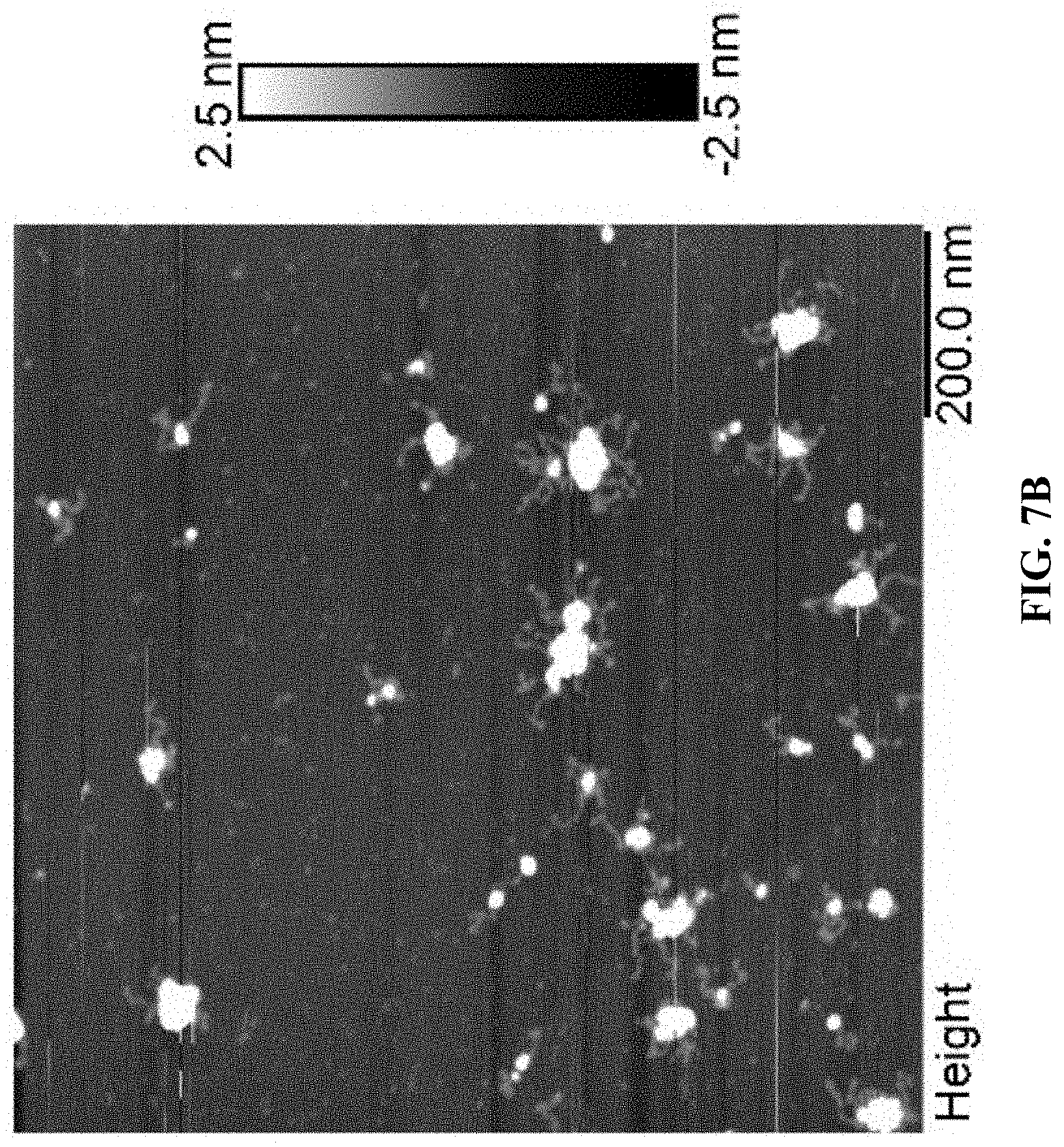
D00016
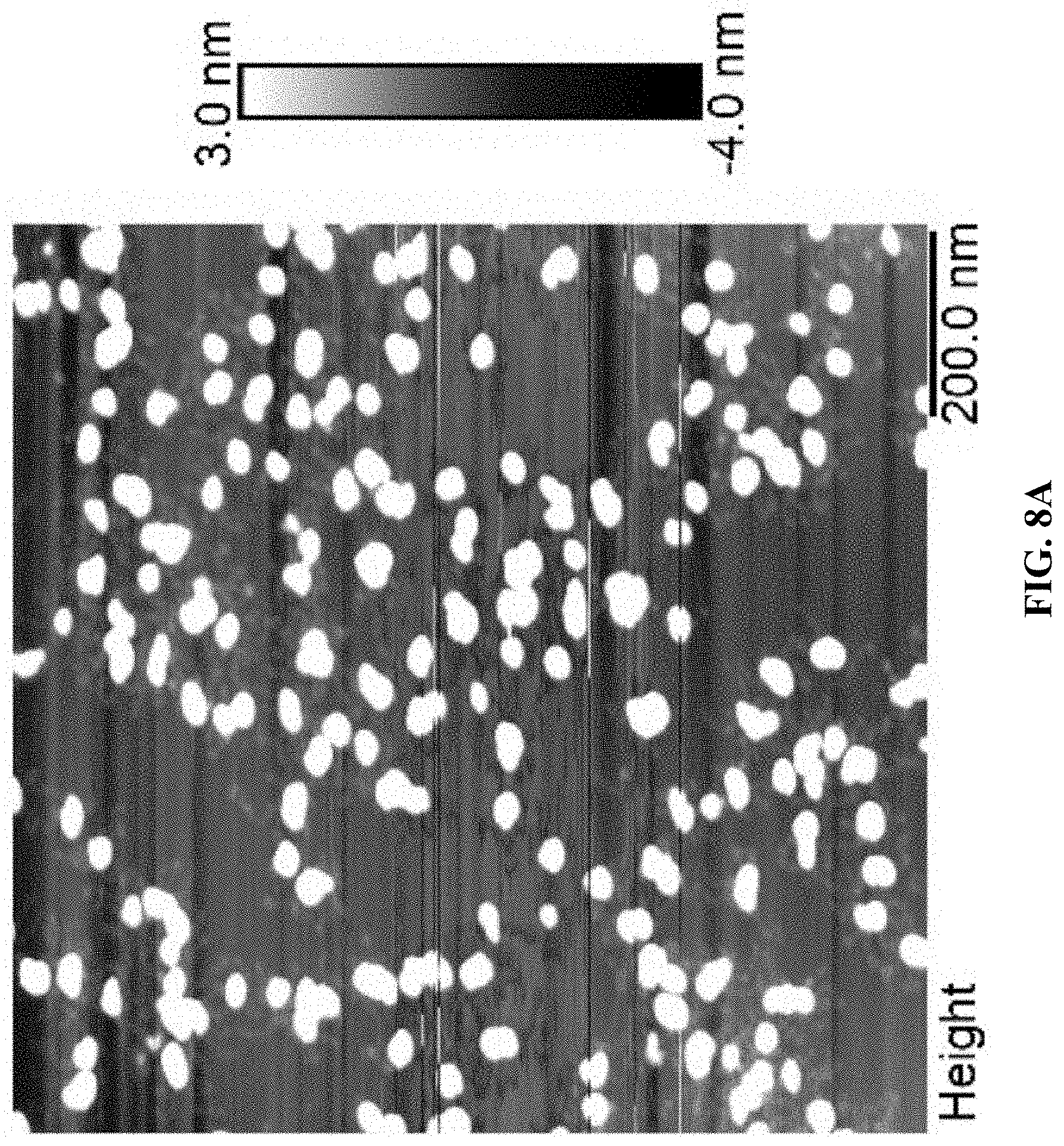
D00017
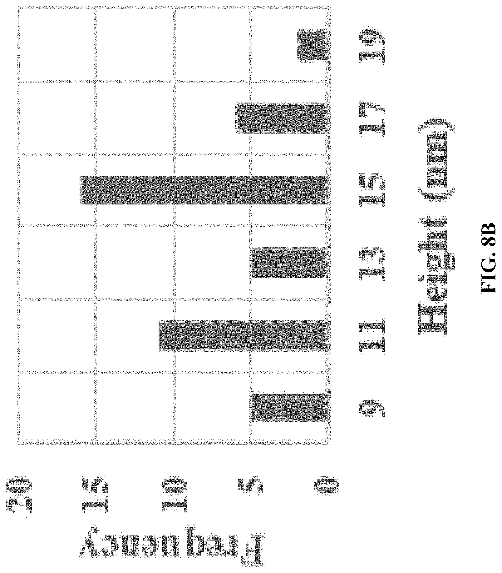
D00018
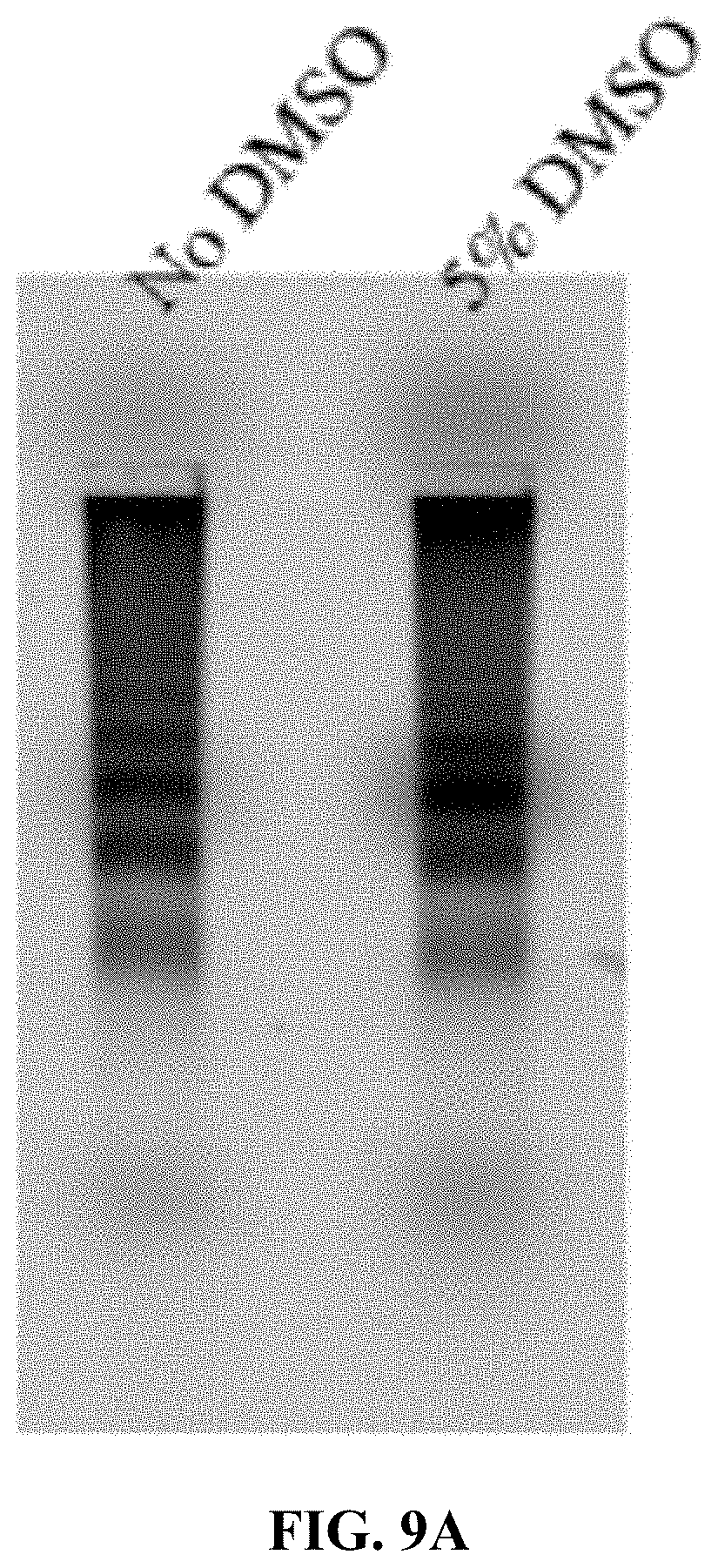
D00019
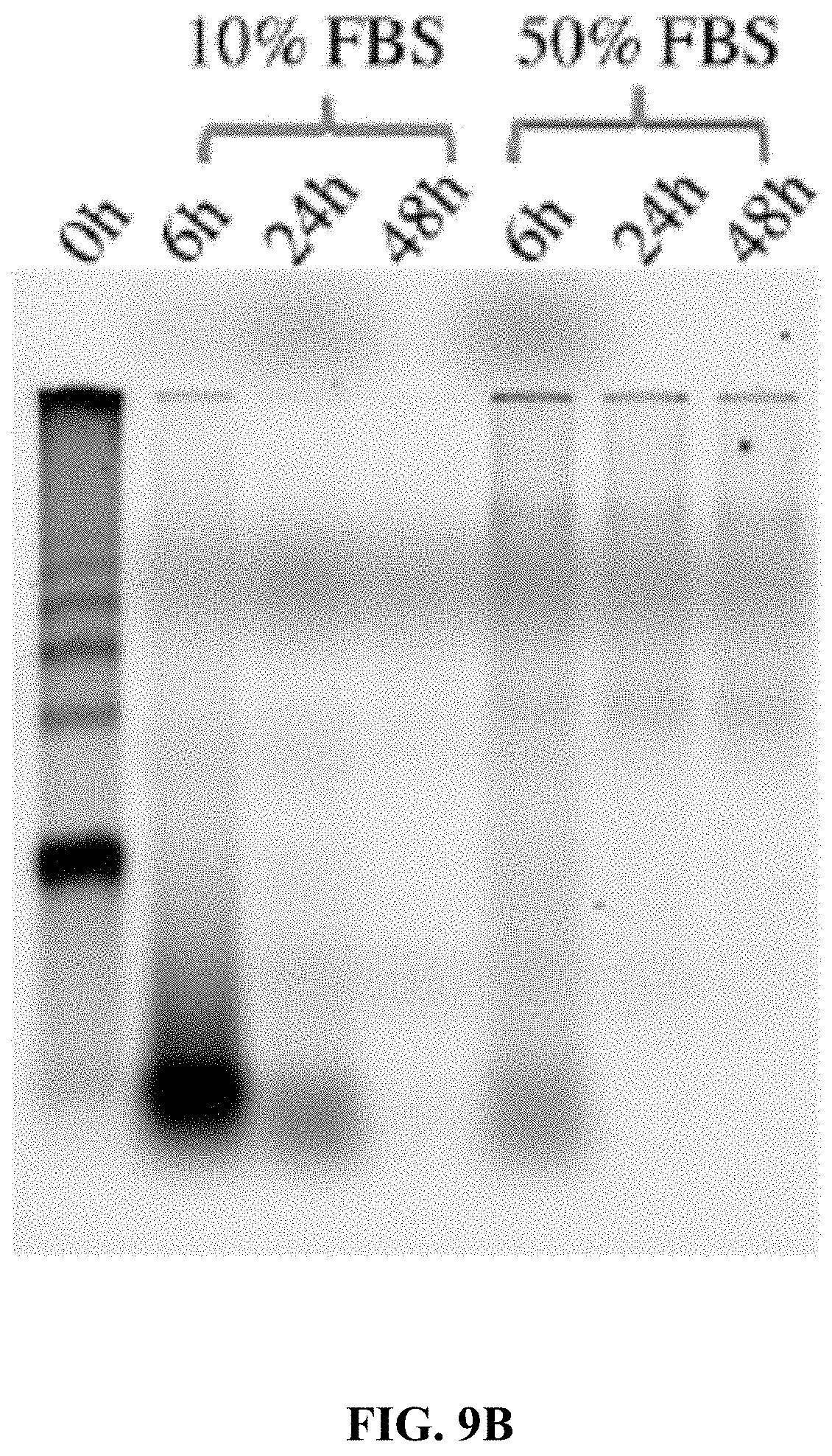
D00020
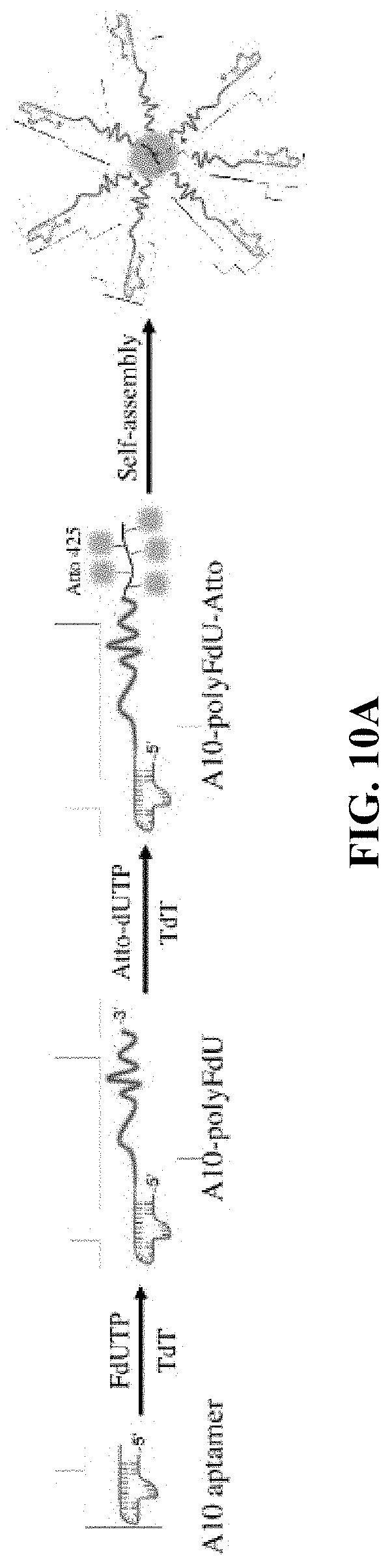
D00021
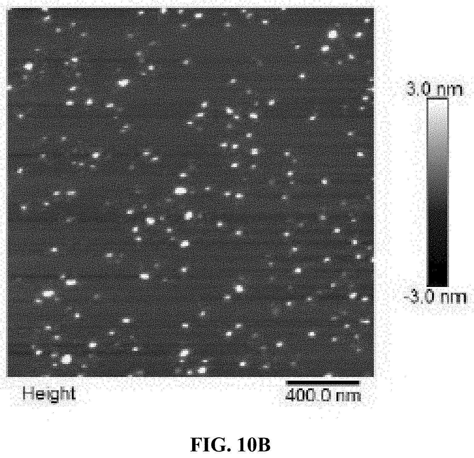
D00022
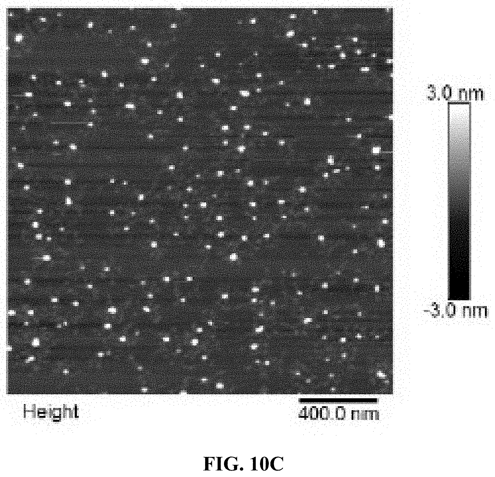
D00023
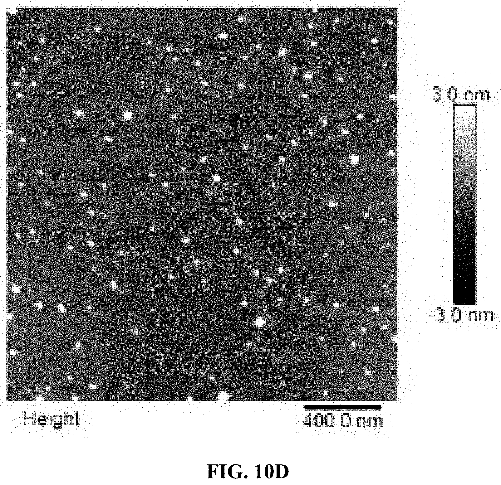
D00024
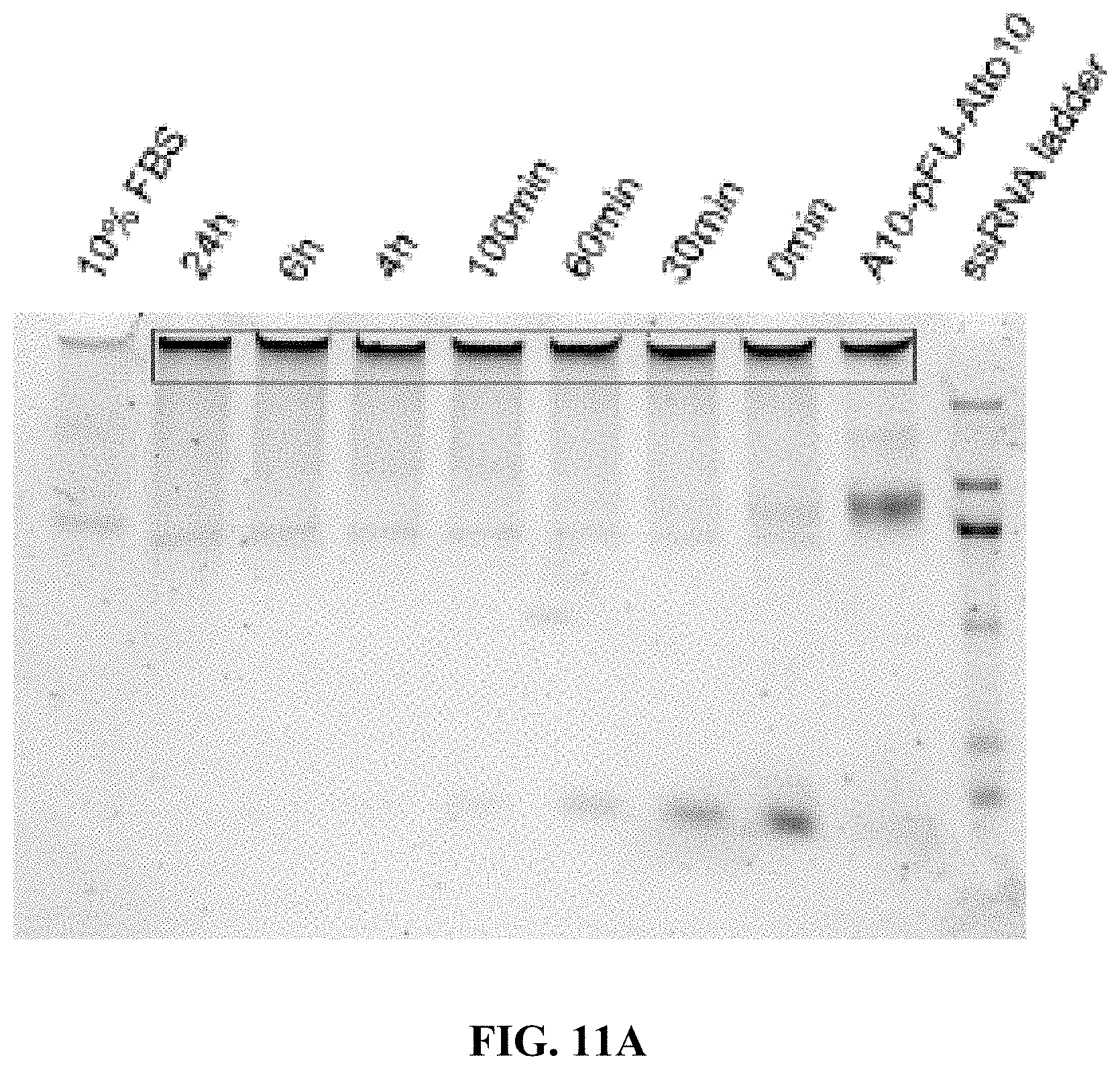
D00025
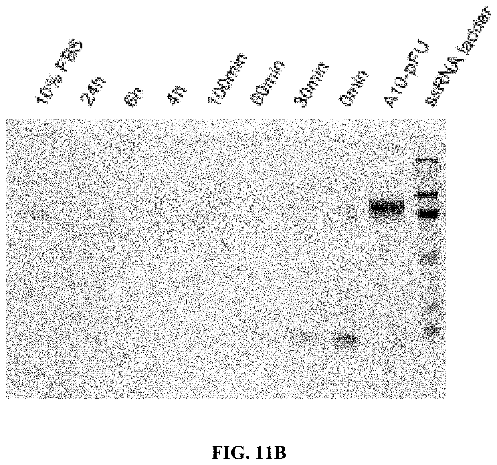
D00026
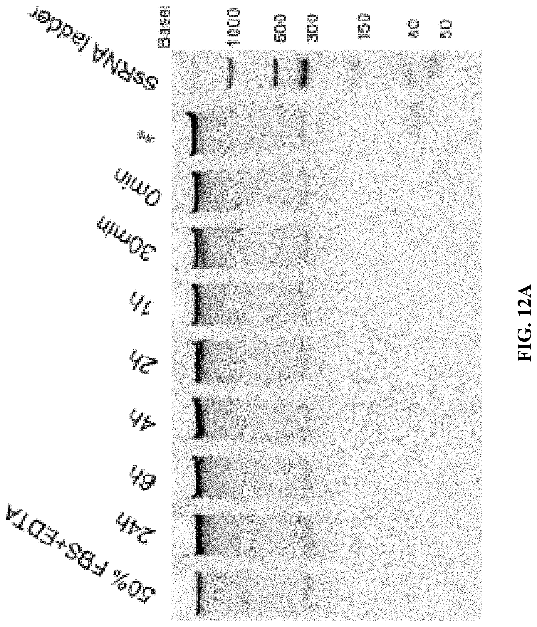
D00027
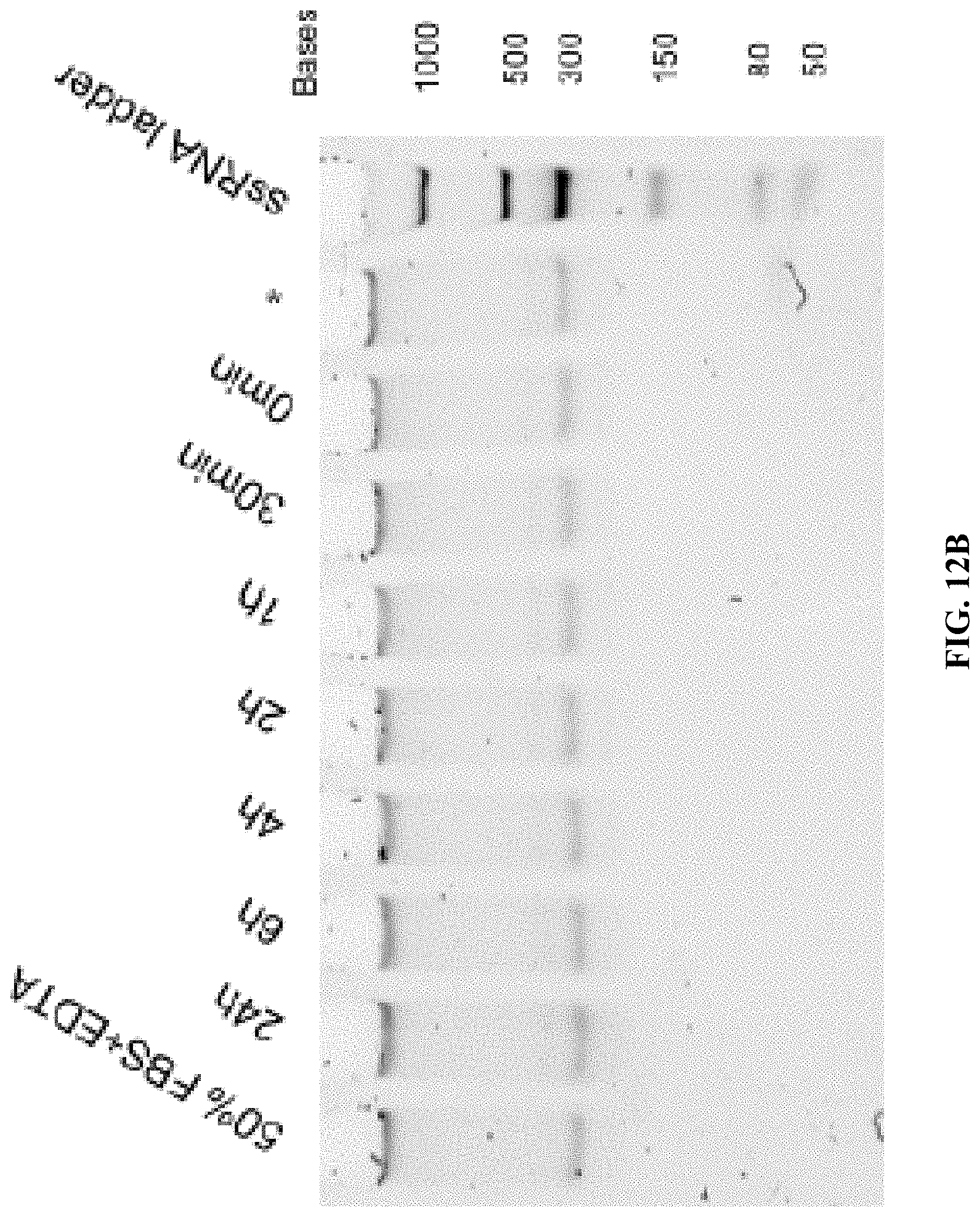
D00028
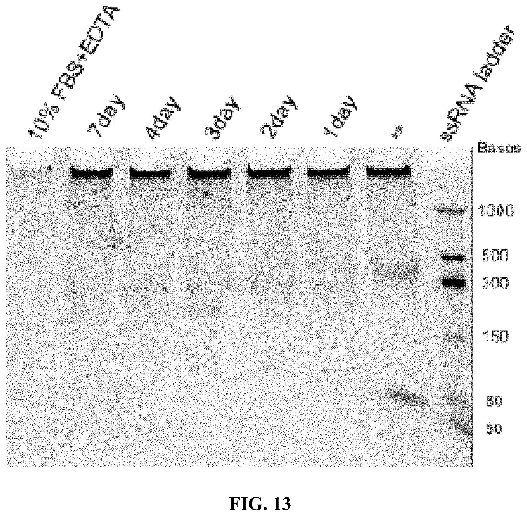
D00029
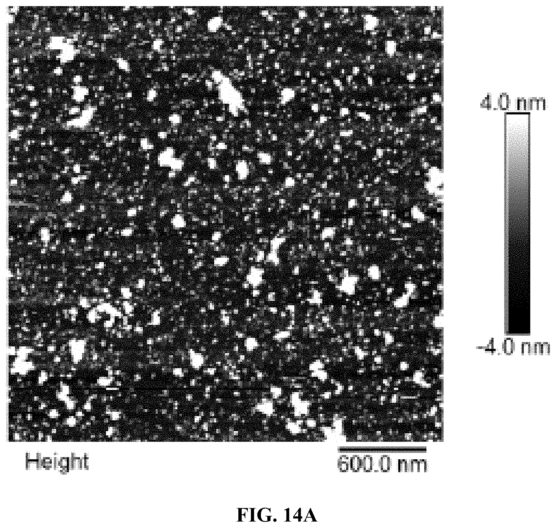
D00030
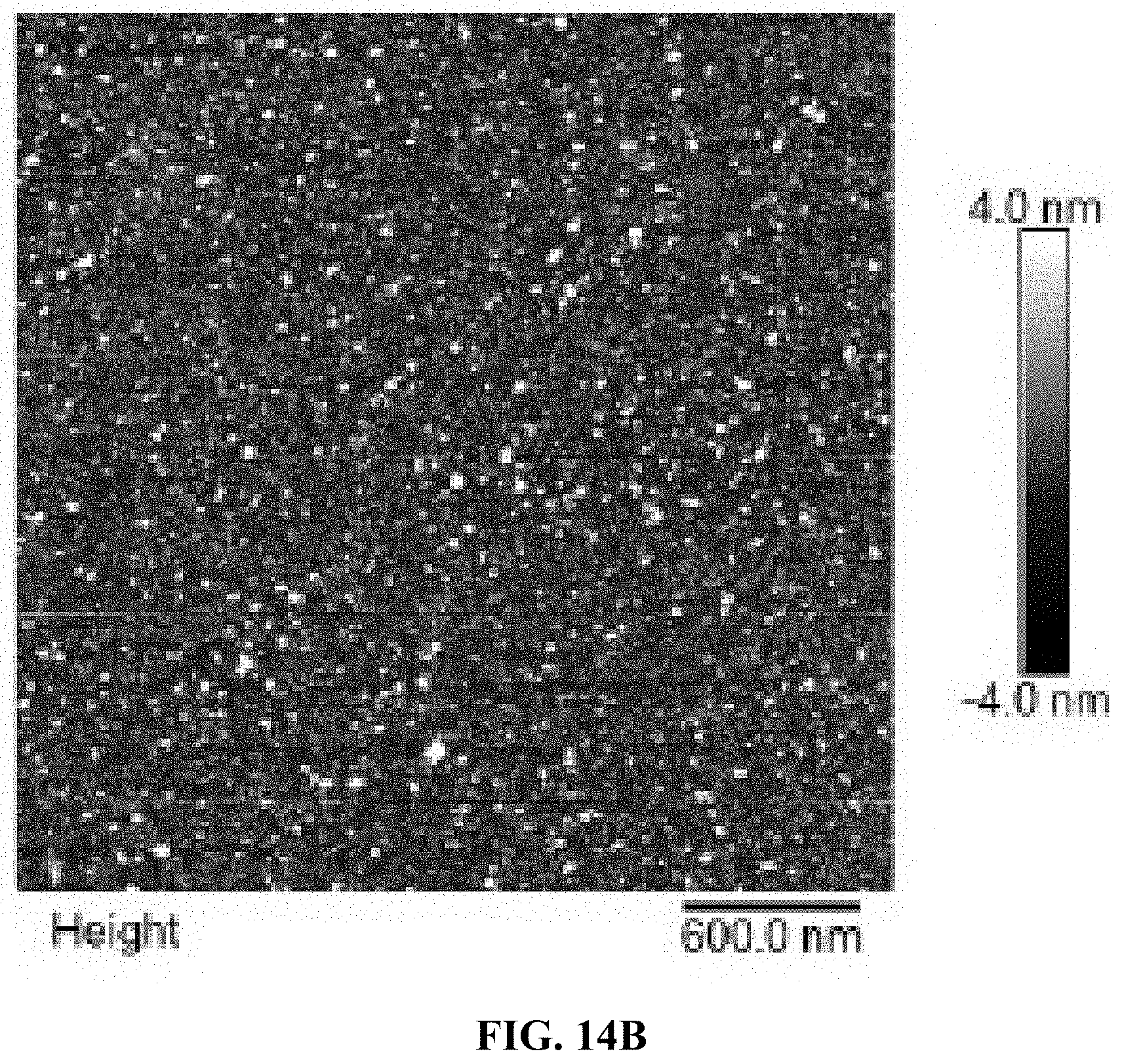
D00031
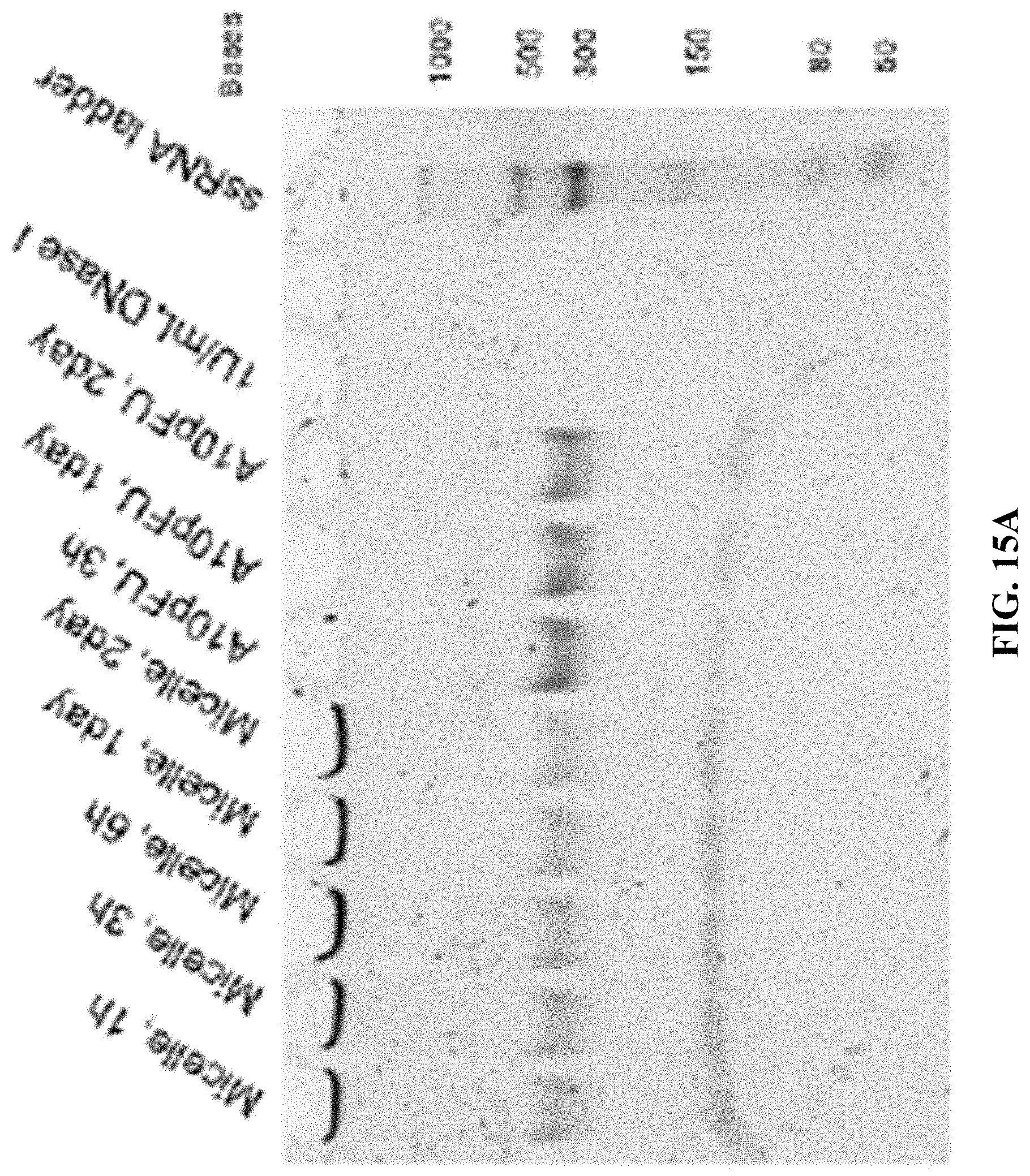
D00032
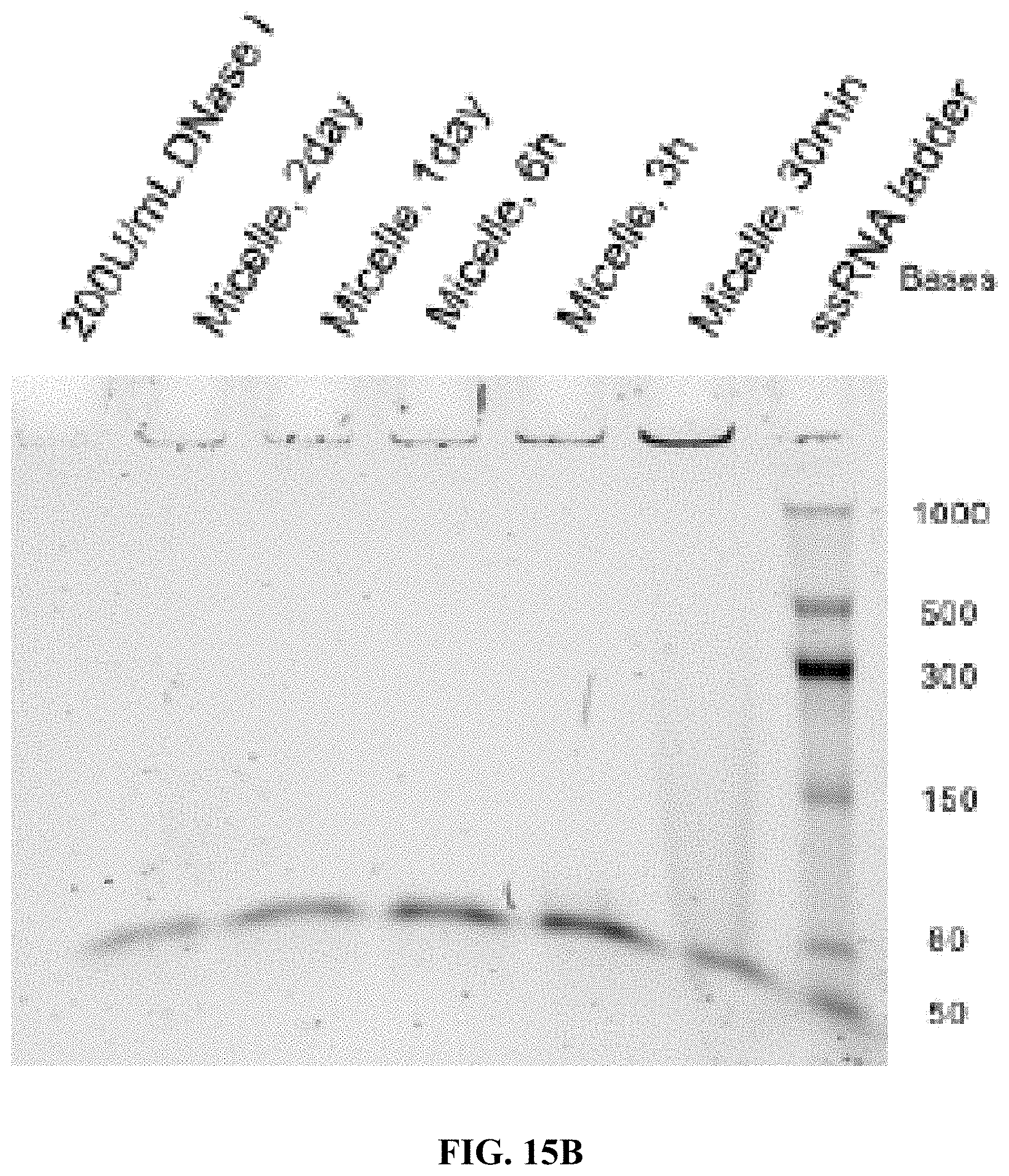
D00033
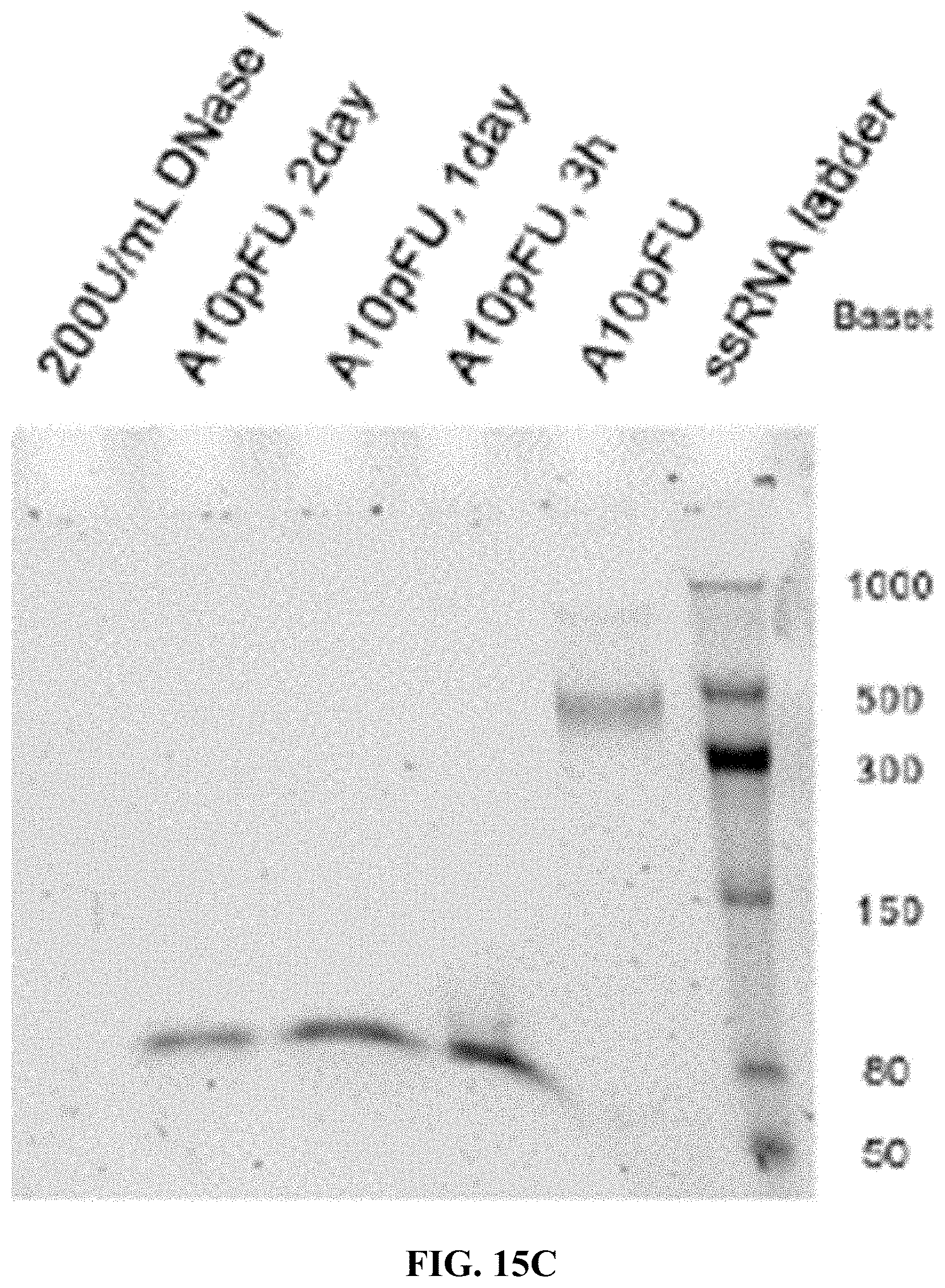
D00034
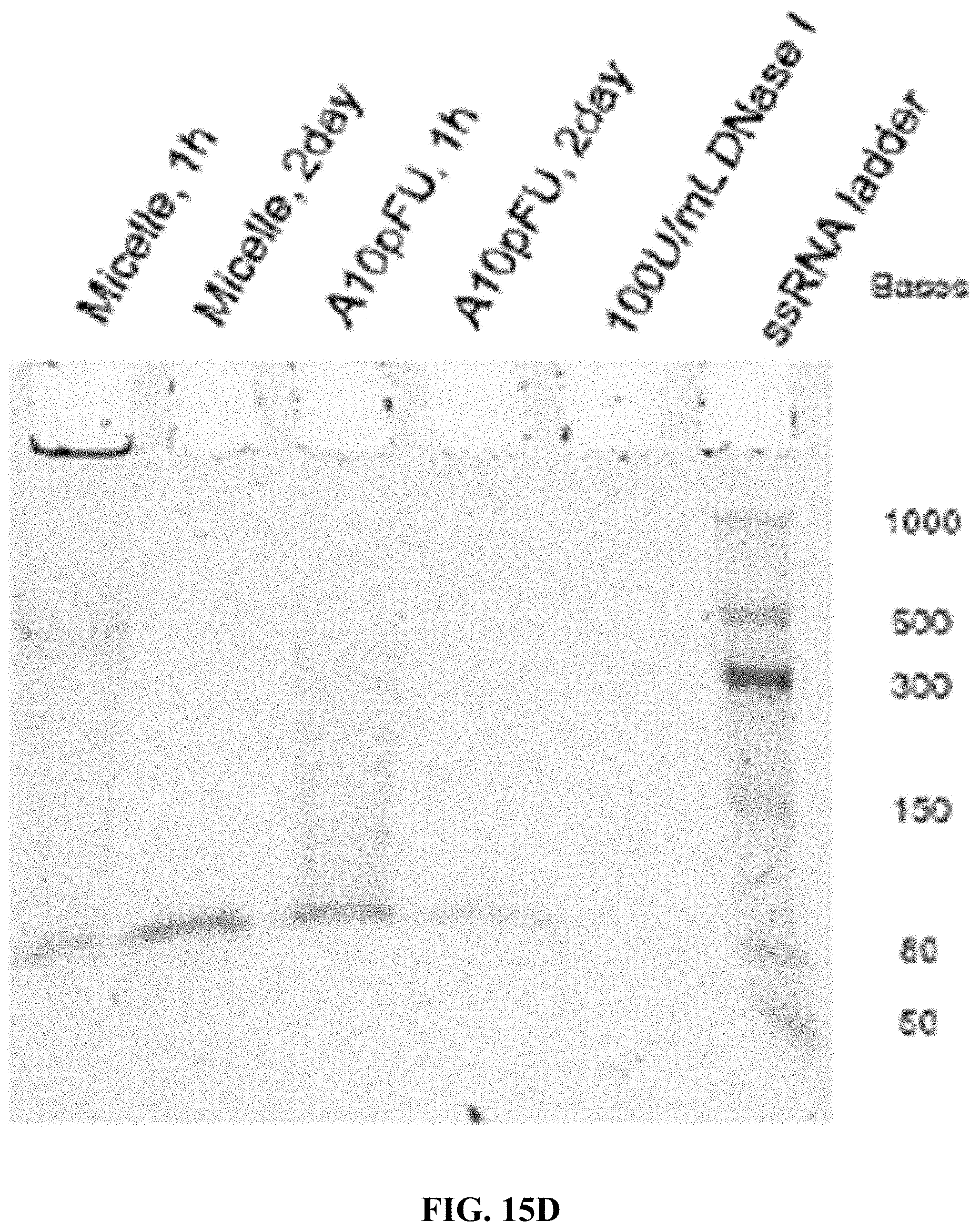
D00035
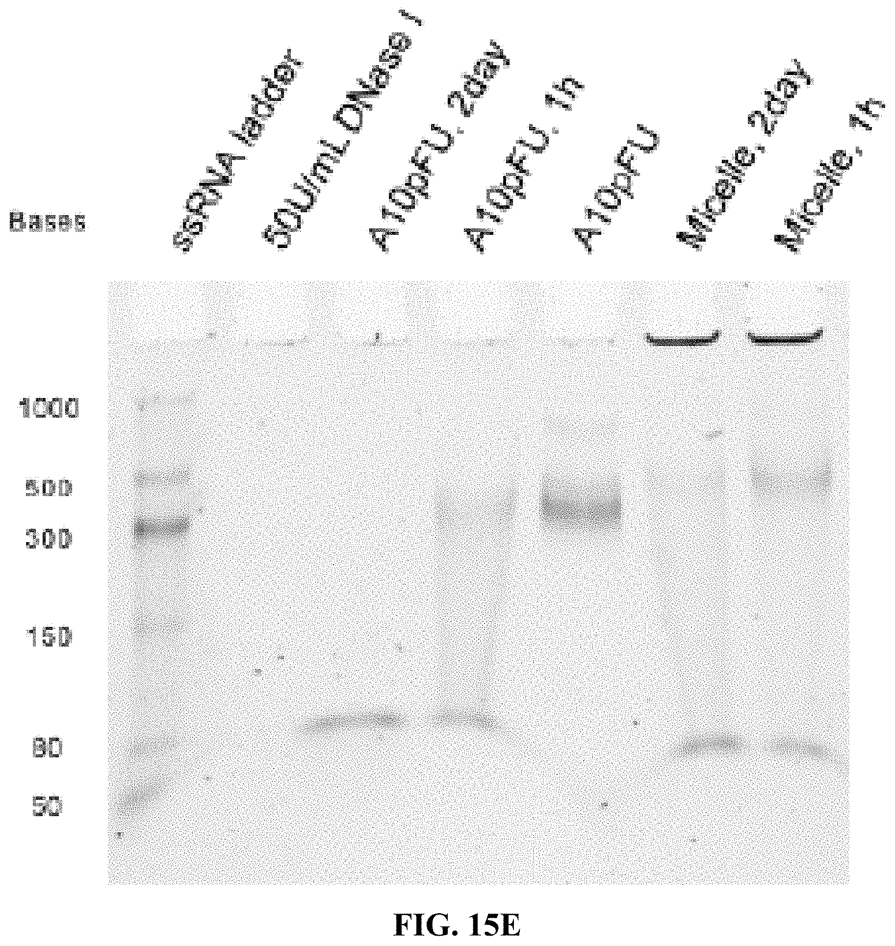
D00036
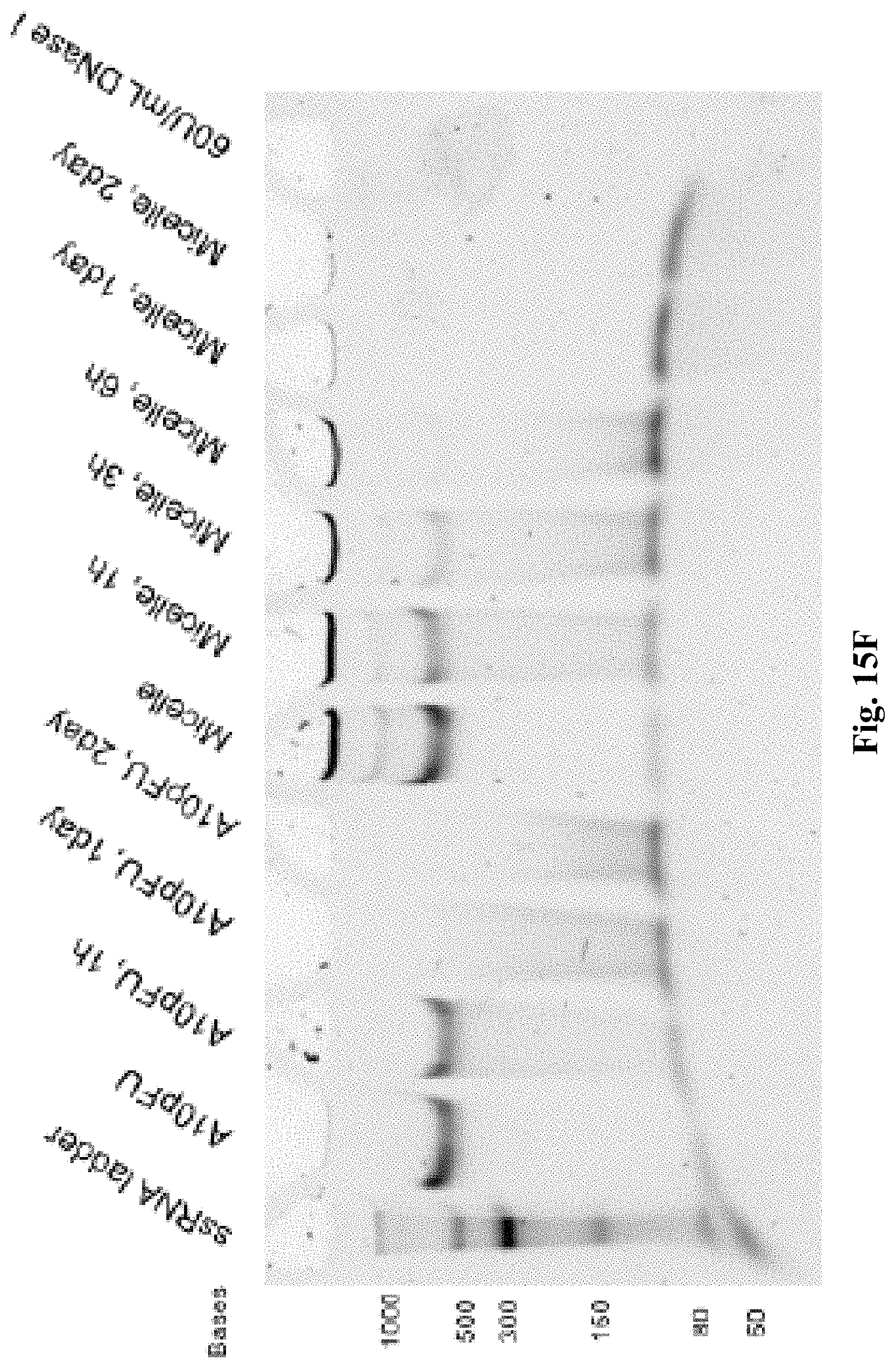
D00037
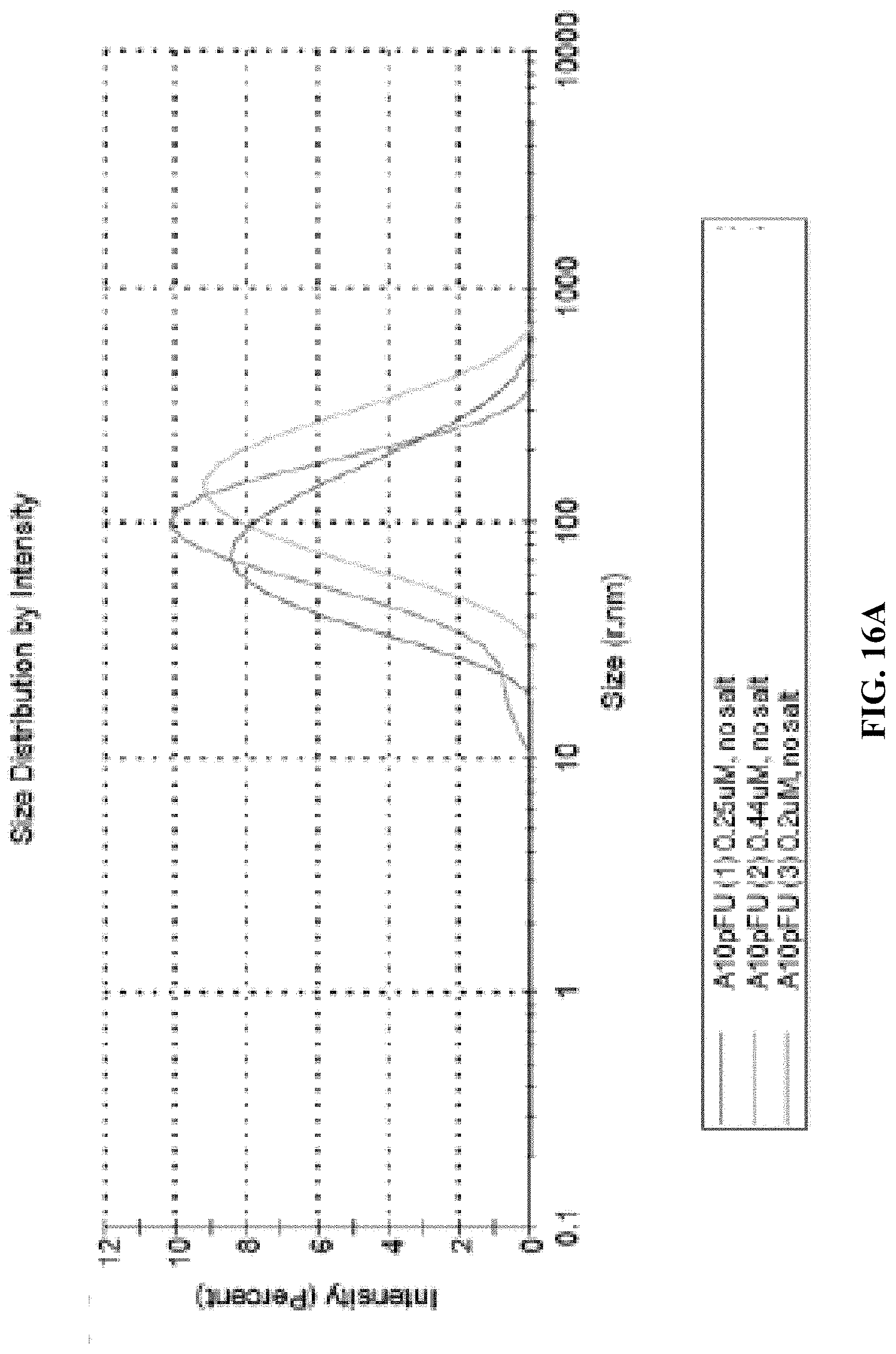
D00038
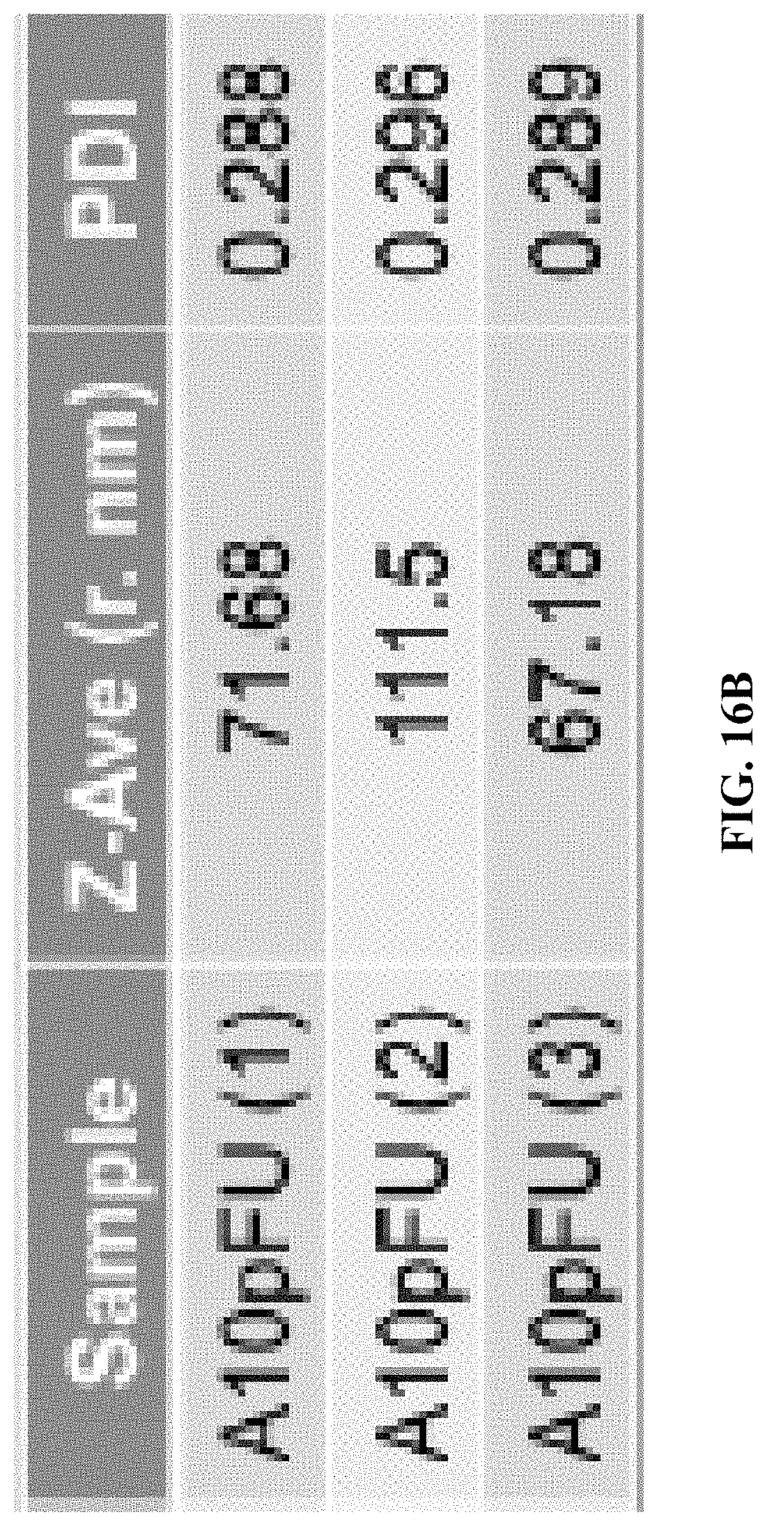
D00039
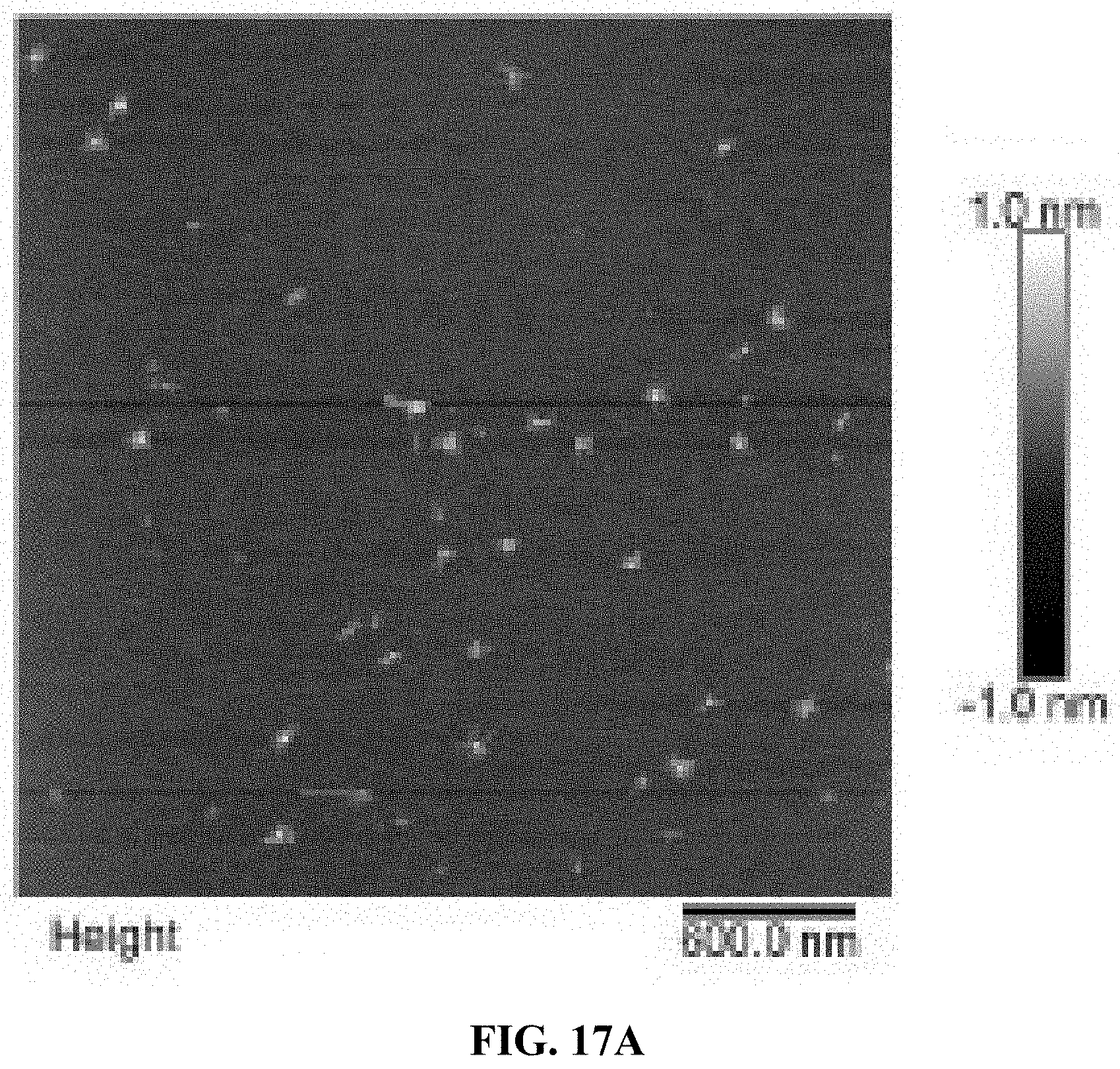
D00040
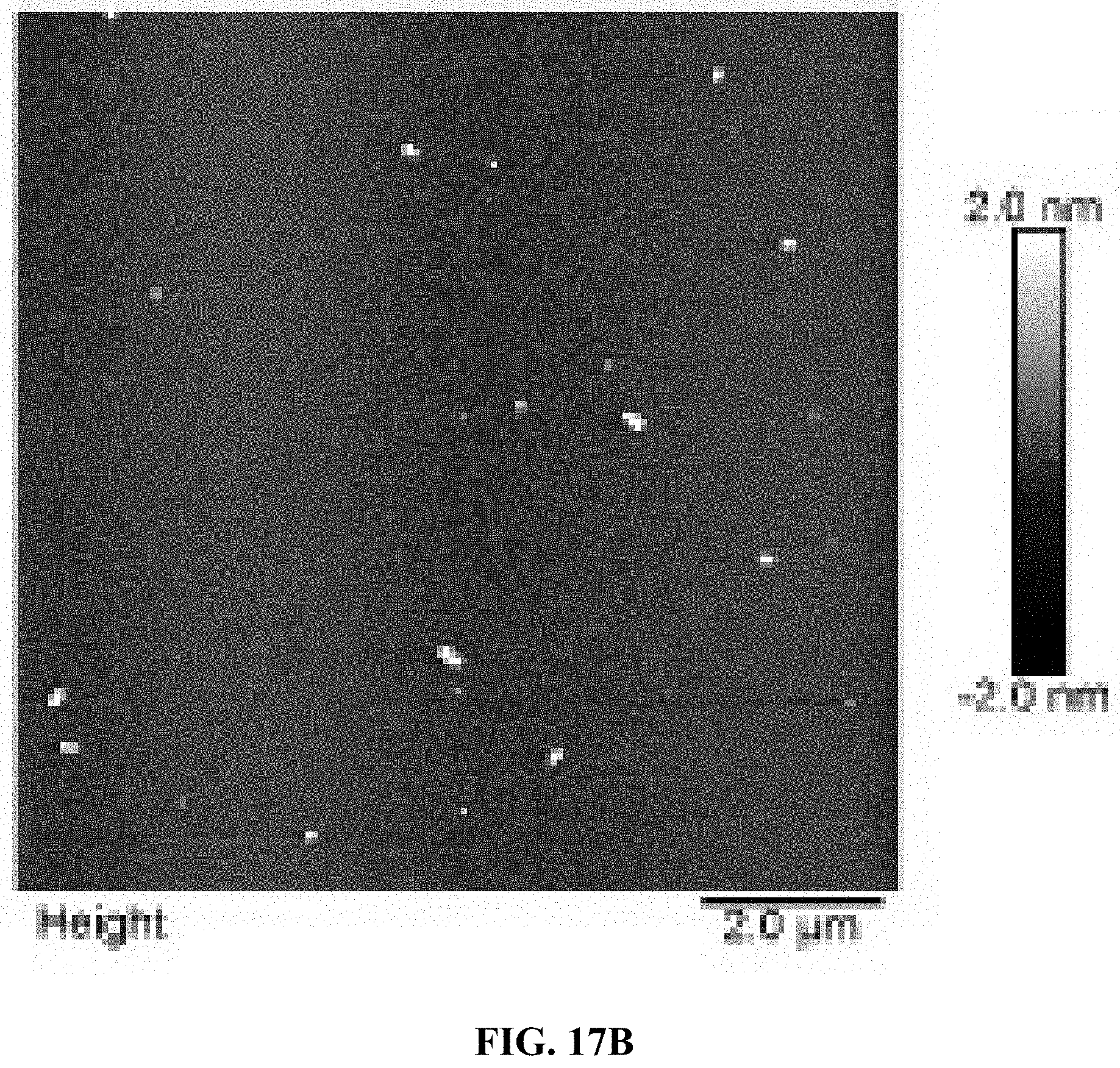
D00041
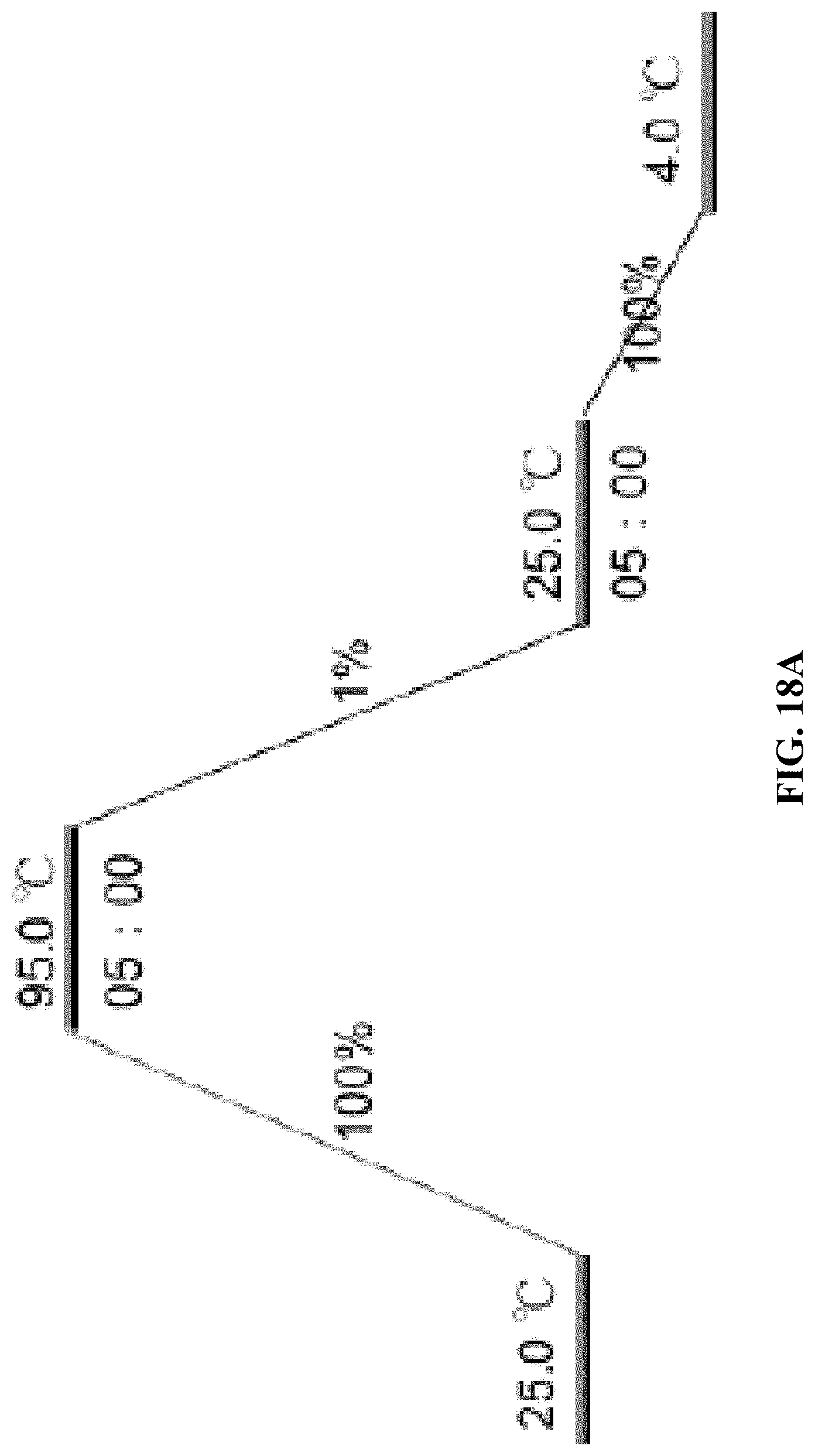
D00042
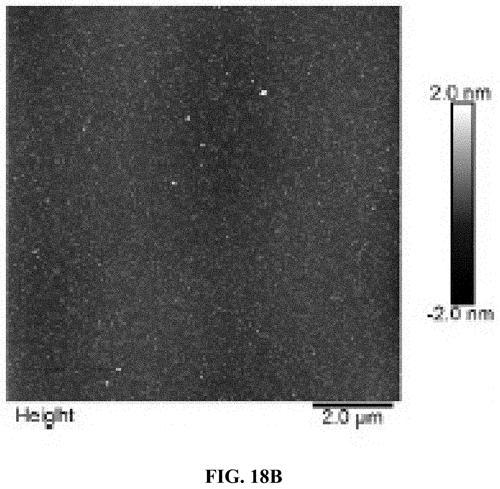
D00043
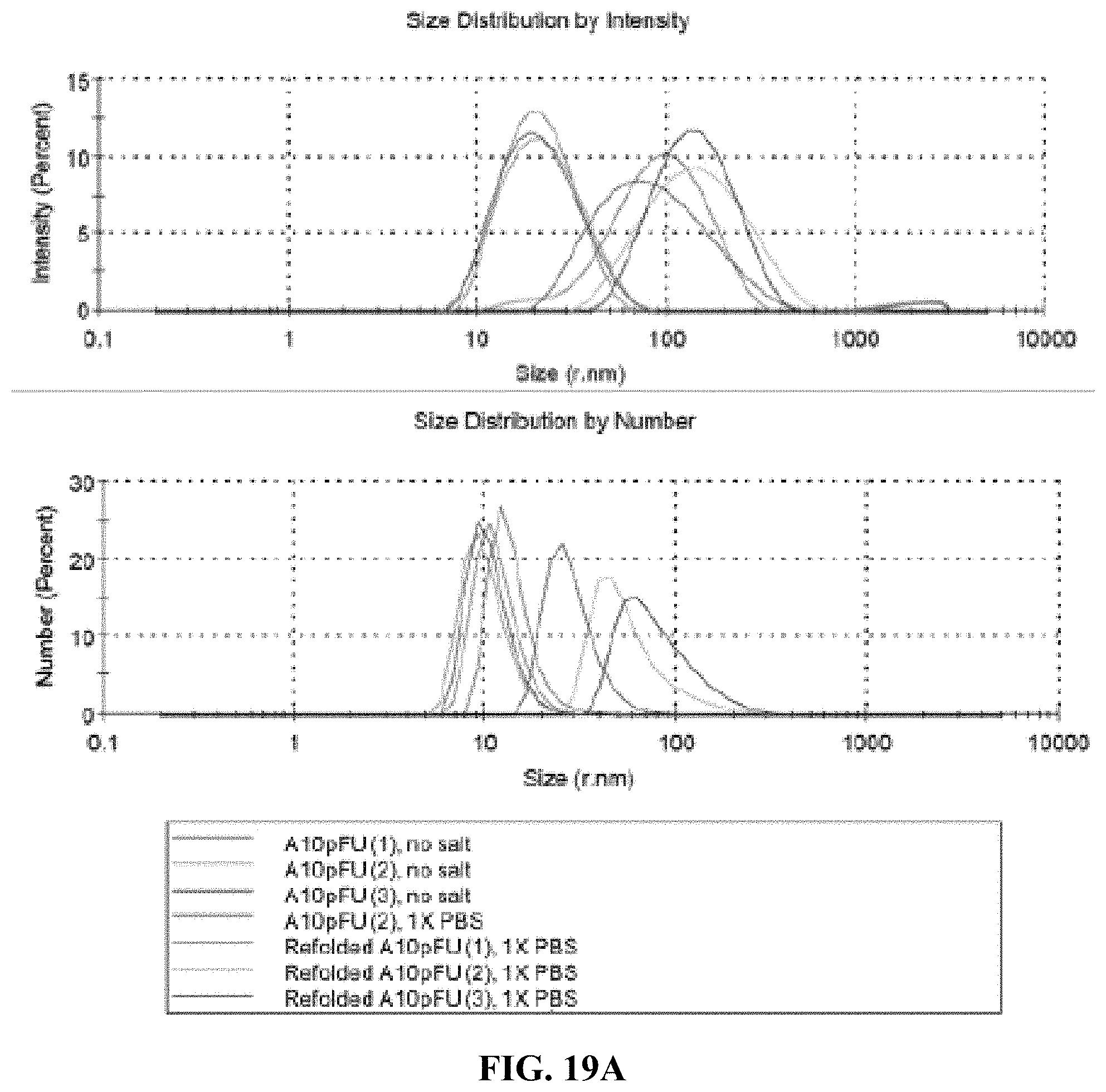
D00044
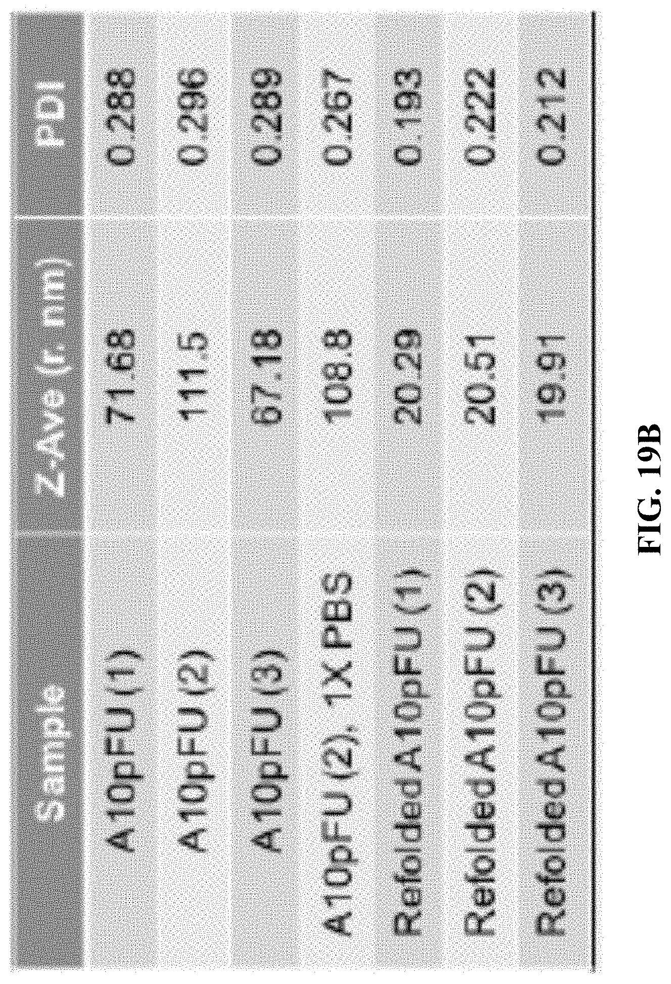
D00045
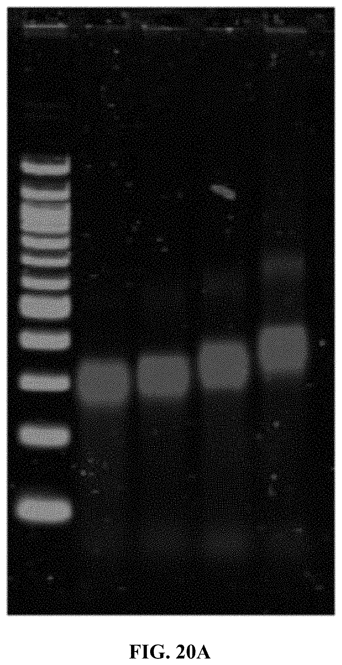
D00046
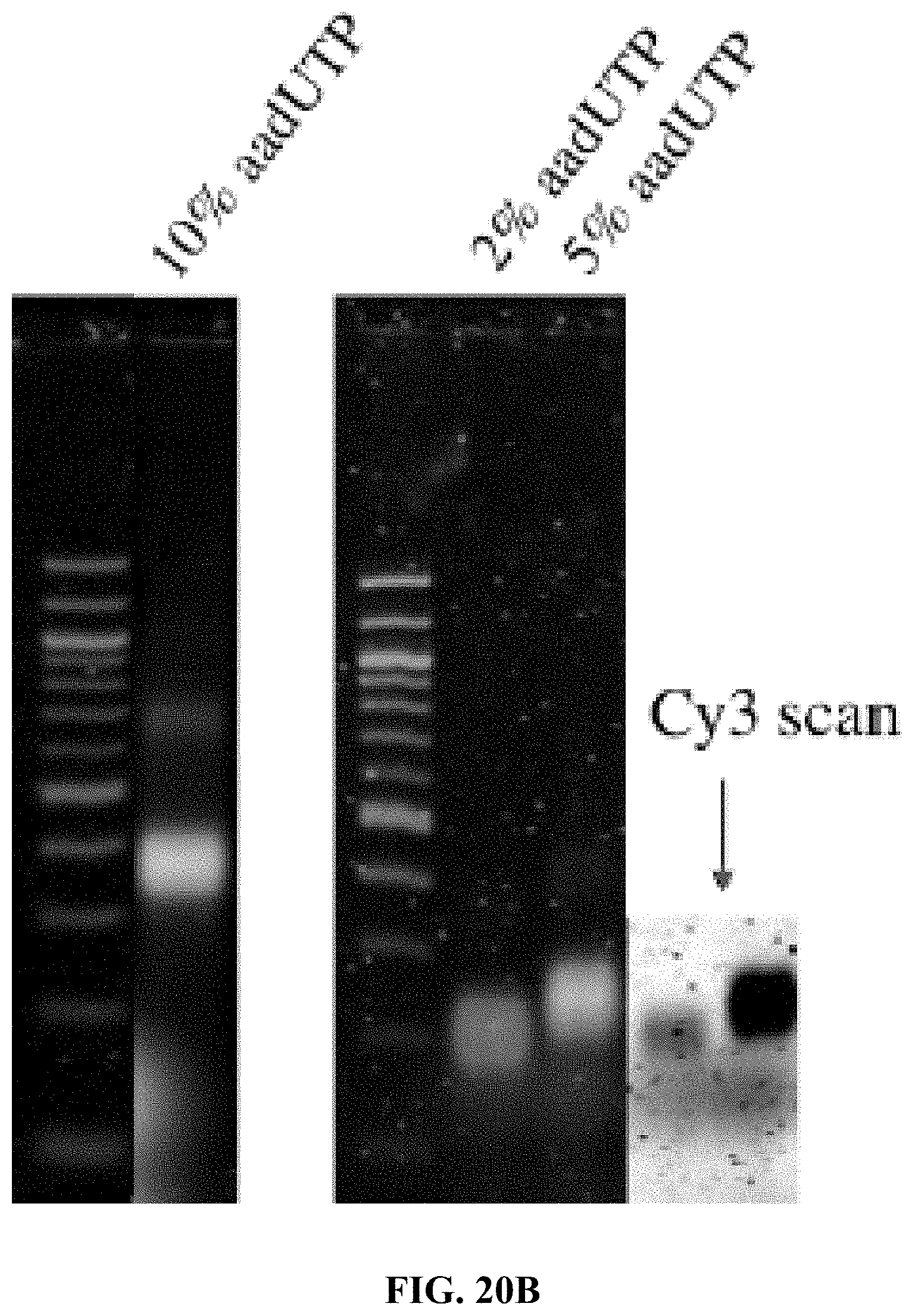
D00047
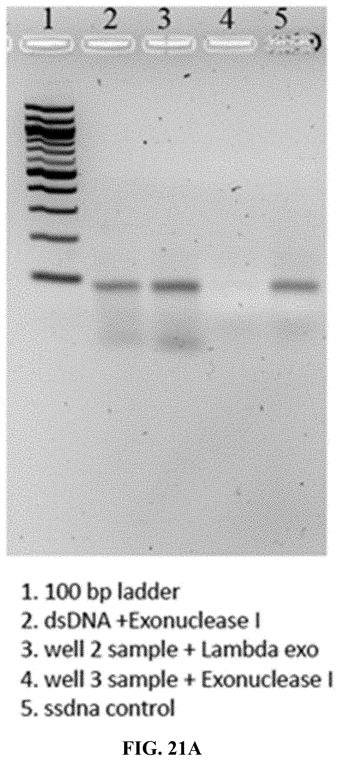
D00048
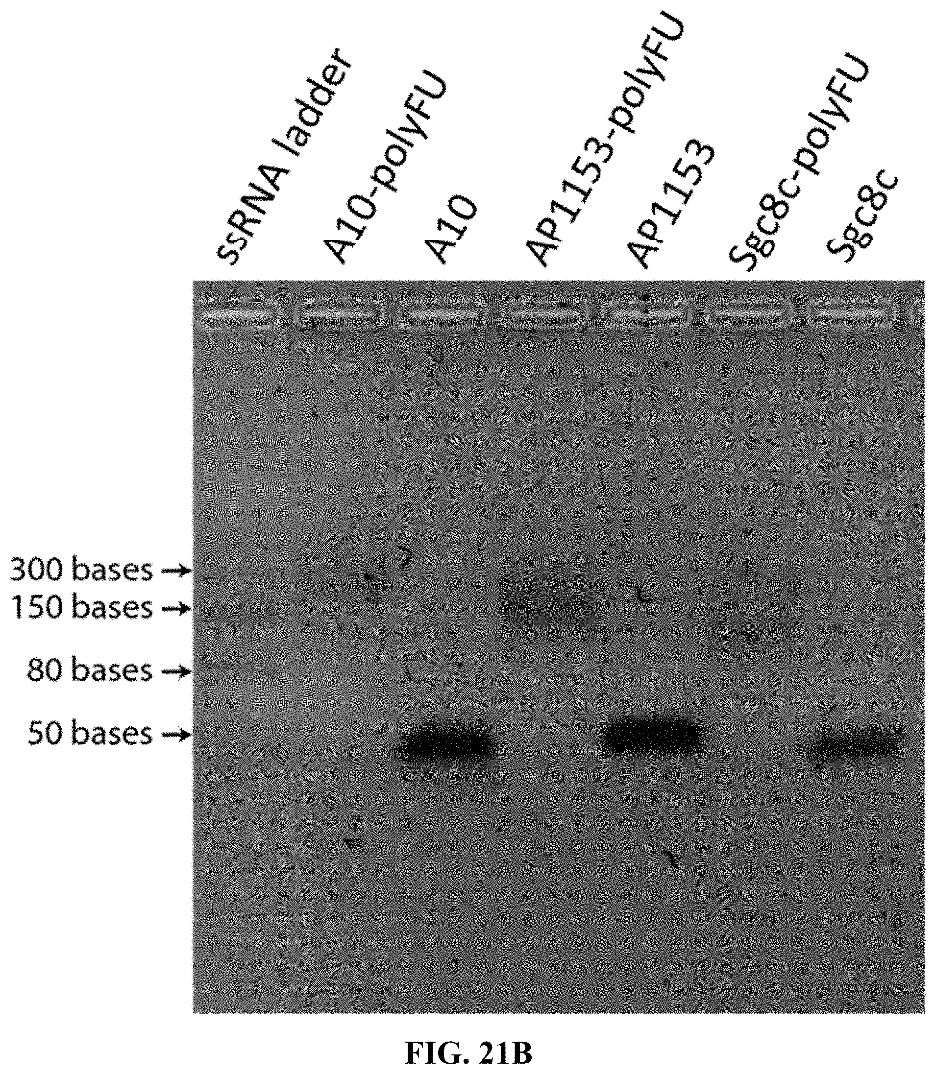
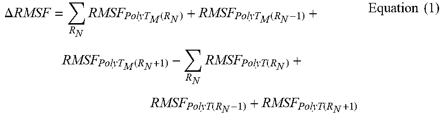
S00001
XML
uspto.report is an independent third-party trademark research tool that is not affiliated, endorsed, or sponsored by the United States Patent and Trademark Office (USPTO) or any other governmental organization. The information provided by uspto.report is based on publicly available data at the time of writing and is intended for informational purposes only.
While we strive to provide accurate and up-to-date information, we do not guarantee the accuracy, completeness, reliability, or suitability of the information displayed on this site. The use of this site is at your own risk. Any reliance you place on such information is therefore strictly at your own risk.
All official trademark data, including owner information, should be verified by visiting the official USPTO website at www.uspto.gov. This site is not intended to replace professional legal advice and should not be used as a substitute for consulting with a legal professional who is knowledgeable about trademark law.